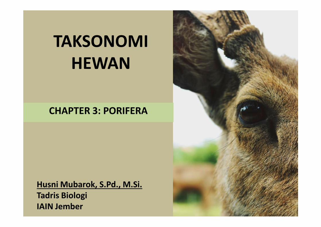
TAKSONOMI HEWAN CHAPTER 3: PORIFERA Husni Mubarok, S.Pd., M.Si. Tadris Biologi IAIN Jember

Welcome message from author
This document is posted to help you gain knowledge. Please leave a comment to let me know what you think about it! Share it to your friends and learn new things together.
Transcript
TAKSONOMI HEWAN
CHAPTER 3: PORIFERA
Husni Mubarok, S.Pd., M.Si.Tadris BiologiIAIN Jember

WHO IS HE ??

Animals in this phylum are informallycalled sponges. Sponges are sessile animalsthat lack tissues. They live as filter feeders,trapping particles that pass through theinternal channels of their body
Kingdom Animalia encompasses 1.3 million known species, and estimates of the total number of
species range as high as 10–20 million
Porifera probably originated from flagellated protozoan like Proterospongia, a colonial
flagellate. The colony of Proterospongia has collared and flagellated cells embedded in a
gelatinous matrix having amoeboid cells.
Phylum ChoanozoaClass Choanoflagellatea
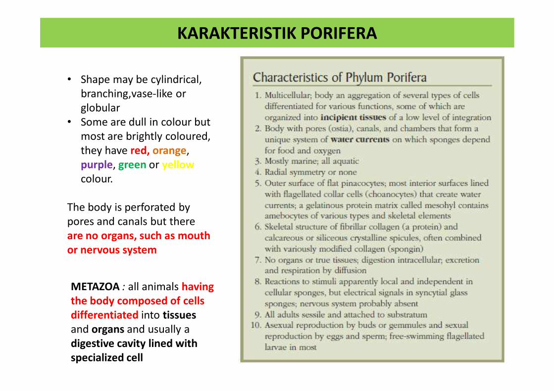
KARAKTERISTIK PORIFERA
• Shape may be cylindrical, branching,vase-like or globular
• Some are dull in colour but most are brightly coloured, they have red, orange, purple, green or yellow colour.
The body is perforated by pores and canals but there are no organs, such as mouth or nervous system
METAZOA : all animals having the body composed of cells differentiated into tissues and organs and usually a digestive cavity lined with specialized cell

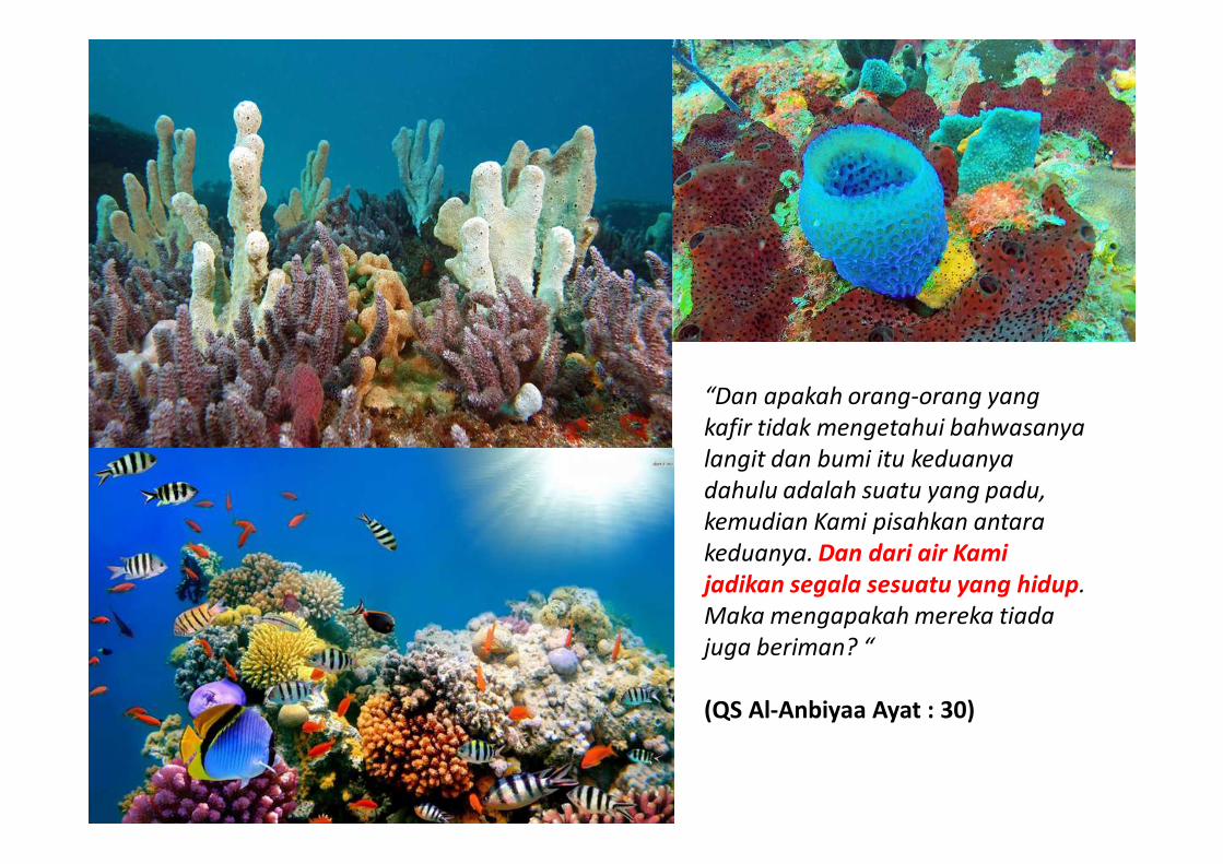
“Dan apakah orang-orang yang kafir tidak mengetahui bahwasanya langit dan bumi itu keduanya dahulu adalah suatu yang padu, kemudian Kami pisahkan antara keduanya. Dan dari air Kami jadikan segala sesuatu yang hidup. Maka mengapakah mereka tiada juga beriman? “
(QS Al-Anbiyaa Ayat : 30)

Phylogeny of Living Animals
Recent molecularstudies indicate that spongesare monophyletic, however, as some studies suggest that sponges are paraphyletic
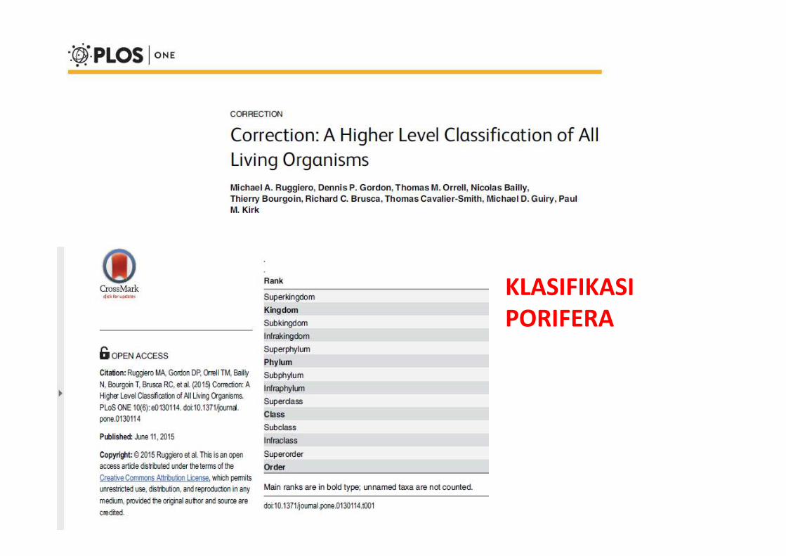
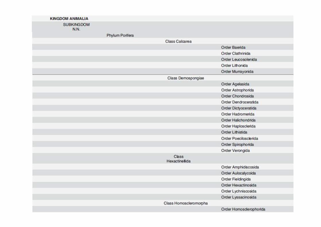
KLASIFIKASI PORIFERA

FORM & FUNCTION
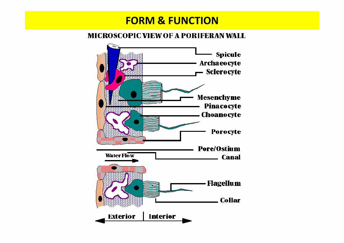
FORM & FUNCTION

FORM & FUNCTION
Archaeocytes are ameboid cells that move in the mesohyl

SKELETON
Skeleton (1) memberi bentuk & struktur, (2) menyokong & melindungi bagian yg lembut (soft) sponge, (3) dasar klasifikasi sponge
Skeleton tertanam di jaringan mesenkimSkeleton trdiri dr spikula (spicules/sclerites) yg terpisah, spongin fibers atau keduanya

SKELETON –SPONGIN FIBERSTRUKTUR• Organik, ber”tanduk”, substansi yg elastis (terkait
dgn silk di komposisi kimia)• Skleroprotein tdr dr sulfur dan sejenis kolagen• Tidak larut dalam air & resisten thd enzim
pendegradasi protein• Mengandung banyak Iodine digunakan sbg obat
pembengkakan pada laring• Class Demospongiae beragam • Ada sbg semen yg berhubungan dgn siliceous spicula• Atau dlm bentuk branching fiber dmana siliceous
spicula tertanam• Di Kertosa spikula absen & hanya bentuk spongin

SKELETON - SPIKULA
CalciumCarbonate
(CaCO3)
STRUKTUR
• Kristal mikroskopik memberi bentuk & padat
• Terdiri dari duri “spines” or “Rays” that radiate from a point
• Terbentuk dari Sel Amoebosit Mesenkim khusus yg disebut Sel Skleroblas
HydratedSilica

SKELETON - SPIKULA
Some Reference: Megasclere spicules: Monaxons, Tetraxons, Triaxons, Polyaxons, Spheres, Desma.
Microsclere spicules: Spires and Asters

SKELETON - SPIKULA
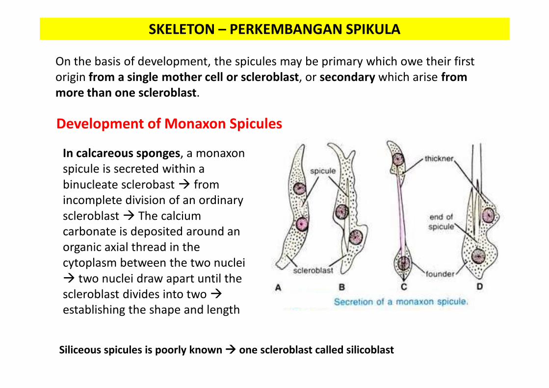
SKELETON – PERKEMBANGAN SPIKULA
On the basis of development, the spicules may be primary which owe their first origin from a single mother cell or scleroblast, or secondary which arise from more than one scleroblast.
Development of Monaxon Spicules
In calcareous sponges, a monaxonspicule is secreted within a binucleate sclerobast from incomplete division of an ordinary scleroblast The calcium carbonate is deposited around an organic axial thread in the cytoplasm between the two nuclei two nuclei draw apart until the scleroblast divides into twoestablishing the shape and length
Siliceous spicules is poorly known one scleroblast called silicoblast

SKELETON – PERKEMBANGAN SPIKULA
Development of Triaxon Spicules
Triaxon or triradiate calcareous spicules are secreted by three scleroblastswhich come together in triangle and divide in two, each into an inner founder and an outer thickner. Each pair secretes a minute spicule and these three rays are early united into a small triradiate spicule. Each ray is then completed in the same manner as a monaxon spicule. Later on, three rays or spicules unite together forming a triaxon or triradiate spicule.

SKELETON – PERKEMBANGAN SPIKULA
Development of Other Spicules
In the formation of quadriradiate or tetraxon spicules, the fourth ray is added to forming triradiate spicule by an additional scleroblast.
The hexactinal spicules of Hexactinellida arise in the centre of a multinucleate syncytial mass which is probably formed by repeated nuclear division of an original silicoblast.

TIPE KANAL SISTEM - ASCONOID• Simplest organization• Air mengalir ke sponge melalui microscopic
dermal pores • Aliran air gerakan flagella yg banyak pd
choanocyte• Choanocytes line internal cavity: Spongocoel• Choanocytesmenyaring air dan mengekstrak
partikel makanan • Used water a single large osculum• This design has distinct limitations
Choanocytes line the spongocoel can collect food only from water directly adjacent to the spongocoel wallSpongocoel to be large, most of the water and food in its central cavity would be inaccessible to choanocytes
• Asconoid sponges: Small & Tube-Shaped• Asconoids are found only in the Class Calcarea

TIPE KANAL SISTEM - ASCONOID
• Example:Leucosolenia (Gr. leukos, white, solen, pipe) slender, tubular individuals grow in groups attached by a commonstolon, or stem, to objects in shallow seawater
• Clathrina (L. clathri, latticework), another asconoid, has brightyellow, intertwined tubes
Leucosolenia Leucosolenia complicata
Clathrina clathrus

TIPE KANAL SISTEM - SYCONOID
• “Larger editions of asconoids”
• Tubular body and single osculum
• The body wall: thicker & more complex
• Folded outward to make choanocyte-lined canals
• Folding increases thesurface area of the wall and thus increases the surface area covered by choanocytes
• The canals are of small diameter most of the water in a canal is accessible to choanocytes
Water Dermal Ostia Incurrent Canalsfilters through Tiny Opening (Prosopyles) the Radial Canals Food is ingested by the choanocytes choanocytes’s flagella forces the used water through Internal Pores (Apopyles)Spongocoel Osculum

TIPE KANAL SISTEM - SYCONOID

TIPE KANAL SISTEM - SYCONOID
• Food capture does not occur in the syconoid spongocoel so it is lined with epithelial typecells
• Example: Sycons , Grantia• During development, syconoid sponges pass
through an asconoid stage, flagellated canals form by evagination of the body wall
• This developmental pattern provides evidencethat syconoid sponges were derived from an ancestor with an asconoid body plan, but the syconoid condition is not homologous among all the sponges that possess it.
• Syconoids : found in class Calcarea and some members of class Hexactinellida
Sycon ciliatum
Grantia

TIPE KANAL SISTEM - LEUCONOID
• Most complex • the surface area of the food-collecting
regions with choanocytes is greatly increased
• choanocytes line the walls of small chambers where they can effectively filter all the water present
• Most sponges are of the leuconoidtype
• Leuconoid bodies account for most species within class Calcarea and are the most common types in other classes.
Euspongia officinalis
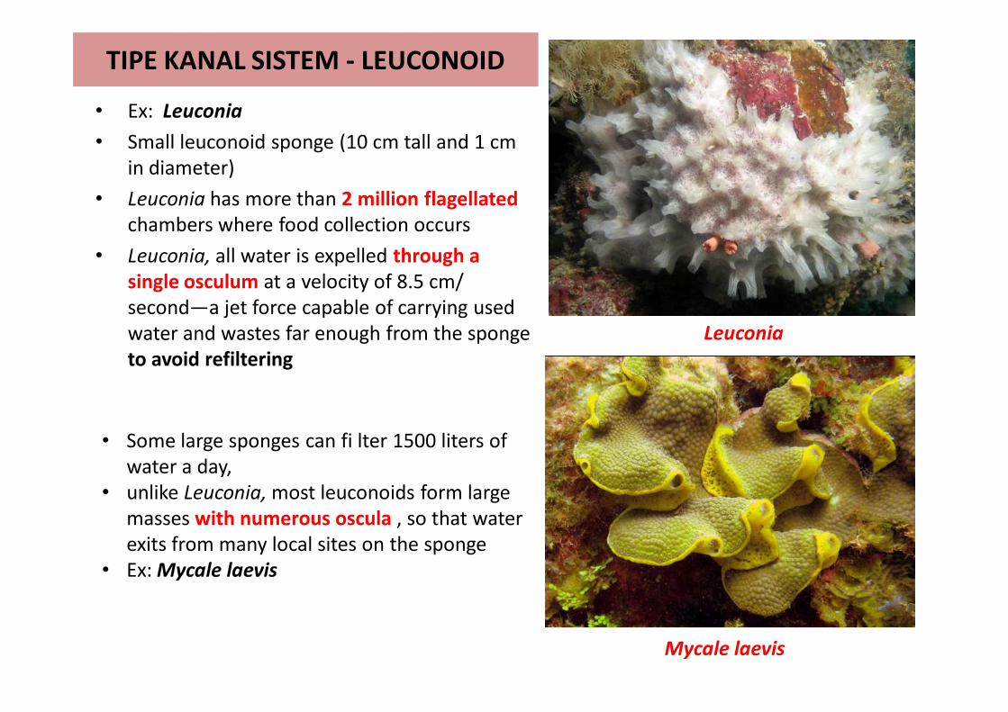
TIPE KANAL SISTEM - LEUCONOID
• Ex: Leuconia• Small leuconoid sponge (10 cm tall and 1 cm
in diameter)• Leuconia has more than 2 million flagellated
chambers where food collection occurs• Leuconia, all water is expelled through a
single osculum at a velocity of 8.5 cm/second—a jet force capable of carrying used water and wastes far enough from the sponge to avoid refiltering
Leuconia
• Some large sponges can fi lter 1500 liters of water a day,
• unlike Leuconia, most leuconoids form large masses with numerous oscula , so that water exits from many local sites on the sponge
• Ex: Mycale laevis
Mycale laevis

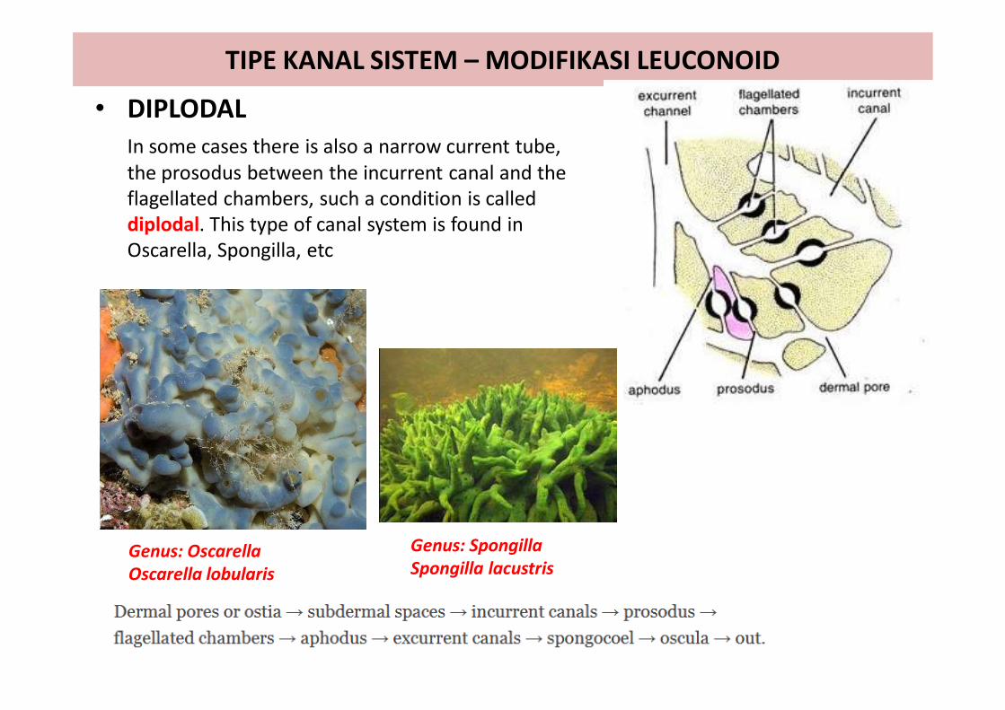
TIPE KANAL SISTEM – MODIFIKASI LEUCONOID
• EURYPYLOUSFlagellated chambers are wide and thimble-shaped, each opening directly into the excurrent canal by a wide aperture called apopyle and receive the water supply direct from the incurrent canal through the prosopyle.
Genus: Plakina Plakina kanaky

TIPE KANAL SISTEM – MODIFIKASI LEUCONOID
• APHODALFlagellated chambers are small and rounded. The opening of each flagellated chamber into the excurrent canal is drawn out into a narrow tube, usually not of great length, termed aphodus.
Genus: GeodiaGeodia neptuni
Genus: StelletaStelleta clavosa
TIPE KANAL SISTEM – MODIFIKASI LEUCONOID
• DIPLODAL In some cases there is also a narrow current tube, the prosodus between the incurrent canal and the flagellated chambers, such a condition is called diplodal. This type of canal system is found in Oscarella, Spongilla, etc
Genus: OscarellaOscarella lobularis
Genus: SpongillaSpongilla lacustris

TIPE KANAL SISTEM - RHAGON TYPE
• A broad base and it is conical in shape with a single osculum at the summit.
• The basal wall is termed the hypophare which is without of flagellated chambers. The upper wall bearing a row of small, oval flagellated chambers is called spongophare.
• Spongocoel is bordered by oval flagellated chambers opening into it by wide apopyles.
• Dermal pores or ostia open into sub-dermal spaces which extend below the entire surface of the body.
• Branching incurrent canals lead from the sub-dermal spaces into small flagellated chambers
• The flagellated chambers alone are lined by choanocytes.
Spongilla

REPRODUCTION - SEXUAL
Sycon
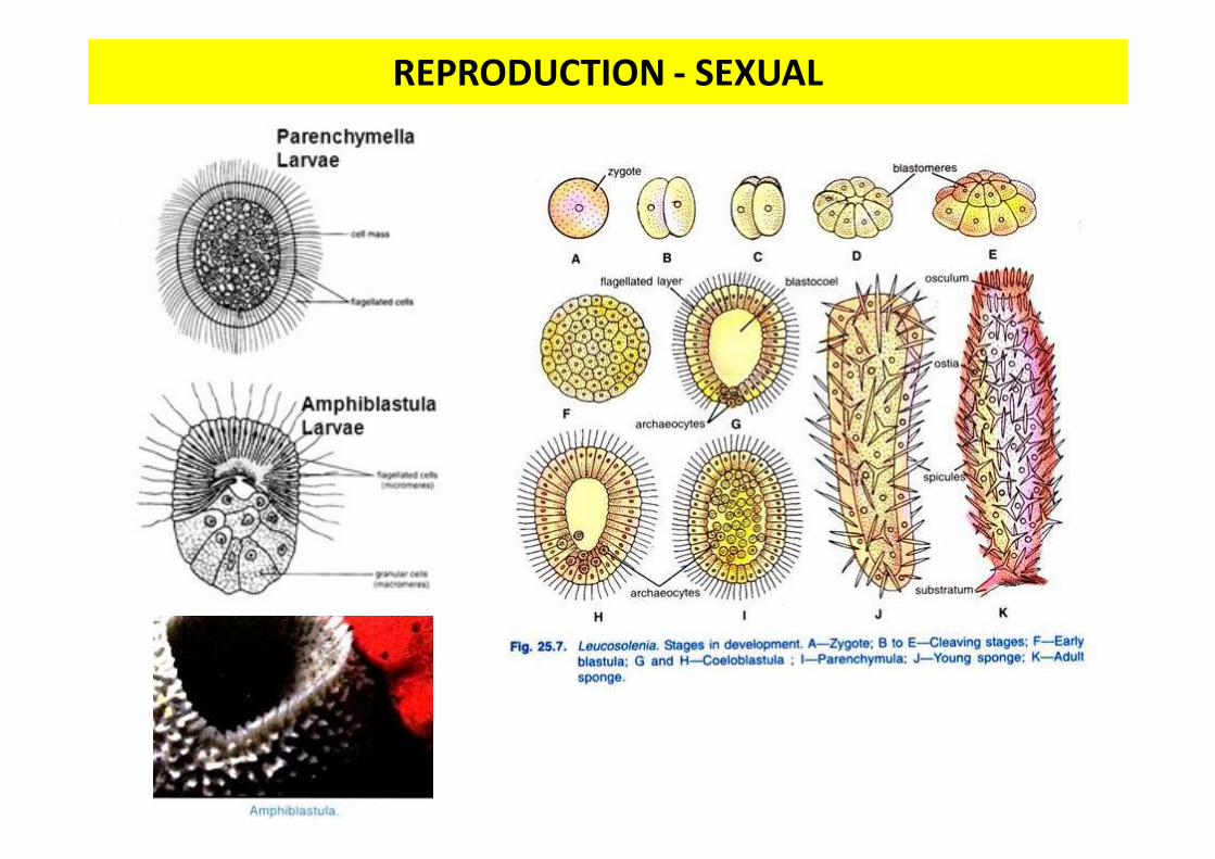
REPRODUCTION - SEXUAL

REPRODUCTION - SEXUAL

REPRODUCTION - ASEXUAL
Exogenous Budding

REPRODUCTION - ASEXUAL
Gemmule : Internal Bud formed internally in all freshwater
sponges and some marine sponges. Response to Hostile environment Resistant to Dessication (Drying
Out), Freezing, and Anoxia (Lack of Oxygen)
Endogenous Budding:
Made up of Amoebcytes surrounded by a layer of spicules

HABITAT - ECOLOGY


http://www.spongeguide.org/index.phpVisit:

Class Calcarea
CATATAN:

Class Demospongiae
CATATAN:

Class Demospongiae

Class HexactinellidaCATATAN:

Class Homoscleromorpha
Read:

PERAN SPONGE DALAM KEHIDUPAN

PERAN SPONGE DALAM KEHIDUPAN

Harmful Sponge

SOFTSKILL
“Aku rasa kau harus berhenti menilai orang dari
penampilannya saja “[Spongebob Squarepants]

TUGAS
TULIS DI BUKU TUGAS
1. Proses terbentuknya gemmule2. Lengkapi catatan
Review Jurnal
Related Documents