For personal use. Only reproduce with permission from The Lancet. SEMINAR Taenia solium infection and the resulting disease neurocysticercosis are endemic in less developed countries where pigs are raised as a food source. 1,2 Neurocysticercosis is common throughout Latin America, most of Asia, sub-Saharan Africa, and parts of Oceania, and is the greatest cause of acquired epilepsy worldwide. 3 It is now increasingly diagnosed in more developed countries owing to immigration of tapeworm carriers from endemic zones. 2,4 T solium has a complex two-host life cycle. Human beings are the only definitive host and harbour the adult tapeworm (taeniasis), whereas both people and pigs can act as intermediate hosts and harbour the larvae or cysticerci (figure 1). Taeniasis Taeniasis occurs only in the human host, after ingestion of undercooked pork infected with cysticerci. Although cysticercosis has been known for ages, its relation to the adult tapeworm was not clear until it was shown by Kuchenmaister in 1855; he fed condemned prisoners with cysticercosis-infected pork and recovered young tapeworms at autopsy. 5 The larvae evaginate in the small intestine; the head (scolex) attaches to the mucosa and begins forming segments (proglottids). T solium has a scolex with four suckers and a double crown of hooks, a narrow neck, and a large strobila measuring 2–4 m and consisting of several hundred proglottids (figure 2). About 2 months after infection, gravid proglottids begin to detach from the distal end and are excreted in the faeces; each segment contains 50–6010 3 fertile eggs. 6 The worm attaches strongly to the mucosa of the upper small intestine by means of its suckers and hooks. The adult tapeworm causes only mild inflammation at the implantation site, 7 without substantial damage to the Lancet 2003; 362: 547–56 Cysticercosis Unit, Instituto Nacional de Ciencias Neurologicas, Jr Ancash 1271, Barrios Altos, Lima, Peru (H H García MD); Department of Microbiology, Universidad Peruana Cayetano Heredia, Lima (H H García, R H Gilman MD); Department of Public Health, School of Veterinary Medicine, Universidad Nacional Mayor de San Marcos, Lima (A E Gonzalez DVM); Department of International Health, Johns Hopkins School of Hygiene and Public Health, Baltimore, MD, USA (A E Gonzalez, R H Gilman); and Imperial College of Science, Technology and Medicine, London, UK (C A W Evans MD) Correspondence to: Dr Héctor H García (e-mail [email protected]) intestine. 4 Taeniasis is characterised by mild symptoms or none at all. 4 Abdominal pain, distension, diarrhoea, and nausea have been attributed to tapeworm infestation, but there are no data from controlled experiments that demonstrate any association, and most patients seem to be free of symptoms. 4,6 In community settings many, if not most, carriers of T solium will neither look for medical care nor notice the tapeworm segments in their stools. Conversely, most patients infected with Taenia saginata notice passage of proglottids, which are motile, more numerous, and larger than those of T solium. 8 Identification of T solium infections is important because of the risk of cysticercosis in the carrier or the immediate environment. Taenia solium cysticercosis Héctor H García, Armando E Gonzalez, Carlton A W Evans, Robert H Gilman, for the Cysticercosis Working Group in Peru The larval stage of the pork tapeworm (Taenia solium) infects the human nervous system, causing neurocysticercosis. This disease is one of the main causes of epileptic seizures in many less developed countries and is also increasingly seen in more developed countries because of immigration from endemic areas. Little information is available on the natural evolution of taeniasis or cysticercosis. Available therapeutic measures include steroids, treatments for symptoms, surgery, and, more controversially, antiparasitic drugs to kill brain parasites. Efforts to control and eliminate this disease are underway through antiparasitic treatment of endemic populations, development of pig vaccines, and other measures. Seminar THE LANCET • Vol 361 • August 16, 2003 • www.thelancet.com 547 Human being (Definitive host) Ingestion of infected pork, poorly cooked: Taeniasis Ingestion of T solium eggs by faecal contamination: Human cysticercosis Ingestion of T solium eggs or proglottids: Porcine cysticercosis Pig (Intermediate host) Search strategy This seminar is based on papers published in the past 15 years in English, Spanish, or Portuguese selected from MEDLINE by use of the PubMed system with keywords “cysticercosis”, “neurocysticercosis”, “Taenia solium”, “epilepsy”, and “seizures”. Older articles were selected from the private collections of the authors and the archives of the Cysticercosis Working Group in Peru. Figure 1: Life cycle of Taenia solium

Welcome message from author
This document is posted to help you gain knowledge. Please leave a comment to let me know what you think about it! Share it to your friends and learn new things together.
Transcript

For personal use. Only reproduce with permission from The Lancet.
SEMINAR
Taenia solium infection and the resulting diseaseneurocysticercosis are endemic in less developed countrieswhere pigs are raised as a food source.1,2 Neurocysticercosisis common throughout Latin America, most of Asia, sub-Saharan Africa, and parts of Oceania, and is thegreatest cause of acquired epilepsy worldwide.3 It is now increasingly diagnosed in more developed countriesowing to immigration of tapeworm carriers from endemiczones.2,4
T solium has a complex two-host life cycle. Humanbeings are the only definitive host and harbour the adulttapeworm (taeniasis), whereas both people and pigs canact as intermediate hosts and harbour the larvae orcysticerci (figure 1).
TaeniasisTaeniasis occurs only in the human host, after ingestion ofundercooked pork infected with cysticerci. Althoughcysticercosis has been known for ages, its relation to theadult tapeworm was not clear until it was shown byKuchenmaister in 1855; he fed condemned prisoners withcysticercosis-infected pork and recovered youngtapeworms at autopsy.5 The larvae evaginate in the smallintestine; the head (scolex) attaches to the mucosa andbegins forming segments (proglottids). T solium has ascolex with four suckers and a double crown of hooks, anarrow neck, and a large strobila measuring 2–4 m andconsisting of several hundred proglottids (figure 2). About2 months after infection, gravid proglottids begin todetach from the distal end and are excreted in the faeces;each segment contains 50–60�103 fertile eggs.6
The worm attaches strongly to the mucosa of the uppersmall intestine by means of its suckers and hooks. Theadult tapeworm causes only mild inflammation at theimplantation site,7 without substantial damage to the
Lancet 2003; 362: 547–56
Cysticercosis Unit, Instituto Nacional de Ciencias Neurologicas, Jr Ancash 1271, Barrios Altos, Lima, Peru (H H García MD);Department of Microbiology, Universidad Peruana CayetanoHeredia, Lima (H H García, R H Gilman MD); Department of PublicHealth, School of Veterinary Medicine, Universidad Nacional Mayorde San Marcos, Lima (A E Gonzalez DVM); Department ofInternational Health, Johns Hopkins School of Hygiene and PublicHealth, Baltimore, MD, USA (A E Gonzalez, R H Gilman); andImperial College of Science, Technology and Medicine, London, UK (C A W Evans MD)
Correspondence to: Dr Héctor H García(e-mail [email protected])
intestine.4 Taeniasis is characterised by mild symptoms ornone at all.4 Abdominal pain, distension, diarrhoea, andnausea have been attributed to tapeworm infestation, butthere are no data from controlled experiments thatdemonstrate any association, and most patients seem tobe free of symptoms.4,6 In community settings many, if notmost, carriers of T solium will neither look for medical carenor notice the tapeworm segments in their stools.Conversely, most patients infected with Taenia saginatanotice passage of proglottids, which are motile, morenumerous, and larger than those of T solium.8
Identification of T solium infections is important becauseof the risk of cysticercosis in the carrier or the immediateenvironment.
Taenia solium cysticercosis
Héctor H García, Armando E Gonzalez, Carlton A W Evans, Robert H Gilman, for the Cysticercosis Working Group in Peru
The larval stage of the pork tapeworm (Taenia solium) infects the human nervous system, causing neurocysticercosis. Thisdisease is one of the main causes of epileptic seizures in many less developed countries and is also increasingly seen inmore developed countries because of immigration from endemic areas. Little information is available on the naturalevolution of taeniasis or cysticercosis. Available therapeutic measures include steroids, treatments for symptoms,surgery, and, more controversially, antiparasitic drugs to kill brain parasites. Efforts to control and eliminate this diseaseare underway through antiparasitic treatment of endemic populations, development of pig vaccines, and other measures.
Seminar
THE LANCET • Vol 361 • August 16, 2003 • www.thelancet.com 547
Human being(Definitive host)
Ingestion of infected pork,poorly cooked: Taeniasis
Ingestion of T solium eggsby faecal contamination:Humancysticercosis
Ingestion of T soliumeggs or proglottids:Porcine cysticercosis
Pig(Intermediate host)
Search strategy
This seminar is based on papers published in the past 15 years in English, Spanish, or Portuguese selected fromMEDLINE by use of the PubMed system with keywords“cysticercosis”, “neurocysticercosis”, “Taenia solium”,“epilepsy”, and “seizures”. Older articles were selected fromthe private collections of the authors and the archives of theCysticercosis Working Group in Peru.
Figure 1: Life cycle of Taenia solium
For personal use. Only reproduce with permission from The Lancet.
The frequency of autoinfection in individuals withtaeniasis is not known. Dixon and Lipscomb9 noted thatnearly 25% of patients with neurocysticercosis either hadharboured or were harbouring a tapeworm. Up to 15%of patients harbour a tapeworm at the time of diagnosisof neurocysticercosis,10 and the proportion withtapeworms is directly related to the number of cerebralparasites,10,11 which strongly suggests autoinfection. Inother cases the tapeworm carrier can be found in the patient’s household.12 Despite the poor sensitivity of stool examinations, parasitological screening of thepatient and household members is recommended so thatsources of infection can be detected and eliminated.
The lifespan of the adult T solium is also unknown.Parasitology textbooks and reviews cite 20–25 years, onthe basis of anecdotal cases.8,13 Age-specific data fromlater studies suggest a much shorter lifespan, probablyless than 5 years.14–16
Diagnosis of taeniasisTwo problems hamper the diagnosis of infection withT solium: the poor sensitivity of stool microscopy, and the morphological similarity between the eggs of T solium and T saginata. On the rare occasions whenthe worm scolex is found, the presence of the doublecrown of hooks (present only in T solium) providesdefinite species identification. The morphology of the genitalia in mature proglottids or that of the graviduterus in the distal proglottids can also enable theidentification of species.4 Haematoxylin-eosin staining of histological sections of proglottids can help.17
Mature, gravid proglottids are rarely available, andmorphological diagnosis of species on the basis ofproglottid materials is technically difficult. In ourexperience, cleansing the intestine with a purgeimmediately before treatment improves the recovery ofparasite material including the scolex, allowing speciesidentification.
Visualisation of taenia eggs by microscopy was theonly diagnostic method available until the early 1990s.Perianal scraping with adhesive tape (Graham’s test) is highly sensitive for T saginata but not for T solium.18,19
The best available diagnostic assay for intestinaltaeniasis is a coproantigen detection ELISA, whichdetects taenia-specific molecules in faecal samples,demonstrating current tapeworm infection.20 It hassensitivity of about 95% and specificity greater than 99% and is an effective tool for epidemiologicalstudies.15,20 Coproantigen detection assays confirmedthat microscopy was poorly sensitive, missing 60–70% of cases. Diverse DNA-based assays, mainly todiscriminate T solium from T saginata infections,17,21 anda serological assay for specific identification of tapewormcarriers22 have been described lately.
Treatment of taeniasisThe two available drugs are niclosamide and praziquantel.Niclosamide is the drug of choice because it is notabsorbed from the intestinal lumen.23 With praziquantelthere is a small risk that asymptomatic viable brain cystsare affected by the drug in serum, causing neurologicalsymptoms (headache, seizures).24 The usual dose is 2 gorally in a single dose for niclosamide, and 5–10 mg/kgorally in a sole dose for praziquantel. Both drugs have verylimited marketing and are difficult to find.
Human cysticercosisCysticercosis is infection with the larval stage of theparasite. Human beings acquire cysticercosis throughfaecal-oral contamination with T solium eggs fromtapeworm carriers.1,4,25 Thus, vegetarians and other people who do not eat pork can acquire cysticercosis.26
Water, wind, flies, and other indirect means of infectionplay little part in transmission.27 Internal autoinfection byregurgitation of proglottids into the stomach in taeniasishas been proposed but not proven.1,27
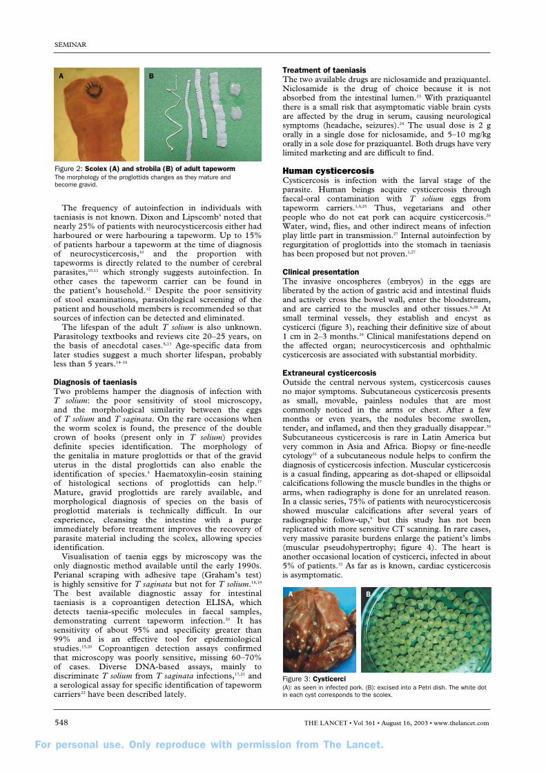
Clinical presentationThe invasive oncospheres (embryos) in the eggs areliberated by the action of gastric acid and intestinal fluidsand actively cross the bowel wall, enter the bloodstream,and are carried to the muscles and other tissues.6,28 Atsmall terminal vessels, they establish and encyst ascysticerci (figure 3), reaching their definitive size of about1 cm in 2–3 months.29 Clinical manifestations depend onthe affected organ; neurocysticercosis and ophthalmiccysticercosis are associated with substantial morbidity.
Extraneural cysticercosisOutside the central nervous system, cysticercosis causesno major symptoms. Subcutaneous cysticercosis presentsas small, movable, painless nodules that are mostcommonly noticed in the arms or chest. After a fewmonths or even years, the nodules become swollen,tender, and inflamed, and then they gradually disappear.30
Subcutaneous cysticercosis is rare in Latin America butvery common in Asia and Africa. Biopsy or fine-needlecytology31 of a subcutaneous nodule helps to confirm thediagnosis of cysticercosis infection. Muscular cysticercosisis a casual finding, appearing as dot-shaped or ellipsoidalcalcifications following the muscle bundles in the thighs orarms, when radiography is done for an unrelated reason.In a classic series, 75% of patients with neurocysticercosisshowed muscular calcifications after several years ofradiographic follow-up,9 but this study has not beenreplicated with more sensitive CT scanning. In rare cases,very massive parasite burdens enlarge the patient’s limbs(muscular pseudohypertrophy; figure 4). The heart isanother occasional location of cysticerci, infected in about5% of patients.32 As far as is known, cardiac cysticercosisis asymptomatic.
SEMINAR
548 THE LANCET • Vol 361 • August 16, 2003 • www.thelancet.com
Figure 2: Scolex (A) and strobila (B) of adult tapewormThe morphology of the proglottids changes as they mature and become gravid.
Figure 3: Cysticerci(A): as seen in infected pork. (B): excised into a Petri dish. The white dotin each cyst corresponds to the scolex.
For personal use. Only reproduce with permission from The Lancet.
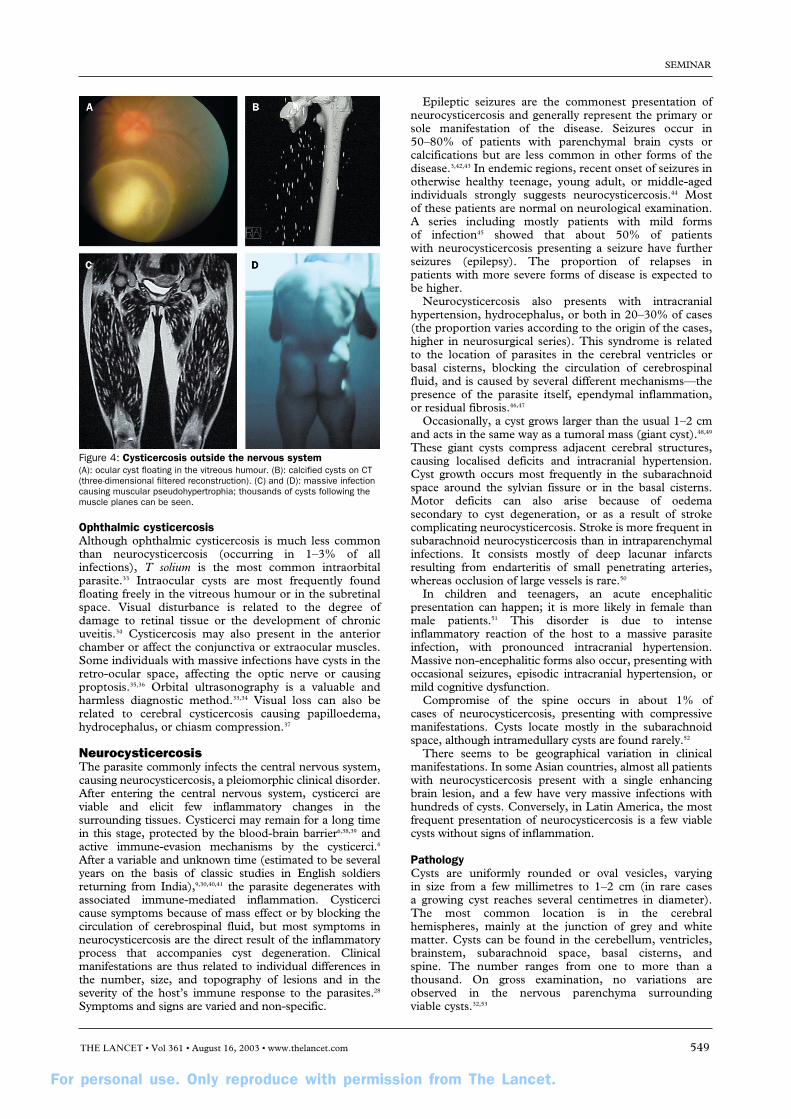
Ophthalmic cysticercosisAlthough ophthalmic cysticercosis is much less commonthan neurocysticercosis (occurring in 1–3% of allinfections), T solium is the most common intraorbitalparasite.33 Intraocular cysts are most frequently foundfloating freely in the vitreous humour or in the subretinalspace. Visual disturbance is related to the degree ofdamage to retinal tissue or the development of chronicuveitis.34 Cysticercosis may also present in the anteriorchamber or affect the conjunctiva or extraocular muscles.Some individuals with massive infections have cysts in theretro-ocular space, affecting the optic nerve or causingproptosis.35,36 Orbital ultrasonography is a valuable andharmless diagnostic method.33,34 Visual loss can also berelated to cerebral cysticercosis causing papilloedema,hydrocephalus, or chiasm compression.37
NeurocysticercosisThe parasite commonly infects the central nervous system,causing neurocysticercosis, a pleiomorphic clinical disorder.After entering the central nervous system, cysticerci areviable and elicit few inflammatory changes in thesurrounding tissues. Cysticerci may remain for a long timein this stage, protected by the blood-brain barrier6,38,39 andactive immune-evasion mechanisms by the cysticerci.6
After a variable and unknown time (estimated to be severalyears on the basis of classic studies in English soldiersreturning from India),9,30,40,41 the parasite degenerates withassociated immune-mediated inflammation. Cysticercicause symptoms because of mass effect or by blocking thecirculation of cerebrospinal fluid, but most symptoms inneurocysticercosis are the direct result of the inflammatoryprocess that accompanies cyst degeneration. Clinicalmanifestations are thus related to individual differences inthe number, size, and topography of lesions and in theseverity of the host’s immune response to the parasites.28
Symptoms and signs are varied and non-specific.
Epileptic seizures are the commonest presentation ofneurocysticercosis and generally represent the primary orsole manifestation of the disease. Seizures occur in50–80% of patients with parenchymal brain cysts orcalcifications but are less common in other forms of thedisease.3,42,43 In endemic regions, recent onset of seizures inotherwise healthy teenage, young adult, or middle-agedindividuals strongly suggests neurocysticercosis.44 Mostof these patients are normal on neurological examination.A series including mostly patients with mild forms of infection45 showed that about 50% of patients with neurocysticercosis presenting a seizure have furtherseizures (epilepsy). The proportion of relapses in patients with more severe forms of disease is expected tobe higher.
Neurocysticercosis also presents with intracranialhypertension, hydrocephalus, or both in 20–30% of cases(the proportion varies according to the origin of the cases,higher in neurosurgical series). This syndrome is relatedto the location of parasites in the cerebral ventricles orbasal cisterns, blocking the circulation of cerebrospinalfluid, and is caused by several different mechanisms—thepresence of the parasite itself, ependymal inflammation,or residual fibrosis.46,47
Occasionally, a cyst grows larger than the usual 1–2 cmand acts in the same way as a tumoral mass (giant cyst).48,49
These giant cysts compress adjacent cerebral structures,causing localised deficits and intracranial hypertension.Cyst growth occurs most frequently in the subarachnoidspace around the sylvian fissure or in the basal cisterns.Motor deficits can also arise because of oedema secondary to cyst degeneration, or as a result of strokecomplicating neurocysticercosis. Stroke is more frequent insubarachnoid neurocysticercosis than in intraparenchymalinfections. It consists mostly of deep lacunar infarctsresulting from endarteritis of small penetrating arteries,whereas occlusion of large vessels is rare.50
In children and teenagers, an acute encephaliticpresentation can happen; it is more likely in female thanmale patients.51 This disorder is due to intenseinflammatory reaction of the host to a massive parasiteinfection, with pronounced intracranial hypertension.Massive non-encephalitic forms also occur, presenting withoccasional seizures, episodic intracranial hypertension, ormild cognitive dysfunction.
Compromise of the spine occurs in about 1% of cases of neurocysticercosis, presenting with compressivemanifestations. Cysts locate mostly in the subarachnoidspace, although intramedullary cysts are found rarely.52
There seems to be geographical variation in clinicalmanifestations. In some Asian countries, almost all patientswith neurocysticercosis present with a single enhancingbrain lesion, and a few have very massive infections withhundreds of cysts. Conversely, in Latin America, the mostfrequent presentation of neurocysticercosis is a few viablecysts without signs of inflammation.
PathologyCysts are uniformly rounded or oval vesicles, varying in size from a few millimetres to 1–2 cm (in rare cases a growing cyst reaches several centimetres in diameter).The most common location is in the cerebralhemispheres, mainly at the junction of grey and whitematter. Cysts can be found in the cerebellum, ventricles,brainstem, subarachnoid space, basal cisterns, and spine. The number ranges from one to more than athousand. On gross examination, no variations areobserved in the nervous parenchyma surrounding viable cysts.32,53
SEMINAR
THE LANCET • Vol 361 • August 16, 2003 • www.thelancet.com 549
Figure 4: Cysticercosis outside the nervous system(A): ocular cyst floating in the vitreous humour. (B): calcified cysts on CT(three-dimensional filtered reconstruction). (C) and (D): massive infectioncausing muscular pseudohypertrophia; thousands of cysts following themuscle planes can be seen.
For personal use. Only reproduce with permission from The Lancet.
Vesicles vary in contents according to their evolutionarystage. Viable cysts have a translucent membrane throughwhich the scolex is visible as a small 2–3 mm nodule.When the cyst starts to degenerate, the vesicular fluidbecomes opaque and dense, and the edges of the cystbecome irregular and shrink. Later, calcification starts inthe cephalic portion and progresses to the vesicular wall,to leave a round, whitish, calcified nodule. In rare cases,all the evolutionary phases can be seen in the sameindividual.54
Intraventricular cysticercosis is generally single and freeinside the cavities. Infrequently, the cyst moves from oneventricular cavity to another.55,56 Classically, cysts aredescribed as locating more frequently in the lower cavities:most frequently in the fourth ventricle, less commonly inthe third, and least frequently in the lateral ventricles.57
Intraventricular parasites can block circulation ofcerebrospinal fluid or produce inflammation of theependyma and choroid plexus.
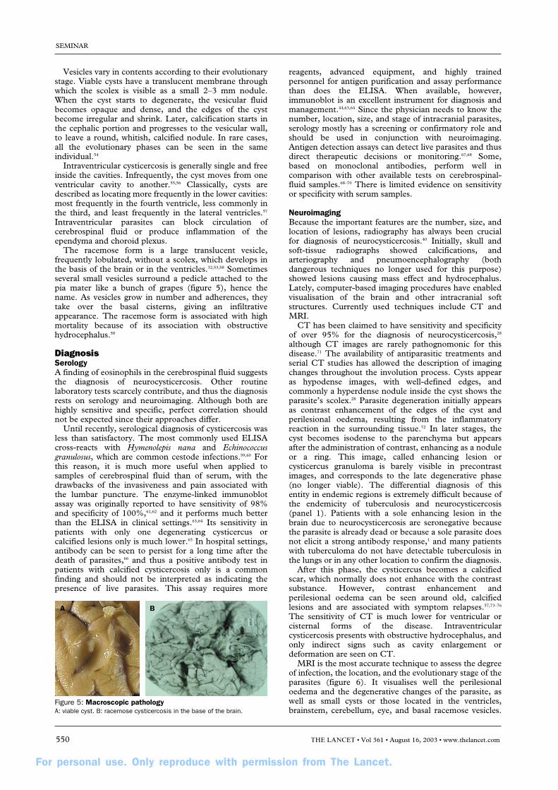
The racemose form is a large translucent vesicle,frequently lobulated, without a scolex, which develops inthe basis of the brain or in the ventricles.32,53,58 Sometimesseveral small vesicles surround a pedicle attached to thepia mater like a bunch of grapes (figure 5), hence thename. As vesicles grow in number and adherences, theytake over the basal cisterns, giving an infiltrativeappearance. The racemose form is associated with highmortality because of its association with obstructivehydrocephalus.58
DiagnosisSerologyA finding of eosinophils in the cerebrospinal fluid suggeststhe diagnosis of neurocysticercosis. Other routinelaboratory tests scarcely contribute, and thus the diagnosisrests on serology and neuroimaging. Although both arehighly sensitive and specific, perfect correlation shouldnot be expected since their approaches differ.
Until recently, serological diagnosis of cysticercosis wasless than satisfactory. The most commonly used ELISAcross-reacts with Hymenolepis nana and Echinococcusgranulosus, which are common cestode infections.59,60 Forthis reason, it is much more useful when applied tosamples of cerebrospinal fluid than of serum, with thedrawbacks of the invasiveness and pain associated withthe lumbar puncture. The enzyme-linked immunoblotassay was originally reported to have sensitivity of 98%and specificity of 100%,61,62 and it performs much betterthan the ELISA in clinical settings.63,64 Its sensitivity inpatients with only one degenerating cysticercus orcalcified lesions only is much lower.65 In hospital settings,antibody can be seen to persist for a long time after thedeath of parasites,66 and thus a positive antibody test inpatients with calcified cysticercosis only is a commonfinding and should not be interpreted as indicating thepresence of live parasites. This assay requires more
reagents, advanced equipment, and highly trainedpersonnel for antigen purification and assay performancethan does the ELISA. When available, however,immunoblot is an excellent instrument for diagnosis andmanagement.44,63,64 Since the physician needs to know thenumber, location, size, and stage of intracranial parasites,serology mostly has a screening or confirmatory role andshould be used in conjunction with neuroimaging.Antigen detection assays can detect live parasites and thusdirect therapeutic decisions or monitoring.67,68 Some,based on monoclonal antibodies, perform well incomparison with other available tests on cerebrospinal-fluid samples.68–70 There is limited evidence on sensitivityor specificity with serum samples.
NeuroimagingBecause the important features are the number, size, andlocation of lesions, radiography has always been crucialfor diagnosis of neurocysticercosis.40 Initially, skull andsoft-tissue radiographs showed calcifications, andarteriography and pneumoencephalography (bothdangerous techniques no longer used for this purpose)showed lesions causing mass effect and hydrocephalus.Lately, computer-based imaging procedures have enabledvisualisation of the brain and other intracranial softstructures. Currently used techniques include CT andMRI.
CT has been claimed to have sensitivity and specificityof over 95% for the diagnosis of neurocysticercosis,28
although CT images are rarely pathognomonic for thisdisease.71 The availability of antiparasitic treatments andserial CT studies has allowed the description of imagingchanges throughout the involution process. Cysts appearas hypodense images, with well-defined edges, andcommonly a hyperdense nodule inside the cyst shows theparasite’s scolex.28 Parasite degeneration initially appearsas contrast enhancement of the edges of the cyst andperilesional oedema, resulting from the inflammatoryreaction in the surrounding tissue.72 In later stages, thecyst becomes isodense to the parenchyma but appearsafter the administration of contrast, enhancing as a noduleor a ring. This image, called enhancing lesion orcysticercus granuloma is barely visible in precontrastimages, and corresponds to the late degenerative phase(no longer viable). The differential diagnosis of this entity in endemic regions is extremely difficult because ofthe endemicity of tuberculosis and neurocysticercosis(panel 1). Patients with a sole enhancing lesion in thebrain due to neurocysticercosis are seronegative becausethe parasite is already dead or because a sole parasite doesnot elicit a strong antibody response,1 and many patientswith tuberculoma do not have detectable tuberculosis inthe lungs or in any other location to confirm the diagnosis.
After this phase, the cysticercus becomes a calcifiedscar, which normally does not enhance with the contrastsubstance. However, contrast enhancement andperilesional oedema can be seen around old, calcifiedlesions and are associated with symptom relapses.57,73–76
The sensitivity of CT is much lower for ventricular orcisternal forms of the disease. Intraventricularcysticercosis presents with obstructive hydrocephalus, andonly indirect signs such as cavity enlargement ordeformation are seen on CT.
MRI is the most accurate technique to assess the degreeof infection, the location, and the evolutionary stage of theparasites (figure 6). It visualises well the perilesionaloedema and the degenerative changes of the parasite, aswell as small cysts or those located in the ventricles,brainstem, cerebellum, eye, and basal racemose vesicles.
SEMINAR
550 THE LANCET • Vol 361 • August 16, 2003 • www.thelancet.com
Figure 5: Macroscopic pathologyA: viable cyst. B: racemose cysticercosis in the base of the brain.

For personal use. Only reproduce with permission from The Lancet.
CT, however, is more sensitive for the detection ofcalcifications.77 The main disadvantages of MRI are itshigh cost and scarce availability.
A set of objective diagnostic criteria has beenproposed.78 Absolute criteria include histologicaldemonstration of the parasite, cystic lesions with scolex onneuroimaging, or direct visualisation of ocular cysts;major criteria include suggestive lesions on neuroimaging,positive serum immunoblot, resolution of cysts afterantiparasitic therapy, or spontaneous resolution ofenhancing lesions; minor criteria are compatible lesionson neuroimaging, compatible clinical manifestations,positive cerebrospinal-fluid ELISA, or cysticercosisoutside the central nervous system; and epidemiologicalcriteria include household contact, living in an endemicarea, or travel to endemic areas. Combinations of thesecriteria provide different degrees of diagnostic certainty.
Prognosis and natural evolutionPanel 2 shows research needs in T solium infection. Thereare few data on the natural evolution of neuro-cysticercosis, and those available are from historicalcontrols in open treatment trials. As far as is known, theevolution of intraparenchymal neurocysticercosis follows afavourable course with degeneration of parasites andformation of residual calcified scars.79 When the parasiteslocate in the subarachnoid space or the ventricles, themorbidity and mortality are much higher because ofparasite growth, intracranial hypertension, arachnoiditis,blockage of cerebrospinal-fluid pathways, hydrocephalus,and other complications.49,80,81
TreatmentCysticercosis outside the nervous system is a benigndisorder and does not merit specific treatment.Neurocysticercosis, however, is associated withsubstantial morbidity and mortality. Until 1978, the onlytreatments available were surgery for cyst excision orventricular shunts or steroids to decrease inflammation.Praziquantel, first used in porcine cysticercosis, was thefirst effective specific antiparasitic drug.82–84 Later,albendazole added a cheaper and more effectivealternative.85,86 Transient worsening of neurologicalsymptoms can be expected during antiparasitic therapy,secondary to the perilesional inflammatory reaction. Inmost cases, symptoms are limited to mild to moderateheadache and are easily controlled with steroids.Although antiparasitic agents in neurocysticercosis havebeen accepted by most of the medical community, somebelieve that, since the parasite starts to cause symptomsat the time of its death, the use of parasiticidal drugsleads to an unnecessary increase in the inflammatoryprocess.79,87 The issue led to heated discussion.88–90 Themain argument is whether antiparasitic treatment ornatural involution of a cyst will lead to reduced scarringand thus improved prognosis in terms of epilepsyevolution.
Many reports have been published on the use ofantiparasitic treatment in neurocysticercosis.82–86,90–94
However, non-treatment comparison groups wereincluded in only four studies, three of which includedpatients with enhancing lesions (parasites that havealready degenerated). A summary of controlled studiescan be found in the Cochrane Library,95 although nooverall conclusion can be reached because the types ofdisease and the findings differ substantially. Two double-blind, placebo-controlled trials assessed patients with asole enhancing lesion. Padma and colleagues93 foundsimilar resolution rates on 3-month follow-up CT ingroups assigned albendazole or placebo.93 By contrast,Baranwal and co-workers91,92 found better resolution onimaging and fewer seizures in the albendazole group thanin controls during follow-up. A third double-blind,placebo-controlled trial in India compared the evolutionof parenchymal neurocysticercosis lesions (including cystsand enhancing lesions) on CT at 3 months aftertreatment94 and showed similar resolution rates. The onlycontrolled trial in non-enhancing intraparenchymal cystshas been an open-label study comparing radiological andclinical (seizure relapses) evolution between patientstreated with albendazole, praziquantel, or no antiparasitic
SEMINAR
THE LANCET • Vol 361 • August 16, 2003 • www.thelancet.com 551
Figure 6: NeuroimagingMRI of viable (A) and degenerating (B) cysts, and CT of calcified cysticerci (C).
Panel 1: Differential diagnosis of neurocysticercosison neuroimaging
Sole non-enhancing cystic lesionHydatid diseaseArachnoid cystsPorencephalyCystic astrocytomaColloid cyst (third ventricle)Several non-enhancing cystic lesionsMultiple metastasesHydatid disease (rare)Enhancing lesionsTuberculosisMycosisToxoplasmosisAbscessEarly gliomaMetastasisArteriovenous malformationCalcificationsTuberous sclerosisTuberculosisCytomegalovirus infectionToxoplasmosis
Panel 2: Research needs in T solium infection
ClinicalImproved knowledge of natural historyConcrete determination of benefits and specific indications ofantiparasitic therapy in neurocysticercosisTesting of new antiparasitic and anti-inflammatory agentsImmunologicalDevelopment of field-applicable, specific assays for humantaeniasisDevelopment of field-applicable, specific assays for viablecysticercosisPrevention and controlSystematic assessment of efficacy and effectiveness of allpotential intervention measures (including pig vaccines)Estimation of infection and disease burden attributable toneurocysticercosisEstimation of economic losses due to porcine cysticercosis

For personal use. Only reproduce with permission from The Lancet.
therapy.90 All three groups received prednisone. The studyfound no differences in the proportions of patients free ofcysts at 6 months and 1 year after treatment, or in therelative reduction in numbers of cysts and no difference inthe proportion of patients free of seizures during the entirefollow-up.
Antiparasitic treatment may be unnecessary or evencontraindicated in some cases. Without doubt however,there are subgroups of patients with neurocysticercosiswho not only benefit from antiparasitic therapy but also might undergo progression of disease and even die if not treated, such as those with giant cysts orsubarachnoid cysticercosis.48,49 Similarly, complications in the treatment of neurocysticercosis with antiparasiticdrugs can be severe, but they are very rare. Until a prospective, randomised study has been completed, asimple summary is that antiparasitic agents are effective to destroy cysticerci in the human brain, at the cost of acute inflammation. Whether this is the most beneficial option for patients with intraparenchymalcysticercosis in the long term is not yet clear. Thecurrently accepted schemes are either 8 days ofalbendazole treatment (15 mg/kg daily) with simultaneousadministration of steroids, or 15 days of praziquantel (50 mg/kg daily).96 Shorter schemes of 1 day ofpraziquantel or 3 days of albendazole seem effective for patients with only one lesion, but not for those withmany cysts.97,98
A panel of experts analysed the current consensus anddisagreements in the management of neurocysticercosis.99
Their main conclusions were that therapeutic decisionsshould be adapted to the individual and should be based on the number, location, and viability of theparasites within the nervous system; that growingcysticerci should be actively managed by eitherantiparasitic drugs or surgical excision; that themanagement of intracranial hypertension secondary toneurocysticercosis should take a high priority; and thatadequate management of seizures should be ensured.Specifically, they agreed on the management of patients with moderate infections and viable cysts(antiparasitic treatment, with steroids); calcifiedcysticercosis (no antiparasitic treatment); ventricularcysticercosis (neuroendoscopic removal, when available);subarachnoid cysts, including giant cysts or racemosecysticercosis, and chronic meningitis (antiparasitictreatment with steroids, ventricular shunt if there ishydrocephalus); cysticercotic encephalitis (no antiparasitictreatment, high-dose steroids, and osmotic diuretics);hydrocephalus with no visible cysts on neuroimaging(ventricular shunt, no antiparasitic treatment); spinalcysticercosis, intramedullary or extramedullary (primarilysurgical); or ophthalmic cysticercosis (surgical resection of cysts). There were disagreements about the use ofantiparasitic drugs (considered unnecessary or of high risk by some experts) in patients with only one or a few viable cysts, patients with massive infections with viable cysts, or patients with many degeneratingcysts.99
Treatment of symptoms has an important role in themanagement of neurocysticercosis. Seizures generallyrespond well to first-line antiepileptic drugs,43,100 butcomplete withdrawal of antiepileptic therapy is difficult to achieve.100 Steroids, dexamethasone or prednisone, are helpful in oedema or intracranial hypertension.
Porcine cysticercosisIn places with inadequate disposal of human faeces, pigsingest stools containing T solium eggs. Cysticerci lodge
anywhere in the body of the pig, most commonly in themuscle and subcutaneous fat. Pigs with cysts in the brainare a subset of those with more cysts in the carcass(Cysticercosis Working Group in Peru 2002, unpublisheddata). Although some pigs have massive infections,porcine cysticercosis is rarely associated with symptoms ofany kind. Most pigs are killed before the age of 9 months,which is too short a time for the cysts to reach thedegenerative stage that is associated with symptoms inhuman beings.
An inexpensive treatment for porcine cysticercosismight increase the value of pigs and block transmission of infection. A veterinary benzimidazole, oxfendazole, ismore than 95% effective in killing the cysts in the pigwhen given in a single dose of 30 mg/kg.101–103 This drughas not yet been tested in human beings.
Immunity to the oncosphere stage plays a central part in regulation of transmission of taeniid cestodes, andoncosphere-based vaccines are highly protective for T ovis, Echinococcus granulosus, and T saginata.104 Partialprotection has been achieved by vaccination of healthypigs with T solium oncospheral antigens.105–107
EpidemiologyCysticercosis affects thousands of individuals in lessdeveloped countries1,3 and in more developed countrieswith a high rate of immigration from endemic areas.2,4 Inendemic countries taeniasis/cysticercosis is extremelycommon, and neurologically symptomatic individuals,although many, represent only the tip of the iceberg. Inmost endemic villages more than 10% of the generalpopulation are seropositive, and the proportion can reach 25%. In population-based studies, 10–18% ofasymptomatic individuals have CT features that suggestneurocysticercosis, mainly brain calcifications inseronegative individuals.108–111
Transient seropositive reactions112 and common cases of asymptomatic calcified neurocysticercosis108–111 areconsistent with a threshold under which parasitedestruction occurs, potentially determined by the numberof parasites, the immune status of the host, the age atinfection, or any combination of these factors. Animalmodels suggest that the host’s immune response canresolve mild infections at early stages.113 If this resolutionoccurs in human neurocysticercosis, it could explain thefrequency of degenerating cysts clustered in younger ages,the relation to infections with a single parasite (90% ofcases with only degenerating cysts), and the frequency inIndia, where, because of vegetarian habits, direct exposureto tapeworm carriers is less frequent.114 Patients withviable cysts only would represent cases in which theparasites survive and establish.
Few data are available on the epidemiology of adulttapeworm infections. Up to 6% of the population inendemic villages may harbour adult T solium tapeworms ata given time.15,115 The few existing community studies intaeniasis, done in Guatemala14,15,116 and Peru,16 show thattapeworm infections are more common in younger thanolder individuals, especially in the female population.Consistently, cases of intestinal taeniasis cluster inhouses,14 probably as a result of similar food consumptionhabits. Clustering of infected pigs or seropositive human beings around tapeworm carriers has also beenshown.14,16
Domestic pig raising, taeniasis, and human cysticercosisare intimately inter-related. Many farmers in lessdeveloped countries cannot afford a cow but can buy afew pigs. These animals are cheap and easily marketable,becoming a convenient source of meat or money.
SEMINAR
552 THE LANCET • Vol 361 • August 16, 2003 • www.thelancet.com

For personal use. Only reproduce with permission from The Lancet.
Even better, no investment is needed to feed them, sincefree pigs roam around and eat pasture and garbage. In endemic areas, few pigs go through the formalslaughterhouse system; moreover, peasants used to screen their pigs for cysticercosis by examining theirtongues117 before taking them to the slaughterhouse.118
Thus, abattoir prevalence statistics are probably a gross underestimation of the real rates.118 Serology hasproven useful to detect infected animals for epidemiologicalpurposes. The rates of porcine infection vary, but inendemic regions 30–60% of pigs are seropositive.117,119
Studies in pigs have provided important data: the highcorrelation between human and porcine prevalence, the useof sentinel pigs to monitor transmission, and the potentialuse of oxfendazole for the control.
Cysticercosis and epilepsyEpilepsy is more prevalent in less developed than in more developed countries.120 Rates such as 17 per 1000 in Ecuador,121 21 per 1000 in Colombia,122 and 37 per1000 in Nigeria123 are three to six times higher than those in more developed countries.3 Serological studies in India,42 South Africa,124 Mexico,108 and Peru125 suggestthat the higher rates of epilepsy are due mainly toneurocysticercosis. In Mexico126 and Peru,44 12% ofneurological hospital beds are occupied by patients with neurocysticercosis. Similar surveys in neurologicalcentres show neurocysticercosis in 30–50% of epilepticindividuals, compared with less than 5% of individualsattending with other neurological symptoms.42,125,127
When correctly compared, seroprevalence is consistentlymuch higher in neurological patients than in the relevantgeneral populations. The risk of epilepsy in seropositiveindividuals is twice to three times that in seronegativecontrols.108,128 The risk increases further for specificsubgroups (people with late-onset epilepsy, for example). There are no data on the risk of neurologicalsymptoms developing in seropositive individuals in thecommunity.
Cysticercosis in non-endemic countriesIn the USA, neurocysticercosis is common in hospitalstreating large Hispanic populations in areas such as Los Angeles, San Diego, New Mexico, and Houston.2,4
The number of cases in the USA (including people born in the country) is increasing, probably as a result of both improved diagnosis and increased immigration of individuals infected with T solium.4 A cluster ofcysticercosis cases arose in an Orthodox Jewish communityin Brooklyn, New York. The only risk to this populationwas their exposure to Latin-American domestic workerswho were probably infected with T solium tapeworms.26
In a recent study in accident and emergency departments of patients who underwent neuroimaging because of a seizure, neurocysticercosis was found in 10% in Los Angeles and 6% in New Mexico.129 Cases ofneurocysticercosis are now also diagnosed more frequentlythan before in Europe, Australia, and other non-endemicareas.130,131
Economic burdenNeurocysticercosis affects mainly older children and adults, so the estimated economic consequences due tochronic disability are heavy. Flisser126 estimated a cost of US$15 million per year only for hospital admission of cases of newly diagnosed neurocysticercosis in Mexico.There are no figures available on the maintenance cost of antiepileptic-drug therapy in epilepsy due toneurocysticercosis, or other long-term costs. Patients
with calcified cysticercosis can have seizures or otherneurological morbidity persisting for years after they are deemed cured. T solium not only produces a severedisease1 but also causes widespread economic losses to thepig industry.132 In Mexico, for example, porcinecysticercosis caused loss of more than half the nationalinvestment in swine production,133 and the lossesoccasioned by the destruction of meat are estimated at $43 million per year.126
Control of taeniasis and cysticercosisT solium is transmitted mainly in rural areas where pigs have access to untreated human sewage or faeces andinfected pork is widely available. An estimated 400 000people have symptomatic neurocysticercosis in LatinAmerica alone.134 More developed countries eradicatedcysticercosis by improving sanitation and controllingdomestic pig-raising.135,136 Since little socioeconomicdevelopment in endemic areas is expected in the nearfuture, intervention measures for control and eradicationare urgently needed.
To date, no intervention has achieved sustainedinterruption of transmission. Abattoir inspection andconfiscation is the only official diagnostic method in mostendemic countries.12 In most of these areas, the majority ofpigs are killed clandestinely in any case. Since only a fewcuts are made in each carcass to preserve its commercialvalue, slaughterhouse inspection probably misses most mildinfections. This system drives farmers to slaughter pigsillegally and does not stop the distribution of infectedmeat.118
Only the tapeworm carriers and the infected pigs areimportant in terms of transmission. Individuals withneurocysticercosis are a health concern; but unless they also carry an intestinal tapeworm, they do not pose apublic-health risk. Rates of human taeniasis can bedecreased either by detection and treatment of thetapeworm carriers, or by treatment of the wholepopulation.12,115,137 Transmission can also be blocked if the sale and consumption of infected pork is prevented by slaughterhouse inspection.12 Health education alone has also been tested in Mexico, with promising butincomplete results. Knowledge about the disease improvedsubstantially, but practices related positively or negativelyto risk (hand-washing, defecating in fields, corralling of pigs) remained unchanged. In favour of health educationis the long-term effect of acquired knowledge.138,139
Porcine infection can be addressed either by massanthelmintic treatment101,102,140 or in the future, if an effectivevaccine becomes available, by immunisation of the pig population.106,141,142 A high-cost vaccine wouldundoubtedly be an impediment for a vaccine strategy, since the target population is rural endemic villages. Any selected combination of interventions would need to ensure community cooperation to achieve sustainablecontrol.
Conflict of interest statementWe have no financial interests in any company or competitor companythat makes the products mentioned in this seminar.
AcknowledgmentsOur work is supported by the National Institute of Allergy and InfectiousDiseases, National Institutes of Health, Bethesda, MD, USA; the Office of Orphan Products Development, Food and Drug Administration,MD, USA, and the Wellcome Trust, London, UK. Figures 3b, 6a, and 6b were facilitated by the Department of Pathology of the Institute of Ciencias Neurologicas, Lima, Peru, and Hermes Escalante,ESCALABS, Trujillo, Peru. The sponsors had no role in the preparationof this review.
SEMINAR
THE LANCET • Vol 361 • August 16, 2003 • www.thelancet.com 553
For personal use. Only reproduce with permission from The Lancet.
References1 Garcia HH, Del Brutto OH. Taenia solium cysticercosis.
Infect Dis Clin North Am 2000; 14: 97–119.2 White AC Jr. Neurocysticercosis: updates on epidemiology,
pathogenesis, diagnosis, and management. Annu Rev Med 2000; 51:187–206.
3 Commission on Tropical Diseases of the International LeagueAgainst Epilepsy. Relationship between epilepsy and tropical diseases.Epilepsia 1994; 35: 89–93.
4 Schantz PM, Wilkins PP, Tsang VCW. Immigrants, imaging andimmunoblots: the emergence of neurocysticercosis as a significantpublic health problem. In: Scheld WM, Craig WA, Hughes JM, eds.Emerging infections 2. Washington: ASM Press, 1998: 213–41.
5 Henneberg R. Die tierischen parasiten des zentralnervensystem. In:Lewandowsky M, ed. Handbuch der neurologie. Berlin: Springer,1912: 642–83.
6 Flisser A. Taeniasis and cysticercosis due to T solium. In: Sun T, ed.Progress in clinical parasitology. New York: CRC Press, 1994:77–116.
7 Merchant MT, Aguilar L, Avila G, Robert L, Flisser A, Willms K.Taenia solium: description of the intestinal implantation sites inexperimental hamster infections. J Parasitol 1998; 84: 681–85.
8 Pawlowski Z, Schultz MG. Taeniasis and cysticercosis (Taeniasaginata). Adv Parasitol 1972; 10: 269–343.
9 Dixon HB, Lipscomb FM. Cysticercosis: an analysis and follow-up of450 cases. London: Medical Research Council, 1961.
10 Gilman RH, Del Brutto OH, Garcia HH, Martinez M. Prevalence oftaeniosis among patients with neurocysticercosis is related to severityof infection. Neurology 2000; 55: 1062.
11 Garcia HH, Del Brutto OH. Heavy nonencephalitic cerebralcysticercosis in tapeworm carriers. Neurology 1999; 53: 1582–84.
12 Gemmell M, Matyas Z, Pawlowsky Z, Soulsby EJL. Guidelines forsurveillance and control of Taeniasis/Cysticercosis. Geneva: WorldHealth Organization, 1983.
13 Richards F Jr, Schantz PM. Cysticercosis and taeniasis. N Engl J Med1985; 312: 787–88.
14 Allan JC, Velasquez-Tohom M, Garcia-Noval J, et al. Epidemiologyof intestinal taeniasis in four, rural, Guatemalan communities. Ann Trop Med Parasitol 1996; 90: 157–65.
15 Allan JC, Velasquez-Tohom M, Torres-Alvarez R, Yurrita P, Garcia-Noval J. Field trial of the coproantigen-based diagnosis ofTaenia solium taeniasis by enzyme-linked immunosorbent assay. Am J Trop Med Hyg 1996; 54: 352–56.
16 Garcia HH, Gilman RH, Gonzalez AE, et al. Hyperendemic humanand porcine Taenia solium infection in Peru. Am J Trop Med Hyg2003; 68: 268–75.
17 Mayta H, Talley A, Gilman RH, et al. Differentiating Taenia solium andTaenia saginata infections by simple hematoxylin-eosin staining andPCR-restriction enzyme analysis. J Clin Microbiol 2000; 38: 133–37.
18 Hall A, Latham MC, Crompton DW, Stephenson LS. Taeniasaginata (Cestoda) in western Kenya: the reliability of faecalexaminations in diagnosis. Parasitology 1981; 83: 91–101.
19 Garcia HH, Gilman RH, Horton J, et al. Albendazole therapy forneurocysticercosis: a prospective double-blind trial comparing 7 versus 14 days of treatment. Neurology 1997; 48: 1421–27.
20 Allan JC, Avila G, Garcia Noval J, Flisser A, Craig PS.Immunodiagnosis of taeniasis by coproantigen detection. Parasitology1990; 101: 473–77.
21 Gonzalez LM, Montero E, Harrison LJ, Parkhouse RM, Garate T.Differential diagnosis of Taenia saginata and Taenia solium infectionby PCR. J Clin Microbiol 2000; 38: 737–44.
22 Wilkins PP, Allan JC, Verastegui M, et al. Development of a serologicassay to detect Taenia solium taeniasis. Am J Trop Med Hyg 1999; 60:199–204.
23 Pearson RD, Hewlett EL. Niclosamide therapy for tapeworminfections. Ann Intern Med 1985; 102: 550–51.
24 Flisser A, Madrazo I, Plancarte A, et al. Neurological symptoms inoccult neurocysticercosis after single taeniacidal dose of praziquantel.Lancet 1993; 342: 748.
25 Garcia HH, Araoz R, Gilman RH, et al. Increased prevalence ofcysticercosis and taeniasis among professional fried pork vendors andthe general population of a village in the Peruvian highlands. Am J Trop Med Hyg 1998; 59: 902–05.
26 Schantz PM, Moore AC, Munoz JL, et al. Neurocysticercosis in anOrthodox Jewish community in New York City. N Engl J Med 1992;327: 692–95.
27 Martinez MJ, De Aluja AS, Gemmell M. Failure to incriminatedomestic flies (Diptera: Muscidae) as mechanical vectors of Taeniaeggs (Cyclophyllidea: Taeniidae) in rural Mexico. J Med Entomol2000; 37: 489–91.
28 Nash TE, Neva FA. Recent advances in the diagnosis and treatmentof cerebral cysticercosis. N Engl J Med 1984; 311: 1492–96.
29 Yoshino K. Studies on the post-embryonal development of Taeniasolium: III on the development of Cysticercus cellulosae within thedefinitive intermediate host. J Med Assoc Formosa 1933; 32: 166–69.
30 Dixon HBF, Smithers DW. Epilepsy in cysticercosis (Taenia solium):a study of seventy-one cases. QJM 1934; 3: 603–16.
31 Sahai K, Kapila K, Verma K. Parasites in fine needle breastaspirates—assessment of host tissue response. Postgrad Med J 2002;78: 165–67.
32 Rabiela MT, Rivas A, Rodriguez J, Castillo S, Cancino FM.Anatomopathological aspects of human brain cysticercosis. In: Flisser A, Willms K, Laclette JP, Larralde C, Ridaura C, Beltran F,eds. Cysticercosis: present state of knowledge and perspectives. NewYork: Academic Press, 1982: 179–200.
33 Rahalkar MD, Shetty DD, Kelkar AB, Kelkar AA, Kinare AS,Ambardekar ST. The many faces of cysticercosis. Clin Radiol 2000;55: 668–74.
34 Cardenas F, Quiroz H, Plancarte A, Meza A, Dalma A, Flisser A.Taenia solium ocular cysticercosis: findings in 30 cases. Ann Ophthalmol 1992; 24: 25–28.
35 Wadia N, Desai S, Bhatt M. Disseminated cysticercosis: newobservations, including CT scan findings and experience withtreatment by praziquantel. Brain 1988; 111: 597–614.
36 Chandra S, Vashisht S, Menon V, Berry M, Mukherji SK. Opticnerve cysticercosis: imaging findings. AJNR Am J Neuroradiol 2000;21: 198–200.
37 Chang GY, Keane JR. Visual loss in cysticercosis: analysis of 23patients. Neurology 2001; 57: 545–48.
38 Evans CAW, the Cysticercosis Working Group in Peru. Theimmunology of the host-parasite relationship in Taenia soliumcysticercosis: implications for prevention and therapy. In: Garcia HH,Martinez SM, eds. Taenia solium taeniasis/cysticercosis, 2nd edn.Lima: Ed Universo, 1999: 25–37.
39 Carpio A. Neurocysticercosis: an update. Lancet Infect Dis 2002; 2:751–62.
40 McArthur WP. Cysticercosis as seen in the British army with specialreference to the production of epilepsy. Trans R Soc Trop Med Hyg1934; 27: 343–63.
41 Dixon HBF, Hargreaves WH. Cysticercosis (Taenia solium): a furtherten years’ clinical study, covering 284 cases. QJM 1944; 13: 107–21.
42 Chopra JS, Kaur U, Mahajan RC. Cysticerciasis and epilepsy: aclinical and serological study. Trans R Soc Trop Med Hyg 1981; 75:518–20.
43 Del Brutto OH, Santibanez R, Noboa CA, Aguirre R, Diaz E,Alarcon TA. Epilepsy due to neurocysticercosis: analysis of 203patients. Neurology 1992; 42: 389–92.
44 Garcia HH, Martinez M, Gilman R, et al. Diagnosis of cysticercosisin endemic regions. Lancet 1991; 338: 549–51.
45 Carpio A, Hauser WA. Prognosis for seizure recurrence in patientswith newly diagnosed neurocysticercosis. Neurology 2002; 59:1730–34.
46 Lobato RD, Lamas E, Portillo JM, et al. Hydrocephalus in cerebralcysticercosis: pathogenic and therapeutic considerations. J Neurosurg1981; 55: 786–93.
47 Sotelo J, Guerrero V, Rubio F. Neurocysticercosis: a newclassification based on active and inactive forms: a study of 753 cases.Arch Intern Med 1985; 145: 442–45.
48 Del Brutto OH, Sotelo J, Aguirre R, Diaz-Calderon E, Alarcon TA.Albendazole therapy for giant subarachnoid cysticerci. Arch Neurol1992; 49: 535–38.
49 Proano JV, Madrazo I, Avelar F, Lopez-Felix B, Diaz G, Grijalva I.Medical treatment for neurocysticercosis characterized by giantsubarachnoid cysts. N Engl J Med 2001; 345: 879–85.
50 Barrinagarrementeria F, Cantu C. Cerebrovascular manifestations ofneurocysticercosis. In: Singh G, Prabhakar S, eds. Taenia soliumcysticercosis: from basic to clinical science. Oxfordshire: CABIPublishing, 2002: 221–28.
51 Rangel R, Torres B, Del Bruto O, Sotelo J. Cysticercotic encephalitis:a severe form in young females. Am J Trop Med Hyg 1987; 36:387–92.
52 Parmar H, Shah J, Patwardhan V, et al. MR imaging inintramedullary cysticercosis. Neuroradiology 2001; 43: 961–67.
53 Trelles JO, Rocca ED, Ravens R. Estudios sobre neurocisticercosis: Isobre la fina estructura de la membrana vesicular quística y racemosa,deducciones patológicas. Rev Neuropsiquiatr (Perú) 1952; 15: 1–35.
54 Escobar A. The pathology of neurocysticercosis. In: Palacios E,Rodriguez-Carbajal J, Taveras JM, eds. Cysticercosis of the centralnervous system. Springfield: Charles C Thomas, 1983: 27–54.
55 Kramer J, Carrazana EJ, Cosgrove GR, Kleefield J, Edelman RR.Transaqueductal migration of a neurocysticercus cyst: case report. J Neurosurg 1992; 77: 956–58.
56 Neal JH. An endoscopic approach to cysticercosis cysts of theposterior third ventricle. Neurosurgery 1995; 36: 1040–43.
SEMINAR
554 THE LANCET • Vol 361 • August 16, 2003 • www.thelancet.com
For personal use. Only reproduce with permission from The Lancet.
57 Zee CS, Go JL, Kim PE, DiGiorgio CM. Imaging ofneurocysticercosis. Neuroimaging Clin N Am 2000; 10: 391–407.
58 Bickerstaff ER, Cloake PCP, Hughes B, Smith WT. The racemoseform of cerebral cysticercosis. Brain 1952; 75: 1–16.
59 Diwan AR, Coker-Vann M, Brown P, et al. Enzyme-linkedimmunosorbent assay (ELISA) for the detection of antibody to cysticerci of Taenia solium. Am J Trop Med Hyg 1982; 31:364–69.
60 Rosas N, Sotelo J, Nieto D. ELISA in the diagnosis ofneurocysticercosis. Arch Neurol 1986; 43: 353–56.
61 Tsang VC, Brand JA, Boyer AE. An enzyme-linkedimmunoelectrotransfer blot assay and glycoprotein antigens fordiagnosing human cysticercosis (Taenia solium). J Infect Dis 1989;159: 50–59.
62 Tsang VC, Pilcher JA, Zhou W, et al. Efficacy of the immunoblotassay for cysticercosis in pigs and modulated expression of distinctIgM/IgG activities to Taenia solium antigens in experimentalinfections. Vet Immunol Immunopathol 1991; 29: 69–78.
63 Proano-Narvaez JV, Meza-Lucas A, Mata-Ruiz O, Garcia-Jeronimo RC, Correa D. Laboratory diagnosis of humanneurocysticercosis: double-blind comparison of enzyme-linkedimmunosorbent assay and electroimmunotransfer blot assay. J Clin Microbiol 2002; 40: 2115–18.
64 Gekeler F, Eichenlaub S, Mendoza EG, Sotelo J, Hoelscher M,Loscher T. Sensitivity and specificity of ELISA and immunoblot fordiagnosing neurocysticercosis. Eur J Clin Microbiol Infect Dis 2002; 21:227–29.
65 Wilson M, Bryan RT, Fried JA, et al. Clinical evaluation of thecysticercosis enzyme-linked immunoelectrotransfer blot in patientswith neurocysticercosis. J Infect Dis 1991; 164: 1007–09.
66 Garcia HH, Gilman RH, Catacora M, Verastegui M, Gonzalez AE,Tsang VC. Serologic evolution of neurocysticercosis patients afterantiparasitic therapy. J Infect Dis 1997; 175: 486–89.
67 Estrada JJ, Kuhn RE. Immunochemical detection of antigens of larval Taenia solium and antilarval antibodies in the cerebrospinalfluid of patients with neurocysticercosis. J Neurol Sci 1985; 71: 39–48.
68 Correa D, Sandoval MA, Harrison LJ, et al. Humanneurocysticercosis: comparison of enzyme immunoassay capturetechniques based on monoclonal and polyclonal antibodies for thedetection of parasite products in cerebrospinal fluid. Trans R Soc Trop Med Hyg 1989; 83: 814–16.
69 Garcia HH, Harrison LJ, Parkhouse RM, et al. A specific antigen-detection ELISA for the diagnosis of human neurocysticercosis. Trans R Soc Trop Med Hyg 1998; 92: 411–14.
70 Garcia HH, Parkhouse RM, Gilman RH, et al. Serum antigendetection in the diagnosis, treatment, and follow-up ofneurocysticercosis patients. Trans R Soc Trop Med Hyg 2000; 94:673–76.
71 Garcia HH, Herrera G, Gilman RH, et al. Discrepancies betweencerebral computed tomography and western blot in the diagnosis ofneurocysticercosis. Am J Trop Med Hyg 1994; 50: 152–57.
72 Vasconcelos D. [From cyst to cysticercus granuloma using cranialcomputerized tomography]. Gac Med Mex 1990; 126: 401–04.
73 Sheth TN, Pillon L, Keystone J, Kucharczyk W. Persistent MRcontrast enhancement of calcified neurocysticercosis lesions. AJNRAm J Neuroradiol 1998; 19: 79–82.
74 Nash TE, Patronas NJ. Edema associated with calcified lesions inneurocysticercosis. Neurology 1999; 53: 777–81.
75 Nash TE, Pretell J, Garcia HH. Calcified cysticerci provokeperilesional edema and seizures. Clin Infect Dis 2001; 33: 1649–53.
76 Gupta RK, Kumar R, Chawla S, Pradhan S. Demonstration of scolexwithin calcified cysticercus cyst: its possible role in the pathogenesis ofperilesional edema. Epilepsia 2002; 43: 1502–08.
77 Martinez HR, Rangel-Guerra R, Elizondo G, et al. MR imaging inneurocysticercosis: a study of 56 cases. AJNR Am J Neuroradiol 1989;10: 1011–19.
78 Del Brutto OH, Rajshekhar V, White AC Jr, et al. Proposeddiagnostic criteria for neurocysticercosis. Neurology 2001; 57:177–83.
79 Kramer LD, Locke GE, Byrd SE, Daryabagi J. Cerebral cysticercosis:documentation of natural history with CT. Radiology 1989; 171:459–62.
80 Cuetter AC, Garcia-Bobadilla J, Guerra LG, Martinez FM, Kaim B.Neurocysticercosis: focus on intraventricular disease. Clin Infect Dis1997; 24: 157–64.
81 Kelley R, Duong DH, Locke GE. Characteristics of ventricular shuntmalfunctions among patients with neurocysticercosis. Neurosurgery2002; 50: 757–61.
82 Robles C, Chavarria Chavarria M. [Report of a clinical case ofcerebral cysticercosis treated medically with a new drug:praziquantel]. Salud Publica Mex 1979; 21: 603–18.
83 Botero D, Castano S. Treatment of cysticercosis with praziquantel inColombia. Am J Trop Med Hyg 1982; 31: 811–21.
84 Sotelo J, Escobedo F, Rodriguez-Carbajal J, Torres B, Rubio-Donnadieu F. Therapy of parenchymal brain cysticercosis withpraziquantel. N Engl J Med 1984; 310: 1001–07.
85 Escobedo F, Penagos P, Rodriguez J, Sotelo J. Albendazole therapyfor neurocysticercosis. Arch Intern Med 1987; 147: 738–41.
86 Sotelo J, Escobedo F, Penagos P. Albendazole vs praziquantel fortherapy for neurocysticercosis: a controlled trial. Arch Neurol 1988;45: 532–34.
87 Moodley M, Moosa A. Treatment of neurocysticercosis: ispraziquantel the new hope? Lancet 1989; 1: 262–63.
88 Kramer LD. Medical treatment of cysticercosis–ineffective. Arch Neurol 1995; 52: 101–02.
89 Del Brutto OH. Medical treatment of cysticercosis–effective. Arch Neurol 1995; 52: 102–04.
90 Carpio A, Santillan F, Leon P, Flores C, Hauser WA. Is the course ofneurocysticercosis modified by treatment with antihelminthic agents?Arch Intern Med 1995; 155: 1982–88.
91 Baranwal AK, Singhi PD, Khandelwal N, Singhi SC. Albendazoletherapy in children with focal seizures and single small enhancingcomputerized tomographic lesions: a randomized, placebo-controlled,double blind trial. Pediatr Infect Dis J 1998; 17: 696–700.
92 Baranwal AK, Singhi PD, Singhi SC, Khandelwal N. Seizurerecurrence in children with focal seizures and single small enhancingcomputed tomographic lesions: prognostic factors on long-termfollow-up. J Child Neurol 2001; 16: 443–45.
93 Padma MV, Behari M, Misra NK, Ahuja GK. Albendazole in singleCT ring lesions in epilepsy. Neurology 1994; 44: 1344–46.
94 Padma MV, Behari M, Misra NK, Ahuja GK. Albendazole inneurocysticercosis. Natl Med J India 1995; 8: 255–58.
95 Salinas R, Prasad K. Drugs for treating neurocysticercosis (tapeworminfection of the brain) (Cochrane Review). Cochrane Database SystRev 2003; 2: CD000215.
96 Sotelo J, del Brutto OH, Penagos P, et al. Comparison of therapeuticregimen of anticysticercal drugs for parenchymal brain cysticercosis. J Neurol 1990; 237: 69–72.
97 Corona T, Lugo R, Medina R, Sotelo J. Single-day praziquanteltherapy for neurocysticercosis. N Engl J Med 1996; 334: 125.
98 Pretell EJ, Garcia HH, Gilman RH, Saavedra H, Martinez M,Cysticercosis Working Group in Peru. Failure of one-day praziquanteltreatment in patients with multiple neurocysticercosis lesions. Clin Neurol Neurosurg 2001; 103: 175–77.
99 Garcia HH, Evans CAW, Nash TE, et al. Current consensus guidelinesfor treatment of neurocysticercosis. Clin Microbiol Rev 2002; 15: 74–56.
100 Del Brutto OH. Prognostic factors for seizure recurrence afterwithdrawal of antiepileptic drugs in patients with neurocysticercosis.Neurology 1994; 44: 1706–09.
101 Gonzales AE, Garcia HH, Gilman RH, et al. Effective, single-dosetreatment or porcine cysticercosis with oxfendazole. Am J Trop Med Hyg 1996; 54: 391–94.
102 Gonzalez AE, Falcon N, Gavidia C, et al. Treatment of porcinecysticercosis with oxfendazole: a dose-response trial. Vet Rec 1997;141: 420–22.
103 Gonzalez AE, Falcon N, Gavidia C, et al. Time-response curve ofoxfendazole in the treatment of swine cysticercosis. Am J Trop Med Hyg 1998; 59: 832–36.
104 Lightowlers MW, Flisser A, Gauci CG, Heath DD, Jensen O, Rolfe R. Vaccination against cysticercosis and hydatid disease. Parasitol Today 2000; 16: 191–96.
105 Verastegui M, Gilman RH, Gonzalez AE, et al. Taenia soliumoncosphere antigen induces immunity in pigs against experimentalcysticercosis. Vet Parasitol 2002; 108: 49–62.
106 Gauci CG, Flisser A, Lightowlers MW. A Taenia solium oncosphereprotein analogous to host-protective Taenia ovis and Taenia saginata18kDa antigens. Int J Parasitol 1998; 28: 757–60.
107 Plancarte A, Flisser A, Gauci CG, Lightowlers MW. Vaccinationagainst Taenia solium cysticercosis in pigs using native andrecombinant oncosphere antigens. Int J Parasitol 1999; 29: 643–47.
108 Schantz PM, Sarti E, Plancarte A, et al. Community-basedepidemiological investigations of cysticercosis due to Taenia solium:comparison of serological screening tests and clinical findings in twopopulations in Mexico. Clin Infect Dis 1994; 18: 879–85.
109 Cruz ME, Schantz PM, Cruz I, et al. Epilepsy and neurocysticercosisin an Andean community. Int J Epidemiol 1999; 28: 799–803.
110 Garcia-Noval J, Moreno E, de Mata F, et al. An epidemiologicalstudy of epilepsy and epileptic seizures in two rural Guatemalancommunities. Ann Trop Med Parasitol 2001; 95: 167–75.
111 Sanchez AL, Lindback J, Schantz PM, et al. A population-based,case-control study of Taenia solium taeniasis and cysticercosis. Ann Trop Med Parasitol 1999; 93: 247–58.
112 Garcia HH, Gonzalez AE, Gilman RH, et al. Short report: transientantibody response in Taenia solium infection in field conditions–amajor contributor to high seroprevalence. Am J Trop Med Hyg 2001;65: 31–32.
SEMINAR
THE LANCET • Vol 361 • August 16, 2003 • www.thelancet.com 555

For personal use. Only reproduce with permission from The Lancet.
113 Verastegui M, Gonzalez A, Gilman RH, et al. Experimental infectionmodel for Taenia solium cysticercosis in swine. Vet Parasitol 2000; 94:33–44.
114 Singh G. Neurocysticercosos in South-Central America and theIndian subcontinent: a comparative evaluation. Arq Neuropsiquiatr1997; 55: 349–56.
115 Cruz M, Davis A, Dixon H, Pawlowski ZS, Proano J. Operationalstudies on the control of Taenia solium taeniasis/cysticercosis inEcuador. Bull World Health Organ 1989; 67: 401–07.
116 Garcia-Noval J, Allan JC, Fletes C, et al. Epidemiology of Taeniasolium taeniasis and cysticercosis in two rural Guatemalancommunities. Am J Trop Med Hyg 1996; 55: 282–89.
117 Gonzalez AE, Cama V, Gilman RH, et al. Prevalence and comparisonof serologic assays, necropsy, and tongue examination for thediagnosis of porcine cysticercosis in Peru. Am J Trop Med Hyg 1990;43: 194–99.
118 The Cysticercosis Working Group in Peru. The marketing ofcysticercotic pigs in the Sierra of Peru. Bull World Health Organ 1993;71: 223–28.
119 Garcia HH, Gilman RH, Gonzalez AE, Pacheco R, Verastegui M,Tsang VC. Human and porcine Taenia solium infection in a village inthe highlands of Cusco, Peru. Acta Trop 1999; 73: 31–36.
120 Nicoletti A, Bartoloni A, Reggio A, et al. Epilepsy, cysticercosis, andtoxocariasis: a population-based case-control study in rural Bolivia.Neurology 2002; 58: 1256–61.
121 Placencia M, Sander JW, Shorvon SD, Ellison RH, Cascante SM.Validation of a screening questionnaire for the detection of epilepticseizures in epidemiological studies. Brain 1992; 115: 783–94.
122 Jimenez I, Mora O, Uribe CS, et al. Factores de riesgo en epilepsia:estudio epidemiologico de cass y controles. Acta Medica Colombiana1991; 16: 5–14.
123 Osuntokun BO, Schoenberg BS. Research protocol for measuring theprevalence of neurological disorders in developing countries: results ofa pilot study in Nigeria. Neuroepidemiology 1982; 1: 143–53.
124 Naidoo DV, Pammenter MD, Moosa A, van Dellen JR, Cosnett JE.Seventy black epileptics: cysticercosis, computed tomography andelectro-encephalography. S Afr Med J 1987; 72: 837–88.
125 Garcia HH, Gilman R, Martinez M, et al. Cysticercosis as a majorcause of epilepsy in Peru. Lancet 1993; 341: 197–200.
126 Flisser A. Neurocysticercosis in Mexico. Parasitol Today 1988; 4:131–37.
127 Medina MT, Rosas E, Rubio-Donnadieu F, Sotelo J.Neurocysticercosis as the main cause of late-onset epilepsy in Mexico.Arch Intern Med 1990; 150: 325–27.
128 Garcia HH, Gilman RH, Tsang VC, Gonzalez AE. Clinicalsignificance of neurocysticercosis in endemic villages. Trans R Soc Trop Med Hyg 1997; 91: 176–78.
129 Ong S, Talan DA, Moran GJ, et al. Neurocysticercosis inradiographically imaged seizure patients in US emergencydepartments. Emerg Infect Dis 2002; 8: 608–13.
130 Dietrichs E, Tyssvang T, Aanonsen NO, Bakke SJ. Cerebral cysticercosis in Norway. Acta Neurol Scand 1993; 88:296–98.
131 Roman G, Sotelo J, Del Brutto O, et al. A proposal to declareneurocysticercosis an international reportable disease. Bull World Health Organ 2000; 78: 399–406.
132 Acevedo A. Economic impact of porcine cysticercosis. In: Flisser A,Willms K, Laclette JP, Larralde C, Ridaura C, Beltran F, ed.Cysticercosis: present state of knowledge and perspectives. NewYork: Academic Press, 1982: 63–68.
133 Murrell KD. Economic losses resulting from food-borne parasiticzoonoses. Southeast Asian J Trop Med Public Health 1991; 22(suppl): 377–81.
134 Bern C, Garcia HH, Evans C, et al. Magnitude of the diseaseburden from neurocysticercosis in a developing country. Clin InfectDis 1999; 29: 1203–09.
135 Gilman R, Garcia HH, Gonzalez AE, et al. Short cuts todevelopment: methods to control the transmission of cysticercosisin developing countries. In: Garcia HH, Martinez SM, eds. Taeniasolium taeniasis/cysticercosis, 2nd edn. Lima: Ed Universo, 1999:313–26.
136 Schantz PM, Cruz M, Sarti E, Pawlowski Z. Potential eradicabilityof taeniasis and cysticercosis. Bull Pan Am Health Organ 1993; 27:397–403.
137 Allan JC, Velasquez-Tohom M, Fletes C, et al. Mass chemotherapyfor intestinal Taenia solium infection: effect on prevalence inhumans and pigs. Trans R Soc Trop Med Hyg 1997; 91: 595–98.
138 Sarti E, Flisser A, Schantz PM, et al. Development and evaluation of a health education intervention against Taenia soliumin a rural community in Mexico. Am J Trop Med Hyg 1997; 56:127–32.
139 Sarti E, Schantz PM, Avila G, Ambrosio J, Medina-Santillan R, Flisser A. Mass treatment against human taeniasis for the control ofcysticercosis: a population-based intervention study. Trans R Soc Trop Med Hyg 2000; 94: 85–89.
140 Gonzalez AE, Gavidia C, Falcon N, et al. Protection of pigs withcysticercosis from further infections after treatment withoxfendazole. Am J Trop Med Hyg 2001; 65: 15–18.
141 Huerta M, de Aluja AS, Fragoso G, et al. Synthetic peptide vaccine against Taenia solium pig cysticercosis: successful vaccination in a controlled field trial in rural Mexico. Vaccine 2001;20: 262–66.
142 Lightowlers MW, Gauci CG. Vaccines against cysticercosis andhydatidosis. Vet Parasitol 2001; 101: 337–52.
SEMINAR
556 THE LANCET • Vol 361 • August 16, 2003 • www.thelancet.com
Related Documents