AD-A250 995 AD T CELL RESPONSES TO ARENAVIRUS INFECTIONS FINAL REPORT VINCENT J. LA POSTA ,EDTI GERALD A. COLE JUNO 2 I NOVEMBER 1, 1991 Supported by U.S. ARMY MEDICAL RESEARCH AND DEVELOPMENT COMMAND Fort Detrick, Frederick, Maryland 21702-5012 Contract No. DAMDl7-87-C-7233 University of Maryland at Baltimore 660 W. Redwood Street Baltimore, Maryland 21201 Approved for public release: distribution unlimited. The findings in this report are not to be construed as an official Department of the Army position unless so designated by other authorized documents 92-14442 P~e0117

Welcome message from author
This document is posted to help you gain knowledge. Please leave a comment to let me know what you think about it! Share it to your friends and learn new things together.
Transcript
AD-A250 995
AD
T CELL RESPONSES TO ARENAVIRUS INFECTIONS
FINAL REPORT
VINCENT J. LA POSTA ,EDTIGERALD A. COLE JUNO 2 I
NOVEMBER 1, 1991
Supported by
U.S. ARMY MEDICAL RESEARCH AND DEVELOPMENT COMMANDFort Detrick, Frederick, Maryland 21702-5012
Contract No. DAMDl7-87-C-7233
University of Maryland at Baltimore660 W. Redwood Street
Baltimore, Maryland 21201
Approved for public release: distribution unlimited.
The findings in this report are not to be construed as anofficial Department of the Army position unless so designated
by other authorized documents
92-14442
P~e0117

SECURITY CLASSIFICATION OF THIS PAGEForm AAmWved
REPORT DOCUMENTATION PAGE OMB No.O 70O1U
ts. REPORT SECURITY CLASSIFICATION lb. RESTRICTIVE MARKINGS
UnclassifiedZa. SECURITY CLASSIFICATION AUTHORITY 3. DISTRIBUTION/AVAILABILITY OF REPORT
Approved for public release;Zb. DECLASSIFICATION I DOWNGRADING SCHEDULE distribution unlimited
4. PERFORMING ORGANIZATION REPORT NUMBER(S) S. MONITORING ORGANIZATION REPORT NUMBER(S)
60. NAME OF PERFORMING ORGANIZATION 6b. OFFICE SYMBOL 7a. NAME OF MONITORING ORGANIZATIONUniversity of Maryland (ff appikable)
at Baltimore I6c. ADDRESS (City, State, and ZIP Code) 7b. ADDRESS (City, State, and ZIP Code)
660 W. Redwood StreetBaltimore, Maryland 21201
Ba. NAME OF FUNDING/SPONSORING 8b. OFFICE SYMBOL 9. PROCUREMENT INSTRUMENT IDENTIFICATION NUMBER
ORGANIZATION U.S. Army Medical (If applicable)Research & Development Command DAMD17-87-C-7233
Sc. ADDRESS(City, State, and ZIP Code) 10. SOURCE OF FUNDING NUMBERSFort Detrick PROGRAM PROJECT I TASK WORK UNITFrederick, Maryland 21702-5012 ELEMENT NO. NO. 3MI- NO. CCESSION NO.
61102A 61102BS12 AB DA313465
11. TITLE (Include Security Classification)
(U) T Cell Responses to Arenavirus Infections
12. PERSONAL AUTHOR(S)Vincent J. La Posta and Gerald A. Cole
13a. TYPE OF REPORT 13b. TIME COVERED 14. DATE OF REPORT (Year, Month, Day) 15. PAGE COUNT
Final FROM8/17/89 T08 / 3 1/91 1991 November 116. SUPPLEMENTARY NOTATION
17. COSATI CODES 18. SUBJECT TERMS (Continue on reverse if necessaty and identify' by block number)FIELD GROUP SUB-GROUP T cells; Arenaviruses; Hemorrhagic fevers, Lassa fever,
06 13 LCMV Lassa fever virus; Synthetic peptides; Vaccine;
I Recombinant vaccinia; GP-C; CD4+ T cell clones; RA 119, ABSTRACT (Continue on reverse if necessary and identify by block number)
See next page
20. DISTRIBUTION /AVAILABILITY OF ABSTRACT 21. ABSTRACT SECURITY CLASSIFICATION03 UNCLASSIFIEDUNLIMITED 12 SAME AS RPT. 0- DTIC USERS Unclassified
22a. NAME OF RESPONSIBLE INDIVIDUAL 22b. TELEPHONE (Include Area Code) 22c. OFFICE SYMBOLMary Frances Bostian 301-619-7325 SGRD-RMI-S
DO Form 1473, JUN S6 Previous editions are obsolete. SECURITY CLASSIFICATION OF THIS PAGE

ABSTRACT
The immunological basis of the reciprocal cross-immunity induced by lymphocytic
choriomeningitis virus (LCMV) and Lassa virus (LV) was investigated. A recombinant vaccinia virus that
expresses the LV envelope glycoprotein precursor molecule, GP-C, (Vac-LV-G) protects C3HIHeJ mice
against lethal challenge with LCMV (UBC). Protection correlated with the induction of T helper cells, but
not cytotoxic T cells, that recognize LCMV antigens in vitro. Using synthetic peptides corresponding to
potential T cell sites on LV GP-C we found that Vac-LV-G and LCMV induce a population of CD4 + T cells
that recognize an epitope located between residues 403-417 of LV GP-C (GP-C403-417). A synthetic
peptic' corresponding to these residues stimulated proliferation and IFNy secretion by T cells primed with
either virus. Five CD4 + T cell clones specific for GP-C403-417 were derived from Vac-LV-G-primed mice.
Of the four clones that secreted IFNy in response to the peptide, three of them also recognized LCMV in
vitro. Two clones (clones 9 and 11) are I-Ak-restricted and lyse target cells bearing the appropriate
restriction elements.in the presence of the peptide. T cell clone 9 mediates a peptide-specific delayed
type hypersensitivity reaction and adoptively protects C3H/HeJ mice against lethal challenge with LCMV.
These findings indicate that CD4 + T cells specific for an epitope conserved between LV and LCMV GP-C
are involved in the reciprocal cross-protection induced by LCMV and LV. A study of the immune response
of rhesus monkeys to infection with LCMV or Vac-LV-G was begun. Monkeys primed with LCMV made a
virus-specific antibody response and the PBL from one of these monkeys responded to stimulation with
LCMV in vitro. Monkeys primed with Vac-LV-G did make an antibody response to LCMV. However, PBL
that responded to LCMV in vitro were detected in one of three monkeys primed with Vac-LV-G. These
findings indicate that primates make T cell responses to conserved epitopes on the GP-C of LV and
LCMV. The epitopes these arenavirus-specific T cells recognize could not be mapped using the synthetic
peptides in our collection.

Contract No. DAMD17-87-C7233Basic
Page No. 13
FOREWORD
Opinions, interpretations, conclusions and recommendations are those of theauthor and are not necessarily endorsed by the US Army.
Where copyrighted material is quoted, permission has been obtained touse such material.
Where material from documents designated for limited distribution isq oted permission has been obtained to use the material.
Citations of commercial organizations and trade names in this reporto constitute an official Department of Army endorsement or approval ofe ducts or services of these organizations.
L nducting research using animals, the investigator(s) adhered tothe "Guide for the Care and Use of Laboratory Animals," prepared by theCommittee on Care and Use of Laboratory Animals of the Institute ofLaboratory Resources, National Research Council (NIH Publication No. 86-23,Revised 1985).
For the protection of human subjects, the investigator(s) adhered tonP-ries of applicable Federal Law 45 CFR 46.
P1-- Signature ,
Acce"'ssion Tor
TIS GRA&IDTIC TAB
Justilaatto
ist Speial

. . - -
TABLE OF CONTENTS
REPORT FRONT COVER 1
REPORT DOCUMENTATION PAGE 2
ABSTRACT 3
FOREWORD 3a
TABLE OF CONTENTS 4
PREFACE 6
INTRODUCTION 7
MATERIALS AND METHODS 9
General comments. 9
Mice. 9
Monkeys. 9
Viruses. 9
Cell lines. 10
Antibodies. 10
Detection of virus-specific antibodies 11
1). Indirect immunofluorescence assays. 11
2). ELISA. 11
Peptides. 11
Immunizations. 11
1). Mice. 11
2). Monkeys. 12
Peptide-specific T cell clones. 12
T cell proliferation assays. 12
1). Murine cells. 12
2). Primate cells. 12
Stimulation of IFNy release. 13
Secondary cytotoxic T cell induction. 13
51 Cr-release cytotoxicity assays. 13
Phenotyping of T cell clones. 14
Detection of Interferon gamma. 14
Delayed-type hypersensitivity reactions. 15
Adoptive protection assays. 15
RESULTS 16
Protection against LCMV challenge by immunization with a recombinant
vaccinia virus expressing LV GP-C. 16
The role of antibodies in cross-protection of Vac-LV-G-primed mice. 16
Lack of cross-reactive cytotoxic T cells In Vac-LV-G-pdmed mice. 17

Demonstration of cross-reactive T cells induced by Vac-LV-G by
assaying lymphocyte proliferation and IFNy secretion. 17
A virs-specific T cell epitope is located between residues 403-417 of Lassa GP-C 18
Cloned T cell lines specific for GP-C403-417 derived from Vac-LV-G-pnmed mice. 19
Clones 9 and 11 are I-Ak-restricted and cytotoxic for antigen-presenting cells. 20
Clone 9 mediates delayed-type hypersensitivity reactions to GP-C403-417. 20
Clone 9 T cells can protect against challenge with LCMV. 21
PRIMATE STUDIES 22
CONCLUSIONS 24
LITERATURE CITED 28
PUBLICATIONS AND MEETING ABSTRACTS 31
PERSONNEL RECEIVING PAY 31
GRADUATE DEGREES RESULTING FROM THIS CONTRACT 32
TABLES 33
TABLE 1. Synthetic peptides corresponding to segments of LV GP-C
containing putative T cell epitopes. 34
TABLE 2. Immunization with Vac-LV-G protects C3HIHeJ mice against
lethal LCMV challenge. 35
TABLE 3. Cross-reactive CTL are not detectable in Vac-LV-G-primed
and LCMV-primed C3H/HeJ mice. 36
TABLE 4. The response of Vac-LV-G-primed and LCMV-primed lymphocytes
to LCMV and the LV GP-C peptide, GP-C403-417. 37
TABLE 5. CD4 + T cells respond to GP-C 403-417 38
TABLE 6. Genetic restriction and cytolytic activity of GP-C403-417 specific T cell clones. 40
TABLE 7. Protection against LCMV challenge with clone 9 T cells 42
TABLE 8. The proliferative response of PBL from monkeys primed
with LCMV or Vac-LV-G to LV GP-C synthetic peptides 43
FIGURES 46
FIGURE 1. Peptide-specific T cell responses 46
FIGURE 2. GP-C403-417-specific IFNy-secreting T cells in Vac-LV-G-primed
populations can be enriched by culturing with LCMV or GP-C403-417 47
FIGURE 3. The response of GP-C403-417-specific cloned T cell lines derived
from Vac-LV-G-primed mice to peptide and LCMV 48
FIGURE 4. Clone 9 T cells mediate peptide-specific DTH reaction 49
FIGURE 5. The response of PBL from rhesus monkeys immunized
with LCMV or Vac-LV-G to stimulation with LCMV in vitro. 50
FIGURE 6. The effect of cell number on the response of LCMV and
Vac-LV-G-pnmed rhesus monkey PBL to LCMV In vitro. 51

DAMD.1787C7M RNW_ Rr (S)
PREFACE
This report summarizes the research findings for the second half of the contract period, i.e., from
February 17, 1989 to August 31, 1991. Data obtained during the first half of the contract period have
been presented in the Midterm Report (report date, May 10, 1989) and where necessary we will refer to
that document when discussing findings prior to Feb 17, 1989. However, some of these data will
presented again in this report if they are central to the themes and conclusions of the research findings of
the second half of the contract period.

DAMD-17-870-7233 F4L FCr (7
INTRODUCTION
Lassa virus (LV) and lymphocytic choriomeningitis virus (LCMV) are members of the Old World
(O.W.) arenavirus family. These viruses exhibit a high degree of structural homology as evidenced by their
serological cross-reactivities (1) and the deduced amino sequence of their structural proteins (2). LCMV
induces neurological disease in mice that is due to a cell-mediated immune mechanisms in which
immunopathology and the clearance of virus are both mediated by CD8 + , virus-specific cytotoxic T cells
(CTL) (3, 4). Whereas LCMV generally causes a mild illness in humans (1), LV causes a severe
hemorrhagic disease (Lassa fever) (5) in the parts of west Africa in which it is endemic. It is estimated that
there are 200,000 to 300,000 infections per year with LV which result in 3,000 to 5,000 deaths (6).
The mechanism of resistance to LV is poorly understood. There is little evidence that antibodies
play a role in recovery from primary infection with LV (7). Cross-protection studies in which animals are
primed with one O.W. arenavirus and challenged with another suggest that resistance to LV may be
mediated by virus-specific effector T cells and that some of the T cell determinants they recognize are
conserved among O.W. arenaviruses. For example, Jahrling and his colleagues showed that strain 13
guinea pigs asymptomatically infected with the Armstrong strain of LCMV (LCMV-Arm) were resistant to
lethal challenge with LV (7). In addition, guinea pigs cured of LV fever by ribavirin survived lethal challenge
with LCMV (WE strain) (7). Cross-protection could be transferred to normal recipients with splenic
lymphocytes, but not serum, from immunized guinea pigs (7) and the protective lymphocytes were
cytolytic for arenavirus-infected target cells. The pattern of cross-lysis and the transfer of cross-immunity
reflected that of the cross-protection induced by virus-priming (7). In other studies, monkeys immunized
with Mopeia virus, which is closely related to LV, were also resistant to otherwise lethal challenge with LV
(8). Recently, cDNA encoding the LV envelope glycoprotein precursor (GP-C) (9) and the nucleocapsid
(N) (10) were inserted into vaccinia virus. These recombinants, each of which expresses the product of
the gene-insert upon replication, are potential vaccine candidates for LV fever. The vaccinia virus
expressing LV GP-C (Vac-LV-G) can protect monkeys and guinea pigs against lethal LV challenge (9, 11).
The long term goal of the work supported by the contract was to understand the nature of the
immune response to Lassa virus with particular reference to the development of a specific vaccination
strategy. Specific aims include the following:

QA4o3177C.7"m RI (:f8)
1) Evaluate the immunogenictty and protective capacity of recombinant vaccinia viruses expressing
the cDNA for structural proteins of Lassa.
2) Produce a series of murine T cell lines specific for structural proteins of LCMV and Lassa virus and
characterize these cell lines as to phenotype, protein specificity and functional activity in vitro and in vivo.
3) Synthesize a series of peptides corresponding to potentially protective epitopes shared
between, or unique to, LCMV and Lassa viruses and use them a) to define the fine specificity of T cells
which might mediate protective immunity and b) as reagents which may have potential use in
immunodiagnosis.
4). Use reactive peptides as potential probes for demonstrating and measuring virus-specific T cell
responsiveness of monkeys which are immune to LCMV and Lassa virus.
This report will show that Vac-LV-G will cross-immunize C3H/HeJ mice against lethal i.c. challenge
with LCMV (UBC). We found that cross-protection correlated with the induction by Vac-LV-G of cross-
reactive T helper/inducer cells that recognize LCMV antigens. Protection did not appear to be due to
cross-reactive antibodies or cross-reactive CD8 + CTL. The cross-reactive T cells were specific for a T cell
epitope located between residues 403-417 of LV GP-C. A synthetic peptide corresponding to these
residues (GP-C403-417) stimulated proliferation and interferon gamma (IFNy) secretion by CD4 + T cells
from Vac-LV-G and LCMV primed C3H mice indicating that the epitope is conserved between LV and
LCMV. A series of cloned CD4 + T cell lines, specific for GP-C403-417, were derived from Vac-LV-G-
primed mice. Most clones secreted IFNy in response to peptide or LCMV. One clone, T cell clone 9, was
cytotoxic in vitro for target cells sensitized with GP-C403-417, it mediated a peptide specific delayed type
hypersensitivity (DTH) reaction and protected normal mice against lethal challenge with LCMV. These data
indicate that CD4 + T cells are involved in cross-protection induced by LV GP-C and that they may play a
role in protection against Lassa fever.
DAMQ.17.8_-7M FA4LRS (9
MATERIALS AND METHODS
General comments. Unless otherwise stated, all the experiments described in this document
were conducted using mice or tissues from mice. The materials and methods described below pertain to
studies with mice, unless otherwise Indicated.
Mice. C3H/HeJ, BALB/c, DBA1 and C57BYI6 mice were purchased from Jackson Laboratories (Bar
Harbor, Maine) and used between 6 and 12 weeks of age. Recombinant inbred mice, B1O.A(4R), A.TL
and C3H.OH originally obtained from Jackson Laboratories were generously provided by Dr. S. Ostrand-
Rosenberg (University of Maryland, Baltimore County). Unless otherwise stated in the text, experiments
were conducted using C3H/HeJ mice.
Monkeys. All work with rhesus monkeys was performed at U.S.A.M.R.I.I.D. In accordance with all
relevant guidelines. .The designations of the monkeys, and the agents they were immunized with, are as
follows:
a). 45J and 46C: unimmunized controls.
b). 4FN, 47G and 4FY: immunized with LCMV (Arm)
c). 927C, 2Y3 and 4G7: immunized with Vac-LV-G.
Monkeys were sedated with ketamine HCI inoculated intramuscularly (7mg/kg) prior to manipulation or
close examination. Blood was obtained from the femoral vein using sterile Vacutainer tubes containing
EDTA or heparin. The maximum volume of blood withdrawn was 5 ml per kg body weight per week.
Viruses. All viruses were stored at -700 C. The recombinant vaccinia viruses expressing the GP-C
(Vac-LV-G) and nucleocapsid (Vac-LV-N) of LV has been described in detail elsewhere (9). Briefly, the
genes under the control of the vaccinia P7.5 promoter were inserted into the thymidine kinase gene of
the Wyeth strain of vaccinia virus. The GP-C is expressed and property post-translationally cleaved to form
the mature envelope glycoproteins GP-1 and GP-2 (9). Large stocks of Vac-LV-G and Vac-LV-N and the

parenL., accinia (Wyeth) were prepared in BSC-40 cells. Semiconfluent monolayers (70%) were infected
with virus at a multiplicity of infection (MOI) - 0.1 to 1, and 2 days later cell-associated virus was obtained by
harvesting the cells and freeze-thawing them three times in a small volume of medium. These preparations
of viruses generally had a titer of 106 pfu/ml. When experiments required virus at higher concentration,
band-purified Vac-LV-G was used. This was kindly prepared for us by Dr. R. Bablanian (SUNY, Brooklyn,
New York) in BSC-40 cells and purified according to the method of Joklik (12). These preparations had a
titer of 1x10 8 to 3x10 8 pfu/ml with a particle to infectivity ratio of 100:1.
The "aggressive' strain of LCMV (UBC) was obtained from Dr. C. Pfau (Rensselaer Polytechnic
Institute, Troy, NY). Seed stock, which had a titer of 2x10 6 pfu/ml when plaqued on Vero cells, was
prepared by passaging the virus once in MDCK cells. Working stocks of LCMV (UBC) were prepared by
infecting BHK cells with MDCK-derived virus at an MOI of 0.1. These preparations had a titer of lx10 5 to
2xl 06 pfu/ml. When used to stimulate monkey PBL in culture, LCMV(UBC) was pelleted by centrifugation
at 200,000 xg in a TLS-55 rotor (Beckman) using TL-100 Tabletop centrifuge. The virus pellet was
resuspend to one fifth the original volume in RPMI-1640 containing 10% heat inactivated, pooled monkey
serum.
The Armstrong (Arm) strain of LCMV was obtained from Dr. P.B. Jahrling (U.S.A.M.R.I.I.D.,
Frederick, MD) and was passaged once in mouse brains. It was stored as a 10% (w/v) mouse brain
suspension in phosphate-buffered saline (PBS).
Cell lines. L cells were used as targets for in vitro cytotoxicity assays. The l-Ak-transfected B cell line,
M12.C3-F6, and the parental line, M12.C3, which does not express cell surface MHC class II antigens
(13,14) were a kind gift from Dr. S. Ostrand-Rosenberg (University of Maryland, Baltimore County).
Antibodies. A rat anti LCMV serum was used to detect LCMV antigens on infected target cells. To
detect LV antigens we used the following reagents obtained from Dr. P.B. Jahrling (U.S.A.M.R.I.I.D.,
Frederick, MD): serum from convalescent LV-infected Rhesus monkeys, monoclonal antibodies to LV
nucleoprotein (L52-93-4 BAO1) and GP-2 (L52-85-6 BG). An anti vaccinia monoclonal antibody was
obtained from Dr. A. Schmaljohn (U.S.A.M.R.I.I.D., Frederick, MD). Hybridoma cells secreting monoclonal
antibodies to murine CD4 (GKI.5) (15), CD8 (116-13.1) (16) and to murine interferon gamma (IFNy)
DIA417-87C.7Mz3~'~r(1
(R46A2) (17) were obtained from the American Type Culture Collection (Rockville, MD). Culture
supematant containing the XMG1.2 antibody to IFNy (18) was a gift from Dr. B. Fox (Dept of Medicine,
University of Maryland at Baltimore). The anti-CD4, anti-CD8 and anti-IFNy antibodies were purified from
spent culture supernatants by affinity chromatography on Protein G columns (Pierce, Rockford 1ll)
according to the manufacturers directions.
Detection of virus-specific antibodies
1). Indirect Immunofluorescence assays. Anti-LCMV antibodies in mouse sera were measured
by the indirect immunofluorescence assay (IFA) using spot slides of LCMV-infected and uninfected L
cells. LV antibodies in mouse sera were assayed by IFA using spotslides of LV-infected Vero cells which
were kindly provided by Dr P.B. Jahrling (U.S.A.M.R.I.hD., Frederick, MD). Serum was diluted in PBS and
reacted with the cells for 1 hour at 370 . Bound antibodies were visualized by using FITC-conjugated goat
anti-mouse secondary antibody and counterstaining the cells with Evans 3lue (0.05% w/v).
2). ELISA. Monkey sera were tested for antibodies to LCMV by an ELISA using lysates of LCMV-
infected cells as antigen. The assays were performed by personnel at the Rapid Diagnosis section of the
Epidemiology Department at U.S.A.M.R.I.I.D.
Peptldes. Synthetic peptides corresponding to segments of LV GP-C were synthesized using an
Applied Biosystems 403A Peptide Synthesizer (Applied Biosystems, Foster City, CA) using the T-boc
method and purified by HPLC. Their sequences are shown in Table 1.
Immunizations.
1). Mice. Mice were immunized Intraperitoneally (i.p.) with 106 or 107 pfu of Vac-LV-G or VAC-LV-N
or with 103 pfu of LCMV. Virus-primed mice were used no earlier than one month after immunization. For
immunization with synthetic peptides, mice were injected in the rear footpads with 100 lig of peptide
emulsified in 50 t1 of complete Freund's adjuvant. Peptide-primed mice were used as a source of primed
lymph node cells 7 to 12 days later.

DAL17-8r7 .7"1 RUIROL R (121
2). Monkeys. Monkeys were injected subcutaneously with 108 pfu of Vac-LV-G per monkey in
a total volume of 0.6 ml or subcutaneously with 103 pfu of LCMV (Arm) in a single 0.5 ml dose.
Peptide-specific T cell clones. A T cell line was established by culturing splenic lymphocytes
from Vac-LV-G -primed mice (3x10 6 cells in 2 ml) with l lg/ml of GP-C403-417 peptide for 7 days. The
medium was replaced and the cells rested for 3 days. The T cells underwent another 5 cycles of
stimulation and rest in which 105 responder cells per well were cultured with 3x10 6 fresh, 'y-irradiated
(3000R) spleen cells and 1 lig/ml of GP-C403-417. The cell line was then cloned by limiting dilution at 3, 1,
0.3 and 0.1 cells/well in flat bottom 96 well trays containing 5x10 5 irradiated spleen cells, lg/ml of
peptide and 10 U/ml of recombinant human IL-2 (Genzyme). Two weeks later cultures containing
proliferating cells were transferred to 24 well trays and stimulated as described above with fresh stimulator
cells, peptide and IL-2. Stably growing clones were restimulated every 10-14 days in this manner.
T cell proliferation assays.
1). Murlne cells. T cell proliferavon assays were conducted in flat bottom 96 well trays (Costar,
Cambridge, MA) using RPMI 1640 medium supplemented with 10% fetal bovine serum (Hyclone, Utah),
glutamine, 10 mM HEPES buffer pH7.5 and 5x10 "5 2-mercaptoethanol. When splenocytes or lymph
node cells were used as responders, triplicate cultures at 4 x 10⁵ cells/well in 200.i1 of medium were
stimulated with peptide or virus for 4 to 5 days and then pulsed with 3 H-thymidine for 4 hrs (1 1.CVwell).
When cloned T cell lines were tested, 5x10 4 to 1x10 5 responder cells were cultured with 5x10 5 Y-
irradiated splenocytes in the presence or absence of peptide for 2 days and then pulsed with 3 H-
thymidine. The cells were collected using a cell harvester and incorporated radioactivity determined by
liquid-scintillation counting. The results are expressed as either counts per minute (C.P.M.) incorporated
into cells, or as a stimulation index (S.I.), which is defined as the ratio of the counts incorporated by cells
cultured with peptide or virus to counts incorporated by cells cultured in medium alone.
2). Primate cells. Monkey peripheral blood leukocytes (PBL) were isolated from whole heparinized
blood by buoyant density centrifugation on Lymphocyte Separation Medium (Organon Tecknica). The
culture medium was RPMI 1640 as described above except that it was supplemented with 10% heat-
DAMQ17-87C-7"M R FZLf :R (13)
inactivated, pooled monkey serum instead of fetal bovine serum. PBL were cultured at lx15 to 4x10⁵
cells per well in round bottomed wells for 4 to 5 days with LCMV or peptides. Cell proliferation was
measured as described above.
Stimulation of IFNy release. The culture conditions employed for stimulating IFNy release
from splenocytes or cloned T cell lines were essentially the same as for the T cell proliferation assays using
murine cells, the exception being that responder splenocytes were cultured at 106 cells/well. When
LCMV was used to stimulate IFNy release from T cell clones, 1-2x10⁵ uninduced peritoneal cells per well
were cultured with lx10 4 pfu of LCMV for 2 days. The trays were then irradiated (1500R), half the medium
removed and 105 responder cells added in 100gd of medium. Cultures were set up in triplicate and 1OOld
of supernatant was harvested at day 3 when splenocytes were tested, or on day 2 when cloned T cells
were the responders. Supematants were kept at -700 until assayed for IFNy by ELISA.
Secondary cytotoxlc T cell Induction. LCMV-specific 20 CTL and vaccinia-specific 20 CTL were
induced by culturing.1 x 107 splenic lymphocytes from mice primed with the appropriate virus with 106 pfu
of LCMV or 106 pfu of Vac-LV-G or vaccinia respectively in 10 ml of medium. Five days later, the cultured
cells were harvested, washed once, assessed for viability (>95%) and resuspended to an appropriate
viable-cell concentration.
5 1Cr-release cytotoxiclty assays. To measure antiviral CTL, L cells were used as targets. They
were infected with LCMV (UBC) at MOI=0.1 for 2 days or with Vac-LV-G or vaccinia for 6hrs (MOI=5) and
labelled with 100-200 lgCi of Na2 5 1CrO4. Graded number of effector cells were cultured with 1-2x10 4
labelled target cells in 200d of medium in round bottom wells for 6 hours at 370. The cytotoxic activity of
GP-C403-417-specific T cell clones against peptide-sensitized B cell lines was measured by culturing
5 1 Cr-labelled M12.C3 and M12.C3-F6 cells with the T cell clones in 200jj1 of medium for 20h in the
presence or absence of 501ig/ml of peptide. At the end of the assay, 100j of culture supematant was
harvested from the wells and the released 51Cr measured in a gamma counter.
Percent cytolysis is defined as: 100 x (E-S)/(T-S)

QD MM .1C~ C. ~4~ 14)
where E, S and T are the experimental counts released, spontaneous release of label and total releasable
counts, respectively. When peptides were included in the cytotoxicity assay, the spontaneous release of
51Cr was determined both in the presence and in the absence of peptide.
Phenotyping of T cell clones. T cell clones were harvested 4 to 5 days after restimulation with
peptide and IL-2 and and the blast cells were purified by buoyant density centrifugation on Lympholyte M
(Cedarlane). They were incubated with the appropriate antibodies diluted in Dulbecco's phosphate
buffered saline supplemented with 2%/ FBS and 0.05% sodium azide for 30 min on ice. After washing 2x
they were incubated for a further 30 min with a FiTC-conjugated goat anti mouse Ig antibody, washed 2x
and fixed with 2% paraformaldehyde in PBS. A minimum of 105 cells from each group was analyzed on an
EPICS flow cytometer (Coulter).
Detection of Interferon gamma. IFNy in culture supematants was detected in a two-site ELISA
using monoclonal antibodies to IFNy, i.e immobilized XMG1.2 and biotinylated R46A2. The protocol is
based on a previously described procedure (19). Unless otherwise stated, reagents and samples were
added to the wells in a volume of 501±l and incubations were for 1 hr at room temperature. Prior to the
addition of each reagent, the wells were washed at least five times with 50mM Tris-buffered saline (pH 7.5)
containing 0.05% Tween 20 (TiS-Tween).
Initially, the trays were coated overnight at 40 with XMG1.2 at Ipg/ml in PBS. Nonspecific binding
sites were subsequently blocked by filling the wells with a mixture of 10% FBS and 10% nonfat milk in TBS
and incubating the trays for a further 2 hr at room temperature. Culture supematants diluted in blocking
solution were added to the wells and, after incubation and washing, were replaced by biotinylated R46A2
(l gg/ml in blocking solution). Subsequently, streptavidin-alkaline phosphatase diluted in TBS-Tween was
added to the wells for 30 minutes. The wells were then washed at least 10 times to remove unbound
streptavidin-alkaline phosphatase. Bound alkaline phosphatase was detected In a 2 step amplified
reaction as described by Stanley et al (20). In the first step 0.0002M NADP (Sigma N-0505) in 0.05M
diethanolamine buffer (pH9.5) was added to the wells. The NADP was dephosphorylated to form NAD.
After 10 min the second step was initiated by adding to each well 500! of a solution containing 3% ethanol,

DAMD-17-87C-7M FIV4LZ:EP (15)
2.8 U/ml diaphorase (Sigma D-2381), 45 U/ml alcohol dehydrogenase (Sigma A-3263) and the
tetrazolium salt, INT violet (0.001 M) (Sigma 1-8377), in 0.025M phosphate buffer (pH7.0). The NAD
formed in the first step activated a redox cycle driven by alcohol dehydrogenase and diaphorase in which
NAD was reduced to NADH. The NADH reduced the INT violet to form a colored product and NAD was
regenerated. After 10 min the second reaction was stopped by adding 0.3M H2SO4 to the wells. The
optical density was measured at 490 nm using a multioptic Vmax ELISA reader (Molecular Devices Corp,
Menlo Park, Ca). Natural murine IFNy(10,000 U/ml), kindly provided by Dr M. Sarzotti-Kelsoe (Veterans
Administration Hospital, Baltimore), was used to generate a standard curve in all experiments. The
supematants were titrated in duplicate 5 fold serial dilutions, and those dilutions whose optical density fell
in the linear portion of the standard curve were used to calculate the IFNy content (expressed as U/ml) of
the supematants. The assay can detect 10 U/ml of IFNy.
Delayed-type hypersensitivity reactions. The ability of a peptide-specific T cell clone to
mediate a DTH response was tested by injecting into each of the two rear footpads of mice a mixture of T
cells, peptide and irradiated normal spleen cells in 300d of Hank's balanced salt solution. The thickness of
the footpad was determined using a dial micrometer (Mitutoyo, Japan) immediately prior to injection of the
mixture and 24 and 48hrs later. Swelling was determined as the percentage increase in footpad thickness
relative to the footpad's thickness prior to injection. Each group contained at least 4 mice.
Adoptive protection assays. To measure the protective activity of T cell clones, lymphocytes were
harvested 10-14 days after the last stimulation with GP-C403-417 and IL-2. The cells were washed 3
times, counted for viability and made up to a concentration of 1.33 x 10⁸ to 6.7 x 10⁶ viable cells/ml in
Hank's balanced salts solution (HBSS). An equal volume of LCMV diluted in HBSS to twice the required
concentration was added to the cell suspension. The cell/virus mixture was kept on ice until mice were
injected i.c. with 30pl of the suspension. The cell dose transferred ranged from 2 x 10⁶ to 1 x 10⁵
cells/mouse. Mice were observed daily for a minimum of 15 days for the development of disease and
deaths.

RESULTS
Protection against LCMV challenge by Immunization with a recombinant vaccinla virus
expressing LV GP-C. In the first half of the contract period we demonstrated the ability of Vac-
LV-G to induce cross-protection against normally lethal i.c. challenge with LCMV. These data are
presented once more in Table 2. C3H/HeJ mice were primed with 106 pfu of Vac-LV-G and challenged at
least one month later with LCMV. The results, cumulated from five independent experiments, show that
89% of construct-primed mice survived challenge compared with 12% of mice primed with parental
vaccinia or with 5% of unprimed controls. However, immunity was not absolute because one week after
challenge Vac-LV-G primed mice showed symptoms characteristic of lymphocytic choriomeningitis.
Increasing the priming dose to 107 pfu of Vac-LV-G did not prevent the development of symptoms after
LCMV challenge, suggesting that in the previous experiment maximal priming had been achieved.
The role of antibodies In cross-protection of Vac-LV-G-prlmed mice. The possibility
that cross-protection, is mediated by cross-reactive antibodies is considered unlikely. The sera of Vac-LV-
G-primed C3H mice were assayed by the indirect immunofluorescence assay (IFA) for antibodies to LV
and LCMV using cells infected with either virus. The sera from mice immunized with a single dose of 106
or 107 pfu of Vac-LV-G did not specifically bind to either LV-infected or LCMV-infected cells. The sera of
mice primed with two doses of 107 pfu of Vac-LV-G contained anti-LV antibodies, however, these sera did
not specifically bind to LCMV-infected L cells (data not shown) suggesting that the construct does not
induce cross-reactive antibodies. Recent reports (21,22) show that antibodies to LCMV can protect mice
against a secondary challenge with LCMV and that protection is associated with the diminution of virus
replication and the virtual abolition of the antiviral CTL response. As indicated in Fig. 1 of the Midterm
Report, the kinetics and magnitude of the LCMV-specific CTL response of Vac-LV-G-primed C3H mice
(immunized with 106 pfu of construct) and normal mice were virtually identical, indicating that if cross-
reactive antibodies were induced by the construct, they were not present in sufficient quantities, or they
were not of the correct specificity, to prevent LCMV replication and subsequent CTL induction.

DAMD-17-87C-7233 Iq_ 'J4L (17)
Lack of cross-reactive cytotoxic T cells In Vac-LV-G-primed mice. Work from this and other
laboratories established that virus-specific CD8 + CTL are essential for protection against LCMV (3, 4, 23,
24). Therefore, we presumed that Vac-LV-G protected mice by inducing LV GP-C-specific CTL that
recognized conserved epitopes on LCMV. Table 3 shows the result of one of several experiments
designed to look for cross-reactive CTL in C3H/HeJ mice. As in other experiments (e.g., Table 5 of the
Midterm Report) we conclude that there is no evidence that Vac-LV-G and LCMV primed detectable levels
of reciprocally cross-reactive CTL in C3H/HeJ mice. Vac-LV-G-primed splenocytes were stimulated with
LCMV for 5 days and then tested for cytolytic activity against LCMV-infected or L cells in a 5 hour 5 1 Cr-
release assay. Vac-LV-G-primed splenocytes stimulated with LCMV lysed neither LCMV-infected nor Vac-
LV-G-infected L cells although, under the same conditions, LCMV-pnmed lymphocytes readily generated
20 LCMV-specific CTL responses. Moreover, 20 LCMV-specific CTL did not lyse Vac-LV-G-infected L cells
in a virus specific manner, nor could they be induced by culturing LCMV-primed splenocytes with Vac-LV-
G. However, Vac-LV-G-primed lymphocytes made potent vaccinia-specific 20 CTL responses when
stimulated with either Vac-LV-G or vaccinia virus in vitro, indicating that the construct was perfectly capable
of stimulate CD8 + T cell response against vaccinia.
Demonstration of cross-reactive T cells Induced by Vac-LV-G by assaying lymphocyte
proliferation and IFNy secretion. In contrast to the results using CTL assays, cross-reactive T cells
that recognized LCMV were readily demonstrable in the spleens of Vac-LV-G-primed mice by assaying the
ability of lymphocytes from such mice to proliferate, or secro' FNy, in response to coculture with LCMV.
IFNy secretion was assayed because this cytokine appears to be important for the control of arenavirus
infections (25, 26, 27). The results in Table 4 show that LCMV induced a modest but reproducible
proliferative response (experiment 1A) and IFNy secretion (experiment 2) in splenic lymphocytes from
Vac-LV-G-primed mice. Clearly, the construct is less effective than LCMV in priming for responses to
LCMV antigens because much stronger proliferative responses (experiment 1A) and higher levels of IFNy
secretion (experiment 2) were obtained from LCMV-primed lymphocytes stimulated with LCMV under the
same conditions. Normal lymphocytes did not respond to LCMV in either assay. These data show that
Vac-LV-G induces a population of cross-reactive, IFN-secreting T helper/inducer cells. The specificity of
the cross-reactive T cells, their cytotoxic potential and their antiviral activity are described below.

i !i~ l ? ! - .;i :i ! , / ............ . - *... '. : ,,. = ,' - - ...
QAA4D-17-A'.72" FMI nr fig8)
A virus-specific T cell epitope Is located between residues 403-417 of Lassa GP-C. As
described in detail in the Midterm Report, the specificity of the T cells primed by Vac-LV-G was mapped
using 11 synthetic peptides corresponding to some of the regions of LV GP-C that, according to
algorithms defined by Delisi and Bersofsky (28) and Rothbard (29), are potential T cell sites. The use of
these algorithms in predicting T cell sites on LV GP-C is described in the Midterm Report (page 10) and
the location of potential T cell determinants on LV GP-C is also shown in Table 7 of the Midterm Report.
The LV GP-C peptides, whose sequences are shown in Table 1, were initially tested for their ability to
prime C3H/HeJ mice for a peptide-specific T cell response. Some peptides was also tested in BALB/c,
C571/6 and DBA/1 mice. Fig 1 summarizes the results of these experiments. Two peptides primed for
strong proliferative responses indicating they represented T cell epitopes on LV GP-C; these were
GP-C170-183 which primed C57BL/6J mice (S.1.=7), and GP-C354-368 which primed BALB/c (S.1.=19),
DBAI1 (S.1.=10) and C57BL/6J mice (S.l.=7), for peptide-specific responses. The other peptides induced
no response, or weak responses (S.I. ranging from 2 to 4), in at least one of the strains of mouse used in
this study.
Even though several peptides contained T cell epitopes, the only "virus-specific" peptide, i.e. a
peptide capable of stimulating Vac-LV-G or LCMV-primed T cells, was GP-C403-417 (amino acid
sequence: IEQQADNMITEMLQK). Table 4 (experiment 18) shows that GP-C403-417 stimulated
proliferation of T cells from mice primed with either Vac-LV-G (S.I. > 4 at 30lg/ml of peptide) or LCMV
(UBC) (S.1.> 20), but not normal mice. As shown in Table 10 of the Midterm Report, lymphocytes from mice
primed with LCMV (Arm) also responded strongly to the peptide (data not shown) but not lymphocytes
from mice primed with the vaccinia-LV nucleoprotein construct (data not shown). None of the other
peptides stimulated Vac-LV-G or LCMV-primed lymphocytes (data not shown). These data indicate that a
conserved, virus-specific T cell epitope is located between residues 403-417 of LV virus GP-C. The
epitope is recognized by CD4+ T helper/inducer cells because the responses of both Vac-LV-G-primed
and LCMV-primed T cells to GP-C 403-417 is blocked by antibodies to CD4 but not CD8 (Table 5). LCMV-
primed lymphocytes from C57BIJ6J (H-2b) and BALB/c (H-2d) did not respond to GP-C403-417
suggesting that recognition of this epitope is probably restricted to mice of the H-2k haplotype (data not
shown). In addition, they did not respond to any of the other peptides indicating that the arenavirus T cell

DAMD.17-87C-7"M3 FAWPIfR (19)
epitopes recognized by C57BL/6J (H-2b) and BALB/c (H-2d) mice primed with LCMV or Vac-LV-G are not
represented in our panel of LV GP-C peptides
In addition to stimulating lymphocyte proliferation, GP-C403-417 induced IFNy-secretion by T
cells from C3H mice immunized with Vac-LV-G or LCMV (Table 4, experiment 3). As with the in vitro
proliferative responses to LCMV, there was a greater peptide-dependent secretion of IFNy by LCMV-
primed lymphocytes (ten-fold above background) than by Vac-LV-G-primed lymphocytes (less than
two-fold above background). The peptide did not stimulate IFNy secretion from normal lymphocytes. If the
cross-reactive T cells primed by Vac-LV-G are indeed specific for GP-C403-417, we reasoned that
culturing Vac-LV-G-primed lymphocytes with LCMV would expand and/or induce the differentiation of
peptide-specific, IFN-secreting T cells. The data depicted in Fig 2 supports this notion. When Vac-LV-G-
primed lymphocytes that had been pre-cultured with either LCMV or GP-C403-417 for 14 days were
restimulated with GP-C403-417, they secreted five to ten fold more IFNy in response to GP-C403-417
than in response to a control peptide (GP-C456-470) or medium alone. Lymphocytes pre-cultured in the
absence of virus or peptide did not secrete IFNy above background levels when stimulated with GP-
C403-417. In summary, these data clearly demonstrate that at least some of the T cells primed by Vac-LV-
G are specific for an epitope(s) that resides within residues 403-417 of LV GP-C and that this epitopes is
conserved on LCMV.
Cloned T cell lines specific for GP-C403-417 derived from Vac-LV-G-primed mice. To
determine the role of GP-C403-417-specific T cells in cross-protection, five GP-C403-417-specific T cell
clones were derived from Vac-LV-G-pnmed mice as described in Materials and Methods. All were Thy 1 +,
CD4 + and CD8" by flow cytometry (data not shown). These cloned T cell lines were tested for their ability
to secrete IFNy in response to GP-C403-417 or LCMV-infected stimulator cells. Clones 1, 3, 9 and 11, but
not r'one 2, secreted IFNy when stimulated with GP-C403-417 (Fig. 3, experiments 1 and 2). No
detectable IFNy was secreted by the cloned T cells in the absence of peptide. In addition, clones 9 and 11
did not respond to stimulation by another peptide, GP-C456-470, confirmirg that they were peptide-
specific (Fig. 3, experiments 4 and 5). Clones 1, 3, and 9 were cross-reactive for LCMV because they
secreted IFNy when cultured with LCMV-infected (but not uninfected) peritoneal cells (Fig. 3,
D tA LE37JC.7 N R R (20)
experiments 2 and 3). Interestingly, clone 11 which responds to GP-C403-417 does not respond to
LCMV.
Clones 9 and 11 are I-Ak-restrlcted and cytotoxic for antigen-presenting cells. The
genetic restriction of T cell clone 9 was mapped by using splenocytes from H-2 recombinant mice to
present GP-C403-417. Clone 9 only responded to peptide presented by splenocytes from mice that
expressed I-Ak i.e. C3H/HeJ (Kk I-Ak I-Ek Dk), BIO.A(4R) (Kk I-Ak I-Eneg Kb) and A.TL (Ks I-Ak I-Ek Dd)
mice but not splenocytes from C3H-OH (Kd I-Ad I-Ed Dk) mice (Table 6). This was confirmed, and shown to
be also true for clone 11, by using a B cell line, M12.C3-F6, that is stably transfected with the az and 13 chain
genes for I-Ak, to present the peptide (13, 14). Clones 9 and 11 secreted large amounts of IFNy when
stimulated by M12.C3-F6 in the presence of GP-C403-417, but virtually no cytokine was secreted when
peptide was omitted, or when the untransfected parental cell line M12.C3 that does not express any
detectable surface MHC class II molecules (13, 14) was used to present the peptide.
Microscopic examination of the cultures showed that M12.C3-F6 cells, but not parental M12.C3
cells, were killed when cultured with either clone 9 or clone 11 T cells in the presence of GP-C403-417.
The cytotoxic activity of clone 9 was confirmed in a 20 hr 51Cr release assay (Table 6). Clone 9 T cells lysed
M12.C3-F6 target cells when GP-C403-417 was present in the culture. In the absence of peptide they
were not lysed. The parental cell line M1 2.C3 was not killed by clone 9 either in the presence or absence
of the peptide. No killing was detected at 4 hr (data not shown). GP-C403-417 did not sensitize normal L
cells (H-2k), which express only class I MHC antigens, for lysis by LCMV-specific 20 CTL (data not shown)
This is consistent with the notion that this epitope is not recognized by CD8 + T cells from H-2k mice.
Clone 9 mediates delayed-type hypersensitivity reactions to GP-C403-417. A
delayed type hypersensitivity (DTH) reaction to LCMV antigens that is mediated by both CD8 + T cells and
CD4 + T cells (30, 31) is a hallmark of the immune response to LCMV. Fig. 6 shows that clone 9 T cells will
mediate a local peptide-specific DTH response when transferred to normal mice. Normal C3H mice
injected in the footpads with a mixture of clone 9 T cells, GP-C403-417 and irradiated normal spleen cells
(added to ensure that antigen-presenting cells were not limiting) showed a peptide-specific footpad

DAMD-17-87C- 7233 FM RT (21)
swelling reaction 24 h later. Little or no response was seen when the peptide or clone 9 T cells were
omitted from the inoculum, or an alternate peptide (GP-C456-470) was used.
Clone 9 T cells can protect against challenge with LCMV. The most interesting function of T
cell clone 9 is its ability to adoptively protect C3H/HeJ mice against a low dose LCMV challenge. In the
experiments presented in Table 7, clone 9 cells were admixed with a lethal dose of LCMV and injected i.c.
into syngeneic and allogeneic mice. C3H mice injected with doses of clone 9 cells ranging from 2 x10 6 to
105 cells per mouse survived challenge with 20 pfu of LCMV (UBC) whereas all but one mouse injected
with virus alone died. The protection is MHC-restricted since clone 9 protected only I of 3 BALB/c (H-2d)
mice against LCMV challenge and CD-1 (H-2q) mice where not protected regardless of the cell dose used.
Clone 9 was ineffective against a 10 fold higher challenge dose of virus.
The possibility that clone 9 T cell cultures were accidentally contaminated with LCMV so that carry-
over virus or antigen immunized against a subsequent LCMV challenge was investigated, as was the
possibility that LCMV was inactivated when preparing the clone 9/LCMV mixtures for i.c. inoculation.
These possibility were not supported by the results of experiments designed to test these alternative
explanations. LCMV could not be recovered from clone 9 T cell cultures (data not shown) and as shown in
Table 8, killed clone 9 T cells were not protective, thus, making it unlikely that immunizing peptides were
carried over from the T cell cultures. In addition, LCMV that was recovered from the LCMV/clone 9 mixture
after one hour still killed mice (data not shown) nor were mice injected a week earlier with either clone 9
cells or culture supematant protected against subsequent LCMV challenge (data not shown). These
results argue against immunization by contaminating virus or antigen in the clone 9 T cell cultures.
PRIMATE STUDIES
The In vitro responses of peripheral blood lymphocytes from rhesus monkeys
vaccinated with either LCMV or Vac-LV-G. Attempts were made to analyze, in vitro, the
immune responses made by primates immunized with either LCMV (Armstrong) or Vac-LV-G. In
collaboration with Dr. C. J. Peters and Dr. P. B. Jahrling at U.S.A.M.R.I.I.D., rhesus monkeys were
immunized with the above viruses. Three monkeys were immunized with 108 pfu of Vac-LV-G
intradermally, three monkeys with 103 pfu of LCMV (Arm) and two monkeys served as unimmunized
controls. The sera from these monkeys were tested 1 year later for antibodies to LCMV by ELISA. All
three LCMV-primed monkeys had detectable serum antibodies to LCMV; none of the sera from Vac-LV-G-
primed monkeys or normal monkeys had antibodies that cross-reacted with LCMV (data not shown).
At various times after immunization PBL from these monkeys were tested for reactivity to LCMV
using in vitro lymphocyte proliferation assays. The results of one such experiment conducted at day 145
post-immunization is shown in Fig 5. Virus-specific, dose-dependent lymphocyte proliferation was
demonstrated in one of three LCMV-primed monkeys (4FY) and one of three Vac-LV-G-primed monkeys
(927C), but not in unimmunized monkeys, indicating that it is possible to demonstrate arenavirus-specific
T cell responses in primates. The PBL from all monkeys responded to mitogenic stimulation with Con A
(data not shown), indicating that the culture conditions could support the proliferation of primate PBL.
However, the fact that LCMV stimulated the PBL from only one of three three LCMV-primed monkeys may
indicate that the conditions used for detecting LCMV-specific T cells were not optimal. The reactivity of
PBL from monkeys 4FY and 927C for LCMV was confirmed in a subsequent experiment Fig. 6 in which
the number of responding cells was titrated and proliferation measured on days 4 and 5. The results show
that at least 2x10 5 cells/well are required in order to observe virus-specific proliferation. Proliferation in
response to LCMV was observed for 4FY and 927C PBL on day 5 although the response of 927C PBL
was detectable on day 4.
Attempts were made to map the epitope recognized by the T cells from monkeys 4FY and 927C
using 5 peptides that spanned the region from residues 383 to 417 and residues 446 to 470 of LV GP-C.
These were the LV GP-C peptides in our panel that had the highest homology with the corresponding
segment from LCMV GP-C (Midterm Report, Table 8). None of these peptides stimulated PBL from either

.a4MD.17-87Q-72?1 MAR9CT(a
monkey 4FY or 927C even though their PBL responded to LCMV (Table 8). These data suggest that the
epitope(s) recognized by monkey GP-C-specific T cells were not represented by these peptides.

fl4AM17.817073 (MFBM24)
CONCLUSIONS
Our knowledge of the immune response to arenaviruses is largely based on studies of LCMV
infection in mice. The role of virus-specific CD8 + CTL in immunity to LCMV and the pathology of LCM
disease is now well established (3, 4, 23, 24) and some of the epitopes they recognize on the GP-C and
nucleoprotein of LCMV have been defined (32, 32a, 33, 34). Little is known of the specificity of CD4 + T
cells in these responses and the nature of the immune response to other arenaviruses such as LV.
LCMV and LV can induce reciprocal cross-protective immunity in rodents and primates (7). Our
finding that C3H/HeJ mice primed with Vac-LV-G are protected against an otherwise lethal challenge with
LCMV clearly demonstrates that cross-protective epitopes reside on LV GP-C. These data extend
previous reports that vaccinia-vectored LV GP-C protects guinea pigs and monkeys against lethal LV
challenge (9,11). Hany et al (35) reported that H-2k mice primed with a vaccinia construct that expresses
LCMV GP-C showed a variable and low degree of protection against LCMV (WE) in virus clearance assays
early, but not late, after immunization. However, in contrast to our results, H-2k mice were not protect
against lethal i.c. challenge with LCMV. The conflicting results may reflect differences in the challenge
doses and strains of.mice and LCMV used in our respective studies.
Vac-LV-G-primed lymphocytes proliferate and secrete IFNy when stimulated with LCMV in vitro,
thus, supporting the hypothesis that Vac-LV-G confers protection against LCMV challenge by priming T
cells that are specific for epitopes common to the envelope glycoproteins of LV and LCMV. The in vitro
experiments utilizing rhesus monkey PBL showed that arenavirus-primed lymphocytes can also be
demonstrated in primates immunized with LCMV or Vac-LV-G. The proliferative response of PBL from at
least one Vac-LV-G-primed monkey to LCMV supports the notion that primates recognize cross-reactive
determinants on the GP-C of LCMV and LV. Immunization of monkeys or mice with Vac-LV-G did not
induce detectable antibodies to LCMV suggesting that cross-protection in our model is not mediated by
antibodies. This is consistent with previous observations that cross-protection does not correlate with the
presence of cross-neutralizing antibodies (7).
Given the central role that CD8 + T cells play in immunity to LCMV (3, 4, 23, 24) it was surprizing to
find that cross-protection did not correlate with the induction of cross-reactive CTL. Reciprocally cross-
reactive CTL were not primed in C3H mice by immunization with LCMV or Vac-LV-G. LCMV-specific 20 CTL
were not induced either when Vac-LV-G-primed lymphocytes were stimulated with LCMV or when LCMV-

DAMD.177C-723 FN49 F RT' t25)
primed lymphocytes were stimulated with Vac-LV-G. Moreover, our attempts at inducing cross-reactive
CTL that recognized both LFV and LCMV GP-C by priming C3H mice with Vac-LV-G and then repeatedly
stimulating their T cells in culture with LCMV invariably led to the generation of non-specific T cell lines (La
Posta and Cole, unpublished). It should be noted that the same approach was successfully used in the
past to generate LCMV-specific CTL from LCMV-immune mice (3). There are two possible reasons why
cross-reactive H-2k-restricted CTL lines could not be prepared. First, C3H mice may be poor responders
to LFV GP-C since mice of the H-2k haplotype appear to make little or no CTL response to LCMV GP-C
(35, 36). Alternatively, H-2k-restricted CTL may recognize sequences on LFV GP-C that are not
conserved on LCMV GP-C and, therefore, cannot be stimulated by LCMV. Type-specific CD8 + CTL
responses for different strains of LCMV have been reported and a clonal analysis of these responses
indicates that a single amino acid difference in a T cell epitope between two strains can result in a lack of
cross-reactivity (36a).
The specificity of the arenavirus-specific T cells induced by Vac-LV-G was tested using synthetic
peptides corresponding to eleven potential T cell sites on LV GP-C. Of these, one peptide corresponding
to residues 403-417 of LV GP-C (IEQQADNMITEMLQK) stimulated CD4 + T cells from Vac-LV-G and
LCMV-primed C3H mice to proliferate. Several of the other peptides e.g., GP-C170-183 and GP-C354-
368 induced peptide-specific T cell and antibody responses but these peptides were not recognized by
LCMV or Vac-LV-G-primed T cells. This is the first description of a "virus-specific" epitope on LV and
LCMV GP-C that is recognized by murine CD4 + T cells. The epitope is distinct from the H-2b MHC class I-
restricted CTL determinants on LCMV GP-C (residues 34-40: AVYNFAT, and residues 278-286:
VENPGGYCL) (32, 32a, 33, 34). Why the immune system selects GP-C403-417 over the other potential
epitopes during virus infection is not known, but it may relate to the efficiency with which the epitope
binds MHC class II molecules and/or possibly the presence of residues outside the sites that may affect
degradation and presentation (reviewed in 37). Similar attempts using peptides to map the epitopes
recognized by LCMV- or Vac-LV-G-immune monkey T cells were not successful. This suggests that the
epitopes they do recognize are not represented among the peptides tested and that peptides
corresponding to other T cell epitopes on LV GP-C should be synthesized and tested.
CD4+ T cell clones specific for GP-C403-417 were established from Vac-LV-G-prmed mice. Three
of the five T cell clones (clones 1, 3, and 9) may be classified as Type 1 T helper (Thi) cells because they

DAMD-17M7C-7233 L4ELPf (26)
secrete IFNy (38) when stimulated by specific peptide or LCMV-infected peritoneal cells. Clone 11 also
secretes IFNy, however, it does not respond to LCMV-infected peritoneal cells. This suggests that there
may be a difference in the fine specificity of T cell clone 11, i.e., the critical residues on GP-C403-417 that
clone 11 recognizes may not be conserved on LCMV (UBC) GP-C.
Clones 9 and 11 are I-Ak-restricted and both are cytolytic for I-Ak-bearing cells in the presence of
GP-C403-417. These results indicate that virus-specific, IFNy-secreting CD4 + T cells can lyse MHC class II-
bearing target cells expressing an arenaviral epitope(s). Cytotoxic class Il-restricted T cells have been
described in other viral infections including influenza virus (39), vesicular stomatitis virus (40) and measles
virus (41). Clone 9 also mediates a peptide-specific local DTH reaction when transferred to normal mice.
This observation is in keeping with previous reports that Thl cells mediate DTH reactions (42). Since DTH
responses to LCMV involve CD4 + T cells (30, 31), it suggests that some of CD4 + T cells that mediate DTH
reactions to LCMV in mice of the H-2k haplotype are specific for GP-C403-417.
T cell clone 9 protects C3H mice against lethal LCMV challenge with a low dose of LCMV. This is
the first demonstration that a CD4 + T cell clone specific for an epitope on GP-C can protect against LCMV.
This finding indicates that CD4 + T cells, while not absolutely necessary for protection against LCMV (23,
24), do have antiviral activity and it supports the notion that the cross-protection engendered by
immunization with Vac-LV-G can be mediated by cross-reactive CD4 + T cells. Earlier evidence indicating a
role for CD4 + T cells in immunity to LCMV include the reduction of CTL responses to LCMV (43) in mice
treated with anti CD4 + antibodies and a diminished clearance of LCMV from the footpads of mice treated
with anti CD4 + (44) or transfused with virus-specific lymphocytes depleted of CD4 + T cells (44). In other
experimental virus infections CD4+ T cells have been shown to prevent neurological disease caused by
coronaviruses (45) although clearance of the virus required both CD4 + and CD8 + T cell subsets (45, 46).
In addition, CD4 + T cells are involved in the pathology of Borna disease (47).
T cell clone 9 may effect its protective activity by (a) secreting antiviral cytokines such as IFNy, (b)
lysing virus-infected MHC class Il-bearing cells, or (c) providing help for a more rapid induction of virus-
specific CD8 + MHC class I-restricted CTL (48). IFNy has been implicated in immune effector mechanisms
against viruses (49), intracellular bacteria (50) and protozoan parasites (51). In addition to its antiviral
activity, IFN7 is a costimulator of CD8+CTL (52), and is also secreted by them (53). Moreover, IFNy
induces or augments the expression of MHC class I (54) and class 11 (55) cell surface molecules that are

DAMD.17-87C.72r1 RVML IBR1 (27
necessary for antigen recognition by CD8 + (56) and CD4 + (57) effector T cells respectively. Of direct
relevance to our studies is the demonstration that LCMV can be cleared from mice by the administration of
IFNy (27). When injected into immunocompetent mice acutely infected with LCMV, antibodies which
neutralize IFNy can prevent virus clearance (25, 26) and the development of lethal CNS disease (25, 26).
Antibodies to IFNy may inhibit immunopathology by neutralizing the IFNy secreted by CTL or by blocking
the induction of such CTL (25) since IFNy is a cofactor in CTL induction and differentiation (52). These
data point to the possibility that the IFNy secreted by T cell clone 9 is responsible for its antiviral activity.
The role of CD8 + T cells in the resistance of Vac-LV-G-primed C3H mice to LCMV challenge
remains enigmatic. Neither LCMV-specific CD8+ CTL, nor their primed precursors, are detectable in Vac-
LV-G-primed mice Rd to challenge but both are readily detectable 7 days after LCMV challenge. The
CTL response appears to be essentially a primary response since the kinetics of induction and the peak
virus-specific cytotoxic activity of splenocytes from Vac-LV-G-prmed mice are similar to that of normal mice
similarly challenged with LCMV. However, we cannot exclude the possibility that cross-reactive CD8 + T
cells were present at extremely low frequencies or that cross-protection is due to both cross-reactive
CD4 + and CD8 + T cells.

DAMl17C4 - RiiLF r (28
LITERATURE CITED
1. Peters C. J. 1984. Textbook of Human Virology, Beishe RB (Editor). PSG Publishing Company
Inc. pp 513-545.
2. Southern P. J. and 0. H. L. Bishop 1987. Curr. Top. Microbiol. Immunol. 133:19-39.
3. Baenziger, J., H. Hengartner, R. M. Zinkemagel, G. A. Cole. 1986. Eur. J. Immu nol. 16: 387-393.
4. Byrne J. A., and M. B. A. Oldstone. 1984. J. Virol. 51: 682-686.
5. Buckley S. M., J Casals, and W. G. Downs. 1970. Nature 227:174-176.
6. McCormick, J. B., P. A. Webb, J. W. Krebs, K. M. Johnson, and E. S. Smith. 1987. J. Inf. Dis. 155:
437-455.
7. Peters, C. J., P.B. Jahrling, C. T. Liu, R. H. Kenyon, K. T. McKee Jr, and J. G. B. Oro. 1987. Curr.
Top. Microbiol. Immunol. 134:4-68.
8. Kiley, M. P., J. V. Lange, and K. M. Johnson. 1979. Lancet ii: 738.
9. Auperin, D.. 0., J. J. Esposito, J. V. Lange, S. P. Bauer, J. Knight, D. R. Sasso, and J. B.
McCormick. 1988. Virus Research 9: 233-248.
10. Clegg J. C. S, and G. Lloyd. 1987. Lancet 8552: 186-188.
11. Fisher-Hoch S. P., J. B. McCormick, 0. Auperin, B. G. Brown, M. Castor, G. Perez, G. Ruo, A.
Conaty, L. Brammer, and S. Bauer. 1989. Proc. Nati. Acad. Sci (USA) 86: 317-321.
12. Joklik, W. 1. 1962. Purif ication of four strains of poxvirus. Virology 18: 9-18.
13. Glimcher, L. H., D. J. McKean, E. Choi, and J. G. Seidman. 1982. J. Immunol. 135.3542-3550.
14. Nabavi, N. Z., Z. Ghogawala, A. Myer, 1. Griff ith, et aL 1989. J. Immunol. 142.1444-1447.
15. Wilde, D. B., P. Marrack, J. Kappler, D. Dialynas, and F. W. Fitch. 1983. J. Immunol. 131: 2178-
16. Shen, F-W. 1981. In: Monoclonal antibodies and T cell hybridomas. Ed: G. J. Hammerling, U.
Hamnmerling, and J. F. Keamney. pp. 25-31
17. Spitalny, G. L., and E. A. Havell. 1984. J. Exp. Med. 159: 1560
18. Cherwinskl H. M., J. 0. Schumacher, K. D. Brown, and T. R. Mosmann. 1987. J. Exp. Med. 166.
1229-1244.
19. Curry, R.C., P.A. Kiener, and G. L. Spitalny. 1987. J. Immunol. Methods. 104:137-142.

QA ML7-8 70 72 R33 EB3L ? (29),
20. Stanley, C. J., A. Johannsson and C. H. Self. 1985. J. Immunol. Methods. 83: 89-95.
21. Thomsen R. A., and 0. Marker. 1988. Immunology. 65. 9-15
22. Wright K. E., and M. J. Buchmeier. 1991. J. Virol. 65.3001-3006.
23. Ahmed, R., L. D., Butler, and L. Bhatti. 1988. J. Virol. 62: 21 02-21 06.
24. Moskophidis 0, S. P. Cobbold, H. Waldmann, and F. Lehmann-Grube. 1987. J. Virol. 61. 1867-
1874.
25. Wille, A, A. Gessner, H. Lother, F. Lehmann-Grube. 1989. Eur J. Immunol. 19:1283-1288.
26. Leist, T. P., M. Eppler, and R. M. Zinkemagel. 1989. J. Virol. 63: 2813-281 9.
27. Klavinskis, L. S., R. Geckeler, M. B. A. Oldstone. 1989. J. Gen. Virol. 70: 3317-3325.
28. Margalit H, J. L. Spouge, J. L. Cornette, K. B. Cease, C De~isi, and J. A. Berzofsky. 1987. J.
Immunol. 138: 2213-2229.
29. Rothbard, J. B., and W. R. Taylor. 1988. EMBO Journal 7: 93-100.
30. Moskophidis, D., and F. Lehmann-Grube. 1989. Proc. Nati. Acad. Sci. 86: 3291 -3295.
31. Moskophidis, D., L. Fang, J. Gossmann, R. Drjupin, J. Lohier, M. Bruns and F. Lehmann-Grube.
1990. J. Immunol. 144:1926-1934.
32. Whitton, J. L., J. R. Gebhard, H. Lewicki, A. Tishon, and M. B. A. Oldstone. 1988. J. Virol. 62:
687-695.
32a. Klavinskis L. S., J. L. Whitton, E. Joly E, and M. B. Oldstone. 1990. Virology 178:393-400.
33. Whitton J. L., A. Tishon, H. Lewicki, J. Gebhard, T. Cook, M. Salvato, E. Joly, and M. B. A.
Oldstone. 1989. J. Virol. 63: 4303-4310.
34. Schulz, M., P. Aichele, M. Vollenweider, F. W. Bobe, F. Cardinaux, H. Hengartner, and R.M.
Zinkernagel. 1989. Eur. J. Immunol. 19:1657-1667.
35. Hany, M., S. Qehen, M. Schulz, H. Hengartner, M. Mackett, D. H. L. Bishop, H. Overton, and R. M.
Zinkemagel. 1989. EurJl. Immunol, 19:417-424.
36. Whitton, J. L., P. J. Southern, and M. B. A. Oldstone. 1988. Virology. 162. 321-327.
36a. Joly E., M. Salvato, J. L. Whitton, and M. B. A. Oldstone. 1989. J. Virol. 63:1845.
37. Gammon, G., N. Shastri, J. Cogswell, S. Wilbur, S.S. Nassen, U. Krzych, A. Miller, E. Sercarz.
1987. Immunol. Rev. 98: 53-73.
38. Mosmann T. R. and R. L. Coffman. 1989. Ann. Rev. Immunol. 7. 145-173.

QA#oQ174723RJM T(=
39. Fludcassi11CiT
40. Arvin, A. M., M. Sharp, S. Smith, C. M. Koropchak et. al. 1991. J. Immunol. 146.257-264.
41. Van Binnendijk, R. S., M. C. M. Poelen, P. De Vries, H.O. Voorma, A. D. M. E. Osterhaus, and F.
G. C. M. UytdeHaag. 1989. J. Immunol. 142. 2847.
42. Cher, D. J. , and T. R. Mosmann. 1987. J. Immunol. 138. 3688-3694.
43. Leist, T. P., M. Kohler, and R. M. Zinkemagel. 1989. Scand. J. Immunol. 30: 679-686.
44. Moskophidis, D., L. Fang, J. Gossmann, and F. Lehmann-Grube. 1989. J. Gen. Virol. 70: 3305-
3316.
45. Stohlman, S. A., G. K. Matsushima, N. Casteel, and L. P. Weiner. J. Immunol. 136. 3052-3056.
46. Williamson J. S. P., and S. A. Stohiman. J. Virol. 64. 4589-4592.
47. Richt, J. A., L. Stitz, H. Wekerle and R. Roft. 1989. J. Exp. Med. 170. 1045-1050.
48. Keene, J-A., and J. Forman. J. Exp. Med. 155. 768-?
49. Stewart, W. E. 1979. The Interferon System. New York: Springer Verlag; Taylor-Papadimtriou, J.
1980. Inteferon 2. pp. 13-46. Ed:.I. Gresser. New York: Academic Press.
50. Kiderlen, A. F., S. H. E. Kaufmann, and M. L. Lohmann-Matthes. 1984. Eur. J. Imrunol. 14: 964.
51. McCabe, R. E., B. J. Luff, and J. S. Remington. 1984. J. Infect. Dis. 150: 961.
52. Maraskovsky, E_, W-F. Chen, and K. Shortman. 1989. J. ImmunoL. 143:1210.
53. Kelso A. and A. L. Glasebrook. 1984. J. Immunol. 132:2924.
54. Momburg F., N. Koch, P. Moller, and G. Moldenhauer. 1986. Eur. J. Immunol. 16:551.
55. Scher M. G., 0. 1. Beller, and E. R. Unanue. 1980. J. Exp. Med. 152: 1684; Scheynius A., C.
Johansson, and P. H. Van Der Meide. 1986. Sr. J. Dermatol. 115:543.
56. Sarmiento M, A. L. Glasebrook, and F. W. Fitch. 1980. J. Immunol. 1252665; Hollander N, E.
Pillemer, and 1. L. Weissman. 1980. J. Exp. Med. 152:674
57. Dialynas 0. P., Z. S. Quan, K. A. Wall, et aI. 1983. J. Immunol. 141:1376.

DAMD-17-87C-7M .W L R (31)
PUBLICATIONS AND MEETING ABSTRACTS
PUBLICATIONS:
1991: Vincent J. La Posta, David D. Auperin, and Gerald A. Cole
CROSS-PROTECTION AGAINST LYMPHOCYTIC CHORIOMENINGITIS VIRUS BY A
CLONED CD4 + T CELL LINE SPECIFIC FOR AN EPITOPE ON LASSA VIRUS
ENVELOPE GLYCOPROTEIN.
Submitted for publication to The Journal of Virology.
MEETING ABSTRACTS:
1991: La Posta V. J. and Cole G.A. A conserved epltope on GP-2 of Lassa and
LCM viruses recognized by cross-protective CD4 + T cells. (Oral
presentation). 10th Annual Meeting of the American Society for Virology. Ft Collins,
Colorado.
1990: La Posta V.J. and Cole G.A. T cell determinants on Lassa virus glyco-
protein (GP-C). (Oral presentation/Abstract# W3-007) VIlIth International
Congress of Virology. Berlin.
1989: La Posta V.J. and Cole G.A. T cell determinants on Lassa virus
glycoproteln. (Oral presentation). Annual Meeting of the American Society for
Tropical Health and Medicine. Hawaii.
PERSONNEL RECEIVING PAY:
Gerald A. Cole SSN: 578-40-3308
Vincent J. La Posta SSN: 218-13-0634
Burlina Newman SSN: 213-40-2330

I~4LC7J~~f32)
GRADUATE DEGREES RESULTING FROM THIS CONTRACT:
No degrees resulted from contract support.

AofD17-877" (33)O~
TABLES

rF;7- -7---
fl4AC7AS7~?m (M)
TABLE 1. Synthetic peptides corresponding to segments of LV GP-C containingputative T cell epitopes.
RESIDUE NUMBERS PEPTIDE SEQUENCE
75-88 METLNMTMPLSCT
170-183 HSYAGDAANHCG
308-320 EFCDMLRLFDFNK
354-368 HLRDIMGIPVCNYSK
383-397 PKCWLVSNGSYLNET
393-407 YLNETHFSDDIEOOA
403-417 IEQQADNMITEMLOK
410-425 MITE MLQK EYME RQGK
436-450 F S TS F YL IS IFL HL V
446-460 FLHLVKIPTHRHIVG
456-470 RH I VG KS C PK PHR LN
DAMD.17-87.7"21 FL4:Lt (35)
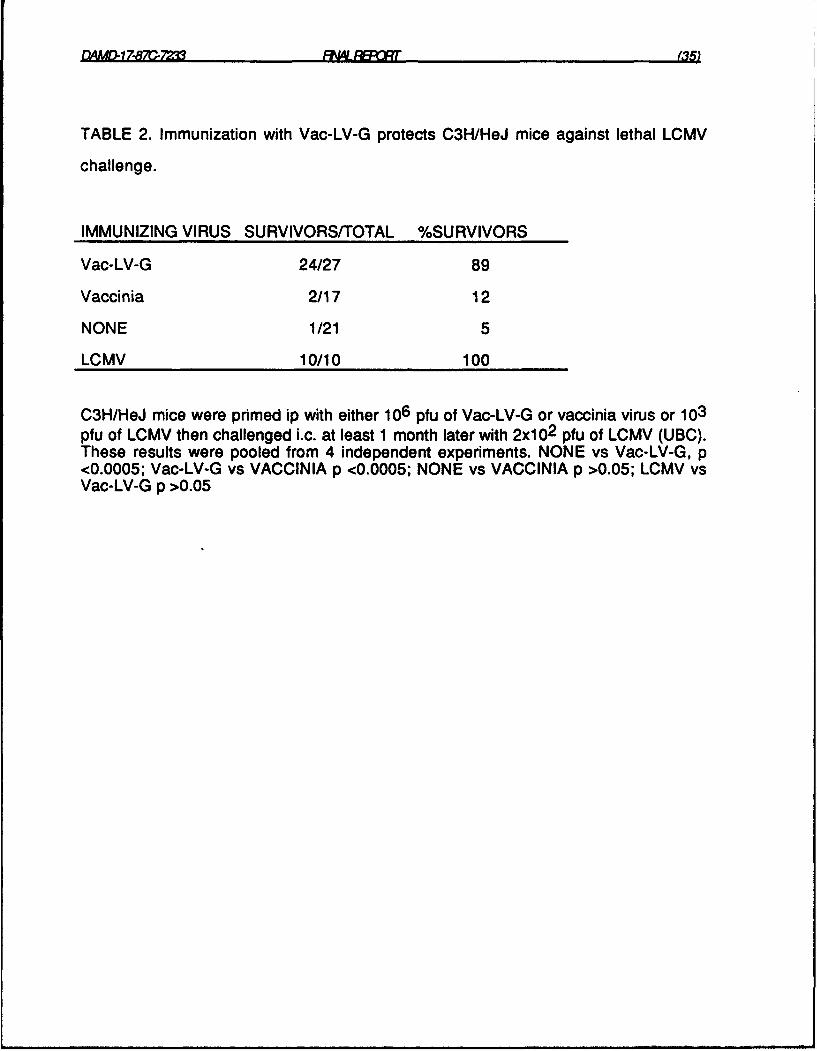
TABLE 2. Immunization with Vac-LV-G protects C3H/HeJ mice against lethal LCMV
challenge.
IMMUNIZING VIRUS SURVIVORS/TOTAL %SURVIVORS
Vac-LV-G 24/27 89
Vaccinia 2/17 12
NONE 1/21 5
LCMV 10/10 100
C3H/HeJ mice were primed ip with either 106 pfu of Vac-LV-G or vaccinia virus or 103pfu of LCMV then challenged i.c. at least 1 month later with 2x1 02 pfu of LCMV (UBC).These results were pooled from 4 independent experiments. NONE vs Vac-LV-G, p<0.0005; Vac-LV-G vs VACCINIA p <0.0005; NONE vs VACCINIA p >0.05; LCMV vsVac-LV-G p >0.05

". ",17S7r _=iM "' '
TABLE 3. Cross-reactive CTL are not detectable in Vac-LV-G-prmed and LCMV-
primed C3H/HeJ mice.
% lysis of L cells infected with:Priming virus Virus in vitro E:T Nothing LCMV Vac-LV-G Vaccinia
Vac-LV-G LCMV 15 1 1 22 155 -1 0 8 5
Vac-LV-G 5 18 15 87 582 2 1 50 36
Vaccinia 5 17 14 86 752 4 4 64 57
Medium 15 1 2 13 10
LCMV LCMV 15 17 62 20 195 3 20 3 52 2 9 2 1
Vac-LV-G 15 6 6 6 8
Vaccinia 15 2 10 6 7
Medium 15 15 22 15 23
Splenocytes from C3H/HeJ mice immunized with Vac-LV-G (twice with 107 pfu one
month apart) or LCMV were stimulated in separate cultures with either LCMV, Vac-LV-
G, vaccinia virus or medium alone for 5 days and the cells tested for cytotoxic activity
against normal L cells and L cells infected with LCMV, Vac-LV-G or vaccinia virus in a
5 hr 5 1 Cr release assay. The target cells were shown by IFA, using the appropriate
antibodies, to be greater than 90% infected with LCMV (rat anti LCMV serum), 70%
infected with Vac-LV-G (rhesus monkey anti Lassa virus serum and anti vaccinia
monoclonal antibody) and 90 % infected with vaccinia (monoclonal anti vaccinia
antibody).
DAMDL17-87C-72'= FN4LFAR 627D
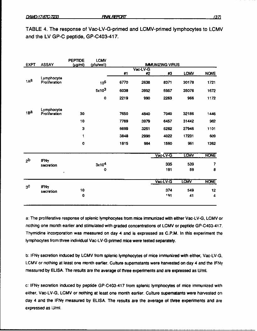
TABLE 4. The response of Vac-LV-G-pdmed and LCMV-primed lymphocytes to LCMVand the LV GP-C peptide, GP-C403-417.
PEPTIDE LCMVEXPT ASSAY (jg/ml) (pfu/well) IMMUNIZING VIRUS
Vac-LV-G#1 #2 #3 LCMV NONE
1Aa LymphocyteProliferation 105 6770 2638 8371 30178 1721
5x10 3 6038 3992 5957 35076 1672
0 2219 990 2263 966 1172
1Ba LymphocyteProliferation 30 7650 4840 7040 32186 1446
10 7769 3979 6457 31442 962
3 6699 3251 5282 27946 1101
1 3848 2990 4022 17231 609
0 1815 984 1560 961 1362
Vac-LV-G LCMV NONE2b IFNy
secretion 3x10 4 335 539 70 191 59 8
Vac-LV-G LCMV NONE3c IFN35
secretion 10 374 549 120 4ql 41 4
a: The proliferative response of splenic lymphocytes from mice immunized with either Vac-LV-G, LCMV or
nothing one month earlier and stimulated with graded concentrations of LCMV or peptide GP-C403-417.
Thymidine incorporation was measured on day 4 and is expressed as C.P.M. In this experiment the
lymphocytes from three individual Vac-LV-G-primed mice were tested separately.
b: IFNy secretion induced by LCMV from splenic lymphocytes of mice immunized with either, Vac-LV-G,
LCMV or nothing at least one month earlier. Culture supematants were harvested on day 4 and the IFNy
measured by ELISA. The results are the average of three experiments and are expressed as U/mI.
c: IFNy secretion induced by peptide GP-C403-417 from splenic lymphocytes of mice immunized with
either, Vac-LV-G, LCMV or nothing at least one month earlier. Culture supernatants were harvested on
day 4 and the IFNy measured by ELISA. The results are the average of three experiments and are
expressed as IJ/ml.

fYAfJ.178C-YM 'I? (8
TABLE 5. CD4 + T cells respond to GP-C 403-417a
3 H-thymidine incorporated (C.P.M.)
Experiment 1 Experiment 2
Ab (jg/ml) LCMV-primed Vac-LV-G-pdmed
mouse 1 mouse 2Peptide only(no antibody) 11,306 10,415 17,782
medium only(no antibody) 1,247 4,791 8,018
anti-CD4 (10) 666 1,791 4,276
anti-CD4 (1) 2,545 N.D.b N.D.
anti-CD8 (10) 8,449 10,839 15,146
anti-CD8 (1) 12,068 N.D. N.D.
a Splenic lymphocytes from LCMV-pndmed mice (a pool of cells from
2 mice) and Vac-LV-G-pnmed mice (cells from 2 mice tested individually)
were tested in two separate experiments. They were cultured for 5 days
with GP-C 403-417 (10ig/ml) in the absence or presence of anti CD4
(GK1.5) or anti CD8 (116-13.1) at concentrations indicated above.
b Not done.

QA4MD-i 7.8 7C-7M FNLAR (39)
ALFN K

-~~~ ~~~~~ Mi -I'm- ~ ~ ~ -, ~ ~ ~~- --r- -
EAPMrJ1787C-7M (40)FC
TABLE 6. Genetic restriction and cytolytic activity of GP-0403-41 7
specific T cell clones.
Experi- T cell Stimulator cell Haplotypement clone (or target cell) K I-A l-E D Assay
IFMy (U/mI)i a 9 C3H/HeJ k k k k 1328
A.TL s k k d 660B1 O.A(4R) k k nag b 290C3H.OH d d d k <LOD b
IFMy (U/mI)2 c 9 M1 2.C3-F6 d k nag d 20,000
M1 2MC d neg neg d 226
11 M12.C3-F6 d k neg d 12,400M1 2MC d nag neg d <LOD
Lysis (%)3 d 9 51Cr M12.C3-F12
plus peptide d k nag d 52.4
510r M12.C3-F12medium control d k nag d 4.3
51 Cr M1 2.0plus peptide d nag nag d 5.0
51 Cr M1 2.0medium control d nag nag d 7.0
a 105 clone 9 T cells were stimulated with S5jg/mI of GP-C403-41 7 in the presence of 5x15 7-
irradiated splenocytes. In the absence of peptide no detectable IFNy was secreted.
b Below the limit of detL on (LOD), which in experiments I and 2 were 75 U/mI and 200U/ml
respeLdively.
c B cell lines M 12.03-F6 and M 1 2M were cultured at 105 cells/well with 105 dlone 9 or clone 11 T
cells in the presence or absence of 5Ojgg/ml of GP-C403-41 7. In the absence of peptide no detectable
IFMy was secreted.

DA MD-i 7-8 70 .2' FNLR (41)
d 2x 10⁴ 51 Cr-Iabelled target cells were cultured with 2x1 05 clone 9 T cells for 2Ohrs in the
presence or absence of 50jgg/ml of GP-C403-417. The spontaneous release of label from each target was
<25% in the presence or absence of peptide.

flAMdQ1782QC-72 RW 4ELCAr (
TABLE 7. Protection against LCMV challenge with clone 9 T ceilsa.
EXPT CELL CHALLENGE SURVIVORSITOTAL
DOSE DOSE (pfu)-- C3H/HeJ Allogeneic
1 1x10 6 20 5/5 N.Db.
NONE 1/5 N.D.
BALB/C (H-2d)
2 2x10⁶ 20 5/5 1/3
NONE 0/5 0/3
2X1 06 200 0/5 N.D.
NONE 0/5 N.D.
CD-i (H-2q)
3 1x10⁶ 20 4/5 0/6
3x10 5 4/5 0/6
lxi0 4/4 0/6
NONE 0/5 0/6
106 killedc 0/5 0/6
a: The indicated amounts of LCMV and clone 9 T cells were injected i.c. as a mixture into mice in 3Oji
of Hanks balanced salt solution. The T cells were used 10-14 days after stimulation. In experiment 3 dead
cells and debris were removed by centrifugation on a Lympholyte M gradient.
b: Not done.
c: Frozen and thawed three times.

DAMD.1787C- 723 WINL PRT (43)
Table 8. The proliferative response of PBL from monkeys primed with LCMV or Vac-
LV-G to LV GP-C synthetic peptidesa.
Monkey designations and immunizing virus
4FY 927C 46C 45J
Antigen in vitro LCMV Vac-LV-G Unimmunized Unimmunized
Medium alone 912 3443 408 307
LCMV 10,341 9,152 1,004 422
GP-C383-397 1,049 2,577 599 234
GP-C393-407 599 4,720 346 254
GP-C403-417 365 4,260 289 478
GP-C446-440 456 2,171 714 200
GP-C456-470 1,051 2,619 321 261
a: 2x1 05 purified PBL were cultured for 5 days with LCMV or LV GP-C peptides at 501gg/ml

f l4QL.17. - 721 RWFFLCn (44)
FIGURE LEGENDS
FIGURE. 1. Peptide-specific T cell responsesa.
a: Peptides encompassing residues 75-88, 170-183, 308-320, 354-368, and 410-425 of LV GP-C were
tested in C3H/HeJ (H-2k), C57BI/6J (H-2b), BALB/cJ (H-2d) and DBA/1J (H-2q ) mice in 5 separate
experiments (one experiment for each peptide). Peptides 383-397, 393-407, 403-417 and 446-460
were all tested in one experiment using C3H/HeJ mice. Mice were immunized with 100 gIg of peptide
emulsified in complete Freund's adjuvant and draining lymph node cells tested 7-12 days latter.
FIGURE. 2. GP-C403-417-specific IFNy-secreting T cells in Vac-LV-G-primed
populations can be enriched by culturing with LCMV or GP-C403-417a.
a: Lymphocytes from mice immunized 1 month earlier with Vac-LV-G were cultured with medium alone,
LCMV (105 pfu/well) or GP-C403-417 (1 Oig/ml) in 24 well trays for 5 days. The medium was replaced and
the cells cultured for a further 9 days. The cells were then harvested and stimulated in 96 well plates for 2
days at 5x10⁴ viable cells/well with medium, GP-C403-417 (10jig/ml) or another peptide GP-C456-470
(101gg/ml) in the presence of fresh, irradiated spleen cells (5xl05 /well). IFNy in the supematants was
measured by ELISA.
FIGURE 3. The response of GP-C403-417-specific cloned T cell lines
derived from Vac-LV-G-primed mice to peptide and LCMV.a
a: Cloned T cell lines were stimulated with the peptides GP-C403-417 or GP-C456-470 (irrelevant peptide)
(5-10 gg/ml) in the presence of irradiated syngeneic splenocytes or with LCMV-infected peritoneal cells.
The clones were used 10 to 14 days after last stimulation, except in experiment 2 where clones 9 and 11
were tested at day 8. In experiments I to 4 IFNy secretion was assayed whereas In experiment 5 the assay

DAMDL17-87C-7233 RN'LR9:Ot (45)
was the proliferation of clone 11 T cells. The horizontal bars indicate the limits of detection for IFNy in the
various experiments. No detectable IFNy was secreted by the T cell clones in the absence of peptide, nor
by splenocytes (with or without peptide) or by peritoneal cells (normal or LCMV-infected) (data not shown).
FIGURE 4. Clone 9 T cells mediate peptide-specific DTH reactiona.
a: Normal C3H/HeJ mice were injected in the footpad with combinations of 106 clone 9 T cells, 4ljg
peptide (GP-C403-417 or GP-C456-470) and 2x10 6 irradiated (1500 R) normal splenocytes in a volume of
30gl. Footpad thickness was measured immediately before injection and 24 h later. Swelling is expressed
as the % increase in thickness. Shown are the results from 2 experiments.
FIGURE 5. The response of PBL from rhesus monkeys immunized with LCMV or Vac-
LV-G to stimulation with LCMV in vitro.a
a: 4 x 10⁵ purified PBL from monkeys immunized with either LCMV (Arm), Vac-LV-G or nothing were
cultured with two doses of LCMV (UBC) or medium alone for 4 days.
FIGURE 6. The effect of cell number on the response of LCMV and Vac-LV-G-primed
rhesus monkey PBL to LCMV in vitro.a
a: Monkey PBL were cultured at 1 x 10⁵ and 2 x 10⁵ celew~ell with LCMV or medium alone for 4 days or 5
days.

FIGURE 1.
EXPT 1: DBA/I: 75-88C57BI/6: 75-88C3H/HeJ: 75-88
BALB/c: 75-88
EXPT 2: C57BLJ6: 170-183C3H/HeJ: 170-183
EXPT 3: DBAI1: 308-320C57BL/6: 308-320C3H/HeJ: 308-320
BALB/c: 308-320
EXPT 4: DBAI1: 354-368C57BL/6: 354-368C3H/"eJ: 354-368
BALB/c: 354-368
EXPT 5: C3H/HeJ: 446-460C3H/HeJ: 403-417C3H/HeJ: 393-407C3H/HeJ: 383-397
EXPT 6: DBAI1: 410-425C3H/HeJ: 410-425BALB/c: 410-425
0 2 4 6 8 10 12 14 16 18 20
STIMULATION INDEX

I (b
zY
z J,~
CL
Own)cv -MiDN4

1 --
7Al
If)
4L
4
U 4
3l
&OO. 3N
_ _ _ _ _ _ __D NN

w wU
w )
o U) -j f
0- w a. +U
+ +LJ + r
h 00U) 0 0LL
UJ CL < a-j CO co U
CL CL +LU CL
a- a. L -i oI-.
-0 M 1: 30
U
(OS -*NVIR
114t~ V S3NIOIJ.U~dLO~ N 3S3~OI

Figure 5.
RESPONSE OF MONKEY P61 TO LCMV IN VITRO
3000-
LCIMV pler well (11fUl
U 15,000Ea 5,000
2000 U None
1000-
45J 46C 4FN 47G 4FY 927C 2Y3 4G7
Unimmunized LCMV-PRIMED Vac-LV-G-PRIMED
MONKEYS

z 9c c-E E >
2 E E E
0
I-z0
M
a=)
II
IldOU
rz~
00-i,,--
z0
ILI
fldO
Related Documents