Synthesis, semipreparative HPLC separation, biological evaluation, and 3D-QSAR of hydrazothiazole derivatives as human monoamine oxidase B inhibitors Franco Chimenti a , Daniela Secci a, * , Adriana Bolasco a , Paola Chimenti a , Arianna Granese a , Simone Carradori a , Elias Maccioni b , M. Cristina Cardia b , Matilde Yáñez c , Francisco Orallo c , Stefano Alcaro d , Francesco Ortuso d , Roberto Cirilli e , Rosella Ferretti e , Simona Distinto d,g , Johannes Kirchmair f , Thierry Langer g a Dipartimento di Chimica e Tecnologie del Farmaco Università degli Studi di Roma ‘La Sapienza’, P.le A. Moro 5, 00185 Roma, Italy b Dipartimento Farmaco Chimico Tecnologico, Università di Cagliari, Via Ospedale 72, 09124 Cagliari, Italy c Departamento de Farmacología and Instituto de Farmacia Industrial, Facultad de Farmacia, Universidad de Santiago de Compostela, Campus Universitario Sur, E-15782 Santiago de Compostela (La Coruña), Spain d Dipartimento di Scienze Farmaco Biologiche ‘Complesso Ninì Barbieri’, Università degli Studi di Catanzaro ‘Magna Graecia’, 88021 Roccelletta di Borgia (CZ), Italy e Istituto Superiore di Sanità, Dipartimento del Farmaco, V.le Regina Elena 299, 00161 Roma, Italy f Department of Pharmaceutical Chemistry, Institute of Pharmacy and Center for Molecular Biosciences (CMBI), University of Innsbruck, Innrain 52, A-6020 Innsbruck, Austria g Inte:Ligand Software-Entwicklungs und Consulting GmbH, Clemens Maria Hofbauer-Gasse 6, A-2344 Maria Enzersdorf, Austria article info Article history: Received 17 March 2010 Revised 25 May 2010 Accepted 26 May 2010 Available online 1 June 2010 Keywords: Hydrazothiazole derivatives 3D-QSAR Human monoamine oxidase B inhibitors abstract The present study reports on synthesis in high yields (70–99%), HPLC enantioseparation, inhibitory activ- ity against human monoamino oxidases, and molecular modeling including 3D-QSAR studies, of a large series of (4-aryl-thiazol-2-yl)hydrazones (1–45). Most of the synthesized compounds proved to be potent and selective inhibitors of hMAO-B isoform in the micromolar or nanomolar range, thus demonstrating that hydrazothiazole could be considered a good pharmacophore to design new hMAO-B inhibitors. Due to the presence in some derivatives of a chiral center, we also performed a semipreparative chro- matographic enantioseparation of these compounds obtained by a stereoconservative pattern. The sepa- rated enantiomers were submitted to in vitro biological evaluation to point out the stereorecognition of the active site of the enzyme towards these structures. Finally, a 3D-QSAR study was carried out using Comparative Molecular Field Analysis (CoMFA), aiming to deduce rational guidelines for the further structural modification of these lead compounds. Ó 2010 Elsevier Ltd. All rights reserved. 1. Introduction Mitochondrial monoamine oxidases (MAOs, EC 1.4.3.4) are flavin-containing enzymes (FAD) that catalyze the oxidative deam- ination of neurotransmitters and exogenous arylalkylamines. In mammals, two different types of MAOs are present in most tissues, namely, MAO-A and MAO-B. MAO-A preferentially deaminates aromatic monoamines such as the neurotransmitters serotonin (5-HT), noradrenaline (NA), and adrenaline (A), while MAO-B mainly oxidizes a-phenylethylamines and benzylamine. Both isoforms act on dopamine (D) and tryptamine. 1 These enzymes are tightly bound to the mitochondrial outer membrane, sharing about 70% amino acid identity. They also differ with respect to substrate specificity, sensitivity to inhibitors, and tissue distribution. Although MAOs are widely distributed in various organs, most of the studies concerning their functional properties and involve- ment in pathological processes have been mainly focused on the central nervous system. In the periphery, MAO-A and MAO-B are differently expressed in a variety of tissues: MAO-A is predominant in heart, adipose tissue, and skin fibroblasts, MAO-B is the major form found in platelets and lymphocytes, whereas both isoen- zymes are expressed in kidney and liver. 2 It has been recently iden- tified an additional MAO mechanism which is involved in reactive oxygen species (ROS) production and the consequent induction of intracellular oxidative stress. Hydrogen peroxide (H 2 O 2 ) is one of the reaction products generated by MAOs during substrate degradation. Except for the potential cytotoxic effect of H 2 O 2 in ni- gral cells in Parkinson’s disease, the cell events following MAO- dependent H 2 O 2 production in physiological conditions are still 0968-0896/$ - see front matter Ó 2010 Elsevier Ltd. All rights reserved. doi:10.1016/j.bmc.2010.05.070 * Corresponding author. Tel.: +39 06 4991 3763; fax: +39 06 49913772. E-mail address: [email protected] (D. Secci). Bioorganic & Medicinal Chemistry 18 (2010) 5063–5070 Contents lists available at ScienceDirect Bioorganic & Medicinal Chemistry journal homepage: www.elsevier.com/locate/bmc

Welcome message from author
This document is posted to help you gain knowledge. Please leave a comment to let me know what you think about it! Share it to your friends and learn new things together.
Transcript
Bioorganic & Medicinal Chemistry 18 (2010) 5063–5070
Contents lists available at ScienceDirect
Bioorganic & Medicinal Chemistry
journal homepage: www.elsevier .com/locate /bmc
Synthesis, semipreparative HPLC separation, biological evaluation, and3D-QSAR of hydrazothiazole derivatives as human monoamine oxidaseB inhibitors
Franco Chimenti a, Daniela Secci a,*, Adriana Bolasco a, Paola Chimenti a, Arianna Granese a,Simone Carradori a, Elias Maccioni b, M. Cristina Cardia b, Matilde Yáñez c, Francisco Orallo c, Stefano Alcaro d,Francesco Ortuso d, Roberto Cirilli e, Rosella Ferretti e, Simona Distinto d,g, Johannes Kirchmair f,Thierry Langer g
a Dipartimento di Chimica e Tecnologie del Farmaco Università degli Studi di Roma ‘La Sapienza’, P.le A. Moro 5, 00185 Roma, Italyb Dipartimento Farmaco Chimico Tecnologico, Università di Cagliari, Via Ospedale 72, 09124 Cagliari, Italyc Departamento de Farmacología and Instituto de Farmacia Industrial, Facultad de Farmacia, Universidad de Santiago de Compostela, Campus Universitario Sur,E-15782 Santiago de Compostela (La Coruña), Spaind Dipartimento di Scienze Farmaco Biologiche ‘Complesso Ninì Barbieri’, Università degli Studi di Catanzaro ‘Magna Graecia’, 88021 Roccelletta di Borgia (CZ), Italye Istituto Superiore di Sanità, Dipartimento del Farmaco, V.le Regina Elena 299, 00161 Roma, Italyf Department of Pharmaceutical Chemistry, Institute of Pharmacy and Center for Molecular Biosciences (CMBI), University of Innsbruck, Innrain 52, A-6020 Innsbruck, Austriag Inte:Ligand Software-Entwicklungs und Consulting GmbH, Clemens Maria Hofbauer-Gasse 6, A-2344 Maria Enzersdorf, Austria
a r t i c l e i n f o a b s t r a c t
Article history:Received 17 March 2010Revised 25 May 2010Accepted 26 May 2010Available online 1 June 2010
Keywords:Hydrazothiazole derivatives3D-QSARHuman monoamine oxidase B inhibitors
0968-0896/$ - see front matter � 2010 Elsevier Ltd. Adoi:10.1016/j.bmc.2010.05.070
* Corresponding author. Tel.: +39 06 4991 3763; faE-mail address: [email protected] (D. Secc
The present study reports on synthesis in high yields (70–99%), HPLC enantioseparation, inhibitory activ-ity against human monoamino oxidases, and molecular modeling including 3D-QSAR studies, of a largeseries of (4-aryl-thiazol-2-yl)hydrazones (1–45). Most of the synthesized compounds proved to be potentand selective inhibitors of hMAO-B isoform in the micromolar or nanomolar range, thus demonstratingthat hydrazothiazole could be considered a good pharmacophore to design new hMAO-B inhibitors.Due to the presence in some derivatives of a chiral center, we also performed a semipreparative chro-matographic enantioseparation of these compounds obtained by a stereoconservative pattern. The sepa-rated enantiomers were submitted to in vitro biological evaluation to point out the stereorecognition ofthe active site of the enzyme towards these structures. Finally, a 3D-QSAR study was carried out usingComparative Molecular Field Analysis (CoMFA), aiming to deduce rational guidelines for the furtherstructural modification of these lead compounds.
� 2010 Elsevier Ltd. All rights reserved.
1. Introduction
Mitochondrial monoamine oxidases (MAOs, EC 1.4.3.4) areflavin-containing enzymes (FAD) that catalyze the oxidative deam-ination of neurotransmitters and exogenous arylalkylamines. Inmammals, two different types of MAOs are present in most tissues,namely, MAO-A and MAO-B. MAO-A preferentially deaminatesaromatic monoamines such as the neurotransmitters serotonin(5-HT), noradrenaline (NA), and adrenaline (A), while MAO-Bmainly oxidizes a-phenylethylamines and benzylamine. Bothisoforms act on dopamine (D) and tryptamine.1
These enzymes are tightly bound to the mitochondrial outermembrane, sharing about 70% amino acid identity. They also differ
ll rights reserved.
x: +39 06 49913772.i).
with respect to substrate specificity, sensitivity to inhibitors, andtissue distribution.
Although MAOs are widely distributed in various organs, mostof the studies concerning their functional properties and involve-ment in pathological processes have been mainly focused on thecentral nervous system. In the periphery, MAO-A and MAO-B aredifferently expressed in a variety of tissues: MAO-A is predominantin heart, adipose tissue, and skin fibroblasts, MAO-B is the majorform found in platelets and lymphocytes, whereas both isoen-zymes are expressed in kidney and liver.2 It has been recently iden-tified an additional MAO mechanism which is involved in reactiveoxygen species (ROS) production and the consequent induction ofintracellular oxidative stress. Hydrogen peroxide (H2O2) is one ofthe reaction products generated by MAOs during substratedegradation. Except for the potential cytotoxic effect of H2O2 in ni-gral cells in Parkinson’s disease, the cell events following MAO-dependent H2O2 production in physiological conditions are still
N N
Cy
Cy
SH2N
N NH
Cy
SH2N
N NH
Cy
NS
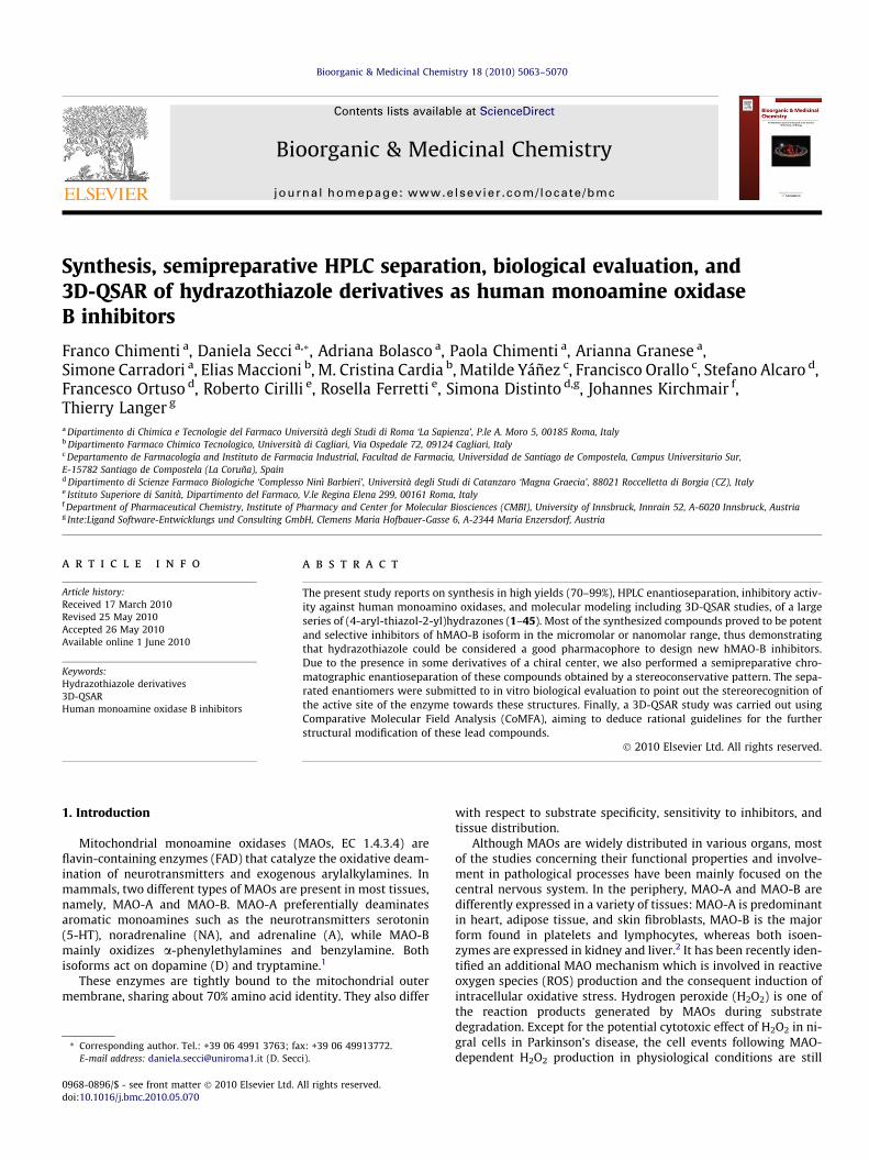
Figure 2. Conversion of N1-thiocarbamoyl-3,5-diaryl-4,5-dihydro-(1H)-pyrazole incycloalkylidenhydrazothiazole derivative.
5064 F. Chimenti et al. / Bioorg. Med. Chem. 18 (2010) 5063–5070
unknown. These findings, along with other results showing theimportant role of ROS generated by MAOs in heart, adipose tissue,macrophages, and skeletal muscle, pointed to the relevance also ofperipheral MAOs in various pathological processes and their poten-tiality as pharmacological agents.3
The discovery of reversible and selective inhibitors has renewedthe interest towards MAO enzymes as drug targets. In fact, irre-versible and/or nonselective inhibitors showed shortcomingsincluding cumulative effects, loss of selectivity after chronictreatment, and interaction with tyramine-containing foods(cheese-effect). Selective and reversible hMAO-A inhibitors(hMAO-AIs) are currently used in the treatment of depressionand anxiety disorders, whereas hMAO-B inhibitors (hMAO-BIs)are used as adjuncts in the treatment of Parkinson’s disease (PD).MAO-B predominates over MAO-A in the human brain especiallyafter the 60th year of life, where hMAO-BIs have proven to be ben-eficial in prolonging the anti-Parkinsonian action of L-DOPA.hMAO-BIs, which decrease the rate of MAO-B catalyzed oxidativedeamination and, consequently, the production of reactive oxygenspecies (ROS), might contribute to the treatment of other neurode-generative diseases, such as Alzheimer’s disease.4–6
The rational design of new agents targeted to MAOs could bebased on the recent description of the crystal structure of humanMAO-B and MAO-A with their corresponding inhibitors and aidedwith theoretical calculations.7,8 The above studies elucidated somefactors responsible for selectivity against the A and B isoforms,such as the lipophilicity of the inhibitor that is important forachieving effective binding to MAO-B, the presence of electron-richaromatic moieties, typical of selective MAO-A inhibitors, and therole played by some amino acid residues in the active sites, suchas Tyr326 for hMAO-B and Ile335 for hMAO-A.9
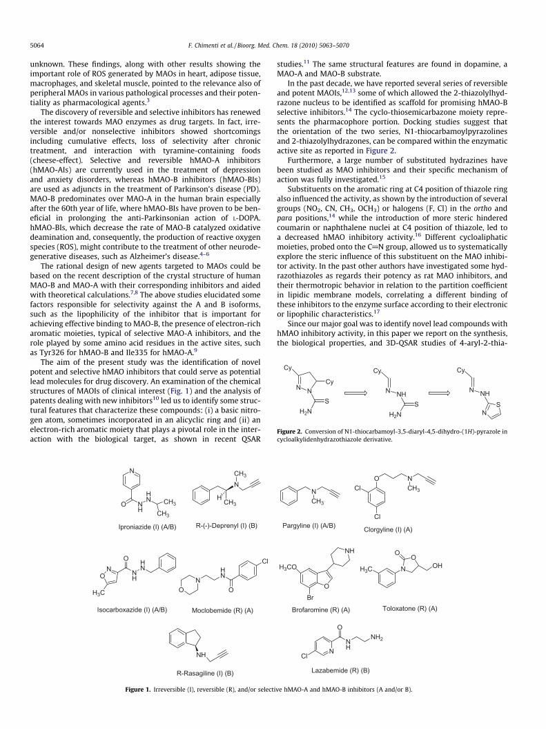
The aim of the present study was the identification of novelpotent and selective hMAO inhibitors that could serve as potentiallead molecules for drug discovery. An examination of the chemicalstructures of MAOIs of clinical interest (Fig. 1) and the analysis ofpatents dealing with new inhibitors10 led us to identify some struc-tural features that characterize these compounds: (i) a basic nitro-gen atom, sometimes incorporated in an alicyclic ring and (ii) anelectron-rich aromatic moiety that plays a pivotal role in the inter-action with the biological target, as shown in recent QSAR
N
O NH
HN CH3
CH3
NCH3
ON
HN
O
Cl
Iproniazide (I) (A/B) R-(-)-Deprenyl (I) (B)
Moclobemide (R) (A)
HCH3
Isocarboxazide (I) (A/B)
HN
NH
ON
O
H3C
R-Rasagiline (I) (B)
NH
Figure 1. Irreversible (I), reversible (R), and/or selecti
studies.11 The same structural features are found in dopamine, aMAO-A and MAO-B substrate.
In the past decade, we have reported several series of reversibleand potent MAOIs,12,13 some of which allowed the 2-thiazolylhyd-razone nucleus to be identified as scaffold for promising hMAO-Bselective inhibitors.14 The cyclo-thiosemicarbazone moiety repre-sents the pharmacophore portion. Docking studies suggest thatthe orientation of the two series, N1-thiocarbamoylpyrazolinesand 2-thiazolylhydrazones, can be compared within the enzymaticactive site as reported in Figure 2.
Furthermore, a large number of substituted hydrazines havebeen studied as MAO inhibitors and their specific mechanism ofaction was fully investigated.15
Substituents on the aromatic ring at C4 position of thiazole ringalso influenced the activity, as shown by the introduction of severalgroups (NO2, CN, CH3, OCH3) or halogens (F, Cl) in the ortho andpara positions,14 while the introduction of more steric hinderedcoumarin or naphthalene nuclei at C4 position of thiazole, led toa decreased hMAO inhibitory activity.16 Different cycloaliphaticmoieties, probed onto the C@N group, allowed us to systematicallyexplore the steric influence of this substituent on the MAO inhibi-tor activity. In the past other authors have investigated some hyd-razothiazoles as regards their potency as rat MAO inhibitors, andtheir thermotropic behavior in relation to the partition coefficientin lipidic membrane models, correlating a different binding ofthese inhibitors to the enzyme surface according to their electronicor lipophilic characteristics.17
Since our major goal was to identify novel lead compounds withhMAO inhibitory activity, in this paper we report on the synthesis,the biological properties, and 3D-QSAR studies of 4-aryl-2-thia-
NCH3
O
H3CO
Br
NH
H3C N
OO
OH
Pargyline (I) (A/B)
Brofaromine (R) (A) Toloxatone (R) (A)
Cl
ClO N
CH3
Clorgyline (I) (A)
NNH
Lazabemide (R) (B)
Cl
ONH2
ve hMAO-A and hMAO-B inhibitors (A and/or B).
F. Chimenti et al. / Bioorg. Med. Chem. 18 (2010) 5063–5070 5065
zolylhydrazone derivatives (1–45) useful to assess a pharmaco-phore motif common to the active compounds.
2. Chemistry
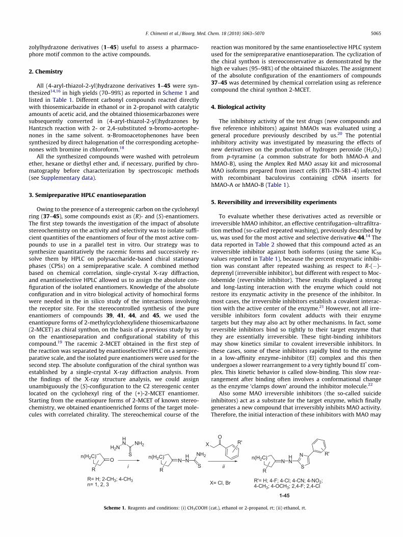
All (4-aryl-thiazol-2-yl)hydrazone derivatives 1–45 were syn-thesized14,16 in high yields (70–99%) as reported in Scheme 1 andlisted in Table 1. Different carbonyl compounds reacted directlywith thiosemicarbazide in ethanol or in 2-propanol with catalyticamounts of acetic acid, and the obtained thiosemicarbazones weresubsequently converted in (4-aryl-thiazol-2-yl)hydrazones byHantzsch reaction with 2- or 2,4-substituted a-bromo-acetophe-nones in the same solvent. a-Bromoacetophenones have beensynthesized by direct halogenation of the corresponding acetophe-nones with bromine in chloroform.18
All the synthesized compounds were washed with petroleumether, hexane or diethyl ether and, if necessary, purified by chro-matography before characterization by spectroscopic methods(see Supplementary data).
3. Semipreparative HPLC enantioseparation
Owing to the presence of a stereogenic carbon on the cyclohexylring (37–45), some compounds exist as (R)- and (S)-enantiomers.The first step towards the investigation of the impact of absolutestereochemistry on the activity and selectivity was to isolate suffi-cient quantities of the enantiomers of four of the most active com-pounds to use in a parallel test in vitro. Our strategy was tosynthesize quantitatively the racemic forms and successively re-solve them by HPLC on polysaccharide-based chiral stationaryphases (CPSs) on a semipreparative scale. A combined methodbased on chemical correlation, single-crystal X-ray diffraction,and enantioselective HPLC allowed us to assign the absolute con-figuration of the isolated enantiomers. Knowledge of the absoluteconfiguration and in vitro biological activity of homochiral formswere needed in the in silico study of the interactions involvingthe receptor site. For the stereocontrolled synthesis of the pureenantiomers of compounds 39, 41, 44, and 45, we used theenantiopure forms of 2-methylcyclohexylidene thiosemicarbazone(2-MCET) as chiral synthon, on the basis of a previous study by uson the enantioseparation and configurational stability of thiscompound.19 The racemic 2-MCET obtained in the first step ofthe reaction was separated by enantioselective HPLC on a semipre-parative scale, and the isolated pure enantiomers were used for thesecond step. The absolute configuration of the chiral synthon wasestablished by a single-crystal X-ray diffraction analysis. Fromthe findings of the X-ray structure analysis, we could assignunambiguously the (S)-configuration to the C2 stereogenic centerlocated on the cyclohexyl ring of the (+)-2-MCET enantiomer.Starting from the enantiopure forms of 2-MCET of known stereo-chemistry, we obtained enantioenriched forms of the target mole-cules with correlated chirality. The stereochemical course of the
NH2
S
HNN
n(H2C)
R
On(H2C)
R
XH2NHN
S
NH2
i
R= H; 2-CH3; 4-CH3n= 1, 2, 3
Scheme 1. Reagents and conditions: (i) CH3COOH (
reaction was monitored by the same enantioselective HPLC systemused for the semipreparative enantioseparation. The cyclization ofthe chiral synthon is stereoconservative as demonstrated by thehigh ee values (95–98%) of the obtained thiazoles. The assignmentof the absolute configuration of the enantiomers of compounds37–45 was determined by chemical correlation using as referencecompound the chiral synthon 2-MCET.
4. Biological activity
The inhibitory activity of the test drugs (new compounds andfive reference inhibitors) against hMAOs was evaluated using ageneral procedure previously described by us.20 The potentialinhibitory activity was investigated by measuring the effects ofnew derivatives on the production of hydrogen peroxide (H2O2)from p-tyramine (a common substrate for both hMAO-A andhMAO-B), using the Amplex Red MAO assay kit and microsomalMAO isoforms prepared from insect cells (BTI-TN-5B1-4) infectedwith recombinant baculovirus containing cDNA inserts forhMAO-A or hMAO-B (Table 1).
5. Reversibility and irreversibility experiments
To evaluate whether these derivatives acted as reversible orirreversible hMAO inhibitor, an effective centrifugation–ultrafiltra-tion method (so-called repeated washing), previously described byus, was used for the most active and selective derivative 44.14 Thedata reported in Table 2 showed that this compound acted as anirreversible inhibitor against both isoforms (using the same IC50
values reported in Table 1), because the percent enzymatic inhibi-tion was constant after repeated washing as respect to R-(�)-deprenyl (irreversible inhibitor), but different with respect to Moc-lobemide (reversible inhibitor). These results displayed a strongand long-lasting interaction with the enzyme which could notrestore its enzymatic activity in the presence of the inhibitor. Inmost cases, the irreversible inhibitors establish a covalent interac-tion with the active center of the enzyme.21 However, not all irre-versible inhibitors form covalent adducts with their enzymetargets but they may also act by other mechanisms. In fact, somereversible inhibitors bind so tightly to their target enzyme thatthey are essentially irreversible. These tight-binding inhibitorsmay show kinetics similar to covalent irreversible inhibitors. Inthese cases, some of these inhibitors rapidly bind to the enzymein a low-affinity enzyme–inhibitor (EI) complex and this thenundergoes a slower rearrangement to a very tightly bound EI* com-plex. This kinetic behavior is called slow-binding. This slow rear-rangement after binding often involves a conformational changeas the enzyme ‘clamps down’ around the inhibitor molecule.22
Also some MAO irreversible inhibitors (the so-called suicideinhibitors) act as a substrate for the target enzyme, which finallygenerates a new compound that irreversibly inhibits MAO activity.Therefore, the initial interaction of these inhibitors with MAO may
N
S
HNN
n(H2C)
R
R'
OR'
ii
X= Cl, Br R'= H; 4-F; 4-Cl; 4-CN; 4-NO2; 4-CH3; 4-OCH3; 2,4-F; 2,4-Cl
1-45
cat.), ethanol or 2-propanol, rt; (ii) ethanol, rt.

Table 1Structures, biological activity (IC50), and hMAO-B selectivity ratios for the inhibitory effects of tested compounds 1–45 and reference drugs on enzymatic activity of humanrecombinant MAO isoforms expressed in baculovirus infected cells
Compd R R0 hMAOA IC50 (nM) hMAOB IC50 (nM) Ratio
1 Cyclopentyl H 7883 ± 91* 296 ± 7 272 Cyclopentyl 4-Cl 7160 ± 640* 262 ± 8 273 Cyclopentyl 4-F 4436 ± 212* 40 ± 0.9 1114 Cyclopentyl 2,4-Cl 54,507 ± 4123* 284 ± 11 1925 Cyclopentyl 2,4-F 2318 ± 161* 3 ± 0.2 7736 Cyclopentyl 4-CH3 2019 ± 74* 134 ± 9 157 Cyclopentyl 4-OCH3 1055 ± 42* 164 ± 13 68 Cyclopentyl 4-NO2 344 ± 22* 94 ± 3 49 Cyclopentyl 4-CN 644 ± 21* 221 ± 2 310 Cyclohexyl H 48,351 ± 1433* 116 ± 5 41711 Cyclohexyl 4-Cl 2911 ± 171* 211 ± 7 1412 Cyclohexyl 4-F 1752 ± 21* 4 ± 0.2 43813 Cyclohexyl 2,4-Cl N.E. 202 ± 16 >495b
14 Cyclohexyl 2,4-F 45,754 ± 143* 652 ± 22 7015 Cyclohexyl 4-CH3 23,731 ± 324* 3689 ± 353 616 Cyclohexyl 4-OCH3 7509 ± 213** 11,956 ± 131 0.617 Cyclohexyl 4-NO2 24,154 ± 824* 86 ± 3 28118 Cyclohexyl 4-CN 4837 ± 183* 53 ± 2 9119 4-Methylcyclohexyl H 34,223 ± 1486* 259 ± 8 13220 4-Methylcyclohexyl 4-Cl 15,217 ± 355* 2411 ± 30 621 4-Methylcyclohexyl 4-F 45,457 ± 3119* 43 ± 1 105722 4-Methylcyclohexyl 2,4-Cl 81,485 ± 3766* 9446 ± 352 923 4-Methylcyclohexyl 2,4-F 19,496 ± 1127* 91 ± 0.8 21424 4-Methylcyclohexyl 4-CH3 8109 ± 442** 5477 ± 461 1.525 4-Methylcyclohexyl 4-OCH3 5763 ± 197* 31,197 ± 1055 0.226 4-Methylcyclohexyl 4-NO2 20,882 ± 788* 362 ± 16 5827 4-Methylcyclohexyl 4-CN 6994 ± 351* 61 ± 0.7 11528 Cycloheptyl H 14,851 ± 739* 27 ± 0.6 55029 Cycloheptyl 4-Cl 6182 ± 173* 477 ± 15 1330 Cycloheptyl 4-F 10,279 ± 812* 4 ± 0.2 257031 Cycloheptyl 2,4-Cl 59,280 ± 2845* 940 ± 13 6332 Cycloheptyl 2,4-F 7229 ± 490* 16 ± 0.4 45233 Cycloheptyl 4-CH3 9612 ± 716* 920 ± 31 1034 Cycloheptyl 4-OCH3 3925 ± 84** 5025 ± 160 0.835 Cycloheptyl 4-NO2 17,853 ± 1026* 11 ± 0.4 162336 Cycloheptyl 4-CN 4327 ± 263* 46 ± 2 9437 2-Methylcyclohexyl H 41,236 ± 3962* 711 ± 37 5838 2-Methylcyclohexyl 4-Cl 35,220 ± 1813* 13,126 ± 517 339 2-Methylcyclohexyl 4-F 43,556 ± 3611* 204 ± 8 214(S)-39 2-Methylcyclohexyl 4-F 4943 ± 92* 47 ± 2 105(R)-39 2-Methylcyclohexyl 4-F 3536 ± 125* 22 ± 1 16140 2-Methylcyclohexyl 2,4-Cl 44,705 ± 5234** 26,811 ± 2745 241 2-Methylcyclohexyl 2,4-F 37,955 ± 3413* 14 ± 0.3 2711(S)-41 2-Methylcyclohexyl 2,4-F 4813 ± 35* 36 ± 3 134(R)-41 2-Methylcyclohexyl 2,4-F 6834 ± 148* 30 ± 3 22842 2-Methylcyclohexyl 4-CH3 N.E. 143 ± 9 >699b
43 2-Methylcyclohexyl 4-OCH3 2762 ± 171 2373 ± 145 1.244 2-Methylcyclohexyl 4-NO2 N.E. 32 ± 2 >3125b
(S)-44 2-Methylcyclohexyl 4-NO2 42,316 ± 2815* 17 ± 0.9 2489(R)-44 2-Methylcyclohexyl 4-NO2 43,953 ± 1087* 10 ± 0.7 439545 2-Methylcyclohexyl 4-CN 31,034 ± 2446* 26 ± 1 1194(S)-45 2-Methylcyclohexyl 4-CN 7221 ± 563* 63 ± 4 115(R)-45 2-Methylcyclohexyl 4-CN 5507 ± 261* 32 ± 2 172C 5 ± 0.3* 61,356 ± 1137 0.00008De 67,250 ± 1025* 20 ± 0.9 3363I 6566 ± 763 7547 ± 361 0.9M 361,382 ± 19,374 N.E. <0.4a
Is N.E. 18,754 ± 1242 >5b
C = clorgyline, De = R-(�)-deprenyl, I = iproniazid, M = moclobemide, Is = isatin. Ratio: hMAO-B selectivity index = IC50 (hMAO-A)/IC50 (hMAO-B). Each IC50 value is the mean ± -S.E.M. from five experiments. Level of statistical significance: *P <0.01 or **P <0.05 versus the corresponding IC50 values obtained against hMAO-B, as determined by ANOVA/Dunnett’s. N.E. = Inactive at 100 lM (highest concentration tested).
a Values obtained under the assumption that the corresponding IC50 against hMAO-B is the highest concentration tested (1 mM).b Value obtained under the assumption that the corresponding IC50 against hMAO-A is the highest concentration tested (100 lM).
5066 F. Chimenti et al. / Bioorg. Med. Chem. 18 (2010) 5063–5070
be different to the interaction obtained after several minutes of theenzyme-inhibitor complex formation. R-(�)-deprenyl, for example,first of all form a non-covalent complex with MAO as an initial,reversible step. The subsequent interaction of R-(�)-deprenyl withMAO leads to a reduction of the enzyme-bound flavin adeninedinucleotide, and concomitant oxidation of the inhibitor. This oxi-dized inhibitor then reacts with FAD at the N-5-position in a cova-
lent manner.23 The initial non-covalent binding to MAO has beenalso described for other MAO inhibitors.24 Finally, it is possiblethat, in some cases of irreversible inhibition, a steric hindrancemay drastically difficult the removal of the inhibitor from the enzy-matic active center (although its interaction with this binding siteis either very weak or reversible). Bearing in mind all the aboveconsiderations and taking into account that the docking studies

Table 2Reversibility and irreversibility of hMAO inhibition of derivative 44 and reference inhibitorsa
Compd % hMAO-A inhibition % hMAO-B inhibition
Before washing After repeated washing Before washing After repeated washing
Moclobemide (500 lM) 86.75 ± 4.34a 10.26 ± 0.6544 (100 lM) 23.41 ± 1.62 26.61 ± 1.54(S)-44 (100 lM) 69.47 ± 3.76 71.47 ± 3.47(R)-44 (100 lM) 60.56 ± 2.98 58.43 ± 2.76R-(�)-Deprenyl (20 nM) 48.67 ± 2.34a 49.14 ± 2.8044 (50 nM) 58.65 ± 2.97 60.19 ± 3.26(S)-44 (50 nM) 74.32 ± 4.14 79.26 ± 4.32(R)-44 (50 nM) 77.83 ± 3.89 81.63 ± 4.25
Each value is the mean ± S.E.M. from five experiments (n = 5).a Level of statistical significance: P <0.01 versus the corresponding % hMAO-A or hMAO-B inhibition before washing, as determined by ANOVA/Dunnett’s.
N
S
R
NH
NR
*
**
*
*
*
*
Scheme 2. Substructure of the common core of the molecules used for rigid fittingto the template.
F. Chimenti et al. / Bioorg. Med. Chem. 18 (2010) 5063–5070 5067
make only a theoretical prediction of the initial possible interactioninhibitor-enzyme, the results obtained in these docking studiesand in the reversibility experiments may be different.
6. Molecular modeling
The CoMFA25 study was performed using Sybyl v. 7.2.26 A well-defined training set of 36 compounds was collected and used formodel generation. Compounds 37–45 show a chiral center as wella E/Z double bond configuration. As the in vitro activity for allrespective configurations is not available and in order to assurehigh quality data for the model generation, these configurationswere excluded for such compounds. Derivatives 39, 41, 44, and45, whose activity data of separated enantiomeric forms are avail-able, were employed as a test set for the model validation. Know-ing that the alignment plays a fundamental role in the COMFAstudies, different alignments have been used to define the bestone for our set of compounds. First of all, we applied substruc-ture-based alignment. All the molecules were submitted to fullgeometry optimization by molecular mechanics using the standardTripos force field27 with a distance dependent dielectric (e = 1) andPowell conjugate gradient algorithm28 (convergence criterion of0.001 kcal/mol ÅA
0
). Atomic charges were calculated using theGasteiger–Hückel method.29 Compound 30 was used as template,and the moiety considered for the alignment is shown with aster-isked atoms in the Scheme 2.
The aligned molecules were included into a sufficiently largebox considering a grid spacing equal to 2.0 ÅA
0
. The steric and elec-trostatic fields were computed using an sp3 C-atom with explicitelectrostatic charge fixed to +1. Partial least square (PLS) was
Table 3Statistical results of different models derived from the training set molecules using differe
Model q2 NOC SEP
Substructure based MAO-A 0.449 5 0.473Substructure based MAO-B 0.356 4 0.829Pharmacophore based MAO-A 0.352 4 0.504Pharmacophore based MAO-B 0.444 4 0.770Receptor based MAO-A 0.811 4 0.273Receptor based MAO-B 0.831 4 0.224
adopted for deriving the linear equation from the resulting fieldmatrix using the cross-validate leave-one-out (LOO) method. Min-imum sigma equal to 2 kcal/mol was applied in order to decreasenoise. Table 3 shows the calculated CoMFA statistical parameters.
The cross-validated analysis, obtaining q2 greater than 0.3, wasconsidered for model generation, as with this q2 cut-off the proba-bility of chance correlation is lower than 5%.25 PLS analysis wasprocessed without validation to derive the square of no-cross-val-idated correlated coefficient (r2). The number of optimum compo-nents (NOC) taken into account for the r2 calculation was assessedby previous cross-validated analysis. This allows to de-noise dataand to avoid over-fitting. For all further analysis the same settingdescribed above were applied. The most important requirementfor CoMFA studies is that the 3D structures to be analyzed arealigned according to a suitable conformational template, which isassumed to be the ‘bioactive’ conformation. The pharmacophore-based alignment represents another commonly used approach, inparticular when the substructure-based alignment methodfails.30–32 We generated the pharmacophore models by means ofthe HipHop algorithm, as included in Catalyst package.33 HipHopgenerates hypotheses which have common feature. Compounds30 (principal2), 12, 5, 35, 21, 19 (principal1) were chosen for themodel generation. The pharmacophores differing from directionof projection points were discarded, the remaining models wereused for the alignment procedure. Once the aligned compoundswere imported in Sybyl, their electrostatic charge distributionwas computed using the Gasteiger–Hückel method. Only the sta-tistical values of the best pharmacophore-based alignment areshown in Table 3.
However the models based on this alignment exhibited onlyminor improvements from the previous one. Therefore we decidedto apply a receptor-based alignment using the docking programGLIDE,34 as we found this approach to work reliably in our previousstudy on the same target13b and in other studies reported in liter-ature.35,36 The conformational properties of compounds have beeninvestigated by Monte Carlo (MC) search applying 5000 iterationsto all rotatable bonds. Each generated structure has been opti-mized with at least 2000 interactions of the Polak Ribiere Conju-gate Gradient algorithm using MMFFs force field and the implicitsolvation model GB/SA water, as implemented in MacroModel
nt alignments
r2 SEE F Steric%
0.752 0.317 18227 570.678 0.586 16286 620.768 0.302 25603 500.790 0.474 29081 510.961 0.125 188759 400.948 0.135 141468 47

Figure 3. Correlation between experimental versus predicted activities of training and test set of MAO-A (a) and MAO-B (b). In addition, beside the line of ideal correlation,dotted lines are given, which indicated deviation from the actual pIC50 by 1 logarithmic unit.
Table 4Comparison between experimental (exp) and predicted (pred) pIC50 by CoMFAmodels obtained with structure-based alignment
Compd MAO-A MAO-B
Config pIC50 exp pIC50 pred Config pIC50 exp pIC50 pred
Training set1 5.10 5.31 6.53 6.672 5.14 5.16 6.58 6.713 5.35 5.57 7.39 7.634 4.26 4.15 6.60 6.455 5.64 5.59 8.49 8.586 5.70 5.67 6.87 6.817 5.98 6.08 6.79 6.728 6.46 6.40 7.02 7.179 6.19 6.14 6.65 6.5510 4.31 4.28 6.93 6.9911 5.54 5.69 6.67 7.0312 5.76 5.39 8.42 8.1813 4.00 3.97 6.69 6.6914 4.34 4.39 6.18 5.8415 4.62 4.71 5.96 6.1216 5.12 5.02 4.92 4.7717 4.62 4.66 7.07 7.2818 5.32 5.32 7.28 7.2119 4.47 4.44 6.59 6.3920 4.82 4.86 5.62 5.7621 4.34 4.60 7.36 7.2322 4.09 4.06 5.02 5.1223 4.71 4.69 7.04 7.2224 5.09 4.92 5.26 5.2825 5.24 5.19 4.50 4.4226 4.68 4.73 6.44 6.4527 5.15 5.05 7.21 7.2928 4.83 4.89 7.56 7.0629 5.21 5.30 6.32 6.2130 4.99 5.26 8.45 7.9031 4.23 4.28 6.03 5.9432 5.14 5.16 7.79 7.6033 5.02 5.00 6.04 6.5534 5.41 5.21 5.30 5.5535 4.75 4.73 7.94 8.2136 5.36 5.28 7.34 7.28
Test set(S)-39 E 5.30 5.29 Z 7.32 7.30(R)-39 E 5.45 5.32 E 7.66 7.25(S)-41 E 5.32 5.43 E 7.18 7.14(R)-41 Z 5.16 4.95 E 7.32 7.10(S)-44 E 4.37 5.49 Z 7.78 7.93(R)-44 E 4.36 5.35 Z 8.00 8.15(S)-45 E 5.14 5.24 E 7.20 7.42(R)-45 Z 5.26 5.36 E 7.49 7.34
5068 F. Chimenti et al. / Bioorg. Med. Chem. 18 (2010) 5063–5070
v.7.2.37 All MC minimum energy conformers were submitted torigid docking using GLIDE. PDB structures 2BXR38 and 1GOS39 wereused as human MAO-A and MAO-B target structures, respectively.A box of about 110 k Å3 centered onto the N5 FAD atom wasdefined as binding site in GLIDE. For each compound, the ten bestdocked poses were considered for the receptor-based alignment.Starting from the highest ranked structure, the pose obtainingthe best q2 value was chosen for the alignment.40 The Figure 3 de-picts the plot of the experimental activities versus the predictedones; the corresponding values are reported in Table 4,respectively.
In order to investigate the robustness of our models, a groupcross-validation (10 groups, 20 times) was processed. The highaverage q2 (Table 5) corroborates the stability of the models. Boot-strapping procedure (100 runs) was considered to exclude the riskof chance correlation.41 Also in this case, the high value of q2 andthe minimal standard deviation confirmed the robustness of thesemodels.
Furthermore, the predictive capability of our models was eval-uated with the test set compounds: 10 conformers of each com-pound, obtained from the docking studies, were considered; theone with the highest predicted activity was defined the putativebioactive conformation. All R/S and E/Z isomers of 39, 41, 44, and45, have been taken into account. As reported in Table 4 and Figure3 due to the possible E/Z interconversion at room temperature,suggested by ab initio calculation,14b and considering that therespective predicted activities of both E and Z configurations arevery similar, only one value was considered for these isomers.The activity of the test set compounds was accurately predictedby both models.
7. Results and discussion
CoMFA results of the best models obtained were analysed inorder to highlight further modification onto the thiazole scaffold.
Table 5Validation of the stability of the receptor-based models
Model Group cross-validation Bootstrapping
Average q2 Average q2 Standard deviation q2
Model MAOA 0.732 0.949 0.019Model MAOB 0.805 0.952 0.019
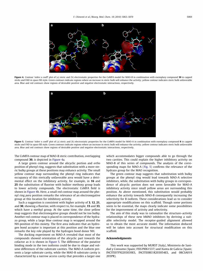
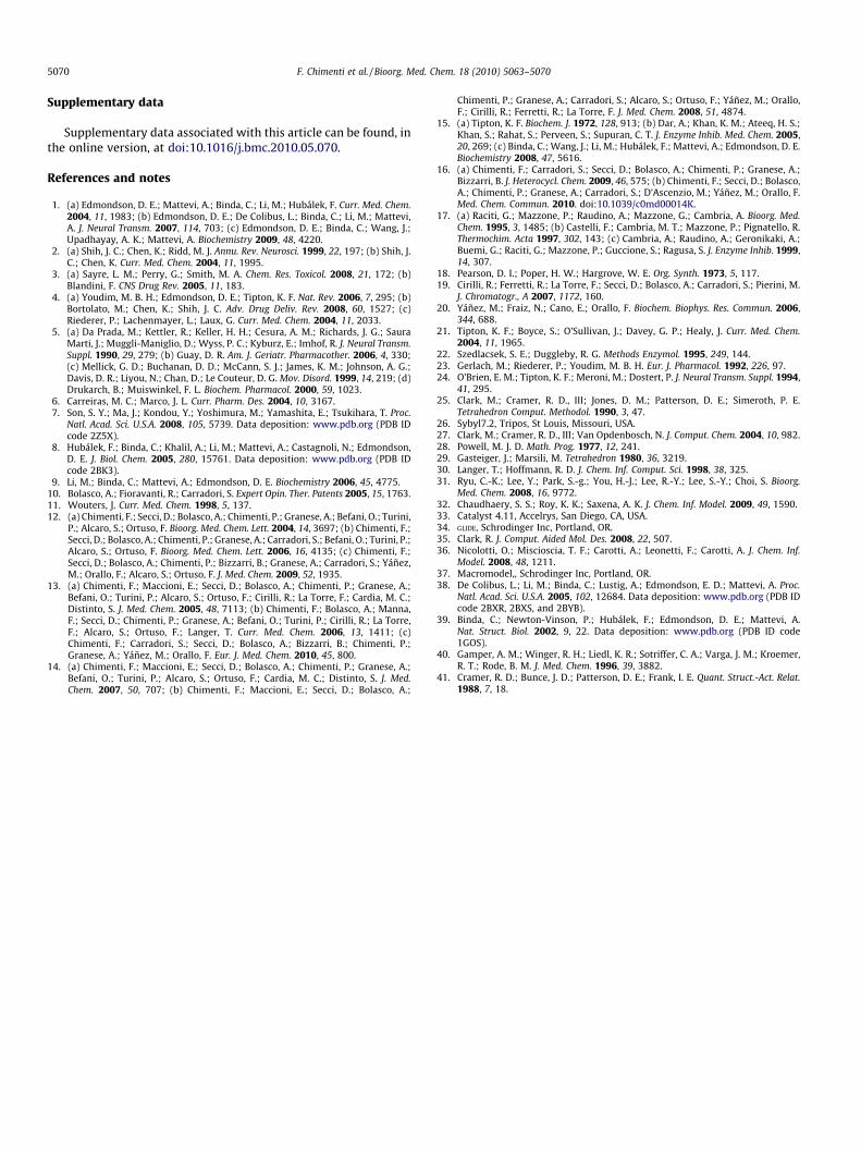
Figure 5. Contour ‘stdev x coeff’ plot of (a) steric and (b) electrostatic properties for the CoMFA model for MAO-A in combination with exemplary compound 8 in cappedsticks and FAD in space fill style. Green contours indicate regions where an increase in steric bulk will enhance the activity; yellow contour indicates steric bulk unfavorablearea. Blue and red contours show regions of desirable positive and negative electrostatic interactions, respectively.
Figure 4. Contour ‘stdev x coeff’ plot of (a) steric and (b) electrostatic properties for the CoMFA model for MAO-B in combination with exemplary compound 30 in cappedsticks and FAD in space fill style. Green contours indicate regions where an increase in steric bulk will enhance the activity; yellow contour indicates steric bulk unfavorablearea. Blue and red contours show regions of desirable positive and negative electrostatic interactions, respectively.
F. Chimenti et al. / Bioorg. Med. Chem. 18 (2010) 5063–5070 5069
The CoMFA contour map of MAO-B steric contribution, overlappingcompound 30, is depicted in Figure 4a.
A large green contour around the alicyclic portion and orthoposition of phenyl ring, suggests that substitution with a more ste-ric bulky groups at these positions may enhance activity. The smallyellow contour map surrounding the phenyl ring indicates thatoccupancy of this sterically unfavorable area would have a detri-mental effect on the inhibitory activity, for example, in 16 and25 the substitution of fluorine with bulkier methoxy group leadsto lower activity compounds. The electrostatic CoMFA field isshown in Figure 4b. Here, a small red contour map around the phe-nyl ring para position remarks the relevance of an electronegativegroup at this location for inhibitory activity.
Such a suggestion is consistent with higher activity of 3, 12, 21,and 30, showing a fluorine, with respect to, for example, 15 and 33,which have a methyl group. At the same time, the close yellowmap suggests that electronegative groups should not be too bulky.Another red contour map is placed in correspondence of the hydra-zo group, while a large blue contour map is wrapped around theNH-position of this moiety. The first area indicates that an hydro-gen bond acceptor is important at this position and the blue oneremarks the key role played by the hydrogen bond donor NH.
The docking experiment on MAO-A revealed that most of thecompounds showed orientation of the alicyclic part towards thecofactor as it is shown in Figure 5. The difference of the putativebinding mode in the two isoforms could be due to shape and vol-ume differences of the substrate cavities. The MAO-A isoform pre-sents a large substrate cavity, while the MAO-B substrate cavity ischaracterized by a narrow access cavity that precedes a larger one
which accommodates bigger compounds able to go through thetwo cavities. This could explain the higher inhibitory activity onMAO-B of this series of compounds. The analysis of the corre-sponding maps for MAO-A (Fig. 5) confirms the relevance of thehydrazo group for the MAO recognition.
The green contour map suggests that substitution with bulkygroups at the phenyl ring would lead towards MAO-A selectiveinhibitors, while, the substitution with bulky groups in correspon-dence of alicyclic portion does not seem favorable for MAO-Ainhibitory activity since small yellow areas are surrounding thisposition. As above mentioned, this substitution would probablyenhance the activity towards MAO-B consequently increasing theselectivity for B isoform. These considerations lead us to considerappropriate modifications on this scaffold. Though some portionsseem to be essential, the maps clearly indicate some possibilitiesfor the improvement of activity and selectivity.
The aim of this study was to rationalize the structure–activityrelationships of these new hMAO inhibitors by deriving a suit-able selectivity model. The receptor-guided alignment allowedus to obtain the most accurate model. The information deducedwill be taken into account for structural modification on thisscaffold.
Acknowledgments
This work was supported by MURST (Italy), Ministerio de Sani-dad y Consumo (Spain; FISS PI061537) and Xunta de Galicia (Spain;INCITE07PXI203039ES, INCITE08E1R203054ES, and 08CSA019203PR).
5070 F. Chimenti et al. / Bioorg. Med. Chem. 18 (2010) 5063–5070
Supplementary data
Supplementary data associated with this article can be found, inthe online version, at doi:10.1016/j.bmc.2010.05.070.
References and notes
1. (a) Edmondson, D. E.; Mattevi, A.; Binda, C.; Li, M.; Hubálek, F. Curr. Med. Chem.2004, 11, 1983; (b) Edmondson, D. E.; De Colibus, L.; Binda, C.; Li, M.; Mattevi,A. J. Neural Transm. 2007, 114, 703; (c) Edmondson, D. E.; Binda, C.; Wang, J.;Upadhayay, A. K.; Mattevi, A. Biochemistry 2009, 48, 4220.
2. (a) Shih, J. C.; Chen, K.; Ridd, M. J. Annu. Rev. Neurosci. 1999, 22, 197; (b) Shih, J.C.; Chen, K. Curr. Med. Chem. 2004, 11, 1995.
3. (a) Sayre, L. M.; Perry, G.; Smith, M. A. Chem. Res. Toxicol. 2008, 21, 172; (b)Blandini, F. CNS Drug Rev. 2005, 11, 183.
4. (a) Youdim, M. B. H.; Edmondson, D. E.; Tipton, K. F. Nat. Rev. 2006, 7, 295; (b)Bortolato, M.; Chen, K.; Shih, J. C. Adv. Drug Deliv. Rev. 2008, 60, 1527; (c)Riederer, P.; Lachenmayer, L.; Laux, G. Curr. Med. Chem. 2004, 11, 2033.
5. (a) Da Prada, M.; Kettler, R.; Keller, H. H.; Cesura, A. M.; Richards, J. G.; SauraMarti, J.; Muggli-Maniglio, D.; Wyss, P. C.; Kyburz, E.; Imhof, R. J. Neural Transm.Suppl. 1990, 29, 279; (b) Guay, D. R. Am. J. Geriatr. Pharmacother. 2006, 4, 330;(c) Mellick, G. D.; Buchanan, D. D.; McCann, S. J.; James, K. M.; Johnson, A. G.;Davis, D. R.; Liyou, N.; Chan, D.; Le Couteur, D. G. Mov. Disord. 1999, 14, 219; (d)Drukarch, B.; Muiswinkel, F. L. Biochem. Pharmacol. 2000, 59, 1023.
6. Carreiras, M. C.; Marco, J. L. Curr. Pharm. Des. 2004, 10, 3167.7. Son, S. Y.; Ma, J.; Kondou, Y.; Yoshimura, M.; Yamashita, E.; Tsukihara, T. Proc.
Natl. Acad. Sci. U.S.A. 2008, 105, 5739. Data deposition: www.pdb.org (PDB IDcode 2Z5X).
8. Hubálek, F.; Binda, C.; Khalil, A.; Li, M.; Mattevi, A.; Castagnoli, N.; Edmondson,D. E. J. Biol. Chem. 2005, 280, 15761. Data deposition: www.pdb.org (PDB IDcode 2BK3).
9. Li, M.; Binda, C.; Mattevi, A.; Edmondson, D. E. Biochemistry 2006, 45, 4775.10. Bolasco, A.; Fioravanti, R.; Carradori, S. Expert Opin. Ther. Patents 2005, 15, 1763.11. Wouters, J. Curr. Med. Chem. 1998, 5, 137.12. (a) Chimenti, F.; Secci, D.; Bolasco, A.; Chimenti, P.; Granese, A.; Befani, O.; Turini,
P.; Alcaro, S.; Ortuso, F. Bioorg. Med. Chem. Lett. 2004, 14, 3697; (b) Chimenti, F.;Secci, D.; Bolasco, A.; Chimenti, P.; Granese, A.; Carradori, S.; Befani, O.; Turini, P.;Alcaro, S.; Ortuso, F. Bioorg. Med. Chem. Lett. 2006, 16, 4135; (c) Chimenti, F.;Secci, D.; Bolasco, A.; Chimenti, P.; Bizzarri, B.; Granese, A.; Carradori, S.; Yáñez,M.; Orallo, F.; Alcaro, S.; Ortuso, F. J. Med. Chem. 2009, 52, 1935.
13. (a) Chimenti, F.; Maccioni, E.; Secci, D.; Bolasco, A.; Chimenti, P.; Granese, A.;Befani, O.; Turini, P.; Alcaro, S.; Ortuso, F.; Cirilli, R.; La Torre, F.; Cardia, M. C.;Distinto, S. J. Med. Chem. 2005, 48, 7113; (b) Chimenti, F.; Bolasco, A.; Manna,F.; Secci, D.; Chimenti, P.; Granese, A.; Befani, O.; Turini, P.; Cirilli, R.; La Torre,F.; Alcaro, S.; Ortuso, F.; Langer, T. Curr. Med. Chem. 2006, 13, 1411; (c)Chimenti, F.; Carradori, S.; Secci, D.; Bolasco, A.; Bizzarri, B.; Chimenti, P.;Granese, A.; Yáñez, M.; Orallo, F. Eur. J. Med. Chem. 2010, 45, 800.
14. (a) Chimenti, F.; Maccioni, E.; Secci, D.; Bolasco, A.; Chimenti, P.; Granese, A.;Befani, O.; Turini, P.; Alcaro, S.; Ortuso, F.; Cardia, M. C.; Distinto, S. J. Med.Chem. 2007, 50, 707; (b) Chimenti, F.; Maccioni, E.; Secci, D.; Bolasco, A.;
Chimenti, P.; Granese, A.; Carradori, S.; Alcaro, S.; Ortuso, F.; Yáñez, M.; Orallo,F.; Cirilli, R.; Ferretti, R.; La Torre, F. J. Med. Chem. 2008, 51, 4874.
15. (a) Tipton, K. F. Biochem. J. 1972, 128, 913; (b) Dar, A.; Khan, K. M.; Ateeq, H. S.;Khan, S.; Rahat, S.; Perveen, S.; Supuran, C. T. J. Enzyme Inhib. Med. Chem. 2005,20, 269; (c) Binda, C.; Wang, J.; Li, M.; Hubálek, F.; Mattevi, A.; Edmondson, D. E.Biochemistry 2008, 47, 5616.
16. (a) Chimenti, F.; Carradori, S.; Secci, D.; Bolasco, A.; Chimenti, P.; Granese, A.;Bizzarri, B. J. Heterocycl. Chem. 2009, 46, 575; (b) Chimenti, F.; Secci, D.; Bolasco,A.; Chimenti, P.; Granese, A.; Carradori, S.; D’Ascenzio, M.; Yáñez, M.; Orallo, F.Med. Chem. Commun. 2010. doi:10.1039/c0md00014K.
17. (a) Raciti, G.; Mazzone, P.; Raudino, A.; Mazzone, G.; Cambria, A. Bioorg. Med.Chem. 1995, 3, 1485; (b) Castelli, F.; Cambria, M. T.; Mazzone, P.; Pignatello, R.Thermochim. Acta 1997, 302, 143; (c) Cambria, A.; Raudino, A.; Geronikaki, A.;Buemi, G.; Raciti, G.; Mazzone, P.; Guccione, S.; Ragusa, S. J. Enzyme Inhib. 1999,14, 307.
18. Pearson, D. I.; Poper, H. W.; Hargrove, W. E. Org. Synth. 1973, 5, 117.19. Cirilli, R.; Ferretti, R.; La Torre, F.; Secci, D.; Bolasco, A.; Carradori, S.; Pierini, M.
J. Chromatogr., A 2007, 1172, 160.20. Yáñez, M.; Fraiz, N.; Cano, E.; Orallo, F. Biochem. Biophys. Res. Commun. 2006,
344, 688.21. Tipton, K. F.; Boyce, S.; O’Sullivan, J.; Davey, G. P.; Healy, J. Curr. Med. Chem.
2004, 11, 1965.22. Szedlacsek, S. E.; Duggleby, R. G. Methods Enzymol. 1995, 249, 144.23. Gerlach, M.; Riederer, P.; Youdim, M. B. H. Eur. J. Pharmacol. 1992, 226, 97.24. O’Brien, E. M.; Tipton, K. F.; Meroni, M.; Dostert, P. J. Neural Transm. Suppl. 1994,
41, 295.25. Clark, M.; Cramer, R. D., III; Jones, D. M.; Patterson, D. E.; Simeroth, P. E.
Tetrahedron Comput. Methodol. 1990, 3, 47.26. Sybyl7.2, Tripos, St Louis, Missouri, USA.27. Clark, M.; Cramer, R. D., III; Van Opdenbosch, N. J. Comput. Chem. 2004, 10, 982.28. Powell, M. J. D. Math. Prog. 1977, 12, 241.29. Gasteiger, J.; Marsili, M. Tetrahedron 1980, 36, 3219.30. Langer, T.; Hoffmann, R. D. J. Chem. Inf. Comput. Sci. 1998, 38, 325.31. Ryu, C.-K.; Lee, Y.; Park, S.-g.; You, H.-J.; Lee, R.-Y.; Lee, S.-Y.; Choi, S. Bioorg.
Med. Chem. 2008, 16, 9772.32. Chaudhaery, S. S.; Roy, K. K.; Saxena, A. K. J. Chem. Inf. Model. 2009, 49, 1590.33. Catalyst 4.11, Accelrys, San Diego, CA, USA.34. GLIDE, Schrodinger Inc, Portland, OR.35. Clark, R. J. Comput. Aided Mol. Des. 2008, 22, 507.36. Nicolotti, O.; Miscioscia, T. F.; Carotti, A.; Leonetti, F.; Carotti, A. J. Chem. Inf.
Model. 2008, 48, 1211.37. Macromodel,, Schrodinger Inc, Portland, OR.38. De Colibus, L.; Li, M.; Binda, C.; Lustig, A.; Edmondson, E. D.; Mattevi, A. Proc.
Natl. Acad. Sci. U.S.A. 2005, 102, 12684. Data deposition: www.pdb.org (PDB IDcode 2BXR, 2BXS, and 2BYB).
39. Binda, C.; Newton-Vinson, P.; Hubálek, F.; Edmondson, D. E.; Mattevi, A.Nat. Struct. Biol. 2002, 9, 22. Data deposition: www.pdb.org (PDB ID code1GOS).
40. Gamper, A. M.; Winger, R. H.; Liedl, K. R.; Sotriffer, C. A.; Varga, J. M.; Kroemer,R. T.; Rode, B. M. J. Med. Chem. 1996, 39, 3882.
41. Cramer, R. D.; Bunce, J. D.; Patterson, D. E.; Frank, I. E. Quant. Struct.-Act. Relat.1988, 7, 18.
Related Documents