Synthesis and photoluminescence properties of microcrystalline Sr 2 ZnWO 6 :RE 3+ (RE = Eu, Dy, Sm and Pr) phosphors K.V. Dabre a , K. Park b , S.J. Dhoble c,⇑ a Department of Physics, Arts, Commerce & Science College, Koradi, Nagpur 441111, India b Faculty of Nanotechnology and Advanced Materials Engineering, Sejong University, Seoul 143-747, Republic of Korea c Department of Physics, R.T.M. Nagpur University, Nagpur 440033, India article info Article history: Received 31 March 2014 Received in revised form 27 July 2014 Accepted 28 July 2014 Available online 7 August 2014 Keywords: Double perovskite Solid state reactions Luminescence X-ray diffraction abstract The novel microcrystalline Sr 2 ZnWO 6 :RE 3+ (RE = Eu, Dy, Sm and Pr) phosphors were synthesized by solid- state reaction method at 1250 °C and their photoluminescence properties were investigated. The Eu 3+ and Dy 3+ activated phosphors show intense red (616 nm) and yellow (574 nm) emission respectively; which indicate that the rare earth ions are substituted at non-centrosymmetric site in the host lattice. Near white (Dy 3+ ) and reddish-orange (Sm 3+ ) emissions of rare earth ions in the host lattice show strong host absorption and energy transfer from the host to activator ion. Pr 3+ activated phosphor shows a series of emission peaks in the visible region with the most intense peak in the blue region at 491 and 499 nm. Ó 2014 Published by Elsevier B.V. 1. Introduction Research to improve the luminescence properties of phosphor material became very important, because of its wide applicability in modern technologies such as lighting (fluorescent lamp, LED), displays (PDP, FED), scintillator, communication (optical fiber), for the sake of growing problem of energy crisis. A phosphor with efficient absorption in near UV or blue spectral region and emit in visible region could be potential material for the application in solid state lighting (LED) (an effective and eco-friendlier replace- ment of fluorescent lamp) [1–6]. The scientific community around the world is continuously exploring the luminescence properties of rare earth ions activated various host materials [7–11], for finding the efficient phosphors with stable optical properties. Tungstate is one of the interesting host materials that have attracted a great deal of interest owing to self-activation, good stability for physical conditions (chemical and thermal) and wider applicability in vari- ous fields such as scintillation, solid state laser, electro-optic, cata- lytic. Tungstate based phosphors show broad excitation and emission bands due to the ligand to metal charge transfer (CT) transition of (WO 4 ) 2 and (WO 6 ) 6 oxyanion complex. Partially filled deep-lying 4f shells of the rare earth ions causes interesting optical properties which make them favorite activators for doping in various hosts. However, the sharp excitation lines of rare earth are less effective and its higher concentration in host lattice leads to quenching of emission. Thus the host sensitization of rare earth ions in tungstate materials is actively studied from last decades [12–16]. The tungstates with double perovskite structure offers various interesting physical properties such as electrical [17], magnetic [18], optical [19] and photocatalytic [20] properties, which has attracted researchers’ attention since 50 years [21,22]. The general formula for double perovskite tungstate is A 2 BWO 6 and its struc- ture could be visualized as three-dimensional network of alternate corner sharing BO 6 and WO 6 octahedra forming FCC lattice (Fig. 1a) and A 2+ cation is at the interstitial space forming cubo-octahedral geometry (Fig. 1b). However, in reality its structure determination became difficult due to presence of several types of distortion such as tilt or rotation of BO 6 and WO 6 octahedra along the crystallo- graphic axis and displacement of A or B cation with respect to rigid polyhedra. These distortions are responsible for lowering symme- try and phase transition in double perovskite due to change in tem- perature and pressure [23–25]. For instance, the detail theoretical and experimental study of temperature induced phase transition in Sr 2 ZnWO 6 [25–27] shows that this double perovskite tungstate exhibits monoclinic phase at room temperature and undergo phase transition to tetragonal and cubic with increase in temperature. The rare earth (especially Eu 3+ ) activation in double perovskite tungstate and their photoluminescence (PL) properties has been actively studied from last few years [28]. Recently, the reports on PL properties of double perovskite structured Ca 3 WO 6 :Eu 3+ [29] http://dx.doi.org/10.1016/j.jallcom.2014.07.205 0925-8388/Ó 2014 Published by Elsevier B.V. ⇑ Corresponding author. E-mail address: [email protected] (S.J. Dhoble). Journal of Alloys and Compounds 617 (2014) 129–134 Contents lists available at ScienceDirect Journal of Alloys and Compounds journal homepage: www.elsevier.com/locate/jalcom

Welcome message from author
This document is posted to help you gain knowledge. Please leave a comment to let me know what you think about it! Share it to your friends and learn new things together.
Transcript
Journal of Alloys and Compounds 617 (2014) 129–134
Contents lists available at ScienceDirect
Journal of Alloys and Compounds
journal homepage: www.elsevier .com/locate / ja lcom
Synthesis and photoluminescence properties of microcrystallineSr2ZnWO6:RE3+ (RE = Eu, Dy, Sm and Pr) phosphors
http://dx.doi.org/10.1016/j.jallcom.2014.07.2050925-8388/� 2014 Published by Elsevier B.V.
⇑ Corresponding author.E-mail address: [email protected] (S.J. Dhoble).
K.V. Dabre a, K. Park b, S.J. Dhoble c,⇑a Department of Physics, Arts, Commerce & Science College, Koradi, Nagpur 441111, Indiab Faculty of Nanotechnology and Advanced Materials Engineering, Sejong University, Seoul 143-747, Republic of Koreac Department of Physics, R.T.M. Nagpur University, Nagpur 440033, India
a r t i c l e i n f o
Article history:Received 31 March 2014Received in revised form 27 July 2014Accepted 28 July 2014Available online 7 August 2014
Keywords:Double perovskiteSolid state reactionsLuminescenceX-ray diffraction
a b s t r a c t
The novel microcrystalline Sr2ZnWO6:RE3+ (RE = Eu, Dy, Sm and Pr) phosphors were synthesized by solid-state reaction method at 1250 �C and their photoluminescence properties were investigated. The Eu3+
and Dy3+ activated phosphors show intense red (616 nm) and yellow (574 nm) emission respectively;which indicate that the rare earth ions are substituted at non-centrosymmetric site in the host lattice.Near white (Dy3+) and reddish-orange (Sm3+) emissions of rare earth ions in the host lattice show stronghost absorption and energy transfer from the host to activator ion. Pr3+ activated phosphor shows a seriesof emission peaks in the visible region with the most intense peak in the blue region at 491 and 499 nm.
� 2014 Published by Elsevier B.V.
1. Introduction
Research to improve the luminescence properties of phosphormaterial became very important, because of its wide applicabilityin modern technologies such as lighting (fluorescent lamp, LED),displays (PDP, FED), scintillator, communication (optical fiber),for the sake of growing problem of energy crisis. A phosphor withefficient absorption in near UV or blue spectral region and emit invisible region could be potential material for the application insolid state lighting (LED) (an effective and eco-friendlier replace-ment of fluorescent lamp) [1–6]. The scientific community aroundthe world is continuously exploring the luminescence properties ofrare earth ions activated various host materials [7–11], for findingthe efficient phosphors with stable optical properties. Tungstate isone of the interesting host materials that have attracted a greatdeal of interest owing to self-activation, good stability for physicalconditions (chemical and thermal) and wider applicability in vari-ous fields such as scintillation, solid state laser, electro-optic, cata-lytic. Tungstate based phosphors show broad excitation andemission bands due to the ligand to metal charge transfer (CT)transition of (WO4)2� and (WO6)6� oxyanion complex. Partiallyfilled deep-lying 4f shells of the rare earth ions causes interestingoptical properties which make them favorite activators for dopingin various hosts. However, the sharp excitation lines of rare earth
are less effective and its higher concentration in host lattice leadsto quenching of emission. Thus the host sensitization of rare earthions in tungstate materials is actively studied from last decades[12–16].
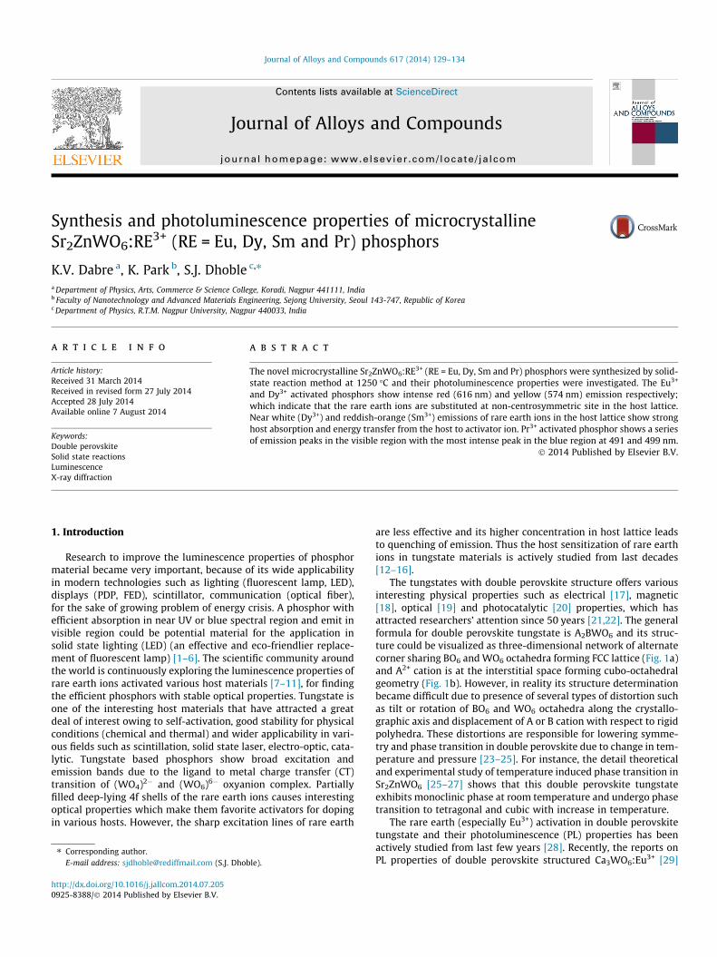
The tungstates with double perovskite structure offers variousinteresting physical properties such as electrical [17], magnetic[18], optical [19] and photocatalytic [20] properties, which hasattracted researchers’ attention since 50 years [21,22]. The generalformula for double perovskite tungstate is A2BWO6 and its struc-ture could be visualized as three-dimensional network of alternatecorner sharing BO6 and WO6 octahedra forming FCC lattice (Fig. 1a)and A2+ cation is at the interstitial space forming cubo-octahedralgeometry (Fig. 1b). However, in reality its structure determinationbecame difficult due to presence of several types of distortion suchas tilt or rotation of BO6 and WO6 octahedra along the crystallo-graphic axis and displacement of A or B cation with respect to rigidpolyhedra. These distortions are responsible for lowering symme-try and phase transition in double perovskite due to change in tem-perature and pressure [23–25]. For instance, the detail theoreticaland experimental study of temperature induced phase transition inSr2ZnWO6 [25–27] shows that this double perovskite tungstateexhibits monoclinic phase at room temperature and undergo phasetransition to tetragonal and cubic with increase in temperature.
The rare earth (especially Eu3+) activation in double perovskitetungstate and their photoluminescence (PL) properties has beenactively studied from last few years [28]. Recently, the reports onPL properties of double perovskite structured Ca3WO6:Eu3+ [29]
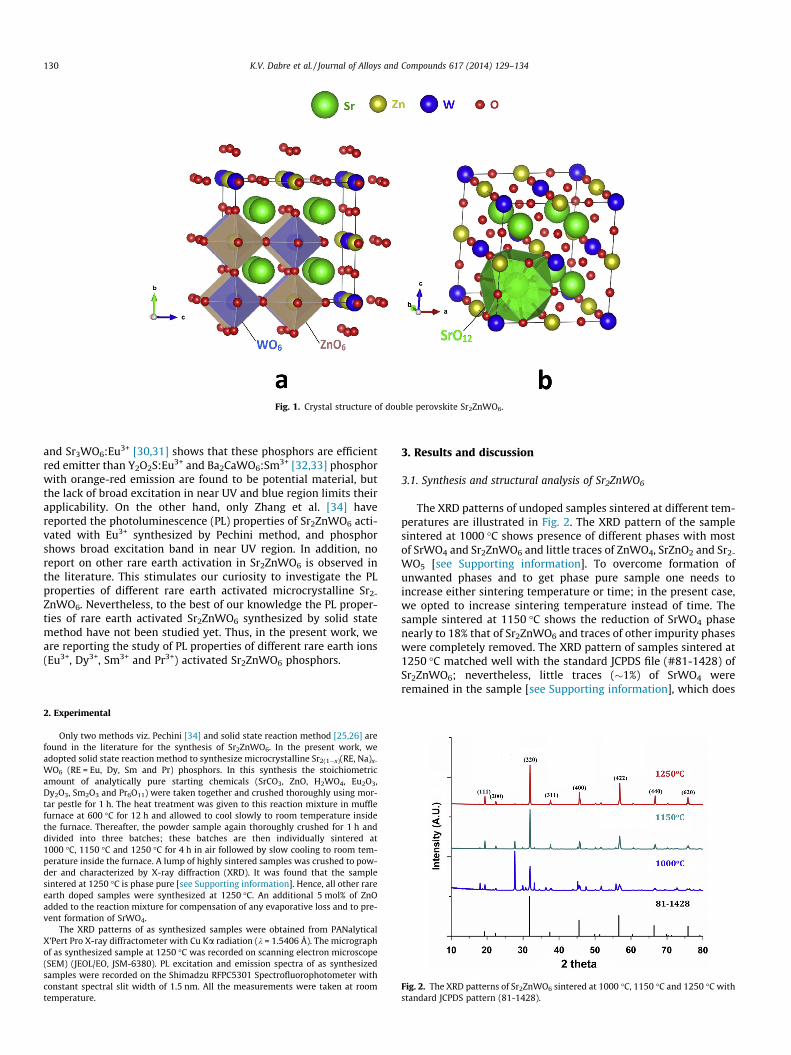
Fig. 1. Crystal structure of double perovskite Sr2ZnWO6.
130 K.V. Dabre et al. / Journal of Alloys and Compounds 617 (2014) 129–134
and Sr3WO6:Eu3+ [30,31] shows that these phosphors are efficientred emitter than Y2O2S:Eu3+ and Ba2CaWO6:Sm3+ [32,33] phosphorwith orange-red emission are found to be potential material, butthe lack of broad excitation in near UV and blue region limits theirapplicability. On the other hand, only Zhang et al. [34] havereported the photoluminescence (PL) properties of Sr2ZnWO6 acti-vated with Eu3+ synthesized by Pechini method, and phosphorshows broad excitation band in near UV region. In addition, noreport on other rare earth activation in Sr2ZnWO6 is observed inthe literature. This stimulates our curiosity to investigate the PLproperties of different rare earth activated microcrystalline Sr2-
ZnWO6. Nevertheless, to the best of our knowledge the PL proper-ties of rare earth activated Sr2ZnWO6 synthesized by solid statemethod have not been studied yet. Thus, in the present work, weare reporting the study of PL properties of different rare earth ions(Eu3+, Dy3+, Sm3+ and Pr3+) activated Sr2ZnWO6 phosphors.
Fig. 2. The XRD patterns of Sr2ZnWO6 sintered at 1000 �C, 1150 �C and 1250 �C withstandard JCPDS pattern (81-1428).
2. Experimental
Only two methods viz. Pechini [34] and solid state reaction method [25,26] arefound in the literature for the synthesis of Sr2ZnWO6. In the present work, weadopted solid state reaction method to synthesize microcrystalline Sr2(1�x)(RE, Na)x-
WO6 (RE = Eu, Dy, Sm and Pr) phosphors. In this synthesis the stoichiometricamount of analytically pure starting chemicals (SrCO3, ZnO, H2WO4, Eu2O3,Dy2O3, Sm2O3 and Pr6O11) were taken together and crushed thoroughly using mor-tar pestle for 1 h. The heat treatment was given to this reaction mixture in mufflefurnace at 600 �C for 12 h and allowed to cool slowly to room temperature insidethe furnace. Thereafter, the powder sample again thoroughly crushed for 1 h anddivided into three batches; these batches are then individually sintered at1000 �C, 1150 �C and 1250 �C for 4 h in air followed by slow cooling to room tem-perature inside the furnace. A lump of highly sintered samples was crushed to pow-der and characterized by X-ray diffraction (XRD). It was found that the samplesintered at 1250 �C is phase pure [see Supporting information]. Hence, all other rareearth doped samples were synthesized at 1250 �C. An additional 5 mol% of ZnOadded to the reaction mixture for compensation of any evaporative loss and to pre-vent formation of SrWO4.
The XRD patterns of as synthesized samples were obtained from PANalyticalX’Pert Pro X-ray diffractometer with Cu Ka radiation (k = 1.5406 Å). The micrographof as synthesized sample at 1250 �C was recorded on scanning electron microscope(SEM) (JEOL/EO, JSM-6380). PL excitation and emission spectra of as synthesizedsamples were recorded on the Shimadzu RFPC5301 Spectrofluorophotometer withconstant spectral slit width of 1.5 nm. All the measurements were taken at roomtemperature.
3. Results and discussion
3.1. Synthesis and structural analysis of Sr2ZnWO6
The XRD patterns of undoped samples sintered at different tem-peratures are illustrated in Fig. 2. The XRD pattern of the samplesintered at 1000 �C shows presence of different phases with mostof SrWO4 and Sr2ZnWO6 and little traces of ZnWO4, SrZnO2 and Sr2-
WO5 [see Supporting information]. To overcome formation ofunwanted phases and to get phase pure sample one needs toincrease either sintering temperature or time; in the present case,we opted to increase sintering temperature instead of time. Thesample sintered at 1150 �C shows the reduction of SrWO4 phasenearly to 18% that of Sr2ZnWO6 and traces of other impurity phaseswere completely removed. The XRD pattern of samples sintered at1250 �C matched well with the standard JCPDS file (#81-1428) ofSr2ZnWO6; nevertheless, little traces (�1%) of SrWO4 wereremained in the sample [see Supporting information], which does
K.V. Dabre et al. / Journal of Alloys and Compounds 617 (2014) 129–134 131
not significantly affect the luminescence properties of phosphor.The double perovskite under study (Sr2ZnWO6) is crystallized inthe cubic phase with space group Fm-3m (Fig. 1), which is in goodagreement with those reported by Zhang et al. [34]. However, tol-erance factor (t = 0.968) and the fitness factor (/ = 0.974) [35] andearlier reports [36,37] claimed the tetragonal phase. Here weexclude the crystallization of the phosphor in tetragonal phase,as no match with standard JCPDS file of tetragonal phase (PDFNo. #15-0572) was obtained and no standard JCPDS file of mono-clinic Sr2ZnWO6 is available. Thus, it is concluded that the phaseof as synthesized Sr2ZnWO6 is the cubic.
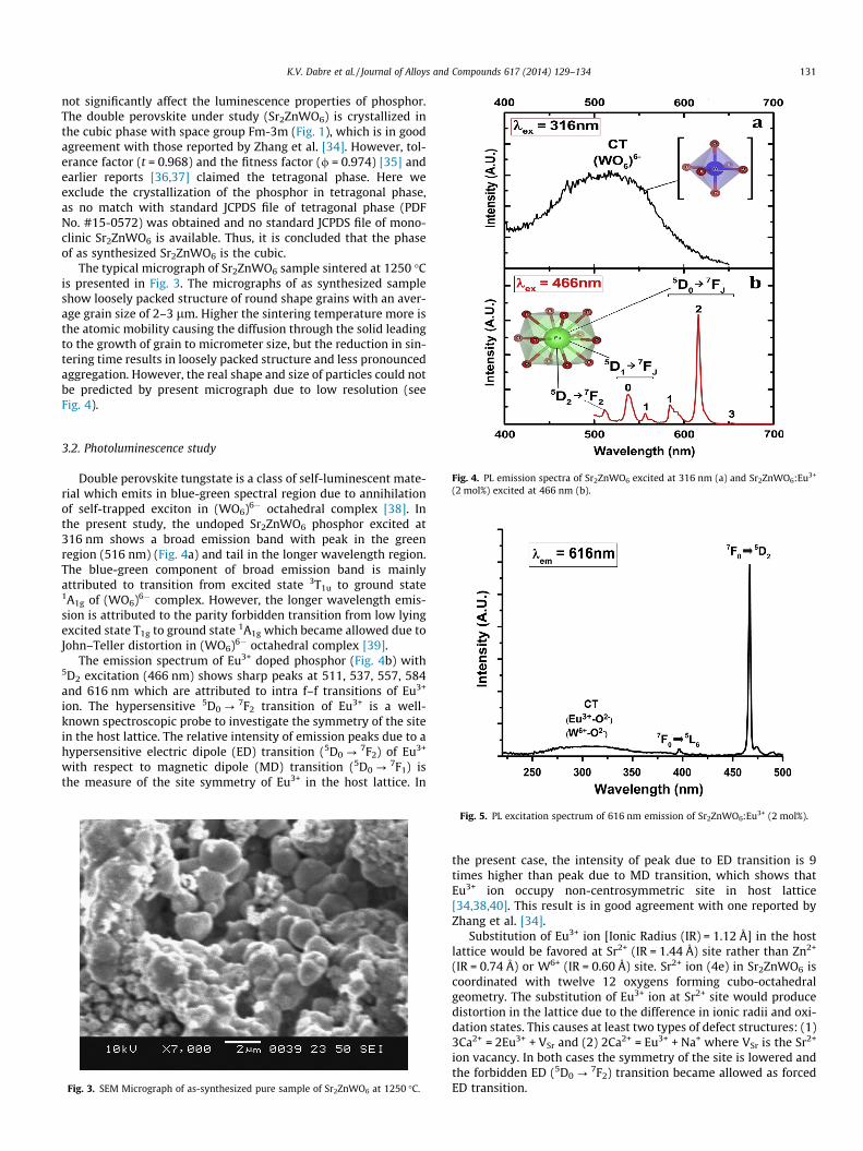
The typical micrograph of Sr2ZnWO6 sample sintered at 1250 �Cis presented in Fig. 3. The micrographs of as synthesized sampleshow loosely packed structure of round shape grains with an aver-age grain size of 2–3 lm. Higher the sintering temperature more isthe atomic mobility causing the diffusion through the solid leadingto the growth of grain to micrometer size, but the reduction in sin-tering time results in loosely packed structure and less pronouncedaggregation. However, the real shape and size of particles could notbe predicted by present micrograph due to low resolution (seeFig. 4).
Fig. 4. PL emission spectra of Sr2ZnWO6 excited at 316 nm (a) and Sr2ZnWO6:Eu3+
(2 mol%) excited at 466 nm (b).
3.2. Photoluminescence study
Double perovskite tungstate is a class of self-luminescent mate-rial which emits in blue-green spectral region due to annihilationof self-trapped exciton in (WO6)6� octahedral complex [38]. Inthe present study, the undoped Sr2ZnWO6 phosphor excited at316 nm shows a broad emission band with peak in the greenregion (516 nm) (Fig. 4a) and tail in the longer wavelength region.The blue-green component of broad emission band is mainlyattributed to transition from excited state 3T1u to ground state1A1g of (WO6)6� complex. However, the longer wavelength emis-sion is attributed to the parity forbidden transition from low lyingexcited state T1g to ground state 1A1g which became allowed due toJohn–Teller distortion in (WO6)6� octahedral complex [39].
The emission spectrum of Eu3+ doped phosphor (Fig. 4b) with5D2 excitation (466 nm) shows sharp peaks at 511, 537, 557, 584and 616 nm which are attributed to intra f–f transitions of Eu3+
ion. The hypersensitive 5D0 ?7F2 transition of Eu3+ is a well-
known spectroscopic probe to investigate the symmetry of the sitein the host lattice. The relative intensity of emission peaks due to ahypersensitive electric dipole (ED) transition (5D0 ?
7F2) of Eu3+
with respect to magnetic dipole (MD) transition (5D0 ?7F1) is
the measure of the site symmetry of Eu3+ in the host lattice. In
Fig. 3. SEM Micrograph of as-synthesized pure sample of Sr2ZnWO6 at 1250 �C.
Fig. 5. PL excitation spectrum of 616 nm emission of Sr2ZnWO6:Eu3+ (2 mol%).
the present case, the intensity of peak due to ED transition is 9times higher than peak due to MD transition, which shows thatEu3+ ion occupy non-centrosymmetric site in host lattice[34,38,40]. This result is in good agreement with one reported byZhang et al. [34].
Substitution of Eu3+ ion [Ionic Radius (IR) = 1.12 Å] in the hostlattice would be favored at Sr2+ (IR = 1.44 Å) site rather than Zn2+
(IR = 0.74 Å) or W6+ (IR = 0.60 Å) site. Sr2+ ion (4e) in Sr2ZnWO6 iscoordinated with twelve 12 oxygens forming cubo-octahedralgeometry. The substitution of Eu3+ ion at Sr2+ site would producedistortion in the lattice due to the difference in ionic radii and oxi-dation states. This causes at least two types of defect structures: (1)3Ca2+ = 2Eu3+ + VSr and (2) 2Ca2+ = Eu3+ + Na+ where VSr is the Sr2+
ion vacancy. In both cases the symmetry of the site is lowered andthe forbidden ED (5D0 ?
7F2) transition became allowed as forcedED transition.
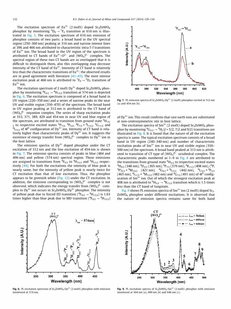
Fig. 7. PL emission spectra of Sr2ZnWO6:Dy3+ (2 mol%) phosphor excited at 312 nm(a) and 454 nm (b).
132 K.V. Dabre et al. / Journal of Alloys and Compounds 617 (2014) 129–134
The excitation spectrum of Eu3+ (2 mol%) doped Sr2ZnWO6
phosphor by monitoring 5D0 ?7F2 transition at 616 nm is illus-
trated in Fig. 5. The excitation spectrum of 616 nm emission ofphosphor consists of two parts: a broad band in the UV spectralregion (250–360 nm) peaking at 316 nm and narrow intense linesat 396 and 466 nm attributed to characteristic intra f–f transitionsof Eu3+ ion. The broad band in the UV region of the spectrum isattributed to CT bands of Eu3+–O2� and (WO6)6� complex. Thespectral region of these two CT bands are so overlapped that it isdifficult to distinguish them, also this overlapping may decreaseintensity of the CT band of Eu3+. Intensity of CT band is relativelyless than the characteristic transitions of Eu3+: the observed resultsare in good agreement with literature [41–43]. The most intenseexcitation peak at 466 nm is attributed to 7F0 ?
5D2 transition ofEu3+ ion.
The excitation spectrum of 2 mol% Dy3+ doped Sr2ZnWO6 phos-phor by monitoring 4F9/2 ?
4H13/2 transition at 574 nm is depictedin Fig. 6. The excitation spectrum is composed of a broad band inUV region (220–350 nm) and a series of narrow peaks in the nearUV and visible region (350–470) of the spectrum. The broad bandin UV region peaking at 312 nm is attributed to the CT band of(WO6)6� oxyanion complex. The series of sharp excitation peaksat 353, 371, 389, 429 and 454 nm in near UV and blue region ofthe spectrum, are attributed to transition from ground state 6H15/
2 to respective excited states 6P7/2, 6P5/2, 4F7/2 + 4I13/2, 4G11/2 and4I15/2 of 4f9 configuration of Dy3+ ion. Intensity of CT band is rela-tively higher than characteristic peaks of Dy3+ ion. It suggests theexistence of energy transfer from (WO6)6� complex to Dy3+ ion inthe host lattice.
The emission spectra of Dy3+ doped phosphor under the CTexcitation of 312 nm and the line excitation of 454 nm is shownin Fig. 7. The emission spectra consists of peaks in blue (484 and496 nm) and yellow (574 nm) spectral region. These emissionsare assigned to transitions from 4F9/2 to 4H15/2 and 4H13/2 respec-tively [44]. For both the excitations the intensity of blue peak isnearly same, but the intensity of yellow peak is nearly twice forCT excitation than that of line excitation. Thus, the phosphorappears to be greenish white (Fig. 12) under the CT excitation. Inaddition, the emission corresponding to (WO6)6� complex is notobserved, which indicates the energy transfer from (WO6)6� com-plex to Dy3+ ion occurs in Sr2ZnWO6:Dy3+ phosphor. The intensityof yellow peak due to forced ED transition (4F9/2 ?
4H13/2) is 1.63times higher than blue peak due to MD transition (4F9/2 ?
4H15/2)
Fig. 6. PL excitation spectrum of Sr2ZnWO6:Dy3+ (2 mol%) phosphor with emissionmonitored at 574 nm.
of Dy3+ ion. This result confirms that rare earth ions are substitutedat non-centrosymmetric site in host lattice.
The excitation spectra of Sm3+ (2 mol%) doped Sr2ZnWO6 phos-phor by monitoring 4G5/2 ?
6HJ (J = 5/2, 7/2 and 9/2) transitions areillustrated in Fig. 8. It is found that the nature of all the excitationspectra is same. The typical excitation spectrum consists of a broadband in UV region (240–340 nm) and number of characteristicexcitation peaks of Sm3+ ion in near UV and visible region (350–500 nm) of the spectrum. A broad band peaked at 313 nm is attrib-uted to transition of CT type of (WO6)6� octahedral complex. Thecharacteristic peaks numbered as 1–9 in Fig. 8 are attributed tothe transitions from ground state 6H5/2 to respective excited states4H9/2 (348 nm), 4D3/2 (365 nm), 6P7/2 (379 nm), 4K11/2 (408 nm), (6P,4P)5/2 + 4M19/2 (421 nm), 4G9/2 + 4I15/2 (442 nm), 4I13/2 + 4F5/2
(465 nm), 4I11/2 + 4M15/2 (482 nm) and 4G7/2 (491 nm) of 4f5 config-uration of Sm3+ ion. Out of which the strongest excitation peak at408 nm is attributed to 6H5/2 ? 4K11/2 transition which is 1.5 timesless than the CT band of tungstate.
Fig. 9 shows PL emission spectra of Sm3+ ion (2 mol%) doped Sr2-
ZnWO6 phosphor under different excitations. It is observed thatthe nature of emission spectra remains same for both band
Fig. 8. PL excitation spectra of Sr2ZnWO6:Sm3+ (2 mol%) phosphor with emissionmonitored at 564 nm (a), 600 nm (b) and 646 nm (c).
Fig. 9. PL emission spectra of Sr2ZnWO6:Sm3+ (2 mol%) phosphor excited at 313 nm(a), 408 nm (b) and 482 nm (c). Fig. 11. PL emission spectra of Sr2ZnWO6:Pr3+ (0.2 mol%) phosphor excited at
315 nm (a) and 450 nm (b). In inset the energy level scheme of 4f2 configuration ofPr3+ ion showing various transitions corresponding to emission peaks is presented.
K.V. Dabre et al. / Journal of Alloys and Compounds 617 (2014) 129–134 133
(313 nm) and line (408 and 482 nm) excitations; this indicates thatSm3+ ion occupy only one site in host lattice. The emission spectraconsists of three emission peaks at 564, 600 and 646 nm, which areattributed to 4G5/2 ?
6HJ (J = 5/2, 7/2 and 9/2 respectively) transi-tions of Sm3+ ion. The intensity of all the peaks for band excitationat 313 nm is nearly 1.35 times higher than line excitations of408 nm and 482 nm. This indicates the energy transfer from tung-state to Sm3+ ion in Sr2ZnWO6:Sm3+ phosphor.
Fig. 10 presents the excitation spectra of Sr2ZnWO6:Pr3+ phos-phor with emission monitored at 490 nm and 653 nm. The excita-tion spectra consist of a broad band (250–350 nm) peaking at315 nm is attributed to the CT band of (WO6)6� octahedral com-plex and a sharp peak at 450 nm is attributed to 3H4 ? 3P2 transi-tion of Pr3+ ion. The relative intensity of CT band of tungstate is 2.5times lower than the sharp characteristic peak of Pr3+ ion. Theemission spectra of Pr3+ (0.2 mol%) doped Sr2ZnWO6 phosphorexcited at 315 nm and 450 nm is presented in Fig. 11. Both theemission spectra are of same nature, but the intensity of blue peak(490 nm) is nearly double for 3P2 excitation (450 nm) than CT bandexcitation (315 nm). This indicates the energy transfer fromtungstate to Pr3+ ion is comparatively less than Dy3+ and Sm3+.
Fig. 10. PL excitation spectra of Sr2ZnWO6:Pr3+ (0.2 mol%) phosphor with emissionmonitored at 491 nm (a) and 653 nm (b).
The typical emission spectrum of Pr3+ doped phosphor consists ofnumber of peaks in blue to red region at 491, 499, 534, 550, 619,631 and 653 nm which are attributed to intra f–f transitions ofPr3+ ion (Inset of Fig. 11).
The color of the phosphor can be frequently expressed by Com-mission Internationale de l’Eclairage (CIE) coordinates. The CIEcoordinates of the rare earth (Eu, Dy, Sm and Pr) doped Sr2ZnWO6
phosphors (Fig. 12) under different excitations are listed in Table 1.Eu3+ shows the yellowish orange color of the phosphor for 466 nmexcitation. The CIE coordinates of Sm3+ doped phosphor with dif-ferent excitations are in orange region of chromaticity diagram. Itshows the variation in color with different excitations due to var-iation in relative intensity of three emission peaks of Sm3+ (at564, 600 and 646 nm). The CIE coordinates of the Pr3+ doped phos-phor excited at 315 and 450 nm show green color. Dy3+ ion is well
Fig. 12. CIE chromaticity coordinate diagram (1931) indicating different colors ofSr2ZnWO6:RE3+ (RE = Eu, Dy, Sm and Pr) phosphors under different excitation. (Thephosphors related to symbols on figure are listed in Table 1).
Table 1CIE coordinate of Sr2ZnWO6:RE3+ (RE = Eu, Dy, Sm and Pr) phosphors under differentexcitation.
Phosphor Excitationwavelength(nm)
Notations onCIE diagram
CIE coordinate
Sr2ZnWO6:Eu3+ 466 a (0.452, 0.467)Sr2ZnWO6:Dy3+ 312 b (0.302, 0.369)
454 c (0.305, 0.438)Sr2ZnWO6:Sm3+ 313 d (0.473, 0.415)
408 e (0.423, 0.371)482 f (0.512, 0.484)
Sr2ZnWO6:Pr3+ 315 g (0.225, 0.486)450 h (0.207, 0.542)
134 K.V. Dabre et al. / Journal of Alloys and Compounds 617 (2014) 129–134
known for its white emission (mixture of blue and yellow). Underthe line excitation of 454 nm Dy3+ doped phosphor shows greenemission however, the phosphor show nearly white color (greenishwhite) for CT band excitation at 312 nm. This shows that the mate-rial could be good candidate for LED applications. However, futurework has to be done in order to improve effective absorption innear UV or blue region.
4. Conclusions
The novel microcrystalline Sr2ZnWO6:RE3+ (RE = Eu, Dy, Sm andPr) phosphors were synthesized by solid state reaction method at1250 �C and their PL properties have been investigated. Theundoped Sr2ZnWO6 phosphor exhibits a broad emission band uponUV excitation in wide range of visible spectra from blue to redpeaking at 516 nm. The dominance of ED transitions in the emis-sion spectra of Eu3+ and Dy3+ doped phosphors indicates that rareearth ions are substituted at non-centrosymmetric site in the hostlattice. The host absorption is dominant in Dy3+ and Sm3+ activatedphosphors and found to be respectively 2 and 1.5 times higher thancorresponding intense line of f–f transition of rare earth ions.Moreover, the host sensitized emission is more intense than thecorresponding intense line excitation; this is conceivable that theenergy transfer takes place from host to rare earth activator(Dy3+ and Sm3+) ion. Each of rare earth exhibits its own character-istic emission in Sr2ZnWO6 phosphor and modifies the color of thephosphor from blue-green to yellowish-orange (Eu3+), greenish-white (Dy3+) orange (Sm3+) and green (Pr3+), making the phosphorattractive candidate for optical application.
Acknowledgement
One of the authors (KVD) is grateful to UGC, India for providingthe financial assistance to carry out this work under the minorresearch project 43-403/12(WRO).
Appendix A. Supplementary material
Supplementary data associated with this article can be found, inthe online version, at http://dx.doi.org/10.1016/j.jallcom.2014.07.205.
References
[1] S. Ye, F. Xiao, Y.X. Pan, Y.Y. Ma, Q.Y. Zhang, Mater. Sci. Eng. R 71 (2010) 1–34.[2] L. Chen, C.C. Lin, C.W. Yeh, R.S. Liu, Materials 3 (3) (2010) 2172–2195.[3] R. Hu, X. Luo, J. Solid State Lighting 1 (3) (2014) 1–9.[4] C. Bois, P. Bodrogi, T.Q. Khanh, H. Winkler, J. Solid State Lighting 1 (5) (2014) 1–
18.[5] X. Zhang, Z. Chi, Y. Zhang, S. Liu, J. Xu, J. Mater. Chem. C 1 (21) (2013) 3376–
3390.[6] C. Liu, S. Zhang, Z. Liu, H. Liang, S. Sun, Y. Taob, J. Mater. Chem. C 1 (7) (2013)
1305–1308.[7] R.J. Xie, N. Hirosaki, Y. Li, T. Takeda, Materials 3 (6) (2010) 3777–3793.[8] P.F. Smet, I. Moreels, Z. Hens, D. Poelman, Materials 3 (4) (2010) 2834–2883.[9] A. Kumar, S.J. Dhoble, D.R. Peshwe, J. Bhatt, J. Alloys Comp. 609 (2014) 100–
106.[10] V. Kumar, A.K. Bedyal, S.S. Pitale, O.M. Ntwaeaborwa, H.C. Swart, J. Alloys
Comp. 554 (2013) 214–220.[11] B.V. Ratnam, M. Jayasimhadri, G.B. Kumar, K. Jang, S.S. Kim, Y.I. Lee, J.M. Lim,
D.S. Shin, T.K. Song, J. Alloys Comp. 564 (2013) 100–104.[12] Z. Mu, Y. Hu, L. Chen, X. Wang, G. Ju, Z. Yang, Y. Jin, J. Lumin. 346 (2014) 33–36.[13] D. Kasprowicz, M.G. Brik, A. Majchrowski, E. Michalski, P. Głuchowski, J. Alloys
Comp. 577 (2013) 687–692.[14] C. Qin, Y. Huang, H.J. Seo, J. Alloys Comp. 534 (2012) 86–92.[15] Y. Chen, H.K. Yang, S.W. Park, B.K. Moon, B.C. Choi, J.H. Jeong, K.H. Kim, J. Alloys
Comp. 511 (2012) 123–128.[16] S.A. Yan, Y.S. Chang, W.S. Hwang, Y.H. Chang, M. Yoshimura, C.S. Hwang, J.
Alloys Comp. 509 (2011) 5777–5782.[17] D.D. Khalyavin, J. Han, A.M.R. Senos, P.Q. Mantas, J. Mater. Res. 18 (11) (2003)
2600–2607.[18] C.A. Lo’pez, J. Curiale, M. del C.Viola, J.C. Pedregosa, R.D. Sanchez, Physica B 398
(2007) 256–258.[19] D.E. Bugaris, J.P. Hodges, A. Huq, H.C. Loye, J. Solid State Chem. 184 (2011)
2293–2298.[20] H. Iwakura, H. Einaga, Y. Teraoka, J. Novel Carbon Resour. Sci. 3 (2011) 1–5.[21] J.H.G. Bode, A.B.V. Oosterhout, J. Lumin. 10 (1975) 237–242.[22] G. Blasse, A.F. Corsmit, J. Solid State Chem. 6 (1973) 513–518.[23] B. Manoun, J.M. Igartua, M. Gateshki, S.K. Saxena, J. Mol. Struct. 888 (2008)
244–252.[24] S. Meenakshi, V. Vijayakumar, S.N. Achary, A.K. Tyagi, J. Phys. Chem. Solids 72
(2011) 609–612.[25] B. Manoun, J.M. Igartua, P. Lazor, A. Ezzahi, J. Mol. Struct. 1029 (2012) 81–85.[26] M. Gateshki, J.M. Igartua, E.H. Bocanegra, J. Phys.: Condens. Matter 15 (2003)
6199–6217.[27] U. Petralanda, I. Etxebarria, Phys. Rev. B 89 (2014) 064107.[28] R. Yu, C. Wang, J. Chen, Y. Wu, H. Li, H. Ma, J. Solid State Sci. Technol. 3 (3)
(2014) R33–R37.[29] X. Zhao, J. Wang, L. Fan, Y. Ding, Z. Li, T. Yu, Z. Zou, Dalton Trans. 42 (2013)
13502–13508.[30] X. Zhao, Y. Ding, Z. Li, T. Yu, Z. Zou, J. Alloys Comp. 553 (2013) 221–224.[31] F.M. Emen, R. Altinkaya, J. Lumin. 134 (2013) 618–621.[32] R. Yu, H.M. Noh, B.K. Moon, B.C. Choi, J.H. Jeong, H.S. Lee, K. Jang, S.S. Yi, J.
Lumin. 152 (2014) 133–137.[33] X. Sun, Z. Hao, C. Li, X. He, H. Qi, L. Yu, Y. Luo, J. Zhang, J. Gao, R. Zhong, J. Lumin.
134 (2013) 191–194.[34] X. Zhang, Z. Li, H. Zhang, S. Ouyanga, Z. Zoua, J. Alloys Comp. 469 (2009) L6–L9.[35] Y. Teraoka, M. Wei, S. Kagawa, J. Mater. Chem. 8 (11) (1998) 2323–2325.[36] E.J. Fresia, L. Katz, R. Ward, J. Am. Chem. Soc. 81 (1959) 4783–4785.[37] Z. Fu, W. Li, Powder Diffr. 7 (1992) 226–227.[38] V. Sivakumar, U.V. Varadaraju, J. Solid State Chem. 181 (2008) 3344–3351.[39] V.B. Mikhailik, H. Kraus, G. Miller, M.S. Mykhaylyk, D. Wahl, J. Appl. Phys. 97
(2005) 083523.[40] M. Guzik, E. Tomaszewicz, S.M. Kaczmarek, J. Cybinska, H. Fuks, J. Non-Cryst.
Solids 356 (2010) 1902–1907.[41] V. Sivakumar, U.V. Varadaraju, J. Electrochem. Soc. 152 (10) (2005) H168–
H171.[42] S.A. Naidu, S. Boudin, U.V. Varadaraju, B. Raveau, J. Solid State Chem. 185
(2012) 187–190.[43] K.V. Dabre, S.J. Dhoble, J. Lumin. 150 (2014) 55–58.[44] Y.C. Li, Y.H. Chang, Y.F. Lin, Y.S. Chang, Y.J. Lin, J. Alloys Comp. 439 (2007) 367–
375.
Related Documents
![MICROCRYSTALLINE WAX - ::krishna::krishna.nic.in/PDFfiles/MSME/Chemical/MICROCRYSTALLINE WAX[1].pdf · Specification of Microcrystalline wax ... MRF Ltd. 1.000 43372 ... The content](https://static.cupdf.com/doc/110x72/5aa76b097f8b9ac5648c1342/microcrystalline-wax-krishna-wax1pdfspecification-of-microcrystalline-wax.jpg)