The Journal of Protozoology Volume 11 November, 1910 Number 4 J. PROTOZOOL. 17(4), 511-517 (1970). Synchronization of Leishmania tarentolae by Hydroxyurea LARRY SIMPSON and PATRICIA BRALY Zoology Dept., Unir~. of California, Los Angeles, Calif. 90024 SYNOPSIS. Leishmania tarentolae cells in brain-heart infusion medium were partially synchronized in terms of DNA synthesis and cell division by a 10 hour period of inhibition in 200 pg/ml hydroxyurea at 27 C. Nuclear and kinetoplast DNA synthesis commenced immediately upon removal of hydroxyurea, and ki- synchronized culture of a hemoflagellate would be a A good tool to study certain aspects of the problem of mitochondrial biogenesis. The hemoflagellates in general probably contain a single mitochondrion (18), a portion of which contains a large amount of mitochondrial DNA. This portion of the mitochondrion is usually a disc-shaped structure situated next to the basal body. Due to the high concentration of mitochondrial DNA, which represents 18- 21% of the total cell DNA in Leishmania tarentolae (14), this region of the mitochondrion stains deep purple with Giemsa dyes and was known historically as the “kineto- plast” due to its proximity to the basal body. The mito- chondrial DNA that is stained by Giemsa dyes is termed kinetoplast DNA (K-DNA) . It has been reported that the S period of the K-DNA (for C. luciliae) is synchronous with the nuclear S period (20), and that the kinetoplast genome physically divides a short time prior to nuclear division (in the case of Try- panosoma mega) (19). This is unlike the situation in Tetrahymena (lo), Physarum (4) and HeLa cells (7), where the mitochondrial and nuclear S periods are not synchronous. The intrinsic synchronicity of the mito- chondrial and nuclear S periods in the hernoflagellates is an interesting problem in the intracellular regulation of organelle biosynthesis and could best be studied by means of a synchronized population of cells. Also, in analogy with recent results on L cells ( 11) , it is possible that mito- chondrial biogenesis occurs at one point in the cell cycle. We have achieved a partial synchronization of DNA synthesis and cell division in L. tarentolae by means of hydroxyurea and have defined several parameters of this system. MATERIALS AND METHODS* Chemicals. Hydroxyurea-A grade, CalBiochem, Los Angeles, Calif. Hemin-2 X crystallized, Mann Research Laboratories, netoplast and nuclear division occurred after about 5 hr. The Index of Synchronization ( 3 ) varied from 33-4176. A moderate decay of the synchronicity was noted by the 2nd cell cycle. Hydroxyurea was selectively lethal to S-phase cells. N.Y., N.Y. Bovine serum albumin-Fraction V, Sigma Chemical Co., St. Louis, Mo. Aminopterin-Lederle Labs., Pearl River, N.Y. MethyL3H-thymidine-Schwartz Bioresearch, Inc. ( 13.4 C/ mM) . L-4 emulsion-Ilford Chemical Co., Essex, England. Bacto- Agar-Difco Laboratories, Detroit, Mich. Permablend-Packard Instrument Co., Downers Grove, 111. All other chemicals were of Reagent grade. Media. Brain-Heart Infusion medium- (BHI ) Difco Laboratories, Detroit, Mich. Dissolved 37 g in 1000 ml redistilled water and autoclaved at 20 Ibs/in2 for 15 minutes. Medium C-defined me- dium for L. tarentolae (Trager, 1957) ; sterilized by filtration thru Millipore HA filter. Hemin-dissolved in 0.05 N NaOH and stored at -20 C in small quantities; added to BHI medium and Medium C to a final concentration of 20 pg/ml just before inocu- lation with cells. Solution SBG: 0.15 M NaC1, 0.02 M glucose, 0.02 M PO, buffer (pH 7.9). Red blood cell extract obtained as de- scribed previously (14). Solution 1A: 0.25 M sucrose-1% BSA- 2% Tergitol TP-9 (Union Carbide Chemical Co.) Solution CA: 0.25 M sucrose-1% BSA-3 mM CaCI?. Agar plating. The medium consisted of 0.58% BHI agar-l.76% red blood cell extract-20 pg hemin/ml. Cells were diluted in sterile SBG and 0.05 ml was spread with a glass rod. Plates were taped, inverted and stored at 27 C. Colonies were counted after 2 weeks incubation. Autoradiography. As previously described ( 14). Cells. A strain of Leishmania tarentolae (Lt-S) was obtained from Dr. S. Krassner of the Univ. of Calif. at Irvine, and maintained both in BHI medium and Medium C at 27 + 0.5 C. Two separate clones (Lt C-1 and Lt C-2) were isolated from colonies on BHI agar and maintained in BHI medium. Experimental cultures were maintained in 18 X 150 mm screw-cap test tubes with a culture volume of 3-10 ml, or in 150 ml bottles with a culture volume of 20-50 ml. Both tubes and bottles were aerated by continuous rotation at 6 RPM. Giemsa-staining. A sample of the culture was mixed with an equal volume of 0.25 M sucrose-1% bovine serum albumin, cooled on ice, and centrifuged at 1500 g for 10 min. The supernatant solu- tion was aspirated off and a thin smear made with a platinum loop and air-dried rapidly. The smears were fixed for 5 min in * Abbreviations: SBG = 0.15 M NaCI-0.02M glucose-0.02 M PO, buffer (pH 7.9). HU = hydroxyurea. BHI = brain-heart infusion medium. K- DNA = kinetoplast DNA. N-DNA = nuclear DNA. 511

Welcome message from author
This document is posted to help you gain knowledge. Please leave a comment to let me know what you think about it! Share it to your friends and learn new things together.
Transcript

The Journal of Protozoology Volume 11 November, 1910 Number 4
J. PROTOZOOL. 17(4), 511-517 (1970).
Synchronization of Leishmania tarentolae by Hydroxyurea LARRY SIMPSON and PATRICIA BRALY
Zoology Dept . , Unir~ . of California, Los Angeles, Calif. 90024
SYNOPSIS. Leishmania tarentolae cells in brain-heart infusion medium were partially synchronized in terms of DNA synthesis and cell division by a 10 hour period of inhibition in 200 pg/ml hydroxyurea at 27 C. Nuclear and kinetoplast DNA synthesis commenced immediately upon removal of hydroxyurea, and ki-
synchronized culture of a hemoflagellate would be a A good tool to study certain aspects of the problem of mitochondrial biogenesis. The hemoflagellates in general probably contain a single mitochondrion (18), a portion of which contains a large amount of mitochondrial DNA. This portion of the mitochondrion is usually a disc-shaped structure situated next to the basal body. Due to the high concentration of mitochondrial DNA, which represents 18- 21% of the total cell DNA in Leishmania tarentolae (14), this region of the mitochondrion stains deep purple with Giemsa dyes and was known historically as the “kineto- plast” due to its proximity to the basal body. The mito- chondrial DNA that is stained by Giemsa dyes is termed kinetoplast DNA (K-DNA) .
It has been reported that the S period of the K-DNA (for C. luciliae) is synchronous with the nuclear S period (20), and that the kinetoplast genome physically divides a short time prior to nuclear division (in the case of T r y - panosoma mega) (19) . This is unlike the situation in Tetrahymena ( lo ) , Physarum (4) and HeLa cells (7) , where the mitochondrial and nuclear S periods are not synchronous. The intrinsic synchronicity of the mito- chondrial and nuclear S periods in the hernoflagellates is an interesting problem in the intracellular regulation of organelle biosynthesis and could best be studied by means of a synchronized population of cells. Also, in analogy with recent results on L cells ( 11) , it is possible that mito- chondrial biogenesis occurs at one point in the cell cycle.
We have achieved a partial synchronization of DNA synthesis and cell division in L. tarentolae by means of hydroxyurea and have defined several parameters of this system.
MATERIALS AND METHODS* Chemicals. Hydroxyurea-A grade, CalBiochem, Los Angeles, Calif. Hemin-2 X crystallized, Mann Research Laboratories,
netoplast and nuclear division occurred after about 5 hr. The Index of Synchronization ( 3 ) varied from 33-4176.
A moderate decay of the synchronicity was noted by the 2nd cell cycle. Hydroxyurea was selectively lethal to S-phase cells.
N.Y., N.Y. Bovine serum albumin-Fraction V, Sigma Chemical Co., St. Louis, Mo. Aminopterin-Lederle Labs., Pearl River, N.Y. MethyL3H-thymidine-Schwartz Bioresearch, Inc. ( 13.4 C/ mM) . L-4 emulsion-Ilford Chemical Co., Essex, England. Bacto- Agar-Difco Laboratories, Detroit, Mich. Permablend-Packard Instrument Co., Downers Grove, 111. All other chemicals were of Reagent grade.
Media. Brain-Heart Infusion medium- (BHI ) Difco Laboratories, Detroit, Mich. Dissolved 37 g in 1000 ml redistilled water and autoclaved at 20 Ibs/in2 for 15 minutes. Medium C-defined me- dium for L. tarentolae (Trager, 1957) ; sterilized by filtration thru Millipore HA filter. Hemin-dissolved in 0.05 N NaOH and stored at -20 C in small quantities; added to BHI medium and Medium C to a final concentration of 20 pg/ml just before inocu- lation with cells. Solution SBG: 0.15 M NaC1, 0.02 M glucose, 0.02 M PO, buffer (pH 7.9). Red blood cell extract obtained as de- scribed previously (14). Solution 1A: 0.25 M sucrose-1% BSA- 2% Tergitol TP-9 (Union Carbide Chemical Co.) Solution CA: 0.25 M sucrose-1% BSA-3 mM CaCI?.
Agar plating. The medium consisted of 0.58% BHI agar-l.76% red blood cell extract-20 pg hemin/ml. Cells were diluted in sterile SBG and 0.05 ml was spread with a glass rod. Plates were taped, inverted and stored at 27 C. Colonies were counted after 2 weeks incubation.
Autoradiography. As previously described ( 14) . Cells. A strain of Leishmania tarentolae (Lt-S) was obtained from Dr. S. Krassner of the Univ. of Calif. at Irvine, and maintained both in BHI medium and Medium C at 27 + 0.5 C. Two separate clones (Lt C-1 and Lt C-2) were isolated from colonies on BHI agar and maintained in BHI medium. Experimental cultures were maintained in 18 X 150 mm screw-cap test tubes with a culture volume of 3-10 ml, or in 150 ml bottles with a culture volume of 20-50 ml. Both tubes and bottles were aerated by continuous rotation at 6 RPM.
Giemsa-staining. A sample of the culture was mixed with an equal volume of 0.25 M sucrose-1% bovine serum albumin, cooled on ice, and centrifuged at 1500 g for 10 min. The supernatant solu- tion was aspirated off and a thin smear made with a platinum loop and air-dried rapidly. The smears were fixed for 5 min in
* Abbreviations: SBG = 0.15 M NaCI-0.02M glucose-0.02 M PO, buffer (pH 7.9).
H U = hydroxyurea. BHI = brain-heart infusion medium. K- DNA = kinetoplast DNA. N-DNA = nuclear DNA.
511
512 Leishmania SYNCHRONIZATION
h
[HYDROXYUREA] ( ( ~ ~ l m l l -
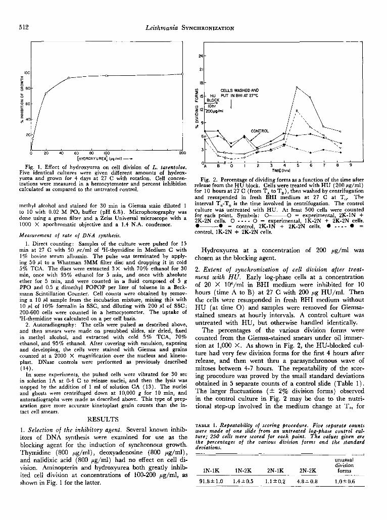
Fig. 1. Effect of hydroxyurea on cell division of L. tarentolae. Five identical cultures were given different amounts of hydrox- yurea and grown for 4 days at 27 C with rotation. Cell concen- trations were measured in a hemocytometer and percent inhibition calculated as compared to the untreated control.
methyl alcohol and stained for 30 min in Giemsa stain diluted 1 to 10 with 0.02 M PO, buffer (pH 6.8). Microphotography was done using a green filter and a Zeiss Universal microscope with a 1000 X apochromatic objective and a 1.4 N.A. condensor.
Measurement of rate of DIVA synthesis. 1. Direct counting: Samples of the culture were pulsed for 15
min at 27 C with 50 pclml of 'H-thymidine in Medium C with 1% bovine serum albumin. The pulse was terminated by apply- ing 50 pl to a Whatman 3MM filter disc and dropping it in cold 5% TCA. The discs were extracted 3X with 70% ethanol for 30 min, once with 95% ethanol for 5 min, and once with absolute ether for 5 min, and were counted in a fluid composed of 5 g PPO and 0.5 g dimethyl POPOP per liter of toluene in a Beck- mann Scintillation Counter. Cell counts were obtained by remov- ing a 10 pl sample from the incubation mixture, mixing this with 10 p1 of 10% formalin in SSC, and diluting with 200 cl of SSC; 200-600 cells were counted in a hemocytometer. The uptake of 3H-thymidine was calculated on a per cell basis.
2. Autoradiography: The cells were pulsed as described above, and then smears were made on presubbed slides, air dried, fixed in methyl alcohol, and extracted with cold 5% TCA, 70% ethanol, and 95% ethanol. After covering with emulsion, exposing and developing, the cells were stained with Giemsa and grains counted at a 2000 X magnification over the nucleus and kineto- plast. DNase controls were performed as previously described (14).
In some experiments, the pulsed cells were vibrated for 30 sec in solution 1A at 0-4 C to release nuclei, and then the lysis was stopped by the addition of 1 ml of solution CA (13) . The nuclei and ghosts were centrifuged down at 10,000 g for 10 min, and autoradiographs were made as described above. This type of prep- aration gave more accurate kinetoplast grain counts than the in- tact cell smears.
RESULTS 1. Selection of the inhibitory agent. Several known inhib- itors of DNA synthesis were examined for use as the blocking agent for the induction of synchronous growth. Thymidine (800 pg/ml) , deoxyadenosine (800 pg/ml) , and nalidixic acid (800 pg/ml) had no effect on cell di- vision. Aminopterin and hydroxyurea both greatly inhib- ited cell division at concentrations of 100-200 pg/ml, as shown in Fig. 1 for the latter.
t CELLS WASHED AND
TIME (hrsl k 6 4
CELLS WASHED AND
TIME (hrsl
Fig. 2. Percentage of dividing forms as a function of the time after release from the H U block. Cells were treated with H U (200 pg/ml) for 10 hours at 27 C (from T, to T,), then washed by centrifugation and resuspended in fresh BHI medium a t 27 C at T,,. The interval T,-To is the time involved in centrifugation. The control culture was untreated with HU. At least 500 cells were counted for each point. Symbols: 0--0 = experimental, 2K-1N + 2K-2N cells. 0 - - - - 0 = experimental, 1K-2N + 2K-2N cells. 0-0 = control, 2K-1N + 2K-2N cells. - - - - = control, 1K-2N + 2K-2N cells.
Hydroxyurea at a concentration of 200 pg/ml was chosen as the blocking agent.
2. Extent of synchronization of cell division after treat- ment with HU. Early log-phase cells at a concentration of 20 X 1O0/ml in BHI medium were inhibited for 10 hours (time A to B) at 27 C with 200 pg HU/ml. Then the cells were resuspended in fresh BHI medium without H U (at time 0) and samples were removed for Giemsa- stained smears at hourly intervals. A control culture was untreated with HU, but otherwise handled identically.
The percentages of the various division forms were counted from the Giemsa-stained smears under oil immer- sion at 1,000 X. As shown in Fig. 2, the HU-blocked cul- ture had very few division forms for the first 4 hours after release, and then went thru a parasynchronous wave of mitoses between 4-7 hours. The repeatability of the scor- ing procedure was proved by the small standard deviations obtained in 5 separate counts of a control slide (Table 1 ) . The larger fluctuations (* 2% division forms) observed in the control culture in Fig. 2 may be due to the nutri- tional step-up involved in the medium change at To, for
TABLE 1. Repeatability of scoring procedure. Five separate counts were made of one slide from an untreated log-phase control cul- ture; 250 cells were scored for each point. The values given are the percentages of the various division forms and the standard deviations. ~ ~ ~ _ _ __
unusual division
IN-1K 1N-2K 2N-1K 2N-2K forms
1.0k0.6 91.8k1.0 1.4k0.5 l . l f0 .2 4.8k0.8
Leishmania SYNCHRONIZATION 513
24
21
18
TIMEfhrs)
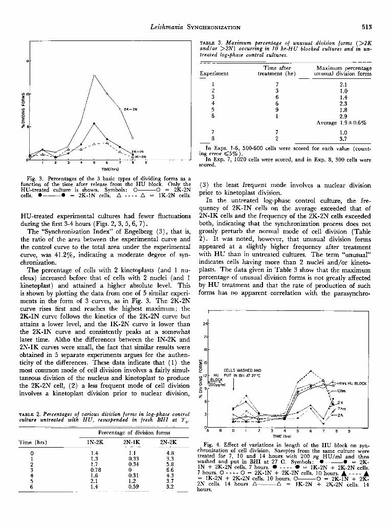
Fig. 3. Percentages of the 3 basic types of dividing forms as a function of the time after release from the H U block. Only the
cells. 0- 0 = 2K-IN cells. A - - - - A = 1K-2N cells. HU-treated culture is shown. Symbols: 0- 0 = 2K-2N
-
-
-
HU-treated experimental cultures had fewer fluctuations during the first 3-4 hours (Figs. 2, 3, 5,6, 7) .
The “Synchronization Index” of Engelberg ( 3 ) , that is, the ratio of the area between the experimental curve and the control curve to the total area under the experimental curve, was 41.2%, indicating a moderate degree of syn- chronization.
The percentage of cells with 2 kinetoplasts (and 1 nu- cleus) increased before that of cells with 2 nuclei (and 1 kinetoplast) and attained a higher absolute level. This is shown by plotting the data from one of 5 similar experi- ments in the form of 3 curves, as in Fig. 3. The 2K-2N curve rises first and reaches the highest maximum; the 2K-IN curve follows the kinetics of the 2K-2N curve but attains a lower level, and the IK-2N curve is lower than the 2K-IN curve and consistently peaks at a somewhat later time. Altho the differences between the IN-2K and 2N-IK curves were small, the fact that similar results were obtained in 5 separate experiments argues for the authen- ticity of the differences. These data indicate that (1) the most common mode of cell division involves a fairly simul- taneous division of the nucleus and kinetoplast to produce the 2K-2N cell, (2) a less frequent mode of cell division involves a kinetoplast division prior to nuclear division,
TABLE 2. Percentages of various division forms in log-phase control culture untreated with H U , resuspended in fresh BHI at To.
Percentage of division forms
Time (hrs) 1N-2K 2N-1K 2N-2K
0 1.4 1 . 1 4.8 1 2 3 4 5 6
1.3 0.33 3.3 1.7 0.34 5.8 0.78 0 6.6 1.6 2.1 1.4
0.31 4.3 1.2 3.7 0.59 3.2
TABLE 3. Maximum percentage of unusual division forms (>2K andlor > 2 N ) occurring in 10 hr-HU blocked cultures and in un- treated log-phase control cultures.
Time after Maximum percentage Experiment treatment (hr) unusual division forms
1 7 2.1 2 3 1 .o 3 6 1.4 4 6 2.3 5 9 1.8 6 1 2.9
Average 1.9 * 0.6%
7 8
7 2
1.0 3.7
In Exps. 1-6, 500-600 cells were scored for each value (count- ing error C5% ).
-In Exp. 3, .lo20 cells were scored, and in Exp. 8, 300 cells were scored.
( 3 ) the least frequent mode involves a nuclear division prior to kinetoplast division.
In the untreated log-phase control culture, the fre- quency of 2K-IN cells on the average exceeded that of 2N-IK cells and the frequency of the 2K-2N cells exceeded both, indicating that the synchronization process does not grossly perturb the normal mode of cell division (Table 2) . It was noted, however, that unusual division forms appeared at a slightly higher frequency after treatment with H U than in untreated cultures. The term “unusual” indicates cells having more than 2 nuclei and/or kineto- plasts. The data given in Table 3 show that the maximum percentage of unusual division forms is not greatly affected by H U treatment and that the rate of production of such forms has no apparent correlation with the parasynchro-
15- m
CELLS WASHED AND e 14hrs HU BLOCK
‘A 8 0 2 3 4 5 6 7 b TlME (hrs)
Fig. 4. Effect of variations in length of the H U block on syn- chronization of cell division. Samples from the same culture were treated for 7 , 10 and 14 hours with 200 pg HU/ml and then
1N + 2K-2N cells. 7 hours. - - - - = 1K-2N + 2K-2N cells. 7 hours. 0 - - - - 0 = 2K-1N + 2K-2N cells. 10 hours. A - - - - A 2N cells. 14 hours A- A = 1K-2N + 2K-2N cells. 14 hours.
washed and put in BHI at 27 C. Symbols: 0- 0 = 2K-
= 1K-2N + 2K-2N cells. 10 hours. 0- 0 = 2K-1N + 2K-
514 Leishmania SYNCHRONIZATION
-
-
-
-
-
- -
-
CELL (XlO')
1000
900
-800
700
600
500
400
300
200
'A "loo 0 0 I 2 3 4 5 6 7 8 9
TIME(hrr)--c
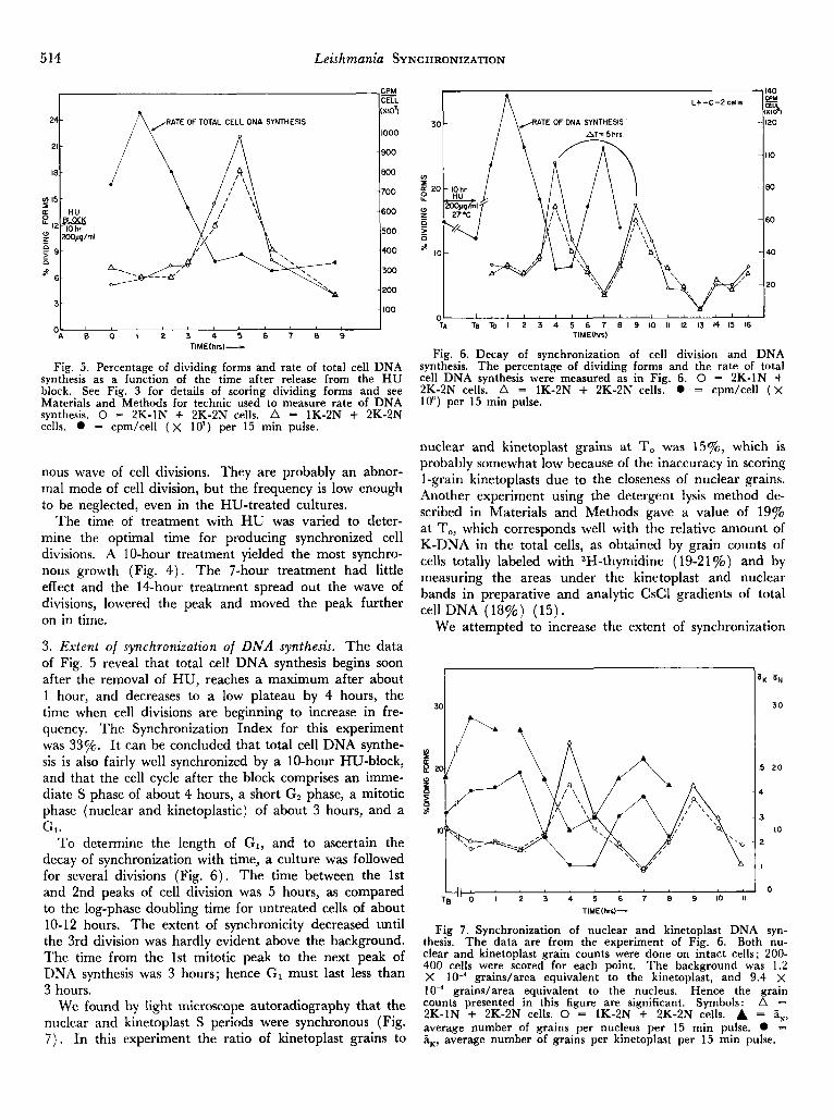
Fig. 5. Percentage of dividing forms and rate of total cell DNA synthesis as a function of the time after release from the H U block. See Fig. 3 for details of scoring dividing forms and see Materials and Methods for technic used to measure rate of DNA synthesis. 0 = 2K-1N + 2K-2N cells. A = 1K-2N + 2K-2N cells. = cpm/cell ( x lo') per 15 min pulse.
nous wave of cell divisions. They are probably an abnor- mal mode of cell division, but the frequency is low enough to be neglected, even in the HU-treated cultures.
The time of treatment with HU was varied to deter- mine the optimal time for producing synchronized cell divisions. A 10-hour treatment yielded the most synchro- nous growth (Fig. 4 ) . The 7-hour treatment had little effect and the 14-hour treatment spread out the wave of divisions, lowered the peak and moved the peak further on in time.
3. Extent of synchronization of DNA synthesis. The data of Fig. 5 reveal that total cell DNA synthesis begins soon after the removal of HU, reaches a maximum after about 1 hour, and decreases to a low plateau by 4 hours, the time when cell divisions are beginning to increase in fre- quency. The Synchronization Index for this experiment was 33%. It can be concluded that total cell DNA synthe- sis is also fairly well synchronized by a 10-hour HU-block, and that the cell cycle after the block comprises an imme- diate S phase of about 4 hours, a short G, phase, a mitotic phase (nuclear and kinetoplastic) of about 3 hours, and a
To determine the length of GI, and to ascertain the decay of synchronization with time, a culture was followed for several divisions (Fig. 6 ) . The time between the 1st and 2nd peaks of cell division was 5 hours, as compared to the log-phase doubling time for untreated cells of about 10-12 hours. The extent of synchronicity decreased until the 3rd division was hardly evident above the background. The time from the 1st mitotic peak to the next peak of DNA synthesis was 3 hours; hence G, must last less than 3 hours.
We found by light microscope autoradiography that the nuclear and kinetoplast S periods were synchronous (Fig. 7 ) . In this experiment the ratio of kinetoplast grains to
30 RATE OF DNA SYNTHESIS
u)
5 20 e w t 0 ij
$ 10
0 TA TB To I 2 3 4 5 6 7 8 9 10 II 12 13 14 15 16
TIME(hS)
Fig. 6. Decay of synchronization of cell division and DNA synthesis. The percentage of dividing forms and the rate of total cell DNA synthesis were measured as in Fig. 6. 0 = 2K-1N + 2K-2N cells. A = 1K-2N + 2K-2N cells. 0 = cpm/cell ( X 1 W ) per 15 min pulse.
nuclear and kinetoplast grains at To was 1570, which is probably somewhat low because of the inaccuracy in scoring 1-grain kinetoplasts due to the closeness of nuclear grains. Another experiment using the detergent lysis method de- scribed in Materials and Methods gave a value of 19% at To, which corresponds well with the relative amount of K-DNA in the total cells, as obtained by grain counts of cells totally labeled with 3H-thymidine (19-21%) and by measuring the areas under the kinetoplast and nuclear bands in preparative and analytic CsCl gradients of total cell DNA (18%) (15).
We attempted to increase the extent of synchronization
1 aK aN i
TB 4'6 ; 2 3 4 5 6 7 Q 9 ;o i l l 0 TlME(hr6)-
Fig 7. Synchronization of nuclear and kinetoplast DNA syn- thesis. The data are from the experiment of Fig. 6. Both nu- clear and kinetoplast grain counts were done on intact cells; 200- 400 cells were scored for each point. The background was 1.2 X lo-' graindarea equivalent to the kinetoplast, and 9.4 x
graindarea equivalent to the nucleus. Hence the grain counts presented in this figure are significant. Symbols: A = 2K-1N + 2K-2N cells. 0 = 1K-2N + 2K-2N cells. A = H,, average number of grains per nucleus per 15 min pulse. 0 = P,, average number of grains per kinetoplast per 15 min pulse.
Leishmania SYNCHRONIZATION 515
25
>
4 2 0 - g 0 15- z 5 a - 10- - 8
5-
20; i 2 i h A 6 ; 8 TIME Ihrsl-
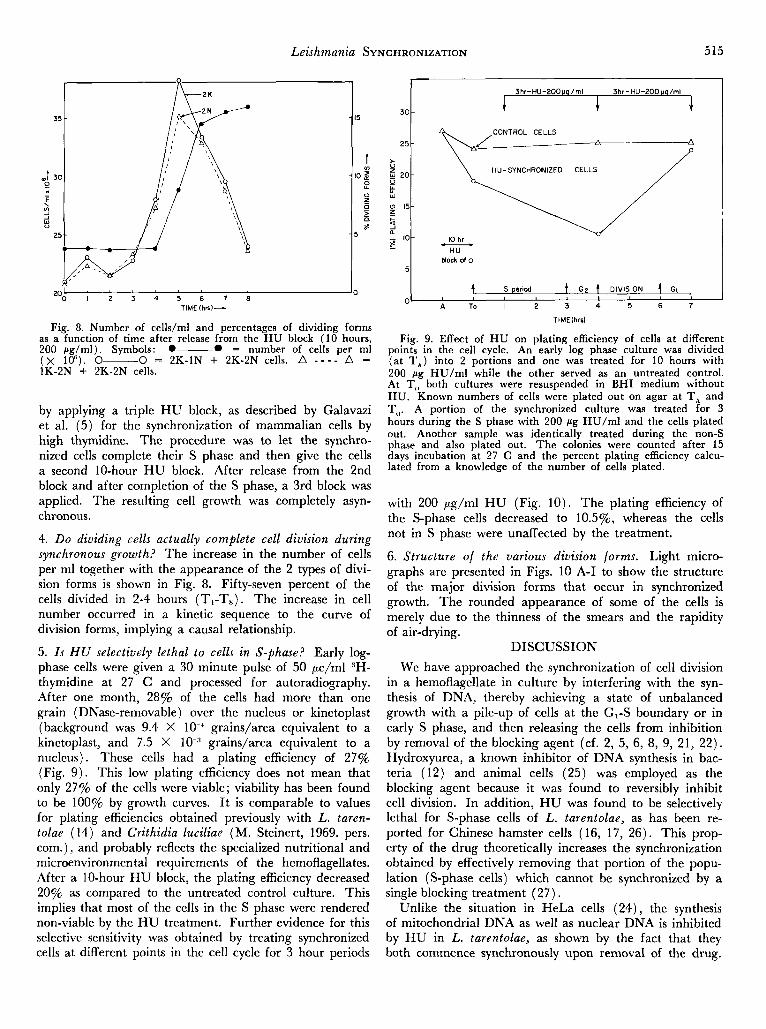
Fig. 8. Number of cells/ml and percentages of dividing forms as a function of time after release from the HU block (10 hours, 200 se/ml) . Svmbols: 0-0 = number of cells Der ml
-
HU-SYNCHRONIZED CELLS
10 hr HU
block 01 o
- t S period t G p t DIVISION 4 GI
. - , , ( x 1P). 0- 0 = 2K-1N + 2K-2N cells. A - - - - A = 1K-2N + 2K-2N cells.
by applying a triple HU block, as described by Galavazi et al. (5) for the synchronization of mammalian cells by high thymidine. The procedure was to let the synchro- nized cells complete their S phase and then give the cells a second 10-hour HU block. After release from the 2nd block and after completion of the S phase, a 3rd block was applied. The resulting cell growth was completely asyn- chronous.
4. Do dividing cells actually complete cell division during synchronous growth? The increase in the number of cells per ml together with the appearance of the 2 types of divi- sion forms is shown in Fig. 8. Fifty-seven percent of the cells divided in 2-4 hours (T,-T,) . The increase in cell number occurred in a kinetic sequence to the curve of division forms, implying a causal relationship.
5. I s HU selectively lethal to cells in S-phase? Early log- phase cells were given a 30 minute pulse of 50 pc/ml 3H- thymidine at 27 C and processed for autoradiography. After one month, 28% of the cells had more than one grain (DNase-removable) over the nucleus or kinetoplast (background was 9.4 X 10.' grains/area equivalent to a kinetoplast, and 7.5 X grains/area equivalent to a nucleus). These cells had a plating efficiency of 27% (Fig. 9 ) . This low plating efficiency does not mean that only 27% of the cells were viable; viability has been found to be 100% by growth curves. It is comparable to values for plating efficiencies obtained previously with L. taren- tolae (14) and Crithidia luciliae (M. Steinert, 1969. pers. corn.), and probably reflects the specialized nutritional and microenvironmental requirements of the hemoflagellates. .4fter a 10-hour H U block, the plating efficiency decreased 20% as compared to the untreated control culture. This implies that most of the cells in the S phase were rendered non-viable by the HU treatment. Further evidence for this selective sensitivity was obtained by treating synchronized cells at different points in the cell cycle for 3 hour periods
i 3 0
3hr-HU-200~!g/ml 3hr-HU-200 ua/ml +
with 200 pg/ml H U (Fig. 10). The plating efficiency of the S-phase cells decreased to 10.5%, whereas the cells not in S phase were unaffected by the treatment.
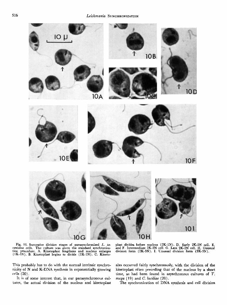
6. Structure of the various division forms. Light micro- graphs are presented in Figs. 10 A-I to show the structure of the major division forms that occur in synchronized growth. The rounded appearance of some of the cells is merely due to the thinness of the smears and the rapidity of air-drying.
DISCUSSION We have approached the synchronization of cell division
in a hemoflagellate in culture by interfering with the syn- thesis of DNA, thereby achieving a state of unbalanced growth with a pile-up of cells at the GI-S boundary or in early S phase, and then releasing the cells from inhibition by removal of the blocking agent (cf. 2, 5, 6, 8, 9, 21, 22) . Hydroxyurea, a known inhibitor of DNA synthesis in bac- teria (12) and animal cells (25) was employed as the blocking agent because it was found to reversibly inhibit cell division. In addition, H U was found to be selectively lethal for S-phase cells of L. tarentolae, as has been re- ported for Chinese hamster cells (16, 17, 26). This prop- erty of the drug theoretically increases the synchronization obtained by effectively removing that portion of the popu- lation (S-phase cells) which cannot be synchronized by a single blocking treatment (27) .
Unlike the situation in HeLa cells (24) , the synthesis of mitochondria1 DNA as well as nuclear DNA is inhibited by H U in L. tarentolae, as shown by the fact that they both commence synchronously upon removal of the drug.
516 Leishmania SYNCHRONIZATION
Fig. 10. Successive division stages of parasynchronized L. ta- rentolae cells. The culture was given the standard synchroniza- tion procedure. A. Kinetoplast lengthens and nucleus enlarges (1K-1N). B. Kinetoplast begins to divide (1K-1N). C. Kineto-
This probably has to do with the normal intrinsic synchro- nicity of N and K-DNA synthesis in exponentially growing cells (20).
It is of some interest that, in our parasynchronous cul- tures, the actual division of the nucleus and kinetopIast
plast divides before nucleus (2K-1N). D. Early 2K-2N cell. E. and F. Intermediate 2K-2N cell. G. Late 2K-2N cell. H. Unusual division form (2K-3N). I. Unusual division form (3K-3N).
also occurred fairly synchronously, with the division of the kinetoplast often preceding that of the nucleus by a short time, as had been found in asynchronous cultures of T . mega (19) and C . Zuciliae (20).
The synchronization of DNA synthesis and cell division
Leishmania SYNCHRONIZATION 517
lasted for 2 cell cycles only (Fig. 6 ) . This could be due to: (1) An innate variability in the length of S periods among individual cells (1) ; ( 2 ) the low Index of Syn- chronicity obtained by this procedure.
The decrease in the interval between successive cell divisions in the parasynchronized culture as compared to asynchronous cells (Fig. 6) is similar to the situation found in human tissue culture cells synchronized by ex- cess thymidine, in which GI and G, phases were shortened after the treatment (6) . This shortening of certain phases of the cell cycle has no ready explanation.
Even tho the extent of synchronization of cell division in L. tarentolae by hydroxyurea is not as high as would be desired, the coordinated synchronization of nuclear and kinetoplastic DNA synthesis is sufficient for a study of the regulation of the synthesis of K-DNA.
The senior author (LS) is indebted to Dr. M. Steinert for kindly communicating his preliminary results on the synchroniza- tion of Crithidia luciliae by hydroxyurea, and thereby stimulating this project. This project was supported by grant AI-09102 from the U. S. Public Health Service, and in part by Research Grant 2456-Zoology, Univ. of California.
REFERENCES 1. Anderson. E. C. & Peterson. D. F. 1964. Svnchronized mam-
malian cells: an experimental test of a model for synchrony de- cay. Exp. Cell Res. 36, 423-7.
2. Bootsma. D.. Budke. L. & Vos. 0. 1964. Studies on svnchro- nous division ‘of tissue culture cells initiated bv excess thvmidine. Exp. Cell Res. 33, 301-9.
3. Engelberg, J. 1961. A method of measuring the degree of synchronization of cell populations. E x p . Cell Res. 23, 218-27.
4. Evans, T. E. 1966. Synthesis of a cytoplasmic DNA during the Gz interphase of Physariim polycephalum. Biochem. Biophys. . . . Res. Corn. &, 678-83. 5. Galavazi, G., Schenk, H. & Bootsma, D. 1966. Synchroniza-
tjon of mammalian cells in vitro by inhibition of the DNA synthe- sis. I. Optimal conditions. E x p . Cell Res. 41, 428-37.
6. Galavazi, G . & Bootsma, D. 1966. Synchronization of mam- malian cells in vitro by inhibition of the DNA synthesis. 11. Popu- lation dynamics. Exp. Cell Res. 41, 438-51.
7. Koch, J. & Stokstad, E. L. R. 1967. Incorporation of (3H) thymidine into nuclear and mitochondria1 DNA in synchronized mammalian cells. Europ. j . Biochem. 3, 1-6.
8. Lambert, W. C. & Studzinski, G. P. 1969. Thymidine as a synchronizing agent. I. Partial recovery of HeLa cells from unbal- anced growth. j . Cell Physiol. 73, 261-6.
9. Littlefield, D. W. 1962. DNA synthesis in partially synchro- nized L cells. Exp. Cell Res. 26, 318-26.
10. Parsons, J. 1965. Mitochondria1 incorporation of tritiated thymidine in Tetrahymena pyriformis. j . Cell Biol. 25, 641-6.
11. Robbins, E. & Morrill, G. A. 1969. Oxygen uptake during the HeLa cell life cvcle and its correlation with macromolecular synthesis. J . Cell Biol. 43, 629-33.
12. Rosenkranz, H . S., Garro, A. J., Levy, J. and Carr, H. 1966. Studies with hydroxyurea. I. The reversible inhibition of bacterial DNA synthesis and the effect of hydroxyurea on the bactericidal action of streptomycin. Biochem. Biophys. Acta 114,
13. Simpson, L. 1968. Behavior of the kinetoplast of Leish- mania tarentolae upon cell rupture. J . Protozool. 15, 132-6.
14. ~ . 1968. Effect of acriflavin on the kinetoplast of Leishmania tarentolae. 1. Cell Biol. 37, 660-82.
15. - . 1970. In preparation. 16. Sinclair, 1965. Hydroxyurea: differential lethal effects on
cultured mammalian cells during the cell cycle. Science 150,
17. Sinclair, 1967. Hydroxyurea: effects on Chinese hamster cells grown in culture. Cancer Res. 27, 297-308.
18. Steinert, M. 1960. Mitochondria associated with the kineto- nucleus (kinetoplast) of Trypanosoma mega. J . Biophys. Biochem.
19. Steinert, M. & Steinert, G. 1962. La synthese de l’acide desoxyribonucleique au cours du cycle de division de Trypano- soma mega. J . Protozool. 9, 203-11.
20. Steinert, M. & Van Assel, S . 1967. Replications coordon- nees des acides desoxyribonucleique nucleaire et mitochondrial chez Crithidia luciliae. Arch. Intern. Physiol. Biochim. 75, 370-1.
21. Studzinski, G. P. & Lambert, W. C. 1969. Thymidine as a synchronizing agent. 11. Nucleic acid and protein formation in synchronous HeLa cultures treated with excess thvmidine. I. Cell
501-15.
1729-31.
Cytol . 8, 542-6.
Physiol. 73, 109-18. 22. Till, J. E., Whitmore, G. R. & Gulyas, S. 1963. DNA syn-
thesis in individual L-strain mouse cells. 11. Effects of thymidine starvation. Biochem. Biophys. Acta 72, 277-89.
23. Trager, W. 1957. Nutrition of a hemoflagellate (Leish- mania tarentolae) having an interchangeable requirement for choline or pyridoxal. j . Protozool. 4, 269.
24. Vesco, C. & Penman, S. 1969. Purified cytoplasmic DNA from HeLa cells: Resistanre to inhibition by hydroxyurea. Bio- chem. Biophys. Res. Commun. a, 249-57.
25. Young, C. W. & Hadas, S. 1964. Hydroxyurea: Inhibitory effect on DNA metabolism. Science 1-16, 1172-4.
26. Yu, C. K. & Sinclair, W. K. 1968. Cytological effects on Chinese hamster cells of synchronizing concentrations of hy- droxyurea. j . Cell Physiol. 72, 39-42.
Related Documents