Swollenin, a Trichoderma reesei protein with sequence similarity to the plant expansins, exhibits disruption activity on cellulosic materials Markku Saloheimo 1 , Marja Paloheimo 1 , Satu Hakola 1 , Jaakko Pere 1 , Barbara Swanson 2 , Eini Nyysso ¨ nen 2 , Amit Bhatia 2, *, Michael Ward 2 and Merja Penttila ¨ 1 1 VTT Biotechnology, Finland; 2 Genencor International, Inc., Palo Alto, CA, USA Plant cell wall proteins called expansins are thought to disrupt hydrogen bonding between cell wall polysaccha- rides without hydrolyzing them. We describe here a novel gene with sequence similarity to plant expansins, isolated from the cellulolytic fungus Trichoderma reesei. The pro- tein named swollenin has an N-terminal fungal type cel- lulose binding domain connected by a linker region to the expansin-like domain. The protein also contains regions similar to mammalian fibronectin type III repeats, found for the first time in a fungal protein. The swollenin gene is regulated in a largely similar manner as the T. reesei cel- lulase genes. The biological role of SWOI was studied by disrupting the swo1 gene from T. reesei. The disruption had no apparent effect on the growth rate on glucose or on different cellulosic carbon sources. Non-stringent Southern hybridization of Trichoderma genomic DNA with swo1 showed the presence of other swollenin-like genes, which could substitute for the loss of SWOI in the disruptant. The swollenin gene was expressed in yeast and Aspergillus niger var. awamori. Activity assays on cotton fibers and filter paper were performed with concentrated SWOI-containing yeast supernatant that disrupted the structure of the cotton fibers without detectable formation of reducing sugars. It also weakened filter paper as assayed by an extensometer. The SWOI protein was purified from A. niger var. awamori culture supernatant and used in an activity assay with Valonia cell walls. It disrupted the structure of the cell walls without producing detectable amounts of reducing sugars. Keywords: cellulase; expansin; cellulose binding domain; Trichoderma; regulation. In the last few years, a new class of proteins called expansins has been discovered in plants (reviewed in [1–3]). A number of expansin genes have been identified from a wide variety of plant species, including cucumber, Arabidopsis, rice [4] and tomato [5]. The expansins were first implicated in loosening the cell wall structure during plant cell growth (the acid- growth response), and the proteins forming a distinct family with high sequence identity and having this type of activity are now classified as a-expansins [6]. The group 1 pollen allergens have approximately 25% amino-acid identity with a-expansins and have been shown to be active in an acid- growth assay on grass cell walls. Along with their vegetative homologues they are designated as b-expansins [6]. Expansins have been proposed to disrupt hydrogen bonding between cellulose microfibrils or between cellulose and other cell wall polysaccharides without having hydro- lytic activity [7,8]. In this way they are thought to allow the sliding of cellulose fibers and enlargement of the cell wall. Purified cucumber expansins have been shown to catalyze extension of isolated plant cell walls such as cucumber hypocotyl walls when assayed using a constant load extensometer [9]. These cucumber expansins have also been shown to weaken filter paper without producing reducing sugar [7]. Some of the a-expansins are functional during fruit ripening, possibly aiding the action of hydrolytic enzymes that degrade the cell wall polymers [5]. Experiments have been made where expansin activity has been over expressed, or reduced by antisense strategy in Arabidopsis thaliana. The results suggest a role for these proteins in the control of plant growth and morphogenesis [10]. A number of saprophytic and pathogenic fungi and bacteria produce a wide range of enzymes designed to break down plant biomass. These enzymes include cellulases that break down cellulose to glucose, and hemicellulases that degrade the different hemicelluloses to monomeric sugars. For the degradation of the insoluble and complex plant cell wall the microbes produce multiple enzyme forms belonging to various enzyme categories. For example, from the fungus Trichoderma reesei, one of the best known saprophytic microbes, genes have been cloned that encode two exo- acting cellulases liberating mainly cellobiose from cellulose chain ends, five endo-acting cellulases hydrolyzing internal linkages of cellulose chains and 10 hemicellulases represent- ing different enzyme activities [11,12]. Most of the cellulases and some of the hemicellulases of this fungus have a Correspondence to M. Saloheimo, VTT Biotechnology, PO Box 1500, 02044 VTT, Finland. Fax: + 358 9 455 2103, Tel.: + 358 9 456 5820, E-mail: Markku.Saloheimo@vtt.fi Abbreviations: CBD, cellulose binding domain; CBHI, CBHII, T. reesei cellobiohydrolases; CREI, regulatory protein involved in catabolite repression in T. reesei; EGI, EGII, EGIV, EGV, T. reesei endoglucanases; FnIII repeats, fibronectin III type repeats; SWOI, T. reesei swollenin I; swo1, gene encoding SWOI; HEC, hydroxyethylcellulose. Enzymes: endo-1,4-glucanase (EC 3.2.1.4), cellobiohydrolase (EC 3.2.1.91). *Present address: EPIcyte Pharmaceutical, Inc., 5810 Nancy Ridge Drive, Suite 150, San, Diego, CA 92121, USA. Note: the complete swo1 sequence data has been submitted to the EMBL database under the accession no. AJ245918. (Received 17 May 2002, revised 3 July 2002, accepted 5 July 2002) Eur. J. Biochem. 269, 4202–4211 (2002) ȑ FEBS 2002 doi:10.1046/j.1432-1033.2002.03095.x

Welcome message from author
This document is posted to help you gain knowledge. Please leave a comment to let me know what you think about it! Share it to your friends and learn new things together.
Transcript

Swollenin, a Trichoderma reesei protein with sequence similarity tothe plant expansins, exhibits disruption activity on cellulosic materials
Markku Saloheimo1, Marja Paloheimo1, Satu Hakola1, Jaakko Pere1, Barbara Swanson2, Eini Nyyssonen2,Amit Bhatia2,*, Michael Ward2 and Merja Penttila1
1VTT Biotechnology, Finland; 2Genencor International, Inc., Palo Alto, CA, USA
Plant cell wall proteins called expansins are thought todisrupt hydrogen bonding between cell wall polysaccha-rides without hydrolyzing them. We describe here a novelgene with sequence similarity to plant expansins, isolatedfrom the cellulolytic fungus Trichoderma reesei. The pro-tein named swollenin has an N-terminal fungal type cel-lulose binding domain connected by a linker region to theexpansin-like domain. The protein also contains regionssimilar to mammalian fibronectin type III repeats, foundfor the first time in a fungal protein. The swollenin gene isregulated in a largely similar manner as the T. reesei cel-lulase genes. The biological role of SWOI was studied bydisrupting the swo1 gene from T. reesei. The disruptionhad no apparent effect on the growth rate on glucose oron different cellulosic carbon sources. Non-stringentSouthern hybridization of Trichoderma genomic DNA
with swo1 showed the presence of other swollenin-likegenes, which could substitute for the loss of SWOI in thedisruptant. The swollenin gene was expressed in yeast andAspergillus niger var. awamori. Activity assays on cottonfibers and filter paper were performed with concentratedSWOI-containing yeast supernatant that disrupted thestructure of the cotton fibers without detectable formationof reducing sugars. It also weakened filter paper asassayed by an extensometer. The SWOI protein waspurified from A. niger var. awamori culture supernatantand used in an activity assay with Valonia cell walls. Itdisrupted the structure of the cell walls without producingdetectable amounts of reducing sugars.
Keywords: cellulase; expansin; cellulose binding domain;Trichoderma; regulation.
In the last few years, a new class of proteins called expansinshas been discovered in plants (reviewed in [1–3]). A numberof expansin genes have been identified from awide variety ofplant species, including cucumber, Arabidopsis, rice [4] andtomato [5]. The expansins were first implicated in looseningthe cell wall structure during plant cell growth (the acid-growth response), and the proteins forming a distinct familywith high sequence identity and having this type of activityare now classified as a-expansins [6]. The group 1 pollenallergens have approximately 25% amino-acid identity witha-expansins and have been shown to be active in an acid-growth assay on grass cell walls. Along with their vegetativehomologues they are designated as b-expansins [6].
Expansins have been proposed to disrupt hydrogenbonding between cellulose microfibrils or between celluloseand other cell wall polysaccharides without having hydro-lytic activity [7,8]. In this way they are thought to allow thesliding of cellulose fibers and enlargement of the cell wall.Purified cucumber expansins have been shown to catalyzeextension of isolated plant cell walls such as cucumberhypocotyl walls when assayed using a constant loadextensometer [9]. These cucumber expansins have also beenshown to weaken filter paper without producing reducingsugar [7]. Some of thea-expansins are functional during fruitripening, possibly aiding the action of hydrolytic enzymesthat degrade the cell wall polymers [5]. Experiments havebeen made where expansin activity has been over expressed,or reduced by antisense strategy inArabidopsis thaliana. Theresults suggest a role for these proteins in the control ofplant growth and morphogenesis [10].A number of saprophytic and pathogenic fungi and
bacteria produce a wide range of enzymes designed to breakdown plant biomass. These enzymes include cellulases thatbreak down cellulose to glucose, and hemicellulases thatdegrade the different hemicelluloses to monomeric sugars.For the degradation of the insoluble and complex plant cellwall themicrobes producemultiple enzyme forms belongingto various enzyme categories. For example, from the fungusTrichoderma reesei, one of the best known saprophyticmicrobes, genes have been cloned that encode two exo-acting cellulases liberating mainly cellobiose from cellulosechain ends, five endo-acting cellulases hydrolyzing internallinkages of cellulose chains and 10 hemicellulases represent-ing different enzyme activities [11,12]. Most of the cellulasesand some of the hemicellulases of this fungus have a
Correspondence toM. Saloheimo, VTT Biotechnology,
PO Box 1500, 02044 VTT, Finland.
Fax: + 358 9 455 2103, Tel.: + 358 9 456 5820,
E-mail: [email protected]
Abbreviations: CBD, cellulose binding domain; CBHI, CBHII,
T. reesei cellobiohydrolases; CREI, regulatory protein involved
in catabolite repression in T. reesei; EGI, EGII, EGIV, EGV,
T. reesei endoglucanases; FnIII repeats, fibronectin III type repeats;
SWOI, T. reesei swollenin I; swo1, gene encoding SWOI; HEC,
hydroxyethylcellulose.
Enzymes: endo-1,4-glucanase (EC 3.2.1.4), cellobiohydrolase
(EC 3.2.1.91).
*Present address: EPIcyte Pharmaceutical, Inc., 5810 Nancy Ridge
Drive, Suite 150, San, Diego, CA 92121, USA.
Note: the complete swo1 sequence data has been submitted to the
EMBL database under the accession no. AJ245918.
(Received 17 May 2002, revised 3 July 2002, accepted 5 July 2002)
Eur. J. Biochem. 269, 4202–4211 (2002) � FEBS 2002 doi:10.1046/j.1432-1033.2002.03095.x
modular structure consisting of a cellulose binding domain(CBD) at either end of the polypeptide chain, connected tothe catalytic domain with a linker region. The role of theCBD is to mediate binding of the enzyme to the insolublesubstrate.In addition to plants, a protein with an endoglucanase
domain and a domain with sequence similarity to expansinshas been reported in the plant pathogen Clavibactermichiganensis ssp. sepedonicus [13]. In this paper, we reportthe discovery of a novel fungal protein having significantsequence identity to plant expansins. Unlike plant expan-sins, this protein has a modular structure with anN-terminal CBD. The protein was named swollenin dueto its ability to swell cotton fibers without producingdetectable amounts of reducing sugars.
E X P E R I M E N T A L P R O C E D U R E S
Strains, vectors and growth conditions
The T. reesei cDNA library in the vector pAJ401 [14] wasscreened in the yeast strain H1152 (a, sso2-1, leu2-3, trp1-1,ura3-1, sso1::HIS3, M. Aalto, unpublished results) on SC-Ura plates with 2% galactose as the carbon source [15] atthe restrictive temperature 31 �C. The yeast strain DBY746(a, his3D, leu2-3, ura3-52, trp1-289, Cyhr) was used forswollenin production. The T. reesei strain QM9414 [16] wasused in Northern studies and for detection of the swolleninprotein. For the Northern studies the strain was cultivatedin shake flasks (28 �C, 200 r.p.m.) in minimal media [17]containing 5% glucose, 2% sorbitol, 2% cellobiose, 2%lactose or 2% Solka floc cellulose for 3 days. Alternatively,the strain was grown on 2% glycerol for 72 h, followed byaddition of sophorose (1 mM final concentration). Afterfurther 15 h the culture was harvested. A culture grown in aminimal medium with 2% glycerol for 87 h was used as acontrol.
Nucleic acid methods
Yeast was transformed with the LiAc method [18] or byelectroporation (Bio-Rad). Plasmid constructs were madeusing standard methodology [19]. Total T. reesei RNA wasisolated as described [20]. The RNA samples (5 lg) weretreated with glyoxal and analyzed in a 1% agarose gel.Northern blotting and hybridization were performed on aHybond-N nylon membrane (Amersham). T. reesei DNAwas isolated as described [21]. Stringent Southern hybrid-ization was performed as described [19]. NonstringentSouthern hybridization was performed in a hybridizationmixture without formamide [19] at 48 �C and the filter waswashed in 2 · NaCl/Cit, 0.1% SDS for 10 min at roomtemperature and for 30 min at 48 �C.
Antibodies and Westerns
Swollenin antibodies were generated in rabbits by immu-nizing with the peptide CDPNYTSSRPQERYGS (aminoacids 422–437 in the swollenin sequence). SDS gel electro-phoresis was performed as described [22] and Westernblotting was performed on a nitrocellulose membrane(Schleicher & Schull) and detection of the swollenin witha secondary antibody-alkaline phosphatase conjugate
(Bio-Rad). Samples of yeast or T. reesei supernatants andpurified SWOI were denatured for Endoglycosidase Htreatment by heating 10 min at 100 �C in 0.5% SDS, 1%2-mercaptoethanol and subsequently treated with Endo Hf
(New England Biolabs) for 3 h at 37 �C in the G5 buffer(50 mMNa-citrate, pH 5.5). 1000 units of EndoHf was usedper 20 lg of purified SWOI and per 20 lL of the T. reeseior concentrated yeast supernatants.
Swo1 gene disruption
The swo1 gene was disrupted from the T. reesei strainQM9414 [16] by replacing it with a hygromycin resistancecassette. The genomic swo1 gene was first subcloned from acosmid library clone into pBluescript SK– as a 5.5 kbEcoRV fragment to obtain the plasmid pSH1. Most of theswollenin-coding region was replaced from pSH1 bydigesting it with NarI and BstEII and ligating with thehygromycin resistance cassette consisting of the Aspergillusnidulans gpdA promoter and trpC terminator and theE. colihygromycin resistance gene derived from the plasmidpBluekan (from P.J. Punt, TNO Nutrition and FoodResearch, Zeist, the Netherlands). The resulting plasmid,pSH9 was digested with EcoRV and transformed intoQM9414 as described [17], and transformants were selectedon 100 lgÆmL)1 hygromycin and purified to uninuclearclones by plating single spores on selective medium.Disruptants of the swo1 gene were screened among thetransformants by Southern hybridization performed asdescribed [19]. Two disruptants obtained were also exam-ined by growing them in shake flasks (28 �C, 200 r.p.m.,5 days) in a medium with 3% whey and 1.5% complexgrain-based nitrogen source [23] and performing Westernanalysis from their culture supernatants as described above.The phenotype of the disruption was studied by platingsingle spores on plates with minimal medium [17] supple-mented with 0, 0.1 or 0.2% proteose peptone and either 2%glucose, 2% Solka floc cellulose, 2%Avicel cellulose or 2%complex grain-based carbon/nitrogen source [23]. An addi-tional test was made on plates where the minimal mediumwith or without peptone andwithout any carbon source hadbeen overlaid by a Whatman 1 filter paper disc. Growth ofcolonies of the swo1 disruptants and the parental strain wasfollowed daily.
Production and characterization of the swolleninpreparations
The yeast strain DBY746 harbouring a plasmid with swo1in the vector pAJ401 [14] or the vector alone were grown inChemap CMF mini 1 L or Biolafitte 14 L bioreactors. Thebioreactor medium was SC-Ura with 2% glucose as thecarbon source [15]. The yeast supernatants were concen-trated 20 times with a Centiprep concentrator (Amicon) forthe treatments of cotton fibers and filter paper. The amountof SWOI was estimated by comparing signals obtained inWesterns from Endo-H-treated yeast supernatants withsignals obtained with known amounts of purified Endo-Htreated SWOI from A. niger var. awamori.The 1.5 kb coding region of the swollenin cDNA clone
was amplified by PCR using the following primers whichwere designed to add BglII and XbaI restriction endonuc-lease sites to the 5¢ and 3¢ ends, respectively. Primer
� FEBS 2002 A fungal expansin homologue (Eur. J. Biochem. 269) 4203
ExAspBgl2: 5¢-CATTAGATCTCAGCAATGGCTGGTAAGCTTATCCTC-3¢. Primer ExAspXba1:5¢-CGACTCTAGAAGGATTAGTTCTGGCTAAAC
TGCACACC-3¢. The DNA sequence of the amplifiedproduct was verified and the swollenin coding region wasinserted into the BglI and XbaI sites between the glaApromoter and terminator of anAspergillus expression vector(pGAPT-PG) to produce pGAPT-exp. The pGAPT-PGvector consists of pUC18 containing the A. nidulans pyrGgene as selectable marker and a 1.1 kb fragment of theA. niger var. awamori glaA promoter and a 0.2 kb fragmentof an A. niger glaA terminator.The expression plasmid pGAPT-exp was transformed
into A. niger var. awamori strain dgr246 P2 as described[24]. Transformants were selected for their ability to growon minimal medium lacking uridine. For swollenin proteinproduction the transformants were grown in liquid mediumas described [25]. Cells were removed and the culturesupernatants were equilibrated with 1 M ammonium sul-phate, 100 mM Tris pH 7. The supernantant was thenapplied to a cellulose (Sigma, St. Louis, MO, USA) affinitycolumn and washed with 1 M ammonium sulphate, 100 mM
Tris pH 7 to remove unbound proteins. The purifiedswollenin was eluted as a single peak in water.The purified swollenin was tested for activity against
hydroxyethylcellulose (HEC), b-glucan, xylan and mannan.The enzymatic activities against HEC (Fluka) and barleyb-glucan (Biocon) were determined according to IUPAC[26] and against birch xylan (Roth) as presented [27].Mannanase activity was assayed according to the procedureof IUPAC (1987) but using 0.5% locust bean gum (Sigma)as a substrate.
Action on solid substrates
Yeast supernatants. Cotton fibers were mercerized bytreating them with 25% NaOH for 15 min at 5 �C andwashing several times with distilled water. The cotton fiberswere suspended in the concentration of 0.5 gÆmL)1 in50 mM sodium acetate, pH 5.0 containing 1/4 of theconcentrated yeast culture media from the swolleninproducing yeast and control strain. Additionally, thepurified T. reesei EG II, CBH I and cellulose bindingdomain (CBD) of CBH I at a concentration of 5 lgÆmL)1
were used as controls for the swollenin [28,29]. Afterincubation for 4 h at 25 �C, the suspended fibers werefiltered off and the amount of reducing sugars released intothe filtrates was determined as described in [30]. The fiberswere rinsed once with buffer and then suspended in distilledwater with glass beads prior to sonication for one minuteusing a probe tip sonicator (Vibra Cell Sonics andMaterialsInc.) The fibers were then stained and visualized by lightmicroscopy to determine gross effects on their structure.For the paper strength test, Whatman no. 3 filter paper
was cut into strips measuring 7 · 2 cm. Sodium acetatebuffer (50 mM, pH 5) was used for all of these experiments.The concentrated yeast samples were sometimes firstdesalted by passage through a Bio-Rad Econo-Pac10 DGcolumn with a molecular mass cut-off of 6000 Da. Afterdesalting, 5 mL of the yeast samples were added to 4 mL ofbuffer in 50 mL disposable conical tubes and the Whatmanstrips were added. At the same time, strips were added tobuffer alone and 8 M urea in buffer. After incubating at
room temperature for 15 min the strips were measured fortheir wet tensile strength. The assay was performed byplacing each wet strip of paper between the Thwing–Alberttensile tester (Model 5564 from Instron Corporation,Canton, MA, USA) clamps spaced 4.5 cm apart. A 250 lbload cell was used. Test speed was 0.1 cmÆmin)1, and thepeak load was measured before breaking; it typically onlytook a minute to reach the paper breaking load.
The purified swollenin. The action of the purified swolleninpreparation on plant cell wall material was followed usingValonia cell wall fragments as substrate. Vesicles of Valoniamacrophysa were purified as described previously [31] andcut to small pieces, 4–5 mg each (dry weight). These cell wallfragments were suspended in 50 mM acetate buffer (pH 5.0)and swollenin was added at the dosages of 10 lgÆmg)1 and100 lgÆmg)1. Treatments with the purified T. reesei CBH I(also known as Cel7A) and EG II (also known as Cel5A)were performed as comparison for swollenin. The sampleswere incubated at +45 �C under stirring for 48 h andthereafter examined under a stereo microscope (Leica, WildM10). The control sample was treated alike but omitting theenzymes and swollenin. In addition the filtrates wereanalysed for solubilized sugars by HPLC.
R E S U L T S
Swollenin has sequence similarity with plant expansins. Theswollenin cDNA was isolated in a screening where compo-nents of the Trichoderma secretory pathway were searchedfor by yeast complementation. The sso2 temperature-sensitiveS. cerevisiae strainwas transformedwith aT. reeseicDNA expression library, and cDNAs derived from clonesable to grow at the restrictive temperature were sequenced.One of them encoded a protein predicted to have anN-terminal signal sequence followed by a cellulose bindingdomain. A major part of the remaining sequence was foundto have sequence similarity with plant expansins in aBLAST database search (Fig. 1C). Based on its swellingactivity on cotton fibers (see below) the protein was namedswollenin (SWOI) and the gene swo1.The genomic copy of the swo1 cDNAwas isolated from a
cosmid library, subcloned and sequenced. The gene containsfive short introns (Fig. 1A). The promoter contains aputative TATA box 90 bp and a putative binding sequenceof the glucose repressor protein CREI [32] 117 bp upstreamfrom the translation start codon (data not shown).The putative swollenin protein starts by a typical signal
sequence. It is followed by two glutamic acids. In theT. reesei cellulases EGI (also known as Cel7B) [33] andEGII [34] there are two glutamic acids at the N-terminusand in CBHI there is one [35], and the N-termini of theseenzymes are blocked by a pyroglutamic acid residue. Byanalogy, it is suggested that the swollenin signal sequencewould be 18 amino acids in length and be cleaved before thetwo glutamines in the sequence (Fig. 1b). The SWOI hasthree potential N-glycosylation sites at positions 160, 336and 406.The swollenin cellulose binding domain (CBD) has the
typical sequence features of fungal CBDs. The amino acidsinvariant in the CBDs of the T. reesei cellulases areconserved in swollenin (Fig. 1B). In the NMR structuresolved from the CBHI CBD there are two disulphide
4204 M. Saloheimo et al. (Eur. J. Biochem. 269) � FEBS 2002

bridges [36]. Based on a close proximity of an additionalcysteine pair in the modelled structures of the CBHII andEGI CBDs it has been suggested that they would have athird disulphide bond [37]. The swollenin CBD has sixcysteines at positions conserved with those of the CBHII(also known as Cel6A) CBD and thus it probably has threedisulphide bridges as well. The residues forming the flatsurface binding to cellulose are conserved in the swolleninCBD with one exception (Fig. 1B). Position 8 in theswollenin sequence has a phenylalanine while the othercellulases have tyrosine or tryptophan. The linker region ofthe swollenin is rich in serines and threonines and isexpected to be heavily O-glycosylated [38]. Without proteinstructure data the length of the linker cannot be determinedunambiguously. However, the region rich in serines andthreonines in SWOI apears to be among the longest found
in T. reesei enzymes, approximately 50 amino acids. Theregion of SWOI between the putative linker and theexpansin-like area shown in Fig. 1c does not match withany sequences in databases.The C-terminal two-thirds of the swollenin show clear
amino-acid similarity with plant expansins (Fig. 1C). Theidentity between swollenin and individual a- or b-expansinsin pairwise comparisons is about 25%over an area of about200 amino acids. The alignment of the swollenin with twoexpansin sequences (Fig. 1C) suggests that two largeinsertions have occurred in the swollenin gene in theN-terminal half of its expansin-like domain. The identitybetween the a- and b-expansins is 20–25% and there are fivesequence elements that are well conserved between theexpansin categories [6]. Four of the elements form the bestconserved parts between swollenin and the expansins and
Fig. 1. The basic structure of the swollenin protein (A), alignment of the swollenin CBD with the CBD sequences of T. reesei cellulases (B), Alignment
of the swollenin with two a-expansins (C) and The alignment of swollenin (SWOI) with FnIII repeat sequences (D). In (A), vertical arrows indicate theintron positions. SS, signal sequence; CBD, cellulose binding domain, QQ, two glutamines suggested to form the N-terminus of the mature
swollenin. The numbering refers to amino-acid positions in themature protein. In (B), the invariant amino acids are boxed and amino acids forming
the flat surface interacting with cellulose are shaded. Lines below the sequences show the proposed disulphide bridges of the SWOI CBD. (C)
Alignment of the swollenin with two a-expansins, LeEx1 of tomato (5) and CuExS2 of cucumber (4). Invariant amino acids are shown by asterisksand conservative substitutions by dots. The regions best conserved between a- and b-expansins are underlined in CuExS2. The conserved cysteinesare shown by arrows and positions with conserved aromatic amino acids in all three sequences by + 0. The regions with homology to the FnIII
repeats in titin are in bold in SWOI. (D) The alignment of swollenin (SWOI) with FnIII repeat sequences of human titin and a consensus sequence
of the bacterial FnIII repeats (BACT). The amino acids strictly conserved between the bacterial and mammalian FnIII repeats are shown by
asterisks.
� FEBS 2002 A fungal expansin homologue (Eur. J. Biochem. 269) 4205
thus they are probably functionally important. TheN-terminal half of the expansins contains eight conservedcysteines with a spacing similar to that of cysteines in thechitin binding domain of wheat germ agglutinin [4]. Sevenof these cysteines are conserved in swollenin. Aromaticamino acids are often important in the interaction ofenzymes and their carbohydrate substrates. In the alignmentbetween swollenin and expansins there are eight positionswhere an aromatic amino acid is conserved (Fig. 1C).Alignments of swollenin with individual expansins sug-gest that it is better conserved with b-expansins thana-expansins.There are two short sequences in swollenin that show
relatively strong conservation with fibronectin type III(FnIII) repeats of mammalian titin proteins (Fig. 1D).Interestingly, such repeats have been found in prokaryotichydrolases such as cellulases, chitinases and amylases [39,40]but thus far not in fungal enzymes. The amino acidsinvariant between the bacterial hydrolases and mammalianFnIII repeats are conserved in swollenin. Unlike thecontinuous FnIII repeats in the bacterial enzymes, theregion with similarity to titin in swollenin is divided into twoparts about 170 amino acids apart.
Regulation of the swollenin gene
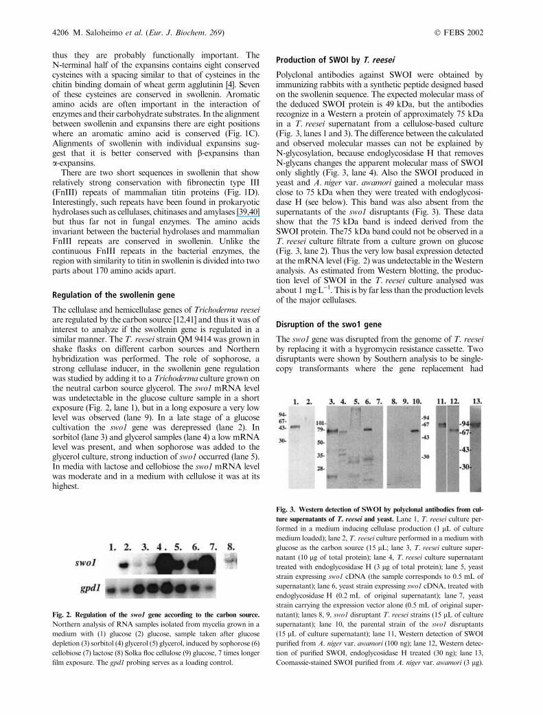
The cellulase and hemicellulase genes of Trichoderma reeseiare regulated by the carbon source [12,41] and thus it was ofinterest to analyze if the swollenin gene is regulated in asimilar manner. The T. reesei strain QM 9414 was grown inshake flasks on different carbon sources and Northernhybridization was performed. The role of sophorose, astrong cellulase inducer, in the swollenin gene regulationwas studied by adding it to a Trichoderma culture grown onthe neutral carbon source glycerol. The swo1 mRNA levelwas undetectable in the glucose culture sample in a shortexposure (Fig. 2, lane 1), but in a long exposure a very lowlevel was observed (lane 9). In a late stage of a glucosecultivation the swo1 gene was derepressed (lane 2). Insorbitol (lane 3) and glycerol samples (lane 4) a low mRNAlevel was present, and when sophorose was added to theglycerol culture, strong induction of swo1 occurred (lane 5).In media with lactose and cellobiose the swo1 mRNA levelwas moderate and in a medium with cellulose it was at itshighest.
Production of SWOI by T. reesei
Polyclonal antibodies against SWOI were obtained byimmunizing rabbits with a synthetic peptide designed basedon the swollenin sequence. The expected molecular mass ofthe deduced SWOI protein is 49 kDa, but the antibodiesrecognize in a Western a protein of approximately 75 kDain a T. reesei supernatant from a cellulose-based culture(Fig. 3, lanes 1 and 3). The difference between the calculatedand observed molecular masses can not be explained byN-glycosylation, because endoglycosidase H that removesN-glycans changes the apparent molecular mass of SWOIonly slightly (Fig. 3, lane 4). Also the SWOI produced inyeast and A. niger var. awamori gained a molecular massclose to 75 kDa when they were treated with endoglycosi-dase H (see below). This band was also absent from thesupernatants of the swo1 disruptants (Fig. 3). These datashow that the 75 kDa band is indeed derived from theSWOI protein. The75 kDa band could not be observed in aT. reesei culture filtrate from a culture grown on glucose(Fig. 3, lane 2). Thus the very low basal expression detectedat the mRNA level (Fig. 2) was undetectable in theWesternanalysis. As estimated from Western blotting, the produc-tion level of SWOI in the T. reesei culture analysed wasabout 1 mgÆL)1. This is by far less than the production levelsof the major cellulases.
Disruption of the swo1 gene
The swo1 gene was disrupted from the genome of T. reeseiby replacing it with a hygromycin resistance cassette. Twodisruptants were shown by Southern analysis to be single-copy transformants where the gene replacement had
Fig. 2. Regulation of the swo1 gene according to the carbon source.
Northern analysis of RNA samples isolated from mycelia grown in a
medium with (1) glucose (2) glucose, sample taken after glucose
depletion (3) sorbitol (4) glycerol (5) glycerol, induced by sophorose (6)
cellobiose (7) lactose (8) Solka floc cellulose (9) glucose, 7 times longer
film exposure. The gpd1 probing serves as a loading control.
Fig. 3. Western detection of SWOI by polyclonal antibodies from cul-
ture supernatants of T. reesei and yeast. Lane 1, T. reesei culture per-
formed in a medium inducing cellulase production (1 lL of culture
medium loaded); lane 2, T. reesei culture performed in a medium with
glucose as the carbon source (15 lL; lane 3, T. reesei culture super-natant (10 lg of total protein); lane 4, T. reesei culture supernatanttreated with endoglycosidase H (3 lg of total protein); lane 5, yeaststrain expressing swo1 cDNA (the sample corresponds to 0.5 mL of
supernatant); lane 6, yeast strain expressing swo1 cDNA, treated with
endoglycosidase H (0.2 mL of original supernatant); lane 7, yeast
strain carrying the expression vector alone (0.5 mL of original super-
natant); lanes 8, 9, swo1 disruptant T. reesei strains (15 lL of culturesupernatant); lane 10, the parental strain of the swo1 disruptants
(15 lL of culture supernatant); lane 11, Western detection of SWOI
purified from A. niger var. awamori (100 ng); lane 12, Western detec-
tion of purified SWOI, endoglycosidase H treated (30 ng); lane 13,
Coomassie-stained SWOI purified from A. niger var. awamori (3 lg).
4206 M. Saloheimo et al. (Eur. J. Biochem. 269) � FEBS 2002

occurred (data not shown). Western analysis of their culturesupernatants further confirmed that they do not produce theSWOI protein (Fig. 3, lanes 8 and 9).We attempted to demonstrate the phenotype of the swo1
disruption, its effect either on the formation of the T. reeseicell wall and growth of the fungal mycelium or on thedegradation of cellulosic carbon sources by the fungus. Thiswas performed by comparing the growth rates of thedisruptants and the parental strain on plates having glucoseor different cellulosic compounds as carbon sources. Thecompounds tested were two commercial celluloses, filterpaper and a complex grain-based carbon/nitrogen source[23]. No significant differences in the growth rates could beobserved between the strains on any of the carbon sourcesand thus swo1 disruption had no apparent phenotype in ourexperiments.Non-stringent hybridization of T. reesei genomic DNA
was performed with a swo1 gene fragment encoding theexpansin-like domain as a probe. Hybridization at 48 �Crevealed several other bands in addition to the onesoriginating from swo1, suggesting that there are other geneshaving expansin-like domains present in the T. reeseigenome in addition to swo1 (Fig. 4). The presence of thesegenes could compensate the lack of swo1 in the disruptantsand thus explain the result of the disruption experiment.
Characterization of the swollenin preparations
When the swollenin cDNA was expressed in S. cerevisiaeunder the PGK1 promoter in a multicopy plasmid, Westernanalysis of bioreactor culture supernatants showed that aheterogeneous high molecular mass protein reacting withthe swollenin antibodies was produced by the yeast (Fig. 3,lane 5). Inmany instances it has been shown that yeast tendsto overglycosylate heterologous proteins, e.g. the T. reeseicellulases CBHI and CBHII [42]. When the swollenin
produced in yeast was treated by endoglycosidase H toremove N-glycans, it gained an apparent molecular massclose to the swollenin produced byTrichoderma (Fig. 3, lane6). The production level of SWOI in yeast was approxi-mately 25 lgÆL)1 as estimated from Western blottingexperiments.Swollenin was also produced inA. niger var. awamori and
after a single step purification procedure the purifiedswollenin protein was obtained for biochemical character-ization. The SWOI expressed in this host migrated as tworelatively diffuse bands with apparent molecular massesbetween 80 and 95 kDa (Fig. 3, lanes 11 and 13), and Endo-H treatment reduced themolecularmass to the same level asSWOI produced byT. reesei (Fig. 3, lane 12). ThusA. nigervar. awamori slightly overglycosylated SWOI. Activities ofpurified swollenin against hydroxyethyl cellulose (HEC),b-glucan, xylan and mannan were measured and the resultsare shown in Table 1. Minor hydrolytic activity onb-glucan, xylan andmannan, but not onHEC,was observedfor the purified swollenin protein expressed in A. niger.
Demonstration of the swollenin activity on solidsubstrates
The swollenin expressed in yeast. The activity of theswollenin produced in yeast towards cellulosic materials wasshown by treatments of cotton fibers and filter paper.Cotton fibers were incubated with concentrated yeastsupernatants from bioreactor cultivations of the SWOI-producing yeast (approximately 0.125 lgÆmL)1 of SWOI)and the control strain with vector alone or, as controls, withthe T. reesei cellulases EGII, CBHI (5 lgÆml)1) and thecellulose binding domain of CBHI. After the treatment thefibers were removed from the reaction mixture by filtering,rinsed, sonicated with glass beads, stained and analyzed bylight microscopy. Soluble reducing sugars were measuredfrom the reaction mixture. Treatment with the control yeastsupernatant did not change the fiber structure (Fig. 5A).The supernatant of the yeast strain producing swollenincaused local disruption of the fiber structure that becamevisible only after sonication. This was seen as swollen areasoccurring along the fibers (Fig. 5B). CBH I caused somelight fibrillation of the fibers (Fig. 5C), whereas thetreatment with EG II resulted in damaged and ruggedoutlook of the fibers accompanied by fiber cutting (Fig. 5E).Din and coworkers [43] have reported on disruption ofcellulosic fibers by a bacterial cellulose binding domain. Nomodification of fiber surface could be detected in ourexperiment with the fungal (CBHI) CBD by the lightmicroscopical method used (Fig. 5D), suggesting that theeffect of SWOI on the fibers is not caused by its CBD. Thetreatment of the cotton fibers with the yeast supernatants or
Table 1. Characteristics of the swollenin preparation purified from
A. niger var. awamori.
Protein
(mgÆmL)1)a
Activity (nkatÆmL)1)
HEC b-glucanase Xylanase Mannanase
SWO1 0.7 0 79 7 5
a Lowry protein.
Fig. 4. Southern hybridization of T. reesei genomic DNA with the
region encoding the expansin-like domain from the swo1 gene, performed
at stringent (68 �C) and nonstringent (48 �C) conditions. The restrictionenzymes used are indicated.
� FEBS 2002 A fungal expansin homologue (Eur. J. Biochem. 269) 4207

with the CBD did not release detectable amounts ofreducing sugars. In contrast, the filtrates from the CBHI-and EGII-treated fibers contained 0.08% and 1.61%reducing sugars of the original dry mass, respectively.
To test the effect of swollenin on paper, filter paper stripswere incubated in concentrated culture supernatants of theswollenin-producing and control yeast strains andmeasuredfor their wet tensile strength. The data shows how muchload each strip of paper could hold before it broke; breakageat a lower mass indicates less tensile strength (Table 2). Theaverage load is only slightly decreased when broth fromyeast which does not contain the swollenin gene is used.However, the same amount of broth from the yeastexpressing the swollenin gene results in a 15–20% decreasein the average load compared to the control broth.Incubation in 8 M urea decreases the average load thepaper can bear by about 40% compared to buffer alone.
The purified swollenin. Fragments of Valonia cell wallswere used as the solid substrate for studies on the mode ofaction of the purified swollenin. This algal cell wall is madeof highly crystalline cellulose with a layered structure asshown in Fig. 6A. Fragments of the cell wall were incubatedindividually with the purified cellulases, CBH I and EG II,and swollenin and alterations in the cell wall structure werefollowed microscopically. The cellulases modified the cellwall fragments with a concomitant release of soluble sugars(Table 3). Treatment with EG II disrupted totally the cellwall structure resulting in a milky solution whereas withCBH I disintegration of cell wall to fibrils was observed(Fig. 6). The action of swollenin resembled that of CBH I,but integrity of the cell wall was partially retained and nosoluble sugars were released.
D I S C U S S I O N
T. reesei produces one of the most powerful mixtures ofextracellular enzymes for efficient hydrolysis of the plantpolysaccharides cellulose and hemicellulose. This fungus hasserved as a model, and extensive studies on the biochem-istry, genetics, regulation, structure-function relationshipsand applications of T. reesei enzymes have been carried out[44]. The discovery of the expansin-like protein SWOI in
Table 2. The average peak load a strip of filter paper could bear before
breakage. The filter paper strips were treated with buffer, yeast culture
supernatants or urea as indicated. The results are the average of 3 or 4
readings.
Sample Average Peak Load (g) SD
Buffer 229 10.9
Control yeast 225 10.4
Swollenin-producing yeast 186 5.0
8 M Urea 132 16.2
Fig. 5. Light microscopy of cotton fibers treated with culture super-
natant of control yeast with the vector alone (A), supernatant of
swollenin-producing yeast (approximately 0.125 lgÆL)1 of SWOI) (B),and isolatedT. reesei cellulases CBHI (C) and EGII (E) and the cellulose
binding domain of CBHI (D) (5 lgÆmL)1). The swollen areas caused bySWOI treatment are shown by arrows. Small fibrils can be seen on the
surface of fibers treated by CBHI. EGII caused modification of the
fibers that can be seen as rugged surface outlook.
4208 M. Saloheimo et al. (Eur. J. Biochem. 269) � FEBS 2002

T. reesei provides new insight to the mechanism of micro-bial lignocellulose degradation, together with the report onan endoglucanase with an expansin-like domain in apathogenic bacterium [13] and the discovery of sequencesimilarity of expansins with family 45 glycosyl hydrolases(see below).Similarly to plant expansins, filter paper was shown to be
weakened by SWOI in an extensometer assay. We alsodemonstrate that the structure of mercerized cotton fiberswas changed upon swollenin treatment in a manner clearlyvisible by light microscopy. It can be assumed that the
swollen areas of the cotton fibers appearing after swollenintreatment coincide with the tilt/twist areas of cotton fibers,where the structure of cellulose is less ordered and moreaccessible for modification than in crystalline regions [45].Both cotton and filter paper consist of relatively purecellulose, and therefore swollenin would appear to be able toopen the crosslinking of cellulose fibers. No reducing sugarformation was detected in the cotton swelling test, which isin accordance with the plant expansin results published.Disruption activity that was detected upon treatment ofValonia cell wall frgaments with purified SWOI is in linewith the results obtained with yeast supernatants containingSWOI. Although theValonia cell wall are not representativeof the higher plant cell walls, the ability of SWOI to disrupttheValonia cell wall without producing reducing sugars is ofspecial interest. Activity of this type has been reported forthe expansins. The SWOI preparate purified from A. nigervar. awamori had a slight activity towards b-glucan,mannan and xylan, but no activity towards hydroxyethylcellulose. The detected enzyme activities were very low, e.g.the specific activity of the T. reesei endoglucanases EGI orEGII against b-glucan are thousands of nkatÆmg)1, whereasthe SWOI preparate had an activity of 79 nkatÆmg)1. Atpresent we can not be sure whether the activities observed inthe SWOI preparation are due to trace amounts ofcontaminating A. niger var. awamori enzyme(s) or to aweak hydrolytic activity of the SWOI protein itself.However, the disruption ability of solid substrate structuresobserved in this work ismost probably not due to hydrolyticactivity of SWOI, as no reducing sugar release from thesolid substrates was detected in these activity tests.It has been reported that expansins have limited sequence
similarity with the family 45 of glycosyl hydrolases, whichincludes the T. reesei endoglucanase EGV [3,14,46]. Thissimilarity is in the same range in identity percentages as thesimilarity in an alignment between swollenin and individualexpansins, but it is limited to a smaller area (data notshown). The sequence conservation between EGV andSWOI is hardly detectable and thus it is weaker thanconservation between EGV and expansins. The sequencemotif HFD forming a part of the active site of the family 45hydrolases [47] is conserved in the expansins, and accordingto the alignment in Fig. 1C would appear to be replaced byHLD in SWOI. The degree of conservation betweenswollenin and the expansin-like domain of celA fromClavibacter michiganensis is lower than conservationbetween swollenin and plant expansins (data not shown).In a recent report the b-expansin of Phleum pratense was
shown to have proteinase activity and to have limitedsequence similarity to papain-type proteinases [48]. Theauthors proposed that expansins loosen the plant cell wallstructure by cleaving cell wall proteins that crosslinkcellulose fibers together rather than by disrupting hydrogenbonding between fibers. The regions around the three activesite residues of papain were suggested to be conserved inboth a- and b-expansins. Some amino-acid similaritybetween papain and swollenin can be detected at one ofthese regions (around Cys256 of swollenin) but the othersare not conserved.An interesting feature of the T. reesei swollenin is that it
has a modular structure typical of fungal cellulases andsome hemicellulases. SWOI has an N-terminal cellulosebinding domain (CBD) that is very well conserved with
Fig. 6. Atomic force microscopy image of the structure of the Valonia
cell wall (A), and light microscopy of Valonia cell wall fragments after
treatment with buffer alone (B), SWOI (C, 10 lgÆmg)1), CBHI (D,100 lgÆmg)1) and EGII (E, 100 lgÆmg)1).
Table 3. Disintegration of Valonia cell walls by the purified swollenin
and T. reesei cellulases. The treatments were performed at a consis-
tency of 0.25%, at + 25 �C for 48 h.
Treatment
Dosage
(lgÆg)1) Effects on cell wall
Solubilized
sugars (% of dw)
Reference – Intact 0
Swollenin 10 Partial disintegration
to fibrils
0
CBH I 10 Total disintegration
to fibrils
0.09
EG II 10 Total disintegration
to milky solution
9.2
� FEBS 2002 A fungal expansin homologue (Eur. J. Biochem. 269) 4209
other fungal CBDs. Thus it can be expected that its functionis to bind the SWOI protein to cellulosic compounds. Another interesting feature, although much less clear in itsfunctional importance, is the sequence similarity to thefibronectin III (FnIII) type repeats of mammalian titinproteins (Fig. 1D). The FnIII repeats of titin form b sand-wich domains that have been suggested to be able to unfoldand refold easily [49] and this would make the protein ableto stretch. The ability to stretch might be important forswollenin, if its function is to allow slippage of cellulosemicrofibrils in plant cell walls as suggested for expansins.Our results suggest that swollenin is a component of the
enzymemixture produced by the fungus which is needed fordegradation of plant biomass and not, e.g. in modifying theTrichoderma cell wall during the growth of the fungus. Theregulation pattern of the T. reesei swo1 gene is highlyreminiscent to that of the cellulase genes of this fungus [41].The gene is induced for instance by plant materials andcertain oligosaccharides. The swo1 gene has a low expres-sion level on glucose, sorbitol and glycerol unlike the moretightly repressed major cellulases [41]. This could imply tothe interesting possibility that swollenin would be amongthe enzymes that, before the onset of massive cellulosedegradation, aid in liberating a soluble inducer when thefungus is encountering the insoluble cellulosic substrate.This soluble inducer would further induce the maincellulolytic machinery.According to early theories on cellulose degradation, the
cellulase system of fungi like T. reesei would comprise twokinds of activities. It was suggested that C1 (�swellingfactor�), a nonhydrolytic component would be needed tomake the substrate more accessible to Cx, the hydrolyticcomponent consisting of the endo- and exo-acting enzymesand b-glucosidases that degrade the substrate to glucose[50]. A large number of hydrolytic enzymes have beencharacterized but so far the C1 factor has remainedunsolved. The Trichoderma C1 has not been well charac-terized, but based on gel filtration it has been reported tohave a molecular mass of 61 kDa [51], not far from 75 kDathat was estimated by SDS gels to be the molecular mass ofSWO1. Based on the properties of swollenin shown in thiswork, it provides a possible candidate for a component ofC1. Our results also point towards the existence of otherproteins with sequence similarity to SWOI in T. reesei(Fig. 4). Thus it is possible that there exist several swollenin-like activities as is the case with the hydrolytic enzymes,which vary somewhat in their modes of action but allcontribute synergistically to the efficient hydrolysis of theplant polysaccharides.
A C K N O W L E D G E M E N T S
We wish to thank Riitta Nurmi and Kati Uotila for excellent technical
assistance. The work was supported by the Finnish National
Technology Agency (Tekes).
R E F E R E N C E S
1. Cosgrove, D.J. (1999) Expansins and other agents that enhance
cell wall extensibility. Ann. Rev. Plant Physiol. Plant Mol. Biol. 50,
391–417.
2. Cosgrove, D.J. (2000) New genes and new biological roles for
expansins. Curr. Opinion Plant Biol. 3, 73–78.
3. Cosgrove, D.J. (2000) Loosening of plant cell walls by expansins.
Nature 407, 321–326.
4. Shcherban, T.Y., Shi, J., Durachko, D., Guiltinan, M.J.,
McQueen-Mason, S., Sheih, M. & Cosgrove, D.J. (1995)
Molecular cloning and sequence analysis of expansins – a highly
conserved, multigene family of proteins that mediate cell wall
extension in plants. Proc. Natl Acad. Sci. USA 92, 9245–9249.
5. Rose, J.K.C., Lee, H.H. & Bennett, A.B. (1997) Expression of a
divergent expansin gene is fruit-specific and ripening-related. Proc.
Natl Acad. Sci. USA 94, 5955–5960.
6. Cosgrove, D.J., Bedinger, P. & Durachko, D.M. (1997) Group I
allergens of grass as cell wall loosening agents. Proc. Natl Acad.
Sci. USA 94, 6559–6564.
7. McQueen-Mason, S. & Cosgrove, D.J. (1994) Disruption of
hydrogenbondingbetweenplant cellwall polymersbyproteins that
induce wall extension. Proc. Natl Acad. Sci. USA 91, 6574–6578.
8. Whitney, S.E., Gidley, M.J. & McQueen-Mason, S.J. (2000)
Probing expansin action using cellulose/hemicellulose composites.
Plant J. 22, 327–334.
9. McQueen-Mason, S., Durachko, D.M. & Cosgrove, D.J. (1992)
Two endogenous proteins that induce cell wall extension in plants.
Plant Cell 4, 1425–1433.
10. Cho, H.-T. & Cosgrove, D.J. (2000) Altered expression of
expansin modulates leaf growth and pedicel abscission in Arabi-
dopsis thaliana. Proc. Natl Acad. Sci. USA 97, 9783–9788.
11. Penttila, M. & Saloheimo, M. (1999) Saprophytism. In:Molecular
Fungal Biology (Oliver, R. & Schweizer, M., eds), pp. 272–293.
Cambridge University Press, Cambridge, UK.
12. Margolles-Clark, E., Ilmen, M. & Penttila, M. (1997) Expression
patterns of ten hemicellulase genes of the fungus Trichoderma
reesei on various carbon sources. J. Biotechnol. 57, 167–179.
13. Laine, M.J., Haapalainen, M., Wahlroos, T., Nissinen, R.,
Kassuwi, S. & Metzler, M.C. (2000) The cellulase encoded by the
native plasmid of Clavibacter michiganensis ssp. sepedonicus plays
a role in virulence and contains an expansin-like domain. Phys.
Mol. Plant Pathol. 57, 221–233.
14. Saloheimo, A., Henrissat, B., Hoffren, A.-M., Teleman, O. &
Penttila, M. (1994) A novel, small endoglucanase gene, egl5, from
Trichoderma reesei isolated by expression in yeast.Mol.Microbiol.
13, 219–228.
15. Sherman, F. (1991) Getting started with yeast.Methods Enzymol.
194, 3–21.
16. Mandels, M., Weber, J. & Parizek, R. (1971) Enhanced cellulase
production by a mutant of Trichoderma viridae. Appl. Microbiol.
21, 152–154.
17. Penttila, M.E., Nevalainen, H., Ratto, M., Salminen, E. &
Knowles, J. (1987) A versatile transformation system for the cel-
lulolytic filamentous fungusTrichoderma reesei.Gene 61, 155–164.
18. Gietz, D., StJean, A., Woods, R.A. & Schiestl, R.H. (1992)
Improved method for high efficiency transformation of intact
yeast cells. Nucleic Acids Res. 20, 1425.
19. Sambrook, J., Fritsch, E.F. & Maniatis, T. (1989) Molecular
Cloning: a Laboratory Manual, 2nd edn. Cold Spring Harbor
Laboratory Press, Cold Spring Harbor, NY.
20. Chirgwin, J.M., Przybyla, A.E., MacDonald, R.J. & Rutter, W.J.
(1979) Isolation of biologically active ribonucleic acid from sour-
ces enriched in ribonuclease. Biochem. J. 18, 5294–5299.
21. Raeder, U. & Broda, P. (1985) Rapid preparation of DNA from
filamentous fungi. Lett. Appl. Microbiol. 1, 17–20.
22. Laemmli, U.K. (1970) Cleavage of structural proteins during the
assembly of of the head of bacteriophage T4.Nature 227, 680–685.
23. Suominen, P., Mantyla, A., Karhunen, T., Hakola, S. & Neva-
lainen, H. (1993) High frequency one-step gene replacement in
Trichoderma reesei. II. Effects of deletions of individual cellulase
genes.Mol. Gen. Genet. 241, 523–530.
24. Ward, M., Wilson, L.J. & Kodama, K.H. (1993) Use of Asper-
gillus overproducing mutants, cured for integrated plasmid, to
4210 M. Saloheimo et al. (Eur. J. Biochem. 269) � FEBS 2002

overproduce heterologous proteins. Appl. Microbiol. Biotechnol.
39, 738–743.
25. Cao, Q.-N., Stubbs, M., Ngo, K.Q.P., Ward, M., Cunningham,
A., Pai, E.F., Tu, G.-C. & Hofmann, T. (2000) Penicillopepsin-
JT2, a recombinant enzyme from Penicillium janthinellum and the
contribution of a hydrogen bond in subsite S3 to Kcat. Protein Sci.
9, 991–1001.
26. Bailey, M.J., Biely, P. & Poutanen, K. (1992) Interlaboratory
testing for assay of xylanase activity. J. Biotechnol. 23, 257–270.
27. IUPAC (1987) Measurement of cellulase activities. Pure Appl.
Chem. 59, 257–268.
28. Pere, J., Siika-aho, M., Buchert, J. & Viikari, L. (1995) Effects of
purified Trichoderma reesei cellulases on the fiber properties of
kraft pulp. Tappi J. 78, 71–78.
29. Linder, M., Salovuori, I., Ruohonen, L. & Teeri, T.T. (1996)
Characterization of a double cellulose binding domain. J. Biol.
Chem. 271, 21268–21272.
30. Sumner, J. & Somers, G. (1949) Dinitrosalicylic method for glu-
cose. Laboratory Experiments in Biological Chemistry, p. 38.
Academic Press, New York.
31. Gardner, K.H. & Blackwell, J. (1974) The structure of native
cellulose. Esiopolymers 13, 1975–2001.
32. Ilmen, M., Thrane, C. & Penttila, M. (1996) The glucose repressor
gene cre1 of Trichoderma: Isolation and expression of a full-length
and a truncated mutant form.Mol. Gen. Genet. 251, 451–460.
33. Penttila, M., Lehtovaara, P., Nevalainen, H., Bhikhabhai, R. &
Knowles, J. (1986) Homology between cellulase genes of Tricho-
derma reesei: complete nucleotide sequence of the endoglucanase I
gene. Gene 45, 253–263.
34. Saloheimo, M., Lehtovaara, P., Penttila, M., Teeri, T.T.,
Stahlberg, J., Johansson, G., Pettersson, G., Clayssens, M.,
Tomme, P. &Knowles, J.K.C. (1988) EGIII, a new endoglucanase
from Trichoderma reesei: the characterization of both gene and
enzyme. Gene 63, 11–21.
35. Shoemaker, S., Schweickart, V., Ladner, M., Gelfand, D., Kwok,
S., Myambo, K. & Innis, M. (1983) Molecular cloning of exo-
cellobiohydrolase derived fromTrichoderma reesei strain L27.Bio/
Technol. 1, 691–696.
36. Kraulis, P.J., Glore, G.M., Nilges,M., Jones, T.A., Pettersson, G.,
Knowles, J. & Gronenborn, A.M. (1989) Determination of the
three-dimensional structure of the C-terminal domain of the cel-
lobiohydrolase I from Trichoderma reesei. Biochemistry 28, 7241–
7257.
37. Hoffren, A.-M., Teeri, T.T. & Teleman, O. (1995) Molecular
dynamics simulations of fungal cellulose binding domains: differ-
ences in molecular rigidity but a preserved cellulose-binding sur-
face. Protein Eng. 8, 443–450.
38. Fagerstam, L.G., Pettersson, L.G. & Engstrom, J.A. (1984) The
primary structure of a 1,4-b-glucan cellobiohydrolase from the
fungus Trichoderma reesei QM9414. FEBS Lett. 167, 309–315.
39. Little, E., Bork, P. & Doolittle, R.L. (1994) Tracing the spread of
fibronectin type III domains in bacterial glycohydrolases. J. Mol.
Evol. 39, 631–643.
40. Hansen, C.K. (1992) Fibronectin type III-like sequences and a new
domain type in prokaryotic depolymerases with insoluble sub-
strates. FEBS Lett. 305, 91–96.
41. Ilmen, M., Saloheimo, A., Onnela, M.-L. & Penttila, M. (1997)
Regulation of cellulase gene expression in the filamentous fungus
Trichoderma reesei. Appl. Environ. Microbiol. 63, 1298–1306.
42. Penttila, M.E., Andre, L., Lehtovaara, P., Bailey, M., Teeri, T. &
Knowles, J. (1988) Efficient secretion of two fungal cellobiohy-
drolases in Saccharomyces cerevisiae. Gene 63, 103–112.
43. Din, N., Gilkes, N.R., Tekant, B., Miller, R.C. Jr, Warren, R.A.J.
& Kilburn, D.G. (1991) Non-hydrolytic disruption of cellulose
fibres by the binding domain of a bacterial cellulase. Bio/Technol.
9, 1096–1099.
44. Harman, G. & Kubicek, C. (1998) Trichoderma and Gliocladium.
Taylor & Francis, London, UK.
45. Rowland, S.P. & Roberts, E.J. (1972) The nature of accessible
surfaces in themicrostructure of cotton cellulose. J. Polym. Sci. 10,
2447.
46. Cosgrove, D.J. (1998) Update: cell wall loosening by expansins.
Plant Physiol. 118, 333–339.
47. Davies, G.J., Tolley, S.P., Henrissat, B., Hjort, C. & Schulein, M.
(1995) Structures of oligosaccharide-bound forms of the
endoglucanase V from Humicola insolens at 1.9 A resolution.
Biochemistry 34, 16210–16220.
48. Grobe, K., Becker, W.-M., Schlaak, M. & Petersen, A. (1999)
Grass group I allergens (b-expansins) are novel, papain-relatedproteinases. Eur. J. Biochem. 263, 33–40.
49. Ericson, H.P. (1994) Reversible unfolding of fibronectin type III
and immunoglobulin domains provides the structural basis for
stretch and elasticity of titin and fibronectin. Proc. Natl. Acad. Sci.
USA 91, 10114–10118.
50. Reese, E.T., Sui, R.G.H. & Levinson, H.S. (1950) The biological
degradation of soluble cellulose derivates and its relationship to
the mechanism of cellulose hydrolysis. J. Bacteriol. 59, 485–497.
51. Selby, K. & Maitland, C.C. (1967) The cellulase of Trichoderma
viride. Separation of the components involved in the solubilization
of cotton. Biochem. J. 104, 716–724.
� FEBS 2002 A fungal expansin homologue (Eur. J. Biochem. 269) 4211
Related Documents


![CellulasesfromThermophilicFungi:RecentInsightsand … · 2019. 7. 31. · NR [21, 22] Melanocarpus albomyces cel7b 7 Trichoderma reesei 6–8 4.23 NR NR 50.0 [23] Melanocarpus albomyces](https://static.cupdf.com/doc/110x72/60d40ab91b88ac6c62145ad0/cellulasesfromthermophilicfungirecentinsightsand-2019-7-31-nr-21-22-melanocarpus.jpg)