Applied Biochemistry and Biotechnology Vol. 91–93, 2001 Copyright © 2001 by Humana Press Inc. All rights of any nature whatsoever reserved. 0273-2289/01/91–93/0099/$12.25 99 *Author to whom all correspondence and reprint requests should be addressed. Fingerprinting Trichoderma reesei Hydrolases in a Commercial Cellulase Preparation T. B. VINZANT, 1 W. S. ADNEY, 1 S. R. DECKER, 1 J. O. BAKER, 1 M. T. KINTER, 2 N. E. SHERMAN, 2 J. W. FOX, 2 AND M. E. HIMMEL* ,1 1 Biotechnology Center for Fuels and Chemicals, National Renewable Energy Laboratory, Golden, CO 80401, E-mail: [email protected]; and 2 Biomolecular Research Facility, University of Virginia Medical School, Charlottesville, VA 22908 Abstract Polysaccharide degrading enzymes from commercial T. reesei broth have been subjected to “fingerprint” analysis by high-resolution 2-D gel electro- phoresis. Forty-five spots from 11 × 25 cm Pharmacia gels have been ana- lyzed by LC-MS/MS and the resulting peptide sequences were compared to existing databases. Understanding the roles and relationships of compo- nent enzymes from the T. reesei cellulase system acting on complex sub- strates is key to the development of efficient artificial cellulase systems for the conversion of lignocellulosic biomass to sugars. These studies suggest follow-on work comparing induced and noninduced T. reesei cells at the proteome level, which may elucidate substrate-specific gene regulation and response. Index Entries: Cellulase; Trichoderma reesei; two-dimensional gel electro- phoresis; liquid chromatography–mass spectrometry/mass spectrometry. Introduction One key technical challenge to enable the production of sugars and ethanol from lost-cost feedstocks remains the reduction in cost of cellulases acting on pretreated biomass (1–3). Our approach to this dilemma is to increase the specific activity of the enzymes in the cellulase complex. Understanding the roles and relationships of cellulase component enzymes acting on specific substrates is vital to the development of an efficient artificial cellulase system for the conversion of cellulosic biomass to sugars. The Trichoderma reesei biomass degrading system consists of many glycosyl hydrolases, of which five β-1,4-endoglucanases (EG I–EG V),

Welcome message from author
This document is posted to help you gain knowledge. Please leave a comment to let me know what you think about it! Share it to your friends and learn new things together.
Transcript

Fingerprinting T. reesei Hydrolases 99
Applied Biochemistry and Biotechnology Vol. 91–93, 2001
Copyright © 2001 by Humana Press Inc.All rights of any nature whatsoever reserved.0273-2289/01/91–93/0099/$12.25
99
*Author to whom all correspondence and reprint requests should be addressed.
Fingerprinting Trichoderma reesei Hydrolasesin a Commercial Cellulase Preparation
T. B. VINZANT,1 W. S. ADNEY,1 S. R. DECKER,1J. O. BAKER,1 M. T. KINTER,2 N. E. SHERMAN,2
J. W. FOX,2 AND M. E. HIMMEL*,1
1Biotechnology Center for Fuels and Chemicals,National Renewable Energy Laboratory, Golden, CO 80401,
E-mail: [email protected]; and 2Biomolecular Research Facility,University of Virginia Medical School, Charlottesville, VA 22908
AbstractPolysaccharide degrading enzymes from commercial T. reesei broth have
been subjected to “fingerprint” analysis by high-resolution 2-D gel electro-phoresis. Forty-five spots from 11 × 25 cm Pharmacia gels have been ana-lyzed by LC-MS/MS and the resulting peptide sequences were comparedto existing databases. Understanding the roles and relationships of compo-nent enzymes from the T. reesei cellulase system acting on complex sub-strates is key to the development of efficient artificial cellulase systems forthe conversion of lignocellulosic biomass to sugars. These studies suggestfollow-on work comparing induced and noninduced T. reesei cells at theproteome level, which may elucidate substrate-specific gene regulationand response.
Index Entries: Cellulase; Trichoderma reesei; two-dimensional gel electro-phoresis; liquid chromatography–mass spectrometry/mass spectrometry.
IntroductionOne key technical challenge to enable the production of sugars and
ethanol from lost-cost feedstocks remains the reduction in cost of cellulasesacting on pretreated biomass (1–3). Our approach to this dilemma is toincrease the specific activity of the enzymes in the cellulase complex.Understanding the roles and relationships of cellulase componentenzymes acting on specific substrates is vital to the development of anefficient artificial cellulase system for the conversion of cellulosic biomassto sugars. The Trichoderma reesei biomass degrading system consists ofmany glycosyl hydrolases, of which five β-1,4-endoglucanases (EG I–EG V),

100 Vinzant et al.
Applied Biochemistry and Biotechnology Vol. 91–93, 2001
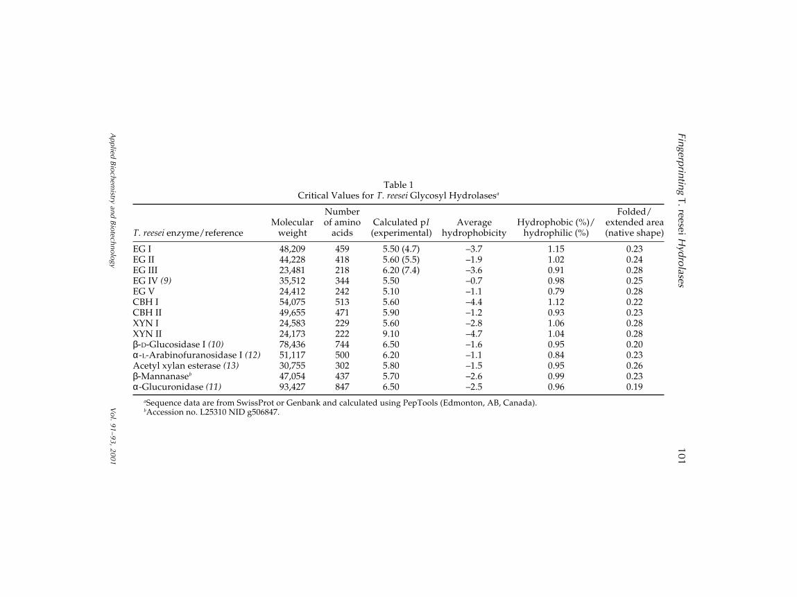
two β-1,4-exoglucanases (cellobiohydrolase [CBH] I and CBH II), twoxylanases (XYN I and XYN II), a β-D-glucosidase, an α-L-arbinoflurano-sidase, an acetyl xylan esterase, a β-mannanase, and an α-glucuronidasehave been sequenced (4) (Fig. 1, Table 1).
Application of preliminary proteonomics analysis to a standard com-mercial cellulase product was judged appropriate for demonstrating fin-gerprinting methodology (5). Although two-dimensional (2D) protein gelshave been used on occasion to follow the expression of selected T. reeseicellulase components (6) and their glycosylated forms (7,8), a systematicdisplay of the entire system of enzymes found in T. reesei culture broth hasnot been reported. We describe here fingerprinting via 2D gel electrophore-sis and internal peptide sequence analysis of a commercial cellulasepreparation. The Biomolecular Research Facility at the University of Vir-ginia Medical School processed gels prepared at National RenewableEnergy Laboratory for spot sequencing and identification. Forty-five spotswere identified by liquid chromatography–mass spectrometry/mass spec-trometry (LC-MS/MS). The experimental results were compared to ~1100known glycosyl hydrolases from all species, with positive hits only arisingfrom T. reesei proteins. Most of these were of known proteins, but severalnovel proteins were detected.
Materials and MethodsEnzyme Sample and Reagents
Genencor (Palo Alto, CA) generously provided a sample of the com-mercial cellulase preparation, Laminex, for testing. Buffers, trypsin,dithiothreitol (DTT), and electrophoresis supplies were obtained fromSigma (St. Louis, MO) and Amersham Pharmacia Biotech (Alameda, CA).
2D Electrophoresis
Characterization of the Laminex cellulase product was performed usingsodium dodecyl sulfate polyacrylamide gel electrophoresis (SDS-PAGE) and
Fig. 1. 2D gel simulation of T. reesei hydrolases based on literature values formolecular weight and isoelectric pH.
Fin
gerp
rintin
g T
. reese
i Hyd
rola
ses
10
1
Ap
plie
d B
ioch
em
istry a
nd
Bio
tech
no
logy
Vo
l. 91
–9
3, 2
00
1
Table 1Critical Values for T. reesei Glycosyl Hydrolasesa
Number Folded/Molecular of amino Calculated pI Average Hydrophobic (%)/ extended area
T. reesei enzyme/reference weight acids (experimental) hydrophobicity hydrophilic (%) (native shape)
EG I 48,209 459 5.50 (4.7) –3.7 1.15 0.23EG II 44,228 418 5.60 (5.5) –1.9 1.02 0.24EG III 23,481 218 6.20 (7.4) –3.6 0.91 0.28EG IV (9) 35,512 344 5.50 –0.7 0.98 0.25EG V 24,412 242 5.10 –1.1 0.79 0.28CBH I 54,075 513 5.60 –4.4 1.12 0.22CBH II 49,655 471 5.90 –1.2 0.93 0.23XYN I 24,583 229 5.60 –2.8 1.06 0.28XYN II 24,173 222 9.10 –4.7 1.04 0.28β-D-Glucosidase I (10) 78,436 744 6.50 –1.6 0.95 0.20α-L-Arabinofuranosidase I (12) 51,117 500 6.20 –1.1 0.84 0.23Acetyl xylan esterase (13) 30,755 302 5.80 –1.5 0.95 0.26β-Mannanaseb 47,054 437 5.70 –2.6 0.99 0.23α-Glucuronidase (11) 93,427 847 6.50 –2.5 0.96 0.19
aSequence data are from SwissProt or Genbank and calculated using PepTools (Edmonton, AB, Canada).bAccession no. L25310 NID g506847.
102 Vinzant et al.
Applied Biochemistry and Biotechnology Vol. 91–93, 2001
2D gels under denatured conditions. Protein sample preparation was initi-ated by trichloroacetic acid precipitation followed by acetone washing anddrying prior to resolublization in nonionic denaturing loading buffer.
All the 2D gel electrophoreses were performed in a horizontal formatutilizing the Pharmacia Multiphor II electrophoresis system and ImmobilineDrystrip kit. The first dimension of isoelectric focusing was carried out inan immobilized pH gradient dry strip gel (pH 3.0–10.0 linear). The seconddimension was run on 8–18% SDS polyacrylamide gradient gel (ExcelGel)and stained with colloidal blue. The standard methods and procedureswere followed directly from the Pharmacia manual.
The precast 180-mm Immobiline Drystrips were hydrated overnightin 8 M urea, 0.5% Triton X-100, Pharmalyte 3-10, and DTT and then wereloaded into the running tray and overlaid with oil. Approximately 75–100 µg of total protein was added to the sample buffer, loaded into theloading cups, and pulled into the first dimension gel at 500 V and 1 mA for5 h and then allowed to run at 3500 V and 1 mA for 14 h (total of 55,250 V-h).After the strips were equilibrated in a Tris-HCl, urea, glycerol, and SDSbuffer, they were placed on the second-dimension SDS-PAGE gel (8–18% SDS ExcelGel; precast 245 × 110 mm) at a 90° angle to the electricalfield. This dimension was run for 1.5–2 h at 600 V and 50 mA. All subse-quent gels were stained with either silver stain or colloidal blue.
Liquid Chromatography–Mass Spectrometry/Mass Spectrometry
The gel piece was precisely cut and transferred to a siliconized tubeand washed and destained in 200 µL of 50% methanol overnight. The pieceswere then dehydrated in acetonitrile, rehydrated in 30 µL of 10 mM DTT in0.1 M ammonium bicarbonate, and reduced at room temperature for 0.5 h.The DTT solution was removed and the sample alkylated in 30 µL of 50 mMiodoacetamide in 0.1 M ammonium bicarbonate at room temperature for0.5 h. The reagent was removed and the gel pieces were dehydrated in100 µL of acetonitrile. The acetonitrile was removed and the gel pieces wererehydrated in 100 µL of 0.1 M ammonium bicarbonate. The pieces werethen dehydrated in 100 µL of acetonitrile, the acetonitrile was removed,and the pieces were completely dried by vacuum centrifugation. The gelpieces were rehydrated in 20 ng/µL of trypsin in 50 mM ammoniumbicarbonate on ice for 10 min. Any excess trypsin solution was removedand 20 µL of 50 mM ammonium bicarbonate added. The sample wasdigested overnight at 37°C and the peptide formed extracted from the poly-acrylamide in two 30-µL aliquots of 50% acetonitrile/5% formic acid. Theseextracts were combined and evaporated to 25 µL for LC-MS analysis.
The liquid chromatography–mass spectrometry system consisted of aFinnigan LCQ ion trap mass spectrometer system with a Protana nanosprayion source interfaced to a self-packed 8 cm × 75 µm id Pnenomenex Jupiter10-µm C18 reversed-phase capillary column. Volumes of the extract (0.5–2 µL) were injected and the peptides eluted from the column by an aceto-nitrile/0.1 M acetic acid gradient at a flow rate of 0.25 µL/min. The

Fingerprinting T. reesei Hydrolases 103
Applied Biochemistry and Biotechnology Vol. 91–93, 2001
nanospray ion source was operated at 2.8 kV. The digest was analyzedusing the double play capability of the instrument acquiring full-scan massspectra to determine peptide molecular weights and product ion spectra todetermine amino acid sequence in sequential scans. This mode of analysisproduces approx 400 collisionally associated desorption (CAD) spectra ofions ranging in abundance over several orders of magnitude. Not all CADspectra are derived from peptides.
The data were analyzed by database searching using the Sequestsearch algorithm. Peptides that were not matched by this algorithm wereinterpreted manually and searched vs the expressed sequence tag data-bases using the Sequest algorithm. Experimental data were compared to adatabase of ~1100 known glycosyl hydrolases plus the NCBI database.Proteins were positively identified by 100% identity to two or more peptidefragments per protein.
Results and Discussion
Recent advances in protein chemistry tools including rapid separationtechniques such as 2D electrophoresis and LC-MS/MS afford uniqueopportunities to examine complex protein mixtures, such as the multicom-ponent mixture of hydrolytic enzymes secreted by T. reesei. These tech-niques permit fingerprinting of mixtures of proteins found in cellulasepreparations and their degradation products over a wide range of pHs andmolecular weights. The capability of 2D electrophoresis to separate severalhundred proteins in a single experiment can ultimately be used to identifyproducts expressed by different genomes as well as to identify uniqueproteins that may be expressed in response to induction by different bio-mass substrates. The data generated from the analysis of complex cellulasemixtures not only gives important information about this complex biologicsystem relevant to biomass conversion, but also may yield informationabout degradation of proteins in commercial preparations.
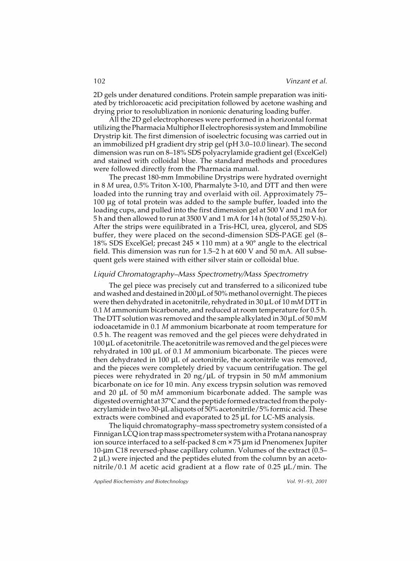
2D gel electrophoresis of a commercial T. reesei cellulase preparationyielded approx 70 discernible spots on a 245 × 110 mm gel (Figs. 2–4). Fifty-five to 60 gel species were observable in the pH and molecular weightwindow we considered appropriate for secreted fungal hydrolases (pH 2.5–9and 20–180 kDa). Thirty-four of these spots were identified as known glyco-syl hydrolases by LC-MS/MS. Eleven spots contained unknown proteins.
Although a number of spots were found by LC-MS/MS to contain asingle protein component (i.e., EG I, EG IV, EG V, CBH I, CBH II, AXE, andβ-D-glucosidase), analysis of many of these spots yielded multiple identi-ties (Table 2). We feel that the sampling and identification conditionsapplied to these gels were stringent enough to rule out contamination ormisidentification of spots. The likely explanations for this observation lie inboth the methodology used and the inherent nature of the sample. Someproteins may still be unresolved under the electrophoretic conditionsapplied (Fig. 3). For example, some improvement in the separation win-

104 Vinzant et al.
Applied Biochemistry and Biotechnology Vol. 91–93, 2001
Fig. 3. Pharmacia 2D gel of T. reesei cellulase preparation (Laminex; Genencor).Closer view of the gel zone defined by pH 3.5–6.85 and molecular weight (MW) of20–150 kDa.
dow may be possible with new recently available “narrow” pH rangeampholines. Also, isozymes or variably glycosylated forms of the proteinsmay explain small changes in molecular weight or isoelectric point (pI);
Fig. 2. Pharmacia 2D gel of T. reesei cellulase preparation (Laminex; Genencor)visualized with Coomassie blue. MW, molecular weight.
Fingerprinting T. reesei Hydrolases 105
Applied Biochemistry and Biotechnology Vol. 91–93, 2001
Fig. 4. Pharmacia 2D gel of T. reesei cellulase preparation (Laminex; Genencor).Analysis of the 45 spots shown was made by LC-MS/MS. MW, molecular weight.
however, this explanation would not account for the large alterations in theproperties observed in the case of EG I, CBH I, and CBH II. Severe proteoly-sis of some proteins during production, storage, or sample preparationmay provide the answer. Except for sample preparation alteration, the pres-ence of these altered proteins raises several interesting possibilities. Canoverall system-specific activity be increased through prevention of thesealtered enzymes? Can purified cellulases be used to reconstitute a cellu-lase mixture that is more active than native? The phenomenon of multipleelectrophoretic forms was most obvious with CBH II. Having manyCBH II spots widely distributed on the gel raises two obvious questions.Is CBH II really needed for efficient hydrolysis? and Can the addition ofholo-CBH II (the intact enzyme) to complex cellulase mixtures enhanceactivity?
It is now logical to compare compositions of variably induced cel-lulase preparations from T. reesei by high-resolution 2D electrophoresisand note the differences as a function of the individual enzyme speciespresent and the overall activity of the cellulase complex on pretreatedbiomass. Special assays such as the diafiltration saccharification assaycan be used to assess overall cellulose digestibility. The results of thesecomparisons may lend yet another perspective to the biochemical inter-actions of the individual enzyme species within the T. reesei cellulasecomplex needed for the rapid and total saccharification of cellulose inbiomass.
106 Vinzant et al.
Applied Biochemistry and Biotechnology Vol. 91–93, 2001
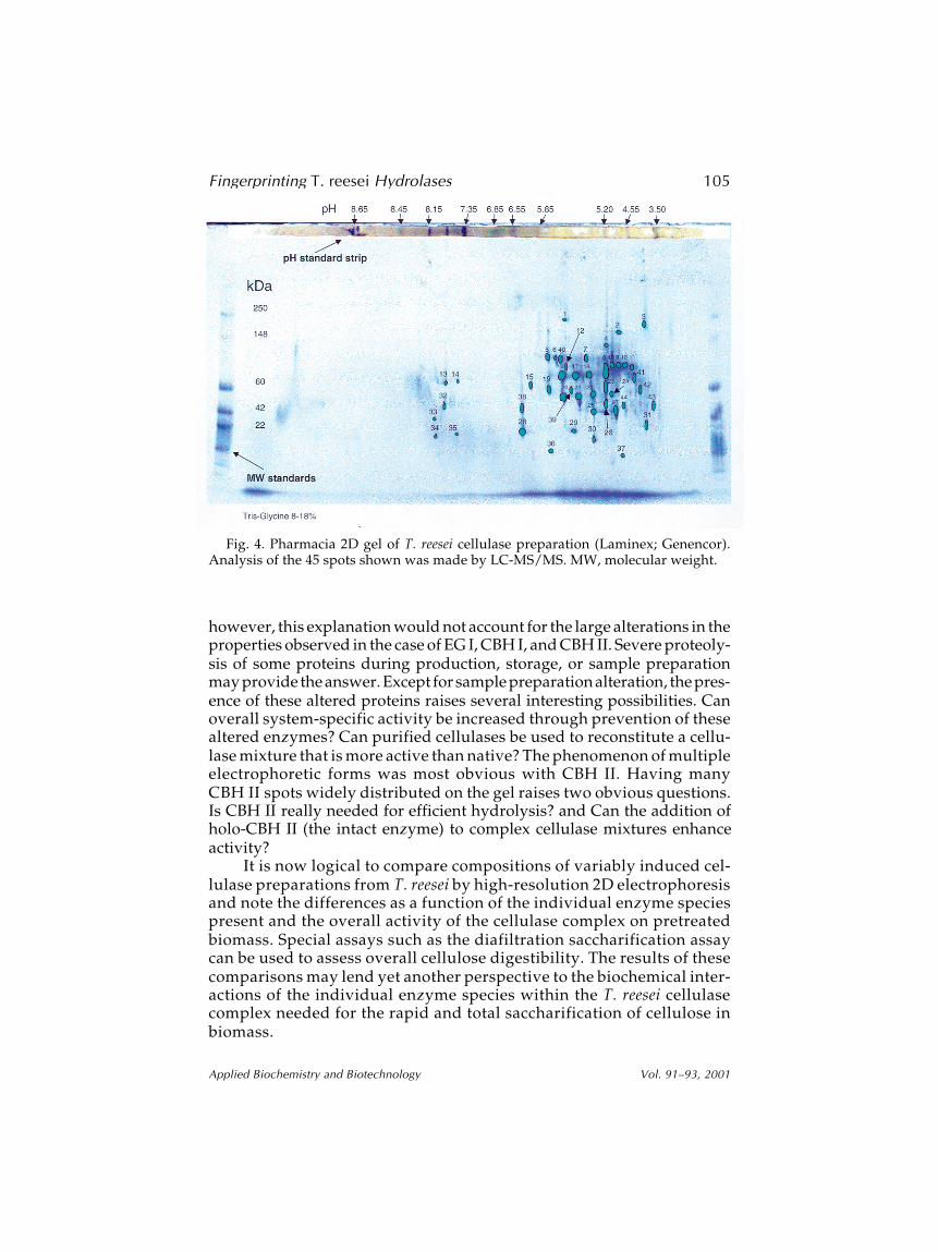
Table 22D Gel Component Analysis
of T. reesei Preparation by LC-MS/MSa
Spot no. Spot assignment
1 CBH II 2 CBH II 3 EG I, CBH II 4 Unknown 1, same as spot 31 5 Unknown 2, same as spot 6 6 Unknown, same as unknown 2 7 EG IV 8 CBH I, CBH II, EG I, EG II 9 CBH I, EG I10 CBH I, EG I11 CBH I, EG I12 Unknown 313 β-D-Glucosidase14 β-D-Glucosidase15 CBH II, β-D-glucosidase16 CBH II17 CBH II18 CBH II, CBH I19 CBH II20 CBH II, CBH I21 CBH II, CBH I22 EG II, CBH II, CBH I23 EG II, CBH II, CBH I24 EG II, CBH II, CBH I25 CBH II26 EG II, CBH II, CBH I27 EG II28 Endo29 Endo, CBH II, EG II30 Acetyl xylan esterase31 Unknown, same as unknown 132 Unknown 4, same as spot 3333 Unknown, same as unknown 434 Unknown 5, same as spot 3535 Unknown, same as unknown 536 Unknown 637 Unknown 738 Endo39 β-D-Mannanase40 EG IV, CBH II41 CBH I42 EG I, CBH I43 EG V44 Unknown 845 EG I
aEG I: 8–11, 42, 45; EG II: 3, 8, 22–24, 26, 27, 29; EG IV:18, 24, 40; EGV: 43; β-glucosidase: 13–15; CBH I: 8–11,20–24, 26, 41, 42; CBH II: 1–3, 8, 15–26, 40; AXE I: 30;β-mannanase: 39; novel: 5–7, 30–36, 44.

Fingerprinting T. reesei Hydrolases 107
Applied Biochemistry and Biotechnology Vol. 91–93, 2001
Acknowledgments
This work was partially funded by the Biochemical Conversion Ele-ment within the Biofuels Systems Program of the Office of Fuels Develop-ment of the US Department of Energy. This work was also funded by theUniversity of Virginia Pratt Committee.
References
1. Wyman, C. E., Bain, R. L., Hinman, N. D., and Stevens, D. J. (1993), Renewable Energy:Sources for Fuels and Electricity, Island Press, Washington, DC.
2. Sheehan, J. J. (1994), in Enzymatic Conversion of Biomass for Fuels Production, vol. 566,Himmel, M. E., Baker, J. O., and Overend, R. P., eds., American Chemical Society,Washington, DC, pp. 1–52.
3. Bergeron, P. (1996), in Handbook on Bioethanol, Wyman, C. E., ed., Taylor & Francis,Washington, DC, pp. 179–195.
4. Swiss-Prot site <http://www.expasy.ch/sprot/>.5. Herbert, B. R., Sanchez, J.-C., and Bini, L. (1997), in Proteonome Research: New Frontiers
in Functional Genomics, Wilkins, M. R., Williams, K. L., Appel, R. D., and Hochstrasser,D. F., eds., Springer Verlag, NY, pp. 13–30.
6. Schmidt, C. S. and Wolf, G. A. (1999), Eur. J. Plant Pathol. 105, 285–295.7. Pakula, T. M., Uusitalo, J., Saloheimo, M., Salonen, K., Aarts, R. J., and Penttila, M.
(2000), Microbiology-UK 146, 223–232.8. Maras, M., De Bruyn, A., Vervecken, W., Uusitalo, J., Penttila, M., Busson, R.,
Herdewijn, P., and Contreras, R. (1999), FEBS Lett. 452, 365–370.9. Saloheimo, M., Nakari-Setala, T., Tenkanen, M., and Penttila, M. (1997), Eur. J.
Biochem. 249, 584–591.10. Barnett, C. C., Berka, R. M., and Fowler, T. (1991), BioTechnology 9, 562–567.11. Margolles-Clark, E., Saloheimo, M., Siika-aho, M., and Penttila, M. (1996), Gene 172,
171, 172.12. Margolles-Clark, E., Tenkanen, M., Nakari-Setala, T., and Penttila, M. (1996), Appl.
Environ. Microbiol. 62, 3840–3846.13. Margolles-Clark, E., Tenkanen, M., Soderlund, H., and Penttila, M. (1996), Eur. J.
Biochem. 237, 553–560.
Related Documents


![CellulasesfromThermophilicFungi:RecentInsightsand … · 2019. 7. 31. · NR [21, 22] Melanocarpus albomyces cel7b 7 Trichoderma reesei 6–8 4.23 NR NR 50.0 [23] Melanocarpus albomyces](https://static.cupdf.com/doc/110x72/60d40ab91b88ac6c62145ad0/cellulasesfromthermophilicfungirecentinsightsand-2019-7-31-nr-21-22-melanocarpus.jpg)
