STUDIES ON PHAKOPSORA PACHYRHIZI, THE CAUSAL ORGANISM OF SOYBEAN RUST by Archana Nunkumar (BSc-Hons), University of Natal Submitted in fulfilment of the requirements for the degree of Master in Science Discipline of Plant Pathology School of Biochemistry, Genetics, Microbiology and Plant Pathology Faculty of Science and Agriculture University of KwaZulu-Natal Pietermaritzburg Republic of South Africa August 2006

Welcome message from author
This document is posted to help you gain knowledge. Please leave a comment to let me know what you think about it! Share it to your friends and learn new things together.
Transcript

STUDIES ON PHAKOPSORA PACHYRHIZI,
THE CAUSAL ORGANISM OF SOYBEAN RUST
by
Archana Nunkumar(BSc-Hons), University of Natal
Submitted in fulfilment of the requirements for the degree of
Master in Science
Discipline of Plant Pathology
School of Biochemistry, Genetics, Microbiology and Plant Pathology
Faculty of Science and Agriculture
University of KwaZulu-Natal
Pietermaritzburg
Republic of South Africa
August 2006

FRONTISPIECE
Electron micrograph showing a uredium with uredospores.
Phakopsora pachyrhizi is one of the few fungi that use direct penetration
through epidermal cells.
Rust never sleepsNeil Young

ABSTRACT
.$' Phakopsora pachyrhizi H. Syd and P. Syd, the causal organism of soybean rust
(SBR) was first reported in Japan in 1902. In 1934 the pathogen was found in several
other Asian countries and as far south as Australia. In India, SBR was first reported
on soybeans in 1951. There have been several early reports of SBR in equatorial
Africa but the first confirmed report of P. pachyrhizi on the African continent was in
1996 from Kenya, Rwanda and Uganda. Since then, the pathogen has spread south
with reports from Zambia and Zimbabwe in 1998 and in Mozambique in 2000.
In February 2001, P. pachyrhizi was first detected on soybeans near Vryheid, in
Northern KwaZulu-Natal, South Africa (SA). As the season progressed, the disease
was observed in other parts of the province, and epidemic levels were found in the
Cedara, Greytown, Howick and Karkloof production regions. Soybean rust
subsequently spread to Amsterdam and Ermelo in the Highveld region of SA. The
disease reappeared in SA in March 2002. It is now established that the pathogen is a
threat to soybean production in the country with yield losses in the region of 10-80%.
A literature review on SBR investigating the taxonomy of the pathogen, its
morphology, symptoms, host range, infection process, epidemiology, control options
and the economic importance of P. pachyrhizi was complied to provide the necessary
background information to conduct research under local conditions and to assist in
interpretation of results of experiments.
Epidemiological trials were conducted at the University of KwaZulu-Natal under
controlled environmental conditions in a dew chamber and conviron. Development of
P. pachyrhizi on the susceptible cultivar (LS5995) was quantified in combinations of
seven temperatures (15,19,21,24,26,28 and 30°C) and five leaf wetness durations
(LWD) (6,9,12,14 and 16hrs) at three relative humidities (RH) (75%, 85% and 95%).
Studies indicate that optimum temperature for uredospore infection is 21-24°C with a
LWD greater than 12hrs and RH 85-95%. The number of pustules as well as lesion

size on the abaxial and adaxial leaf surface increased with increasing LWD at all the
RH values tested. Infection did not occur on plants incubated at 15°C and 30°C at
85% or 95%RH whereas at 75%RH infection did not occur on plants incubated at
15°C, 19°C and 30°C regardless of LWD. Number of pustules per lesion produced at
75%, 85% and 95%RH was highest at 24°C and showed a gradual increase with
increasing LWD. Lesion size on both leaf surfaces increased after 12hrs LWD at 24°C
at 75% and 85%RH whereas at 95%RH lesion size increased after 14hrs LWD at
24°C.
Exposure of uredospores to ultraviolet light which is equivalent to ultraviolet C
(sunlight) which is < 280nm, shows a decrease in germination (7%). Under
continuous darkness, the germination percentage was found to range from 58% after
48 hrs. Germination was found to peak at 16hrs in darkness with a gradual decrease
as time increased whereas germination under ultraviolet light was highest after 6hrs
with a gradual decrease with increased exposure to light. Germ tube lengths were
found to be shorter when exposed to ultraviolet light (1071-1m) compared to controls
kept in the dark (181I-1m). Results obtained clearly show a negative effect of ultraviolet
light on the germination and germ tube length of uredospores. A 0.1 ml suspension of
uredospores on 1.25% water agar Petri dishes was exposed to cycles of 14h
ultraviolet light and 10h darkness for 48h. Results indicate an increase in germination
percentage of uredospores when exposed to 10h of darkness following a 14h period
under ultraviolet light.
Controlled environmental studies were conducted to determine alternative hosts of P.
pachyrhizi in SA. The control used in this experiment was Prima 2000, a susceptible
cultivar to soybean rust. Seven legume plants [Cajanus cajan (L.) Huth, Glycine max
(L.) Merr, Lablab purpureus (L.) Sweet, Lupinus angustifolius (L.) Finnish, Phaseolus
vulgaris (L.), Pueraria lobata (M&S) Willd and Vigna unguiculata (L.) Walp] and three
dry bean lines (Bonus; OPS-RS2 and PAN 159) showed typical SBR symptoms when
rated after 21 days post inoculation with uredospores for percentage disease severity.
Disease severity was significantly different within the alternative hosts, but G. max,
11

P. vulgaris and P. lobata were not significantly different from Prima 2000 (control). A
uredospore suspension of 2.5 x 10⁵ uredospores mr1 from plants that showed typical
SBR symptoms was made and inoculated onto Prima 2000, a susceptible soybean
cultivar. Uredospores from pustules on G. max, L. purpureus, L. angustifolius, P.
vulgaris, P. lobata, V. unguiculata, Bonus and PAN 159 produced viable uredospores
on PRIMA 2000. These plants are considered alternative hosts of P. pachyrhizi.
Effect of leaf age on susceptibility of soybean to SBR was tested under controlled
environmental conditions. Mean number of lesions as well as lesion size were greater
on younger leaves than on older leaves of plants at the same physiological age.
Plants at the early vegetative and reproductive stages had a significantly lower
number of lesions as well as a smaller lesion size. Plants at the V6 and R1 growth
stages were significantly more susceptible to P. pachyrhizi than plants at other
developmental stages.
Trichoderma harzianum Rifai, Eco-77® a commercial biological control product, was
evaluated for its efficacy as a biological control agent of P. pachyrhizi. Trichoderma
harzianum sprayed at the standard concentration on infected soybean plants was
significantly more effective in controlling P. pachyrhizi than plants sprayed at 1/2X
and 2x the standard concentration. This was noted in both Trial 1 and 2. Data indicate
that spraying the filtrate two days after inoculation produces less disease.
111

DECLARATION
I, Archana Nunkumar, declare that the research reported in this thesis, except where
otherwise indicated, is my own original research. This thesis has not been submitted
for any degree or examination at any other university.
Archana Nunkumar (Candidate)
Dr P.M. Caldwell (Supervisor)
IV

TABLE OF CONTENTS
ABSTRACT
DECLARATION .
TABLE OF CONTENTS
ACKNOWLEDGEMENTS
DEDICATION
FOREWORD
iv
v
xi
xiii
xiv
CHAPTER ONE . 1
LITERATURE REVIEW 1
1.1 INTRODUCTION . 1
1.2 BACKGROUND INFORMATION. 3
1.2.1 HISTORY 31.2.2 GEOGRAPHIC DISTRIBUTION 41.2.3 ECONOMIC IMPORTANCE 6
1.3 THE PATHOGEN 7
1.3.1 TAXONOMY AND MORPHOLOGY 71.3.2 SYMPTOMS. 101.3.3 HOST RANGE 13
1.4 INFECTION PROCESS AND EPIDEMIOLOGY 15
1.4.1 LIFE CYLCE AND INFECTION PROCESS 151.4.2 EPIDEMIOLOGY OF SOYBEAN RUST . 18
1.5 DISEASE MODELLING 20
1.6 DISEASE MANAGEMENT. 23
1.6.1 CULTURAL CONTROL 231.6.2 CHEMICAL CONTROL 241.6.3 BIOLOGICAL CONTROL . 271.6.4 RESISTANCE 27
1.7 SUMMARY. 31
1.8 REFERENCES 32
v

CHAPTER TWO. 41
DEVELOPMENT OF PHAKOPSORA PACHYRHIZI AT DIFFERENT
TEMPERATURES, RELATIVE HUMIDITIES AND LEAF WETNESS
DURATIONS 41
ABSTRACT . 41
2.1 INTRODUCTION 42
2.2 MATERIALS AND METHODS 43
2.2.1 Host material 432.2.2 Inoculum production 442.2.3 Spore concentration 442.2.4 Uredospore germination tests 452.2.5 Inoculation . 452.2.6 Experimental design 452.2.7 Disease assessment 452.2.8 Statistical analyses 46
2.3 RESULTS 46
2.3.1 Spore concentration 462.3.2 Uredospore germination tests 462.3.3 Number of pustules per lesion 472.3.4 Lesion size (mm) on the abaxial leaf surface. 492.3.5 Lesion size (mm) on the adaxial leaf surface. 51
2.4 DISCUSSION 53
2.5 REFERENCES 55
CHAPTER THREE 57
EFFECT OF ULTRAVIOLET LIGHT ON THE GERMINATION
OF UREDOSPORES OF PHAKOPSORA PACHYRHIZI . 57
ABSTRACT . 57
3.1 INTRODUCTION
VI
58

3.2 MATERIALS AND METHODS 59
3.2.1 Fungal inoculum . 593.2.2 Uredospore exposure to ultraviolet light 593.2.3 Uredospore exposure to cycles of ultraviolet light and
darkness 603.2.4 Statistical analyses 60
3.3 RESULTS 60
3.3.1 Uredospore exposure to ultraviolet light 603.3.2 Uredospore germination when exposed to cycles of ultraviolet
light and darkness. 61
3.4 DISCUSSION 64
3.5 REFERENCES 67
CHAPTER FOUR 70
ALTERNATIVE HOST STUDY OF PHAKOPSORA PACHYRHIZI
IN SOUTH AFRICA 70
ABSTRACT . 70
4.1 INTRODUCTION 71
4.2 MATERIALS AND METHODS 72
4.2.1 Inoculum sources . 724.2.2 Plant production 724.2.3 Inoculation and incubation 744.2.4 Uredospore germination tests . 754.2.5 Ratings scale used to differentiate between a host and
non-host 754.2.6 Re-inoculation and re-infection studies 754.2.7 Statistical analyses 76
4.3 RESULTS 784.3.1 Uredospore germination tests 784.3.2 Initial host reactions observed . 784.3.3 Re-inoculation and re-infection studies 79
4.4 DISCUSSION 80
vii

4.5 REFERENCES 81
CHAPTER FIVE. 83
EFFECT OF LEAF AGE ON SUSCEPTIBILTV OF SOYBEAN
TO PHAKOPSORA PACHYRHIZI. 83
ABSTRACT. 83
5.1 INTRODUCTION 84
5.2 MATERIALS AND METHODS 87
5.2.1 Test plants . 875.2.2 Inoculum 875.2.3 Inoculation . 875.2.4 Uredospore germination tests 885.2.4 Post-inoculation treatment 885.2.5 Disease rating 885.2.6 Statistical analyses 88
5.3 RESULTS 89
5.3.1 Viability of inoculum 895.3.2 Number of lesions per leaf produced by Phakopsora
pachyrhizi at the different developmental stages ofsoybean plants 89
5.3.3 Lesion size of Phakopsora pachyrhizi produced at thedifferent developmental stages of soybean plants . 91
5.4 DISCUSSION 93
5.5 REFRENCES 95
CHAPTER SIX . 98
EVALUATION OF TRICHODERMA HARZIANUM AS A
POSSIBLE BIOLOGICAL CONTROL AGENT FOR PHAKOPSORA
PACHYRHIZI 98
ABSTRACT.
Vlll
98

6.1 INTRODUCTION 99
6.2 MATERIALS AND METHODS 101
6.2.1 Test plants . 1016.2.2 Inoculum 1016.2.3 Inoculation . 1026.2.4 Uredospore germination tests 1026.2.5 Eco-77® germination tests 1026.2.6 Determination of the optimum concentration of Eco-77®
for the control of soybean rust . 1026.2.7 Determination of the correct time of application of Eco-77®
filtrates 1036.2.7 Ratings 1046.2.8 Statistical analyses 104
6.3 RESULTS 104
6.3.1 Viability of biological control agent and inoculum 1046.3.2 Final percentage disease severity and area under disease
progress curve (AUDPC) of infected plants subjected todifferent concentrations of Eco-77® 105
6.3.3 Final percentage disease severity and area under diseaseprogress curve (AUDPC) of infected plants subjected to thefiltrate of Eco-77® at different times 107
6.4 DISCUSSION 109
6.5 REFERENCES 111
CHAPTER SEVEN 114
GENERAL OVERVIEW 114
7.1 Research Conducted, But Not Yet Reported.
7.2 Proposed Future Research Priorities
7.3 REFRENCES
APPENDIX 1
Appendix 1a.
Appendix 1b
Appendix 1c.
IX
119
119
121
124
124
125
126

APPENDIX 2
Appendix 2a
Appendix 2b
APPENDIX 3
127
127
128
129
APPENDIX 4 130
Appendix 4a. 130
Appendix 4b 131
APPENDIX 5 132
Appendix Sa. 132
Appendix Sb 133
Appendix Se. 134
Appendix Sd 13S
Conference papers 136
x

ACKNOWLEDGEMENTS
I gratefully acknowledge:
Or P.M. Caldwell, my supervisor, for her support, sound advice, assistance,
encouragement and for introducing me to this unique pathogen. Her enthusiasm for
the project, her willingness to share information and the stimulation she provided in
many discussions were invaluable. I am grateful to her for reviewing and editing this
thesis.
Professor Z.A. Pretorius for his help and advice as co-supervisor of this thesis.
Mr. L. de Klerk at CERU for his assistance in maintaining the equipment used.
The staff of the Centre for Electron Microscopy for technical assistance with electron
microscopy.
Mr. E. Kidane for his help with statistical analyses and his willingness to give up his
time has been greatly appreciated.
Mr. P. Herbst of Link Seed (Pty) Ltd. for a constant supply of soybean seed.
Or A. Liebenberg and Department of Agriculture and Environmental Affairs, Cedara,
for providing me with seed for the alternative host study.
The University of KwaZulu-Natal for providing facilities for this research.
The Protein Research Foundation for their financial support that made this research
possible.
Xl

Staff and postgraduate students of microbiology and plant pathology for their help and
encouragement throughout this study. To my friends Dael Visser, Benice Sivparsad,
Krishna Naicker, Sanisha Naidu and Tanya Naidoo thank you for providing a shoulder
of strength and for always finding time to make me laugh when I wanted to cry.
My mother for sacrifices made for my education and always believing in me.
My family for their support and encouragement during my studies. To Achal and
Suman may this study be an example for you to do better.
Mandhira and Shraddha Gokul for your love, support and encouragement.
Rishi Sooklall for always being there.
Finally, I would like to thank God for seeing me through this study.
XlI

DEDICATION
To my mother, Kocellia Nunkumar,
for her support, encouragement
and understanding
during my years of study
Xlll

FOREWORD
The research presented in this thesis was undertaken in the Discipline of Plant
Pathology, University of KwaZulu-Natal, Pietermaritzburg under the supervision of Or
P.M. Caldwell.
Since the first report of Phakopsora pachyrhizi Syd., the causal organism of soybean
rust (SBR) on soybeans, in South Africa (SA) during March 2001 much research has
been undertaken in the study of this unique fungal pathogen. Soybean rust is a new
disease in SA and it has been established that P. pachyrhizi is now endemic to SA.
Very little knowledge about this pathogen in SA is available.
Research on SBR pathogen has been carried out by institutes including the University
of KwaZulu-Natal, University of the Free State, Protein Research Foundation,
Department of Agriculture and Environmental Affairs, Cedara and Agricultural
Research Council to find effective solutions to what has become a serious yield
reducing pathogen before it causes irreversible damage to the soybean industry in
SA.
The extent of this research is broad, traversing seven chapters. The main objectives
of the research in this thesis were: a review of the literature on the history and
geographic distribution of P. pachyrhizi, the economic importance and pathogen
taxonomy and morphology, the symptoms, host range, infection process and
epidemiology, and control options available for the control of P. pachyrhizi (Chapter
One); the epidemiology of P. pachyrhizi under controlled environmental conditions
(Chapter Two); in vitro screening of P. pachyrhizi uredospores to the effects of
ultraviolet light (Chapter Three); an alternative host study (Chapter Four); an
evaluation of soybean plants at different developmental stages to SBR (Chapter
Five); an evaluation of a commercial biological control product to control SBR
(Chapter Six) and a review of the experimental results, conclusions and
recommendations for future research on SBR in SA (Chapter Seven).
XIV

Chapter Two and Three will aid in the production of a disease prediction model for
SBR. Chapter Four gives an indication of which legume plants are alternative hosts of
this pathogen in SA. Chapter Five gives an indication at which time plants are more
susceptible to the pathogen, hence, providing a more accurate time to apply chemical
sprays for control. Chapter Six will help determine if P. pachyrhizi can be controlled
by alternative methods, as at present chemical applications are the only effective
means for control.
xv
CHAPTER ONE
LITERATURE REVIEW
1.1 INTRODUCTION
Soybean, Glycine max (L.) Merril. is an ancient crop with numerous food, feed and
industrial uses. This crop is native to eastern Asia and has been used for thousands
of years for human and animal food as well as medicine to treat many human
diseases (Hartman et al., 1999). Soybeans provide protein and are the primary
source of vegetable oil, which constitutes 40% of the world's edible vegetable oil.
Soybean products are economically important due to their ability to produce new, low
cost, nutritionally balanced, high- protein foods, fit for human consumption (Sinclair,
1982).
Soybeans are grown from temperate to tropical regions of the world, with production
being highest in Brazil, China and the United States of America (U.S.A). Emphasis in
research has been on breeding of soybeans, appropriate for tropical environments
(Hartman et al., 1999). Due to an increase in soybean production throughout the
world, diseases that affect this crop have also, therefore, increased in number and
severity.
In Africa, soybean cultivation has increased in the last four decades from 72 000
tonnes on 191 OOOha in 1961 to 989 000 tonnes on 1 090 OOOha in 2002. However, it
accounts for only 0.5% of the annual global production of 179 917000 tonnes (Singh
et al., 2004). In South Africa (SA) soybean is a strategically important crop and is
grown under natural rainfall and irrigated conditions, usually in summer rainfall areas
(Bell et al., 1990). In KwaZulu-Natal (KZN) SA, approximately 30 000-35 OOOha of
soybeans are grown annually. Due to an ever-increasing demand for soybeans,
expansion of production is still possible in the northern and midland areas of KZN
(Ward,2003).
1

Soybeans are affected by more than 100 pathogens, with approximately 35 of
economic importance (Earthington et al., 1993). All parts of the soybean plant are
susceptible to numerous pathogens, resulting in a reduction in quality and quantity of
seed yields (Sinclair and Backman, 1989).
Phakopsora pachyrhizi Sydow, the causal organism of soybean rust (SBR) is one of
the major disease problems limiting soybean yield. In February 2001, P. pachyrhizi
was detected for the first time on soybeans near Vryheid in Northern KZN, SA
(Pretorius et al., 2001). As the season progressed, the disease was observed in other
parts of the province, and epidemic levels were found in the Cedara, Greytown,
Howick and Karkloof production regions. Soybean rust subsequently spread to
Amsterdam and Ermelo in the Highveld region of SA (Caldwell et al., 2002).
The disease reappeared in SA in March 2002. It is clear that the pathogen is now an
established threat to soybean production in the country. Yield losses in SA are
reported to be in the region of 10-80% (Caldwell and Laing, 2002).
Soybean rust has spread around the globe causing extensive damage to soybean
crops throughout the Southern hemisphere. Apparently it is able to travel great
distances via wind-borne spores. Also known as Asian rust, this fungal infection can
defoliate soybean fields rapidly, often resulting in severe and sometimes total loss
(Stewart et al., 2005).
2
1.2 BACKGROUND INFORMATION
1.2.1 HISTORY
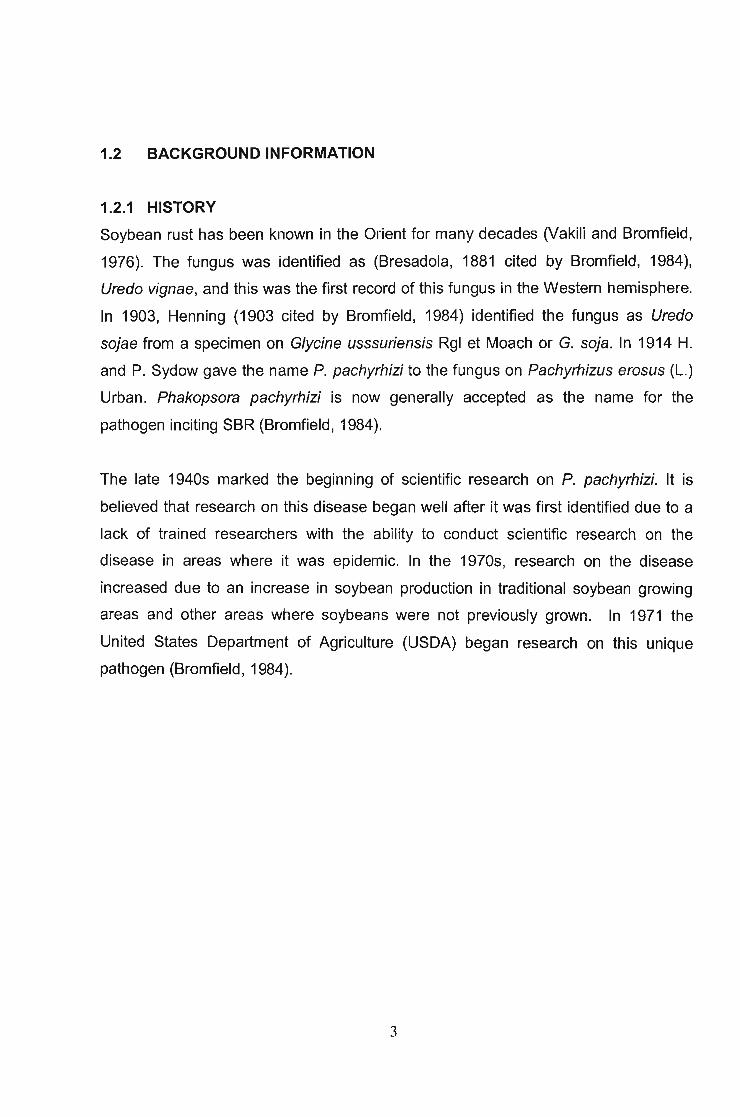
Soybean rust has been known in the Orient for many decades (Vakili and Bromfield,
1976). The fungus was identified as (Bresadola, 1881 cited by Bromfield, 1984),
Uredo vignae, and this was the first record of this fungus in the Western hemisphere.
In 1903, Henning (1903 cited by Bromfield, 1984) identified the fungus as Uredo
sojae from a specimen on Glycine usssuriensis Rgl et Moach or G. soja. In 1914 H.
and P. Sydow gave the name P. pachyrhizi to the fungus on Pachyrhizus erosus (L.)
Urban. Phakopsora pachyrhizi is now generally accepted as the name for the
pathogen inciting SBR (Bromfield, 1984).
The late 1940s marked the beginning of scientific research on P. pachyrhizi. It is
believed that research on this disease began well after it was first identified due to a
lack of trained researchers with the ability to conduct scientific research on the
disease in areas where it was epidemic. In the 1970s, research on the disease
increased due to an increase in soybean production in traditional soybean growing
areas and other areas where soybeans were not previously grown. In 1971 the
United States Department of Agriculture (USDA) began research on this unique
pathogen (Bromfield, 1984).
3

1.2.2 GEOGRAPHIC DISTRIBUTION
The first report of the disease was from Japan in 1902 (Figure 1.1). By 1934 the
pathogen had been found in several Asian countries and as far south as Australia
(Bromfield and Hartwig, 1980). In India, SBR was first reported on soybeans in 1951
(Sharma and Mehta, 1996). There have been several early reports of SBR in
equatorial Africa (Javaid and Ashraf, 1978; Bromfield, 1980), but the first confirmed
report of P. pachyrhizi on the African continent was in 1996 from Kenya, Rwanda, and
Uganda. Since then, the pathogen has spread south with reports from Zambia and
Zimbabwe in 1998, Mozambique in 2000 and SA in 2001 (Pretorius et al., 2001; Levy
et al., 2002). The westward movement of the pathogen on the African continent was
reported from Nigeria in 1999 (Akinsanmi et al., 2001).
In South America the first report of P. pachyrhizi was from Paraguay in March 2001
(Morel et al., 2004). It was subsequently reported in the state of Parana, Brazil in
2001 (Yorinori, 2004). The disease was found in Hawaii in 1994 on cultivated
soybeans on the islands of Hilo, Kakaha, Kauai and Oahu. (Kilgore and Heu, 1994).
By 2002, SBR was widespread throughout Paraguay and in limited areas of Brazil
bordering Paraguay, with reports of severe disease in some fields in both countries
(Morel and Yorinori, 2002). The pathogen also was found in a limited area in northern
Argentina (Rossi, 2003).
In August 2004, the USDA and the Animal Plant Health Inspection Service (APHIS)
confirmed a report of SBR in Colombia (Caspers-Simmet, 2004). On the 10
November 2004, the USDA issued a press release on the first report of SBR on the
USA mainland (Rogers and Redding, 2004). It is now established that SBR occurs in
all major soybean producing areas around the world.
4

Figure 1.1 Worldwide distribution of SBR caused by Phakopsora pachyrhizi
(Miles et al., 2003).
5
1.2.3 ECONOMIC IMPORTANCE
Soybean rust causes severe economic losses in many parts of the world where
soybeans are grown on a large commercial scale and is considered the most
destructive foliar disease of soybeans (Miles et al., 2003). In 1973 APHIS declared
SBR to be one of the hundred most dangerous exotic pests and diseases and a
number one threat to soybeans (Hershman, 2003).
Soybean rust reduces yield through premature defoliation, decreasing the number of
filled pods and by reducing the weight of seeds per plant. It also lowers the quality of
seed produced. The severity of loss and the particular components of yield affected
depend primarily on the time of disease onset and the intensity of disease at
particular growth stages of the soybean crop (Bromfield, 1984). When early infection
and unfavourable environmental conditions exist, yield losses of 50-60% can be
experienced (Kloppers, 2002). "
Yield losses as high as 10-50% have been reported in Southern China, 40% in
Japan,10-40% in Thailand, and 25-90% in Taiwan (Sinclair and Backman, 1989).
Nearly complete yield losses can occur in limited areas in most of these countries.
Yield losses in Uganda were estimated to be 22.9% in 2003 (Kawuki et al., 2003).
South Africa produces 208 000 tonnes of soybean seed on 193 OOOha of land.
Farmers in KZN plant about 35 OOOha, i.e., about 18% of arable land is planted to
soybeans in SA. In SA yield losses of 10-80% were reported, with losses of up to
100% where monocropping was practiced (Caldwell and Laing, 2002).
Yang et al. (1991) reported that the number of pods per plant at growth stage R6 was
reduced by as much as 40%, but the number of seeds per pod was not affected, i.e.,
the disease affected the attainable yield by reducing pod set. From growth stage R6
to R7, percentage of pod abortion was high for severely diseased plants. Seed
growth rate (grams per day) from R4 to R7 was reduced by 40-80% in diseased
6

plants. The. time for diseased plants to grow from R4 to R7 was reduced by as many
as 16 days compared to protected plants (Yang et al., 1991).
In 1984, an economic risk analysis projected that the potential losses in the U.S.A
would be $7.1 billion per year, once SBR was established in the main soybean
growing area of the U.S.A. (Kuchler et al., 1984). A conservative prediction indicated
yield losses greater than 10% in nearly all the U.S.A growing areas with losses of
50% in the Mississippi delta and the southeastern coastal states (Yang, 1995).
1.3 THE PATHOGEN
1.3.1 TAXONOMY AND MORPHOLOGY
Phakopsora pachyrhizi belongs to the Phylum Basidiomycota (Alexopoulos and
Mims, 1979), the Class Uredinomycetes, the Order Uredinales, the Family
Melampsoraceae and the Genus Phakopsora (Agrios, 1997). A related rust fungus,
Phakopsora meibomiae Arthur, also infects soybeans but is considered less virulent
than P. pachyrhizi (Caldwell et al., 2002).
Phakopsora pachyrhizi and P. meibomiae may only be differentiated based upon the
morphological characteristics of telia. However, primers have been developed
specifically for the two species to facilitate a polymerase chain reaction (PCR) so that
the two species can be accurately and quickly identified (Frederick et al., 2002).
Uredia are globose, subepidermal, and erumpent and light cinnamon to reddish
brown. They form abundantly on the abaxial leaf surface, where they range from 100
to 200l..lm in diameter (Sinclair and Backman, 1989). Pycnia and aecia are unknown.
Uredospores are globose, subglobose, ovate or ellipsoidal and are essentially hyaline
to light yellow-brown and open through a central pore to form a germ tube. The size
of the spores is highly variable, in the range of 18-45 x 13-28I..1m, depending on the
host and environmental conditions (Figure 1.2). Paraphyses, which are found
surrounding the inner wall of the uredia, unite at the base, forming a domelike
7

covering over the sporophores. The paraphyses are inward curving, hyaline to
subhyaline, prominently capitate at the apex, with a narrow lumen, and measure
about 7-151Jm toward the apex (Figure 1.3) (Sinclair and Backman, 1989).
Phakopsora pachyrhizi is one of five rust fungi that can infect without the formation of
an appressorium. Direct penetration is usually observed. Telia are rare but
occasionally form subepidermally, mostly on the abaxial leaf surface, among the
uredia and at the edges of lesions. They are orange-brown or light brown when
young, and become dark-brown to black with age. They are crustose, irregular to
round, sparse to aggregate, and about 150-2501Jm in diameter, with 3-5j..tm irregular
layers of teliospores (Sinclair and Backman, 1989).
8

Figure 1.2 Electron micrograph showing soybean rust uredospores and germ tubes
(Nunkumar, A).
Figure 1.3 Paraphyses surrounding the inner wall of a uredium
(Nunkumar, A).
9

1.3.2 SYMPTOMS
Soybean rust is an obligate parasite and a low sugar disease (Caldwell et al., 2002).
This is indicated by the fact that infection usually begins on the older, lower leaves of
plants at, or after the flowering stage, but is generally not noticed until the pods are
set. Early symptoms appear as small water-soaked lesions, which gradually increase
in size, turning from grey to tan or brown and are restricted by leaf veins. The release
of visible clouds of rust spores is another identifying characteristic of SBR (Caldwell
and Laing, 2002). Initially SBR symptoms may be confused with bacterial pustule
(Xanthomonas axonopodis pv. glycines Nakano). However, bacterial pustule has
water-soaked lesions containing mucilaginous or sticky bacteria (Caldwell and Laing,
2002). This bacterial pathogen also causes defoliation of plants, but no pustules are
visible.
Lesions are found mainly on the leaves, where they are common on the abaxial leaf
surface exuding clumps of brownish spores called uredospores (Bromfield et al.,
1980; Sinclair and Backman, 1989) (Figure 1.4). However, in severe cases, lesions
can also be found on pods, stems and petioles (Caldwell et al., 2002). Within each of
the lesions is one to many erumpent, globose uredia. Reddish-brown lesions appear
to indicate a semi-compatible reaction, while those with a tan coloration, without
extensive necrosis indicate a compatible interaction (Caldwell et al., 2002).
Once lesions appear, premature yellowing occurs and defoliation is rapid, resulting in
fewer pods and seeds, lower seed weight and early maturity (Caldwell et al., 2002).
The infected leaves turn bronze or yellow and these patches in the field are known as
"hot spots" (Figure 1.5). Once these hot spots are observed in the field, it is usually
too late to apply fungicides.
Although quantitative data are lacking, it is generally thought that leaf yellowing and
defoliation are correlated with the number of lesions per leaflet. As the number of
lesions per unit area increases, yellowing and defoliation becomes more pronounced.
10

The rate of severity of these processes may be influenced by the host variety and the
pathogen isolate involved (Bromfield, 1984).
11
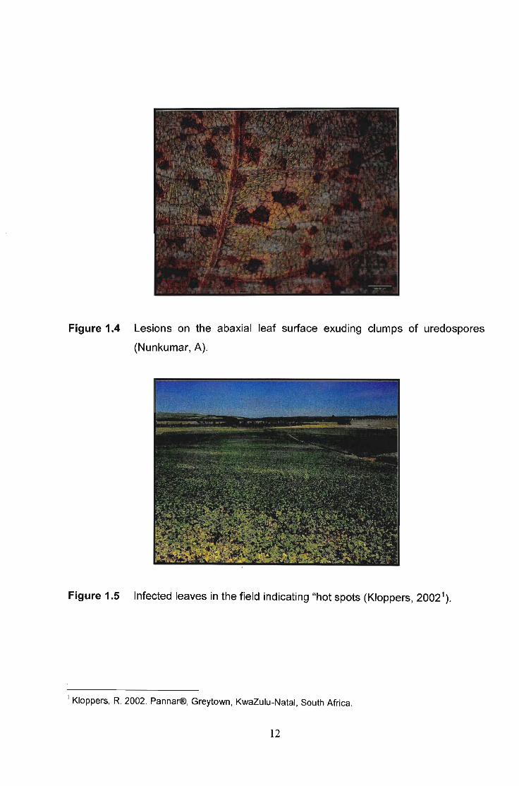
Figure 1.4 Lesions on the abaxial leaf surface exuding clumps of uredospores
(Nunkumar, A).
Figure 1.5 Infected leaves in the field indicating "hot spots (Kloppers, 2002\
1 Kloppers. R. 2002. Pannar®. Greytown. KwaZulu-Natal, South Africa.
12

1.3.3 HOST RANGE
Phakopsora pachyrhizi is an obligate parasite and cannot survive independently of its
hosts or on debris. It must, therefore, find alternate hosts on which to survive under
host-free conditions.
This pathogen has an unusually wide host range. Phakopsora pachyrhizi has been
reported to produce natural infections on 31 plant species in 17 genera of legumes
and 60 species of plants in 26 additional genera when inoculated (Chu and Chuang,
1961 ).
Many researchers have proposed lists of alternate hosts, but some lists require
cautious interpretation. This is because many researchers do not state the criteria
used to determine a "host'. The host in question should only be considered as an
alternate host if the fungus sporulates on it. In some instances, hosts, which do not
support sporulation, have been included in lists of alternate hosts (Bromfield, 1984).
A full host range has, therefore, not been clearly identified (Miles et al., 2003) and is
complicated by pathotypes or races of the fungus and strains or varieties of the host.
The same legume species may support sporulation of the fungus in one region but
not in another due to differences in races of the pathogen (Bromfield, 1984).
Rytter et al. (1984) tested 35 species within 23 genera of legumes for reactions to
three races of P. pachyrhizi. Twelve species were found to be new alternative hosts,
inclUding Coronilla varia (L.) Koch, Lespedeza striata (H&A) Thunb, Lupinus luteus
(L.) Finnish, Sesbania sericea (Willd.) Link and Trifolium repens (L.).
Shinde and Thakare (2000) tested various leguminous and pulse crops under
glasshouse conditions during 1997-1999 to determine possible hosts of P. pachyrhizi.
It was found that Vigna unguiculata (L.) Walp. (cowpea); Phaseolus vulgaris (L.)
(French bean), Phaseolus aureus Roxb. (blackgram), Cajanus cajan (L.) Huth.
13

(pigeon pea) and Cicer arietinum (L.) (chickpea) and Glycine wightii (Wight & Am.)
Verdc. (perennial soybean) were infected by the pathogen.
Some common hosts include Melilotus indica Color. (yellow sweet clover), Lupinus
angustifolius (L.) Finnish (lupin), Phaseolus vulgaris (L.) (green/kidney bean),
Phaseolus lunatus (L.) (lima/butter bean. One of the rather important alternate host is
Pueraria lobata (M&S) Willd. (kudzu vine), which is widespread in the U.S.A and
South America (Miles et al., 2003).
14

1.4 INFECTION PROCESS AND EPIDEMIOLOGY
1.4.1 LIFE CYCLE AND INFECTION PROCESS
Successful infection of a host by a pathogen is the culmination of a series of events
that must occur in sequence, Le., spore germination, appressorium formation, and
penetration. Each of these steps and the subsequent ones of colonization and
sporulation are influenced by biotic factors of the pathogen and host, and abiotic
factors of the environment.
Two spore types are known in P. pachyrhizi. The uredospore is the common spore
type found throughout the season. Uredospores are readily dispersed by wind and
multiple spore cycles occur throughout the season. Telia and teliospores have been
found on infected plants late in the season (Miles et al., 2003). Since no alternate
host has been identified, there has been no further characterization of the life cycle
(Miles et al., 2003). The infection process starts when uredospores germinate to
produce a single germ tube that grows across the leaf surface, 5 to 400 IJm, until an
appressorium forms. Appressoria form over anticlinal walls or over the center of
epidermal cells, but rarely over stomata. Penetration of epidermal cells is by direct
penetration through the cuticle by an appressorial peg. When appressoria form over
stomata, the hyphae penetrate one of the guard cells rather than entering the leaf
through the stomatal opening.
This rust and related species are unique in their ability to directly penetrate the
epidermis; most rust pathogens enter the leaf through stomatal openings and
penetrate cells once inside the leaf. The direct penetration of the epidermal cells and
the non-specific induction of appressoria in the infection process of P. pachyrhizi may
aid in understanding the broad host range of the pathogen. Under dry conditions this
extended sporulation capacity allows the pathogen to persist and remain a threat. If
conditions for re-infection are sporadic throughout the season, significant inoculum
potential still remains from the initial infection to reestablish an epidemic. A general
life cycle of the heterocious rust is presented in Figure 1.6.
15

5pennCltia 'reftiUzt!. COl'l'lPillible
1-l;.;"~lr;.~.:i=·~;.;~=~~,~,", .' , --..
. : ,:'-"""-~'--"
Oilo<ll)'6tlo:"l)'<~IiLfm .
~~i\lm prImordiUJll
lJrroillm on Whfl31
Figure 1.6 A general life cycle of the heterocious rust (Agrios, 1997).
Successful infection is dependant on the availability of moisture on plant surfaces. At
least 6 hours of free moisture is needed for infection with maximum infections
occurring with 10 to 12 hours of free moisture. Temperatures between 15 and 28°C
are ideal for infection (Miles et al., 2003). The infection process of P. pachyrhizi and
the developmental stages of SBR are given in Table 1.1 (Marchetti et al., 1975;
Bonde et al., 1976; McLean, 1979; Koch et al., 1983; Miles et al., 2003).
16

Figure 1.6 A general life cycle of the heterocious rust (Agrios, 1997).
Successful infection is dependant on the availability of moisture on plant surfaces. At
least 6 hours of free moisture is needed for infection with maximum infections
occurring with 10 to 12 hours of free moisture. Temperatures between 15 and 28°C
are ideal for infection (Miles et al., 2003). The infection process of P. pachyrhizi and
the developmental stages of SBR are given in Table 1.1 (Marchetli et al., 1975;
Bonde et al., 1976; McLean, 1979; Koch et al., 1983; Miles et al., 2003).
16

Table 1.1 Sequence of events over time in the development of Phakopsora pachyrhizi
(Marchetti et al., 1975; Bonde et al., 1976; McLean, 1979; Koch et al.,
1983; Miles et al., 2003).
Sequence of events Time
1. A uredospore lands on soybean leaf surface over epidermal cell ohpi
2. Germ-tube development (5-400IJm) 12 hpi
3. An appressorium-cone formed 16 hpi
4. Penetration hyphae formed 16 hpi
5. First hyphal septum formed 18-20 hpi
6. Primary hyphae produced 18-20 hpi
7. Collapse of epidermal cell 24 hpi
8. Haustorium formed 24-48 hpi
9. Branching into secondary hyphae 48-72 hpi
10. Mycelial development inside spongy mesophyll and intercellular space 3 dpi
11. Collapse of appressorium and penetration hyphae 4 dpi
12. Necrotic lesions appear on leaf 6 dpi
13. Runner hyphae passing through mesophylls 7 dpi
14. Hyphae aggregate, uredial primordia formed 9 dpi
15 Uredospore mature 11-12 dpi
hpl- hours post Infection
dpi- days post infection
17

1.4.2 EPIDEMIOLOGY OF SOYBEAN RUST
The ability of P. pachyrhizi to cause an epidemic in soybeans depends on a number
of factors. Two of these factors are temperature and leaf wetness duration, which
together determine the suitability of infection periods. Another factor affecting
epidemic development is the timing of the first rain on the crop and the amount of rain
(Tschanz et al., 1984).
Studies have shown that the rate of SBR development is closely associated with the
development and maturation of the soybean plant. Delayed rust onset, therefore
results in less serve infection levels. Therefore, the effect of soybean development
and maturation on rust development has to be accounted for in epidemiological and
host resistance studies (Tschanz and Tsai, 1982; Tschanz et al., 1984).
Rust epidemics are most severe during extended periods of leaf wetness when the
average daily temperature is less than 28°C (Hartman et al., 1999) with relative
humidities of 75-80% (Caldwell et al., 2002). Dry conditions, excessive precipitation or
daily mean temperatures greater than 30°C or less than 15°C inhibit rust development
(Sinclair and Backman, 1989). Moisture on plant surfaces is crucial for germination to
occur (Caldwell et al., 2002), Hence areas where prolonged periods of leaf wetness
due to dew, mist and light rain occurs provide optimum conditions for germination
(Kloppers, 2002). Temperatures above 2rC for extended periods retard rust
development even with adequate free moisture on the leaf surface (Casey, 1979).
In areas where rainfall occurs more evenly throughout the season, SBR develops
more rapidly as opposed to areas where rainfall occurs in uneven patterns. Hence
rust development varies according to prevailing rainfall patterns. To determine the
effect of precipitation and irrigation on rust development, field soybeans were watered
with overhead irrigation and furrow irrigation (Wang and Hartman, 1992). Results
indicate that rust was more prevalent in overhead-irrigated plots.
Field studies by Casey (1979) demonstrated that the development of a severe rust
epidemic requires about 10 h d-1 of leaf wetness and a daily mean temperature of 18-
18
26°C. In another field study, mean night temperatures consistently below 14°C
prevented or greatly inhibited rust development, while mean night temperatures
above 25.5°C had little effect on rust development when they occurred in conjunction
with frequent, long leaf wetness periods (Tschanz et al., 1984).
At optimum temperatures between 20°C and 25°C, infection of a susceptible host can
occur during 6hrs of leaf wetness. Within this temperature range, maximum infection
occurs within 1-12hr of leaf wetness. Increased periods of leaf wetness are necessary
for infection when temperatures fall outside the optimum temperature range
(Marchetti et al., 1976).
Phakopsora pachyrhizi telia and teliospores are induced by low temperatures and
occur when night temperatures are between 5 and 15°C and day temperatures
between 20 and 25°C (Kitani and Inoue, 1960; Hsu and Wu, 1968; Bromfield, 1980).
Yeh et al. (1981) also induced formation of telia and teliospores on a number of other
leguminous hosts. So far teliospore germination has not been observed, and its role
in the epidemiology of the disease is unknown.
19
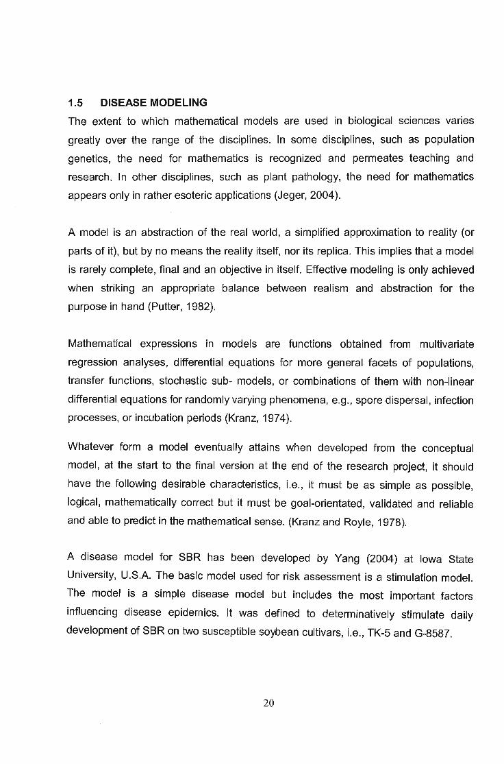
1.5 DISEASE MODELlNG
The extent to which mathematical models are used in biological sciences varies
greatly over the range of the disciplines. In some disciplines, such as population
genetics, the need for mathematics is recognized and permeates teaching and
research. In other disciplines, such as plant pathology, the need for mathematics
appears only in rather esoteric applications (Jeger, 2004).
A model is an abstraction of the real world, a simplified approximation to reality (or
parts of it), but by no means the reality itself, nor its replica. This implies that a model
is rarely complete, final and an objective in itself. Effective modeling is only achieved
when striking an appropriate balance between realism and abstraction for the
purpose in hand (Putter, 1982).
Mathematical expressions in models are functions obtained from multivariate
regression analyses, differential equations for more general facets of populations,
transfer functions, stochastic sub- models, or combinations of them with non-linear
differential equations for randomly varying phenomena, e.g., spore dispersal, infection
processes, or incubation periods (Kranz, 1974).
Whatever form a model eventually attains when developed from the conceptual
model, at the start to the final version at the end of the research project, it should
have the following desirable characteristics, Le., it must be as simple as possible,
logical, mathematically correct but it must be goal-orientated, validated and reliable
and able to predict in the mathematical sense. (Kranz and Royle, 1978).
A disease model for SBR has been developed by Yang (2004) at Iowa State
University, U.S.A. The basic model used for risk assessment is a stimulation model.
The model is a simple disease model but includes the most important factors
influencing disease epidemics. It was defined to determinatively stimulate daily
development of SBR on two susceptible soybean cultivars, Le., TK-5 and G-8587.
20

The model consists of a main program, an input and an initiation program, and a
graphic and statistic output program. The main program has five subroutines with 10
state variables, and some constants. Models of infection rate, latent period, and
senescence have been developed. The model of latent period explains up to 98.7%
of the variation, with no uredia present until 6.35 physiological days after inoculation.
Sixteen physiological days after inoculation, up to 95% of the lesions became
infectious (Kranz and Royle, 1978).
The rationale for modeling and analysis of disease progress data derives from the
desire to compare epidemics. The comparison may be years, locations or
environments, management practices or pathosystems. The goals of such
comparisons are to identify environmental factors that influence epidemic
development, to decide upon the efficacy of specific management practices. The
ultimate goal, of course, is to manage diseases the best way possible to minimize the
impact of the disease on the plants (Sail, 1980).
Previous soybean rust risk assessments with an assumption of availability of spores
early in a season showed that weather conditions (dew and temperature) during a
growing season, in general, are suitable for disease development in U.S.A soybean
growing regions. Predicting the time of rust appearance in a field is critical to
determining the destructive potential of rusts, including soybean rust. Epidemiology is
most likely used to assess rust incipient time (Pivonia and Yang, 2006).
Few biologically based models to assess the risk of soybean rust have been
developed because of difficulty in estimating variables related to infection rate of the
disease. A fuzzy logic system, however, can estimate apparent infection rate by
combining meteorological variables and biological criteria pertinent to SBR severity. A
fuzzy logic apparent infection rate (FLAIR) model was developed to simulate severity
of SBR and validated using data from field experiments. The FLAIR model estimated
daily apparent infection rates of SBR and simulated disease severity based on
population dynamics. In weekly simulation, the FLAIR model explained >85% of
variation in disease severity. In simulation of an entire epidemic period, the FLAIR
21

model was able to predict disease severity accurately once initial values of disease
severity were predicted accurately. Results suggest that a model could be developed
to determine apparent infection rate and an initial value of disease severity in
advance using forecasted weather data, which would provide accurate prediction of
severity of SBR before the start of a season (Kim et al., 2005).
22

1.6 DISEASE MANAGEMENT
Successful SBR management can be expected to result from the skillful utilization of
appropriate fungicides applied when necessary, the establishment of effective
biological control agents and deployment of disease resistant and tolerant varieties. In
each of these areas, additional research is required to provide the "manager" with
more powerful tools to accomplish the job (Bromfield, 1984).
1.6.1 CULTURAL CONTROL
Modifications to present day cultural practices, or adoption of new ones, frequently
prevent or reduce the incidence or progress of a disease. Modification of planting
dates, ulitilization of early maturing varieties, utilization of varieties with a short pod
filling stage, control of weed hosts and selection of planting sites may be used to
effectively control or reduce SBR (Bromfield, 1984).
It is recommended that soybeans be grown far from pastures containing Glycine
wightii (Wight & Arn.) Verdc., a common pasture legume known to be an alternative
host of SBR. Production of cultivated crops such as Phaseolus spp., which are also
alternative hosts of P. pachyrhizi, should be limited in soybean growing areas.
Although the destruction of weed hosts may reduce the level of inoculum, weed hosts
are extensive in range and the pathogen has the ability to travel long distances.
Crops should be irrigated in the middle of the day, allowing leaves to dry before dew
sets in, or at night, thereby preventing extension of the dew period (Caldwell and
Laing, 2002).
Field observations at the Asian Vegetable Research and Development Center
(AVRDC), Taiwan, demonstrated that the physiological age of the soybean plant
plays a role in SBR development. It was observed that later maturing cultivars are
less affected by SBR on the same day than earlier maturing, susceptible cultivars.
This indicates that development is slower on late maturing cultivars on a calendar
scale and is affected by the physiological growth stage of the host
(Tschanz and Tsai, 1982). Dadke and Kachapur (1997) observed that 30 and 45 day
23

old plants were highly susceptible whereas 15-day-old plants were less susceptible to
SBR. The transport of susceptible wild or crop host material from known areas of
infestation should be limited (Sinclair, 1978).
1.6.2 CHEMICAL CONTROL
The first report of chemical control of SBR was during the 1960s. Since the
pioneering work of Kitane and colleagues on the effectiveness of lime-sulphur,
Bordeaux mixture, mercurials and zineb for the control of SBR in Japan, numerous
protectant and eradicant fungicides have been tested (Bromfield, 1984). In the 1970s
systemic fungicides in the form of Plantvax® (oxycarboxin) and Benlate® (benomyl)
were tested (du Preez and Caldwell, 2004).
At present, fungicides remain the most effective means of control of SBR. Mancozeb
is widely used as a protectant spray. However, frequent applications (4 applications
per season) are necessary for highly effective control, and the spray schedule has to
be initiated before symptoms appear. Triadimefon also gives good control and can be
applied less frequently than mancozeb. Preventative spraying is said to be far more
effective than curative spraying and, if possible, is recommended especially and
specifically for areas where disease occurred the previous season (Hartman et al.,
1999).
In Zimbabwe, chemicals have been used to effectively control SBR. The Chemical
Registration Authority has approved various chemicals for the control of SBR in
Zimbabwe (Table 1.2) (Anonymous, 2003a).
Since the first occurrence of soybeans in SA during 2000/01 growing season, several
fungicides have received emergency registration (Table 1.3), thereby providing an
initial measure of control (Anonymous, 2003b). These fungicides received emergency
registration due to the strength of their use in Zimbabwe as well as their use on other
crops, such as beans (Caldwell and Laing, 2002).
24

Table 1.2 Fungicides and rates registered for Phakopsora pachyrhizi control in
Zimbabwe (Anonymous, 2003a).
Trade Name Active Ingredient Application Rate (ml ha-1)
Alto® cyproconazole 300
Folicur® tebuconazole 1000
Funginex® triforine 1500
Impact® flutriafol 800
Punch Xtra® carbendazim/flusilazole 350/500'
Score® difenoconazole 300/500..
Shavit® triadimenol 500
Tilt® propiconazole 500
·Lower rate for ground application, "Higher rate for aerial application
25

Table 1.3 Emergency fungicides and rates registered for Phakopsora pachyrhizi
control in South Africa for the 2002/03 growing season (Anonymous,
2003b).
Trade Name Active Ingredient Dosage Rate (ml ha-1)
Ground Aerial
Bayfidan® triadimenol 500 625
Capitan® f1usilazole 400 500
Denanin® triforine 1500 -
Folicur® tebuconazol 750 1000
Impact® flutriafol 1000 1250
Punch C® carbendazium/flusilazole 400 500
Punch Xtra® carbendazium/flusilazole 600 750
Score® difenoconazole 325 -
Shavit® triadimenol 500 625
The registered fungicides in SA all belong to the same chemical group, Le., the sterol
biosynthetic inhibitors (SBl's) (Caldwell et al., 2002). Within the SBl's the registered
fungicides mostly belong to the triazole sub-group (Anonymous, 2003b). Thus, if
resistanGe developed to one of these fungicides due to use at lower rates than
recommended (Caldwell et al., 2002), P. pachyrhizi resistance would easily be
conferred to the other fungicides. Therefore, these fungicides should be used
cautiously to prevent fungicide-resistance problems emerging.
26

1.6.3 BIOLOGICAL CONTROL
More than 30 genera of fungi have been found inhabiting pustules on rust infected
plants (Uttlefield, 1981), but it is uncertain as to how many of these are truly parasitic
on the rust fungus. Eudarluca caricis (Fr.) DE Erikss., Tuberculina vinosa (Saac.) and
Lecanicillium lecanii (Zimm.) Gams and Zare were listed as the most important
hyperparasitic fungi on rust by Blakeman and Fokkema (1982). Eudarluca caricis has
not been reported on P. pachyrhizi, but Naidu (1978) has reported its parasitism of P.
elettariae (Racib) Cummins, the causal agent of cardamom rust in India.
Pon et al. (1954) described a soilborne bacterium, Xanthomonas parasitica Dastur,
disseminated by rain splash, which parasitizes uredia of various cereal rust fungi and
causes uredospore lysis. The genus Bacillus has also been implicated in uredospore
lysis and in the inhibition of uredospore germination (Uttlefield, 1981).
Urocladium spp. and Sphaerolopsis spp. may be effective as biological control agents
of SBR. Verlicillium psalliotae Treschow, a mycoparasite, has the ability to infect and
colonize uredospores of SBR. Verlicillium psalliotae forms appressorium-like
structures at infection sites. Uredospores are not penetrated by Verlicillium psalliotae,
but appear degraded and eventually burst to produce lytic enzymes (Saksirirat and
Hoppe, 1990).
1.6.4 RESISTANCE
Host plant resistance was first reported in the 1960s from field evaluations in Taiwan.
Physiological races of P. pachyrhizi were first described in 1966 when a set of nine
single uredospore isolates was inoculated onto six soybean and five legume
accessions (Un, 1966). Reactions of the nine isolates were similar on all six of the
soybean genotypes, but six pathotypes were identified based upon their reactions on
the legume accessions. The first example of virulence diversity on soybean cultivars
was described in Queensland, Australia (McLean et al., 1976) where one rust isolate
was found to be virulent on the cultivar 'Wills' but avirulent on the accession
P1200492. Another isolate was virulent on both soybean genotypes. Several other
27

studies have also shown considerable variation in virulence among isolates from the
same field as well as isolates collected from wide geographical areas (Anonymous,
1983; Poonpolgul and Surin, 1985; Shin and Tschanz, 1986).
Specific resistance to P. pachyrhizi is known, and four single dominant genes have
been identified as RPP1, RPP2, RPP3 and RPP4. These four genes condition resistance
to a limited set of rust isolates (Table 1.4). RPP1 was described as having an immune
reaction when inoculated with a few isolates, including India 731. Inoculation of most
rust isolates on plants containing RPP1 produces a resistant red-brown (RB) lesion
with no or sparsely sporulating uredia. The RB lesion type is considered to be a
resistant lesion type when compared to a fully susceptible TAN lesion (Miles et al.,
2003).
Single gene resistance has not been durable, and the usefulness of the single genes
was lost soon after the sources were identified (Kochman, 1977). For example, the
accession PI230970 was identified as resistant in field evaluations in 1971-1973. In
1976, a few susceptible lesions were observed on plants in the field and by 1978,
most of the lesions were of the susceptible TAN type (Bromfield, 1984). By 1966,
susceptible lesions were found on plants of Komata in field trials, and by the mid
1970s the line was not considered to be a useful source of resistance (Kochman,
1977). Resistance in Ankur, identified in the early 1970s (Singh et al., 1975) was
overcome in the early 1980s (Bromfield, 1984), and provided another example where
a single gene for resistance was not useful. Only Bing Nang, the source of the RPP4
gene, has not been reported to be defeated in the field.
28

Table 1.4 Named single genes, original sources and Phakopsora pachyrhizi isolates used in
studies of inheritance of resistance to soybean rust (Miles et al., 2003)
Phakopsora pachyrhizi
isolates
Named single gene Accession number and cultivar Resistant reaction Susceptible
name of original source reaction
Rpp1 PI200492 IN 73_1°C TW 72-1
Komata TW 80-2
Rpp2 PI230970 AU 72-1 c TW 80-2
IN 73-1 C
PH 77-1 C
TW 72-1 C
Rpp3 PI462312 IN 73-1 C TW 72-1
Ankur TW 80-2
Rpp4 PI459025 IN 73-1 C
Sing Nang TW 72-1 C
TW 80-2 c
a AU=Australia, IN=lndia, PH=Philippines, TW=Taiwan
b Immune reaction type
C Isolates used in original inheritance studies to examine segregation patterns
29

Partial resistance, or rate reducing resistance, is also known in soybeans (Wang and
Hartman, 1992). Lines with partial resistance in field evaluations were rated as
moderately resistant, since fewer lesions developed on plants throughout the season.
In greenhouse studies, host-pathogen combinations that resulted in RB reaction types
tended to have longer latent periods, lower rates of increase in pustule number over
time, and smaller lesions compared to susceptible interactions that resulted in a TAN
reaction type (Marchetti et al., 1975; Bromfield et al., 1980).
Identification and utilization of partial resistance in breeding programmes has been
limited. Evaluation methods are time consuming and difficult to incorporate into
breeding programmes and have been limited to use with advanced generations.
These difficulties, at least in part, lead to the development of a strategy to select
genotypes with tolerance (Singh et al., 1975, Anonymous, 1992, Wang and Hartman,
1992, Hartman, 1995).
Tolerance is the strategy to select genotypes with high yield potential that have less
yield loss from SBR. Screening for tolerance to SBR was started at the AVRDC
(Anonymous, 1992; Hartman, 1995), where yields from paired plots, with and without
the fungicide Dithane M-45® applied every 2 weeks, were compared to determine
losses due to rust. High yielding cultivars with less yield loss under severe rust
conditions were considered to be tolerant. Rust development rates and estimates of
rust severity on foliage were not correlated with yield loss in tolerant material. Using
fungicide protected plots as yield checks, tolerant lines from breeding populations
were identified as early as the F5 generation without having to take detailed notes on
rust severity (Anonymous, 1983; Hartman, 1995).
30

1.7 SUMMARY
Soybean rust caused by P. pachyrhizi is an important disease in many parts of the
world where soybeans are grown on a large commercial scale. SBR is a devastating
disease that causes large economic losses worldwide. It is for this reason that major
research programs on SBR are conducted yearly in many countries.
Soybean producing countries now free from SBR are understandably concemed
about the possible introduction and establishment of the causal pathogen within their
borders. Similarly, countries contemplating production expansion or the initiation of
soybean production must consider the possibility of SBR as one of the many
variables impinging on decisions (Bromfield, 1984). For now, the U.S.A is relying on
the application of fungicides as a control measure. Incorporation of resistance or
tolerance into commercial germplasm may also occur (Miles et al., 2003).
In SA, a number of organizations, including the Department of Agriculture and
Environmental Affairs, the Protein Research Foundation, the Agricultural Research
Council and private companies, together with the University of KwaZulu-Natal and the
University of the Free State, are all involved in collaborative research programmes to
find solutions to this devastating disease on soybeans (Caldwell and McLaren, 2004).
The development of resistant varieties will take several years. Fungicides provide a
short-to-medium term solution to the problem (Caldwell et al., 2002). Early recognition
of the disease is imperative for implementation of successful control. Fungicides at
the recommended rates should be applied as soon as the disease is observed
(Kloppers, 2002), thereby ensuring a high-yield (Caldwell et al., 2002).
31

1.8 REFERENCES
Agrios, G.E. 1997. Plant Pathology. Academic Press, New York, U.S.A
Anonymous. 1983. Progress Report: Asian Vegetable Research and
Development Center. Taiwan.
Anonymous. 1992. Breeding for high yield potential combined with soybean rust
tolerance. Shanhua, Tainan, Taiwan: Asian Vegetable Research and Development
Center.
Anonymous. 2003a. Pestalert: soybean rust.
http://www.aphis.usda.gov/ppg/ep/soybean rust! Accesed 31/08/2004.
Anonymous. 2003b. Soybean rust recommendations. Crop Production Newsletter
1:2. Asian Vegetable Research and Development Center. Taiwan.
Akinsanmi, O. A, Ladipo, J. L., and Oyekan, P. O. 2001. First report of soybean
rust (Phakopsora pachyrhiz/) in Nigeria. Plant Disease 85:97.
Alexopoulos, C.J. and Mims, C.W. 1979. Introductory Mycology. John Wiley &
Sons, New York, U.S.A
Bell, R.A, Birch, E.B., Chadwick, J.B., Chapman, J., Dunn, I.D.S., Duxbury, MR.,
Farina, M.P.W., Greenfield, P.L., Le Roux, S.D., Muirhead, AP., Neville, W.G.,
Parsons, M.J. and Smit, M.A 1990. Soybeans in KwaZulu-Natal. Department of
Agricultural Development, Pretoria, South Africa.
Blakeman, J.P. and Fokkema, N.J. 1982. Potential for biological control of plant
diseases on the phylloplane. Annual Review of Phytopathology 20:167-192.
32

Bonde, M. R, Melching, J. S., and Bromfield, K. R 1976. Histology of the suscept
pathogen relationship between Glycine max and Phakopsora pachyrhizi, the
cause of soybean rust. Phytopathology 66:1290-1294.
Bromfield, K. R 1984. Soybean rust, Monograph (American Phytopathological
Society), No. 11. American Phytopathological Society. St. Paul, Minnesota, U.S.A.
Bromfield, K. R 1980. Soybean rust: some considerations relevant to threat
analysis. Protection Ecology 2:251-257.
Bromfield, K. R, and Hartwig, E. E. 1980. Resistance to soybean rust
[Phakopsora pachyrhiz/l and mode of inheritance. Crop Science 20: 254-255.
Bromfield, K. R, Melching, J. S., and Kingsolver, C. H. 1980. Virulence and
aggressiveness of Phakopsora pachyrhizi isolates causing soybean rust.
Phytopathology 70: 17-21.
Caldwell, P.M. and Laing, MD. 2002. Soybean rust- A new disease on the move.
http://www.saspp.org/achived articles/FeatureMarch.php Acessed 02/02/2004.
Caldwell, P.M., Laing, M.D. and Ward, J. 2002. The threat to South Africa soya
crop continues. Farmer's Weekly, Republican Press, Johannesburg, South Africa.
Caldwell, P.M. and McLaren, N.W. 2004. Soybean rust research in South Africa.
In: Proc. VII World Soybean Research Conference. (Eds. F. Moscardi and M.C.
Panizzi). 29 February-5 March 2004, Foz do Iguassu, Brazil. 354-360.
Casey, P.S. 1979. The epidemiology of soybean rust, Phakopsora pachyrhizi
Syd. Ph.D. Thesis. University of Sydney, Australia.
33

Caspers-Simmet, J. 2004. Asian Soybean rust moving north. Agricultural News.
http://wedstar.postbulletin.com/agrinews/9807300756308.bsp Acessed 25/06/2005
Chu, H.T. and Chuang, Y.C. 1961. Investigation on soybean diseases. Taiwan
Sugar Experiment Station Report 25: 11-15.
Dadke, M.S. and Kachapur, M.R 1997. Influence of crop age on the soybean
infection by uredospores of Phakopsora pachyrhizi. Karnataka Journal of
Maharashtra Agricultural Universities 10: 922-923.
du Preez, E.D. and Caldwell, P.M. 2004. Chemical control of Soybean rust
(Phakopsora pachyrhizi Syd.) in South Africa In: Proc. VII World Soybean
Research Conference. (Eds. F. Moscardi and M.C. Panizzi). 29 February-5 March
2004, Foz do Iguassu, Brazil. 431-435.
Earthington, SR., Um, S.M., Nickell, C.D., Pataky, J.K. and Esgar, RW. 1993.
Disease pressure on soybeans in Illinois. Plant Disease 77:1136-1139.
Frederick, RD., Syner, C., Preterson, G.L., and Bonde, M.R 2002. Polymerase
chain reaction assays for the detection and discrimination of the soybean rust
pathogens Phakopsora pachyrhizi and P. meibomiae. Phytopathology 92:217
227.
Hartman, G. L. 1995. Highlights of Soybean rust research at the Asian Vegetable
Research and Development Center. In: SBR Workshop. (Ed J. B. Sinclair and G. L.
Hartman). 9-11 August 1995 College of Agriculture, Consumer, and Environmental
Sciences, National Soybean Research Laboratory Publication Number 1, Urbana,
Illinois, U.S.A. 19-28
Hartman, G.L., Sinclair, J.B., and Rupe, J.C. 1999. Compendium of soybean
disease. 4th edition. American Phytopathological Society, Minnesota. U.S.A.
34

Hershman, D.E. 2003. Australasian soybean rust: an exotic pest threat.
http://www.ca.uky.edu/agcollege/plantpathology/PPAExten/ppfsags21.pdf Accessed
06/10/2005
Hsu, C.M. and Wu, L.C. 1968. Study on soybean rust. Science Agriculture 16: 186
188.
Javaid, I., and Ashraf, M. 1978. Some observations on soybean diseases in
Zambia and occurrence of Pyrenochaeta-Glycines on certain varieties. Plant
Disease Reporter 62:46-47.
Jeger, M.J. 2004. Analysis of disease progress as a basis for evaluating disease
management practices. Annual review of Phytopathology 42:61-82.
Kawuki, RS., Adipala, E. and Tukamuhabwa, P. 2003. Yield loss associated with
soybean rust (Phakopsora pachyrhizi Syd.) in Uganda. Journal of Phytopathology
151:7-12.
Killgore, E., and Heu, R 1994. First report of soybean rust in Hawaii. Plant
Disease 78:1216.
Kim, K., Wang, T.C. and Yang, XB. 2005. Simulation of apparent infection rate to
predict severity of soybean rust. Phytopathology 95: 1122-1131.
Kitani, K. and Inoue, Y. 1960. Studies on soybean rust and its control measures.
Shiloku Agricultural Experiment Station Bulletin 5:319-342. Japan.
Kloppers, R 2002. New soybean disease in South Africa.
http://www.saspp.org/new disease/soybean 2001.php Accessed 03/04/2004.
35

Koch, E., Ebrahim Nesbat, F., and Hoppe, H. H. 1983. Light and electron
microscopic studies on the development of soybean rust (Phakopsora
pachyrhizi Syd.) in susceptible soybean leaves. Phytopathology 106:302-320.
Kochman, J. K. 1977. Soybean rust in Australia. In: Rust of Soybean--The problem
and research needs. (Eds. R. E. Ford and J. B. Sinclair). International Agricultural
Publications, Manila, the Philippines. 44-48.
Kranz, J. 1974. Comparison of epidemics. Annual review of Phytopathology
12:355-374.
Kranz, J. and Royle, D.J. 1978. Perspectives in mathemathical modeling of plant
disease epidemics. In: Plant disease epidemiology (Eds. P.R.Scott and A
Bainbridge) Blackwell, Oxford. 111-119.
Kuchler, F., Duffy, M., Shrum, R. D., and Dowler, W. M. 1984. Potential economic
consequences of the entry of an exotic fungal pest: the case of soybean rust.
Phytopathology 74:916-920.
Levy, C., Techagwa, J. S., and Tattersfield, J. R. 2002. The status of Soybean rust
in Zimbabwe and SA. In: Proc. VII World Soybean Research Conference. (Eds. F.
Moscardi and M.C. Panizzi). 29 February-5 March 2004, Foz do Iguassu, Brazil.
Un, S. Y. 1966. Studies on the physiologic races of soybean rust fungus,
Phakopsora pachyrhizi Syd. Journal of Taiwan Agricultural Research 15:24-28.
Uttlefield, L.J.1981. Biology of the plant rusts. Iowa State University Press, U.S.A
Marchetti, M. A, Uecker, F. A, and Bromfield, K. R. 1975. Uredial development of
Phakopsora pachyrhizi in soybeans. Phytopathology 65:822-823.
Marchetti, M. A, Melching, J. S., and Bromfield, K. R. 1976. The effects of
temperature and dew period on germination and infection by uredospores of
Phakopsora pachyrhizi. Phytopathology 66:461-463.
36

McLean, R J. 1979. Histological studies of resistance to soybean rust,
Phakopsora pachyrhizi Syd. Australian Journal of Agricultural. Research 30:77-84.
Miles, MR., Frederick, RD. and Hartman, G.L 2003. Soybean rust: Is the U.S.
soybean crop at risk? http://www.aspent.org/online/feature/rust Accesed
13/09/2004.
Morel, W., and Yorinori, J. T. 2002. Situacion de la roja de la soja en el Paraguay.
Bol de Diulgacion No. 44. Ministerio de Agricultura y Granaderia, Centro Regional de
Investigacion Agricola, Capitan Miranda, Paraguay.
Morel, W., Scheid, N., Amarilla, V., and Cubilla, L.E. 2004. Soybean rust in
Paraguay, evolution in the past three years. In: Proc. VII World Soybean Research
Conference. (Eds. F. Moscardi and M.C. Panizzi). 29 February-5 March 2004, Foz do
Iguassu, Brazil. 361-364.
Naidu, R 1978. Parasitism of Darluca filum (Biv) Cast. on cardamom. Journal of
Plant Crops 6:46.
Pivona, Sand Yang XB. 2006. Relating epidemic progress from a general
disease model to seasonal appearance time of rusts in the United States:
implications for soybean rust. Phytopathology 96: 400-407
Poonpolgul, S., and Surin, P. 1985. Physiological races of soybean rust in
Thailand. Thailand Phytopathology 5:119-120.
Pon, D.S., Townsend, C.E., Wessman, G.E., Schmitt, C.G. and Kingsclover, C.H.
1954. A Xanthomonas parasitic on uredia of cereal rusts. Phytopathology 44:707
710.
Pretorius, Z.A., Kloppers, RJ. and Frederick, RD. 2001. First report of soybean
rust in South Africa. Plant Disease 85:1288.
37

Putter, C.A.J. 1982. An epidemiological analysis of the Phytophtora and
Alternaria blight pathosystem in the Natal Midlands. PH.D. Thesis, University of
Natal, South Africa.
Rogers, J. and Redding, J. 2004. USDA confirms soybean rust in United States.
USDA APHIS News Release No. 0498.04.
http://www.usda.gov/wps/portalllut/p/ s.7 0 Al7 0 10B? Accessed 15/11/2004.
Rossi, R. L. 2003. First report of Phakopsora pachyrhizi, the causal organism of
soybean rust in the Provence of Misiones, Argentina. Plant Disease 87:102.
Rytter, J. L., Dowler, W. M., and Bromfield, K. R. 1984. Additional alternative hosts
of Phakopsora pachyrhizi, causal agent of soybean rust. Plant Disease 68:818
819.
Saksirirat, W., and Hoppe, H. H. 1990. Light and scanning electron microscopic
studies on the development of the mycoparasite Verticil/urn psalliotae
Treschow on uredospores of the soybean rust (Phakopsora pachyrhizi Syd.).
Journal of Phytopathology 128:340-344.
Sail, J. 1980. Epidemiology of grape powdery mildew: A model. Phytopathology
70:338-342.
Sharma, N. D., and Mehta, S. K. 1996. Soybean rust in Madhya Pradesh. Acta
Botanica Indica 24:115-116.
Shin, D. C., and Tschanz, A. T. 1986. Studies on physiological reactions of
soybean cultivars tolerant and susceptible to rust (Phakopsora pachyrhizi
Syd.). Korean Journal of Crop Science 31 :440-446.
Shinde, V.K. and Thakare, C.S. 2000. Studies on the host range of Phakopsora
pachyrhizi Syd. Journal of Maharashtra Agricultural Universities 25:94-95.
38

Sinclair, J. B. 1978. Chemical control of soybean rust. In: Proceedings of
Workshop on soybean rust in the Western Hemisphere. (Ed. N.G. Vakili). 14
November-17 November 1976, Mayaguez Institute of Tropical Agriculture, Mayaguez,
Puerto Rico. 30-34.
Sinclair, J. B. 1982. Compendium of soybean diseases. 2nd ed. American
Phytopathological Society, St. Paul, Minnesota, U.S.A
Sinclair, J.B. and Backman, P.A. 1989. Compendium of soybean diseases. 3rd ed.
American Phytopathological Society, St. Paul, Minnesota: U.S.A
Singh, B.B., Hakizimana, F., Kueneman, E.A, and Ortiz, R. 2004. Soybean
production and utilization in Africa.. In: Proc. VII World Soybean Research
Conference. (Eds. F. Moscardi and M.C. Panizzi). 29 February-5 March 2004, Foz do
Iguassu, Brazil. 56-70.
Singh, B. B., Gupta, S. C., and Singh, B. D. 1975. Sources of field resistance to
rust [Phakopsora pachyrhizl] and yellow mosaic diseases of soybean. Indian
Journal of Genetics and Plant Breeding 34:400-404.
Stewart, S., Guillin, E.A and Diaz, L. 2005. First report of soybean rust caused by
Phakopsora pachyrhizi in Uruguay. Plant Disease 89:909.
Vakili, N. G. and Bromfield, K.R. 1976. Phakopsora rust on soybean and other
legumes in Puerto Rico. Plant Disease Reporter 60:995-999.
Tschanz, AT. and Tsai, B.Y. 1982. Effect of maturity on soybean rust
development. Soybean rust Newsletter 5:39-41.
Tschanz, AT., Wang, T.e. and Tsai, B.Y. 1984. Recent advances in soybean rust
research at AVRDC. In: Soybeans in Tropical and Subtropical Cropping
Systems.(Ed. Shanmugasundaram and SUlzberger). Asian Vegetable Research and
Development Center. Taiwan.
39

Wang, T. C., and Hartman, G. L. 1992. Epidemiology of soybean rust and
breeding for host resistance. Plant Protection Bulletin 34: 109-124.
Ward, J. 2003. Chinese fungus arrives in Natal.
http://www.ppath.unp.ac.za/publications.asp?nav=detail&id=1 Accessed 20/06/2004.
Yang, X.B. 2004. Epidemiological approaches and methods for modeling of
soybean rust. In: Proc. VII World Soybean Research Conference. (Eds. F. Moscardi
and M.C. Panizzi). 29 February-5 March 2004, Foz do Iguassu, BraziI.436-439.
Yang, X. B. 1995. Assessment and management of the risk of soybean rust. In:
Proceeding of the SBR workshop. (Eds. J. B. Sinclair and G. L. Hartman). 9-11
August 1995. National Soybean Research Laboratory, Urbana, Illinois, U.S.A. 29-33.
Yang, X.B., Royer, M.H., Tschanz, A.T. and Tsai, B.Y. 1991. Analysis and
quantification of soybean rust epidemics from seventy-three sequential
planting experiments. Phytopathology 80:1421-1427.
Yeh, C. C., Tschanz, A. T., and Sinclair, J. B. 1981. Induced teliospore formation
by Phakopsora pachyrhizi on soybeans and other hosts. Phytopathology
71 :1111-1112.
Yorinori, J.T. 2004. Country report and rust control strategies in Brazil. In: Proc.
VII World Soybean Research Conference. (Eds. F. Moscardi and M.C. Panizzi). 29
February-5 March 2004, Foz do Iguassu, Brazil. 447-455.
40

CHAPTER TWO
DEVELOPMENT OF PHAKOPSORA PACHYRHIZI AT DIFFERENT
TEMPERATURES, RELATIVE HUMIDITIES AND LEAF WETNESS
DURATIONS
A. Nunkumar1, P.M. Caldwell\ Z.A. Pretorius2
1Discipline of Plant Pathology, University of KwaZulu-Natal, Private Bag XO1,
Scottsville 3209, South Africa
2Deparlment of Plant Sciences, University of the Free State, Bloemfontein, 9300,South Africa
ABSTRACT
In order to successfully control a plant pathogen, its epidemiology must be well
understood. To investigate the interaction between temperature, relative humidity
(RH) and leaf wetness duration (LWD), infection studies of rust (Phakopsora
pachyrhizi Syd.) on soybean plants (Glycine max (L.) Merr.) were carried out
under controlled environmental conditions in a dew chamber and conviron TM.
Development of P. pachyrhizi on the susceptible cultivar (LS5995) was quantified
in combinations of seven temperatures (15, 19, 21, 24, 26, 28 and 30°C) and five
LWD (6, 9, 12, 14 and 16h) at three relative humidities (RH) (75%, 85% and
95%RH). Following the temperature, RH and LWD treatments, plants were
removed from the dew chamber and placed in a conviron™ (21-22°C, 80%RH,
14h photoperiod and a photosynthetic active radiation (PAR) of 260IJmol/m2sec-1).
Studies indicated that the optimum temperature for uredospore germination was
21-24°C, a LWD greater than 12h and RH 85-95%. Number of pustules per lesion
(abaxial leaf surface) and lesion size (abaxial and adaxial leaf surfaces) were
calculated at 21 days post-inoculation. Infection did not occur on plants incubated
at 15°C and 30°C at 85% or 95%RH whereas at 75% infection did not occur on
plants incubated at 15°C, 19°C and 30°C regardless of LWD. Number of pustules
per lesion on the abaxial leaf surface as well as lesion size on both leaf surfaces
increased with increasing LWD at all RH tested. At 75% and 85%RH, lesion size
on the abaxial leaf surfaces increased after 12h LWD at 24°C. At 75%RH lesion
41

size on the adaxial leaf surface increased significantly after 14h LWD with the
highest lesion size developing at 24°C whereas at 85%RH and 95%RH lesion size
increased significantly after 14h LWD with the highest lesion size developing at
21°C. Number of pustules per lesion on the abaxial leaf surface produced at 75%,
85% and 95%RH was highest at 24°C and showed a gradual increase with
increasing LWD.
2.1 INTRODUCTION
Successful infection of a host by a pathogen is the culmination of a series of
events that must occur in sequence. These events are spore germination,
appressorium formation and penetration of the fungus into the host. Each of these
steps and subsequent ones of colonization and sporulation are influenced by biotic
factors of the host and pathogen, and abiotic factors of the environment.
Therefore, it is essential that these factors and their complex inter-relationships be
understood to provide the rationale for ultimate management of soybean rust
(SBR), Phakopsora pachyrhizi Syd. (Bromfield, 1984).
SBR epidemics are most severe during long periods of leaf wetness when the
average daily temperature is between 15-28°C with 75-80%RH (Hartman et al.,
1999 and Caldwell et al., 2002). Dry conditions, excessive precipitation or daily
mean temperatures > 30°C or < 15°C inhibit rust development (Sinclair and
Backman, 1989). Moisture on the plant surface is crucial for germination to occur
(Caldwell et al., 2002). Hence areas where prolonged periods of leaf wetness due
to dew, mist and light rain occur provide optimum conditions for germination
(Kloppers, 2002).
Epidemiology is essential to obtain a better knowledge of all aspects of plant
disease epidemics. Through plant epidemiological studies the process of pathogen
dispersal, infection, colonization, reproduction and disease spread can be better
understood, providing precise information on which disease models can be
developed, disease forecasting schemes devised, diversification strategies
formulated and an integrated approach to plant disease management put into
practice (Wang and Hartman, 1992).
42

Most knowledge of the epidemiology of P. pachyrhizi is from field observations of
the pathogen under natural infection. Studies conducted in controlled
environments make it possible to isolate the effect of specific factors and remove
confounding stresses imposed under field conditions.
The aim of this study was to investigate the optimum conditions under which SBR
caused by P. pachyrhizi infects soybean plants (Glycine max (L.) Merr.). In this
study, effects of temperature, RH and leaf wetness duration (LWD) on infection
and lesion development were investigated under controlled environmental
conditions.
2.2 MATERIALS AND METHODS
2.2.1 Host material
Single soybean plants (LS59951) were grown in seedling containers (35mm x
95mm)· (Clausen Plastics2) placed in plastic containers filled with water (Figure
2.1). Composted pine bark was the growth medium for the duration of the study.
Due to the fact that pine bark is nutritionally poor and has a poor water holding
capacity, plants were hand watered once a day and fertilized every two weeks with
Nitroso/® (N:P:K) (8:2:5.8). Plants were grown in a growth room at 25°C, 600/0RH,
a photoperiod of 14h and a light intensity of 347.17IJmol/sec/m2.
: Link Seed® (~ty) Ltd, p.a. Box 755, Greytown, KwaZulu-Natal, Republic of South AfricaClausen Plastlcs®, Johannesburg, Republic of South Africa
43

Figure 2.1 Plants were grown in a growth roOm to avoid natural infection by
wind-blown uredospores in 35mm x 95mm seedling containers in
plastic dishes of water.
2.2.2 Inoculum production
Uredospores of P. pachyrhizi were obtained from naturally infected soybean plants
grown in a tunnel (20-30°C, 50-100%RH with a 12-14h photoperiod) at the
University of KwaZulu-Natal, Pietermartizburg, South Africa. Uredospores of P.
pachyrhizi were collected from the uredia on the abaxial leaf surfaces of naturally
infected soybean plants using a wet paintbrush and suspended in distilled water.
Uredospore concentration was adjusted to 5.5 x10⁵ spores mr1 using a
haemocytometer.
2.2.3 Spore concentration
Three spore concentrations, i.e., 1.37 x 10⁵, 2.75 X 10⁵ and 5.5 x 10⁵ spores mr1
were inoculated onto soybean plants to determine which spore concentration gave
the best distribution of pustules on the leaf surface of the soybean cultivar LS5995,
so that they could be easily counted and also to ensure there was no competition
between pustules for spore germination. Plants were inoculated at the V3 growth
stage. Plants were grown in a growth room at 25°C, 60%RH, a photoperiod of 14h
and a light intensity of 347.17I..1mollsec/m2. Five plants with three replications were
used.
44

2.2.4 Uredospore germination tests
Uredospores were plated onto 1.25% water agar at the beginning, middle and end
of each inoculation period to determine any possible differences in spore
germination during the course of the inoculation period which was 3h. Five plates
with three replicates were used. At least 150 uredospores from each plate were
counted. Petri dishes were incubated in the dark at 21°C for 16h and after this
period germinating uredospores were counted using a compound microscope
under X40 and the germination percentage determined.
2.2.5 Inoculation
Uredospores were applied to the abaxial leaf surface of the third trifoliate leaf from
a distance of 5-10cm using a modified Andres and Wilcoxson (1984) inoculator.
The suspension was deposited as a uniform layer of droplets at the centre of the
leaf. Plants were left to dry for 15 minutes before placing them in a dew chamber.
Leaves were sprayed with distilled water to ensure the start of the leaf wetness
period. Plants were placed in a dew chamber at the required temperature, RH and
LWD in continuous darkness for infection to take place. The dew chamber was set
at the required temperature and RH and allowed to stabilize two hours before
plants were placed inside. After this plants were transferred to a conviron™ (21
22°C, 80%RH, 14h photoperiod and a light intensity of 66.4lJrllOl/sec/m2) for 21
days.
2.2.6 Experimental design
Five plants with three replicates were used in each trial. Development of P.
pachyrhizi on the susceptible soybean cultivar (LS5995) was quantified in
combinations of seven temperatures (15, 19, 21, 24, 26, 28 and 30°C) and five
LWD (6, 9, 12, 14 and 16h) at 75%,85% and 95%RH in a dew chamber. The trial
was repeated with replication.
2.2.7 Disease assessment
Number of pustules per lesion on the abaxial leaf surface and lesion size on the
abaxial and adaxial leaf surface was determined weekly for 21 days post
inoculation (dpi). A template was designed for counting number of pustules as well
as lesion size at six sites on the leaf surface. These results were then averaged
45

and used in the analyses. The number of pustules per lesion was counted using a
compound microscope under 40X magnification. Lesion size was calculated
according to the following equation: Area = length x width x 0.76 (Melching et al.,
1988).
2.2.8 Statistical analyses
Experiments were treated as a randomized complete block design. All data were
subjected to a three-way analysis of variance test using GenStat® Executable
Release 7.1 Statistical Analysis (Lawes Agricultural Trust, 2003) software to
determine the difference between treatment means. All least significant differences
were determined at P<0.05. Means from the ANOVA results were used to plot the
3-D graphs.
2.3 RESULTS
Similar trends and patterns were noticed in both Trials. According to the ANOVA,
experiments did not differ, and data were therefore pooled.
2.3.1 Spore concentration
The best spore concentration was found to be 5.5 x 10⁵ spore's mr1. This
concentration was significantly different from the other concentrations used. At this
concentration pustules produced on the leaves were evenly distributed, and easy
to count. Therefore this was the concentration used to inoculate plants in all
experiments carried out in these trials.
2.3.2 Uredospore germination tests
The germination percentages determined on agar plates at the start of inoculation,
halfway through inoculation, and at the finish of inoculation were not significantly
different from one another within the study in both Trial 1 and 2. This indicates that
the inoculum germination remained constant throughout the inoculation period.
46

2.3.3 Number of pustules per lesion
The interaction of temperature, RH and LWD had a significantly different
interaction. No infection occurred on plants subjected to temperatures of 15°C and
30°C at 85%RH and 95%RH, whereas at 75%RH no infection occurred at 15°C,
19°C and 30°C regardless of the LWD (Appendix 1a). At 75%RH and 85%RH
number of pustules per lesion increased significantly after 12h LWD with the
optimum number of pustules produced at 24°C (Figures 2.2i and ii). At 95%RH an
increase in the number of pustules was found after 14h LWD at 24°C (Figure
2.2iii). However, the number of pustules per lesion was significantly higher at
85%RH and 95%RH than 75%RH (Appendix 1a).
47

(i) 75%RH
(ii) 85%RH
(iii) 95%RH
Figure 2.2 Effect of temperature (QC), relative humidity and leaf wetness
duration on number of pustules/lesion on abaxial leaf surface at 75%
(i), 85% (ii) and 95% (iii) relative humidity.
48

O-~-2.3.4 Lesion size (mm) on the ~xial leaf surface
Results obtained for both trials showed similar trends (Appendix 1b). The
interaction of temperature, RH and LWD had a significantly different interaction
(Appendix 1b). No infection occurred on plants subjected to temperatures of 15°C
and 30°C at 85%RH and 95%RH, whereas at 75%RH no infection occurred at
15°C, 19°C and 30°C regardless of the LWD (Appendix 1,b). At 75%RH and
85%RH lesion size increased significantly after 12h leaf wetness duration with the
highest lesion size produced at 24°C (Figures 2.3i and ii). At 95%RH an increase
in lesion size was found after 12h leaf wetness at 24°C (Figure 2.3iii).
49

(i) 75%RH
(ii) 85%RH
(iii) 95%RH
Figure 2.3 Effect of temperature (OC), relative humidity and leaf wetness
duration on lesion size (mm) on abaXial leaf surface at 75% (i), 85%
(ii) and 95% (iii) relative humidity. o-~e.~( (> ~
50

c1:,2.3.5 Lesion size (mm) on the ~xial leaf surface
The interaction of temperature, RH and LWD had a significantly different
interaction (Appendix 1c). No infection occurred on plants subjected to
temperatures of 15°C and 30°C at 85%RH and 95%RH, whereas at 75%RH no
infection occurred at 15°C, 19°C and 30°C regardless of the LWD. Hence no
lesion development was seen on plants at these temperatures (Figures 2.4i, ii and
iii). At 75%RH lesion size increased significantly after 14h LWD with the highest
lesion size developing at 24°C whereas at 85%RH and 95%RH lesion size
increased significantly after 14h LWD with the highest lesion size developing at
21°C (Figures 2.4i, ii and iii). Lesion size on the adaxial leaf surface was lower
than the lesion size found on the abaxial leaf surface. I
51

(i) 75% RH
(ii) 85% RH
(iii) 95%RH
Figure 2.4 Effect of temperature eC), relative h~midity and leaf wetnesso--bO-."t<'o....'
duration on lesion size (mm) on ~xjal leaf surface developed at
75% (i), 85% (ii) and 95% (iii) relative humidity.
52

2.4 DISCUSSION
Germination of uredospores used as inoculum was statistically unchanged during
the inoculation period, as estimated from the germination trials on agar plates.
These results correlate with the results obtained by Melching et al. (1988), which
also indicated no change in germination of uredospores during the inoculation
period.
The ability of P. pachyrhizi to cause an epidemic in soybeans is dependent on a
number of factors. In these trials temperature, RH and LWD indicate that these
factors play an important role in the development of P. pachyrhizi. Although the
frequency and duration of infection periods appear to be useful parameters in
predicting SBR development, sufficient data are unavailable to accurately predict
the effects of temperature, RH and LWD on the infection efficiency of SBR
(Marchetti et al., 1976). It has been shown in the field that extended periods of
LWD of approximately 10h per day and moderate temperatures (18-26°C) are
necessary for severe epidemics (Casey, 1979).
Successful infections were obtained within the temperature range of 19-24°C
when leaves were wet for more than 9h. This was also noticed by Gottwald (1985).
From the data obtained it can be suggested that infection of soybeans by P.
pachyrhizi occurs at night during cooler temperatures when leaf wetness is
present. However, when extrapolating results from controlled environmental
conditions to field conditions for which experimental data still remain to be
collected, caution should be observed.
Marchetti et al. (1976) observed that at 20°C-25°C a minimum LWD of 6h was
required for infection to occur. Increasing the LWD results in an increase in
disease. Marchetti et al. (1976), as well as Wang and Hartman (1992), fou!1d this
to be true. Temperatures above 2rC inhibited infection with no infection occurring
at 30°C. These results are similar to those found by Marchetti et al. (1976), and
Wang and Hartman (1992).
53

In the trails conducted in this thesis, the humidifier produced fine droplets of mist
that could be seen on the leaf surface at 85%RH and 95%RH. At 75% RH, the
humidifier still produced the mist droplets which produced a thin film of water on
the leaf surface throughout the trial. According to Stromberg3 (pers. comm.),
because the leaf is always transpiring, it always has a thin film of water on its
surface.
Wang and Hartman (1992) found that temperatures that were favourable for
growth and development of soybean plants, in general, favoured infection in the
field. Therefore environmental conditions in KwaZulu-Natal should be conducive to
promoting SBR. Wang and Hartman (1992) found that an increase in disease in
the field was influenced by temperature and leaf wetness. Fewer infection periods
were associated with low temperatures; low night temperatures < 15°C reduced
disease and adversely affected the growth and development of soybean plants.
Results obtained in these trials indicate that no infection occurred at 15°C. Wang
and Hartman (1992) found that extended periods of leaf wetness >10h and
moderate temperatures (18-26°C) increased disease. Results obtained in this
study showed that LWD > 12h as well as temperatures of 21-24°C increased
disease.
This study will contribute accurate data to develop a model for early prediction and
subsequent fungicide applications for SBR. Spraying at the correct time, rather
than on a calendar-based schedule, will improve yields and profit margins for
farmers as well as optimize labour usage.
The epidemiology of SBR has been a major focus of research in South Africa. A
number of important discoveries related to the interaction of soybean, P.
pachyrhizi and the environment have been made. However, there are still some
questions to answer with regard to the origin of the initial inoculum that starts the
epidemic, the races· that predominate and the utilization of environmental
parameters to forecast disease outbreaks.
3 Virginia Polytechnic Institute and Stat University, Blacksburg
54

2.5 REFERENCES
Andres, M.W. and Wilcoxson, RD. 1984. A device for uniform deposition of
Iiquid-suspendedurediospores on seedling and adult cereal plants.
Phytopathology 74 :550-552.
Anonymous. 2003. GenStat® Executable Release 7.1. Lawes Agricultural Trust,
Rothamstead Experimental Station, Harpenden, U.K. Clarendon Press, London,
U.K.
Bromfield, K. R 1984. Soybean rust, Monograph No 11. American
Phytopathological Society. St. Paul, Minnesota, U.S.A
Caldwell, P.M., Laing, M.D. and Ward, J. 2002. The threat to SA soya crop
continues. Farmer's Weekly, Republican Press, Johannesburg, South Africa.
Casey, P.S. 1979. The epidemiology of soybean rust, Phakopsora pachyrhizi
Syd. PhD Thesis. University of Sydney, Australia.
Gottwald, T.R 1985. Influence of temperature, leaf wetness period, leaf age,
and spore concentration on infection of Pecan leaves by conidia of
Cladosporium caryigenum. Phytopathology 75:190-194.
Hartman, G.L., Sinclair, J.B., and Rupe, J.C. 1999. Compendium of soybean
disease. 4th edition. American Phytopathology Society, Minnesota, U.S.A
Kloppers, R 2002. New soybean disease in South Africa.
http://www.saspp.org/new disease/soybean 2001.php. Accessed 19/08/2005
Marchetti, M. A, Melching, J. S., and Bromfield, K. R 1976. The effects of
temperature and dew period on germination and infection by uredospores of
Phakopsora pachyrhizi. Phytopathology 66:461-463.
Melching, J.S. Dowler, W.M., Koogle, D.L. and Royer, M.H. 1988. Effect of plant
and leaf age on susceptibility of soybean to soybean rust. Canadian Journal
of Plant Pathology 10:30-35.
55

Sinclair, J.B. and Backman, P.A. 1989. Compendium of soybean diseases.
American Phytopathology Society, St Paul, Minnesota, U.S.A.
Wang, T.e. and Hartman, G.L. 1992. Epidemiology of soybean rust and
breeding for host resistance. Plant Protection Bulletin 34:109-124.
56

CHAPTER THREE
EFFECT OF ULTRAVIOLET LIGHT ON THE GERMINATION OF
UREDOSPORES OF PHAKOPSORA PACHYRHIZI
A. Nunkumar1, P.M. Caldwell1 and Z.A. Pretorius2
1Discipline of Plant Pathology, University of KwaZulu-Natal, Private Bag X01,
Scottsville, 3209, South Africa
2Deparlment of Plant Sciences, University of the Free State, Bloemfontein, 9300,South Africa
ABSTRACT
The effect of ultraviolet light «280nm) on uredospore germination of Phakopsora
pachyrhizi Syd. the causal organism of soybean rust on soybeans [Glycine max
(L.) Merr.] was studied. A 0.1 ml uredospore suspension was plated onto 1.25%
water agar Petri dishes. Control plates were covered in black plastic bags and also
placed under ultraviolet light. Percentage germination and germ tube length were
measured after 6, 9, 12, 14, 16, 20, 24, 36 and 48h. Uredospore germination
under ultraviolet light was 7% after 48h. However, under continuous darkness, the
germination percentage was 58% after 48 h. Germination was found to peak at
16h in darkness with a gradual decrease as time increased whereas germination
under ultraviolet light was highest at 6h with a gradual decrease with increased
exposure to light. Germ tube lengths were shorter when exposed to ultraviolet light
(107IJm) compared to controls kept in the dark (181IJm). Germ tube lengths were
longest at 6h when exposed to ultraviolet light with a gradual decrease with
increased exposure to light whereas germ tube lengths were highest at 16-20h in
darkness. Results obtained clearly show a negative effect of ultraviolet light on the
germination and germ tube length of uredospores. In a second experiment a 0.1 ml
suspension of uredospores on 1.25% water agar Petri dishes was exposed to
cycles of 14h ultraviolet light and 10h darkness for 48h. Results indicate an
increase in germination percentage of uredospores when exposed to 10h of
darkness following a 14h period under ultraviolet light. A hypothetical explanation
of this phenomenon is outlined in this chapter.
57

3.1 INTRODUCTION
Fungi are greatly influenced by light, as seen in the formation of reproductive
structures, pigment biosynthesis, phototaxis, and phototropism (Tan, 1978;
Kumagai, 1988). Light can have a stimulatory or inhibitory effect on many
processes of the growth, development, reproductive and behavioral processes of
fungi (Koch and Hoppe, 1987).
Phototropism of germ tubes of plant pathogenic fungi is important in the plant
infection process (Yarwood, 1932; Koch and Hoppe, 1987; Honda et al., 1992 and
Islam and Honda, 1996). Among these are positive or negative phototropic
reactions, which are significant in relation to spore dispersal and spore
germination (Koch and Hoppe, 1987).
Phycomyces blakesleeanus Burgeff, when induced for germination by blue light,
shows positive phototropism of sporangiophores and is one of the best-studied
examples. The side of the sporangiophores nearest to the light source grows more
slowly than the far side. This results in positive phototropism during exposure to
unilateral blue light (Banbury, 1959, DelbrOck, 1963 and Gressel and Rau, 1983).
Compared to positive phototropism, negative phototropism is rare in nature (Koch
and Hoppe, 1987). Germ tubes of several rust species and Botrytis cinerea Pers.,
Fr show negative phototropism to light (Gettkandt, 1954; Jaffe and Etzold, 1962
and Carlile, 1965). Negative phototropism has been observed in uredospore germ
tubes of several rust species. The most intensively studied examples include
Puccinia graminis f. sp. tritici Pers, P. recondita f. sp. tritici Rob. ex Des. and P.
recondita f. sp. secalis Rob. ex Des (Koch and Hoppe, 1987).
A decrease in germination of uredospores of P. tritici and P. recondita when
exposed to ultraviolet light and an increase when exposed to darkness was shown
by Givan and Bromfield (1964a and b) and Koch and Hoppe (1987). Light
inhibition of uredospore germination has previously been reported for Puccinia
spp. (Subrahmanyam et al., 1988; Tapsoba and Wilson, 1997 and Mueller and
Buck, 2003). These studies have evaluated shorter incubation periods and
58

suggest that the first 2h of incubation may be the most important (Tapsoba and
Wilson, 1997). High intensity light inhibited uredospore germination of P. graminis
f. sp. trifici (Weston, 1932; Givan and Bromfield, 1964a and Lucas ef al., 1975).
The aim of this trial was to determine the effect of ultraviolet light on germination
and germ tube length of uredospores of Phakopsora pachyrhizi H. Syd and P.
Syd, the causal organism of soybean rust (SBR) on soybeans [Glycine max (L.)
Merr.]. Results from this experiment will facilitate our understanding of uredospore
germination when exposed to conditions that favour germination under natural
environmental conditions. These results will help with the production of a disease
prediction model, which is the ultimate aim of this research.
3.2 MATERIALS AND METHODS
3.2.1 Fungal inoculum
Uredospores of P. pachyrhizi were collected from naturally infected leaves of
soybean plants which were grown in a tunnel (20-25°C, 80-90%RH and a
photoperiod of 14h) at the University of KwaZulu-Natal, Pietermaritzburg, South
Africa. Uredospores of P. pachyrhizi were collected from the abaxial leaf surfaces
using a wet paintbrush and suspended in distilled water. The uredospore
concentration was adjusted to 5.5 x 10⁵spores mr1 using a haemocytometer.
3.2.2 Uredospore exposure to ultraviolet light
A 0.1 ml uredospore suspension was plated onto 1.25% water agar in Petri dishes.
Plates were exposed to ultraviolet light for 6, 9, 12, 14, 16, 20, 24, 36 and 48h.
Control plates were covered in black plastic bags and also placed under ultraviolet
light «280nm). Petri dishes were placed directly below (27cm) the ultraviolet light
source. For each treatment, five plates with four replicates were used. The number
of germinated and non-germinated uredospores from each plate was counted with
the aid of a compound microscope at 40X magnification. Uredospores with germ
tUbe lengths greater than or equal to the diameter of the uredospore were
considered germinated. At least 150 uredospores from each plate were counted.
Data were converted to percentage germination. A thermometer was placed in the
59

ultraviolet light cabinet 48h prior to the experiment to determine if the temperature
was constant. The trial was repeated once.
3.2.3 Uredospore exposure to cycles of ultraviolet light and darkness
A 0.1 ml uredospore suspension was pipetted onto 1.25% water agar in Petri
dishes. Plates were exposed to ultraviolet light for 14h followed by 10h of darkness
for a 48h period. Plates exposed to darkness were covered in black plastic. For
each treatment, five plates with four replicates were used. The number of
germinated uredospores from each plate was counted after each light and
darkness treatment with the aid of a compound microscope at 40X magnification.
Data were converted to percentage germination. The trial was repeated once.
3.2.4 Statistical analyses
Treatments were arranged in a Randomized Complete Block Design (RCBD). All
data were subjected to analysis of variance (ANOVA) using GenStat® Executable
Release 7.1 Statistical Analysis Software (Lawes Agricultural Trust, 2003) to
determine differences between treatment means. All least significant differences
were determined at P<0.05.
3.3 RESULTS
3.3.1 Uredospore exposure to ultraviolet light
Similar trends and patterns were noticed in both Trials. According to the ANOVA,
experiments did not differ, and data were therefore pooled. Exposure of
uredospores to ultraviolet light and darkness did significantly influence the
germination and germ tube lengths of SBR uredospores.
Germination of uredospores under darkness was found to increase steadily with
optimum germination occurring at 16h (Appendix 2a). Uredospores exposed toI
ultraviolet light showed a gradual decrease in germination percentage with the
lowest germination percentage (7%) occurring at 48h (Figure 3.1). Uredospores
that were exposed to a longer uv light period were shown to have germinated but
these spores had germ tubes being shriveled and detached therefore it was
regarded as non-viable spores.
60

No significant differences were found in germination of uredospores between 9
and 12h, 16 and,20h, 24 and 36h and 36 and 48h but the germination % at 48h
was significantly different from 24h, when exposed to ultraviolet light (Appendix
2a). No significant differences in germination were found between 12, 14,20, 24
and 36h exposure to darkness.
Uredospore germ tube length increased with prolonged to darkness increased
from 169-181tJm in 48h. The highest germ tube lengths (190-193tJm) were found
at 16 and 20h darkness (Figure 3.2). Germ tube lengths were highest at 6h
(172tJm) when exposed to ultraviolet light with a gradual decrease with increased
exposure to light (Appendix 2a and Figure 3.2). At 48h exposure to ultraviolet light
germ tube lengths were decreased from 172 to 107tJm. Significant differences
between germ tube lengths were found between the ultraviolet light and darkness
treatments. When exposed to ultraviolet light germ tubes were found to have
dehydrated and withered and were much shorter. However, when exposed to
darkness they started growing normally again. Therefore dehydrated germinated
uredospores and germ tubes were not counted with increased exposure to
ultraviolet light.
3.3.2 Uredospore germination when exposed to cycles of ultraviolet light
and darkness
Similar trends and patterns were noticed in both Trials. According to the ANOVA,
experiments did not differ, and data were therefore pooled. Uredospore
germination under darkness (51-55%) was significantly higher following exposure
to ultraviolet light (37-40%) (Appendix 2b and Figure 3.3). Exposure to ultraviolet
light decreased germination of uredospores while exposure to darkness increased
germination.
It was observed that germinating uredospores exposed to ultraviolet light became
dehydrated and appeared to be dying but once exposed to darkness they were
subsequently rehydrated. Recovery from dehydration was much quicker in the
dark. Therefore dehydrated germinated uredospores and germ tubes were not
counted with increased exposure to ultraviolet light.
61
-.- Ultra'04olet light_____ Darkness90- 80~0-c 70
0+:i 60CGc-- 50E...CD
40mCDm 30Sc 20CD~
10CDQ.
06 9 12 14 16 20 24 36 48
Time (h)
Figure 3.1 Effect of ultraviolet light on uredospore germination of Phakopsora
pachyrhizi after 48h. Bars represent the standard deviation of the
treatment mean of pooled data.
250
-E 200~--~en 150c
.!!CD.c 100:::3-E...CD 50
C)
0-16 9 12 14
-+- Ultraviolet light
--- Darkness
16 20 24 36 48
Time (h)
Figure 3.2 Effect of ultraviolet light on germ tube length (IJm) of uredospores of
Phakopsora pachyrhizi after 48h. Bars represent the standard
deviation of the treatment mean of pooled data.
62

70
_ 60~o-g 50;:;cac.~ 40G)
0) 30G)0)
Sc 20G)
l:!:. 10
o
III 14h Light
11 10h Darkness
III 14h Light
11 10h Darkness
14h Light 10hDarkness
14h Light 10hDarkness
Time (h)
Figure 3.3 Germination of uredospores of Phakopsora pachyrhizi as affected by
14h cycles of ultraviolet light and 10h darkness for a 48h period. Bars represent
the standard deviation of the treatment mean of pooled data.
63

3.4 DISCUSSION
Under natural conditions, illumination will always be variable both in intensity and
duration. Therefore, the study of the interaction of time and light intensity is
informative in the study of germination of uredospores (Tapsoba and Wilson,
1997).
Germination of uredospores of P. pachyrhizi is significantly affected by
environmental factors acting on spores during the germination process (Tapsoba
and Wilson, 1997), e.g., if temperature does not remain constant it would have an
effect on uredospore germination. Inhibition of uredospore germination by visible
light has been studied in detail with cereal rusts. Light inhibition of uredospore
germination has previously been reported for Puccinia spp. (Subrahmanyam et al.,
1988; and Tapsoba and Wilson, 1997). High intensity light inhibited uredospore
germination of P. graminis f. sp. tritici (Weston, 1932; Givan and Bromfield, 1964a
and Lucas et al., 1975).
Germination of uredospores shows a negative response to ultraviolet light and a
positive response to darkness. Complete suppression of uredospore germination
when exposed to ultraviolet light was not noted. However, at 48h exposure to
ultraviolet light percentage germination was low (Appendix 2a and Figure 3.1). A
decrease in germination when exposed to ultraviolet light and an increase when
exposed to darkness is similar to the results found by Givan and Bromfield (1964a
and b) and Koch and Hoppe (1987) when working on P. tritici, P. recondita and P.
pachyrhizi.
Germ tube length decreased when exposed to increased durations of ultraviolet
light and increased when exposed to darkness. These results are similar for those
obtained by Koch and Hoppe (1987). They hypothesized that the reduced germ
tube length could be due to a delay in germination or a reduction in growth rates.
From observations it was noted that germ tubes usually grew away from the light
source. These results are similar to that found by Koch and Hoppe (1987).
64

Givan and Bromfield (1964a and b) observed that uredospores of P. graminis var.
tritici and P. graminis var. recondita incubated under 1h of light followed by 1h of
darkness showed considerably better germination than those incubated in
continuous light for 2h. Recovery is clearly much more rapid in the dark than in the
light. This was observed in the trial conducted. These studies have evaluated
shorter incubation periods and suggest that the first 2h of incubation may be the
most important as found by Tapsoba and Wilson (1997). In this trial it was noted
that uredospores exposed to ultraviolet light are dehydrated but once exposed to
darkness these spores are subsequently rehydrated. Germination of uredospores
was not checked < 6h as literature indicates that uredospore germination is more
prevalent at >6h (Marchetti et al., 1976).
Givan and Bromfield (1964a and b) suggested the following hypothesis to account
for the difference in recovery rates in darkness and in light: formation of an
inhibitory germination substance proceeds rapidly when spores are placed in the
light, with the rate of inhibitor formation remaining constant throughout the
incubation period for any given light intensity. There is a delay, however, in
initiation of reactions breaking down the inhibitor, so that initially there is
accumulation of the inhibitor within the spores. After several hours, reactions
breaking down the inhibitor move rapidly enough to deplete the endogenous
concentration, so that germination can proceed. Transfer of spores to darkness
terminates production of the inhibitor, so that the concentration decreases rapidly
with a concomitant increase in germination rate. However, it must be emphasized
that there is no direct physiological or biochemical evidence for occurrence of such
an inhibitor and inhibitory effects of light may not involve inhibitor formation (Givan
and Bromfield, 1964).
These results indicate that uredospores of P. pachyrhizi in the air/field during the
day only have a small germination potential, implying that more uredospores
germinate and infect at night. Caution should be observed, however, when
extrapolating the results of germination on an artificial moist medium to field
conditions. The highly variable levels of light, temperature and moisture in the field
may impose different constraints on germination than the relatively constant
65

conditions provided by artificial light thus, moisture during daytime is not
epidemiologically important as moisture at night.
66

3.5 REFERENCES
Anonymous. 2003. GenStat® Executable Release 7.1. Lawes Agricultural Trust,
Rothamstead Experimental Station, Harpenden, U.K. Clarendon Press, London,
U.K.
Banbury, G.H. 1959. Phototropism of lower plants. Handbuch der
Pflanzenphysiologie XVI//1 (Ed W. Rowland) Springer Verlag, Berlin, Germany.
530-578.
Carlile, M.J. 1965. The photobiology of fungi. Annual Review of Plant
Physiology 16:175-202.
DelbrOck, M. 1963. Der Lichtsinn von Phycomyces. Botany 75:411-430.
Gettkandt, G. 1954. Zur Kenntnis des phototropismus der keimmyzelien
einiger parasitischer. Pilze Unversitiet, Berlin, Germany. 691-710.
Givan, CV. and Bromfield, K.R. 1964a. Light inhibition of uredospore
germination in Puccinia graminis var. tritici. Phytopathology 54:382-384.
Givan, CV. and Bromfield, K.R. 1964b. Light inhibition of uredospore
germination in Puccinia recondita. Phytopathology 54:116-117.
Gressel, J. and Rau, W. 1983. Photocontrol of fungal development. In:
Encyclopedia of plant physiology 16 B, Photomorphogenesis. (Eds. W. Shropshire
and H. Mohr). Springer Verlag, Berlin, Germany.603-639.
Honda, Y., Kashima, T. and Kumagai, 1. 1992. Suppression of brown spot
disease of cultivated chrysanthemum by manipulating phototropic response
of conidium germ tubes of Septoria obesa. Journal of Phytopathology 136:270
278.
Islam, S.Z. and Honda, Y. 1996. Influence of phototropic response of spore
germ tubes on infection process in Colletotrichum lagenarium and Bipolaris
oryzae. Mycoscience 37:331-337.
67

Jaffe, L. and Etzold, E. 1962. Orientation and locus of tropic photoreceptor
molecules in spores of Botrytis and Osmunda. Journal of Cellular Biology
13:13-31.
Kumagai, T. 1988. Photocontrol of fungal development. Photochemical
Photobiology 47:889-896.
Koch, E. and Hoppe, H.H. 1987. Effect of light on uredospore germination and
germ tube growth of soybean rust (Phakopsora pachyrhizi Syd.). Journal of
Phytopathology 119:64-74.
Lucas, J.A, Kendrick, RE. and Givan, CV. 1975. Photocontrol of fungal spore
germination. Plant Physiology 56:847-849.
Marchetti, M. A, Melching, J. S., and Bromfield, K. R 1976. The effects of
temperature and dew period on germination and infection by uredospores of
Phakopsora pachyrhizi. Phytopathology 66:461-463.
Mueller, D.S. and Buck, J.W. 2003. Effects of Light, temperature, and leaf
wetness duration on Daylily rust. Plant Disease 87:442-445.
Subrahmanyam, P., Reddy, P.M. and McDonald, D. 1988. Photosensitivity of
urediniospore germination in Puccinia arachidis. Transition British Mycological
Society 90:229-232.
Tan, K.K. 1978. Light induced fungal development. In: The filamentous fungi.
(Eds. J.E. Smith and D.R Berry). Edward Arnold Ltd., London, U.K. 334-357.
Tapsoba, H. and Wilson, J.P. 1997. Effects of temperature and light on
germination of urediniospores of pearl millet rust pathogen, Puccinia
substriata var. indica. Plant Disease 81:1049-1052.
Weston Dillon, W.AR. 1932. The reaction of disease organisms to certain
wavelengths in the visible and invisible spectrum. Journal of Phytopathology
4:229-246.
68
Yarwood, C.E. 1932. Reversal phototropism of the germ tubes of clover
powdery mildew. Phytopathology 22:31.
69

CHAPTER FOUR
ALTERNATIVE HOST STUDY OF PHAKOPSORA PACHYRHIZ/IN
SOUTH AFRICA
A.Nunkumar1, P.M.CaldweIl1and Z.A.Pretorius2
1Discipline of Plant Pathology, University of KwaZulu-Natal, Private Bag X01,
Scottsville, 3209, South Africa
2Deparlment of Plant Sciences, University of the Free State, Bloemfontein, 9300,
South Africa
ABSTRACT
Controlled environmental studies were conducted to determine alternative hosts of
Phakopsora pachyrhizi Syd., the causal agent of soybean rust (SBR). Twenty
legume plants and kudzu vine from the Department of Agriculture and
Environmental Affairs at Cedara, South Africa (SA) and 15 experimental dry bean
lines from the Agricultural Research Council, SA, were tested for susceptibility to
SBR. Plants at the V3 leaf stage were inoculated with a 5.5 x 10⁵mr1 suspension
of uredospores and placed under continuous darkness in a dew chamber set at
24°C, 85%RH and 16h leaf wetness duration (LWD). Following incubation in the
dew chamber, plants were placed in a Conviron™ (21-22°C, 80%RH, 14h
photoperiod and a photosynthetic active radiation of 260IJmol/m2sec-1) for 21 days.
Host reaction was recorded 21 days post inoculation (dpi). Seven legume species
[Cajanus cajan (L.) Huth, Glycine max (L.) Merr, Lablab purpureus (L.) Sweet,
Lupinus angustifolius (L.) Finnish, Phaseolus vulgaris (L.), Pueraria lobata (M&S)
Willd and Vigna unguiculata (L.) Walp] and three dry bean lines [Bonus; OPS-RS2
and PAN 159] showed typical SBR symptoms. Disease severity was significantly
different within the alternative hosts, with Glycine max, Phaseolus vulgaris,
Lupinus angustifolius and Pueraria lobata not being significantly different from
Prima 2000 (control). A uredospore suspension of 2.5 x 10⁵ uredospores mr1from
plants that showed typical SBR symptoms was made and inoculated onto Prima
2000, a susceptible soybean cultivar. Prima 2000 was placed in a dew chamber at
70

24°C, 85%RH and 16h LWD under continuous darkness. Following incubation in
the dew chamber, plants were placed in a Conviron TM. Uredospores from species
that infected Prima 2000 were considered alternative hosts of P. pachyrhizi.
Uredospores from pustules on G. max, L. purpureus, L. angustifolius, P. vulgaris,
P. lobata, V. unguiculata, Bonus and PAN 159 produced viable uredospores on
Prima 2000. These plants are considered alternative hosts of P. pachyrhizi.
4.1 INTRODUCTION
Phakopsora pachyrhizi Syd., the causal organism of soybean rust (SBR) is an
obligate parasite and cannot survive independently of its hosts or on debris. It
must, therefore, find alternate ways in which to survive unfavourable conditions
and over season between soybean cropping cycles. Phakopsora pachyrhizi does
this by living on alternative hosts (Caldwell et al., 2002). Host range studies have
been conducted by many researchers. Reviews and additions to the host range of
P. pachyrhizi have most notably been made by Sinclair (1982), Tschanz (1982),
Bromfield (1984) and Rytter et al. (1984). Complications in identifying these
alternative hosts have risen through the renaming of host plant species, host
species tested and rust pathotypes. A full host range has therefore not been
clearly identified (Miles et al., 2003) and is complicated by pathotypes or races of
the fungus and strains or varieties of the hosts.
Many researchers have proposed possible alternative hosts, but some references
require cautious interpretation. The host in question should only be considered an
alternative host if the fungus sporulates on it. In some instances, hosts, which do
not support sporulation, have been included in lists of alternative hosts (Bromfield,
1984).
Phakopsora pachyrhizi has an unusually wide host range. This pathogen has been
reported to produce infections on 31 plant species in 17 genera of legumes and 60
species of plants in 26 additional genera (Chu and Chuang, 1961).
71

The purpose of this research was to identify alternative hosts of this pathogen that
could provide primary inoculum for soybean crops in South Africa (SA) or serve as
overwintering sources for the pathogen. Legumes chosen for screening have been
reported to be alternative hosts in other areas of the world.
4.2 MATERIALS AND METHODS
4.2.1 Inoculum sources
Phakopsora pachyrhizi was initially established from uredospores on leaves from
naturally infected plants which were grown in a tunnel (20-25°C, 80-90%RH and a
photoperiod of 14h) at the University of KwaZulu-Natal, Pietermaritzburg, SA.
Uredospores of P. pachyrhizi were collected from the abaxial leaf surfaces using a
wet paintbrush and suspended in distilled water and the concentration was
adjusted to 5.5 x 10⁵ spores mr1 using a haemocytometer.
4.2.2 Plant production
Twenty legume seeds and young kudzu vine plants were obtained from the
Department of Agriculture and Environmental Affairs at Cedara1 (Table 4.1) and 15
experimental dry bean lines, either susceptible or resistant to Uromyces
appendiculatus Pers.:Pers., were obtained from the Agricultural Research Council2
(Table 4.2). Plants were grown in seedling containers (Clausen Plastics3) in a
growth room at 21-22°C, 60%RH, a photoperiod of 14h and a light intensity of
347.17IJmol/sec/m2 (Figure 2.1). Plants were fertilised every two weeks with
Nitrosol® (8:2:5.8) (N: P: K). Once plants reached the third trifoliate stage (V3)
they were inoculated with soybean rust (SBR) uredospores. Five plants with three
replications were used.
I Department of Agriculture and Environmental Affairs, Cedara, Private Bag X9059,Pietermaritzburg, KwaZulu-Natal , Republic of South Africa2 Or. Andries Liebenberg, Agricultural Research Council, Private Bag X1251, Pothchefstroom,RepUblic of South Africa3 . .
Clausen PlastiCS, Johannesburg, Republic of South Africa
72

Table 4.1 Potential alternative hosts obtained from the Department of
Agriculture and Environmental Affairs, Cedara, South Africa
Latin name
Cajanus cajan (L) Huth
Cajanus cajan (L) Huth
Cajanus cajan (L) Huth
Cajanus cajan (L) Huth
Canavalia ensiformis (L) DC
Coronilla varia (L) DC
Glycine max (L) Merr
Lablab purpureus (L) Sweet
Lespedeza cuneata (Dum.-Cours)
Lupinus angustifolius (L) Finnish
Lupinus angustifolius (L) Finnish
Medicago sativa (L) DC
Mucuna pruriens (L) DC
Mucuna pruriens (L) DC
Phaseolus vulgaris (L) DC
Pueraria lobata (M&S) Willd
Trifolium repens (L) DC
Vigna unguiculata (L) Walp
73
Common name
Pigeon pea
Pigeon pea Line MN5
Pigeon pea ICPL 85010
Pigeon pea Line 87
Jack bean
Crown vetch
Vegetable soybean
Lablab
Sericea lespedeza
Lupin
Lupin (Cedara cultivar)
Lucerne
Macuna velvet bean
Macuna velvet bean
Dry bean
Kudzu vine
Clover (crimson)
Cowpea

Table 4.2 Experimental dry bean cultivars and lines obtained from the
Agricultural Research Council, South Africa
Cultivar Seed type Resistance to dry bean
rust
Teebus Small white bean Susceptible
PAN 159 Redspec~edsugarbean Susceptible
Bonus Redspec~edsugarbean Susceptible
Teebus-RR1 Small white bean Resistant
OPS-KW1 Small white bean Resistant
PAN 185 Small white bean Resistant
Mkuzi Carioca bean Resistant
PAN 150 Carioca bean Resistant
PAN 116 Red speckled sugar bean Resistant
Kranskop Red speckled sugar bean Moderately resistant
Jenny Red speckled sugar bean Moderately resistant
OPS-RS 1 Redspec~edsugarbean Moderately resistant
OPS-RS 2 Red speckled sugar bean Moderately resistant
PAN 148 Red speckled sugar bean Moderately resistant
PAN 128 Red speckled sugar bean Moderately resistant
4.2.3 Inoculation and incubation
A concentration 5.5 x 10⁵ spores mr1 was used to inoculate the plants. A drop of
Tween 20 was added to the uredospore suspension to ensure uredospores
adhered to the leaf surface. The abaxial leaf surface of the V3 growth stage was
inoculated using an Andres and Wilcoxson inoculator (1984). The suspension was
deposited as a uniform layer of droplets on the centre of the leaf. Plants were left
to dry for 15 minutes before placing them in a dew chamber. Leaves were sprayed
with distilled water to ensure the start of the leaf wetness period. Plants were
placed in a dew chamber under continuous darkness at 24°C, 85%RH and 16h
LWD. The dew chamber was set at the required temperatures and RH and
allowed to stabilize 2h before plants were placed inside. After completion of the
LWD, plants were transferred to a Conviron™ (21-22°C, 80%RH, a photoperiod of74

14h and a light intensity of 66.4~mollsec/m2) for 21 days. Prima 2000, a
susceptible soybean cultivar were used as control plants.
4.2.4 Uredospore germination tests
Uredospores were plated onto 1.25% water agar at the beginning, middle and end
of each inoculation period (3h) to determine any possible differences in spore
germination during the course of the inoculation period. Five plates with three
replicates were used. Petri dishes were incubated in the dark at 21°C for 16h and
after this period germinating uredospores were counted using a compound
microscope at 40X magnification. At least 150 uredospores from each plate were
counted.
4.2.5 Rating scale used to differentiate between a host and non-host
Host reaction was recorded 21 days post inoculation. Plants that produced no
infection were left for a further seven days. If no infection was found after this time,
then these plants were classified as resistant. The modification of a scheme
proposed by Vakili and Bromfield (1976) was adopted to record the following
reactions: NI = no infection, R = resistant, necrotic flecks, light brown to dark
brown or purple, no uredia, S =susceptible, uredia found on the leaf. Plants that
produced typical SBR symptoms were rated for disease severity as proposed by
the Asian Vegetable Research Development Centre, Tainan, Taiwan (Figure 4.1).
4.2.6 Re-inoculation and re-infection studies
Uredospores from alternative host plants that produced uredia and showed
sporulation, were inoculated onto Prima 2000. Five plants with three replications
were used. Soybean plants were placed in a dew chamber at the required
temperature, RH and LWD under continuous darkness for infection to take place
(24°C, 85%RH and 16h LWD). After 16h LWD, plants were transferred to a
Conviron™ (21-22°C, 80%RH, a photoperiod of 14h and a light intensity of
66.4~mol/sec/m2)for 21 days. Uredospores from species that were able to infect
Prima 2000 were considered alternative hosts.
75

4.2.7 Statistical analyses
Treatments were arranged in a Randomized Complete Block Design (RCBD). All
data were subjected to analysis of variance (ANQVA) using GenStat® Executable
Release 7.1 Statistical Analysis Software (Lawes Agricultural Trust, 2003) to
determine differences between treatment means. All least significant differences
were determined at P<0.05. The experiment was repeated once.
76

0: NO SYMPTOMS
40%: LIGHT INFECTION
80%: HEAVY INFECTION
20%: VERY LIGHT INFECTION
60%: MEDIUM INFECTION
100%: LEAF AREA COVERED WITH
PUSTULES
Figure 4.1 Rating scale used to determine percentage leaf area infected with
uredia of Phakopsora pachyrhizi (Asian Vegetable Research
Development Centre).
77

4.3 RESULTS
Trial 2 confirmed results that were obtained in Trial 1. According to the ANOVA,
experiments did not differ, and data were therefore pooled.
4.3.1 Uredospore germination tests
Germination percentages determined on agar plates at the start of inoculation,
halfway through inoculation, and at the end of inoculation were not significantly
different from one another. This indicates that the germination percentage of the
inoculum remained constant throughout the inoculation period.
4.3.2 Initial host reactions observed
Table 4.3 shows the initial reaction of the host plants to P. pachyrhizi after 21dpi.
Trifolium repens (clover), M. sativa (lucerne), Lespedeza cuneata (Sericea
lespedeza), OPS-RS 1, PAN 128 ,PAN 148 and Teebus-RR1 did not show any
reactions on the leaf surface. After a further seven days in the Conviron TM, these
plants still did not show symptoms of infection and were classified as resistant.
Canavalia ensiformis Uack bean), C. varia (crown vetch), M. pruriens (macuna
velvet bean) and the experimental dry bean lines Jenny, Kranskop, Mkuzi, OPS
KW1, PAN 116, Pan 150, PAN 185 and Teebus were also classified as resistant to
SBR as the leaf surfaces of these plants showed necrotic flecks, light brown to
dark brown or purple lesions with no uredia on the leaf surface. The remainder of
the plants had a susceptible reaction to SBR inoculations (Table 4.3). Table 4.4
shows the disease severity ratings obtained on the alternative hosts. Disease
severity was significantly different within the alternative hosts, with vegetable
soybean, kudzu vine and dry beans not being significantly different from PRIMA
2000 (control) (Table 4.4). These alternative hosts and PRIMA 2000 had the
highest disease severity compared to the other plants (Appendix 3). Pigeon pea
(Line MN5) had the lowest disease severity (Appendix 3) and was significantly
different from the rest of the plants.
78

Table 4.3 Host reactions produced after inoculation with uredospores of
Phakopsora pachyrhizi
Host plant common name Host plant Latin name Host reactionLEGUME PLANTS
Cowpea Vigna unguiculata SusceptibleDry bean Phaseolus vulgaris Susceptible
Kudzu vine Pueraria lobata SusceptibleLablab Lablab purpureus SusceptibleLupin Lupinus angustifolius Susceptible
Lupin (Cedara cultivar) Lupinus angustifolius SusceptiblePigeon pea MN5 Cajanus cajan Susceptible
Pigeon pea ICPL 85010 Cajanus cajan SusceptiblePigeon pea 87 Cajanus cajan Susceptible
Pigeon pea Cajanus cajan SusceptibleVegetable soybean Glycine max Susceptible
Crown vetch Coronilla varia ResistantJack bean Canavalia ensiformis Resistant
Macuna velvet bean Mucuna pruriens ResistantClover (crimson) Trifolium repens No reaction/ resistant
Lucerne Medicago sativa No reaction/ resistantSericea lespedeza Lespedeza cuneata No reaction/ resistantDRY BEAN LINES
Bonus Phaseolus vulgaris SusceptibleOPS-RS 2 Phaseolus vulgaris SusceptiblePAN 159 Phaseolus vulgaris Susceptible
Jenny Phaseolus vulgaris ResistantKranskop Phaseolus vulgaris Resistant
Mkuzi Phaseolus vulgaris ResistantOPS-KW1 Phaseolus vulgaris ResistantPAN 116 Phaseolus vulgaris ResistantPAN 150 Phaseolus vulgaris ResistantPAN 185 Phaseolus vulgaris ResistantTeebus Phaseolus vulgaris Resistant
OPS-RS 1 Phaseolus vulgaris No reaction/ resistantPAN 128 Phaseolus vulgaris No reaction/ resistantPAN 148 Phaseolus vulgaris No reaction/ resistant
Teebus-RR1 Phaseolus vulgaris No reaction/ resistantCONTROL-SOYBEAN
PRIMA 2000 Glycine max Susceptible
78

Table 4.4 Disease severity ratings of alternative hosts that showed
susceptibility to soybean rust
Host plant Disease severity %
Lupin (Cedara) 379h
Cowpea 26c
Pigeon pea MN5 17a
Pigeon pea ICPL 85010 22b
Pigeon pea ICPL 87 23bc
Pigeon pea 20ab
Lablab 36f9
Lupin 23bc
Vegetable soybean 40h
Dry beans 399h
Kudzu vine 379h
Bonus 20ab
OPS-RS 2 22b
PAN 159 31 e
Prima 2000 41 h
F. test (0.001 )
I.s.d 3.244
s.e.d 1.584
cv% 6.5
Means with same letter are not significantly different at P<O.005.
4.3.3 Re-inoculation and re-infection studies
Cowpea, dry beans, kudzu vine, lablab, lupin, vegetable soybeans, Bonus and
PAN 159 produced an infection on PRIMA 2000 with uredospores being produced
when they were inoculated with uredospores produced from the initial inoculation
. trials. These plants were defined as alternative hosts of P. pachyrhizi in this trial.
79

4.4 DISCUSSION
Six legume species and 12 dry bean lines were classified as resistant to P.
pachyrhizi (Table 4.3). However, these plants may possibly be hosts of P.
pachyrhizi in other areas in SA which provide a longer LWD or higher
temperatures. The possibility of these plants being alternative hosts must not be
ruled out before further field trials are conducted. The physiological age or maturity
of soybean plants also affects the rate of SBR development (Melching et al.,
1988). While the physiological age of some host plants seems to determine when
lesions and uredospores appear, as well as how fast they develop, it is not known
how widespread these phenomena are among alternative host plants (Melching et
al., 1988). Experimental and field observations by Vakili and Bromfield (1976)
indicated that sporulation and appearance of lesions and uredial development
might be predetermined by the physiological age of at least some hosts. As
availability of space was a problem, plants were inoculated at the V3 growth stage.
Lesions with uredia were observed on eleven legume species and three dry bean
lines (Table 4.3). However, not all uredospores from these lesions infected PRIMA
2000. Eight of these, Le., cowpea, dry beans, kudzu vine, lablab, lupin, vegetable
soybeans, Bonus and PAN 159, caused infection on PRIMA 2000 and have been
identified as possible alternative hosts of P. pachyrhizi (Table 4.3). du Preez et al.
(2005), found dry beans to be alternative host of P. pachyrhizi in KwaZulu-Natal.
Kudzu vine is regarded as the prime alternative host in South America (Miles et
al., 2004). In South Africa, SBR symptoms on kudzu vine growing naturally in the
Mpumalanga province have been found (Pretorius pers. comm.4). It is speculated
that the uredospores on the kudzu vine are blown into KZN every season.
However, the kudzu vine at Cedara is grown in a small area which is used for
research. It does not grow naturally in this province and may only be providing a
little inoculum for the next season. The alternative hosts grown widely in KZN,
need to be followed up with field observations in summer.
4 Professor Z.A. Pretorius, Department of Plant Sciences, University of the Free-State,Bloemfontein, 9300, South Africa.
80

4.5 REFERENCES
Andres, M.W. and Wilcoxson, RD. 1984. A device for uniform deposition of
liquid-suspended urediospores on seedling and adult cereal plants.
Phytopathology 74:550-552.
Anonymous. 2003. GenStat® Executable Release 7.1. Lawes Agricultural Trust,
Rothamstead Experimental Station, Harpenden, U.K. Clarendon Press, London,
U.K.
Bromfield, K. R 1984. Soybean rust, Monograph No 11. American
Phytopathology Society, St. Paul, Minnesota, U.S.A.
Caldwell, P.M., Laing, M.D. and Ward, J. 2002. The threat to SA soya crop
continues. Farmer's Weekly, Republican Press, Johannesburg, South Africa.
Chu, H.T. and Chuang, Y.C. 1961. Investigation on soybean diseases. Taiwan
Sugar Experiment Station Report 25. Taiwan.
Hershman, D.E. 2003. Australasian soybean rust: an exotic pest threat.
http://www.ca.uky.edu/agcollege/plantpathology/PPAExten/ppfsags21.pdf
Accessed 06/10/2005.
Melching, J.S. Dowler, W.M., Koogle, D.L. and Royer, M.H. 1988. Effect of plant
and leaf age on susceptibility of soybean to soybean rust. Canadian Journal
of Plant Pathology 10:30-35.
Miles, MR., Frederick, RD. and Hartman, G.L 2003. Soybean rust: Is the U.S.
soybean crop at risk? http://www.aspent.org/online/feature/rust Accessed
13/09/2004.
Preez, E.D.du, Rij, N.C.van and Lawerence, K.F. 2005. First report of soybean
rust caused by Phakopsora pachyrhizi on dry beans in South Africa. Plant
Disease 89:206.
81

Rytter, J.L., Dowler, W.M. and Bromfield, K.R. 1984. Additional alternate hosts
of Phakopsora pachyrhizi, causal agent of soybean rust. Plant Disease
68:818-819.
Sinclair, J.B. 1982. Compendium of soybean diseases. 2nd edition. American
Phytopathology Society, St. Paul, Minnesota, U.S.A.
Tschanz, A.T. 1982. Soybean rust epidemiology. Final Report. AVRDC,
Shanhua, Tainan, Taiwan.
Vakili, N. G. and Bromfield, K.R. 1976. Phakopsora rust on soybean and other
legumes in Puerto Rico. Plant Disease Reporter 60:995-999.
82

CHAPTER FIVE
EFFECT OF LEAF AGE ON SUSCEPTIBILITY OF SOYBEAN TO
PHAKOPSORA PACHYRHIZI
A.Nunkumar1, P.M.CaldweIl1and Z.A.Pretorius2
1Discipline of Plant Pathology, University of KwaZulu-Natal, Private Bag X01,
Scottsville, 3209, South Africa
2Deparlment of Plant Sciences, University of the Free State, Bloemfontein, 9300,
South Africa
ABSTRACT
Soybeans (Prima 2000) were grown in a growth room (25°C, a photoperiod of 14h
at 60%RH). At different growth stages (V1, V3, V6, R1, R3 and R6) soybean
plants (Glycine max (L.) Merr.) were inoculated with a concentration of 5.5 x 10⁵
uredospores mr1 of Phakopsora pachyrhizi Syd and placed under continuous
darkness in a dew chamber set at 24°C, 85%RH and 16h leaf wetness duration
(LWD). Following incubation in the dew chamber, plants were placed in a
Conviron™ (21-22°C, 80%RH, a photoperiod of 14h and a photosynthetic active
radiation of 260\Jmol/m2sec-1) for 21 days. Numbers of lesions as well as lesion
size were assessed on the abaxial leaf surface at 8, 12, 16 and 20 days post
inoculation. Mean number of lesions and lesion sizes were greater on younger
leaves than on older leaves of plants at the same physiological age. This was
noticed at all growth stages of the soybean plants. Plants at the early vegetative
(V1) and late reproductive stages (R6) had a significantly lower number of lesions
as well as lesion size compared to plants at the other growth stages. Plants at the
V6 and R 1 growth stages were significantly more susceptible to P. pachyrhizi
Syd., than plants at other developmental stages.
83

5.1 INTRODUCTION
Soybean (Glycine max [L.] Merr.) is considered a "wonder crop" due to its dual
qualities, Le., high protein (40%) and oil (20%) content. This two-in-one crop has
gained considerable importance in agricultural economy (Dadke et al., 1997).
In Africa, soybean cultivation has increased in the last four decades from 72 000
tonnes on 191 OOOha in 1961 to 989 000 tonnes on 1 090 OOOha in 2002.
However, it accounts for only 0.5% of the annual global production of 179 917 000
tonnes (Singh et al., 2004). South Africa (SA) produces 208 000 tonnes of
soybean seed on 193 OOOha of land tonnes (Singh et al., 2004).
In SA soybean is a strategically important crop and is grown under natural rainfall
and irrigated conditions, usually in summer rainfall areas (Bell et al., 1990). More
than 100 pathogens are known to affect soybean, of which 35 are economically
important (Earthington et al., 1993). All parts of the soybean plant are susceptible
to numerous pathogens, resulting in a reduction in quality and quantity of seed
yields (Sinclair and Backman, 1989). Phakopsora pachyrhizi H. Syd and P. Syd,
the causal organism of soybean bean rust (SBR) is one of the major disease
problems limiting soybean yield. Yield losses of 50-60% are common, as well as
complete crop losses where early infection and favourable environmental
conditions exist (Kloppers, 2002). Yield losses of up to 40% have been reported in
Japan, 10-50% in southern China, 10-40% in Thailand and 23-90% in Taiwan
(Sinclair and Backman, 1989). Zimbabwe has experienced yield loss of 60-80% in
commercial crops (Caldwell et al., 2002). In SA yield losses of 10-80% were
reported, with losses of up to 100% where monocropping with no rotation was
practiced (Caldwell and Laing, 2002).
The soybean plant is highly sensitive to its environment. The total growth and yield
of the crop may vary widely depending upon location, soil, planting date, variety
selection, weed competition, diseases, pest injury, and fertility levels. This level of
environmental sensitivity often varies according to the particular growth stage of
the crop. Therefore, careful manipulation of these factors and good
troubleshooting techniques are necessary to get top yields and profits (Naeve,
84

2006). Development of the soybean plant begins at germination and ends when
the mature seed is ready for harvest (Figure 5.1). The plant's total growth and
lifespan, including length of vegetative growth, time of flowering, and maturity, are
greatly influenced by photoperiod and day-night temperatures. Most varieties have
a "critical threshold" night length requirement for floral initiation and development.
Plants grow vegetatively during periods with shorter nights, and start reproductive
development when nights exceed the critical threshold length. Thus, a variety with
a short night length requirement flowers and matures earlier than varieties with
long night length requirements. The amount of vegetative growth before flowering
depends on the variety and environment. The amount of vegetative growth
occurring after flower initiation depends on either environment and growth habit,
which may be determinate, or indeterminate (Bell et al., 1990).
85

tH'Ol.lAfetr,.ft.l"
Figure 5.1 Diagram showing soybean growth and development (Naeve, 2006).
The present investigation was carried out on the soybean variety PRIMA 2000 at
different growth stages of the crop to determine the relationship between plant
development and leaf age and susceptibility to P. pachyrhizi.
86

5.2 MATERIALS AND METHODS
5.2.1 Test plants
Soybean plants (Prima 20001) were grown singly in seedling containers (3 x 3 x 5)
(Clausen Plastics2) placed in plastic containers filled with water in a growth room
(25°C, 60%RH, a photoperiod of 14h and a light intensity of 347.17~mol/sec/m2)
(Figure 2.1). Plants were fertilized twice a week with Nitrosol® (8:2:5.8) (N: P: K).
Prima 2000 was selected because it is a cultivar of commercial importance and is
susceptible to SBR. Plantings were made at 5 to 7 day intervals to provide plant
populations of different ages and stages of development. Five plants with three
replications were used for each physiological age group.
5.2.2 Inoculum
Uredospores of P. pachyrhizi were obtained from naturally infected soybean plants
grown in a tunnel (20-30°C, 50-100%RH and a photoperiod of 12-14h) at the
University of KwaZulu-Natal, Pietermartizburg, SA. Uredospores of P. pachyrhizi
were collected from the uredia on the abaxial leaf surfaces of naturally infected
soybean plants using a wet paintbrush and suspended in distilled water.
Uredospore concentration was adjusted to 5.5 x10⁵ uredospores mr1 using a
haemocytometer.
5.2.3 Inoculation
Selected plants were inoculated with uredospores suspended in distilled water
containing Tween 20 (polyoxyethlene sorbitan monlaurate, 0.25% v/v) to facilitate
the adherence of the uredospores to the leaf surface. Plants at the V1, V3, V6, R1,
R3 and R6 growth stages were inoculated using an Andres and Wilcoxson (1984)
inoculator.
~ Pannar Seed ~Pty) P.O, Box 19, Greytown 3250, Republic of South Africa.Clausen Plastlcs®, Johannesburg, Republic of South Africa
87

5.2.4 Uredospore germination tests
Uredospores were plated onto 1.25% water agar at the beginning, middle and end
of each inoculation period to determine any possible differences in spore
germination during the course of the inoculation period which was 3h. Five plates
with three replicates were used. Petri dishes were incubated in the dark at 21°C
for 16h and after this period germinating uredospores were counted using a
compound microscope at 40X magnification and the germination percentage
determined. At least 150 uredospores from each plate were counted.
5.2.5 Post-inoculation treatment
Following inoculation, plants were left to dry for 15 minutes before placing them in
a dew chamber [22-24°C, 85%RH and 16h leaf wetness duration (LWD)] under
continuous darkness. Leaves were sprayed with distilled water to ensure the start
of the leaf wetness period. The dew chamber was set at the required temperatures
and RH and allowed to stabilize 2h before plants were placed inside. Following the
16h LWD plants were transferred to a Conviron™ (21-22°C, 80%RH, a
photoperiod of 14h and a light intensity of 66.4lJmol/sec/m2) for 21 days.
5.2.6 Disease rating
A template was designed for counting number of lesions and lesion size at six
sites on the abaxial leaf surface. These results were then averaged and used in
the analyses. Area of leaflets was measured and lesion size was estimated by use
of the equation: area = length x width x 0.76 (Melching et al., 1988). Number of
lesions was calculated at 8, 12, 16 and 20 days post inoculation (dpi). Ten lesions
on the abaxial leaf surface of each leaf were measured and the average lesion
size (IJm) and number of lesions determined. Lesion size and number of lesions
were determined using a compound microscope at 40X magnification for each age
group.
5.2.7 Statistical analyses
The trial was repeated once with treatments arranged in a Randomized Complete
Block Design (RCBD). All data were subjected to analysis of variance (ANOVA)
using GenStat® Executable Release 7.1 Statistical Analysis Software (Lawes
88

Agricultural Trust, 2003) to determine differences between treatment means. All
least significant differences were determined at P<0.05.
5.3 RESULTS
Trial 2 confirmed results that were obtained in Trial 1. According to the ANOVA,
experiments did not differ, and data were therefore pooled.
5.3.1 Viability of inoculum
Germination percentages determined on agar plates at the start of inoculation,
halfway through inoculation, and at the finish of inoculation were not significantly
different from one another. This indicates that the germination percentage of the
inoculum remained constant throughout the inoculation period.
5.3.2 Number of lesions per leaf produced by Phakopsora pachyrhizi at the
different developmental stages of soybean plants
ANOVA indicated significant differences between the different plant stages and
time. Number of lesions that developed at 8 dpi was significantly higher on plants
at the V6 and R1 growth stages (Appendix 4a). Number of lesions was found to be
significantly higher on plants at the V6 and R1 growth stages at 20 dpi (Figure
5.2).
89

30
-as 25.!!'"CDn. 20(I)r:0'in 15.!!-0'" 10CD.cE:J 5 -z
0 ±8 12 16
T
20
-.-V1
---V3
V6
~R1
-.....R3
.....;fI- R6
Time (days post- inoculation)
Figure 5.2 Effect of growth stages on number of lesions per leaf of Phakopsora
pachyrhizi. Bars represent the standard deviation of the treatment
mean of pooled data.
90

5.3.3 Lesion size (mm) of Phakopsora pachyrhizi produced at the different
developmental stages of soybean plants
ANOVA indicated significant differences between the different plant stages and
time. Lesion sizes produced by P. pachyrhizi at 16 dpi and 20 dpi were
consistently higher on plants at the V6 and R1 growth stages. (Appendix 4b and
Figure 5.3). Lesion sizes were significantly larger on the younger than on the older
leaves. Lesion sizes were found to be highest on plants at the V6 and R1 growth
stages at 20 dpi (Figure 5.3). At growth stages V1 and R6 lesion size was
relatively smaller compared to the V6 and R1 growth stages, Le., plants are most
susceptible to infection at the V6 and R1 growth stages.
91

........... V1
---V3
V6
---*- R1
~R3
-+-R6
2016
.L
~ ---i"-.L
8 12
25
20-EE 15-Cl)N 10.-fRc.2 5fRCl)
..J0-
-5
Time (days post inoculation)
Figure 5.3 Effect of plant growth stages on lesion size (mm) per leaf of
Phakopsora pachyrhizi. Bars represent the standard deviation of the
treatment mean of pooled data.
92

5.4 DISCUSSION
Adequate standardization of plant age, inoculum density and quality, and
environmental conditions is required to recognize true differences in susceptibility
to pathogens (Yarwood, 1959; Schein, 1965; Populer, 1978 and Melching et al.,
1988). In the research conducted, environmental conditions during plant
propagation prior to inoculation and during post-dew development were sufficiently
defined and controlled to provide an acceptable level of variation in disease
development that attributed solely to environmental factors.
For fungal pathogens, susceptibility of a host to a disease may be caused by any
one of the following four components of susceptibility: larger number of infections
resulting from a given amount of inoculum, larger lesions or lesions expanding
more rapidly, a shorter latent period and a higher yield of spores per unit area of
tissue per lesion (Yarwood, 1959; Populer, 1978; Parlevliet, 1979, Zadoks and
Schein, 1979 and Melching et al., 1988).
Results obtained from the current study indicate that the number of lesions and
lesion size showed significantly different levels of susceptibility to SBR associated
with differences in plant age. Once again these results are similar with those
obtained by Melching et al. (1988). Yang et al. (1991) reported that P. pachyrhizi
causes the number of soybean pods per plant at growth stage R6 to be reduced
by as much as 40%, but the number of seeds per pod was not affected, indicating
that disease affected the attainable yield by reducing pod set. The time for
diseased plants to grow from R4 to R7 was reduced by as many as 16 days
compared to protected plants (Yang et al., 1991). From growth stage R6 to R7,
percentage pod abortion was high for severely diseased plants. Seed growth rate
(grams per day) from R4 to R7 was reduced by 40-80% in diseased plants. The
study of susceptible stages of the crop helps producers to take precautionary
measures to avoid disease incidence. It also helps in the screening of genotypes
for resistance.
Susceptibility of soybean plants to SBR appears to change with the growth stage
of the plants. Low disease severity was observed on plants that were at the V1
93

and R6 developmental stage. Overall, plants at the V6 and R1 growth stages were
more susceptible to SBR than plants at the other growth stages tested in terms of
number of lesions and lesion size, Le., plant age has an influence on susceptibility
and resistance to SBR.
Environment may have some effect on the susceptibility of plants in the field.
Attack by P. pachyrhizi may occur at any stage of growth but older leaves appear
have a defence mechanism that inhibits disease development. Data obtained
emphasize the importance of considering the age of soybean plants in screening
for resistance as well as in determining the most effective timing for use of
chemicals.
94

5.5 REFRENCES
Andres, M.W. and Wilcoxson, RD. 1984. A device for uniform deposition of
liquid-suspended urediospores on seedling and adult cereal plants.
Phytopathology, 74:550-552.
Anonymous. 2003. GenStat® Executable Release 7.1. Lawes Agricultural Trust,
Rothamstead Experimental Station, Harpenden, U.K. Clarendon Press, London,
U.K.
Bell, RA, Birch, E.B., Chadwick, J.B., Chapman, J., Dunn, I.D.S., Duxbury, M.R,
Farina, M.P.W., Greenfield, P.L., Le Roux, S.D., Muirhead, AP., Neville, W.G.,
Parsons, M.J. and Smit, M.A 1990. Soybeans in KwaZulu Natal. Department of
Agricultural Development, Pretoria, South Africa.
Caldwell, P.M. and Laing, M.D. 2002. Soybean rust- A new disease on the
move. http://www.saspp.org/achived articles/FeatureMarch.php Accessed
10/05/04.
Caldwell, P.M., Laing, MD. and Ward, J. 2002. The threat to South Africa soya
crop continues. Farmer's Weekly, Republican Press, Johannesburg, South
Africa.
Dadke, M.S., Kachapur, M.R and Anahosur, K.H. 1997. Influence of crop age on
the soybean infection by uredospores of Phakopsora pachyrhizi. Karnataka
Journal of Agricultural Science 10:922-923.
Earthington, SR., Um, S.M., Nickell, C.D., Pataky, J.K. and Esgar, RW. 1993.
Disease pressure on soybeans in Illinois. Plant Disease 77:1136-1139.
Kloppers, R 2002. New soybean disease in South Africa.
http://www.saspp.org/new disease/soybean 2001.php Accessed 03/04/2004.
95

Melching, J.S., Dowler, W.M., Koogle, D.L. and Royer, M.H. 1988. Effect of plant
and leaf age on susceptibility of soybean to soybean rust. Canadian Journal
of Plant Pathology 10:30-35.
Naeve, S. 2006. Soybean growth and development. www.
soybeans.umn.edu/crop/growth/index.htm. Acessed 16/08/06
Parlevliet, J.E. 1979. Components of resistance that reduce the rate of
epidemic development. Annual Review of Phytopathology 17:203-222.
Populer, C. 1978. Changes in host susceptibility with time. In: Plant Disease:
An Advanced Treatise, Vol 2. (Eds J.G. Horsfall and E.B. Cowling). Academic
Press, New York, U.S.A
Schein, R.D. 1965. Age-correlated changes in susceptibility of bean leaves to
Uromyces phaseoli and tobacco mosaic virus. Phytopathology 76:455-458.
Sinclair, J.B. and Backman, P.A 1989. Compendium of soybean diseases. 3rd
ed. American Phytopathological Society, St. Paul, Minnesota: U.S.A
Singh, B.8., Hakizimana, F., Kueneman, E.A, and Ortiz, R. 2004. Soybean
production and utilization in Africa. In: Proc. VII World Soybean Research
Conference. (Eds. F. Moscardi and M.C. Panizzi). 29 February-5 March 2004, Foz
do Iguassu, Brazil. 56-70.
Yang, X.B., Royer, M.H., Tschanz, AT. and Tsai, B.Y. 1991. Analysis and
quantification of soybean rust epidemics from seventy-three sequential
planting experiments. Phytopathology 80: 1421-1427.
Yarwood, C.E. 1959. Predisposition. In: Plant Pathology: An Advanced Treatise,
Vol 1. (Eds J.G.Horsfall and AE. Dimond). Academic Press, New York, U.S.A.
96

Zadoks, J.C. and Schein, R.D. 1979. Epidemiology and plant disease
management. Oxford University Press, New York, U.S.A.
97

CHAPTER SIX
EVALUATION OF TRICHODERMA HARZIANUM AS A POSSIBLE
BIOLOGICAL CONTROL AGENT FOR PHAKOPSORA
PACHYRHIZI
A. Nunkumar1, P.M. Caldwell 1and Z.A. Pretorius2
1Discipline of Plant Pathology, University of KwaZulu-Natal, Private Bag X01,
Scottsville, 3209, South Africa
2Deparlment of Plant Sciences, University of the Free State, Bloemfontein, 9300,South Africa
ABSTRACT
Trichoderma harzianum Rifai, Eco-77® a commercial biological control product,
was evaluated for efficacy as a biological control agent of Phakopsora pachyrhizi
(P. and H. Syd.) the causal organism of soybean rust (SBR). Eco-77® was
evaluated at three concentrations [standard (1 g in 2L water), Y2 standard (0.5g in
2L water) and 2x standard (2g in 2L water] as well as in a filtrate form. To evaluate
which concentration of Eco-77® controlled P. pachyrhizi, uredospores (5.5 x 10⁵
uredospores mr1) were inoculated onto soybean plants (Glycine max (L.) Merril) at
the V3 growth stage two days before spraying with the biological control agent.
Five plants with five replicates were used for each trial. Plants were placed in a
dew chamber (21-23°C, 16h leaf wetness duration and 85%RH) and transferred to
a Conviron™ (21-22°C, 80%RH, with a photoperiod of 14h and a light intensity of
66.4lJmol/sec/m2). Liquid paraffin and distilled water were applied as controls. To
evaluate if Eco-77® was effective in the filtrate form, T. harzianum was grown in
potato dextrose broth for seven days. It was then centrifuged and the filtrate
sprayed onto soybean plants two days before and two days after inoculation with
uredospores of P. pachyrhizi. Potato dextrose broth and distilled water were
applied as controls. Soybean plants were evaluated weekly for leaf area infected
on a rating scale of 0-100%. Data indicated that plants sprayed with the standard
concentration of Eco-77® after inoculation with SBR uredospores had the least
leaf area infected by P. pachyrhizi. No statistical differences were found between
98

paraffin and distilled water treatments but were significantly higher from plants
sprayed with Eco-77®. The area under disease progress curve shows that plants
sprayed with 2x the standard concentration had significantly higher disease
compared to the standard concentration treatments. Data indicate that spraying
the filtrate two days after inoculation results in lower disease severity. No statistical
differences were found between potato dextrose broth and distilled water but there
were statistical differences between the potato dextrose broth, distilled water and
the biological control agent, Eco-77®.
6.1 INTRODUCTION
Microorganisms, as naturally occurring resident antagonists, play an important role
in plant disease control and therefore can be managed or exploited to achieve the
desired results (Mathre et al., 1999). In intensive agricultural production systems it
is significant to protect plants from adverse biotic factors which affect the efficiency
and microbiological quality of crops as raw materials. Available plant protection
methods have been reviewed in the past decade due to the renewed emergence
on sustainable agricultural production systems. Therefore, the importance of using
environmentally-friendly and food-hygienically safe plant protection methods, and
plant-protecting agents of biological origin, has been greatly emphasized in recent
years (Foldes et al., 2000).
Fungi belonging to the Basidiomycetes, particularly the rusts, have been frequently
noted as hosts of other parasites. The control of rust diseases is usually carried
out using resistant varieties (Johnson, 1992 and Kolmer, 1995) and application of
synthetic fungicides (Oalal and Singh, 1994; Harko et al., 1994; Hofle et al., 1995).
Since rusts produce external structures as secondary inoculum aiding disease
spread, they are liable to be controlled more effectively by hyperparasites than
other diseases such as leaf spots. Both primary and secondary inoculum can be
parasitized, thereby affecting disease at the time of infection, and later, its
subsequent spread (Sharma and Sankaran, 1988).
99

Hyperparasites present an attractive alternative to fungicides in control of
biotrophic plant pathogens. There are probably no environmental hazards involved
in using these widespread enemies of powdery mildew and rusts to reduce
disease losses (Sundheim, 1986).
More than 30 genera of fungi have been found inhabiting pustules on rust infected
plants (Littlefield, 1981), but it is uncertain as to how many of these are truly
parasitic on the rust fungus. Eudarluca caricis (Fr.) a.E. Erikss. and Lecanicillium
lecanii (Zimm.) Gams and Zare were listed as the most important hyperparasitic
fungi on rust by Blakeman and Fokkema (1982). Eudarluca caricis has not been
reported on Phakopsora pachyrhizi Syd., but Naidu (1978) has reported its
parasitism of P. elettariae (Racib) Cummins, the causal organism of cardamomn
rust in India.
Pon et al. (1954) described a soilborne bacterium, Xanthomonas parasitica,
disseminated by rain splash, which parasitizes uredia of various cereal rust fungi
and causes uredospore lysing. The genus Bacillus has also been implicated in
uredospore lysing and in the inhibition of uredospore germination (Littlefield,
1981 ).
Urocladium and Sphaerolopsis may be effective as biological control agents of
soybean rust (SBR). Verlicillium psalliotae, a mycoparasite, has the ability to infect
and colonize uredospores of SBR. Verlicillium psalliotae forms appressorium-Iike
structures at infection sites. Uredospores are not penetrated by V. psalliotae, but
appear degraded and eventually burst to form lytic enzymes (Saksirirat and
Hoppe, 1990).
Trichoderma spp. are fungi that are present in nearly all agricultural soils and in
other environments such as decaying wood. The antifungal abilities of these
beneficial microbes have been known since the 1930s, and there have been
extensive efforts to use them for plant disease control since then. These fungi
grow tropically toward hyphae of other fungi, coil about them in a lectin-mediated
reaction, and degrade cell walls of the target fungi by the secretion of different lytic
enzymes. This process (mycoparasitism) limits growth and activity of plant
pathogenic fungi. Specific strains of fungi in the genus Trichoderma colonize and
penetrate plant root tissues and initiate a series of morphological and biochemical
100

changes in the plant, considered to be part of the plant defense response, which in
the end leads to induced systemic resistance (ISR) in the entire plant (Yedidia et
al., 1999).
Pesticide hazards and resistance problems, as well as effects on non-target plants
and pests, have produced renewed interest in naturally occurring pesticides and
biological control agents. These natural compounds are often less toxic and less
persistent and are assumed to be environmentally more acceptable and less
hazardous to humans and animals (Eldoksch et al., 2001).
The present investigation aimed to study the antifungal activity of the formulated
product Eco-77®1 (Trichoderma harzianum Rifai) against SBR under controlled
environmental conditions.
6.2 MATERIALS AND METHODS
6.2.1 Test plants
Single soybean plants (Prima 20002) were grown in seedling containers (3 x 3 5c)
(Clausen Plastics3) placed in plastic containers, filled with water, in a growth room
(25°C, 60%RH, a photoperiod of 14h and a light intensity of 347. 17l.Jmol/sec/m2)
(Figure 2.1). PRIMA 2000 was selected because it is a cultivar of commercial
importance, and it exhibits a susceptible reaction to SBR.
6.2.2 Inoculum
Uredospores of P. pachyrhizi were obtained from naturally infected soybean plants
grown in a tunnel (20-30°C, 50-100%RH and with a photoperiod of 12-14h) at the
University of KwaZulu-Natal, Pietermartizburg, South Africa. Uredospores of P.
pachyrhizi were collected from the uredia from the abaxial leaf surfaces of
naturally infected soybean plants using a wet paintbrush and suspended in
distilled water. The uredospore concentration was adjusted to 5.5 x 10⁵
uredospores mr1using a haemocytometer.
~ Plant Health Products (Pty) Ltd, p.a. Box 207, Nottingham Road, Republic of South AfricaPannar Seed (Pty), p.a. Box 19, Greytown 3250, Repbulic of South Africa
3 Clausen Plastics®, Johannesburg, Republic of South Africa
101

6.2.3 Inoculation
Five plants with five replications were used for each trial. Eight tagged leaves of
plants in each replicate were inoculated with SBR uredospores in distilled water
containing Tween 20 (polyoxyethlene sorbitan monlaurate, 0.25% v/v). Tween 20
was used to allow the uredospores to adhere to the abaxial leaf surface. Plants
were inoculated with an Andres and Wilcoxson (1984) inoculator.
6.2.4 Uredospore germination tests
Uredospores were plated onto 1.25% water agar at the beginning, middle and end
of each inoculation period to determine any possible differences in spore
germination during the course of the inoculation period of 3h. Five plates with
three replicates were used. Petri dishes were incubated in the dark at 21°C for 16h
and after this period germinating uredospores were counted using a compound
microscope at 40X magnification and the germination percentage determined. At
least 150 uredospores from each plate were counted.
6.2.5 Eco-77® germination tests
Before the start of the experiment Eco-77®, (Trichoderma harzianum) , at the
standard concentration, was suspended in distilled water and plated onto potato
dextrose agar (PDA). This was done to determine the viability of the biological
control agent. Petri dishes were incubated at room temperature and spores were
counted after 16h using a compound microscope at 40X magnification. Five plates
with three replicates were used.
6.2.6 Determination of the optimum concentration of Eco-77® for the
control of soybean rust
Eco-77® was evaluated at the following concentrations: standard (1g in 2L water),
% standard (O.5g in 2L water) and 2x standard (2g in 2L water). Liquid paraffin
was mixed (4ml in 1L water) with Eco-77® before suspending the mixture in
distilled water. Eco-77® was sprayed onto the plants in the form of a spore
suspension. 2 x 10⁹
spores per gram were applied to the leaf surface. This was
done to help Eco-77® adhere to the leaf surface. Two days after inoculation plants
were sprayed with the different concentrations of Eco-77®. The volume of Eco
77® sprayed onto the plants was 2ml. Leaves at the V3 growth stage were
102
inoculated with uredospores at 5.5 x 10⁵spores mr1 and placed in a dew chamber
[21-23°C, 85% RH and 16h leaf wetness duration (LWD)]. Once inoculated plants
were left to dry for 15 minutes before placing them in a dew chamber. Leaves
were sprayed with distilled water to ensure the start of the leaf wetness period and
were placed in a dew chamber at the required temperature, RH and LWD in
continuous darkness for infection to take place. The dew chamber was set at the
required temperature and RH and allowed to stabilize 2h before plants were
placed inside. After this, plants were transferred to a Conviron TM, (21-22°C,
80%RH, 14h photoperiod and a light intensity of 66.4l.Jmol/sec/m2) for 21 days.
Liquid paraffin and distilled water were used as controls and were sprayed onto
the plant two days after inoculation.
6.2.7 Determination of the correct time of application of Eco-77® filtrates
An Eco-77® suspension at the standard concentration was spread over PDA in
Petri dishes. Plates were incubated at 25°C in an incubator for a period of 14 days.
Mycelial plugs (3mmx 3mm) were cut and inoculated in potato dextrose broth
(PDB) and incubated at 25°C in a water-bath shaker4 at 120 oscillations min-1 for
seven days. The PDB containing Eco-77® was filtered through cheesecloth to
remove the mycelial mat that had grown. The liquid content was centrifuged using
a Beckman J2 HS Centrifuge at a speed of 9000xg for 25 minutes at 4°C. Plants
at the V3 growth stage were sprayed with the filtrate two days before inoculation
(2DB) and two days after inoculation (2DA) with uredospores at 5.5 x 10⁵ spores
mr1
. Following inoculation, plants were left to dry for 15 minutes before placing
them in a dew chamber at 22-24°C, 85%RH and 16h LWD under continuous
darkness for infection to take place. Leaves were sprayed with distilled water to
ensure the start of the leaf wetness period. The dew chamber was allowed to
stabilize 2h before plants were placed inside. Following the 16h LWD plants were
transferred to a conviron (21-22°C, 80%RH, with a photoperiod of 14h and a light
intensity of 66.4l.Jmollsec/m2) for 21 days. Distilled water and PDB were used as
controls and sprayed onto the plants 2DB and 2DA inoculation. Plants that were
sprayed with the filtrate 2DA were also exposed to conditions in the dew chamber.
4 Gesellschaft fOr Labortechnik, mbH,D-30938, Burgwedel, Germany
103

6.2.7 Ratings
Rating for leaf area infected (Figure 4.1) was carried out on the plants weekly for a
period of 21 days. The rating scale is used by the Asian Vegetable Research
Development Centre, Tainan, Taiwan but has been modified for the purposes of
this work. A rating scale of 0-100% of percentage leaf area infected with
uredospore pustules was used.
6.2.8 Statistical analyses
The trial was repeated once with treatments arranged in a Randomized Complete
Block Design (RCBD). Area under disease progress curve (AUDPC) was
calculated for the different concentrations of Eco-77® (6.2.6) and filtrate trials
(6.2.7). All data were subjected to analysis of variance (ANOVA) using GenStat®
Executable Release 7.1 Statistical Analysis Software (Lawes Agricultural Trust,
2003) to determine differences between treatment means. All least significant
differences were determined at P<0.05.
6.3 RESULTS
Trial 2 confirmed results that were obtained in Trial 1. According to the ANOVA,
experiments did not differ, and data were therefore pooled.
6.3.1 Viability of biological control agent and inoculum
Germination of Eco-77® was checked on PDA and it was found that there was
100% germination of the biological control agent used.
Germination percentages of uredospores determined on agar plates at the start of
inoculation, halfway through inoculation, and at the end of inoculation were not
significantly different from one another within the study. This indicates that
germination remained constant throughout the inoculation period. These results
were constant in both Trials.
104

6.3.2 Final percentage disease severity and area under disease progress
curve (AUDPC) of infected plants subjected to different concentrations
of Eco-77®
Results show significant differences between each treatment for final percentage
disease severity and AUDPC (Tables 6.1). Plants sprayed with the standard
concentration had a significantly lower final percentage disease severity and
AUDPC than the other treatments. Plants treated with liquid paraffin (control) were
not significantly different from plants sprayed with distilled water only (control) but
were significantly different from plants sprayed with Eco-77® at all concentrations
(Appendix 5a). Therefore, it appears that Eco-77® is responsible for the control of
P. pachyrhizi. Plants sprayed at ~ the standard concentration had a significantly
lower percentage disease severity than plants sprayed at 2x the standard
concentration (Table 6.1). AUDPC results indicate that plants sprayed at the
standard concentration had a significantly lower AUDPC than other treatments
(Table 6.1 and Appendix 5b).
105

Table 6.1 Final percentage disease severity and Area under disease progress curve (AUDPC) of plants inoculated with
uredospores of Phakopsora pachyrhizi and sprayed with different concentrations of Eco-77®
Concentration of Eco- Final disease severity (Ufo) Area under disease progress
77® curve
Standard 26a 368a
~ Standard 31 b 430b
2x Standard 41 c 578c
Liquid paraffin 49d 681 d
Distilled water 49d 693d
F test <0.001 <0.001
I.s.d. 0.746 15.44
s.e.d. 0.358 7.4
cv% 1.4 2.1
Means with same letter are not significantly different at P<O.005
106

6.3.3 Final percentage disease severity and area under disease progress
curve (AUDPC) of infected plants subjected to the filtrate of Eco-77®
at different times
Results show significant differences between each treatment for final percentage
disease severity and AUDPC (Tables 6.2). Plants sprayed with Eco-77® filtrate
2DA inoculation had a significantly lower final percentage disease severity and
AUDPC values than all other treatments. This indicates that plants sprayed with
the filtrate 2DA inoculation can control SBR. Plants sprayed 2DB inoculation with
Eco-77® filtrate had a significantly higher final percentage disease severity than
plants sprayed 2DA inoculation (Table 6.2). Plants sprayed with PDB (control)
2DB and 2DA inoculation were not significantly different from plants sprayed with
distilled water (control) 2DB and 2DA inoculation (Appendix 5c and d).
107

Table 6.2 Final percentage disease severity and Area under disease progress curve of plants inoculated with uredospores
Phakopsora pachyrhizi and spayed with Eco-77®, at the standard concentration, at different times
Intervals of spraying Eco-77® Final disease severity (%) Area under disease progress curve
2 Days before inoculation 27° 3350
2 Days after inoculation 23a 317a
2 Days before inoculation-potato dextrose broth 48c 676c
2 Days after inoculation-potato dextrose broth 48c 671 c
2 Days before inoculation-distilled water 48c 670c
2 Days after inoculation-distilled water 47fc 662c
F test <0.001 <0.001
I.s.d. 1.261 59.44
s.e.d. 0.611 28.80
cv% 2.4 8.2
Means with same letter are not significantly different at P<O.005.
108

6.4 DISCUSSION
Natural products and non-pathogenic fungi, bacteria and yeasts have proved to be
potential sources of environmentally safe antimicrobial agents useful in plant
protection (Biles and Hills, 1988; Bar-Nun and Mayer, 1990; Abdel-Moity et al.,
1993; Eldoksch and Abdel-Moity, 1997; Hassanein and Eldoksch, 1997 and
Hammouda et al., 1999). Therefore, the importance of using environmentally
friendly and hygienically safe plant protection methods, and plant protecting
agents of biological origin, have been greatly emphasized in recent years (F61des
et al., 2000).
Eco-77® (T. harzianum) was evaluated for its efficacy to reduce SBR under
greenhouse conditions. Percentage disease severity, area under disease progress
curve (AUDPC) and yield have been used to evaluate the effect of biological
treatments on crops (Hassanein and Eldoksch, 1997). In this study percentage
final disease severity and AUDPC were used to evaluate the biocontrol (Eco-77®)
treatments on soybeans infected with P. pachyrhizi.
Data in Tables 6.2 and 6.3, generally, showed that soybean treatments with Eco
77® at the standard concentration were more effective in controlling the pathogen
than spraying the plants with Eco-77® at ~ standard and 2x standard
concentrations. Plants that were sprayed with the filtrate of Eco-77® 2DA
inoculation had a significantly lower percentage disease than plants sprayed with
the filtrate 2DB inoculation with uredospores of P. pachyrhizi. This indicates the
plants need to be infected with P. pachyrhizi before spraying with the filtrate of
Eco-77®. Neither the PDB nor the distilled water had any effect on controlling the
disease and therefore Eco-77® was responsible for the control of this pathogen.
Under greenhouse conditions Eldoksch et al. (2001) found T. harzianum to be
effective in controlling leaf wheat rust compared to the fungicide Sumi-8
diniconazole. The inhibition of rust severity by antagonistic species when they are
applied to the leaves may be due to inhibitory substances produced by these
biocontrol agents or competition for nutrients and space (Eldoksch et al., 2001).
109

Many soybean pathogens are present in SA; most occur every season but are
seldom of economic importance. Soybean rust has resulted in significant
economic losses in commercial soybean production since 2001 and has emerged
as a major threat to the soybean industry. Fungicides are the general control
measure used at present. However, Eco-77® has shown initial control of this
pathogen. A further study is needed to evaluate the economic impact of Eco-77®
compared to various fungicide treatments to establish differences in gross margins
between the two control measures.
It can be concluded that the use of Eco-77® as a non-chemical approach to
managing SBR exhibited effective control in reducing disease severity under
greenhouse conditions. Field evaluations need to be conducted before this product
can be registered as an effective biological control agent against P. pachyrhizi.
110

6.5 REFERENCES
Abdel-Moity, S.M.H., Abdalla, M.Y., and Shehata, M.R.A 1993. Evaluation of
certain microorganisms for the biological control of chocolate spot diseases
on faba bean. Applied Science 11: 17-32.
Andres, M.W. and Wilcoxson, R.D. 1984. A device for uniform deposition of
liquid-suspended urediospores on seedling and adult cereal plants.
Phytopathology 74:550-552.
Anonymous. 2003. GenStat® Executable Release 7.1. Lawes Agricultural Trust,
Rothamstead Experimental Station, Harpenden, U.K. Clarendon Press, London,
U.K.
Bar-Nun, Nand Mayer, AM. 1990. Cucurbitacins protect cucumber tissue
against infection by Botrytis cinerea. Phytochemistry 29:787-791.
Blakeman, J.P. and Fokkema, N.J. 1982. Potential for biological control of
plant diseases on the phylloplane. Annual Review of Phytopathology 20:167.
Biles, C.L and Hill, J.P. 1988. Effect of Trichoderma harzianum on sporulation
of Cochliobolus sativus on excised wheat seedling leaves. Phytopathology
78:656-659.
Dalal, S.K and Singh, S. 1994. Genetics of slow rusting in wheat. Indian Journal
of Genetics and Plant Breeding 54:45-49.
Eldoksch, H.A and Abdel-Moity, S.M.H. 1997. Antifungal activity of some
medicinal plant extracts against the soil-borne fungi Rhizoctonia solani and
Fusarium oxysporum. Journal of Agricultural Science 22:2803-2811.
Eldoksch, H.A, Atteia, M.F. and Abdel-Moity, S.M.H. 2001. Management of
brown leaf rust, Puccinia recondita of wheat using natural products and
biocontrol agents. Pakistan Journal of Biological Sciences 4:550-553.
F6ldes, T., Banhegyi, I., Herpai, Z., Varga, L. and Szigeti, J. 2000. Isolation of
Bacillus strains from the rhizosphere of cereals and in vitro screening for
111

antagonism against phytopathogenic, food-borne pathogenic and spoilage
microorganisms. Journal of Applied Microbiology 89:840-846.
Harko, AA, Aslam, M. and Khanzada, AK. 1994. Leaf rust development in
various cultivars of wheat carrying different known genes for resistance.
Pakistan Journal of Botany 26:173-176.
Hammouda, AM., Abdel-Moneim, I., Haifaa, S. and Heweidy, M.A 1999. Induced
resistance in Faba bean against rust disease by using natural products.
Egyptian Journal of Applied Science 14:15-26.
Hassanien, F.M. and Eldoksch, H.A 1997. Antibacterial action of carvone and
some plant extracts on certain phytopathogenic bacteria and pathogenicity
of Agrobacterium tumefaciens. Alexander Journal of Agriculture 42:127-136.
Holfe, G., Sullivan 6 AC. and Sutter, M. 1995. The synthesis and fungicidal
activity of derivatives of Soraphen A substituted at position 12. Pesticide
Science 43:358-361.
Johnson, R, 1992. Past, present and future opportunities in breeding for
disease resistance with examples from wheat. Euphytica 63:3-22.
Kolmer, J.A 1995. Selection of Puccinia recondita f. sp. tritici virulence
phenotypes in three multilines of Thatcher wheat lines near isogenic for leaf
rust resistance genes. Canadian Journal of Botany 73:1081-1088.
Littlefield, L.J. 1981. Biology of the Plant Rust. Iowa State University Press,
Iowa, U.S.A
Mathre, D.E., Cook, RJ. and Callan, N.W. 1999. From discovery to use:
Traversing the world of commercializing biological agents for plant disease
control. Plant Disease 83:972-983.
Naidu, R 1978. Parasitism of Darluca filum (Biv) Cast. on cardaman. Journal
of Plantation Crops 6:46.
112

Pon, D.S., Townsend, C.E., Wessoman, G.E., Schmidt, C.G. and Kingslover, C.H.
1954. A Xanhomonas parasitic on uredia of cereal rusts. Phytopathology
44:707.
Saksirirat, W. and Hoppe, H.H. 1990. Light and scanning electron microscopic
studies on the development of the mycoparasite Verticillium psalliotae
Treschou on uredospores of the soybean rust (Phakopsora pachyrhizi Syd.).
Journal of Phytopathology 128:348-344.
Sharma, J.K and Sankaran, K.V. 1988. Biocontrol of rust and leaf spots
diseases: In Biocontrol of plant disease. Vol 2. (Eds K.G. Mukerjiand K.L Garg)
CRC Press, Inc. Florida, U.S.A.
Sundheim, L. 1986. Hyperparasites of biotrophic pathogens. In: Microbiology of
the phyllosphere. (Eds N.J. Fokkema and J. Van Den Heuvel) Cambridge
University Press, Cambridge, U.K.
Yedidia I., Benhamou, N. and Chet, I. 1999. Induction of defense responses in
cucumber plants (Cucumis sativus L.) by the biocontrol agent Trichoderma
harzianum. Applied Environmental Microbiology 65:1061-1070.
113

CHAPTER SEVEN
GENERAL OVERVIEW
In Africa, soybean cultivation has increased in the last four decades from 72 000
tonnes on 191 OOOha in 1961 to 989 000 tonnes on 1 090 OOOha in 2002.
However, it accounts for only 0.5% of the annual global production of 179 917000
tonnes (Singh et al., 2004). In South Africa (SA) soybeans are a strategically
important crop and are grown under natural rainfall and irrigated conditions,
usually in summer rainfall areas (Bell et al., 1990). In KwaZulu-Natal (KZN) SA
approximately 30 000-35 OOOha of soybeans are grown annually. Due to an ever
increasing demand for soybeans, expansion of soybean production is still possible
in the northern and midland areas of KZN (Ward, 2003).
Soybeans are affected by more than 100 pathogens, with approximately 35 of
economic importance (Earthington et al., 1993). All parts of the soybean plant are
susceptible to numerous pathogens, resulting in a reduction in quality and quantity
of seed yields (Sinclair and Backman, 1989). Phakopsora pachyrhizi Sydow, the
causal organism of soybean rust (SBR) is one of the major disease problems
limiting soybean yield. Soybean rust is not unique as it mirrors outbreaks of other
plant diseases like those of barley yellows, wheat stripe rust and karnal bunt.
The first report of the disease was from Japan in 1902 (Bromfield, 1984). By 1914,
the pathogen appeared in numerous eastern countries, with an epidemic occurring
in south-east Asia (Caldwell and Laing, 2002). By 1934, the pathogen was
reported as far east as Australia (Miles et al., 2003), but epidemic proportions
were never reached (Caldwell and Laing, 2002). The first report of the disease in
India was in 1951 (Miles et al., 2003).
There have been several early reports of SBR in equatorial Africa (Javaid and
Ashraf, 1978; Bromfield, 1980), but the first confirmed report of P. pachyrhizi on
the African continent was in 1996 from Kenya, Rwanda, and Uganda (Levy et al.,
114

2000). Since then, the pathogen has spread south with reports from Zambia and
Zimbabwe in 1998 and Mozambique in 2000 (Caldwell and Laing, 2002).
It is believed that the pathogen was wind-borne from Asia to Africa (Caldwell and
Laing, 2002). In South Africa (SA) the first report of this disease was near Vryheid
in Northern KwaZulu-Natal, (Pretorius et al., 2001). As the season progressed, the
disease was observed in other parts of the province, and epidemic levels were
found in the Cedara, Greytown, Howick and Karkloof production regions. Soybean
rust subsequently spread to Amsterdam and Ermelo in the Highveld region of SA
(Caldwell and Laing, 2002). The disease reappeared in SA in March 2002. It is
clear that the pathogen is now an established threat to soybean production in the
country.
In South America the first report of P. pachyrhizi was from Paraguay in March
2001 (Morel et al., 2004). It was subsequently reported in the state of Parana,
Brazil in 2001 (Yorinori, 2004). By 2002, SBR was widespread throughout
Paraguay and in limited areas of Brazil bordering Paraguay, with reports of severe
disease in some fields in both countries (Morel and Yorinori, 2002). The pathogen
also was found in a limited area in northern Argentina (Rossi, 2003). In August
2004, the United States Department of Agriculture (USDA) and the Animal Plant
Health Inspection Service (APHIS) confirmed a report of SBR in Colombia
(Caspers-Simmet, 2004).
Soybean rust was not present in the United States until the USDA confirmed its
presence in Louisiana in early November 2004. Within a matter of days the
disease had been found in eight more states in the south (Anonymous a). The
arrival of SBR in the US was not unexpected (Anonymous a). USDA researchers
believe the fungus arrived from Brazil on the hurricane winds of September 2004.
During October-November 2004 conditions became ideal for SBR spores. Wind
conditions out of Colombia, South America, associated with the hurricane activity
would have brought spores into the southern states of America in jet stream winds
(Anonymous b). Yield loss of soybeans due to soybean rust in Brazil totalled some
US$ 2 billion in 2003 (Wolfgang and Butzen, 2006).
115

The fungus attacks a broad range of host plants including kudzu, an invasive vine
that covers much of South America and the southern states of the U.S. Kudzu vine
could thus provide an overwintering source for the fungus to multiply and spread.
Efforts to control the disease around the world have focused on educating farmers
to identify it accurately and early, thus enabling them to spray only when needed.
Several fungicides can control P. pachyrhizi but they are expensive to apply and
thus reduce profit (Wolfgang and Butzen, 2006). Even with the application of
fungicides, the USDA states that loss due to SBR could be between US$ 240
million and US$ 2 billion a year (Wolfgang and Butzen, 2006).
Long term, the solution for control must be some form of genetic resistance.
Enhanced diversity would almost certainly offer some protection against SBR,
especially to farmers who cannot afford to spray with fungicides (Wolfgang and
Butzen, 2006). Genetic resistance has not yet been found, but will almost certainly
be present in existing soybean genebanks (Wolfgang and Butzen, 2006).
Resistance will eventually be found among commercial varieties or wild relatives
and will be bred into commercial varieties, saving the harvest and reducing the
cost of plant protection (Anonymous a and Wolfgang and Butzen, 2006).
Apart from genetic breeding, researchers are trying to implement disease models.
The disease model will capture the dynamic nature of SBR which will provide
disease warning to guide fungicide applications (Yang, 2004).
Current disease prediction models are computer-based to cope with large
amounts of data collection, analysis and prediction. Disease prediction models
have been developed for soybean rust from results collected in China and Taiwan
during the 1990s (Yang, 2004). Two types of prediction models have been
developed:
1. Simulation models - these models require daily input of weather information
for accurate disease prediction
2. Neutral network models - this type of model also requires daily input of
weather information
116

The establishment of a disease prediction model will allow for correct timing of
application of fungicides thus improving yields and profit margins for farmers as
well as optimizing labour usage.
Much of the research in this thesis focused on the epidemiology of this pathogen,
determining the sustainability of uredospores when exposed to ultraviolet light,
determining possible alternative hosts of SBR in SA, finding the correlation
between susceptibility and growth stage of the soybean plant and to determine if
Trichoderma harzianum Rifai, Eco-77®, a commercial biological control product is
effective in controlling SBR. The research has confirmed the following:
1. Phakopsora pachyrhizi requires temperatures between 21-24°C with a leaf
wetness duration greater than 12h and RH 85%-95% for infection to occur.
2. Temperatures> 26°C will result in a low infection percentage regardless of
the leaf wetness duration and RH.
3. Exposure of uredospores to ultraviolet light has a negative effect on the
germination percentage.
4. Seven legume plants [Cajanus cajan (L.) Huth, Glycine max (L.) Merr,
Lablab purpureus (L.) Sweet, Lupinus angustifolius (L.) Finnish, Phaseolus
vulgaris (L.), Pueraria lobata (M&S) Willd and Vigna unguiculata (L.) Walp]
and three dry bean lines (Bonus; OPS-RS2 and PAN 159) showed typical,
SBR symptoms. When inoculated with uredospores from pustules on G.
max, L. purpureus, L. angustifolius, P. vulgaris, P. lobata, V. unguiculata,
Bonus and PAN 159 produced viable uredospores on PRIMA 2000. These
plants are considered alternative hosts of P. pachyrhizi.
5. Soybean plants at the V6 and R1 growth stages were significantly more
susceptible to P. pachyrhizi than plants at other developmental stages.
6. Trichoderma harzianium sprayed at the standard concentration on infected
soybean plants was significantly more effective in controlling P. pachyrhizi
than plants sprayed at 1/2X and 2X the standard concentration.
7. Trichoderma harzianium sprayed in the form of a filtrate two days after
inoculation with P. pachyrhizi resulted in a decrease in disease severity
compared to the filtrate sprayed two days before inoculation with P.
pachyrhizi.
117

Data from the epidemiology and exposure of uredospores to ultraviolet light will aid
in a future disease prediction model. Simulation models can be developed to
determine the effect of SBR on soybean yield; but these models are based on the
assumption that rust uredospores are available early in a growing season. The
earlier the disease occurs in a soybean field, the greater the potential impact on
yield (Pivonia and Yang, 2006).
Assessment of epidemiological factors (e.g. infection efficiency, latent period,
spore production and infection period) are needed for the prediction of epidemic
progress (Pivonia and Yang, 2006). Measured disease data may contain
considerable errors even under well-designed and executed experiments (Kim et
al., 2005). Fuzzy logic can be applied to develop a model without relying on
precise and quantitative measurements (Kim et al., 2005). Fuzzy logic is an
extension of classic logic. Classic logic deals with exact logical reasoning using
binary values whereas fuzzy logic handles imprecise representation of knowledge
using a degree of truth (Kim et al., 2005).
When environmental conditions are favourable for airborne disease development
on a susceptible host, a delay in disease onset is probably due to the lack of
inoculum. This means that spore concentration is below a threshold for disease
onset. The threshold level should not be a constant value but a variable depending
on environmental conditions and plant growth stage. Future studies needs to be
conducted to include more factors into a disease stimulation model might reduce
difference between model predictions and actual disease onset (Pivonia and
Yang, 2006).
Despite its simplicity the general disease model should provide a tool to compare
suitability of environmental conditions to the development of rust in time over
soybean production regions in SA.
More extensive research needs to be conducted on the alternative hosts present
in SA. The present study laid the foundation as to determine which alternative
plants may be possible hosts. Results obtained in this work were from controlled
environmental studies therefore field evaluations need to be conducted as different
118
plants could react differently under different environmental conditions. These
alternative hosts will indicate the inoculum source of P. pachyrhizi in SA.
Field trials need to be conducted to determine if SBR can be controlled by Eco
77®. More isolation and intensive screening methods needs to be conducted on
other possible biological control agents. This screening will be undergone in a
greenhouse and then taken out into the field. A combination of these biological
agents as well as adjuvants needs to be screened onto infected soybean plants.
7.1 Research Conducted, But Not Yet Reported
Trials that have been conducted at the University of KwaZulu-Natal, but not yet
reported include:
• Application of potassium silicate to control SBR. A 50-60% reduction in
SBR was found in these initial pot trials.
7.2 Proposed Future Research Priorities
• Results from the epidemiology and exposure to ultraviolet light will be used
to develop a disease prediction model.
• The alternative host study has provided some information on the alternative
hosts used by P. pachyrhizi in SA. This will help in determining the source
of inoculum during the winter months and will enable one to determine
which province is a high inoculum carrier and source of P. pachyrhizi. Field
results and molecular techniques need to be conducted to determine more
alternative hosts present in SA.
• Data from the biological control trial has shown some potential. Therefore
more greenhouse and field trials need to be conducted to evaluate the
efficacy of this biological control agent. Greenhouse and field trials using
integrating cultural practices, biocontrol agents, resistant cultivars and
chemical fungicides are necessary.
• Breeding lines for tolerance against SBR
119

• Mapping disease incidence.
120

7.3 REFERENCES
Anonymous a 2004. ProMEO-PLANT Digest 86.
http://wwww.hortnet.co.nz/publications/hortfacts/images/90502005.gif>-Mod.OH
Anonymous b 2004. ProMED-PLANT Digest 60.
http://wwww.hortnet.co.nz/publications/hortfacts/images/90502005.gif>-Mod.DH
Bell, RA, Birch, E.B., Chadwick, J.B., Chapman, J., Dunn, I.D.S., Duxbury, M.R,
Farina, M.P.W., Greenfield, P.L., Le Roux, S.o., Muirhead, AP., Neville, W.G.,
Parsons, M.J. and Smit, M.A 1990. Soybeans in KwaZulu-Natal. Department of
Agricultural Development, Pretoria, South Africa.
Bromfield, K. R 1980. Soybean rust: some considerations relevant to threat
analysis. Protection Ecology 2:251-257.
Bromfield, K. R 1984. Soybean rust, Monograph (American Phytopathological
Society), No. 11. American Phytopathological Society. St. Paul, Minnesota, U.S.A
Caldwell, P.M., Laing, M.D. and Ward, J. 2002. The threat to South Africa soya
crop continues. Farmer's Weekly, Republican Press, Johannesburg, South
Africa.
Caspers-Simmet, J. 2004. Asian Soybean rust moving north. Agricultural News.
http://wedstar.postbulletin.com/agrinews/9807300756308.bsp Acessed 25/06/2005
Earthington, S.R, Um, S.M., Nickell, C.o., Pataky, J.K. and Esgar, RW. 1993.
Disease pressure on soybeans in Illinois. Plant Disease 77:1136-1139.
Javaid, I., and Ashraf, M. 1978. Some observations on soybean diseases in
Zambia and occurrence of Pyrenochaeta-Glycines on certain varieties. Plant
Disease Reporter 62:46-47.
Kim, K., Wang, T.C. and Yang, X.B. 2005. Simulation of apparent infection rate
to predict severity of soybean rust. Phytopathology 95: 1122-1131.
121

Levy, C., Techagwa, J. S., and Tattersfield, J. R 2002. The status of Soybean
rust in Zimbabwe and SA. In: Proc. VII World Soybean Research Conference.
(Eds. F. Moscardi and M.C. Panizzi). 29 February-5 March 2004, Foz do Iguassu,
Brazil.
Miles, M.R, Frederick, RD. and Hartman, G.L 2003. Soybean rust: Is the U.S.
soybean crop at risk? http://www.aspent.org/online/feature/rust Accesed
13/09/2004.
Morel, W., and Yorinori, J. T. 2002. Situacion de la roja de la soja en el
Paraguay. Bol de Diulgacion No. 44. Ministerio de Agricultura y Granaderia,
Centro Regional de Investigacion Agricola, Capitan Miranda, Paraguay.
Morel, W., Scheid, N., Amarilla, V., and Cubilla, L.E. 2004. Soybean rust in
Paraguay, evolution in the past three years. In: Proc. VII World Soybean
Research Conference. (Eds. F. Moscardi and M.C. Panizzi). 29 February-5 March
2004, Foz do Iguassu, Brazil. 361-364.
Pivona, Sand Yang XB. 2006. Relating epidemic progress from a general
disease model to seasonal appearance time of rusts in the United States:
implications for soybean rust. Phytopathology 96: 400-407
Pretorius, Z.A., Kloppers, RJ. and Frederick, RD. 2001. First report of soybean
rust in South Africa. Plant Disease 85:1288.
Rossi, R L. 2003. First report of Phakopsora pachyrhizi, the causal organism
of soybean rust in the Provence of Misiones, Argentina. Plant Disease 87:102.
Sinclair, J.B. and Backman, P.A. 1989. Compendium of soybean diseases. 3rd
ed. American Phytopathological Society, St. Paul, Minnesota: U.S.A.
Singh, B.8., Hakizimana, F., Kueneman, E.A., and Ortiz, R 2004. Soybean
production and utilization in Africa.. In: Proc. VII World Soybean Research
122

Conference. (Eds. F. Moscardi and M.C. Panizzi). 29 February-5 March 2004, Foz
do Iguassu, Brazil. 56-70.
Ward, J. 2003. Chinese fungus arrives
http://www.ppath.unp.ac.za/publications.asp?nav=detail&id=1
2010612004.
in Natal.
Accessed
Wolfgang, Sand Butzen S. 2006. Asian soybean rust.
http://www.pioneer.com/usa/agronomy/soybeans/asiansoybeanrust.htm
Accessed 29/03/2006.
Yang, X.B. 2004. Epidemiological approaches and methods for modeling of
soybean rust. In: Proc. VII World Soybean Research Conference. (Eds. F.
Moscardi and M.C. Panizzi). 29 February-5 March 2004, Foz do Iguassu, Brazil.
436-439.
Yorinori, J.T. 2004. Country report and rust control strategies in Brazil. In:
Proc. VII World Soybean Research Conference. (Eds. F. Moscardi and M.C.
Panizzi). 29 February-5 March 2004, Foz do Iguassu, Brazil. 447-455.
123

APPENDIX 1
Appendix 1a
Number of pustules per lesion on the abaxial leaf surface produced at different temperatures and leaf wetness durations at 75%,
85% and 95%RH
75% RH 85% RH 95% RH
Leaf wetness duration (h) Leaf wetness duration (h) Leaf wetness duration (h)
6 9 12 14 16 6 9 12 14 16 6 9 12 14 16
Temperature (0C)
15 Oa Oa Oa Oa Oa Oa Oa Oa Oa Oa Oa Oa Oa Oa Oa
19 Oa Oa Oa Oa Oa 3c 2bC 3c 5de yf 1a 1a 3bc 4c 4c
21 3bc 4c 5de 5de 6e 3c 6e 99 11 i 13ji 4c 5d 7e 99 11 i
24 3bc 5de 6et yf yf 6e 6e 99 1i 17m 3bc 4c 8e 10h 14k
26 2b 3bc 3bc 4c 5de 2bc 3c 3c 4d 5de 2b 3bC 3bc 4c 4c
28 1a 2b 2b 3bC 3bC 1ab 2bC 2bc 3c 3c 1a 1a 2b 3bC 3bC
30 Oa Oa Oa Oa Oa Oa Oa Oa Oa Oa Oa Oa Oa Oa Oa
F test <0.001 <0.001 <0.001
I.s.d. 1.3713 1.3713 1.3713
s.e.d. 0.6956 0.6956 0.6956
cv% 27 27 27
Means with the same letter are not significantly different at P<0.005.
124

Appendix 1b
d..~ CA-r- ~cl.Lesion sizes (mm) on the (;l~xialleaf surface produced at different temperatures and leaf wetness durations at 75%,85% and 95%
RH
75% RH 85% RH 95% RH
Leaf wetness duration (h) Leaf wetness duration (h) Leaf wetness duration (h)
6 9 12 14 16 6 9 12 14 16 6 9 12 14 16
Temperature (0C)
1S 08 08 08 08 08 08 08 08 08 08 08 08 08 08 08
19 08 08 08 08 08 28 4bC 7C ad 9d 3b SC 7C ad gd
21 18 28b 3b 4bc SC 18 6C 7c 10e 1i SC 7C ad 10e 10e
24 18 28b 4bc SC Q~ 18 7c 9d 10e 10e 6c aC gd gd 1i26 28b 3b 3b 4bc SC 28 28 3b 4bC SC 28 3b 3b 3b SC
2a 08 18 18 28b 3b 08 28 28 3b 4bc 18 28 28 3b SC
30 08 08 08 08 08 08 08 08 08 08 08 08 0.08 08 08
F test <0.001 <0.001 <0.001
I.s.d. 0.023974 0.023974 0.023974
s.e.d. 0.012161 0.012161 0.012161
cv% 34.9 34.9 34.9
Means with the same letter are not significantly different at P<O.OOS
125

Appendix 1c o.~
Lesion size (mm) on the~xialleaf surface produced at different temperatures and leaf wetness durations at 75%, 85% and 95%
RH
75% RH 85% RH 95 RH
Leaf wetness duration (h) Leaf wetness duration (h) Leaf wetness duration (h)
Temperature (0C) 6 9 12 14 16 6 9 12 14 16 6 9 12 14 16
1S Oa Oa Oa Oa Oa Oa Oa Oa Oa Oa Oa Oa Oa Oa Oa
19 Oa Oa Oa Oa Oa 1a 2a Sa 7a 9ba 1a 3a Sa 8b 10b
21 1a 2a 3a 4a Sa 3a 6a 12b 14f Hi 2a 4a 10b 1S9 1i24 3a 4a 6a 7a 7a 4a Sa 7a 138 14f 3a 3a 10b 14f 1S9
26 2a 2a 2a 2a 4a 1a 2a 2a 3a 3a 2a 2a 3a 3a 3a
28 Oa Oa 1a 2a 3a Oa Oa 2a 3a 4a Oa 1a 2a 3a 4a
30 Oa Oa Oa Oa Oa Oa Oa Oa Oa Oa Oa Oa Oa Oa Oa
F test <0.001 <0.001 <0.001
I.s.d. 0.07979 0.07979 0.07979
s.e.d. 0.04048 0.04048 0.04048
cv% 18.3 18.3 18.3
Means with the same letter are not significantly different at P<O.OOS
126

APPENDIX 2
Appendix 2a
Effect of ultraviolet light «280nm) on uredospore germination and germ tube
length
Means with the same letter are not significantly different at P<O.005
Germination (%) Germ tube length (J,lm)
Time (h) Light Dark Time (h) Light Dark
6 659n 58)' 6 1721 1691
9 50ef 54f9 9 163h 177j
12 46e 60hi 12 1559 182k
14 35d 63hi 14 151 f 1861m
16 26c 77j 16 145e 193n
20 22c 62hi 20 138d 190n
24 13b 60hi 24 132c 188mn
36 11 ab 60hi 36 119b 1851
48 7a 589h 48 107a 181 k
F test «0.001 ) «0.001 F test «0.001 ) «0.001 )
I.s.d. 5.767 I.s.d 3.155
s.e.d. 2.844 s.e.d 1.556
cv% 7.5 cv% 1.2..
127

Appendix 2b
Uredospore germination of Phakopsora pachyrhizi as affected by cycles of
ultraviolet light and darkness
Germination (%)
Time (h)
14h Light
10h Darkness
14h Light
10h Darkness
F test
I.s.d
s.e.d
cv%
«0.001 )
4.939
2.018
5.4
Means with the same letter are not significantly different at P<O.005
128

APPENDIX 3
- 50 ]~0 45-
rE ~Q)
~C) 40
~ m ~.sc 35
mQ)0I- 30
rnQ)c.
25~ ~ rf~ m m m"i: 20
rfQ)
~ 15fI)
Q) 10fI)cuQ) 5fI)
C 01 2 3 4 5 6 7 8 9 10 11 12 13 14 15
Host plants
Disease severity percentages obtained when host plants were inoculated with
soybean rust uredospores. Bars represent the standard deviation of the treatment
mean of pooled data.
1 =Lupin (Cedara cultivar)
2 =Cowpea
3 =Pigeon pea MN5
4::: Pigeon pea ICPL 85010
5 = Pigeon pea ICPL 87
6 =Pigeon pea
7 =Lablab
8 = Lupin
9 =Vegetable soybean
10 =Dry beans
11 ;: Kudzu vine
12 =Bonus (Dry bean lines from Agricultural Research Council)
13 ;: OPS-RS 2 (Dry bean lines from Agricultural Research Council)
14 =PAN 159 (Dry bean lines from Agricultural Research Council)
15 =PRIMA 2000 (Control soybean plants)
129

APPENDIX 4
Appendix 4a
Effect of plant developmental stage on number of lesions produced by Phakopsora
pachyrhizi over a 21 day period
Number of lesions
Developmental stages Time (dpi)
8 12 16 20
V1 3a 6bc gCd 13ef
V3 Sab 6bc 11 d 16g
V6 7c 11 d 1Sfg 2ii
R1 7c 13ef 1Sfg 17i
R3 Sab 8c 11 d 1Sfg
R6 Sab 8c 11 d 16g
F test «0.001 )
I.s.d 2.S7S
s.e.d 1.371
cv% 1S.4
Means with the same letter are not significantly different at P<O.OO5
130

Appendix 4b
Effect of plant developmental stage on lesion size (mm) produced by Phakopsora
pachyrhizi over a 21 day period
Lesion size (mm)
Developmental stages Time (dpi)
8 12 16 20
V1 1a 1.4a 2a 7.56
V3 4a 7b 12ba 20e
V6 6a 15ba 17de 20e
R1 8b 13ba 14b 15ba
R3 6a 13ba 16d 20e
R6 6a 17de 20e 20e
F test «0.001 )
I.s.d 0.010824
s.e.d 0.005383
cv% 8.6
Means with the same letter are not significantly different at P<O.OO5
131

APPENDIX 5
Appendix 5a
60-~- 50CDC)
~ 40CDf8. 30CDfI)
: 20.!l-cca 10.511.
oStandard 1/2 Standard 2x Standard Liquid
paraffin
Concentration of Eco-77<q
oistiUedwater
Final percentage disease severity of plants inoculated with uredospores of
Phakopsora pachyrhizi and sprayed with different concentrations of Eco-77®. Bars
represent the standard deviation of the treatment mean of pooled data.
132

Appendix5b
800
700
600
o 500Q.C 400:::>« 300
200
100
oStandard 1/2 2x Standard Liquid
Standard paraffin
Concentration of Eco-n®
Distilledwater
Area under disease progress curve of plants inoculated with uredospores of
Phakopsora pachyrhizi and sprayed with different concentrations of Eco-77®. Bars
represent the standard deviation of the treatment mean of pooled data.
133

Appendix5c
60-'::!e..o-SOCl)C)
~40B..Cl) .c.30Cl)Cl)
:8 20.!!."Ci 10c
u:::O+--=
2DB 2DA 2DBPD 2DAPD 2DBDW 2DADW
Intervals at which Eco-77® filtrate was sprayed
Final percentage disease severity of plants inoculated with uredospores of
Phakopsora pachyrhizi and sprayed with Eco-n ® filtrate at different times. Bars
represent the standard deviation of the treatment mean of pooled data.
20B :: Sprayed with Eco-77® filtrate 2 days before inoculation
2DA =Sprayed with Eco-77® filtrate 2 days after inoculation
2DBPD =Sprayed with potato dextrose broth 2 days before inoculation
2DAPD =Sprayed with potato dextrose broth 2 days after inoculation
2DBDW = Sprayed with distilled water 2 days before inoculation
2DADW =Sprayed with distilled water 2 days after inoculation
134

Appendix5d
800
700
600
0 500Q.Q 400::J« 300
200
100
02DB 2DA 2DBPD 2DAPD 2DBDW 2DADW
Intervals atwhich Eco-n® filtrate was sprayed
Area under disease progress curve of plants inoculated with uredospores of
Phakopsora pachyrhizi and sprayed with Eco-77 ® filtrate at different times. Bars
represent the standard deviation of the treatment mean of pooled data.
20B = Sprayed with Eco-77® firtrate 2 days before inoculation
20A = Sprayed with Eco-77® filtrate 2 days after inoculation
2DBPD =Sprayed with potato dextrose broth 2 days before inoculation
20APO =Sprayed with potato dextrose broth 2 days after inoculation
2DBOW = Sprayed with distilled water 2 days before inoculation
20ADW = Sprayed with distilled water 2 days after inoculation
135

MICROSCOPY STUDIES OF Phakopsora pachyrhizi AND Sclerotinia sclerotiorum: TWO IMPORTANT YIELDLIMITING SOYBEAN DISEASES
D.D. Visser, A. Nunkumar and P.M. Caldwell
School of Biochemistry, Genetics, Microbiology and Plant Pathology,University of KwaZulu-Natal, Pietermaritzburg
Soybeans, Glycine max (L.) Merrill. are a major sourceof vegetable oil and protein in the world1. Consumptiontrends in SA for soybean derived commodities farexceeds production trends, resulting in an annual importof 600 000-800 000 tonnes (almost $200 million) ofoilcake in order to meet local demands2
. Sclerotinia stemrot (SSR), caused by Sclerotinia sclerotiorum (Lib.) deBary has recently emerged from a previously minorpathogen of soybeans in SA, to a major pathogen,causing significant yield losses (0-100%)3 particularly inthe wetter production areas. Soybean rust (SBR) causedby Phakopsora pachyrhizi H. & P. Sydow was firstidentified in SA in 2001, causing yield losses of 10-60%.Together these pathogens threaten the viability ofsoybean production. Light and environmental scanningelectron microscopy (ESEM) were used to study thesetwo pathogens.
ESEM revealed that 5-day-old sclerotia of SSR consistedof a mass of interwoven mycelial strands. As sclerotiacontinued to develop, subsurface mycelial cells swelledto form bulbous rind cells, which darkened with age.Initially rind cells were rough in appearance, due tomembranous material appressed to the rind cell surface,and later became smooth as sclerotia matured. In vitrodual culture bioassays of both hyphae and sclerotia ofSSR were performed to identify possible bio-controlmechanisms of EcoT® and Eco77®. ESEM studiesshowed that hyphae of EcoT® and Eco77® coiled aroundhyphae of SSR (Fig. 1), i.e. mycoparasitism occurred.ESEM also showed that both biocontrol agents colonizedsclerotial surfaces by forming dense, branched mycelia ina thin mucilage, causing the sclerotia to disintegrate.
ESEM confirmed optimum conditions necessary forgermination and illustrated the infection process of P.pachyrhizi. Uredospores were found to germinate on theleaf surface to form a short germ tube that terminated inthe formation of an appressorium. The majority ofgermlings developed appressoria at the junction betweenepidermal cells. Appressoria were often sessile to theparent uredospore.Under optimal conditions, (21-24°Cand 85% RH), germination commenced 6 hrs postinoculation (hpi), while appressoria formed 6-10 hpi.Results indicated that a minimum of 12 hrs leaf wetnessat optimum temperature and relative humidity wererequired for penetration to occur. The pathogen wasshown to penetrate the host leaf directly, through thecuticle (Fig. 2), as opposed to the more conventional
stomatal penetration employed by many rust fungi in theuredial stage.
References1. Singh, RR et al. (2004) Proc. of the VII WorldSoybean Res. Conf. Iguayu Falls, Brazil, 29 Jan-05 Mar2004,56.2. Joubert, lS.G. (2004) http://www.proteinresearch.net/?dimame=html_docs/-550-protein%20 statistics.3. Purdy, L.H. (1979) Phytopathology. 69, 875.
Figure 1. Mycoparasitism of Sclerotinia sclerotiorumhyphae (S) by hyphae of Trichoderma (T).
Figure 2. Penetration hole of Phakopsora pachyrhizithrough soybean epidermal cells.
e-mail: [email protected]
'J'J
Related Documents