STUDIES ON ANTIBODY AFFINITY AT THE CELLULAR LEVEL CORRELATION BETWEEN BINDING PROPERTIES OF SECRETED ANTIBODY AND CELLULAR RECEPTOR FOR ANTIGEN ON IMMUNOLOGICAL MEMORY CELLS* B'~ BIRGER ANDERSSON (From the Department of Tumor Biology, Karolinsk~ Instituter, Sloc/,'holm, Sweden) (Received for publication 13 October 1971) Similarities between cell-associated antigen-specific receptor and the antibody re- leased by the cell after stimulation have been postulated (1, 2) and such a similarity will, among other things, offer an explanation of the effect of variations in antigen concentration on the affinity of antibodies. If a certain critical concentration of antigen on the cell surface is needed to trigger a precursor cell to high-rate antibody formation, then the antigen concentration will exert a selective pressure during the immune re- sponse. The following indirect evidence for such a cellular selection, mainly obtained from studies on intact animals, is available: (a) antibody affinity increases with time after immunization (3) ; (b) the immunizing dose regulates the antibody affinity early in the immune response (3, 4); (c) high affinity antibody formation is relatively resistant to suppression by administered antibody (4); (d) high affinity antibody formation is more susceptible to immunological paralysis than low affinity antibody formation (5). The following indirect evidences for the above outlined postulate obtained from cellular in vitro studies are available: (a) there is a correlation between the affinity of serum antibody of immune celI donor animals and the concentration of antigen needed to trigger the cells into mitosis in vitro (6); (b) an increase with time in immune-cell avidity for antigen has been observed in vitro (7). However, so far no functional test has been performed showing directly that a cell with a high affinity receptor is also determined to produce a high affinity antibody. The present paper contributes with direct evidence for a parallelism between affinity of cell-associated receptors and antibody released by the same cell after stinmlation. Affinity of membrane-located antigen-specific receptors was analyzed by cell fractionation on plastic bead inmmnoadsorbent columns coated with hapten-protein conjugates (8, 9). Affinity of antibody was deter- mined after transfer to sublethally irradiated host animals and stimulation with antigen. The antibody affinity was studied at the cellular level, using the sensitivity of hemolytic plaque-forming cells to hapten inhibition as a measure of affinity (10, 11). * This work was supported by the Swedish Cancer Society, the Cancer Association of Stockholm, and Karolinska Institutets Forskningsfonder. 312 THE JOURNAL OF EXPERIMENTAL MEDICINE • VOLL'ME 135, 1972

Welcome message from author
This document is posted to help you gain knowledge. Please leave a comment to let me know what you think about it! Share it to your friends and learn new things together.
Transcript
S T U D I E S ON A N T I B O D Y A F F I N I T Y AT T H E C E L L U L A R L E V E L
CORRELATION BETWEEN BINDING PROPERTIES OF SECRETED ANTIBODY AND
CELLULAR RECEPTOR FOR ANTIGEN ON IMMUNOLOGICAL MEMORY CELLS*
B'~ BIRGER ANDERSSON
(From the Department of Tumor Biology, Karolinsk~ Instituter, Sloc/,'holm, Sweden)
(Received for publication 13 October 1971)
Similarities between cell-associated antigen-specific receptor and the antibody re- leased by the cell after stimulation have been postulated (1, 2) and such a similarity will, among other things, offer an explanation of the effect of variations in antigen concentration on the affinity of antibodies. If a certain critical concentration of antigen on the cell surface is needed to trigger a precursor cell to high-rate antibody formation, then the antigen concentration will exert a selective pressure during the immune re- sponse.
The following indirect evidence for such a cellular selection, mainly obtained from studies on intact animals, is available: (a) antibody affinity increases with time after immunization (3) ; (b) the immunizing dose regulates the antibody affinity early in the immune response (3, 4); (c) high affinity antibody formation is relatively resistant to suppression by administered antibody (4); (d) high affinity antibody formation is more susceptible to immunological paralysis than low affinity antibody formation (5).
The following indirect evidences for the above outlined postulate obtained from cellular in vitro studies are available: (a) there is a correlation between the affinity of serum antibody of immune celI donor animals and the concentration of antigen needed to trigger the cells into mitosis in vitro (6); (b) an increase with time in immune-cell avidity for antigen has been observed in vitro (7).
However, so far no functional test has been performed showing direct ly tha t a cell with a high affinity receptor is also determined to produce a high affinity ant ibody. The present paper contr ibutes with direct evidence for a parallel ism between affinity of cell-associated receptors and ant ibody released by the same cell after st inmlation. Affinity of membrane- located antigen-specific receptors was analyzed by cell fract ionation on plastic bead inmmnoadsorbent columns coated with hapten-prote in conjugates (8, 9). Affinity of an t ibody was deter- mined after transfer to sublethally i r radiated host animals and s t imulat ion with antigen. The an t ibody affinity was studied at the cellular level, using the sensi t ivi ty of hemolyt ic plaque-forming cells to hapten inhibition as a measure of affinity (10, 11).
* This work was supported by the Swedish Cancer Society, the Cancer Association of Stockholm, and Karolinska Institutets Forskningsfonder.
312 THE JOURNAL OF EXPERIMENTAL MEDICINE • VOLL'ME 135, 1972
BII~GER ANDERSSON 313
Materials and Methods
Animals.--Adult inbred mice of (C3H)< C57BL)F1 genotype were used. In each experi- ment mice of the same sex and approximately the same age were used.
Antigens and Immunization.--Bovine serum albumin (BSA) 1 (Cohn fraction V, Koch- Light Laboratories Ltd., Colnbrook, Buckinghamshire, England) and ovalbumin (OA) (Worthington Biochemical Corp., Freehold, N. J., 3)< crystallized) were used as antigens in their native form or labeled with 4-hydroxy-3-iodo-5-nitrophenylacetic acid (NIP) according to Brownstone et al. (12) or labeled with 2,4-dinitrophenyl (DNP) according to Little and Eisen (13). The procedures for preparation of conjugates of different extents of substitution were as described by these workers. NIP10OA and DNP10OA were used for immunization. For priming of animals, 100/~g of antigen was injected into each hind footpad in Freund's complete adjuvant (Difco Laboratories, Inc., Detroit, Mich.). Challenge after cell transfer was with 10 #g of the antigen dissolved in a balanced salt solution (BSS) given mixed with the injected cells.
Preparation of Cell Suspensions.--Adult immune mice were used as cell donors. Spleen and lymph nodes were gently teased and cut so that their content of cells was collected in BSS with 5% rabbit normal serum (RNS). After decantation and washing by centrifugation, the cells were resuspended in BSS with 5% RNS. The cell suspensions were then kept at +4°C until tested or injected.
Transfer of Immunological Memory Cells.--Cell donor animals were immunized as described above and 2-3 months later lymphoid cells were harvested. Recipients were mice that, 1-2 hr before, had been given 600 R whole body irradiation with the following physical factors: 200 kv, 15 ma, focal distance 60 cm, 1.5 mm A1 inherent filter, added filter 0.5 mm Cu, half-value layer (HVL) 0.95 mm Cu. The dose rate was 65 R/rain. Thereafter lymphoid cells from primed animals were injected intravenously together with antigen dissolved in the buffer. Cell doses and antigen doses were as described for the different experiments in Results.
Column Separation of Lymphoid Cells. -Polymetacrylic plastic bead particles (Dega]an) (Wolfgang Degussa Gesellschaft, Hanau, West Germany) were labeled with DNP40BSA by the following procedure. After washing in ordinary tap water the beads were suspended in phosphate-buffered saline (PBS), pH 7.4. The antigen was then added at a concentration of 10 mg/ml. After incubation at 45°C for 1 hr the beads were placed in a refrigerator at +4°C for 18 hr. To the column material, which was approximately 150 ml of plastic beads, was then added 6--10 ml of fetal calf serum. After 1 hr at room temperature the beads were packed in a glass column 1.5 )< 90 cm and free antigen and protein were washed off with PBS. The column was eventually filled with Eagle's medium in Earle's salt solution (EME) and placed at +4°C. A lymphoid cell suspension in 6-10 ml of EME was placed on top of the column and allowed to pass through at a flow rate of 1-2 ml/min. The column separation procedure was essentially the same as that described in the original paper (8).
Assay for Single Antibody-Forming Cells (PFC's).--The hemolytic plaque assay (14) was modified to detect cells producing hapten-specific antibody plaques. Sheep erythrocytes (SRBC) were labeled with NIP-hapten by incubation with 0.5 mg/ml of NIP-azide prepared according to Brownstone et al. (12) in a 0.12 ~ bicarbonate buffer for 45 rain at room tempera- ture. The NIP-SRBC were threafter washed three times by centrifugation in PBS and finally a 15% suspension in PBS was prepared. Target cells in the DNP-specific plaque assay were
1 Abbreviations used in this paper: AFCP, antibody-forming cell precursor; ARC, antigen- reactive cell; BSA, bovine serum albumin; BSS, balanced salt solution; DNP, 2,4-dinitro- phenyl; EACA, e-aminocaproic acid; EME, Eagle's medium in Earle's; L0, hapten concentra- tion inhibiting 50% of PFC's; NIP, 4-hydroxy-3-iodo-5-phenylacetic acid; OA, ovalbumin; PBS, phosphate-buffered saline; PFC, antibody plaque-forming cell; RNS, rabbit normal serum; SRBC, sheep erythrocytes.
314 ANTIBODY AFFINITY AT TIIE CELLULAR LEVEL
SRBC labeled with DNP40BSA prepared according to Little and Eisen (13). The SRBC were incubated with 10 mg/ml of DNP40BSA and 0.275 mg/ml of bisdiazotized benzidine in PBS for 20 min at room temperature. After washing by centrifugation three times in PBS, a 15% suspension was made in PBS containing 10 mg/ml of free BSA. The plaque assay was carried out according to standard procedures (14) using 1.9 ml total volume of 0.7% top-layer agar (Difco Bacto-Agar, Difco Laboratories, Inc., Detroit, Mich.) in EME. 1 mg of diethyl- aminoethyl (DEAE)-dextran was added per plate. Target erythroeytes were 0.1 ml of a 15% suspension. In the DNP40BSA-SRBC test 1 mg of free BSA was added to the agar together with the erythrocytes. This is a concentration of BSA that will completely inhibit BSA-specific plaques (11). Antibody-forming cell suspension was added in 0.1 ml of EME. After 1 hr at 37°C 3 ml of a rabbit-anti-mouse immunoglobulin serum diluted 1:50 in BSS was added to each plate. After another 15 min at room temperature, 2 ml of fresh guinea pig serum diluted 1 : 15 in BSS was added to each plate. After 30 rain at 37°C the plaques were counted.
During the 1st 2 wk of the primary anti-NIP response, direct plaques were obtained. Since
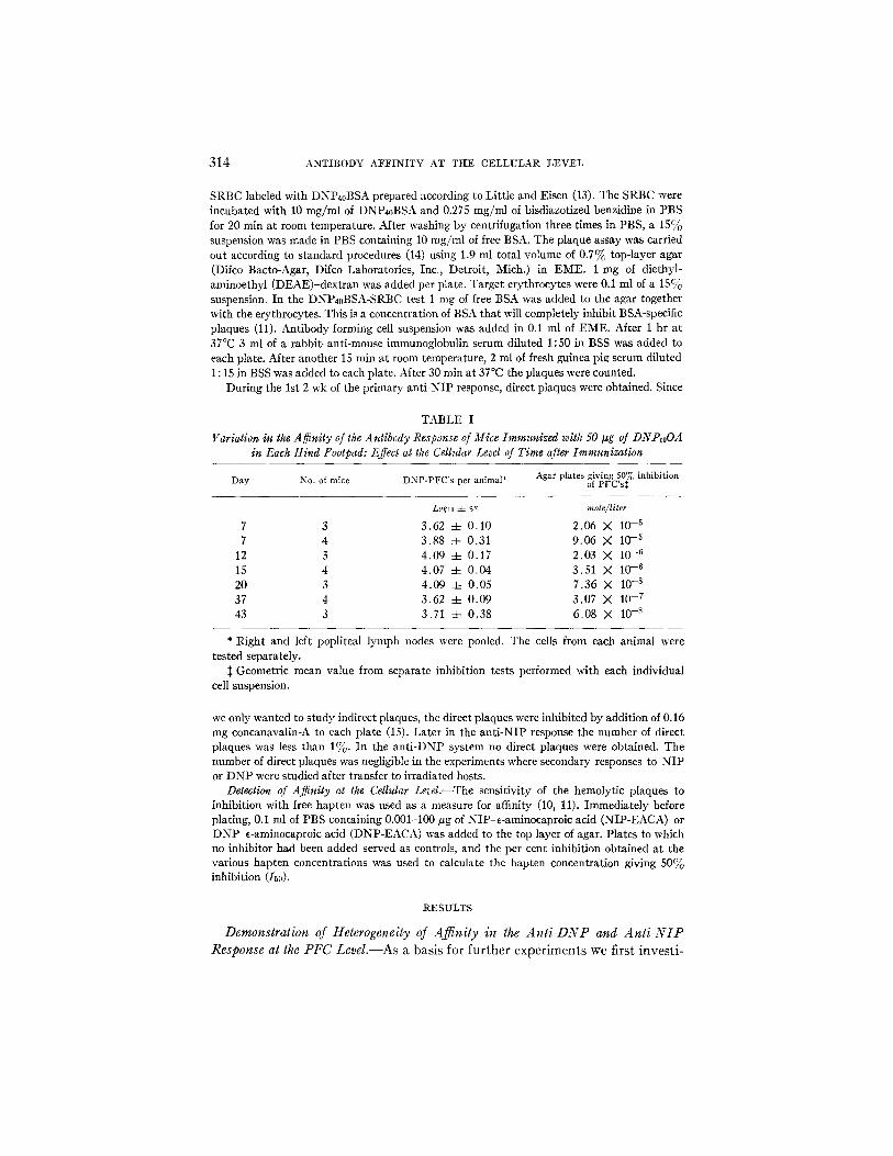
TABLE I
Variation in the Aginity of the Antibody Response of Mice Immunized with 50 Izg of DNPloOA in Each Hind Footpad: Effect at the Cellular Level of Time after Immunization
Agar plates giving 50% inhibition Day No. of mice DNP-PFC's per animal* of PFC's~;
Loglo -4- s~ molefliter
7 3 3.62 -4- 0.10 2,06 X 10 -5 7 4 3.88 -4- 0.31 9.06 X 10 -5
12 3 4.09 + 0.17 2.03 X 10 -6 15 4 4.07 4- 0.04 3.51 X 10 -6 20 3 4.09 4- 0.05 7.36 X 10 -s 37 4 3.62 4- 0.09 3.07 M 10 -7 43 3 3.71 4- 0.38 6.08 X 10 -s
* Right and left popliteal lymph nodes were pooled. The ceils from each animal were tested separately.
:~ Geometric mean value from separate inhibition tests performed with each individual cell suspension.
we only wanted to study indirect plaques, the direct plaques were inhibited by addition of 0.16 mg concanavalin-A to each plate (15). Later in the anti-NIP response the number of direct plaques was less than 1%. In the anti-DNP system no direct plaques were obtained. The number of direct plaques was negligible in the experiments where secondary responses to NIP or DNP were studied after transfer to irradiated hosts.
Detection of A~nity at the Cellular Level.--The sensitivity of the hemolytic plaques to inhibition with free hapten was used as a measure for affinity (10, 11). Immediately before plating, 0.I ml of PBS containing 0.001-100 pg of NIP-e-aminocaproic acid (NIP-EACA) or DNP-e-aminocaproic acid (DNP-EACA) was added to the top layer of agar. Plates to which no inhibitor had been added served as controls, and the per cent inhibition obtained at the various hapten concentrations was used to calculate the hapten concentration giving 50% inhibition (/50).
R E S U L T S
Demonstration of Heterogeneity of AJfinity in the An t i -DNP and A n t i - N I P Response at the PFC LeveL--As a basis for f u r t h e r e x p e r i m e n t s we first inves t i -
B I R G E R ANDERSSON 3 1 5
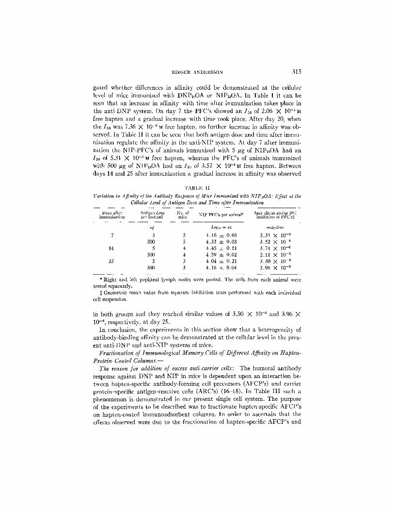
gated whether differences in affinity could be demonst ra ted a t the cellular level of mice immunized with DNP10OA or N1P10OA. In Table I i t can be seen tha t an increase in affinity with time after immunizat ion takes place in the an t i -DNP system. On day 7 the P F C ' s showed an I50 of 2.06 X 10⁻⁵~t free hapten and a gradual increase with t ime took place. After day 20, when the I50 was 7.36 X 10. 8 M free hapten, no further increase in affinity was ob- served. In Table I I i t can be seen tha t both antigen dose and t ime after immu- nization regulate the affinity in the an t i -NIP system. At day 7 after immuni- zat ion the N I P - P F C ' s of animals immunized with 5 /~g of NIP10OA had an 150 of 5.31 X 10.5~t free hapten, whereas the P F C ' s of animals immunized with 500 #g of NIP10OA had an I50 of 3.52 3( 10.aM free hapten. Between days 14 and 25 after immunizat ion a gradual increase in affinity was observed
TABLE II
Variation in AjTnily o[ the Antibody Response of Mice Immunized with NIPloOA: Effect at the Cellular Level of Antigen Dose and Time after Immunization
Days after Antigen dose No. of NIP PFC's per animal* Agar plates giving 50% immunization per footpad mice inhibition of PFC's:~
vg Logic :t= sE mole~liter
7 5 5 4.16 -4- 0.05 5.31 )< 10 -5 500 5 4.32 4- 0.03 3.52 X 10 -4
14 5 4 4.45 -4- 0.11 3.74 >( 10 -6 500 4 4.59 4- 0.02 2.11 X 10 -5
25 5 3 4.04 4- 0.21 3.50 X 10 -6 500 3 4.16 ::t= 0.04 3.96 X 10 -6
* Right and left popliteal lymph nodes were pooled. The cells from each animal were tested separately.
:~ Geometric mean value from separate inhibition tests performed with each individual cell suspension.
in both groups and they reached similar values of 3.50 X 10 .6 and 3.96 ;< 10 .6 , respectively, a t day 25.
I n conclusion, the experiments in this section show tha t a heterogeneity of ant ibody-binding affinity can be demonst ra ted at the cellular level in the pres- ent an t i -DNP and an t i -NIP systems of mice.
Fractionalion of Immunological Memory Cells of Different Affinity on Hapten- Protein-Coated C o l u m n s . -
The reason for addition of excess anti-carrier cells: The humoral an t ibody response against D N P and N I P in mice is dependent upon an interact ion be- tween hapten-specific ant ibody-forming cell precursors (AFCP's ) and carrier protein-specific ant lgen-react ive cells (ARC's) (16-18). In Table I I I such a phenomenon is demonst ra ted in our present single cell system. The purpose of the experiments to be described was to fract ionate hapten-specific A F C P ' s on hapten-coated immunoadsorbent columns. I n order to ascertain tha t the effects observed were due to the fract ionat ion of hapten-specific A F C P ' s and

316 A N T I B O D Y A F F I N I T Y AT T H E C E L L U L A R L E V E L
not, for example, due to variations in the number of ARC's, an excess number of anti-carrier cells were added to all cell suspensions before injection, together with antigen, into sublethally irradiated animals, i.e. the animals were in- jected with hapten coupled to the carrier, against which an excess of immune cells had been added.
A2~nity fraclionation of DNP-specific immunological memory cells on B S A - or DNP-BSA-coated columns: Spleen and lymph node cells from mice primed with I)NP-OA 3-4 months earlier were pooled and allowed to pass through columns coated with DNP-BSA and control columns coated with BSA. In Table IV is listed the affinity of antibody released by the cells after transfer to sublethally irradiated recipient mice together with 107 anti-OA cells and 10
TABLE III
ttapten-Specific Immunological Memory of Immune Cells Transferred to Sublethally Irradiated Syngeneic Mice: the Effect of Cells Immune to the Protein Carrier
on Which the Hapten is Presented
Hapten-speclfic cells* Carrier-specific cells* Immunogen:~ NIP-PFC§ DNP-PFC§
Loglo -4- S E Loglo 4- S R
Exp. 1 Anti NIP-OA + anti 106 - - 10 tzg NIP-BSA 1.44 ~: 0.14 1.63 :t: 0.16
DNP-OA -7 10 #g DNP-BSA A n t i N I P O - A + anti l0 s A n t i B S A 2 X 10⁷ 10/~gNIP-BSA 2.73 =l= 0.13 2,09 ~ 0.20
DNP-OA + 10 #g DNP-BSA Exp. 2 Anti NIP-OA 106 -- 10/zg NIP-BSA 1.53 :t: 0.23 1.56 =h 0.14
+ anti DNP-OA + 10 #g DNP-BSA Anti NIP OA 106 Anti BSA 2 X 10⁷ 10 #g NIP-BSA 2.66 =1= 0.I0 2.97 -- o . n
q- anti DNP-OA -7 i0 #g DNP-BSA
* Lymph node and spleen cells from mice immunized with 100 IZg of antigen in Freund's complete adjuvant 2-3 months earlier, The animals were immunized with one antigen only and mixing was performed in vitro.
~c Antigen given in soluble form mixed with the injected cells. § PFC's per spleen of the recipients tested 7 days after 600 R and infusion of cells and ant[gem Each value
is the mean from six animals.
#g of DNP-OA. Unpassed cells and cells passed through the BSA-coated columns released antibody with similar affinity, I50 being 6.3 X 10 -7 and 2.2 X 10 7 and 2.2 X 10 .7 ~ free hapten, respectively. The cells passed through the DNP-BSA-coated column showed a reduced affinity, with an 150 of 2.4 X 10⁻⁶~ free hapten. Furthermore, the number of PFC's was also reduced in the DNP-BSA-passed cells, indicating that a hapten-specific elimination of immunological memory cells had taken place, preferentially affecting the pre- cursors of high affinity PFC's.
Ajfinity fractionation of DNP-specific and NIP-specific immunological memory cells on DNP-BSA-coaled columns: The specificity of the elimination on DNP-BSA-coated columns was studied by allowing passage of a mixture of DNP-OA-primed cells and NIP-OA-primed cells. The affinity of antibodies produced after transfer to subletha]ly irradiated recipients was thereafter
B I R G E R A N D E R S S O N 3 1 7
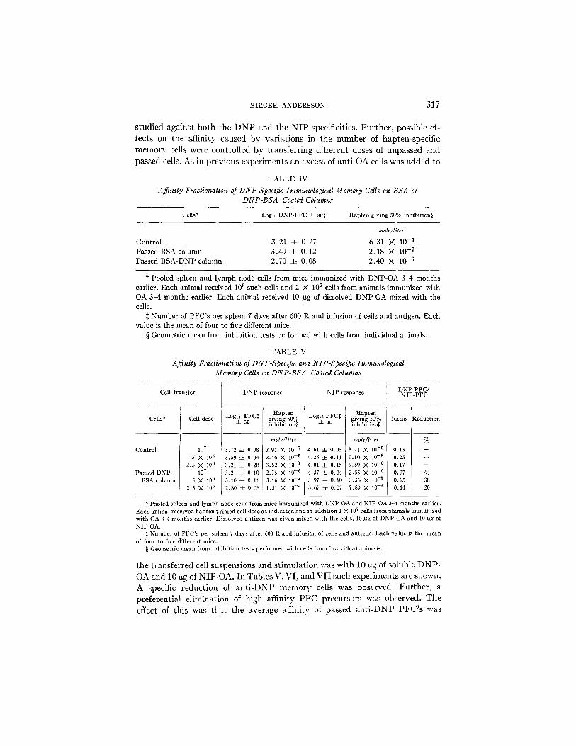
studied against both the D N P and the N I P specificities. Further, possible ef- fects on the affinity caused by variations in the number of hapten-specific memory cells were controlled by transferring different doses of unpassed and
passed cells. As in previous experiments an excess of anti-OA cells was added to
TABLE IV
Affinity Fractionation of DNP-Specific Immunological Memory Cells on BSA or DNP-BSA-Coated Columns
Cells* Logt0 DNP-PFC 4- sr$ Hapten giving 50% inhibition§
mole/liter
Control 3.21 4- 0.27 6.31 X 10 -7 Passed BSA column 3.49 -4- 0.12 2.18 X 10 -7 Passed BSA-DNP column 2.70 4- 0.08 2.40 X 10 -6
* Pooled spleen and lymph node cells from mice immunized with DNP-OA 3-4 months earlier. Each animal received 106 such cells and 2 X 10⁷ cells from animals immunized with OA 3-4 months earlier. Each animal received 10 #g of dissolved DNP-OA mixed with the cells.
:~ Number of PFC's per spleen 7 days after 600 R and infusion of cells and antigen. Each value is the mean of four to five different mice.
§ Geometric mean from inhibition tests performed with cells from individual animals.
TABLE V
Affinity Fractionation of DNP-Specific and NIP-Specific Immunological Memory Cells on DNP-BSA-Coated Columns
Cell transfer
Cells*
Control
Passed DNP- BSA column
Cell dose
107 5 X 10⁶
2.5 X 10⁶ 107
5 X 10⁶ 2.5 X 10⁶
DNP response
Hapten Logi0 PFC~ giving 50%
4- sE inhibition§
mole~liter
3.72 4- 0.08 2.91 N 10 -7 3.59 4- 0.04 2.46 X l0 -6 3.21 -4- 0.28 3.52 X 10 --8 3.21 4- 0.10 2.75 X 10 -6 3.10 4- 0.11 3.16 X 10 -~ 2.80 4- 0.08 1.51 X 10 -4
NIP response
Hapten Logl~ PFC:~ giving 50%
4- sz inhibition§
mole/l*ter
4,61 4- 0.05 8.71 X 10 -6 4.25 :t: 0.11 9.80 X 10 - s 4.01 -4- 0.15 9.59 X 10 - s 4.37 4- 0,04 2.55 X 10 -6 3.97 4- 0.10 3.56 X 10 -5 3,67 4- 0.07 7.89 X 10 -6
DNP-PFC/ NIP-PFC
Ratio Reduction
% 0.13 0.23 0.17 0.07 44 0.15 38 0.14 20
* Pooled spleen and lymph node cells from mice immunized with DNP-OA and NIP-OA 3-4 months earlier. Each animal received hapten primed cell dose as indicated and in addition 2 X 10⁷ cells from animals immunized with OA 3-4 months earlier. Dissolved antigen was given mixed with the cells, 10/zg of DNP-OA and 10 #g of NIP-OA.
:~ Number of PFC's per spleen 7 days after 600 R and infusion of cells and antigen. Each value is the mean of four to five different mice.
§ Geometric mean from inhibition tests performed with cells from individual animals.
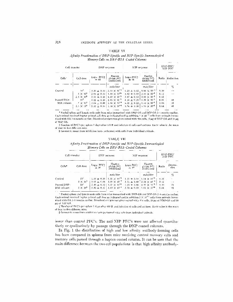
the transferred cell suspensions and stimulation was with 10 ~g of soluble DNP- OA and 10 vg of NIP-OA. In Tables V, VI, and VII such experiments are shown. A specific reduction of an t i -DNP memory cells was observed. Further, a preferential elimination of high affinity PFC precursors was observed. The effect of this was that the average affinity of passed an t i -DNP PFC's was
318 A N T I B O D Y A F F I N I T Y A T T H E C E L L U L A R L E V E L
TABLE VI Affinity Fractionation of DNP-Speclfic and NIP-Specific Immunological
Memory Cells on DNP-BSA-Coated Columns
Cell transfer
Cells*
Control
Passed DNP- BSA column
Cell dose
107
5 X 10 fi 2.5 X 10⁶
107
X 1116 i 2 . 8 X 10⁶
DNP response
LogLo PFC* 4- SE
3.51/ 4- 0.14 2.94 4- 0.13 2.55 4- 0.10 3.11 4- 0.10 3.04 4- 0.09 2.32 4- 0.14
N I P response
Hapten giving .50% Log~0 PFC:~ inhibition§ 4- s~;
mole~liter
3.33 X 10 - 7 4.23 4- 0.12 1.1g X 10 -5 3.82 4- 0.10 6.19 X I0 -5 3.47 =I= 0.10 2.83 X 10 -5 4.34 4- 0.10 5.16 X I0 -4 4.31 ::t= 0.12 i 1.36 M 10 -4 3.74 4- 0.08 I,
Hapten giving 50% inhibition§
mole~liter
8.98 X 10 -6 2.43 X 10 -5 3.03 X 10 -5 1.29 ),( 10 -5
1.43 X 10 -5 3.50 X 1o 5
] D N P - P F C / N I P - P F C
l- Ratio Reduction
0.19 - - 0.13 0.12 {I.06 68
0.06 ,59 0.04 68
* Pooled spleen and lymph node cells from mice immunized with DNP-OA and NIP-OA 3 4 months earlier. Each animal received hapten primed cell dose as indicated and in addit ion 2 X 10 -7 cells from animals immu- nized with OA 3 ~ months earlier. Dissolved antigen was given mixed with the cells, 10 p,g of DNP-OA and 10 #g of NIP-OA.
:~ Number of PFC's per spleen 7 days after 600 R and infusion of cells and antigen. Each value is the mean of four to five different mice.
§ Geometric mean from inhibition tests performed with cells from indivMual animals.
TABLE VII Affnity Fractionatlon of DNP-Specific and N1P-Spec{[ic Immunological
Memory Cells on DNP-BSA Coated Columns
D N P - P F C / Cell t ransfer DNP response NIP response N I P - P F C
Cells* Cell dose
Control
Passed DNP- BAS column
107
3 X 706 107
3 X 10⁶
Hapten Loglo PFC~ giving 50%
4- sE inlfibition§
mole~liter 4.10 4- 0.10 1.68 X 10 ~8 3.05 4- 0.08 3.01 M 10 ~7 3.46 4- 0.12 3.37 X 10 -5 2.36 4- 0.17 3.83 X 10 "6
Logto PFCt i ~ sF I
4.55 -t_ 0.10 3.34 4- 0,09 4,45 -c- 0.04 3.78 4- (I.70
I
Hapten Ratio giving 50% Depres- inhibition§ slon
mole/liler %
1.14 X 10 -~ O.35 -~- 2.36 X 10 - s 0.52 - - 4.20 X 70 -5 0.10 71 5.03 X 10 -5 0.04 92
* Pooled spleen and lymph node cells from mice immunized with DNP-OA and NIP-OA 3 -4 months earlier. Fach animal received hapten primed cell close as indicated and in addition 2 X 1(I -7 cells from animals immu- nized with OA 3-4 months earlier. Dissolved antigen was given mixed with the cells, 10Ng of DNP-OA and 70 #g of NIP OA.
~: Number of PFC's per spleen 7 days after 600 R and infusion of cells and antigen. Each value is the mean of four to five different mice.
§ Geometric mean from inhibition tests performed with cells from individual animals.
lower than control PFC's. The anti-NIP PFC's were not affected quantita- tively or qualitatively by passage through the DNP-coated columns.
In Fig. 1 the distribution of high and low affinity antibody-forming cells has been compared in spleens from mice receiving control memory cells and memory cells passed through a hapten-coated column. I t can be seen that the main difference between the two cell populations is that high affinity antibody-
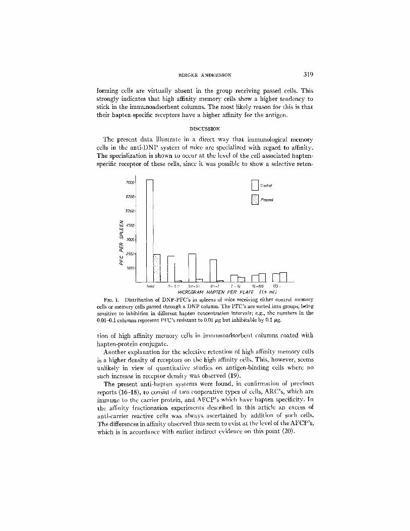
BIRGER ANDERSSON 319
forming cells are virtually absent in the group receiving passed cells. This strongly indicates that high affinity memory cells show a higher tendency to stick in the immunoadsorbent columns. The most likely reason for this is that their hapten-specific receptors have a higher affinity for the antigen.
DISCUSSION
The present data illustrate in a direct way that immunological memory cells in the an t i -DNP system of mice are specialized with regard to affinity. The specialization is shown to occur at the level of the cell-associated hapten- specific receptor of these cells, since it was possible to show a selective reten-
7000-
6000-
5000.
laj 4000- Lu
3000-
2000-
1000
E Confrol
D Pctssed
Total 0- 0~ Oot-Ot Ol -I I - I0 I0-I00 I00 -
MICROGRAM HAPTEN PER PLATE (1.9 ml) Fro. 1. Distribution of DNP-PFC's in spleens of mice receiving either control memory
cells or memory cells passed through a DNP column. The PFC's are sorted into groups, being sensitive to inhibition in different hapten concentration intervals; e.g., the numbers in the 0.01-0.1 columns represent PFC's resistant to 0.01 #g but inhibitable by 0.1 #g.
tion of high affinity memory cells in imnmnoadsorbent columns coated with hapten-protein conjugate.
Another explanation for the selective retention of high affinity memory cells is a higher density of receptors on the high affinity cells. This, however, seems unlikely in view of quantitative studies on antigen-binding cells where no such increase in receptor density was observed (19).
The present anti-hapten systems were found, in confirmation of previous reports (16-18), to consist of two cooperative types of cells, ARC's, which are immune to the carrier protein, and AFCP ' s which have hapten specificity. I n the affinity fractionation experiments described in this article an excess of anti-carrier reactive cells was always ascertained by addition of such cells. The differences in affinity observed thus seem to exist at the level of the AFCP's , which is in accordance with earlier indirect evidence on this point (20).
320 ANTIBODY AFFINITY AT THE CELLULAR LEVEL
Experiments are also in progress to determine whether nonimmune AFCP's as well can be functionally fractionated on immunoadsorbent columns. There is reason to believe that the performance of such a fractionation will be pos- sible, since it has recently been shown that antigen-binding lymphocytes of nonimmune, as well as immune, guinea pigs are heterogenous with regard to binding affinity of their membrane receptors (21, 22).
In summary the direct evidence for similarities between membrane-located receptor antibody of lymphocytes (AFCP's) and the humoral antibody released by the cell are the following. (a) The receptors have been shown to have the same specificity as the hnmoral antibody released by the cells (8). (b) The receptors are inmmnoglobulins and express the same class specialization as the immunoglobulins released by the cells (23). (c) The receptor discriminates a haptenic site on a hapten-protein conjugate to the same extent as does the humoral antibody released by the cell (9). (d) The receptor expresses the affinity for the antigen, and immunological memory cells express similar affinity of their membrane receptor and their humoral antibody released after stimu- lation (as shown in this article).
SUMMARY
Heterogeneity with regard to affinity of anti-hapten antibody was demon- strated at the cellular level in mice. The heterogeneity was shown at the level of single antibody-forming cells using hapten inhibition of hemolvtic antibody plaque formation as a measure of affinity. The affinity increased with time after immunization. A high antigen dose initially resulted in relatively low affinity antibody production as compared to the affinity of the antibody pro- duction in animals immunized with a low dose.
Affinity specialization of immunological memory cells was demonstrated, since it was possible to specifically fractionate such cells with regard to affinity on hapten-protein-coated plastic bead colmnns. High affinity memory cells showed a higher tendency to become retained in the columns than did low affinity memory cells.
The data in a direct way demonstrate that memory cells carrying membrane- associated receptors of a certain affinity for the antigen are determined to release antibody of a similar affinity after stimulation with antigen.
The author is grateful to Mrs. M. L. Larsson and K. Sk~nberg for their excellent technical assistance.
REFERENCES
1. Ehrlich, P. 1900. On immunity with special reference to cell life. Proc. Roy. Soc. Ser. B Biol. Sci. 66:424.
2. Burnet, F. M. 1959. The Clonal Selection Theory of Acquired Immunity. Cam- hridge University Press, New York.
3. Ei~en, H. N., and G. W. Siskind. 1964. Variations in affinities of antibodies during the immune response. Biockemistry. 3:996.
BIRGER ANDERSSON 321
4. Siskind, G. W., P. Dunn, and J. G. Walker. 1968. Studies on the control of anti- body synthesis. II . Effect of antigen dose and of suppression by passive anti- body on the affinity of antibody synthesized. J. Exp. Med. 127:55.
5. Theis, G. A., and G. W. Siskind. 1968. Selection of cell populations in induction of tolerance: affinity of antibody produced in partially tolerant rabbits. J. Im- munol. 100:138.
6. Paul, W. E., G. W. Siskind, and B. Benacerraf. 1968. Specificity of cellular immune responses. Antigen concentration dependence of stimulation of DNA synthesis in vitro by specifically sensitized cells, an expression of the binding characteris- tics of cellular antibody. J. Exp. Med. 127:25.
7. Bulloch, W. W., and M. B. Rittenberg. 1970. In vitro-initiated secondary anti- hapten response. II . Increasing cell avidity for antigen. J. Exp. Med. 132:926.
8. Wigzell, H., and B. Andersson. 1969. Cell separation on antigen-coated columns. Elimination of high rate antibody-forming cells and immunological memory cells. J. Exp. Med. 129:23.
9. Wigzell, H., and O. M~kel/i. 1970. Separation of normal and immune lymphoid cells by antigen-coated columns. Antigen-binding characteristics of membrane antibodies as analyzed by hapten-protein antigens. J. Exp. Med. 132:110.
10. Hraba, T., F. Havas, and A. R. Pickard. 1970. Partial tolerance to the dinitro- phenyl group in neonatal and adult mice. Int. Arch. Allergy Appl. Immunol. 38:635.
11. Andersson, B. 1970. Studies on the regulation of avidity at the level of the single antibody-forming cell. The effect of antigen dose and time after immunization. J. Exp. Med. 139.:77.
12. Brownstone, A., N. A. Mitchison, and R. Pitt-Rivers. 1966. Chemical and sero- logical studies with an iodine-containing synthetic immunological determinant 4-hydroxy-3-iodo-5-nitrophenylacetic acid (NIP) and related compounds. Immunology. 10:465.
13. Little, J. R., and H. N. Eisen. 1967. In Methods in Immunology and Immuno- chemistry. C. A. Williams and M. W. Chase, editors. Academic Press, Inc., New York. 128.
14. Jerne, N. K., and A. A. Nordin. 1963. Plaque formation in agar by single antibody producing cells. Science (Washington). 140:505.
15. Nordin, A. A., H. Cosenza, and W. Hopkins. 1969. The use of concanavalin-A for distinguishing IgM from IgG antibody-producing cells. J. Immunol. 103:859.
16. Ovary, Z., and B. Benacerraf. 1963. Immunological specificity of the secondary response to dinitrophenylated proteins. Proc. Soc. Exp. Biol. Mecl. 114:72.
17. Rajewsky, K., V. Schirrmacher, S. Nase, and N. K. Jerne. 1969. The requirement of more than one antigenic determinant for immunogenicity. J. Exp. Med. 129:1131.
18. Mitchison, N. A., K. Rajewsky, and R. B. Taylor. 1970. Cooperation of antigenic determinants and of cells in the induction of antibodies. In Developmental Aspects of Antibody Formation and Structure. J. Sterzl, editor. Academic Press, Inc., New York. 547.
19. Ada, G. L., P. Byrt, T. Mandel, and N. Warner. 1970. A specific reaction between antigen labelled with radioactive iodine and lymphocyte-like cells from normal, tolerant and immunized mice or rats. In Developmental Aspects on Antibody Formation and Structure. J. Sterzl, editor. Academic Press, Inc., New York. 503.
322 ANTIBODY AFFINITY AT TIlE CELLULAR LEVEL
20. Katz, I). H., W. E. Paul, E. A. GoidI, and B. Benacerraf. 1970. Enhancement of primary and secondary anti-hapten antibody responses by carrier preim- munization. J. Exp. Med. 132:261.
21. Davie, J. M., and W. F.. Paul. t971. Receptors on immunocompetent cells. II . Specificity and nature of receptors on dinitrophenylated guinea pig albumin 12~'l-binding lymphocytes of normal guinea pigs. J. Exp. Med. 134:495.
22. Davie, J. M., A. S. Rosenthal, and W. E. Paul. 1971. Receptors on immunocom- petent cells, l l i . Specificity and nature of receptors on dinitrophenylated guinea pig albumin 12aI-binding cells of immunized guinea pigs. J. Exp. Med. 134:517.
23. Wakers, S. C., and H. Wigzell. 1970. 1)emonstration of heavy and light chain antigenic determinants on the cell-bound receptor for antigen. Similarities be- tween membrane attached and humoral antibodies produced by the same cell. J . Exp. Med. 132:1233.
Related Documents


