J~~~A THE BRITISH IJOURNAL OF OPHTHALMOLOGY JUNE, 1948 COMMUNICATIONS OX VITREOUS HUMOUR. 1.-THE RESIDUAL PROTEIN* BY A. PIRIE, G. SCHMIDT and J. W. WATERS NUFFIELD LABORATORY.OF OPHTHALMOLOGY AND DEPARTMENT OF CRYSTALLOGRAPHY, OXFORD WHEN a vitreous humour is removed intact from an eye and put on a filter or hung from a clamp, a viscous fluid drips out of it until finally all that remains is a thin- membranous wisp of material. Robertson and Duke-Elder (1933) describe the vitreous humour as " a gel composed of a meshwork of elastic fibrillae suspended in a viscous fluid," and we may suppose that the membranous stuff left after filtration forms the elastic fibrils of the intact vitreous body. This - insoluble residue was first studied by Young (1894) and Mdrner (1894), *who considered that it was collagen because it dissolved in boiling water and the solution set to a gel on cooling. MTd6rner (l.c.) termed it the residual protein, and it has since been * Received for publication, March 24, 1948.

Welcome message from author
This document is posted to help you gain knowledge. Please leave a comment to let me know what you think about it! Share it to your friends and learn new things together.
Transcript

J~~~A
THE BRITISH IJOURNALOF
OPHTHALMOLOGYJUNE, 1948
COMMUNICATIONS
OX VITREOUS HUMOUR. 1.-THE RESIDUALPROTEIN*
BY
A. PIRIE, G. SCHMIDT and J. W. WATERSNUFFIELD LABORATORY.OF OPHTHALMOLOGY AND DEPARTMENT OF
CRYSTALLOGRAPHY, OXFORD
WHEN a vitreous humour is removed intact from an eye and puton a filter or hung from a clamp, a viscous fluid drips out of ituntil finally all that remains is a thin- membranous wisp of material.Robertson and Duke-Elder (1933) describe the vitreous humour as" a gel composed of a meshwork of elastic fibrillae suspended in aviscous fluid," and we may suppose that the membranous stuff leftafter filtration forms the elastic fibrils of the intact vitreous body.This - insoluble residue was first studied by Young (1894) andMdrner (1894), *who considered that it was collagen because itdissolved in boiling water and the solution set to a gel on cooling.MTd6rner (l.c.) termed it the residual protein, and it has since been
* Received for publication, March 24, 1948.

A. PIRIE, G. SCHMIDT antd J. W. WATERS
known by this name. Duke-Elder (1930) described it as like gelatin,but Meyer (1945) classified it as an insoluble mucoid of unknowncomposition. The analyses of the insoluble residue show that it isa protein. Krause (1934) found 1416 per cent. N and gave ana-lytical figures for -arginine, histidine, lysine, cystine, tryosine andtryptophane, all of which were present in the material analysed.The experiments described in this paper bring forward further
evidence in support of the view that the residual protein of vitreoushumour contains a protein like collagen and describe the liquefyingeffect of preparations containing the enzyme collagenase on thevitreous body. These results are considered in relation to the wellknown acid and alkaline shrinkage of the vitreous humour, andare'used to formulate a theory of the way-in which the residualprotein is laid down.
It is difficult to collect sufficient of the insoluble residue from thevitreous humour to identify it by analysis of the constituent amino-acids. One ox vitreous humour with a volume between 12-15 ml.yields only 2-3 mg. of insoluble residue. We have, therefore,attempted to identify this residue through its general properties, byamino-acid chromatography of hydrolysed material, by X-rayanalysis and by enzyme analysis. The material appears to be fairlyuniform, judged by solubility tests and enzyme digestibility, butwe realise that the results we have obtained do not in any wayprove that the insoluble residue of the vitreous humour consists ofa single substance.
METHODS
Material. The experiments have been done using ox vitreoushumours obtained within 1-2 hours after death of the animal.The humours were removed from the e$e by making a completeequatorial cut through sclera, choroid and retina and allowing thevitreous humour to fall away from the retina, while still attachedto the anterior half of the eye. It was then gently cut and pressedapart from the ciliary body and lens. The posterior lens capsulesometimes adheres to the vitreous humour, but can be removedwith forceps.
Prebaration of residual Protein fromn vitreous huntour. Theinsoluble residue may be separated from the soluble constituents bysuspending the humour in saline and allowing the soluble sub-stances to diffuse out into the surrounding fluid. Friedenwaldand Stiehler (1935) found that the vitreous humour retained itsform during prolonged washing in saline and that when it wasfinally filtered, the filtrate contained no organic material. Wefound that if an ox vitreous humour were left in the ice chest in saline
322

OX VITREOUS HUMOUR
which was changed every few days, all detectable protein andhyaluronic acid had diffused out of the humour within 8-12 weeks.Such washed humours are more flaccid than when fresh and wehave taken the insoluble residue remaining after their filtration asthe residual protein. Salt may be removed from this by a fewdays' wash in distilled water.The insoluble residue may also be prepared by filtration of the
fresh vitreous humour followed by washing. This might seem thequicker method, but we found that very prolonged washing, bvsuspension in saline or water, was necessary to remove the smallamount of soluble material remaining after the first filtration ofthe fresh humour.
Total nitrogen. Total nitrogen was estimated in 5-10 mg. samples frozendried over P9,;0. The samples were incinerated with 2 ml. H,SO, + 0.5 g. catalyst(40 g. K2SO4, 10 g. CUSO4, 0-17 g. SeO2) incineration being continued for 5-8hours after clearing. The ammonia was estimated by Markham's (1942) method.
Total carbohydrate. This was estimated by the orcin colorimetric method,described by N. W. Pirie (1)936).Hexosamine. This was estimated by the method described by Elson and Morgan
(1933).Amino-acid chromatography. The method of Consden, Gordon and Martin
(1944) was used.
GENERAL PROPERTIES OF INSOLUBLE RESIDUE
The residues from vitreous humour are grey and fibrous, show-ing considerable tensile strength and some anisotropy, particularlywhen stretched. When dried, each residue is a dark grey hornystring. The colour seems to be due to adsorbed uveal pigment,cotton fibres and dust from the air and from the washing solutions.We found that the total nitrogen'of such residues was near 12 p'ercent. and that they contained no hexosamine and from 3-7 percent. total carbohydrate. Further purification, of the residualprotein while it was in this insoluble state was difficult owing to itscapacity to adsorb other insoluble materials, such as cotton fibres,etc. From the general properties one may say that the vitreousresidue appears to be made up largely of fibrous protein, but wefelt that for further purification it was essential to get the protein,or proteins, into solution, even if this involved some degree ofhydrolysis. We have, therefore, made a soluble preparation fromthe vitreous humour residues.
Preparation and properties of soluble protein from vitreous humour residues.Ox vitreous humours were washed as free as possible of soluble material bysuspension in saline for long periods followed by filtration. The residues fromabout 50 humours were dissolved in 60 ml. HCI pH 1-8 on a boiling water bath.The cloudy solution was centrifuged and the small black sediment discarded.The supematant was brought to pH 4 and the srriall flocculent precipitate that
323L
A. PIRIE, G. SCHMIDT and J. W. WATERS
came out was centrifuged off and has not been further investigated. It seemsprobable that this is an " artificial mucin," similar to those prepared by Meyer andSmyth (1937) and formed in this case from hyaluronic acid and any of the proteinspresent. The supernatant fluid from this precipitate was half saturated withammonium sulphate by addition of an equal, volume of saturated solution. Animmediate copious precipitate came down. This was centrifuged out, re-dissolvedin water and dialysed against distilled water in the ice chest. No further precipitatecame out when the supematant fluid from half saturation was wholly saturatedwith ammonium sulphate.
After several days' dialysis in the ice chest the solution of the precipitate got by50 per cent. saturation with ammonium sulphate was frozen and dried. Thepreparation was a white fibrous material and had a total N 13 0-13-5 per cent.Further purification by dissolving in warm water, bringing the solution to pH 4,centrifuging and precipitation from the supernatant fluid by half saturation withammonium sulphate gave a product that formed a firm jelly in the dialysis sac andhad no ash, 14-8 per cent. N, no hexosamine and 6 per cent. total carbohydrate.We found that the vitreous humour residual protein lost 3-3 per cent. of its totalnitrogen when taken into solution in dilute HCl. This loss probably representsloss of the amide group of glutamine, as Thierfelder and v. Cramm (1919) haveshown that glutamine peptides are unstable under such conditions. If we correctfor the loss of N on solution in acid the total nitrogen of the soluble product fromox vitreous humour residual protein is then 15-3 per cent.
The characteristic insoluble proteins of the animal body arecollagen, elastih and keratin, with reticulin as a rather nebuloussubstance of unknown composition. The method of preparationand the general properties of the soluble stuff from ox vitreoushumour residue suggests that the original material is like collagenand that it-dissolves to form gelatin. Solubility in dilute acid is aproperty of collagen and precipitability by half saturation withammonium sulphate and ability'to form a gel are properties ofgelatin. Yet the final value of 15 3 per cent. N is low for typical-collagen or for gelatin. Bergmann and Stein (1939) found thatcollagen from ox Achilles tendon had 18&6 per cent. N. Fewcollagens have been analysed, other than those from skin andtendon; Morner (1894) gave N '17 03 per cent. for ox corneacollagen and we have found that-collagen prepared from ox corneaby the method of Bergmann and Stein (l.c.) had N 161 per cent.uncorrected for ash.Both histologists and chemists accept cornea collagen as a
typical collagen, so we felt that it would be valuable to compare itin some detail with the vitreous humour material. We have, there-fore, prepared a soluble gelatin from ox cornea collagen and havecompared its general properties with those of the soluble materialfrom vitreous humour. We have also compared the amino-acidchromatograms of ox cornea gelatin and vitreous humour residueswith that of commercial gelatin.We found that we could prepare a soluble protein from ox
cornea collagen by the same method as we had used with vitreoushumour residue. Th'is had no ash or hexosamine, total N 155per cent. and 6 per cent. total carbohydrate, and in appearance and
.324
OX VITREOUS HUMOUR
properties was similar to the preparation from vitreous humour.If we correct the total N for the nitrogen lost on solution in diluteacid, the final figure is 16 0 per cent. total N.
AMINO-ACID CHROMATOGRAM OF OX VITREOUS HUMOUR RESIDUEAND OF CORNEA GELATIN COMPARED WITH COMMERCIAL GELATIN
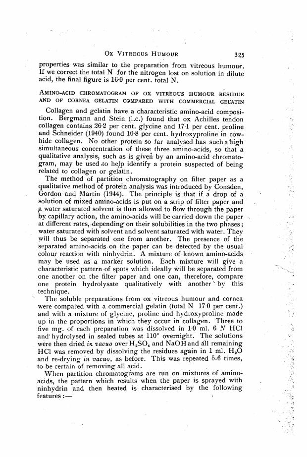
Collagen and gelatin have a characteristic amino-acid composi-tion. Bergmann and Stein (l.c.) found that ox Achilles tendoncollagen contains 262 per cent. glycine and 171 per cent. prolineand Schneider (1940) found 10 8 per cent. hydroxyproline in cow-hide collagen. No other protein so far analysed has such a highsimultaneous concentration of these three amino-acids, so that aqualitative analysis, such as is giveA by an amino-acid chromato-gram, may be'used .to help identify a protein suspected of beingrelated to collagen or gelatin.The method of partition chromatography on filter paper as a
qualitative method of protein analysis was introduced by Consden,Gordon .and Mlartin (1944). The principle is that if a drop of asolution of mixed amino-acids is put on a strip of filter paper and,a water saturated solvent is then allowed to flow through the paperby capillary action, the amino-acids will be carried down the paperat different rates, -depending on their solubilities in the two phases;water saturated with solvent and solvent saturated with water. Theywill thus be separated one from another. The presence of theseparated amino-acids on the paper can be detected by the usualcolour reaction with ninhydrin. A mixture of known amino-acidsmay be used as a marker solution. Each mixture will give acharacteristic pattern of spots which ideally will be separated fromone another on the filter paper and one can, therefore, compareone protein hydrolysate qualitatively with another' by thistechnique.The soluble preparations from ox vitreous humour and cornea
were compared with a commercial gelatin (total N 17 0 per cent.)and with a mixture of glycine, proline and hydroxyproline madeup in the proportions in which they occur in collagen. Three tofive mg. of each preparation was dissolved in 10 ml. 6 N HCIand' hydrolysed in sealed tubes at 1100 overnight. The solutionswere then dried in vacuo over H2SO4 and NaOH and ill remainingHCl was removed by dissolving the residues again in 1 ml. H2Oand re-drying in vacuo, as before. This was repeated 5-6 times,to be certain of removing all acid.When partition chromatograms are run on mixtures of amino-
acids, the pattern which results when the paper is sprayed withninhydrin and then heated is characterised by the followingfeatures:
325

326 A. PIRIE, G. SCHMIDT anrnd J. W. WATERS
1. The presence of spots at different positions along the lengthof the run. The exact position of each spot depends upon thenature of the amino-acid component (Consden, Gordon andMartin (l.c.)), and to a certain extent also upon the amount ofamino-acid contained in it (Fisher, Parsons and iMlorrison, 1948).
2. Each spot is coloured. Most amino-acids give spots withninhydrin which are coloured rose or pink; some, e.g., alanine,are rather more blue, while proline and hydroxyproline areinstantly recognised by a characteristic yellow colour.
3. The size of each coloured spot varies.' It has been shownthat there exists a relatively simple relationship between the area ofa spot and the amount of amino-acid in it (Fisher, Parsons andMorrison (l.c.)).One dimensional partition chromatograms were run on solutions
of the hydrolysates prepared as described above from ox vitreoushumour and cornea, from commercial gelatin and on a marketsolution of a mixture of glycine, proline and hydroxyproline madeup in the proportions in which these amino-acids occur in collagen.The dilutions were so made that the volume of each hydrolysateput on the paper was 3 ul and contained 30 ,g total N. Chromato-grams of the three hydrolysates and of the mixture' of amino-acidswere run on the same sheet so that the' pattern would not be affectedby such adventitious sources of variation as those due to tempera-ture changes and'length of run. The chromatograms were run intwo different types of solvent, so that two different types of patterncould be obtained, since the positions of the amino-acids in adeveloped chromatogram relative to each other and to the solventfront depend upon the solvent used. We used either phenol with07 per cent. NH3 or a pyridine-amyl alcohol mixture (Edman,1945).When the chromatograms were develope'd very striking similari-
ties in the pattern exhibited by the three hydrolysates were at onceevident (Fig. 1). The components in all three mixtures were com-parable not only in position but in shades of colour and in spotarea. This was true for chromatograms developed in both types ofsolvent. No attempt was made to identify all the amino-acidcomponents of the hydrolysates, e.g., by running two dimensi'onalruns or by running hydrolysates in parallel with various syntheticmixtures; it was, however, evident that all three hydrolysatescontained roughly comparable amounts of proline, hydroxyproline,glycine, glutamic and aspartic acids, and alanine.Using the one dimensional technique and dealing with complex
mixtures of components it is not possible' to say whether or notsmall amounts of other amino-acid residues are present in one of
Ox VITREOUS HUMOUR
0
ASPARTAT'I(iOnk spct)
G I. t F AM AT' E.
(phink spot
G L YC I N E->-
(p)ink spsot)
AILANI NE
(pink, spot)
;YIeROLINF-St~yellow spot)
C-r,-1
<: 1
.W _
:4_ X_ A _f~~
H
O
'4-
7-U4~~~~~~~~~~~~~~~~.L4~~~~~~~~~~~~~~~~~~~~~~~~~~~~~~~~~~~~~~~~~~~~~~~~~~~~~~~~.
a:;X**;s~~~~
Pno.I)NN11-lt'(yellow Spot)
FIG. 1.
Partition chromatograms of hydrolysates of commercial gelatin, cornea
gelatin, vitreous humour residual protein and a mixture of glycine,proline and hydroxy-proline. Whatman filter paper No. 1. Solvent
saturated solution of phenol in water plus 017 per cent. NH4OH.
the hydrolysates, but not in the others. While, therefore, there iino justification on the evidence of partition chromatography alonfor identifying the nature and relative amounts of all the residue:in the hydrolysates from the three sources, the evidence is sufficiento suggest that the vitreous humour protein might be a member Q
the collagen gelatin-group.
27 .:
)
I' ';,
., fi
:--
,
.;',,
,, -.Pis
w1.-. J a8
, F..., ..y;s
.
> ..,
Di., .f .,. . .. .
o..;
.,.. ....
*. w
.; o.
S9 -
S *tZ,0.
'i't . s_, ass-.;
a, '.S .' :,,4,<
e <s:s ,8; ,1
tt .- -i,t b
Xir, <-* } ,- >liv lERO
0-, I
I
1, I- .-I ;,1.
3'/

328 A. PIRIE, G. SCHMIDT antd J. W. WATERS
X-RAY ANALYSIS OF RESIDUAL PROTEIN
We have taken X-ray photographs of vitreous humour residues,both in the unstretched and the stretched state. Material wasprepared for X-ray analysis by combined washing and filtration.Vitreous humours were suspend'ed in- saline and left in the icechest for several days with frequent changes of saline. Thispreliminary washing removed most of the soluble protein and somehyaluronic acid. The humours were next suspended and washedin successive changes of distilled water in order to remove most ofthe salt, which would interfere with the X-ray photograph of theprotein. The humours were then filtered br suspension on a glassfilter and, after a further wash, the residue on the filter was dried.This residue, collected from several vitreous humours, was photo-graphed without being stretched. In a further experiment and inorder to get some orientation of the material, the vitreous humourswere only partly filtered after being washed and while still wetwere bunched together with a loop of cotton and hung on a clampto drip further. When nearly all the contained fluid had drippedout, a loop of cotton was tied round the lower end of the bunch ofhumours and a 50 g. weight was attached. The bunch was nowhung in a dessicator and allowed to dry completely while undertension. This dried material was always deep grey or black,probably owing to the presence of uveal pigment.About 20 mg. of dry material, as prepared above, were com-
pressed into a round specimen and photographed on a flat plate ata specimen-to-film distance of 4 0 cm. with an X-ray beam capableof recording up to 60 A. A powder diagram was obtained whosespacings are recorded in Table I. The close resemblance of thesespacings to the figures given by Astbury (1943) for collagensuggest that the protein of the vitreous humour can be classified asa collagen type.
Confirmation of this view has come from a study of stretchedmaterial prepared as already described. This method producedsome degree of orientation, as was shown by the birefringence ofthe stretched residual protein which had been isotropic in theunstretched state. The X-ray photograph (Fig. 2), taken in thiscase on a cylindrical camera of radius 2 00 cm. and with the" fibre " axis of the specimen perpendicular to the X-ray beam,also demonstrates a considerable degree of orientation. Thegeneral agreement of the spacings of the X-ray reflections withthose of oriented collagen fibres provides additional support forthe view that the protein of the vitreous humour belongs to the
, collagen class.

- Ox VITREOUS HUMOUR
TABLE ISPACINGS GIVEN BY INSOLUBLE PROTEIN OF OX VITIIEOUS HUMOUR
Spacings obtained from Spacings obtained from stretchedunstretched specimen specimen (calculated from Fig. 1)
Equatorial Meridian
A A A
2'9 2 1 2v9 (2 86; 2'91)
39 4-3 (44) 91 (95)
12-1 5*9 (562)
ca *50 118 (109)-
The figures -in brackets are those given by Astbury (l.c.) for collagen.
329/
.\ ..
.1
FIG. 2.
X-ray diagram of insoluble protein of ox vitreous humour. Stretchedpreparation showing ozientation of fibres. Photograph taken withcylindrical camera, radius 3YOO cm. Cu radiation, Ni filtered.
ENZYME ANALYSIS OF VITREOUS RESIDUAL PROTEIN
The simplest test to find whether a given enzyme digests theresidual protein of the vitreous humour is to see whether theenzyme takes the protein into solution. Previous workers haveshown that the residual\ protein of the vitreous humour is, likecollagen, dissolved by pepsin, but not by trypsin. We haveconfirmed this with our own preparations.
I

330 A. PIRIE, G. SCHMIDT and J. W. WATERS
Through the kindness of Dr. W. E. van Heyningen we wereable to test the effect of preparations of collagenase from Cl.Welchii. Bidwell and van Heyningen (1948) have shown that thisenzyme digests only collagen and reticulin of all proteins so fartested, and this remarkable specificity makes it extrem'ely useful forpurposes of identification.We found that a preparation of the enzyme readily dissolved the
isolated residual protein of the vitreous humour. Small volumesof undiluted enzyme solution were next injected into the vitreoushumour and Table II shows that collagenase preparations willliquefy the vitreous humour either in the eye or after extraction ofthe humour from the eye. Liquefaction was more complete afterextraction, possibly because there was inevitable damage to thestructure during removal from the eye, which made diffusion ofthe enzyme through the humour more rapid. After incubation withenzyme the vitreous humours were filtered and the residues on thefilters-if any-were washed by suspension in water for some hoursor days and were then dried in vacuo and weighed. In all experi-ments vitreous humours that had been injected with saline wereused as controls.
TABLE I IEFFECT OF Cl. Welchii COLLAGENASE ON OX VITREOUS HUMOUR
Vitreouspreparation
in ox eve
in ox eye
freshlyextracted
after sixdays insaline
residueprotein fron
filteredvitreoushumour
Enzyme Saline Timeaddition addition at 370t ml. ml. hr.
04 - 16C04
0 2 - 24
04 0-2 20
-- .0 6 ,,
0*4 - 22
- 0 4 24
0 1 09 24
Gross effect
intci liquidintact
i liquidintact
liquid
intact
dliquid
intact
dissolved
Weight ofresiduemg.
-P43.4
1*73-6
none
3 6
none
none
Experi-mentNo.
I
4,.
5.
NOTE: Cl. Welchii collagenase preparation contained 220 Q enzyme units/ml.Eight ml. of enzyme preparation were mixed with 2 ml. antiserum containing 75 aunits/ml., 70 0 units/ml. and 320 antihyaluronidase (Lister) units/ml. before use inthese experiments.
OX VITREOUS HUMAOUR 331
The table shows that the collagenase preparation had an obviousliquefying effect on the vitreous humouYr and, in conjunction withthis, the weight of insoluble residue remaining after filtration wassmaller than in the controls. In some cases the residual proteinwas completely dissolved. The weights of the control residues wererather high, probably because they were only washed in water fora day or two before drying. We feared that prolonged washingwould disintegrate the residues from the enzyme treated vitreoushumours and so restricted the washing of all residues to shortperiods.The evidence given by the various experimental methods
described all' suggests that the insoluble residue of the ox vitreoushumour is largely made up of a protein of the collagen class. Tothe ophthalmologist, knowledge of the chemistry of the vitreous-hKumour is mainly of interest if it increases knowledge of itsdevelopment and of the normal and abnormal behaviour andappearance of the vitreous humour during life. The experimentsreported here tell us nothing of the development of the vitreoushumour, but we think that the results can be useful in formulatinga more precise picture of the " meshwork of elastic fibrillaesuspended in a viscous fluid," which Robertson and Duke-Elder(l.c.) considered to be the structure of the vitreous humour, as aresult of their work on its physical properties. In the followingparagraphs we have, therefore, attempted an interpretation of thechemical results in terms of the behaviour of the vitreous humourunder various conditiorns and we report experiments which webelieve support this interpretation.
COMPARISON OF VITREOUS HUMOUR WITH COLLAGEN GEL
The insoluble protein in the'vitreous humour appears to be laiddown in a state of extremely fine division, either as a very finemeshwork of fibres or as extremely thin sheets. Such a networkof molecular fineness is on the borderline of what is usually con-sidered to be the structure of a gel.We have, therefore, compared the properties of the vitreous
humour with the properties of a collagen gel. Soluble collagenwas first described by Nageotte (1927), who prepared it by treatingrat tail tendon with very dilute acetic acid. Most of the tendongoes into solution and collagen may be re-precipitated either asmacroscopic fibres, by neutralising the solution, or as a firm gelby dialysing the solution against distilled water to remove saltsand acid. Nageotte and Guyon (1931) found that rat tail tendoncollagen is the only collagen that goes into solution in this way.Tendons from the ox areinsoluble.'

332 A. PIRIE, G. SCHMIDT anid J. W. WATE-RS
We have prepared collagen gels of varying concentration fromfiltered solutions of rat tail tendon and have compared these withthe vitreous humour. G'els in which the concentration of collagenis 0-3 per cent, or o-ver do not break down when put on a filter,but gels of 0-07 per cent. or lo'wer concentrations, could be filteredin exactly the same way as the vitreous humour, the collagen beingleft behind on the filter as a wisp-like residue.. This collagenresidue showed considerable anisotropy, particularly wihenstretched, rather like the residue from the vitreous humour and itstained&brown with silver, again like the vitreous humour residue.There were, however, two important differences between the gel
residue and the vitreous humour residue. In the fir'st place, thevitreous humour residue showed considerable tensile strength,even when wet, while the gel residue showed little or none. Amore important difference was that when 'the gel residuie wassuspended in water it swelled and finally went completely intosolution, whereas the vitreous humour residue showed no sign ofdissolving under such conditions and remained intact for .manymonths.We consider, therefore, that the structure of the vitreous humour
is rather different from a collagyen gyel, the differences between themperhaps showing that there is a firmer, more stable arrangement inthe vitreous humour, which may perhaps be co'nsidered as a net-work of submicroscopic fibres:. This picture, if correct, mustconform with what is known of the properties of collagen andCollagen fibres obtained from other sources.-
SWELLING OF COLLAGEN AND ~SHRINKAGE OF VITREOUS HUMOUR,
One of, the most characteristic properties of collagen is its capa-city to sw'ell, the degree of swelling -varying with the acidity oralkalinity of the solution. Fig. 3 gives the swelling curve of fresh,undried ox cornea collagen (Pinie, 1947) and shows that there ismaximal swelling in acid, minimal at neutrality and intermediateswelling in alkaline Solutions-.- Highberger (1939) has found thatthe- isoelectric point of native skin collagen is pH 7-4-7-6, whichcorresponds with the zone of minimal swelling of cornea collagen.
If we apply thes-e results to the vitreous humour we see that thefibr'es in it will be minimally swollen under normal conditions ofneutrality, but will swell with any change,.either towards;acidityor alkalinity. This is exactly the reverse of the effect-of acidity oral-kalinity on the size of the vitreous humour as a whole. Manythe vitreous humour in either acid or alkali and we found that thesame is truie for vitreous humour residues washed free from the

-/
xiIT
OX VITREOUS HIUMOUR
7000
333
65000
5000
0
x
4000
~'3000
2000
0 1500
j 2 3 4 5 .6 7 8 910 I 12pH
FiG. 3.
Swelling curve of ox cornea collagen.
soluble contents. A vitreous humour residue may have a wetweight of 10-15 g. at neutrality and one of 1-2 g. after suspen-
Nsion in HCi.
100
We think that shrinkage of the vitreous humour and of theisolated vitreous humour residues in acid and alkali may beexplained on the-assumption'that the fibres in it are arranged in a
network. iKuntzel (1926) showed that collagen fibres from rat tailshortened in length as they swelled in acid, the shortening beingdirectly proportional to the degree of swelling of the fibre. Thiswas confirmed by Jordan-Lloyd and Marriott (1935). If we considera loose network of collagen fibres, swelling of these fibres willcause a shortening of each individual one. If the nodes are fixed,the shortening of each fibre will cause an over-all contraction ofthe whole structure because the lateral swelling of the fibres simplyfills up the interfibrillar spaces which originally are very largecompared to, the volume of the fibres.We ,suggest that the shrinkage of the vitreous humour in acid or
alkali is due to the swelling and shortening of the fibres within it.Some evidence in favour of this view may be got from a study ofthe effect of salts on the volume changes in acid and alkali of thevitreous humour and of the isolated residues. Loeb (1920) showedthat-acid swelling of gelatin was depressed by salts, the degree ofdepression at any particular pH and-salt concentration depending
NJ
A. PIRIE, G. SCHMIDT and J.. W. WATERS
upon the valency of the anion. Alkaline swelling of gelatin wasalso depressed by salts, the depression depending on the valencyof the cation.
If salts act in this way on the fibres of the vitreous humour weshould expect that the volume of the vitreous humour, which wesuggest is inversely related to the volume, or degree of swelling ofthe fibres within it, will be greater in acid plus salts than in acidalone. We should also expect that salts with di- or tri-valentanions will be more effective in reducing the shrinkage of thevitreous humour in acid than salts with monovalent anions.A great deal of work on swelling and shrinkage of vitreous
humour has already been done, in some of which the effect of saltswas examined. A difficulty in assessing these results is -that thefresh vitreous humour contains both soluble and insoluble consti-tuents and when suspended in acid or alkali the soluble tonstituents-if not precipitated-will diffuse into the surrounding fluid, thusaltering its composition. The amount of salts diffusing out of thehumour may appreciably change the concentration in the solutionunless the volume of solution is very great or it is renewed aftersome time.
EFFECT OF SALTS ON VOLUME OF WASHED
VITREOUS HUMOUR RESIDUES
We have examined the effect of salts on acid and alkaline swell-ing of the washed vitreous humour residues. By using suchwashed residues we have avoided any complication due to precipi-tation of vitreous mucin by acid, and lack of equilibrium owing tooutward diffusion of soluble constituents, including salts. Theresults we obtained were the same as those for fresh, vitreoushumours, but we found that the final volume was much morerapidly obtained and, in general, the valency effect was moreclearly shown.
Each washed residue or fresh vitreous humour was put in a stoppered flaskcontaining 200 ml. of the appropriate solution and left on the bench for 24-48hours. The volume of the vitreous residue or fresh vitreous humour was deter-mined both before and after suspension in fluid by placing in a measuring cylinderof appropriate size and reading off the volume. The pH of the solutions wasdetermined electrometrically before and after immersion of the vitreous preparationsin them.
Table III shows the percentage decrease in volume of washedvitreous residues after immersion in different salt-solutions at eitheracid or alkaline pH. The first experiment shows that in acid themonovalent anion Cl- has practically no effect on vitreous shrink-age, the divalent anion SO;-- diminishes vitreous shrinkage con-siderably and the trivalent anion Fe(CN)6--- in a much lower con-centration almost abolishes the shrinkage. Experiment two shows
334
Ox \I H
T1TREOUS HUMOUR
TABLE IIIIEFFECT OF SALTS ON ACID AND ALKALINE SHRINKAGE OF WASHED
VITREOUS HUMOUR RESIDUES
SaltExpt. added
a Per cent.1 decrease
in volume
M.Nolie -NaCl
128
82 79
HC1 at pH 20
M1 Na9SO,
128
36
M _-K3Fe(CN)61024
14
* ~~~~~HC1at pH 1V8
Saltd None -CaCi2-A1C13 -K2SO4 - K2SOL - K3Fe(CN)r;added 64 64 64 1024 1024Per cent..
2' decreasein volume
Saltadded
'Per cent.3 change
in volume
94 92 92 50 85 30
NaOH at pH 111
M MNone -NaCl CaC1l
512 512
-78 -63 +18
SaltMadded None ---KC 1
256Per cent.
4 decrease 61in volume
NaOH at pH 11-4
MA M- K2SO4' CaCl2256 256
77 56 23 36
that calcium chloride and aluminium chloride do not depressshrinkage in acid, which is in favour of the view that this effect inacid is dependent on the valency of the anion not the cation. It
M *Malso shows that although K2SO depresses shrinkage,
64 1024M
has little effect, while K3Fe(CN)6 a salt with a trivalent anion,1024
Mhas a greater effect than K2SO4. Experiments three and four
64show that in alkali CaCI2 reduces shrinkage-in fact, in one casethere is slight expansion-while KCl and NaCl haye no effect. Inereiss ,e no,effect. In~~~~~~~~~~~~~~~~~~~~~~~~~~~~~~~~~
,, . ; ,r
>
8
.' ;2\t'..1':
a.5
.E.>
a-
.'t.
o.' ".d,/ ;-o_,
6- .; sh .t
Ff_ ,,,
f '.
- ;, 9,., ,_j_ s
s;
'0/.&5.. > ,>
r 4
__, 4!
.:
!_ ;F ,i'
- @&
i
.s
_ D)
_
.,,,-,e
g G
.:
t
>, p.
sj -o. j
_
,i.
, fdK,
z K; ,. 8
.e,
.',f, .q
.t.' j:,;.. At./ L . ! .
/
r',_ ^-
335
M
32
I
v
336 A. PIRIE, G. SCHMIDT and J. W. WATERS
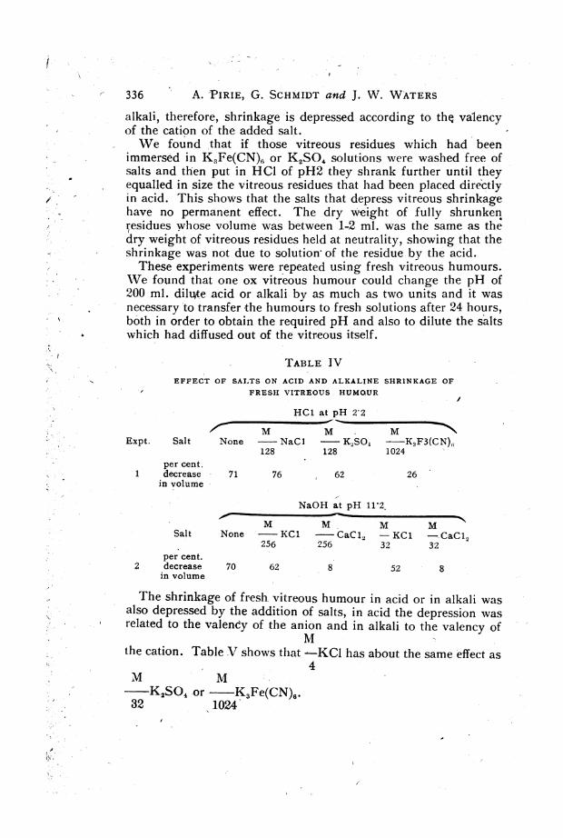
alkali, therefore, shrinkage is depressed according to the valencyof the cation of the added salt.We found that if those vitreous residues which had been
immersed in KsFe(CN)6 or K2SO4 solutions were washed free ofsalts and then put in HC1 of pH2 they shrank further until theyequalled in size the vitreous residues that had been placed directlyin acid. This shows that the salts that depress vitreous shrinkagehave no permanent effect. The dry weight of fully shrunkenresidues whose volume was between 1-2 ml. was the same as thedry weight of vitreous residues held at neutrality, showing that theshrinkage was not due to solution of the residue by the acid.These experiments were repeated using fresh vitreous humours.
We found that one ox vitreous humour could change the pH of-2"00 ml. dilute acid or alkali by as much as two units and it was
necessary to transfer the humours to fresh solutions after 24 hours,both in order to obtain the required pH and also to dilute the saltswhich had diffused out of the vitreous itself.
TABLE IVEFFECT OF SALTS ON ACID AND ALKALINE SHRINKAGE OF
FRESH VITREOUS HUMOUR
HC1 at pH 2 2
_M
Expt. Salt None NaCl K2S04 K3F3(CN),;128 128 1024
per cent.1 decrease 71 76 62 26
in volume
NaOH at pH 11'2.
- ~ ~ ~ ~ ~~~MM M MSalt None -KO1 -CaC12 -KC1 -CaCi2
256 256 32 32per cent.
2 decrease 70 62 8 52 8in volume
The shrinkage of fresh vitreous humour in acid or in alkali wasalso depressed by the addition of salts, in acid the depression wasrelated to the valency of the anion and in alkali to the valency of
Mthe cation. Table V shows that -KCI has about the same effect as
4M M
K2SO4 or K3Fe(CN)6.32 1024
.. >~~~~~~~~~~~~~~~~~~~~~~~~~~~
OX VITREOUS HUMOUR
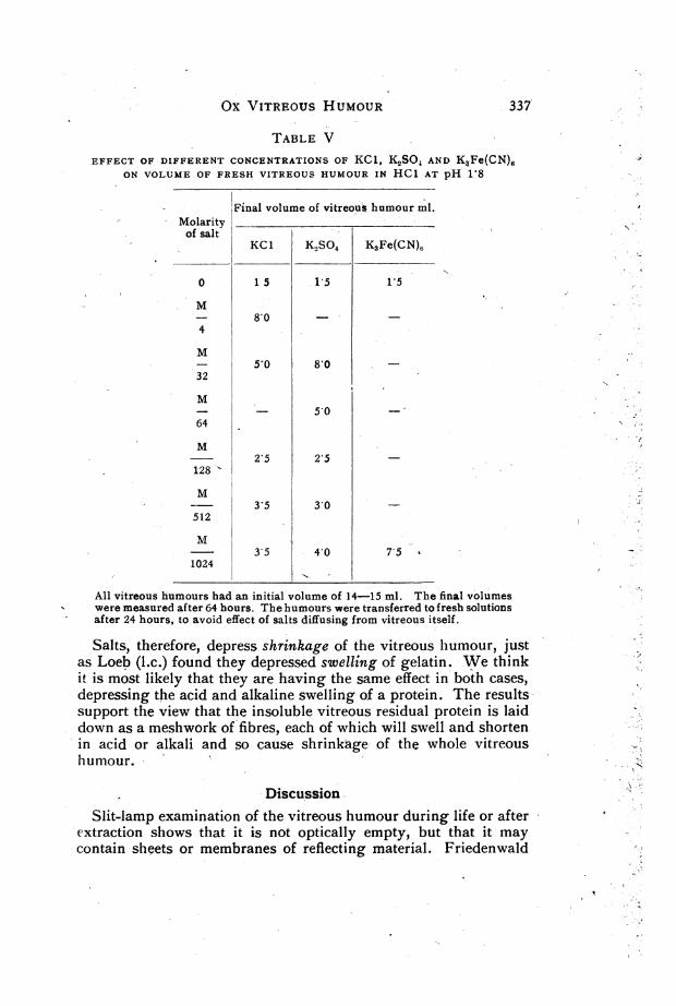
TABLE VEFFECT OF DIFFERENT CONCENTRATIONS OF KC1, K2SO AND K3Fe(CN)6
ON VOLUME OF FRESH VITREOUS HUMOUR IN HC1 AT pH 1-8
Final volume of vitreous humour ml.Molarity _
of saltKC1 K.SO4 K3Fe(CN),
0 1 5 1-5 1,5
M- 8 04
M- 5 0 80 -
32
M- 5o-064
2 5 2 5128
3-5 30 -512
M3*5 4*0 7*5
1024
All vitreous humours had an initial volume of 14-15 ml. The final volumeswere measured after 64 hours. The humours were transferred to fresh solutionsafter 24 hours, to avoid effect of salts diffusing from vitreous itself.
Salts, therefore, depress shrinkage of the vitreous humour, justas Loeb (l.c.) found they depressed swelltng of gelatin. We thinkit is most likely that they are having the same effect in both cases,depressing the acid and alkaline swelling of a protein. The resultssupport the view that the insoluble vitreous residual protein is laiddown as a meshwork of fibres, each of which will swell and shortenin acid or alkali and so cause shrinkSige of the whole vitreoushumour.
DiscussionSlit-lamp examination of the vitreous humour during life or after
extraction shows that it is not optically empty, but that it maycontain sheets or membranes of reflecting material. Friedenwald
337'

338 A. PIIRIE, G. SCHMIDT aind J. \V. WATERS
and Stiehler (1935), using the slit ultra-microscope to examine theextracted vitreous humour of the ox, observed fairly regular brightreflections which they interpreted as due to very thin sheets of theresidual protein. The bright reflections ran roughly parallel to thesurface of the vitreous humour and were separated from oneanother by spaces of 0-5-3 microns. Friedenwald and Stiehlerconsidered that the thickness of the sheets was of the same orderof magnitude as the wave length of light and they thought thatcohesion between sheets might form the visible -opa6ities of thedegenerating humour. Vogt (1941), who has studied the vitreoushumour in the living human eye, speaks of the scaffolding of thevitreous body and states that it is predominantly lamellar instructure, that is, membranous, but that a fibrillar structure in themembranes can often be made out. Vogt's very beautiful picturesof vitreous opacities fit with'the idea that these are formed bycohesion between membranes or fibres.The vitreous humour of the ox appears to be a more rigid
structure than that of any other animal so far examined. i\Ieyer,Smyth and Gallardo (1938) have found that ox vitreous humourcontains a great deal more hexosamine than the humours' of otheranimals. This indicates that the hyaluroniG acid content of oxvitreous 'humour is greater and probably explains its greaterrigidity. In spite of such quantitative differences between animals,there appears to be a qualitative similarity, both in the slit-lampappearance and in the chemistry 'of the vitreous 'humours ofdiffer nt species.
If the experimental evidence given here is accepted the picture ofthe structure of the vitreous- humour becomes more complex. Itcannot be considered as uniform, but must be made up of at leasttwo " structures," the collagen-like network and the hyaluronicacid and protein jelly. Liquefaction of the humour follows enzymichydrolysis of the network and therefore one may say that this net-work is essential for maintenance of a normal vitreous humour.Yet it does not form the humour, being minimally swollen atneutrality and occupying only a very small part of the totalvolume. It seems to us that it is the relation of the network to thejelly which is of first importance and. must be taken intoaccount in any theories of the causes of vitreous swelling andvitreous degenerations.
SUMMARY
1. X-ray photography, amino acid chromatography andenzyme analysis provide evidence that the residual' protein of theox vitreous humour is largely a collagen type.

OX VITREOUS HUMOUR -339
2. Enzyme preparations containing collagenase liquefy the oxvitreous humour.
3. The bearing of this result on the conception of vitreoushumour structure is discussed.
We are deeply indebted to Dr. D. S. Parsons and Dr. R. B. Fisher for carryingout the amino-acid partition chromatograms on our material and for their inter-pretation of them. We also wish to thank Mr. Tugwell, photographer to theRadcliffe Infirmary, for the care he has taken in preparing the photographs of thechromatograms.
REFERENCES
ASTBURY, W. T. (1943)-Adv. in Enzymol., Vol. III, p. 6?.BERGMANN, M. and STEIN, W. H. (1939)-Jl. Biol.. Chem., Vol. CXXVIII, p. 217.BIDWEILL, E. and VAN HEYNINGEN, W. X. (1948)-Biochem. JI., Vol. XLII, p. 140.CONSIEN, R., GORDON, A. H. & MARTIN, A. J. P. (1944)-Biochem. Ji.,
Vol.'XXXVIII, p.224.-DUKE-ELDER, W. S.-Brit. JI. Opbhthal., Monograph Suppl;, No'. 4.EDMAN, P. (1945)-Arkiv.f, Kemi Mineral. Och. Geol. 22A, No. 3.ELSON, L. A. & MORGAN, W. T. J. (1933)-Biochem. JI., Vol. XXVII, p. 1824.FISHER, R. B., PARSONS, D. S. & MORRISON, G. A. (1948) In press.FRIEDENWALD, J. S. and STIEHLER, R. D. (1935)-Arch. of Ofihthal., Vol. XIV,
p. 789.HIGHBERGER, J. H. (1939)-Jl. Amer. Chemn. Soc., Vol. LXI, p. 2302.JORDAN-LLOYD, D. & MARRIOTT, R. H. (1935).-Proc. Roy. Soc. Loutd. B.,
Vol. CXVIII, p. 439.KRAUSE, A. C. (1934)-Biochemistry of the Eye. The Johns Hopkins Press, Baltimore.KUNTZEL, A. (1926)-Koll. Zeits., Vol. XL, p. 264.LOEB, J. (1920-21)-Jl. Gen. Physiol., Vol. III, p. 391.MARKHAM, R. (1942)-Biochein. JI., Vol. XXXII, p. 790.MEYER, K. and SMYTH, E. M. (1937)-Jl. Biol. Chem., Vol. CXIX, p. 507.MEYER, K., SMYTH, E. M. and GALLARDO, E. (1938),-Amer. JI. Ophthal., Vol.
XXI, P. 1084.MEYER, K. (1945)-Advances in Protein Chem., Vol. II,-p. 250.MORNER, C. T. (1894)-Zeits. f. Physiol. Chem., Vol. XVIII. p. 233.NAGEOTTE, J. (1927)-Comnbt. Rend. Soc. Biol., Vol. XCVI, p. 172.NAGEOTTE, J. and GUYON, L. (1931)-Arch. de. Biol., Vol. XLI, p. 1.PIRIE, A. (1947)-Biochem. Ji., Vol. XLI, p. 185.PIRIE, N. W. (1936)-Brit. Ji. e4j. Path., Vol. XVII, p. 274..ROBERTSON, E. B. and DUKE-ELDER, WV. S. (1933).-Proc. Roy. Soc. Lond. B.,
Vol. CXII, p. 215.SCHNEIDER, F. (1940)-Collegiumn, p. 97.THIERFELDER, H. & VON CRAMM, E. (1919)-Zeits. Physiol. Chem., Vol. CV,
p.58.VOGT, A. (1941)-Slit-lamp Microscopy of the Living Eye, Vol. III. Schwezer,
Druck und Verlagshaus, Zurich.YOUNG, R. A. (1894)-Ji. Physiol., Vol. XVI. p. 325.
Related Documents