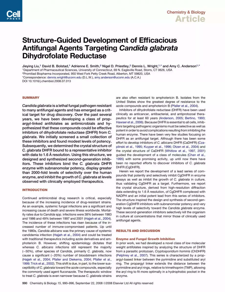
Chemistry & Biology Article Structure-Guided Development of Efficacious Antifungal Agents Targeting Candida glabrata Dihydrofolate Reductase Jieying Liu, 1 David B. Bolstad, 1 Adrienne E. Smith, 2 Nigel D. Priestley, 2 Dennis L. Wright, 1, * and Amy C. Anderson 1, * 1 Department of Pharmaceutical Sciences, University of Connecticut, 69 N. Eagleville Road, Storrs, CT 0626, USA 2 Promiliad Biopharma Incorporated, 950 West Fork Petty Creek Road, Alberton, MT 59820, USA *Correspondence: [email protected] (D.L.W.), [email protected] (A.C.A.) DOI 10.1016/j.chembiol.2008.07.013 SUMMARY Candida glabrata is a lethal fungal pathogen resistant to many antifungal agents and has emerged as a crit- ical target for drug discovery. Over the past several years, we have been developing a class of prop- argyl-linked antifolates as antimicrobials and hy- pothesized that these compounds could be effective inhibitors of dihydrofolate reductase (DHFR) from C. glabrata. We initially screened a small collection of these inhibitors and found modest levels of potency. Subsequently, we determined the crystal structure of C. glabrata DHFR bound to a representative inhibitor with data to 1.6 A ˚ resolution. Using this structure, we designed and synthesized second-generation inhib- itors. These inhibitors bind the C. glabrata DHFR enzyme with subnanomolar potency, display greater than 2000-fold levels of selectivity over the human enzyme, and inhibit the growth of C. glabrata at levels observed with clinically employed therapeutics. INTRODUCTION Continued antimicrobial drug research is critical, especially because of the increasing incidence of drug-resistant strains. As an example, systemic fungal infections are a significant and increasing cause of death and severe illness worldwide. Mortal- ity rates due to Candida spp. infections were 38% between 1983 and 1986 and 49% between 1997 and 2001 (Hajjeh et al., 2004). The incidence of these infections has risen because of the in- creased number of immune-compromised patients. Up until the 1980s, Candida albicans was the primary cause of systemic candidemia infection (Hajjeh et al., 2004) and could be treated with traditional therapeutics including azole derivatives and am- photericin B. However, shifting epidemiology dictates that whereas C. albicans infections still represent the majority (50%), other species of Candida, primarily C. glabrata, now cause a significant (20%) number of bloodstream infections (Hajjeh et al., 2004; Pfaller and Diekema, 2004; Pfaller et al., 1999; Trick et al., 2002). This shift is due, in part, to the lower sus- ceptibility of C. glabrata toward the azole compounds, especially the commonly used agent fluconazole. The therapeutic window to treat C. glabrata is even narrower because C. glabrata strains are also often resistant to amphotericin B. Isolates from the United States show the greatest degree of resistance to the azole compounds and amphotericin B (Pfaller et al., 2004). Inhibitors of dihydrofolate reductase (DHFR) have been used clinically as anticancer, antibacterial, and antiprotozoal thera- peutics for at least 60 years (Anderson, 2005; Bertino, 1993; Hawser et al., 2006). Because DHFR is essential to all cells, inhib- itors targeting pathogenic organisms must be selective as well as potent in order to avoid complications resulting from inhibiting the human enzyme. There have been very few studies focusing on DHFR as an antifungal target. Although there has been some effort to develop inhibitors of C. albicans DHFR (CaDHFR) (Cza- plinski et al., 1995; Kuyper et al., 1996; Otzen et al., 2004) and the crystal structure of CaDHFR (Whitlow et al., 1997, 2001) guided the development of a class of molecules (Chan et al., 1995) with some promising activity, up until now there have been no reported efforts to discover inhibitors of C. glabrata DHFR (CgDHFR). Herein we report the development of a lead series of com- pounds that potently and selectively inhibit CgDHFR in enzyme assays as well as inhibit the growth of C. glabrata in culture, thus validating CgDHFR as a target. Furthermore, we report the crystal structure, derived from high-resolution diffraction data extending to 1.6 A ˚ resolution, of CgDHFR complexed with NADPH and an initial potent lead from this series of inhibitors. The structure inspired the design and synthesis of second-gen- eration CgDHFR inhibitors with subnanomolar potency and very high levels of selectivity toward the Candida glabrata enzyme. These second-generation inhibitors selectively kill the organism in culture at concentrations that mirror those of clinically used antifungal agents. RESULTS AND DISCUSSION Enzyme and Fungal Growth Inhibition In prior work, we had developed a novel class of low molecular weight antifolates inspired by analyzing the structure of DHFR from a parasitic protozoan, Cryptosporidium hominis (ChDHFR) (Pelphrey et al., 2007). This series is characterized by a prop- argyl-based linker between the pyrimidine and substituted aryl ring. The propargyl linker extends the distance between the pyrimidine and aryl rings, relative to trimethoprim (TMP), allowing the aryl ring to fit more optimally in a hydrophobic pocket in the enzyme. 990 Chemistry & Biology 15, 990–996, September 22, 2008 ª2008 Elsevier Ltd All rights reserved

Welcome message from author
This document is posted to help you gain knowledge. Please leave a comment to let me know what you think about it! Share it to your friends and learn new things together.
Transcript
Chemistry & Biology
Article
Structure-Guided Development of EfficaciousAntifungal Agents Targeting Candida glabrataDihydrofolate ReductaseJieying Liu,1 David B. Bolstad,1 Adrienne E. Smith,2 Nigel D. Priestley,2 Dennis L. Wright,1,* and Amy C. Anderson1,*1Department of Pharmaceutical Sciences, University of Connecticut, 69 N. Eagleville Road, Storrs, CT 0626, USA2Promiliad Biopharma Incorporated, 950 West Fork Petty Creek Road, Alberton, MT 59820, USA
*Correspondence: [email protected] (D.L.W.), [email protected] (A.C.A.)DOI 10.1016/j.chembiol.2008.07.013
SUMMARY
Candida glabrata is a lethal fungal pathogen resistantto many antifungal agents and has emerged as a crit-ical target for drug discovery. Over the past severalyears, we have been developing a class of prop-argyl-linked antifolates as antimicrobials and hy-pothesized that these compounds could be effectiveinhibitors of dihydrofolate reductase (DHFR) from C.glabrata. We initially screened a small collection ofthese inhibitors and found modest levels of potency.Subsequently, we determined the crystal structure ofC. glabrata DHFR bound to a representative inhibitorwith data to 1.6 A resolution. Using this structure, wedesigned and synthesized second-generation inhib-itors. These inhibitors bind the C. glabrata DHFRenzyme with subnanomolar potency, display greaterthan 2000-fold levels of selectivity over the humanenzyme, and inhibit the growth of C. glabrata at levelsobserved with clinically employed therapeutics.
INTRODUCTION
Continued antimicrobial drug research is critical, especially
because of the increasing incidence of drug-resistant strains.
As an example, systemic fungal infections are a significant and
increasing cause of death and severe illness worldwide. Mortal-
ity rates due to Candida spp. infections were 38% between 1983
and 1986 and 49% between 1997 and 2001 (Hajjeh et al., 2004).
The incidence of these infections has risen because of the in-
creased number of immune-compromised patients. Up until
the 1980s, Candida albicans was the primary cause of systemic
candidemia infection (Hajjeh et al., 2004) and could be treated
with traditional therapeutics including azole derivatives and am-
photericin B. However, shifting epidemiology dictates that
whereas C. albicans infections still represent the majority
(�50%), other species of Candida, primarily C. glabrata, now
cause a significant (�20%) number of bloodstream infections
(Hajjeh et al., 2004; Pfaller and Diekema, 2004; Pfaller et al.,
1999; Trick et al., 2002). This shift is due, in part, to the lower sus-
ceptibility of C. glabrata toward the azole compounds, especially
the commonly used agent fluconazole. The therapeutic window
to treat C. glabrata is even narrower because C. glabrata strains
990 Chemistry & Biology 15, 990–996, September 22, 2008 ª2008 E
are also often resistant to amphotericin B. Isolates from the
United States show the greatest degree of resistance to the
azole compounds and amphotericin B (Pfaller et al., 2004).
Inhibitors of dihydrofolate reductase (DHFR) have been used
clinically as anticancer, antibacterial, and antiprotozoal thera-
peutics for at least 60 years (Anderson, 2005; Bertino, 1993;
Hawser et al., 2006). Because DHFR is essential to all cells, inhib-
itors targeting pathogenic organisms must be selective as well as
potent in order to avoid complications resulting from inhibiting the
human enzyme. There have been very few studies focusing on
DHFR as an antifungal target. Although there has been some
effort to develop inhibitors of C. albicans DHFR (CaDHFR) (Cza-
plinski et al., 1995; Kuyper et al., 1996; Otzen et al., 2004) and
the crystal structure of CaDHFR (Whitlow et al., 1997, 2001)
guided the development of a class of molecules (Chan et al.,
1995) with some promising activity, up until now there have
been no reported efforts to discover inhibitors of C. glabrata
DHFR (CgDHFR).
Herein we report the development of a lead series of com-
pounds that potently and selectively inhibit CgDHFR in enzyme
assays as well as inhibit the growth of C. glabrata in culture,
thus validating CgDHFR as a target. Furthermore, we report
the crystal structure, derived from high-resolution diffraction
data extending to 1.6 A resolution, of CgDHFR complexed with
NADPH and an initial potent lead from this series of inhibitors.
The structure inspired the design and synthesis of second-gen-
eration CgDHFR inhibitors with subnanomolar potency and very
high levels of selectivity toward the Candida glabrata enzyme.
These second-generation inhibitors selectively kill the organism
in culture at concentrations that mirror those of clinically used
antifungal agents.
RESULTS AND DISCUSSION
Enzyme and Fungal Growth InhibitionIn prior work, we had developed a novel class of low molecular
weight antifolates inspired by analyzing the structure of DHFR
from a parasitic protozoan, Cryptosporidium hominis (ChDHFR)
(Pelphrey et al., 2007). This series is characterized by a prop-
argyl-based linker between the pyrimidine and substituted aryl
ring. The propargyl linker extends the distance between the
pyrimidine and aryl rings, relative to trimethoprim (TMP), allowing
the aryl ring to fit more optimally in a hydrophobic pocket in the
enzyme.
lsevier Ltd All rights reserved
Chemistry & Biology
Structure of C. glabrata DHFR and Potent Inhibitor
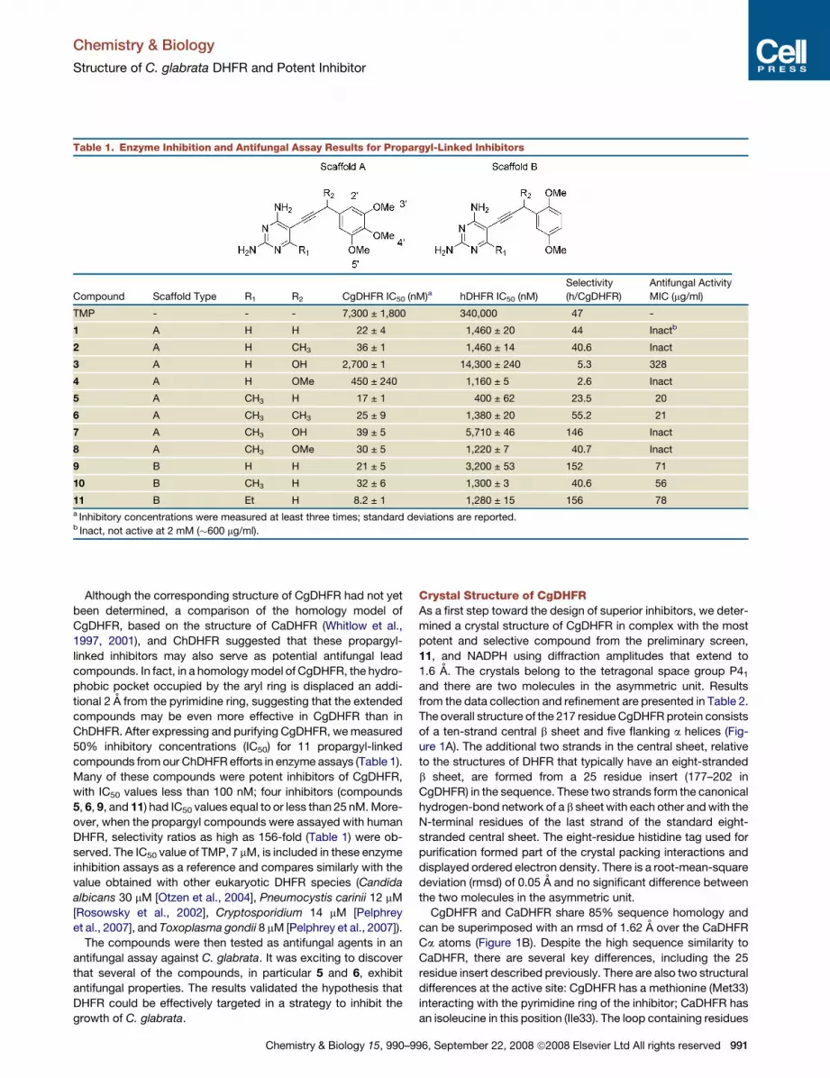
Table 1. Enzyme Inhibition and Antifungal Assay Results for Propargyl-Linked Inhibitors
Compound Scaffold Type R1 R2 CgDHFR IC50 (nM)a hDHFR IC50 (nM)
Selectivity
(h/CgDHFR)
Antifungal Activity
MIC (mg/ml)
TMP - - - 7,300 ± 1,800 340,000 47 -
1 A H H 22 ± 4 1,460 ± 20 44 Inactb
2 A H CH3 36 ± 1 1,460 ± 14 40.6 Inact
3 A H OH 2,700 ± 1 14,300 ± 240 5.3 328
4 A H OMe 450 ± 240 1,160 ± 5 2.6 Inact
5 A CH3 H 17 ± 1 400 ± 62 23.5 20
6 A CH3 CH3 25 ± 9 1,380 ± 20 55.2 21
7 A CH3 OH 39 ± 5 5,710 ± 46 146 Inact
8 A CH3 OMe 30 ± 5 1,220 ± 7 40.7 Inact
9 B H H 21 ± 5 3,200 ± 53 152 71
10 B CH3 H 32 ± 6 1,300 ± 3 40.6 56
11 B Et H 8.2 ± 1 1,280 ± 15 156 78a Inhibitory concentrations were measured at least three times; standard deviations are reported.b Inact, not active at 2 mM (�600 mg/ml).
Although the corresponding structure of CgDHFR had not yet
been determined, a comparison of the homology model of
CgDHFR, based on the structure of CaDHFR (Whitlow et al.,
1997, 2001), and ChDHFR suggested that these propargyl-
linked inhibitors may also serve as potential antifungal lead
compounds. In fact, in a homology model of CgDHFR, the hydro-
phobic pocket occupied by the aryl ring is displaced an addi-
tional 2 A from the pyrimidine ring, suggesting that the extended
compounds may be even more effective in CgDHFR than in
ChDHFR. After expressing and purifying CgDHFR, we measured
50% inhibitory concentrations (IC50) for 11 propargyl-linked
compounds from our ChDHFR efforts in enzyme assays (Table 1).
Many of these compounds were potent inhibitors of CgDHFR,
with IC50 values less than 100 nM; four inhibitors (compounds
5, 6, 9, and 11) had IC50 values equal to or less than 25 nM. More-
over, when the propargyl compounds were assayed with human
DHFR, selectivity ratios as high as 156-fold (Table 1) were ob-
served. The IC50 value of TMP, 7 mM, is included in these enzyme
inhibition assays as a reference and compares similarly with the
value obtained with other eukaryotic DHFR species (Candida
albicans 30 mM [Otzen et al., 2004], Pneumocystis carinii 12 mM
[Rosowsky et al., 2002], Cryptosporidium 14 mM [Pelphrey
et al., 2007], and Toxoplasma gondii 8 mM [Pelphrey et al., 2007]).
The compounds were then tested as antifungal agents in an
antifungal assay against C. glabrata. It was exciting to discover
that several of the compounds, in particular 5 and 6, exhibit
antifungal properties. The results validated the hypothesis that
DHFR could be effectively targeted in a strategy to inhibit the
growth of C. glabrata.
Chemistry & Biology 15, 990–
Crystal Structure of CgDHFRAs a first step toward the design of superior inhibitors, we deter-
mined a crystal structure of CgDHFR in complex with the most
potent and selective compound from the preliminary screen,
11, and NADPH using diffraction amplitudes that extend to
1.6 A. The crystals belong to the tetragonal space group P41
and there are two molecules in the asymmetric unit. Results
from the data collection and refinement are presented in Table 2.
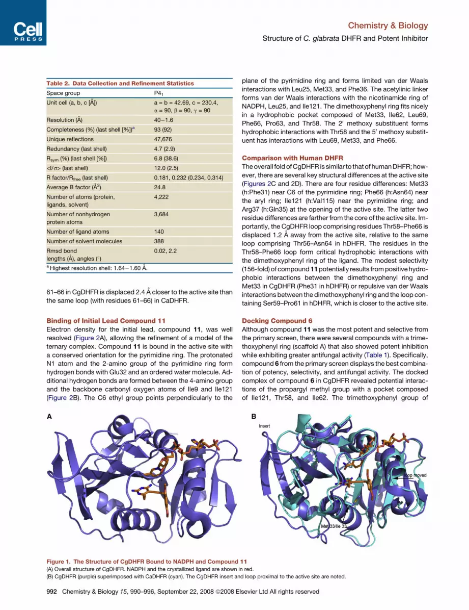
The overall structure of the 217 residue CgDHFR protein consists
of a ten-strand central b sheet and five flanking a helices (Fig-
ure 1A). The additional two strands in the central sheet, relative
to the structures of DHFR that typically have an eight-stranded
b sheet, are formed from a 25 residue insert (177–202 in
CgDHFR) in the sequence. These two strands form the canonical
hydrogen-bond network of a b sheet with each other and with the
N-terminal residues of the last strand of the standard eight-
stranded central sheet. The eight-residue histidine tag used for
purification formed part of the crystal packing interactions and
displayed ordered electron density. There is a root-mean-square
deviation (rmsd) of 0.05 A and no significant difference between
the two molecules in the asymmetric unit.
CgDHFR and CaDHFR share 85% sequence homology and
can be superimposed with an rmsd of 1.62 A over the CaDHFR
Ca atoms (Figure 1B). Despite the high sequence similarity to
CaDHFR, there are several key differences, including the 25
residue insert described previously. There are also two structural
differences at the active site: CgDHFR has a methionine (Met33)
interacting with the pyrimidine ring of the inhibitor; CaDHFR has
an isoleucine in this position (Ile33). The loop containing residues
996, September 22, 2008 ª2008 Elsevier Ltd All rights reserved 991
Chemistry & Biology
Structure of C. glabrata DHFR and Potent Inhibitor
61–66 in CgDHFR is displaced 2.4 A closer to the active site than
the same loop (with residues 61–66) in CaDHFR.
Binding of Initial Lead Compound 11Electron density for the initial lead, compound 11, was well
resolved (Figure 2A), allowing the refinement of a model of the
ternary complex. Compound 11 is bound in the active site with
a conserved orientation for the pyrimidine ring. The protonated
N1 atom and the 2-amino group of the pyrimidine ring form
hydrogen bonds with Glu32 and an ordered water molecule. Ad-
ditional hydrogen bonds are formed between the 4-amino group
and the backbone carbonyl oxygen atoms of Ile9 and Ile121
(Figure 2B). The C6 ethyl group points perpendicularly to the
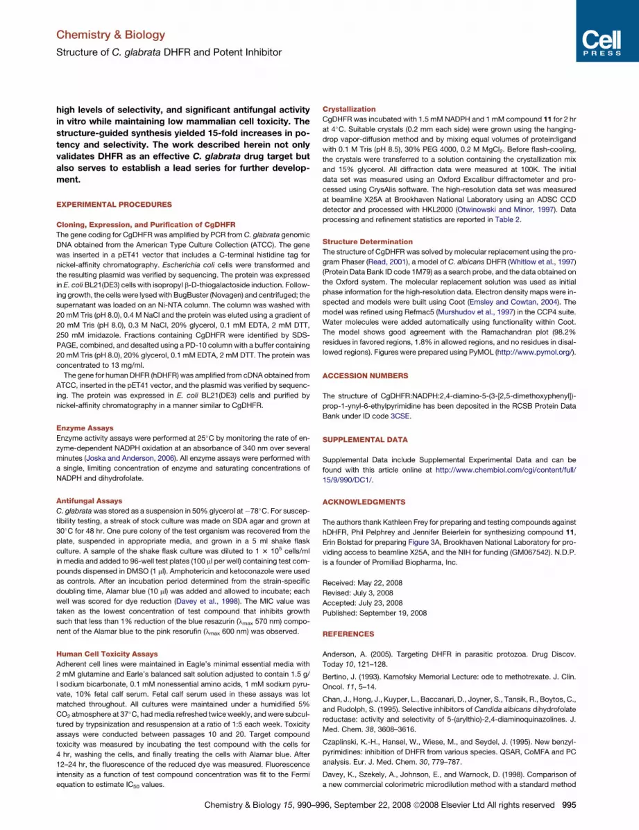
Table 2. Data Collection and Refinement Statistics
Space group P41
Unit cell (a, b, c [A]) a = b = 42.69, c = 230.4,
a = 90, b = 90, g = 90
Resolution (A) 40�1.6
Completeness (%) (last shell [%])a 93 (92)
Unique reflections 47,676
Redundancy (last shell) 4.7 (2.9)
Rsym (%) (last shell [%]) 6.8 (38.6)
<I/s> (last shell) 12.0 (2.5)
R factor/Rfree (last shell) 0.181, 0.232 (0.234, 0.314)
Average B factor (A2) 24.8
Number of atoms (protein,
ligands, solvent)
4,222
Number of nonhydrogen
protein atoms
3,684
Number of ligand atoms 140
Number of solvent molecules 388
Rmsd bond
lengths (A), angles (�)
0.02, 2.2
a Highest resolution shell: 1.64�1.60 A.
plane of the pyrimidine ring and forms limited van der Waals
interactions with Leu25, Met33, and Phe36. The acetylinic linker
forms van der Waals interactions with the nicotinamide ring of
NADPH, Leu25, and Ile121. The dimethoxyphenyl ring fits nicely
in a hydrophobic pocket composed of Met33, Ile62, Leu69,
Phe66, Pro63, and Thr58. The 20 methoxy substituent forms
hydrophobic interactions with Thr58 and the 50 methoxy substit-
uent has interactions with Leu69, Met33, and Phe66.
Comparison with Human DHFRThe overall fold of CgDHFR is similar to that of human DHFR; how-
ever, there are several key structural differences at the active site
(Figures 2C and 2D). There are four residue differences: Met33
(h:Phe31) near C6 of the pyrimidine ring; Phe66 (h:Asn64) near
the aryl ring; Ile121 (h:Val115) near the pyrimidine ring; and
Arg37 (h:Gln35) at the opening of the active site. The latter two
residue differences are farther from the core of the active site. Im-
portantly, the CgDHFR loop comprising residues Thr58–Phe66 is
displaced 1.2 A away from the active site, relative to the same
loop comprising Thr56–Asn64 in hDHFR. The residues in the
Thr58–Phe66 loop form critical hydrophobic interactions with
the dimethoxyphenyl ring of the ligand. The modest selectivity
(156-fold) of compound 11 potentially results from positive hydro-
phobic interactions between the dimethoxyphenyl ring and
Met33 in CgDHFR (Phe31 in hDHFR) or repulsive van der Waals
interactions between the dimethoxyphenyl ring and the loop con-
taining Ser59–Pro61 in hDHFR, which is closer to the active site.
Docking Compound 6Although compound 11 was the most potent and selective from
the primary screen, there were several compounds with a trime-
thoxyphenyl ring (scaffold A) that also showed potent inhibition
while exhibiting greater antifungal activity (Table 1). Specifically,
compound 6 from the primary screen displays the best combina-
tion of potency, selectivity, and antifungal activity. The docked
complex of compound 6 in CgDHFR revealed potential interac-
tions of the propargyl methyl group with a pocket composed
of Ile121, Thr58, and Ile62. The trimethoxyphenyl group of
Figure 1. The Structure of CgDHFR Bound to NADPH and Compound 11(A) Overall structure of CgDHFR. NADPH and the crystallized ligand are shown in red.
(B) CgDHFR (purple) superimposed with CaDHFR (cyan). The CgDHFR insert and loop proximal to the active site are noted.
992 Chemistry & Biology 15, 990–996, September 22, 2008 ª2008 Elsevier Ltd All rights reserved

Chemistry & Biology
Structure of C. glabrata DHFR and Potent Inhibitor
Figure 2. Potent and Selective Interactions of the Ligand
(A) Electron density for the active site and inhibitor contoured at 2s.
(B) Interactions of compound 11 with CgDHFR, with hydrogen bonds shown as dashed lines.
(C) A sequence alignment of Candida glabrata (Cg), Candida albicans (Ca), and human DHFR (H).
(D) Comparison of CgDHFR (purple) and hDHFR (salmon). Only key residues in hDHFR at the active site are shown for clarity.
compound 6 presents a 30 methoxy group to interact with Ser61
and Leu25, a 40 methoxy group that has very few interactions
with the protein, and a 50 methoxy group that interacts with
Ile62, Pro63, Met33, and, distally, Phe66.
Second-Generation CompoundsSeveral moieties of the first-generation inhibitors (compounds
1–11), including a small alkyl group at the C6 position of the py-
rimidine ring, the propargyl methyl group of compound 6, and the
30 methoxy group on the phenyl ring, yielded favorable van der
Waals interactions with CgDHFR. However, additional interac-
tions, relative to those used by compounds 11 and 6, appeared
to be available in the ligand binding pocket. It seemed that larger
lipophilic moieties at the 50 position on the aryl ring, relative to the
simple methoxy group of compounds 11 and 6, could take better
advantage of the hydrophobic interactions available from Ile62,
Pro63, Met33, and Phe66 (Figure 3A). A bulkier hydrophobic
group at this position, designed to increase potency against
Chemistry & Biology 15, 990–9
CgDHFR, was also predicted to decrease potency (and increase
selectivity) against hDHFR. The loop containing residues 56–64
in hDHFR is closer to the active site, and modeling suggested
that it would need to relocate to accommodate steric bulk at
the 50 position of a second-generation inhibitor.
Therefore, using our high-resolution structure of CgDHFR and
features of compounds 11 and 6, second-generation inhibitors
were designed to be significantly more potent and selective
(Figure 3B). The series of second-generation derivatives main-
tain a methyl group at the C6 position of the pyrimidine, a prop-
argyl methyl group, and the original 30 methoxy-substituted
phenyl ring. However, the new compounds also include a second
aryl ring at the meta (50) position of the first phenyl ring (see
scaffold in Table 3). Because substituents on the second aryl
ring were envisioned to project bulk toward the loop region
and increase potency by interacting with residues 63–66 in
CgDHFR and selectivity by interfering with residues 61–64 in
hDHFR, biphenyl derivatives with methyl groups at the meta
96, September 22, 2008 ª2008 Elsevier Ltd All rights reserved 993

Chemistry & Biology
Structure of C. glabrata DHFR and Potent Inhibitor
(compound 12) and para (compound 13) positions on the second
ring were designed.
In addition to the predicted new interactions with the enzyme,
the design of the biphenyl compounds presents a facile route for
synthesis. The synthesis of these compounds relies on two pow-
erful palladium-mediated cross-coupling reactions that allow for
the assembly of a potential library of ligands in a modular fashion.
The original propargyl design takes advantage of a key Sonaga-
shira coupling to join an iodinated pyrimidine with an acetylene
partner (Pelphrey et al., 2007). The synthesis of the biphenyl
compounds expands on this modular design to include a Suzuki
coupling to install the second aryl ring. The wide availability of
several diverse boronic acid coupling partners assures direct
access to a wide variety of analogs. Building upon our previous
methodology (Pelphrey et al., 2007), compounds 12 and 13 were
synthesized for biological assay. Characterization details for
compounds 12 and 13 can be accessed in Supplemental Data
available online.
The second-generation compounds were tested in enzyme
assays using CgDHFR and human DHFR as well as in antifungal
assays (Table 3). Not only are these compounds extremely
potent, with IC50 values in the subnanomolar range against
CgDHFR, they also exhibit very strong selectivity for the fungal
enzyme. Significantly, these second-generation compounds
Figure 3. Structural Features Used for the
Design of Second-Generation Inhibitors
(A) A surface representation of CgDHFR at the
active site with compound 11 bound. The surface
is colored using a gradient that extends from lipo-
philic (red) to neutral (green) to hydrophilic (blue).
The view is the same as that shown in Figure 2B.
Residues near the 50 position of the aryl ring are
labeled, as are the propargyl, 30, and 50 pockets.
(B) A docked complex of compound 13 (cyan) in
CgDHFR (purple).
also displayed superior antifungal activity
in vitro, with levels of inhibition compara-
ble to clinically used therapeutics (Pfaller
and Diekema, 2004). Evaluation of com-
pounds 12 and 13 in human cell lines
yielded an assessment of overt toxicity.
We measured IC50 values in MCF-10 cells and then calculated
selectivity ratios by dividing the IC50 value in the human cell
line by the MIC value, the lowest concentration of compound
(in micromolar units) that inhibits at least 99% of fungal cell
growth (Table 3). We were very excited to find that the best
lead compound is 13.5-fold selective as an antifungal agent.
SIGNIFICANCE
Candida glabrata is an emerging fungal pathogen that
currently causes at least 20% of all candidemia infections.
Unfortunately, C. glabrata is resistant to the majority of clin-
ically approved antifungal agents. In this work, we describe
a full cycle of structure-based drug design resulting in a lead
compound effective against DHFR from Candida glabrata.
Initial assays of a group of propargyl-based DHFR inhibitors
identified two compounds with nanomolar potency against
the enzyme and moderate antifungal activity. The high-reso-
lution structure of the CgDHFR enzyme bound to one of
these initial leads revealed a hydrophobic pocket near the
50 site of the aryl ring that could be exploited for increased
potency and selectivity for the pathogenic enzyme. We
designed and synthesized second-generation compounds
and show that they exhibit subnanomolar potency, very
Table 3. Enzyme Inhibition and Antifungal Properties of Second-Generation Compounds
Compound R
CgDHFR
IC50 (nM)
hDHFR
IC50 (nM)
Enzyme Selectivity
(h/CgDHFR)
Antifungal
Activity MIC
Human Cell
Toxicity IC50 (mM)
Cellular
Selectivity
12 30, 50 (meta) Me 0.55 750 1364 3 mg/ml (8 mM) 47 5.9
13 40 (para) Me 0.6 1410 2350 1.5 mg/ml (4 mM) 54 13.5
994 Chemistry & Biology 15, 990–996, September 22, 2008 ª2008 Elsevier Ltd All rights reserved
Chemistry & Biology
Structure of C. glabrata DHFR and Potent Inhibitor
high levels of selectivity, and significant antifungal activity
in vitro while maintaining low mammalian cell toxicity. The
structure-guided synthesis yielded 15-fold increases in po-
tency and selectivity. The work described herein not only
validates DHFR as an effective C. glabrata drug target but
also serves to establish a lead series for further develop-
ment.
EXPERIMENTAL PROCEDURES
Cloning, Expression, and Purification of CgDHFR
The gene coding for CgDHFR was amplified by PCR from C. glabrata genomic
DNA obtained from the American Type Culture Collection (ATCC). The gene
was inserted in a pET41 vector that includes a C-terminal histidine tag for
nickel-affinity chromatography. Escherichia coli cells were transformed and
the resulting plasmid was verified by sequencing. The protein was expressed
in E. coli BL21(DE3) cells with isopropyl b-D-thiogalactoside induction. Follow-
ing growth, the cells were lysed with BugBuster (Novagen) and centrifuged; the
supernatant was loaded on an Ni-NTA column. The column was washed with
20 mM Tris (pH 8.0), 0.4 M NaCl and the protein was eluted using a gradient of
20 mM Tris (pH 8.0), 0.3 M NaCl, 20% glycerol, 0.1 mM EDTA, 2 mM DTT,
250 mM imidazole. Fractions containing CgDHFR were identified by SDS-
PAGE, combined, and desalted using a PD-10 column with a buffer containing
20 mM Tris (pH 8.0), 20% glycerol, 0.1 mM EDTA, 2 mM DTT. The protein was
concentrated to 13 mg/ml.
The gene for human DHFR (hDHFR) was amplified from cDNA obtained from
ATCC, inserted in the pET41 vector, and the plasmid was verified by sequenc-
ing. The protein was expressed in E. coli BL21(DE3) cells and purified by
nickel-affinity chromatography in a manner similar to CgDHFR.
Enzyme Assays
Enzyme activity assays were performed at 25�C by monitoring the rate of en-
zyme-dependent NADPH oxidation at an absorbance of 340 nm over several
minutes (Joska and Anderson, 2006). All enzyme assays were performed with
a single, limiting concentration of enzyme and saturating concentrations of
NADPH and dihydrofolate.
Antifungal Assays
C. glabrata was stored as a suspension in 50% glycerol at�78�C. For suscep-
tibility testing, a streak of stock culture was made on SDA agar and grown at
30�C for 48 hr. One pure colony of the test organism was recovered from the
plate, suspended in appropriate media, and grown in a 5 ml shake flask
culture. A sample of the shake flask culture was diluted to 1 3 105 cells/ml
in media and added to 96-well test plates (100 ml per well) containing test com-
pounds dispensed in DMSO (1 ml). Amphotericin and ketoconazole were used
as controls. After an incubation period determined from the strain-specific
doubling time, Alamar blue (10 ml) was added and allowed to incubate; each
well was scored for dye reduction (Davey et al., 1998). The MIC value was
taken as the lowest concentration of test compound that inhibits growth
such that less than 1% reduction of the blue resazurin (lmax 570 nm) compo-
nent of the Alamar blue to the pink resorufin (lmax 600 nm) was observed.
Human Cell Toxicity Assays
Adherent cell lines were maintained in Eagle’s minimal essential media with
2 mM glutamine and Earle’s balanced salt solution adjusted to contain 1.5 g/
l sodium bicarbonate, 0.1 mM nonessential amino acids, 1 mM sodium pyru-
vate, 10% fetal calf serum. Fetal calf serum used in these assays was lot
matched throughout. All cultures were maintained under a humidified 5%
CO2 atmosphere at 37�C, had media refreshed twice weekly, and were subcul-
tured by trypsinization and resuspension at a ratio of 1:5 each week. Toxicity
assays were conducted between passages 10 and 20. Target compound
toxicity was measured by incubating the test compound with the cells for
4 hr, washing the cells, and finally treating the cells with Alamar blue. After
12–24 hr, the fluorescence of the reduced dye was measured. Fluorescence
intensity as a function of test compound concentration was fit to the Fermi
equation to estimate IC50 values.
Chemistry & Biology 15, 990–
Crystallization
CgDHFR was incubated with 1.5 mM NADPH and 1 mM compound 11 for 2 hr
at 4�C. Suitable crystals (0.2 mm each side) were grown using the hanging-
drop vapor-diffusion method and by mixing equal volumes of protein:ligand
with 0.1 M Tris (pH 8.5), 30% PEG 4000, 0.2 M MgCl2. Before flash-cooling,
the crystals were transferred to a solution containing the crystallization mix
and 15% glycerol. All diffraction data were measured at 100K. The initial
data set was measured using an Oxford Excalibur diffractometer and pro-
cessed using CrysAlis software. The high-resolution data set was measured
at beamline X25A at Brookhaven National Laboratory using an ADSC CCD
detector and processed with HKL2000 (Otwinowski and Minor, 1997). Data
processing and refinement statistics are reported in Table 2.
Structure Determination
The structure of CgDHFR was solved by molecular replacement using the pro-
gram Phaser (Read, 2001), a model of C. albicans DHFR (Whitlow et al., 1997)
(Protein Data Bank ID code 1M79) as a search probe, and the data obtained on
the Oxford system. The molecular replacement solution was used as initial
phase information for the high-resolution data. Electron density maps were in-
spected and models were built using Coot (Emsley and Cowtan, 2004). The
model was refined using Refmac5 (Murshudov et al., 1997) in the CCP4 suite.
Water molecules were added automatically using functionality within Coot.
The model shows good agreement with the Ramachandran plot (98.2%
residues in favored regions, 1.8% in allowed regions, and no residues in disal-
lowed regions). Figures were prepared using PyMOL (http://www.pymol.org/).
ACCESSION NUMBERS
The structure of CgDHFR:NADPH:2,4-diamino-5-(3-[2,5-dimethoxyphenyl])-
prop-1-ynyl-6-ethylpyrimidine has been deposited in the RCSB Protein Data
Bank under ID code 3CSE.
SUPPLEMENTAL DATA
Supplemental Data include Supplemental Experimental Data and can be
found with this article online at http://www.chembiol.com/cgi/content/full/
15/9/990/DC1/.
ACKNOWLEDGMENTS
The authors thank Kathleen Frey for preparing and testing compounds against
hDHFR, Phil Pelphrey and Jennifer Beierlein for synthesizing compound 11,
Erin Bolstad for preparing Figure 3A, Brookhaven National Laboratory for pro-
viding access to beamline X25A, and the NIH for funding (GM067542). N.D.P.
is a founder of Promiliad Biopharma, Inc.
Received: May 22, 2008
Revised: July 3, 2008
Accepted: July 23, 2008
Published: September 19, 2008
REFERENCES
Anderson, A. (2005). Targeting DHFR in parasitic protozoa. Drug Discov.
Today 10, 121–128.
Bertino, J. (1993). Karnofsky Memorial Lecture: ode to methotrexate. J. Clin.
Oncol. 11, 5–14.
Chan, J., Hong, J., Kuyper, L., Baccanari, D., Joyner, S., Tansik, R., Boytos, C.,
and Rudolph, S. (1995). Selective inhibitors of Candida albicans dihydrofolate
reductase: activity and selectivity of 5-(arylthio)-2,4-diaminoquinazolines. J.
Med. Chem. 38, 3608–3616.
Czaplinski, K.-H., Hansel, W., Wiese, M., and Seydel, J. (1995). New benzyl-
pyrimidines: inhibition of DHFR from various species. QSAR, CoMFA and PC
analysis. Eur. J. Med. Chem. 30, 779–787.
Davey, K., Szekely, A., Johnson, E., and Warnock, D. (1998). Comparison of
a new commercial colorimetric microdilution method with a standard method
996, September 22, 2008 ª2008 Elsevier Ltd All rights reserved 995
Chemistry & Biology
Structure of C. glabrata DHFR and Potent Inhibitor
for in-vitro susceptibility testing of Candida spp. and Cryptococcus neofor-
mans. J. Antimicrob. Chemother. 42, 439–444.
Emsley, P., and Cowtan, K. (2004). Coot: model-building tools for molecular
graphics. Acta Crystallogr. D Biol. Crystallogr. 60, 2126–2132.
Hajjeh, R., Sofair, A., Harrison, L., Lyon, G.M., Arthington-Skaggs, B., Mirza,
S., Phelan, M., Morgan, J., Lee-Yang, W., Ciblak, M., et al. (2004). Incidence
of bloodstream infections due to Candida species and in vitro susceptibilities
of isolates collected from 1998 to 2000 in a population-based active surveil-
lance program. J. Clin. Microbiol. 42, 1519–1527.
Hawser, S., Lociuro, S., and Islam, K. (2006). Dihydrofolate reductase inhibi-
tors as antibacterial agents. Biochem. Pharmacol. 71, 941–948.
Joska, T., and Anderson, A. (2006). Structure-activity relationships of Bacillus
cereus and Bacillus anthracis dihydrofolate reductase: toward the identifica-
tion of new potent drug leads. Antimicrob. Agents Chemother. 50, 3435–3443.
Kuyper, L., Baccanari, D., Jones, M., Hunter, R., Tansik, R., Joyner, S., Boytos,
C., Rudolph, S., Knick, V., Wilson, H.R., et al. (1996). High-affinity inhibitors of
dihydrofolate reductase: antimicrobial and anticancer activities of 7,8-dialkyl-
1,3-diaminopyrrolo[3,2-f ]quinazolines with small molecular size. J. Med.
Chem. 39, 892–903.
Murshudov, G., Vagin, A., and Dodson, E. (1997). Refinement of macromolec-
ular structures by the maximum-likelihood method. Acta Crystallogr. D Biol.
Crystallogr. 53, 240–255.
Otwinowski, Z., and Minor, W. (1997). Processing of X-ray diffraction data
collected in oscillation mode. In Methods in Enzymology, C.W. Carter and
R.M. Sweet, eds. (New York: Academic Press), pp. 307–326.
Otzen, T., Wempe, E., Kunz, B., Bartels, R., Lehwark-Yvetot, G., Hansel, W.,
Schaper, K., and Seydel, J. (2004). Folate-synthesizing enzyme system as tar-
get for development of inhibitors and inhibitor combinations against Candida
albicans—synthesis and biological activity of new 2,4-diaminopyrimidines
and 40-substituted 4-aminodiphenyl sulfones. J. Med. Chem. 47, 240–253.
Pelphrey, P., Popov, V., Joska, T., Beierlein, J., Bolstad, E., Fillingham, Y.,
Wright, D., and Anderson, A. (2007). Highly efficient ligands for DHFR from
996 Chemistry & Biology 15, 990–996, September 22, 2008 ª2008 E
Cryptosporidium hominis and Toxoplasma gondii inspired by structural analy-
sis. J. Med. Chem. 50, 940–950.
Pfaller, M., and Diekema, D. (2004). Rare and emerging opportunistic fungal
pathogens: concern for resistance beyond Candida albicans and Aspergillus
fumigatus. J. Clin. Microbiol. 42, 4419–4431.
Pfaller, M., Messer, S., Hollis, R., Jones, R., Doern, G., Brandt, M., and Hajjeh,
R. (1999). Trends in species distribution and susceptibility to fluconazole
among blood stream isolates of Candida species in the United States. Diagn.
Microbiol. Infect. Dis. 33, 217–222.
Pfaller, M., Messer, S., Boyken, L., Tendolkar, S., Hollis, R., and Diekema, D.
(2004). Geographic variation in the susceptibilities of invasive isolates of
Candida glabrata to seven systemically active antifungal agents: a global
assessment from the ARTEMIS antifungal surveillance program conducted
in 2001 and 2002. J. Clin. Microbiol. 42, 3142–3146.
Read, R. (2001). Pushing the boundaries of molecular replacement with
maximum likelihood. Acta Crystallogr. D Biol. Crystallogr. 57, 1373–1382.
Rosowsky, A., Forsch, R., and Queener, S. (2002). Inhibition of Pneumocystis
carinii, Toxoplasma gondii and Mycobacterium avium dihydrofolate reduc-
tases by 2,4-diamino-5-[2-methoxy-5-(w-carboxyalkoxy)benzyl]pyramidines:
marked improvement in potency relative to trimethoprim and species selectiv-
ity relative to piritrexim. J. Med. Chem. 45, 233–241.
Trick, W., Fridkin, S., Edwards, J., Hajjeh, R., and Gaynes, R. (2002). Secular
trend of hospital-acquired candidemia among intensive care unit patients in
the United States during 1989–1999. Clin. Infect. Dis. 35, 627–630.
Whitlow, M., Howard, A., Stewart, D., Hardman, K., Kuyper, L., Baccanari, D.,
Fling, M., and Tansik, R. (1997). X-ray crystallographic studies of Candida
albicans dihydrofolate reductase. J. Biol. Chem. 272, 30289–30298.
Whitlow, M., Howard, A., Stewart, D., Hardman, K., Chan, J., Baccanari, D.,
Tansik, R., Hong, J., and Kuyper, L. (2001). X-ray crystal structures of Candida
albicans dihydrofolate reductase: high resolution ternary complexes in which
the dihydronicotinamide moiety of NADPH is displaced by an inhibitor.
J. Med. Chem. 44, 2928–2932.
lsevier Ltd All rights reserved
Related Documents