Citation: Swaminathan, N.; Anderson, K.; Nosanchuk, J.D.; Akiyama, M.J. Candida glabrata Empyema Thoracis—A Post-COVID-19 Complication. J. Fungi 2022, 8, 923. https://doi.org/ 10.3390/jof8090923 Academic Editor: Sofia Costa de Oliveira Received: 9 August 2022 Accepted: 29 August 2022 Published: 30 August 2022 Publisher’s Note: MDPI stays neutral with regard to jurisdictional claims in published maps and institutional affil- iations. Copyright: © 2022 by the authors. Licensee MDPI, Basel, Switzerland. This article is an open access article distributed under the terms and conditions of the Creative Commons Attribution (CC BY) license (https:// creativecommons.org/licenses/by/ 4.0/). Fungi Journal of Case Report Candida glabrata Empyema Thoracis—A Post-COVID-19 Complication Neeraja Swaminathan 1, * , Katherine Anderson 2 , Joshua D. Nosanchuk 1 and Matthew J. Akiyama 1 1 Department of Medicine, Division of Infectious Diseases, Albert Einstein College of Medicine and Montefiore Medical Center, Bronx, NY 10467, USA 2 Case Western Reserve University School of Medicine, Cleveland, OH 44106, USA * Correspondence: nswaminath@montefiore.org Abstract: The COVID-19 pandemic is associated with a significant increase in the incidence of invasive mycosis, including pulmonary aspergillosis, mucormycosis, and candidiasis. Fungal empyema thoracis (FET) is an uncommon clinical presentation of invasive fungal disease (IFD) associated with significant mortality. Here, we describe the first report of a patient with post-COVID-19 multifocal necrotizing pneumonia complicated by a polymicrobial empyema that included Candida glabrata. Candida empyemas represent another manifestation of a COVID-19-associated fungal opportunistic infection, and this infrequently encountered entity requires a high degree of clinical suspicion for timely identification and management. Therapy for empyemas and other non-bloodstream Candida infections may differ from candidemia due to several pharmacokinetic parameters impacting bioavailability of the antifungal in the affected tissue (e.g., pleural space) and is an area that needs more investigation. Keywords: COVID-19; Candida glabrata; Candida empyema 1. Introduction Invasive candidiasis, especially candidemia, has increasingly been recognized as a complication of COVID-19 and is associated with an estimated mortality of 19–40% overall, with as high as 70% in critically ill patients [1]. Candida empyemas are a rare but severe manifestation of invasive candidiasis with scarce data on their optimal management [2]. Additionally, the landscape of Candida infections continues to shift with a growing number of non-albicans Candida (NAC) cases being reported. This is clinically relevant because of both the intrinsic and acquired resistance to antifungals seen in these organisms [3]. The goals of this case report are to review our current knowledge of Candida empyemas pertain- ing to risk factors, such as COVID-19, and to discuss the pathogenesis and management of this uncommon mycoses. 2. Case Presentation A female in her 60s with poorly controlled diabetes mellitus (hemoglobin A1c > 14 g/dL) presented 10 days after her discharge from another facility where she was diagnosed with COVID-19. She was incompletely vaccinated. During the initial admission, she was hypoxic and required oxygen supplementation via nasal cannula. She was treated with a 5-day course of remdesivir (200 mg on the first day, followed by 100 mg daily) and 10 days of dexamethasone (6 mg daily) and was discharged home after 7 days on two liters of supplemental oxygen via nasal canula. The patient noted a worsening dry cough, dyspnea, and fatigue after her discharge and endorsed a loss in appetite and a loss in weight. She denied difficulty or pain while swallowing, nausea, vomiting, abdominal pain, and diarrhea. She had no significant travel or exposure to sick contacts and no substance use or underlying lung disease. Her home medications included only insulin and metformin for diabetes. J. Fungi 2022, 8, 923. https://doi.org/10.3390/jof8090923 https://www.mdpi.com/journal/jof

Welcome message from author
This document is posted to help you gain knowledge. Please leave a comment to let me know what you think about it! Share it to your friends and learn new things together.
Transcript

Citation: Swaminathan, N.;
Anderson, K.; Nosanchuk, J.D.;
Akiyama, M.J. Candida glabrata
Empyema Thoracis—A
Post-COVID-19 Complication. J.
Fungi 2022, 8, 923. https://doi.org/
10.3390/jof8090923
Academic Editor: Sofia Costa de
Oliveira
Received: 9 August 2022
Accepted: 29 August 2022
Published: 30 August 2022
Publisher’s Note: MDPI stays neutral
with regard to jurisdictional claims in
published maps and institutional affil-
iations.
Copyright: © 2022 by the authors.
Licensee MDPI, Basel, Switzerland.
This article is an open access article
distributed under the terms and
conditions of the Creative Commons
Attribution (CC BY) license (https://
creativecommons.org/licenses/by/
4.0/).
FungiJournal of
Case Report
Candida glabrata Empyema Thoracis—APost-COVID-19 ComplicationNeeraja Swaminathan 1,* , Katherine Anderson 2, Joshua D. Nosanchuk 1 and Matthew J. Akiyama 1
1 Department of Medicine, Division of Infectious Diseases, Albert Einstein College of Medicine and MontefioreMedical Center, Bronx, NY 10467, USA
2 Case Western Reserve University School of Medicine, Cleveland, OH 44106, USA* Correspondence: [email protected]
Abstract: The COVID-19 pandemic is associated with a significant increase in the incidence of invasivemycosis, including pulmonary aspergillosis, mucormycosis, and candidiasis. Fungal empyemathoracis (FET) is an uncommon clinical presentation of invasive fungal disease (IFD) associated withsignificant mortality. Here, we describe the first report of a patient with post-COVID-19 multifocalnecrotizing pneumonia complicated by a polymicrobial empyema that included Candida glabrata.Candida empyemas represent another manifestation of a COVID-19-associated fungal opportunisticinfection, and this infrequently encountered entity requires a high degree of clinical suspicionfor timely identification and management. Therapy for empyemas and other non-bloodstreamCandida infections may differ from candidemia due to several pharmacokinetic parameters impactingbioavailability of the antifungal in the affected tissue (e.g., pleural space) and is an area that needsmore investigation.
Keywords: COVID-19; Candida glabrata; Candida empyema
1. Introduction
Invasive candidiasis, especially candidemia, has increasingly been recognized as acomplication of COVID-19 and is associated with an estimated mortality of 19–40% overall,with as high as 70% in critically ill patients [1]. Candida empyemas are a rare but severemanifestation of invasive candidiasis with scarce data on their optimal management [2].Additionally, the landscape of Candida infections continues to shift with a growing numberof non-albicans Candida (NAC) cases being reported. This is clinically relevant because ofboth the intrinsic and acquired resistance to antifungals seen in these organisms [3]. Thegoals of this case report are to review our current knowledge of Candida empyemas pertain-ing to risk factors, such as COVID-19, and to discuss the pathogenesis and management ofthis uncommon mycoses.
2. Case Presentation
A female in her 60s with poorly controlled diabetes mellitus (hemoglobin A1c > 14 g/dL)presented 10 days after her discharge from another facility where she was diagnosedwith COVID-19. She was incompletely vaccinated. During the initial admission, shewas hypoxic and required oxygen supplementation via nasal cannula. She was treatedwith a 5-day course of remdesivir (200 mg on the first day, followed by 100 mg daily)and 10 days of dexamethasone (6 mg daily) and was discharged home after 7 days ontwo liters of supplemental oxygen via nasal canula. The patient noted a worsening drycough, dyspnea, and fatigue after her discharge and endorsed a loss in appetite and a lossin weight. She denied difficulty or pain while swallowing, nausea, vomiting, abdominalpain, and diarrhea. She had no significant travel or exposure to sick contacts and nosubstance use or underlying lung disease. Her home medications included only insulinand metformin for diabetes.
J. Fungi 2022, 8, 923. https://doi.org/10.3390/jof8090923 https://www.mdpi.com/journal/jof
J. Fungi 2022, 8, 923 2 of 8
When she came to our center, she appeared chronically ill, was tachycardic (108 beatsper minute), and required 4 L of oxygen via nasal canula to achieve oxygen saturation > 95%.Upon respiratory exam, she had coarse rhonchi bilaterally with reduced breath sounds inthe left lower zone. She had mild anasarca and pitting pedal edema. The remainder of hercardiovascular, abdominal, and neurological examination was unremarkable.
3. Investigations
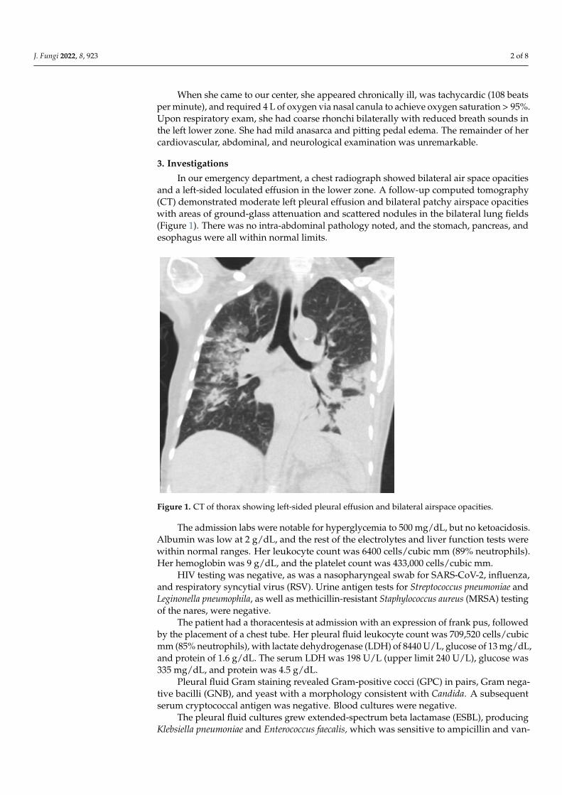
In our emergency department, a chest radiograph showed bilateral air space opacitiesand a left-sided loculated effusion in the lower zone. A follow-up computed tomography(CT) demonstrated moderate left pleural effusion and bilateral patchy airspace opacitieswith areas of ground-glass attenuation and scattered nodules in the bilateral lung fields(Figure 1). There was no intra-abdominal pathology noted, and the stomach, pancreas, andesophagus were all within normal limits.
J. Fungi 2022, 8, 923 2 of 8
When she came to our center, she appeared chronically ill, was tachycardic (108 beats per minute), and required 4 L of oxygen via nasal canula to achieve oxygen saturation >95%. Upon respiratory exam, she had coarse rhonchi bilaterally with reduced breath sounds in the left lower zone. She had mild anasarca and pitting pedal edema. The re-mainder of her cardiovascular, abdominal, and neurological examination was unremark-able.
3. Investigations In our emergency department, a chest radiograph showed bilateral air space opaci-
ties and a left-sided loculated effusion in the lower zone. A follow-up computed tomog-raphy (CT) demonstrated moderate left pleural effusion and bilateral patchy airspace opacities with areas of ground-glass attenuation and scattered nodules in the bilateral lung fields (Figure 1). There was no intra-abdominal pathology noted, and the stomach, pancreas, and esophagus were all within normal limits.
Figure 1. CT of thorax showing left-sided pleural effusion and bilateral airspace opacities.
The admission labs were notable for hyperglycemia to 500 mg/dL, but no ketoacido-sis. Albumin was low at 2 g/dL, and the rest of the electrolytes and liver function tests were within normal ranges. Her leukocyte count was 6400 cells/cubic mm (89% neutro-phils). Her hemoglobin was 9 g/dL, and the platelet count was 433,000 cells/cubic mm.
HIV testing was negative, as was a nasopharyngeal swab for SARS-CoV-2, influenza, and respiratory syncytial virus (RSV). Urine antigen tests for Streptococcus pneumoniae and Leginonella pneumophila, as well as methicillin-resistant Staphylococcus aureus (MRSA) test-ing of the nares, were negative.
The patient had a thoracentesis at admission with an expression of frank pus, fol-lowed by the placement of a chest tube. Her pleural fluid leukocyte count was 709,520 cells/cubic mm (85% neutrophils), with lactate dehydrogenase (LDH) of 8440 U/L, glucose of 13 mg/dL, and protein of 1.6 g/dL. The serum LDH was 198 U/L (upper limit 240 U/L), glucose was 335 mg/dL, and protein was 4.5 g/dL.
Pleural fluid Gram staining revealed Gram-positive cocci (GPC) in pairs, Gram neg-ative bacilli (GNB), and yeast with a morphology consistent with Candida. A subsequent serum cryptococcal antigen was negative. Blood cultures were negative.
Figure 1. CT of thorax showing left-sided pleural effusion and bilateral airspace opacities.
The admission labs were notable for hyperglycemia to 500 mg/dL, but no ketoacidosis.Albumin was low at 2 g/dL, and the rest of the electrolytes and liver function tests werewithin normal ranges. Her leukocyte count was 6400 cells/cubic mm (89% neutrophils).Her hemoglobin was 9 g/dL, and the platelet count was 433,000 cells/cubic mm.
HIV testing was negative, as was a nasopharyngeal swab for SARS-CoV-2, influenza,and respiratory syncytial virus (RSV). Urine antigen tests for Streptococcus pneumoniae andLeginonella pneumophila, as well as methicillin-resistant Staphylococcus aureus (MRSA) testingof the nares, were negative.
The patient had a thoracentesis at admission with an expression of frank pus, followedby the placement of a chest tube. Her pleural fluid leukocyte count was 709,520 cells/cubicmm (85% neutrophils), with lactate dehydrogenase (LDH) of 8440 U/L, glucose of 13 mg/dL,and protein of 1.6 g/dL. The serum LDH was 198 U/L (upper limit 240 U/L), glucose was335 mg/dL, and protein was 4.5 g/dL.
Pleural fluid Gram staining revealed Gram-positive cocci (GPC) in pairs, Gram nega-tive bacilli (GNB), and yeast with a morphology consistent with Candida. A subsequentserum cryptococcal antigen was negative. Blood cultures were negative.
The pleural fluid cultures grew extended-spectrum beta lactamase (ESBL), producingKlebsiella pneumoniae and Enterococcus faecalis, which was sensitive to ampicillin and van-

J. Fungi 2022, 8, 923 3 of 8
comycin. The yeast noted in the culture was initially identified as Candida firmetaria by ourmicrobiology laboratory based on an automated biochemical method (BD Phoenix). How-ever, analysis by matrix-assisted laser desorption/ionization time-of-flight mass spectrom-etry (MALDI-TOF MS) identified the isolate as Candida glabrata. The minimum inhibitoryconcentrations (MICs) for this organism are summarized in Table 1, as follows.
Table 1. MICs for the isolated C. glabrata.
DRUG MIC
Voriconazole 0.12 ug/mL
Anidulafungin 0.06 ug/mL
Caspofungin 0.06 ug/mL
Fluconazole 4 ug/mL
Itraconazole 0.5 ug/mL
Isavuconazole 0.12 ug/mL
Posaconazole 0.5 ug/mL
Micafungin 0.015 ug/mL
Amphotericin B (E-test) 0.19 ug/mL
5-Fluorocytosine (E-test) 0.016 ug/mL
C. glabrata is susceptible to fluconazole, voriconazole, micafungin, and amphotericin Baccording to the Clinical and Laboratory Standards Institute (CLSI) criteria.
4. Differential Diagnosis
The patient presented with a subacute course of respiratory and constitutional symp-toms 2 weeks after an initial diagnosis of COVID-19. Her physical examination wasconsistent with a consolidation in the lower left posterior lung. A radiographic examinationrevealed the presence of a multifocal necrotizing pneumonia with a left-sided empyemathoracis. An analysis of the pleural fluid revealed a polymicrobial infection, including ESBLK. pneumoniae, E. faecalis, and C. glabrata.
5. Treatment
The patient was initially started on empiric intravenous (IV) vancomycin at 1 g every24 h and piperacillin-tazobactam at 4.5 g every 8 h. IV micafungin at 100 mg every 24 hwas added on day two once the pleural fluid Gram stain was reviewed and the yeastswere identified. On day three of admission, IV meropenem at 500 mg every 6 h wasstarted in place of piperacillin-tazobactam in response to the K. pneumoniae susceptibilities.After 4 days of micafungin, the patient was switched to an oral voriconazole tablet of300 mg every 12 h for two loading doses, followed by 200 mg every 12 h for enhanced drugbioavailability in the pleural space. Voriconazole was empirically selected, as C. glabrataare generally susceptible to this azole, even in the setting of fluconazole resistance. Thepatient’s respiratory symptoms improved on this antimicrobial regimen.
6. Outcome and Follow-Up
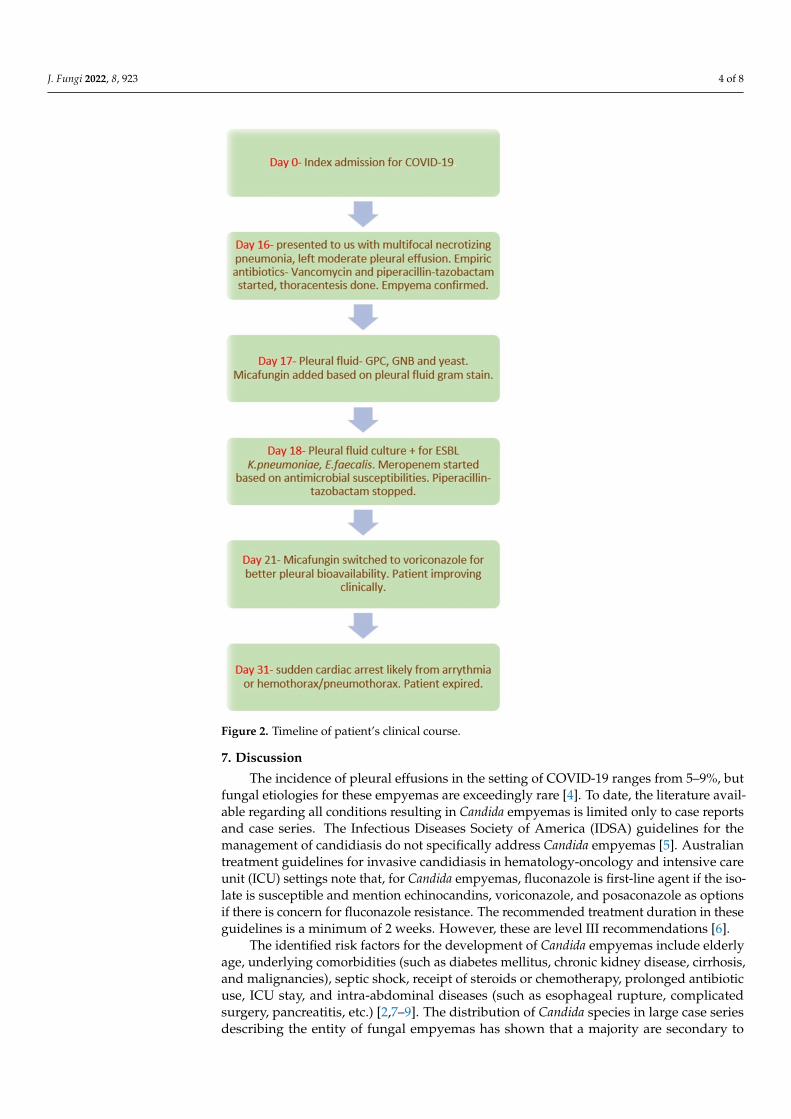
Unfortunately, after her initial clinical improvement, the patient developed an acutecardiac arrest on day ten of admission. Despite resuscitative efforts, the patient expired.The most likely cause of death was thought to be an arrythmia or a sudden hemothorax orpneumothorax. The patient’s family declined an autopsy. Figure 2 summarizes the clinicalcourse of this patient.
J. Fungi 2022, 8, 923 4 of 8J. Fungi 2022, 8, 923 4 of 8
Figure 2. Timeline of patient’s clinical course.
7. Discussion The incidence of pleural effusions in the setting of COVID-19 ranges from 5–9%, but
fungal etiologies for these empyemas are exceedingly rare [4]. To date, the literature availa-ble regarding all conditions resulting in Candida empyemas is limited only to case reports and case series. The Infectious Diseases Society of America (IDSA) guidelines for the man-agement of candidiasis do not specifically address Candida empyemas [5]. Australian treat-ment guidelines for invasive candidiasis in hematology-oncology and intensive care unit (ICU) settings note that, for Candida empyemas, fluconazole is first-line agent if the isolate is susceptible and mention echinocandins, voriconazole, and posaconazole as options if there is concern for fluconazole resistance. The recommended treatment duration in these guidelines is a minimum of 2 weeks. However, these are level III recommendations [6].
The identified risk factors for the development of Candida empyemas include elderly age, underlying comorbidities (such as diabetes mellitus, chronic kidney disease, cirrhosis,
Figure 2. Timeline of patient’s clinical course.
7. Discussion
The incidence of pleural effusions in the setting of COVID-19 ranges from 5–9%, butfungal etiologies for these empyemas are exceedingly rare [4]. To date, the literature avail-able regarding all conditions resulting in Candida empyemas is limited only to case reportsand case series. The Infectious Diseases Society of America (IDSA) guidelines for themanagement of candidiasis do not specifically address Candida empyemas [5]. Australiantreatment guidelines for invasive candidiasis in hematology-oncology and intensive careunit (ICU) settings note that, for Candida empyemas, fluconazole is first-line agent if the iso-late is susceptible and mention echinocandins, voriconazole, and posaconazole as optionsif there is concern for fluconazole resistance. The recommended treatment duration in theseguidelines is a minimum of 2 weeks. However, these are level III recommendations [6].
The identified risk factors for the development of Candida empyemas include elderlyage, underlying comorbidities (such as diabetes mellitus, chronic kidney disease, cirrhosis,and malignancies), septic shock, receipt of steroids or chemotherapy, prolonged antibioticuse, ICU stay, and intra-abdominal diseases (such as esophageal rupture, complicatedsurgery, pancreatitis, etc.) [2,7–9]. The distribution of Candida species in large case seriesdescribing the entity of fungal empyemas has shown that a majority are secondary to

J. Fungi 2022, 8, 923 5 of 8
C. albicans, while NAC are less common [2,7,8]. However, the ongoing shift in prevalenceof NAC may alter the epidemiology of this condition in the future. Concomitant bacterialempyemas are frequently present in patients with fungal empyemas. For example, in acase series from Taiwan with 63 patients, 49% had a bacterial infection, and 14% also hadfungemia [8]. Mortality rates are variable in these case series, ranging from 27 to 73% [2,7–9]. Itappears that pleural drainage, particularly in video-assisted thoracoscopic surgery (VATS),reduced the risk of mortality. In addition, higher mortality is seen in older studies, whereasnewer case series have improved outcomes, which could reflect improvements in overallICU care [2].
There are no studies that compare antifungals in empyema to demonstrate the supe-riority of one agent. This is unlike candidemia, where echinocandins are recommendedas empiric first-line management [5]. In a retrospective case series of eighty-one patientswith fungal empyemas at two tertiary care centers, echinocandins were identified as anindependent predictor of increased risk for mortality [2]. While the exact reasons for azolesbeing superior to echinocandins were not clear, the authors attributed the outcomes tothe antifungal pharmacokinetics of azoles being superior to echinocandins in achievingadequate levels in pleural fluid. The factors that influence drug concentration includepH, oxygen concentration, protein binding, degree of pleural inflammation, size of theeffusion, and presence of loculations [10]. In a study that looked at anidulafungin in themanagement of Candida empyemas, the ratio of the area under the concentration (AUC) tothe time curve between pleural fluid and plasma was only 12.5% [11]. Voriconazole andfluconazole were noted to have good penetration into the pleural fluid, producing troughconcentrations similar to paired plasma concentrations [2,12]. In contrast, echinocandinconcentrations within infected pleural fluid were between 9–15% and 57–67% of those inplasma [2,10]. However, the evidence at present remains insufficient to make any definitiverecommendation favoring azoles in the management of fungal empyemas. There are alsoscant data on the utility of pleural irrigation, although an isolated case report documentedthe instillation of antifungals [9]. Amphotericin B also reportedly had a poor pleural-to-serum concentration ratio [10]. Apart from these pharmacokinetic parameters, anotherimportant consideration in the choice of antifungals is that of antimicrobial resistance.Several NAC species are associated with resistance to azoles, especially fluconazole, butthese isolates are often susceptible to newer azoles, such as voriconazole, Posaconazole,and isavuconazole [3].
Another aspect of this case that merits attention is the challenges associated with the accurateidentification of Candida species—especially NAC species. Although colony color and morphologyon a chromogenic medium (CHROMagar) allow the ready differentiation of C. albicans and NACspecies, distinguishing specific NAC species is more difficult [13,14]. Identification panelssuch as BD Phoenix, Vitek, AuxaColor, etc. are miniaturized biochemical-based tests thatcontain a series of conventional, chromogenic, and fluorogenic tests, as well as growth-and enzymatic-based substrates. The microbial utilization and degradation of substratesare detected by chromogenic and fluorogenic indicators. The results are compared againsta standard. However, not all organisms of a particular strain are 100% uniform withrespect to some of the results of fermentation, oxidation, hydrolysis. Moreover, these testsmay also not be able to distinguish closely related organisms that may produce similarresults [15,16]. Thus, there is a small chance of erroneous identification, as encounteredin our patient. MALDI-TOF MS is a rapid and more reliable means of definitive Candidaspecies identification [17,18]. C. firmetaria, formerly known as C. lambica, which was theinitial identification by our laboratory, is a rare pathogen in humans with only a handful ofcase reports noted in the literature. It bears morphological similarity to C. kruseii [19–21].C. glabrata is a more common variety of NAC and has previously been described as a causeof Candida empyemas in patients without COVID-19 [2,7–9].
The fungal infections commonly described in the setting of COVID-19 includeCOVID-19-associated pulmonary aspergillosis (CAPA), COVID-associated mucormycosis(CAM), and COVID-associated candidiasis (CAC). CAPA has a highly variable cumulative

J. Fungi 2022, 8, 923 6 of 8
incidence in the ICU (from 1 to 40%) and an attributable mortality of around 33%, and thesecases have predominantly been due to Aspergillus fumigatus [22,23]. In the case of CAM,while the exact incidence is not known, it is close to 7 per 1000 COVID-19 cases, whichis over 50 times higher than the prior highest incidence data available. There has been aclear rise in cases globally during the pandemic, especially in developing countries, such asIndia, which already have a high baseline prevalence of mucormycosis. Most of these caseshave been noted in the diabetic population, and the mortality rates have ranged from 25 to50% [24].
COVID-19 as a risk factor for invasive candidiasis has only recently emerged. Ameta-analysis looking at COVID-19-associated candidiasis (CAC) found that the pooledprevalence of resistant NAC, specifically Candida auris, was 5.7% but with a mortality ashigh as 67% [25]. CAC can be due to two major groups of risk factors. The first groupincludes generic risk factors such as age, premorbid comorbidities, critical illness withartificial lines and tubes, parenteral nutrition, etc. The second group comprises moreCOVID-19-specific risks, which include the use of extracorporeal membrane oxygenation(ECMO), the administration of corticosteroids, and notably, COVID-19-mediated damage tothe lung epithelium. There are also reports of an association between the use of Interleukin-6 inhibitors, such as tocilizumab, and the development of candidiasis [1]. Candida colo-nization of the airway is also common in critically ill patients. One study looked at thecharacterization of Candida colonization and COVID-19, and polymerase chain reaction(PCR) sequencing demonstrated that sixty-nine out of one hundred patients had Candidaspecies in bronchoalveolar lavage fluid (BAL) specimens. While C. albicans colonization hadno discernable impact on COVID-19 severity, C. glabrata colonization did [26]. Moreover,respiratory Candida colonization was present in over one-third of patients with Candidaempyemas within 4 weeks of disease manifestation [7]. The growth of Candida in BALand sputum cultures is usually deemed to be from oral colonizers and, therefore, Candidapneumonitis is often untreated [26]. However, when Candida grows in blood cultures orpleural cultures, it cannot be disregarded as a colonizer or contaminant.
COVID-19-related Candida empyemas have been infrequently reported in the literature,and we identified three other cases, each with C. albicans. Table 2 summarizes the patientcharacteristics.
Table 2. Three prior reports of C. albicans empyemas associated with COVID-19.
Study Age, Gender ComorbidConditions Fungus Isolated COVID-19
ManagementEmpyemaTreatment
ConcomitantBacterialInfection
Outcome
Sharma et al. [27] 55, male Hypertension C. albicans Not mentionedTube
thoracostomy,micafungin
MRSA inrespiratory
culture
Unclear: stilladmitted at the
time ofpublication
Qasem et al. [28] 52, male None C. albicans ECMO Chest tube,decortication - Expired
Glendening et al. [29] 73, male Congestiveheart failure C. albicans Hydroxychloroquine
intubatedChest tube,fluconazole
Moraxellabacteremia Discharged
Our patient is the first description of empyema thoracis due to C. glabrata followingCOVID-19. Her risk factors included age, uncontrolled diabetes mellitus, and a receiptof steroids for COVID-19 treatment. She presented with a post-COVID-19 necrotizingbacterial pneumonia and pleural effusion that became super-infected with Candida. We didnot have a prior sputum culture on this patient to assess if she had known colonizationwith Candida. There was also no documentation of her exposure to any antimicrobials,except remdesivir, prior to her admission.
Our patient underscores the importance of considering fungal empyema in COVID-19patients who present with complicated pneumonias, as this disease requires prompt pleuraldrainage and antifungal treatment.

J. Fungi 2022, 8, 923 7 of 8
8. Conclusions
• Candida empyema thoracis is an invasive candidiasis that frequently occurs in theabsence of candidemia;
• Fungal empyema thoracis commonly presents as a polymicrobial infection, predomi-nantly with a concomitant bacterial infection;
• COVID-19 is a newly identified risk factor for fungal empyema;• The management of Candida empyemas involves prompt pleural drainage and sys-
temic antifungals;• Optimum treatment for non-bloodstream candidiasis may vary in important ways
from the management of candidemia, and there are pharmacologic reasons for predict-ing that azoles are superior to echinocandins in the management of Candida empyemas.
Author Contributions: N.S. and K.A.: drafted the paper; J.D.N. and M.J.A.: edited and proofreadthe manuscript. All authors have read and agreed to the published version of the manuscript.
Funding: This research received no external funding.
Institutional Review Board Statement: Not applicable.
Informed Consent Statement: Written informed consent was obtained from patient’s family (next-of-kin)since patient was deceased.
Data Availability Statement: Not applicable.
Conflicts of Interest: The authors declare no conflict of interest.
References1. Arastehfar, A.; Carvalho, A.; Nguyen, M.H.; Hedayati, M.T.; Netea, M.G.; Perlin, D.S.; Hoenigl, M. COVID-19-associated
candidiasis (CAC): An underestimated complication in the absence of immunological predispositions? J. Fungi 2020, 6, 211.Available online: https://pubmed.ncbi.nlm.nih.gov/33050019/ (accessed on 14 April 2022). [CrossRef] [PubMed]
2. Senger, S.S.; Thompson, G.R., 3rd; Samanta, P.; Ahrens, J.; Clancy, C.J.; Nguyen, M.H. Candida empyema thoracis at two academicmedical centers: New insights into treatment and outcomes. Open Forum Infect. Dis. 2021, 8, ofaa656. [CrossRef] [PubMed]
3. Kaur, H.; Singh, S.; Rudramurthy, S.M.; Ghosh, A.K.; Jayashree, M.; Narayana, Y.; Ray, P.; Chakrabarti, A. Candidaemia in atertiary care centre of developing country: Monitoring possible change in spectrum of agents and antifungal susceptibility. IndianJ. Med. Microbiol. 2020, 38, 109–116. [CrossRef] [PubMed]
4. Shi, H.; Han, X.; Jiang, N.; Cao, Y.; Alwalid, O.; Gu, J.; Fan, Y.; Zheng, C. Radiological findings from 81 patients with COVID-19pneumonia in Wuhan, China: A descriptive study. Lancet Infect. Dis. 2020, 20, 425–434. Available online: https://www.thelancet.com/article/S1473-3099(20)30086-4/fulltext (accessed on 14 April 2022). [CrossRef]
5. Pappas, P.G.; Kauffman, C.A.; Andes, D.R.; Clancy, C.J.; Marr, K.A.; Ostrosky-Zeichner, L.; Reboli, A.C.; Schuster, M.G.;Vazquez, J.A.; Walsh, T.J.; et al. Executive summary: Clinical practice guideline for the management of candidiasis: 2016 updateby the infectious diseases society of America. Clin. Infect. Dis. 2016, 62, 409–417. [CrossRef]
6. Keighley, C.; Cooley, L.; Morris, A.J.; Ritchie, D.; Clark, J.E.; Boan, P.; Worth, J.L.; Australasian Antifungal Guidelines SteeringCommittee. Consensus guidelines for the diagnosis and management of invasive candidiasis in haematology, oncology andintensive care settings, 2021. Intern. Med. J. 2021, 51, 89–117. [CrossRef]
7. Nigo, M.; Vial, M.R.; Munita, J.M.; Jiang, Y.; Tarrand, J.; Jimenez, C.A.; Kontoyiannis, D.P. Fungal empyema thoracis in cancerpatients. J. Infect. 2016, 72, 615–621. [CrossRef]
8. Lin, K.H.; Liu, Y.M.; Lin, P.C.; Ho, C.M.; Chou, C.H.; Wang, J.H.; Chi, C.-Y.; Ho, M.-W.; Wang, J.-H. Report of a 63-case seriesof Candida empyema thoracis: 9-year experience of two medical centers in central Taiwan. J. Microbiol. Immunol. Infect. 2014,47, 36–41. [CrossRef]
9. Ko, S.C.; Chen, K.Y.; Hsueh, P.R.; Luh, K.T.; Yang, P.C. Fungal empyema thoracis: An emerging clinical entity. Chest 2000,117, 1672–1678. [CrossRef]
10. Felton, T.; Troke, P.F.; Hope, W.W. Tissue penetration of antifungal agents. Clin. Microbiol. Rev. 2014, 27, 68–88. [CrossRef]11. Moriyama, B.; Ditullio, M.; Wilson, E.; Henning, S.A.; Penzak, S.R.; Danner, R.L.; Pennic, G.; Rinaldi, M.G.; Zelazny, A.M.;
Gea-Banacloche, J.; et al. Pharmacokinetics of anidulafungin in pleural fluid during the treatment of a patient with Candidaempyema. Antimicrob. Agents Chemother. 2011, 55, 2478–2480. [CrossRef] [PubMed]
12. Rieder-Nelissen, C.M.; Hasse, J.; Yeates, R.A.; Sarnow, E. Fluconazole concentrations in pulmonary tissue and pericardial fluid.Infection 1997, 25, 192–194. [CrossRef] [PubMed]
13. Mulet Bayona, J.V.; Salvador García, C.; Tormo Palop, N.; Valentín Martín, A.; González Padrón, C.; Colomina Rodríguez, J.;Peman, J.; Gimeno Cardona, C. Novel chromogenic medium CHROMagarTM Candida Plus for detection of Candida auris andother Candida species from surveillance and environmental samples: A multicenter study. J. Fungi 2022, 8, 281. [CrossRef]
J. Fungi 2022, 8, 923 8 of 8
14. Hospenthal, D.R.; Beckius, M.L.; Floyd, K.L.; Horvath, L.L.; Murray, C.K. Presumptive identification of Candida species otherthan C. albicans, C. krusei, and C. tropicalis with the chromogenic medium CHROMagar Candida. Ann. Clin. Microbiol. Antimicrob.2006, 5, 1. [CrossRef]
15. Posteraro, B.; Ruggeri, A.; De Carolis, E.; Torelli, R.; Vella, A.; De Maio, F.; Ricciardi, W.; Postteraro, P.; Sanguinetti, M. ComparativeEvaluation of BD Phoenix and Vitek 2 Systems for Species Identification of Common and Uncommon Pathogenic Yeasts. J. Clin.Microbiol. 2013, 51, 3841–5384. [CrossRef] [PubMed]
16. Posteraro, B.; Efremov, L.; Leoncini, E.; Amore, R.; Posteraro, P.; Ricciardi, W.; Sanguinetti, M. Are the Conventional CommercialYeast Identification Methods Still Helpful in the Era of New Clinical Microbiology Diagnostics? A Meta-Analysis of Their Accuracy.J. Clin. Microbiol. 2015, 53, 2439–2450. Available online: https://jcm.asm.org/content/53/8/2439 (accessed on 14 April 2022).[CrossRef] [PubMed]
17. Samantaray, S.; Singh, R. Evaluation of MALDI-TOF MS for identification of species in the Candida parapsilosis complex fromcandidiasis cases. J. Appl. Lab. Med. 2022, 7, 889–900. [CrossRef]
18. Xie, T.-A.; Liu, Y.-L.; Liang, C.; Huang, Y.-Y.; Li, J.-W.; Li, Z.-W.; Fan, S.-J.; Chen, J.-T.; Xia, Y.; Li, X.-Y.; et al. Accuracy ofmatrix-assisted LASER desorption ionization-time of flight mass spectrometry for identification of Candida. Biosci. Rep. 2019,39, BSR20190859. [CrossRef]
19. Vervaeke, S.; Vandamme, K.; Boone, E.; De Laere, E.; Swinne, D.; Surmont, I. A case of Candida lambica fungemia misidentifiedas Candida krusei in an intravenous drug abuser. Med. Mycol. 2008, 46, 853–856. [CrossRef]
20. Trowbridge, J.; Ludmer, L.M.; Riddle, V.D.; Levy, C.S.; Barth, W.F. Candida lambica polyarthritis in a patient with chronicalcoholism. J. Rheumatol. 1999, 26, 1846–1848.
21. Krüger, W.H.; Kröger, N.; Rüssmann, B.; Renges, H.; Kabisch, H.; Zander, A.R. Treatment of mycotic infections after haemopoieticprogenitor cell transplantation with liposomal amphotericin-B. Bone Marrow Transplant. 1998, 22, S10–S13. [PubMed]
22. Salmanton-García, J.; Sprute, R.; Stemler, J.; Bartoletti, M.; Dupont, D.; Valerio, M.; Garcia-Vidar, C.; Falces-Romero, I.;Machado, M.; de la Vila, S.; et al. COVID-19-Associated Pulmonary Aspergillosis, March-August 2020. Emerg. Infect. Dis.2021, 27, 1077–1086. [CrossRef] [PubMed]
23. Lamoth, F. Invasive aspergillosis in coronavirus disease 2019: A practical approach for clinicians. Curr. Opin. Infect. Dis. 2022,35, 163–169. [CrossRef] [PubMed]
24. Chandley, P.; Subba, P.; Rohatgi, S. COVID-19-Associated Mucormycosis: A Matter of Concern Amid the SARS-CoV-2 Pandemic.Vaccines 2022, 10, 1266. [CrossRef]
25. Vaseghi, N.; Sharifisooraki, J.; Khodadadi, H.; Nami, S.; Safari, F.; Ahangarkani, F.; Meis, J.M.; Badali, H.; Morovati, H. Globalprevalence and subgroup analyses of coronavirus disease (COVID-19) associated Candida auris infections (CACa): A systematicreview and meta-analysis. Mycoses 2022, 65, 683–703. [CrossRef]
26. Erami, M.; Raiesi, O.; Momen-Heravi, M.; Getso, M.I.; Fakhrehi, M.; Mehri, N.; Yarahmadi, M.; Amiri, S.; Raissi, V.; Hashemi, S.J.Clinical Impact of Candida Respiratory Tract Colonization and Acute Lung Infections in Critically Ill Patients with COVID-19Pneumonia. Microb. Pathog. 2022, 166, 105520. Available online: https://pubmed.ncbi.nlm.nih.gov/35405278/ (accessed on 14April 2022). [CrossRef]
27. Sharma, A.; Mehta, N.; Pirrotta, S.; Sheldon, D.; Juarez, R. A tale of two species: First reported case of an empyema secondary toco-infection with SARS-CoV-2 and candida albicans. Chest 2020, 158, A717. [CrossRef]
28. Qasem, N.M.; Barakat, M.; Elamin, N.; Bashir, M.; Hassan, I.F. Candida albicans empyema in COVID-19 infected patient: The firstreported case in Qatar. J. Emerg. Med. Trauma Acute Care 2022, 2022, 10.
29. Glendening, J.; Koroscil, M. A Report of Fungal Empyema Following Recovery of Severe SARS-CoV-2 Infection. Chest 2020,158, A566. Available online: https://journal.chestnet.org/article/S0012-3692(20)32718-5/fulltext (accessed on 14 April 2022).[CrossRef]
Related Documents