1 Structure-Based Design of 2-Arylamino-4-cyclohexylmethoxy-5- nitroso-6-aminopyrimidine Inhibitors of Cyclin-Dependent Kinase 2 Ian R. Hardcastle, a * Kerry L. Sayle, a Johanne Bentley, b A. Hilary Calvert, b William Clegg, c Nicola J. Curtin, b Jane A. Endicott, d Bernard T. Golding, c Karen Haggerty, a Ross W. Harrington, c Francesco Marchetti, a Veronique Mesguiche, a David R. Newell, b Martin E. M. Noble, d Rachel J. Parsons, a David J. Pratt, d Lan Z. Wang b and Roger J. Griffin a a Northern Institute for Cancer Research, School of Natural Sciences – Chemistry, Bedson Building, Newcastle University, Newcastle Upon Tyne NE1 7RU, UK b Northern Institute for Cancer Research, Paul O’Gorman Building, Medical School, Framlington Place, Newcastle University, Newcastle Upon Tyne, NE2 4HH, UK c School of Natural Sciences – Chemistry, Bedson Building, Newcastle University, Newcastle Upon Tyne NE1 7RU, UK d Laboratory of Molecular Biophysics and Department of Biochemistry, University of Oxford, Oxford OX1 3QU, UK *corresponding author: e-mail [email protected], tel 0191 222 6645

Welcome message from author
This document is posted to help you gain knowledge. Please leave a comment to let me know what you think about it! Share it to your friends and learn new things together.
Transcript
1
Structure-Based Design of 2-Arylamino-4-cyclohexylmethoxy-5-
nitroso-6-aminopyrimidine Inhibitors of
Cyclin-Dependent Kinase 2
Ian R. Hardcastle,a* Kerry L. Sayle,a Johanne Bentley,b A. Hilary Calvert,b William Clegg,c Nicola
J. Curtin,b Jane A. Endicott,d Bernard T. Golding,c Karen Haggerty,a Ross W. Harrington,c
Francesco Marchetti,a Veronique Mesguiche,a David R. Newell,b Martin E. M. Noble,d Rachel J.
Parsons,a David J. Pratt,d Lan Z. Wangb and Roger J. Griffina
aNorthern Institute for Cancer Research, School of Natural Sciences – Chemistry, Bedson Building, Newcastle University, Newcastle Upon Tyne NE1 7RU, UK
bNorthern Institute for Cancer Research, Paul O’Gorman Building, Medical School, Framlington Place, Newcastle University, Newcastle Upon Tyne, NE2 4HH, UK
cSchool of Natural Sciences – Chemistry, Bedson Building, Newcastle University, Newcastle Upon Tyne NE1 7RU, UK
dLaboratory of Molecular Biophysics and Department of Biochemistry, University of Oxford, Oxford OX1 3QU, UK
*corresponding author: e-mail [email protected], tel 0191 222 6645
2
ABSTRACT
An efficient synthesis of 2-substituted O4-cyclohexylmethyl-5-nitroso-6-aminopyrimidines from 6-amino-2-mercaptopyrimidin-4-ol has been developed and used to prepare a range of derivatives for evaluation as inhibitors of cyclin-dependent kinase 2 (CDK2). The structure activity relationships (SARs) are similar to those observed for the corresponding O6-cyclohexylmethoxypurine series with the 2-arylsulfonamide and 2-arylcarboxamide derivatives showing excellent potency. Two compounds, 4-(6-amino-4-cyclohexylmethoxy-5-nitrosopyrimidin-2-ylamino)-N-(2-hydroxyethyl)benzenesulfonamide (7q) and 4-(6-amino-4-cyclohexylmethoxy-5-nitrosopyrimidin-2-ylamino)-N-(2,3-dihydroxypropyl)benzenesulfonamide (7s), were the most potent with IC50 values of 0.7 ± 0.1 and 0.8 ± 0.0 nM against CDK2, respectively. The SARs determined in this study are discussed with reference to the crystal structure of 4-(6-amino-4-cyclohexylmethoxy-5-nitrosopyrimidin-2-ylamino)-N-(2,3-dihydroxypropyl)benzenesulfonamide (7j) bound to phosphorylated CDK2/cyclin A.
INTRODUCTION
The progression of cells through the cell-cycle is a highly ordered process, which is strictly controlled by the cyclin-dependent kinase (CDK) family of enzymes and their cyclin partners. Regulation of the serine-threonine kinase activity of specific CDKs, necessary to allow the cells to pass through cell-cycle checkpoints, is achieved by the binding of the cyclin partner and phosphorylation to produce the fully activated protein kinase complex.1 In cancer, the presence of oncogenic signalling pathways, or the absence of control resulting from the loss or mutation of tumour suppressor genes, inevitably results in abnormal cell cycle control and increased CDK/cyclin activity.2, 3 For this reason, the development of potent and selective CDK inhibitors has become an important therapeutic goal.4-6
The development of potent and selective small-molecule ATP-competitive kinase inhibitors has been successful for a number of therapeutic targets. New drugs have entered clinical use, notably imatinib that inhibits Bcr-Abl-kinase and c-Kit kinase, gefitinib and erlotinib which target the EGFR tyrosine kinase, and lapatinib, a dual EGFR, Her2 tyrosine kinase inhibitor.7 A large number small molecule inhibitors of CDK2 have been reported based on a structurally diverse range of scaffolds. These include derivatives of the monoheterocycles - pyrimidine,8 [1,2,4]triazole,9 pyrazole,10 pyridine11; biheterocycles – purine,12-15 [1,3,5]triazine-pyridine,16 1,4,5,6-tetrahydropyrrolo[3,4-c]pyrazole,17 pyrazolo[3,4-c]pyridazine,18 triazolo[1,5-a]pyrimidine,19 pyrazolo[1,5a]pyrimidine;20 triheterocycles - benzodipyrazole,21 aminoimidazo[1,2a]pyridine.22 All the inhibitors are competitive with ATP, and vary in their potency and selectivity among the other members of the CDK family and also other unrelated kinases.
The biological effects of inhibition of CDK2 have been studied using molecular genetic approaches in cell lines and knockout animals. Experiments in cell-lines have produced conflicting results, suggesting that CDK2 is not essential for proliferation in all situations.23, 24 Additionally, CDK2-knockout mice have been generated which are viable and apparently normal, other than being infertile.25, 26 These results cast doubt over the inhibition of CDK2 as a valuable therapeutic target. However, it should be noted that the absence or attenuation of a protein through genetic knockdown or knockout does not exactly mirror the situation where a protein is present at its usual cellular concentration, but is inactivated through a small-molecule inhibitor. Furthermore, a number of studies to date have demonstrated anti-tumour activity in tumour models with CDK2 inhibitors that have significant activity against other CDKs, including CDK4, CDK7 and CDK9,27-29 and the first generation of CDK inhibitors has entered clinical trials as anti-tumour agents.30
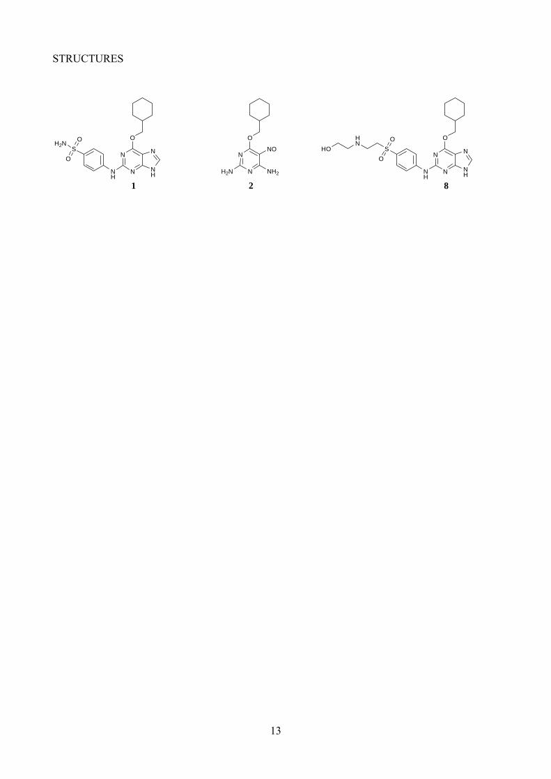
We have previously reported the purine NU6102 (1) as a highly potent and selective ATP-competitive inhibitor of CDK2 (Ki = 6 nM), developed from the lead compound O6-cyclohexylmethoxyguanine NU2058 (CDK2 Ki = 12 μM).31, 32 The X-ray crystal structure of 1
3
bound to CDK2 shows the purine core making a triplet of hydrogen bonds within the ATP binding site and two hydrogen bonds between the sulfonamide group and Asp86 of CDK2. We have also demonstrated previously that a series of nitrosopyrimidines are CDK2 inhibitors, e.g. NU6027 (2; CDK2 Ki = 1.3 μM). The nitroso group induces a ‘purine-mimetic’ conformation by hydrogen bonding to the adjacent NH2 group. The X-ray structure of 2 in complex with CDK2 shows the pyrimidine binding in a similar orientation to NU2058 and 1.12 A limited series of analogues of 2 that explored similar structural modifications to those leading to the development of 1 has been communicated.33 In this paper we report the synthesis of an expanded range of 2-arylamino-4-cyclohexylmethyl-5-nitroso-6-aminopyrimidines, based on 2. The structure-activity relationships for the inhibition of CDK2 are discussed with reference to an X-ray crystal structure of a key inhibitor bound to the CDK2/cyclinA protein complex.
<structures 1 and 2 here>
RESULTS AND DISCUSSION
Chemical Synthesis
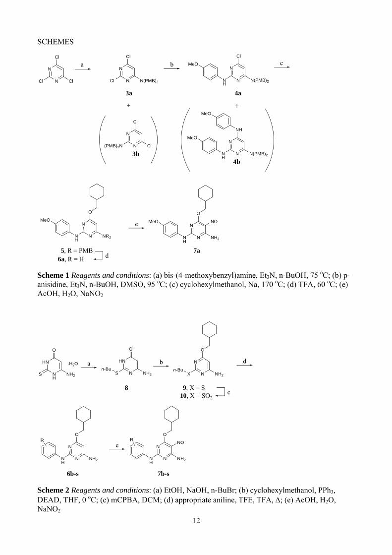
The initial route to the target 2,4-disubstituted pyrimidines used 2,4,6-trichloropyrimidine as the starting material. Substitution with bis-p-methoxybenzylamine in refluxing ethanol, gave both the 2- and 4-substituted pyrimidines (3a and 3b), which were separable by chromatography (Scheme 1). Unambiguous structure determination for 3a and 3b was not possible from their 1H and 13C NMR spectra, so an X-ray crystal structure was obtained for 3a that confirmed it as the desired isomer (Figure 1). A second substitution of the 2-substituted pyrimidine 3a with p-methoxyaniline was achieved under more forcing conditions, in refluxing n-butanol with triethylamine as base, giving the 2,4-disubstituted pyrimidine 4a, accompanied by the 2,6-disubstituted product 4b, in a 2:3 ratio. Again, X-ray crystallography was used to unambiguously assign the structure of 4a (Figure 1). The final substitution was carried out using sodium cyclohexylmethoxide in cyclohexylmethanol, giving the trisubstituted pyrimidine 5. Removal of the p-methoxybenzyl protecting groups with TFA, followed by nitrosation, gave 6-amino-2-anilino-4-cyclohexylmethoxy-5-nitrosopyrimidine (7a). An attempt to follow the same reaction scheme using 4-amino-N,N-dimethylbenzenesulfonamide, however, met with failure. The lack of regioselectivity in the initial substitution, and the formation of unwanted substitution products in the second step of Scheme 1, rendered this route unsuitable for the synthesis of a series of analogues.
<Scheme 1 here>
<Figure 1 here>
We considered that a more efficient synthesis could be achieved from 6-amino-2-mercaptopyrimidin-4-ol, which would allow the introduction of the anilino and cyclohexylmethoxy substituents and avoid the complications of regioselectivity seen in Scheme 1. The mercaptopyrimidine was alkylated to give the n-butyl sulfide (8) in excellent yield (Scheme 2), and then the cyclohexylmethoxy group was introduced under Mitsonubu conditions to give 9 in good yield. Direct displacement of the butylsulfide group from 9 with anilines proved unsuccessful, so the leaving group ability was improved by oxidation to the sulfone (10) using mCPBA, prior to displacement with the appropriate anilines to give the 2-arylaminopyrimidines 6b-s. The optimum conditions for the sulfone displacement were found to be with trifluoroethanol as solvent and five equivalents of TFA as catalyst, as described previously.34 Under these conditions, the displacements with various anilines proceeded in moderate to good yields. For the final step, nitrosation under standard conditions gave the desired 5-nitrosopyrimidines (7b-s).12
<Scheme 2 here>
4
<Table 1 here>
SAR Discussion
A series of 2-arylamino-5-nitrosopyrimidines was prepared and evaluated for CDK2 inhibitory activity and the results are shown in Table 1. Compounds lacking the 5-nitroso group (6b, 6e, 6g, 6h, and 6j) are at least 103 times less potent as CDK2 inhibitors compared with their 5-nitroso counterparts (7b, 7e, 7g, 7h, and 7j). These results are consistent with previous findings which showed that the 5-nitroso group forms an intramolecular hydrogen bond with the 6-amino group and orientates one of the amino NH bonds correctly to interact with the backbone carbonyl of Glu 81 of CDK2.12, 35
As predicted from the results in the comparable purine series,32 the nature and position of the substituent on the N2-aryl moiety has a profound effect on the CDK inhibitory activity. Small-lipophilic substituents at the 3-position (7b, 7d, 7e) produced modest improvements in activity compared with the parent compound (2, IC50 = 2.2 ± 0.6 μM for CDK2), whereas polar substituents attached by a methylene group (7g and 7i) resulted in up to 60-fold improvements in potency. In line with the purine series, the introduction of polar substituents at the 4-aryl position proved favourable. Compounds bearing 4-hydroxy or 4-carboxamido substituents (7c and 7j) showed 100-fold improvement in activity, whereas the 4-sulfonamido substituent 7m gave a 2000-fold improvement in activity over the parent 2.
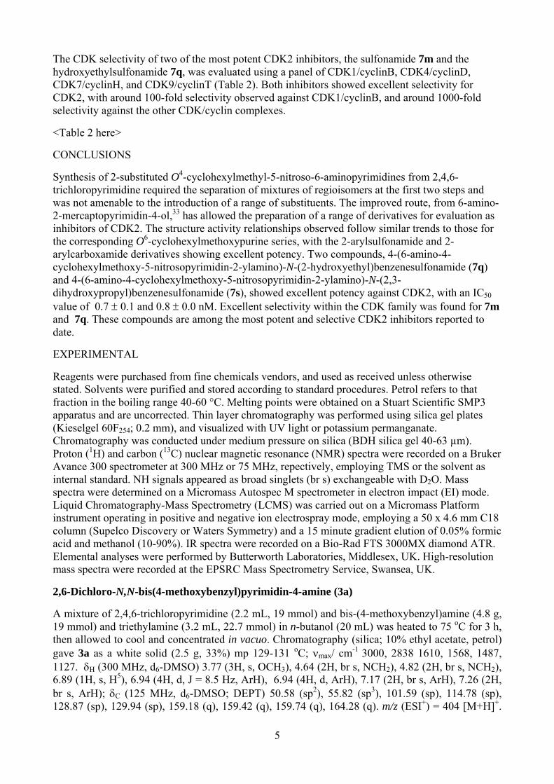
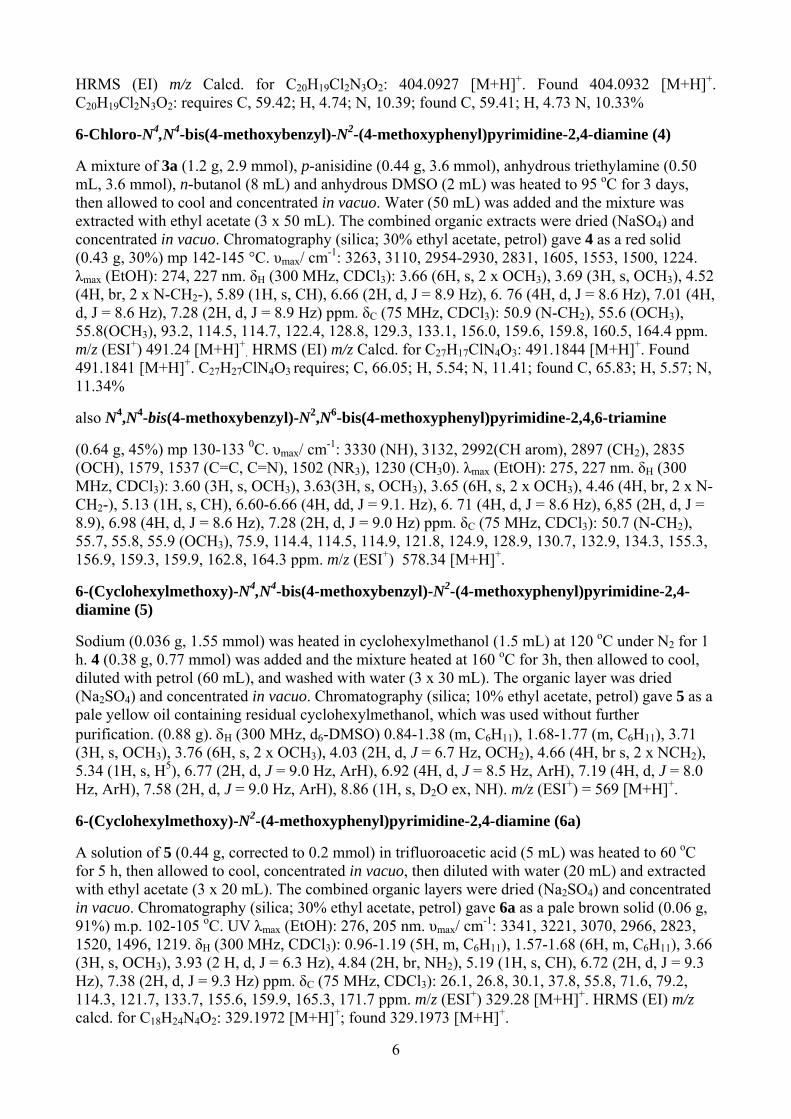
We have previously reported the X-ray crystal structure of carboxamide 7j bound to the CDK2/cyclin A phosphorylated on Thr160 (T160pCDK2/cyclin A) (Figure 2, PDB accession code 1OGU).8 Compounds 7j and 2 bind in a similar orientation within the CDK2 ATP-binding site. Compound 7j forms three hydrogen bonds with the hinge region of the enzyme, namely N6 to the peptide carbonyl of Glu81, and N1 and N2 to the backbone amide and carbonyl groups of Leu83, respectively. The 5-nitroso group forms the anticipated intramolecular hydrogen bond to N6, locking the molecule into a purine-like conformation. The N2 aryl ring stacks against the peptide backbone between His84 and Gln85. The orientation of the carboxamide substituent could not be determined unambiguously. However, the formation of an additional hydrogen bond from a carboxamide NH to the side chain of Asp86 is a reasonable interpretation of the data and is consistent both with the SARs and the observed structure of the sulfonamide 2. The interaction of at least one carboxamide or sulfonamide NH with CDK2 can be inferred from the loss of activity observed for the dialkylcarboxamides (7k and 7l) and the diethylsulfonamide (7n), consistent with the structural data for 7j.
<Figure 2 here>
The X-ray crystal structure of the T160pCDK2/cyclin A/purine 2 complex, shows that only one of the sulfonamide protons makes a hydrogen bond to Asp68, suggesting that substitution of the other position may be favourable.32 It was anticipated that the pyrimidines would behave similarly. The monoalkylsulfonamides (7o-s) retained potent activity, in particular against CDK2. The 2-hydroxyethyl- and 2,3-dihydroxypropylsulfonamides (7q and 7s) displayed a modest improvement in activity compared with the parent 7m, and are amongst the most potent CDK2 inhibitors reported to date. The improvement in activity suggests that additional interactions may be formed between the additional hydroxyl group(s) and the enzyme. The formation of additional favourable hydrogen bonding interactions, in the region accessed by the sulfonamide substituents, has been observed in the purine series. For example, the X-ray structure of sulfoxide 8 bound to CDK2 shows the amine and hydroxyl groups bound to the side-chain carboxyl group of Asp86.36
<Structure 8 here>
5
The CDK selectivity of two of the most potent CDK2 inhibitors, the sulfonamide 7m and the hydroxyethylsulfonamide 7q, was evaluated using a panel of CDK1/cyclinB, CDK4/cyclinD, CDK7/cyclinH, and CDK9/cyclinT (Table 2). Both inhibitors showed excellent selectivity for CDK2, with around 100-fold selectivity observed against CDK1/cyclinB, and around 1000-fold selectivity against the other CDK/cyclin complexes.
<Table 2 here>
CONCLUSIONS
Synthesis of 2-substituted O4-cyclohexylmethyl-5-nitroso-6-aminopyrimidines from 2,4,6-trichloropyrimidine required the separation of mixtures of regioisomers at the first two steps and was not amenable to the introduction of a range of substituents. The improved route, from 6-amino-2-mercaptopyrimidin-4-ol,33 has allowed the preparation of a range of derivatives for evaluation as inhibitors of CDK2. The structure activity relationships observed follow similar trends to those for the corresponding O6-cyclohexylmethoxypurine series, with the 2-arylsulfonamide and 2-arylcarboxamide derivatives showing excellent potency. Two compounds, 4-(6-amino-4-cyclohexylmethoxy-5-nitrosopyrimidin-2-ylamino)-N-(2-hydroxyethyl)benzenesulfonamide (7q) and 4-(6-amino-4-cyclohexylmethoxy-5-nitrosopyrimidin-2-ylamino)-N-(2,3-dihydroxypropyl)benzenesulfonamide (7s), showed excellent potency against CDK2, with an IC50 value of 0.7 ± 0.1 and 0.8 ± 0.0 nM. Excellent selectivity within the CDK family was found for 7m and 7q. These compounds are among the most potent and selective CDK2 inhibitors reported to date.
EXPERIMENTAL
Reagents were purchased from fine chemicals vendors, and used as received unless otherwise stated. Solvents were purified and stored according to standard procedures. Petrol refers to that fraction in the boiling range 40-60 °C. Melting points were obtained on a Stuart Scientific SMP3 apparatus and are uncorrected. Thin layer chromatography was performed using silica gel plates (Kieselgel 60F254; 0.2 mm), and visualized with UV light or potassium permanganate. Chromatography was conducted under medium pressure on silica (BDH silica gel 40-63 µm). Proton (1H) and carbon (13C) nuclear magnetic resonance (NMR) spectra were recorded on a Bruker Avance 300 spectrometer at 300 MHz or 75 MHz, repectively, employing TMS or the solvent as internal standard. NH signals appeared as broad singlets (br s) exchangeable with D2O. Mass spectra were determined on a Micromass Autospec M spectrometer in electron impact (EI) mode. Liquid Chromatography-Mass Spectrometry (LCMS) was carried out on a Micromass Platform instrument operating in positive and negative ion electrospray mode, employing a 50 x 4.6 mm C18 column (Supelco Discovery or Waters Symmetry) and a 15 minute gradient elution of 0.05% formic acid and methanol (10-90%). IR spectra were recorded on a Bio-Rad FTS 3000MX diamond ATR. Elemental analyses were performed by Butterworth Laboratories, Middlesex, UK. High-resolution mass spectra were recorded at the EPSRC Mass Spectrometry Service, Swansea, UK.
2,6-Dichloro-N,N-bis(4-methoxybenzyl)pyrimidin-4-amine (3a)
A mixture of 2,4,6-trichloropyrimidine (2.2 mL, 19 mmol) and bis-(4-methoxybenzyl)amine (4.8 g, 19 mmol) and triethylamine (3.2 mL, 22.7 mmol) in n-butanol (20 mL) was heated to 75 oC for 3 h, then allowed to cool and concentrated in vacuo. Chromatography (silica; 10% ethyl acetate, petrol) gave 3a as a white solid (2.5 g, 33%) mp 129-131 oC; νmax/ cm-1 3000, 2838 1610, 1568, 1487, 1127. δH (300 MHz, d6-DMSO) 3.77 (3H, s, OCH3), 4.64 (2H, br s, NCH2), 4.82 (2H, br s, NCH2), 6.89 (1H, s, H5), 6.94 (4H, d, J = 8.5 Hz, ArH), 6.94 (4H, d, ArH), 7.17 (2H, br s, ArH), 7.26 (2H, br s, ArH); δC (125 MHz, d6-DMSO; DEPT) 50.58 (sp2), 55.82 (sp3), 101.59 (sp), 114.78 (sp), 128.87 (sp), 129.94 (sp), 159.18 (q), 159.42 (q), 159.74 (q), 164.28 (q). m/z (ESI+) = 404 [M+H]+.
6
HRMS (EI) m/z Calcd. for C20H19Cl2N3O2: 404.0927 [M+H]+. Found 404.0932 [M+H]+. C20H19Cl2N3O2: requires C, 59.42; H, 4.74; N, 10.39; found C, 59.41; H, 4.73 N, 10.33%
6-Chloro-N4,N4-bis(4-methoxybenzyl)-N2-(4-methoxyphenyl)pyrimidine-2,4-diamine (4)
A mixture of 3a (1.2 g, 2.9 mmol), p-anisidine (0.44 g, 3.6 mmol), anhydrous triethylamine (0.50 mL, 3.6 mmol), n-butanol (8 mL) and anhydrous DMSO (2 mL) was heated to 95 oC for 3 days, then allowed to cool and concentrated in vacuo. Water (50 mL) was added and the mixture was extracted with ethyl acetate (3 x 50 mL). The combined organic extracts were dried (NaSO4) and concentrated in vacuo. Chromatography (silica; 30% ethyl acetate, petrol) gave 4 as a red solid (0.43 g, 30%) mp 142-145 °C. υmax/ cm-1: 3263, 3110, 2954-2930, 2831, 1605, 1553, 1500, 1224. λmax (EtOH): 274, 227 nm. δH (300 MHz, CDCl3): 3.66 (6H, s, 2 x OCH3), 3.69 (3H, s, OCH3), 4.52 (4H, br, 2 x N-CH2-), 5.89 (1H, s, CH), 6.66 (2H, d, J = 8.9 Hz), 6. 76 (4H, d, J = 8.6 Hz), 7.01 (4H, d, J = 8.6 Hz), 7.28 (2H, d, J = 8.9 Hz) ppm. δC (75 MHz, CDCl3): 50.9 (N-CH2), 55.6 (OCH3), 55.8(OCH3), 93.2, 114.5, 114.7, 122.4, 128.8, 129.3, 133.1, 156.0, 159.6, 159.8, 160.5, 164.4 ppm. m/z (ESI+) 491.24 [M+H]+
. HRMS (EI) m/z Calcd. for C27H17ClN4O3: 491.1844 [M+H]+. Found 491.1841 [M+H]+. C27H27ClN4O3 requires; C, 66.05; H, 5.54; N, 11.41; found C, 65.83; H, 5.57; N, 11.34%
also N4,N4-bis(4-methoxybenzyl)-N2,N6-bis(4-methoxyphenyl)pyrimidine-2,4,6-triamine
(0.64 g, 45%) mp 130-133 0C. υmax/ cm-1: 3330 (NH), 3132, 2992(CH arom), 2897 (CH2), 2835 (OCH), 1579, 1537 (C=C, C=N), 1502 (NR3), 1230 (CH30). λmax (EtOH): 275, 227 nm. δH (300 MHz, CDCl3): 3.60 (3H, s, OCH3), 3.63(3H, s, OCH3), 3.65 (6H, s, 2 x OCH3), 4.46 (4H, br, 2 x N-CH2-), 5.13 (1H, s, CH), 6.60-6.66 (4H, dd, J = 9.1. Hz), 6. 71 (4H, d, J = 8.6 Hz), 6,85 (2H, d, J = 8.9), 6.98 (4H, d, J = 8.6 Hz), 7.28 (2H, d, J = 9.0 Hz) ppm. δC (75 MHz, CDCl3): 50.7 (N-CH2), 55.7, 55.8, 55.9 (OCH3), 75.9, 114.4, 114.5, 114.9, 121.8, 124.9, 128.9, 130.7, 132.9, 134.3, 155.3, 156.9, 159.3, 159.9, 162.8, 164.3 ppm. m/z (ESI+) 578.34 [M+H]+.
6-(Cyclohexylmethoxy)-N4,N4-bis(4-methoxybenzyl)-N2-(4-methoxyphenyl)pyrimidine-2,4-diamine (5)
Sodium (0.036 g, 1.55 mmol) was heated in cyclohexylmethanol (1.5 mL) at 120 oC under N2 for 1 h. 4 (0.38 g, 0.77 mmol) was added and the mixture heated at 160 oC for 3h, then allowed to cool, diluted with petrol (60 mL), and washed with water (3 x 30 mL). The organic layer was dried (Na2SO4) and concentrated in vacuo. Chromatography (silica; 10% ethyl acetate, petrol) gave 5 as a pale yellow oil containing residual cyclohexylmethanol, which was used without further purification. (0.88 g). δH (300 MHz, d6-DMSO) 0.84-1.38 (m, C6H11), 1.68-1.77 (m, C6H11), 3.71 (3H, s, OCH3), 3.76 (6H, s, 2 x OCH3), 4.03 (2H, d, J = 6.7 Hz, OCH2), 4.66 (4H, br s, 2 x NCH2), 5.34 (1H, s, H5), 6.77 (2H, d, J = 9.0 Hz, ArH), 6.92 (4H, d, J = 8.5 Hz, ArH), 7.19 (4H, d, J = 8.0 Hz, ArH), 7.58 (2H, d, J = 9.0 Hz, ArH), 8.86 (1H, s, D2O ex, NH). m/z (ESI+) = 569 [M+H]+.
6-(Cyclohexylmethoxy)-N2-(4-methoxyphenyl)pyrimidine-2,4-diamine (6a)
A solution of 5 (0.44 g, corrected to 0.2 mmol) in trifluoroacetic acid (5 mL) was heated to 60 oC for 5 h, then allowed to cool, concentrated in vacuo, then diluted with water (20 mL) and extracted with ethyl acetate (3 x 20 mL). The combined organic layers were dried (Na2SO4) and concentrated in vacuo. Chromatography (silica; 30% ethyl acetate, petrol) gave 6a as a pale brown solid (0.06 g, 91%) m.p. 102-105 oC. UV λmax (EtOH): 276, 205 nm. υmax/ cm-1: 3341, 3221, 3070, 2966, 2823, 1520, 1496, 1219. δH (300 MHz, CDCl3): 0.96-1.19 (5H, m, C6H11), 1.57-1.68 (6H, m, C6H11), 3.66 (3H, s, OCH3), 3.93 (2 H, d, J = 6.3 Hz), 4.84 (2H, br, NH2), 5.19 (1H, s, CH), 6.72 (2H, d, J = 9.3 Hz), 7.38 (2H, d, J = 9.3 Hz) ppm. δC (75 MHz, CDCl3): 26.1, 26.8, 30.1, 37.8, 55.8, 71.6, 79.2, 114.3, 121.7, 133.7, 155.6, 159.9, 165.3, 171.7 ppm. m/z (ESI+) 329.28 [M+H]+. HRMS (EI) m/z calcd. for C18H24N4O2: 329.1972 [M+H]+; found 329.1973 [M+H]+.
7
6-(Cyclohexylmethoxy)-N2-(4-methoxyphenyl)-5-nitrosopyrimidine-2,4-diamine (7a)
To a solution of 6a (0.05 g, 0.15 mmol) in 30% acetic acid and water (5 mL) at 80 oC was added sodium nitrite (0.014 g, 0.2 mmol) in water (0.2 mL) giving a brown precipitate. Heating was continued 2 h then the mixture was allowed to cool to rt, and concentrated in vacuo. The residues were disolved in ethyl acetate (40 mL) and washed with Na2CO3 solution (3 x 20 mL) and water (2 x 20 mL). The combined organic layers were dried (Na2SO4) and concentrated in vacuo. Chromatography (silica; 5% methanol, ethyl acetate) followed by HPLC (C18, acetonitrile, water) gave 7a as a dark green solid (0.044 g, 26%) m.p. 168-171 oC. υmax/ cm-1: 3288, 3070, 2923, 2851, 1557, 1496, 1446, 1240. λmax (EtOH): 368, 292, 244 nm. δH (300 MHz, d6-acetone) 0.74-1.79 (11H, m, C6H11), 3.67 (3H, s, OCH3), 4.24 (2H, s, OCH2), 6.76 (2H, br s, ArH), 7.32 (1H, br s, D2O ex, NH), 7.66(2H, br s, ArH), 9.07 (1H, s, D2O ex, NH), 10.33 (1H, s, D2O ex, NH). m/z (ESI+) = 358 [M+H]+. δC (75 MHz, DMSO): 25.4, 26.3, 29.5, 37.2, 55.7, 72.3, 114.2, 123.5, 131.9, 140.4, 150.3, 156.5, 160.2, 171.6 ppm. m/z (ESI+) 358.26 [M+H]+. HRMS (ESI+) m/z calcd. for C18H23N5O3: 358.1874 [M+H]+; found 358.1878 [M+H]+. C18H23N5O3. requires C, 60.49; H, 6.49; N, 19.59; found C, 60.87; H, 6.49, N, 18.95%
6-Amino-2-n-butylsulfanyl-H3-pyrimidin-4-one (8)
6-Amino-2-mercaptopyrimidin-4-ol monohydrate (5.0 g, 31 mmol) was slurried in EtOH (30 mL) at 50 oC, and treated with NaOH (3.25 M, 10 mL, 32.1 mmol) and the mixture stirred 30 min. 1-Bromobutane (3.45 mL, 32.1 mmol) was added dropwise to the slurry and stirring continued 18 h. Water (10 mL) was added and the mixture stirred 30 min. After cooling to room temperature, the mixture was concentrated in vacuo giving 8 as an off-white solid (5.70g, 92%): m.p. 183-186 oC. Lit. 175 oC 37; νmax/ cm-1 1570, (C=N), 1601 (C=C), 2858-2928 (CH3, CH2), 3271, 3464 (OH, NH2). δH (300 MHz, d6-DMSO) 0.89 (3H, t, J = 7 Hz, CH3), 1.31-1.43 (2H, m, CH3CH2), 1.53-1.63 (2H, m, CH3CH2CH2), 3.06 (2H, t, J = 7 Hz, SCH2), 4.87 (1H, s, H5), 6.43 (2H, br s, NH2, exchangeable with D2O); δC (125 MHz, d6-DMSO) 13.5 (CH3), 21.3 (CH3CH2), 29.0 (CH3CH2CH2CH2S), 31.0 (CH3CH2CH2CH2S), 81.1 (C5), 163.5 (C6). m/z (ESI+) = 200 [M+H]+.
2-n-Butylsulfanyl-4-cyclohexylmethoxypyrimidin-6-ylamine (9)
To a mixture of 8 (1.50 g, 7.53 mmol), cyclohexylmethanol (1.39 mL, 11.30 mmol), and PPh3 (2.96 g, 11.30 mmol) in THF (50 mL) was added DEAD (1.78 mL, 11.30) dropwise over 20 min at 0 oC, and stirring continued 24 h. The mixture was concentrated in vacuo yielding a yellow oil which was stirred in diethyl ether at 0 oC, forming a white precipitate, which was removed by filtration. The filtrate was collected and concentrated in vacuo. Chromatography (silica;20% ethyl acetate, petrol) followed by recrystallisation (MeOH) gave 9 as a white solid (1.60 g, 72%): m.p. 79-82 oC; νmax/ cm-1 1547, 1583 (C=N), 1631 (C=C), 2850-3156 (CH3, CH2), 3294-3425 (NH2). δH (200 MHz, d6-DMSO) 1.08 (3H, t, J = 7 Hz, CH3), 1.22-1.62 (9H, m, C6H11+ CH3CH2CH2), (6H, m, C6H11), 3.12 (2H, t, J = 7 Hz, SCH2), 4.18 (2H, d, J = 6 Hz, OCH2), 5.57 (1H, s, H5), 6.80 (2H, br s, NH2, exchangeable with D2O) ppm; δC (50 MHz, d6-DMSO) 13.5 (CH3), 21.5 (CH3CH2), 25.5 (C6H11), 26.3 (C6H11), 29.3 (C6H11), 31.6 (C6H11), 32.1 (CH3CH2CH2CH2S), 36.89 (CH3CH2CH2CH2S), 66.6, 70.3 (OCH2), 81.57 (C5), 165.14 (C2), 168.79 (C6), 169.17 (C4) ppm. m/z (ESI+) = 296 [M+H]+. C15H25N3OS requires: C, 60.98; H, 8.53; N, 14.22; S, 10.85%; found: C, 61.01; H, 8.52; N, 13.85; S, 10.42.
2-(n-Butane-1-sulfonyl)-4-cyclohexylmethoxypyrimidin-6-ylamine (10)
8
To a stirred solution of 9 (1.0 g, 3.38 mmol) in DCM (20 mL) was added m-CPBA (2.34 g, 13.5 mmol) and stirring continued for 17 h. The mixture was concentrated in vacuo giving a yellow solid which was extracted into ethyl acetate (2 x 20 mL). The combined extracts were washed with saturated sodium sulfite solution (30 mL) and aqueous NaHCO3 solution (30 mL), then dried (Na2SO4) and concentrated in vacuo yielding 10 as an off-white solid (0.99 g, 89%). Recrystallisation (ethyl acetate, petrol) gave a white solid: m.p. 141-143 oC; νmax/ cm-1 1130 (SO2 sym str.), 1300 (SO2 asym. str.) 1595 (C=N), 1635 (C=C), 2852-3212 (CH3, CH2), 3323, 3424 (NH2). δH (300 MHz, CDCl3) 0.89 (3H, t, J = 7 Hz, CH3), 1.15-1.18 (5H, m, C6H11), 1.41 (2H, q, J = 7, 15 Hz, CH3CH2CH2), 1.66-1.79 (8H, m, C6H11+ CH3CH2CH2), 3.32-3.38 (2H, m, SO2CH2), 4.05 (2H, d, J = 6 Hz, OCH2), 5.48 (2H, br s, NH2, exchangeable with D2O), 5.75 (1H, s, H5) ppm; δC (75 MHz, CDCl3) 14.0 (CH3), 22.2 (CH3CH2), 24.5 (CH3CH2CH2), 26.7 (C6H11), 27.0 (C6H11), 30.1 (C6H11), 37.7 (C6H11), 51.1 (CH2SO2), 73.0 (OCH2), 89.1 (C5), 164.7 (C2), 165.5 (C6), 171.2 (C4). m/z (ESI+) = 328 [M+H]+. C15H25N3O3S requires: C, 55.02; H, 7.70; N, 12.83; found C, 54.88; H, 7.71; N, 12.63%.
General Procedure A.
To solution of 10 (0.20 g, 0.61 mmol) and the appropriate aniline (2 mol. eq.) in TFE (4 mL) was added TFA (0.24 mL, 3.05 mmol). The mixture was stirred for 10 min at rt, then heated under reflux for a further 2 h, and concentrated in vacuo yielding a white solid. The solid was extracted into ethyl acetate (2 x 20 mL). The combined extracts were washed with copious amounts of water (100 mL), then dried (Na2SO4), and concentrated in vacuo.
4-(6-Amino-4-cyclohexylmethoxypyrimidin-2-ylamino)benzenesulfonamide (6m)
General Procedure A: C (0.30 g, 0.92 mmol), 4-aminobenzenesulfonamide (0.32 g, 1.84 mmol), TFE (3 mL), TFA (0.38 mL, 4.60 mmol). HPLC (C18; methanol, water)gave 6m as a white solid (0.24 g, 69%): m.p. 177-179 oC; νmax/ cm-1 1564 (C=C, C=N str.), 1620 (SO2NH2, NH2 def.), 2853-2925 (CH2), 3104-3216 (=C-H str.), 3346 (asym + sym N-H str.), 3471 (NH2); λmax = 210 and 296 nm. δH (300 MHz d6-DMSO) 0.98-1.26 (5H, m, C6H11), 1.68-1.78 (6H, m, C6H11), 4.01 (2H, d, J = 6 Hz, CH2O), 5.29 (1H, s, H5), 6.43 (2H, s, NH2 exchangeable with D2O), 7.13 (2H, s, NH2 exchangeable with D2O), 7.63 (2H, d, J = 9 Hz, ArH), 7.93 (2H, d, J = 9 Hz, ArH), 9.34 (NH, exchangeable with D2O) ppm. m/z (ESI+) = 378 [M+H]+. C17H23N5O3S·0.33CH3OH requires: C, 53.54; H, 6.32; N, 18.04; found: C, 53.50; H, 6.20; N, 18.04%.
4-(6-Amino-4-cyclohexylmethoxypyrimidin-2-ylamino)-N-(2-hydroxyethyl)benzenesulfonamide (6q)
General Procedure A: 9 (0.11 g, 0.33 mmol), amino-N-(2-hydroxyethyl)benzenesulfonamide (0.14 g, 0.65 mmol) TFE (3 mL), TFA (0.13 mL, 1.63 mmol). HPLC (C18; methanol, water) gave 6q as a white solid (0.09 g, 0.22 mmol, 66%). m/z (ESI+) = 422 [M+H]+.
General Procedure B: Nitrosation.
The appropriate pyrimidine was dissolved in aqueous acetic acid (30%; 3 mL) and the solution was heated to 80 oC, then sodium nitrite (0.02 g, 0.35 mmol) in water (0.2 mL) was added dropwise. The mixture was stirred for a further 2 h, then extracted into ethyl acetate (30 mL). The extract was washed with water (30 mL), dried (Na2SO4) and concentrated in vacuo.
4-(6-Amino-4-cyclohexylmethoxy-5-nitrosopyrimidin-2-ylamino)benzenesulfonamide (7m)
General Procedure B: 6m (0.08 g, 0.20 mmol). HPLC (C18; methanol, water) gave 7m as a green solid (0.02 g, 25%): m.p. 159-162 oC; νmax/ cm-1 1528 (N=O str.), 1633 (NH2 def.), 2846-3040 (-CH2-), 3280 (asym + sym N-H str.), 3364 (NH2); λmax = 362 nm. δH (300 MHz d6-DMSO) 1.00-
9
1.23 (5H, m, C6H11), 1.71-1.88 (6H, m, C6H11), 4.43 (2H, d, J = 6 Hz, CH2O), 7.29 (2H, s, NH2 exchangeable with D2O), 7.73 (2H, d, J = 8 Hz, ArH), 8.00 (2H, s br, ArH), 8.60 (NH, exchangeable with D2O), 10.25 (1H, s, NH, exchangeable with D2O), 10.60 (NH, exchangeable with D2O) ppm. m/z (ESI+) = 407 [M+H]+. C17H22N6O4S·0.5CH3CO2H requires: C, 49.53; H, 5.54; N, 19.25; found: C, 49.44; H, 5.37; N, 19.53%.
4-(6-Amino-4-cyclohexylmethoxy-5-nitrosopyrimidin-2-ylamino)-N-(2-hydroxyethyl)benzenesulfonamide (7q)
General Procedure B: 6q (0.09 g, 0.22 mmol). Recrystallisation (MeOH) gave 7q as a green solid: m.p. 193-195 °C; νmax/ cm-1 1522 (N=O str.), 1576 (C=N, C=C), 2852-2926 (-CH2-), 3100-3267 (NH2, OH); λmax = 362, 263, 239 and 208 nm. δH (300 MHz d6-DMSO) 1.05-1.30 (5H, m, C6H11), 1.50-1.88 (6H, m, C6H11), 2.78 (2H, q, J = 6 Hz, NHCH2), 3.36 (2H, t, CH2OH), 4.39 (2H, d, J = 6 Hz, CH2O), 4.68 (1H, t, OH exchangeable with D2O), 7.40 (1H, t, NHCH2 exchangeable with D2O), 7.71 (2H, d, J = 8 Hz, ArH), 7.97 (2H, d, J = 8 Hz, ArH) 8.50 (1H, s, NH exchangeable with D2O) ppm. m/z (ESI+) = 451 [M+H]+. C19H26N6O5S·0.5CH3CO2H requires: C, 49.99; H, 5.87; N, 17.49; found: C, 49.67; H, 5.26; N, 17.60%.
Biological Evaluation
Compounds were assayed for the inhibition of human cyclin-dependent kinases 1 and 2, as described previously.12 The final ATP concentration within the assay was 12.5 μM.
X-Ray Crystallography§
Crystals of 3a and 4a were small and weakly diffracting, and data were collected at 120 K with synchrotron radiation (λ = 0.8462 Å) at station 16.2SMX of the Synchrotron Radiation Source at Daresbury Laboratory, through the EPSRC National Crystallography Service. Crystal data for 3a: C20H19Cl2N3O2, Mr = 404.3, monoclinic, space group P21/c, a = 21.9143(10), b = 10.1032(5), c = 8.7088(4) Å, β = 98.099(1)°, V = 1908.94(16) Å3, Z = 4, μ = 0.36 mm−1, 13035 data measured, 3886 unique (Rint = 0.029), 246 refined parameters, R (F, F2 > 2σ) = 0.038, Rw (F2, all data) = 0.098, S = 1.03, final difference map within ±0.29 e Å−3. Crystal data for 4a: C27H27ClN4O3, Mr = 491.0, monoclinic, space group C2/c, a = 16.9620(14), b = 10.7877(9), c = 27.228(2) Å, β = 98.375(1)°, V = 4929.0(7) Å3, Z = 8, μ = 0.19 mm−1, 15124 data measured, 5026 unique (Rint = 0.042), 323 refined parameters, R (F, F2 > 2σ) = 0.045, Rw (F2, all data) = 0.117, S = 1.06, final difference map within ±0.31 e Å−3.
ACKNOWLEDGEMENTS
The authors thank Cancer Research UK, AstraZeneca, the EPSRC (Studentship to K.L.S.), the
BBSRC (Studentship to D.J.P.), and the MRC for financial support, and the EPSRC and CCLRC
for funding of the National Crystallography Service and access to SRS diffraction facilities. We
also acknowledge the use of the EPSRC Mass Spectrometry Service at the University of Wales
(Swansea).
§ CCDC reference numbers 000000–000000. See http://www.rsc.org/suppdata/ob/00/00000000/ for
crystallographic data in .cif or other electronic format.
REFERENCES
1. D. O. Morgan, Annual Review of Cell and Developmental Biology, 1997, 13, 261-291.
10
2. G. DelSal, M. Loda and M. Pagano, Crit. Rev. Oncog., 1996, 7, 127-142. 3. C. J. Sherr, Science, 1996, 274, 1672-1677. 4. T. M. Sielecki, J. F. Boylan, P. A. Benfield and G. L. Trainor, J. Med. Chem., 2000, 43, 1-
18. 5. G. I. Shapiro, J Clin Oncol, 2006, 24, 1770-1783. 6. G. K. Schwartz and M. A. Shah, J Clin Oncol, 2005, 23, 9408-9421. 7. J. Dancey and E. A. Sausville, Nat. Rev. Drug Disc., 2003, 2, 296-313. 8. K. L. Sayle, J. Bentley, F. T. Boyle, A. H. Calvert, Y. Z. Cheng, N. J. Curtin, J. A. Endicott,
B. T. Golding, I. R. Hardcastle, P. Jewsbury, V. Mesguiche, D. R. Newell, M. E. M. Noble, R. J. Parsons, D. J. Pratt, L. Z. Wang and R. J. Griffin, Bioorg. Med. Chem. Lett., 2003, 13, 3079-3082.
9. R. H. Lin, P. J. Connolly, S. L. Huang, S. K. Wetter, Y. H. Lu, W. V. Murray, S. L. Emanuel, R. H. Gruninger, A. R. Fuentes-Pesquera, C. A. Rugg, S. A. Middleton and L. K. Jolliffe, J. Med. Chem., 2005, 48, 4208-4211.
10. P. Pevarello, M. G. Brasca, P. Orsini, G. Traquandi, A. Longo, M. Nesi, F. Orzi, C. Piutti, P. Sansonna, M. Varasi, A. Cameron, A. Vulpetti, F. Roletto, R. Alzani, M. Ciomei, C. Albanese, W. Pastori, A. Marsiglio, E. Pesenti, F. Fiorentini, J. R. Bischoff and C. Mercurio, J. Med. Chem., 2005, 48, 2944-2956.
11. R. H. Lin, Y. H. Lu, S. K. Wetter, P. J. Connolly, I. J. Turchi, W. V. Murray, S. L. Emanuel, R. H. Gruninger, A. R. Fuentes-Pesquera, M. Adams, N. Pandey, S. Moreno-Mazza, S. A. Middleton and L. K. Jolliffe, Bioorg. Med. Chem. Lett., 2005, 15, 2221-2224.
12. C. E. Arris, F. T. Boyle, A. H. Calvert, N. J. Curtin, J. A. Endicott, E. F. Garman, A. E. Gibson, B. T. Golding, S. Grant, R. J. Griffin, P. Jewsbury, L. N. Johnson, A. M. Lawrie, D. R. Newell, M. E. M. Noble, E. A. Sausville, R. Schultz and W. Yu, J. Med. Chem., 2000, 43, 2797-2804.
13. Y. T. Chang, N. S. Gray, G. R. Rosania, D. P. Sutherlin, S. Kwon, T. C. Norman, R. Sarohia, M. Leost, L. Meijer and P. G. Schultz, Chem. Biol., 1999, 6, 361-375.
14. P. Imbach, H. G. Capraro, P. Furet, H. Mett, T. Meyer and J. Zimmermann, Bioorg. Med. Chem. Lett., 2000, 10, 1001-1001.
15. M. Legraverend, P. Tunnah, M. Noble, P. Ducrot, O. Ludwig, D. S. Grierson, M. Leost, L. Meijer and J. Endicott, J. Med. Chem., 2000, 43, 1282-1292.
16. G. H. Kuo, A. DeAngelis, S. Emanuel, A. H. Wang, Y. Zhang, P. J. Connolly, X. Chen, R. H. Gruninger, C. Rugg, A. Fuentes-Pesquera, S. A. Middleton, L. Jolliffe and W. V. Murray, J. Med. Chem., 2005, 48, 4535-4546.
17. P. Pevarello, D. Fancelli, A. Vulpetti, R. Amici, M. Villa, V. Pittala, P. Vianello, A. Cameron, M. Ciomei, C. Mercurio, J. R. Bischoff, F. Roletto, M. Varasi and M. G. Brasca, Bioorg. Med. Chem. Lett., 2006, 16, 1084-1090.
18. M. F. Brana, M. Cacho, M. L. Garcia, E. P. Mayoral, B. Lopez, B. de Pascual-Teresa, A. Ramos, N. Acero, F. Llinares, D. Munoz-Mingarro, O. Lozach and L. Meijer, J. Med. Chem., 2005, 48, 6843-6854.
19. C. M. Richardson, D. S. Williamson, M. J. Parratt, J. Borgognoni, A. D. Cansfield, P. Dokurno, G. L. Francis, R. Howes, J. D. Moore, J. B. Murray, A. Robertson, A. E. Surgenor and C. J. Torrance, Bioorg. Med. Chem. Lett., 2006, 16, 1353-1357.
20. D. S. Williamson, M. J. Parratt, J. F. Bower, J. D. Moore, C. M. Richardson, P. Dokurno, A. D. Cansfield, G. L. Francis, R. J. Hebdon, R. Howes, P. S. Jackson, A. M. Lockie, J. B. Murray, C. L. Nunns, J. Powles, A. Robertson, A. E. Surgenor and C. J. Torrance, Bioorg. Med. Chem. Lett., 2005, 15, 863-867.
21. R. D'Alessio, A. Bargiotti, S. Metz, M. G. Brasca, A. Cameron, A. Ermoli, A. Marsiglio, P. Polucci, F. Roletto, M. Tibolla, M. L. Vazquez, A. Vulpetti and P. Pevarello, Bioorg. Med. Chem. Lett., 2005, 15, 1315-1319.
11
22. C. Hamdouchi, B. Zhong, J. Mendoza, E. Collins, C. Jaramillo, J. E. De Diego, D. Robertson, C. D. Spencer, B. D. Anderson, S. A. Watkins, F. M. Zhang and H. B. Brooks, Bioorg. Med. Chem. Lett., 2005, 15, 1943-1947.
23. O. Tetsu and F. McCormick, Cancer Cell, 2003, 3, 233-245. 24. J. Du, H. R. Widlund, M. A. Horstmann, S. Ramaswamy, K. Ross, W. E. Huber, E. K.
Nishimura, T. R. Golub and D. E. Fisher, Cancer Cell, 2004, 6, 565-576. 25. C. Berthet, E. Aleem, V. Coppola, L. Tessarollo and P. Kaldis, Current Biology, 2003, 13,
1775-1785. 26. S. Ortega, I. Prieto, J. Odajima, A. Martín, P. Dubus, R. Sotillo, J. L. Barbero, M.
Malumbres and M. Barbacid, Nature Genetics, 2003, 35, 25 - 31. 27. S. J. McClue, D. Blake, R. Clarke, A. Cowan, L. Cummings, P. M. Fischer, M. MacKenzie,
J. Melville, K. Stewart, S. D. Wang, N. Zhelev, D. Zheleva and D. P. Lane, Int. J. Cancer, 2002, 102, 463-468.
28. R. N. Misra, H. Y. Xiao, K. S. Kim, S. F. Lu, W. C. Han, S. A. Barbosa, J. T. Hunt, D. B. Rawlins, W. F. Shan, S. Z. Ahmed, L. G. Qian, B. C. Chen, R. L. Zhao, M. S. Bednarz, K. A. Kellar, J. G. Mulheron, R. Batorsky, U. Roongta, A. Kamath, P. Marathe, S. A. Ranadive, J. S. Sack, J. S. Tokarski, N. P. Pavletich, F. Y. F. Lee, K. R. Webster and S. D. Kimball, J. Med. Chem., 2004, 47, 1719-1728.
29. X. J. Chu, W. DePinto, D. Bartkovitz, S. S. So, B. T. Vu, K. Packman, C. Lukacs, Q. J. Ding, N. Jiang, K. Wang, P. Goelzer, X. F. Yin, M. A. Smith, B. X. Higgins, Y. S. Chen, Q. Xiang, J. Moliterni, G. Kaplan, B. Graves, A. Lovey and N. Fotouhi, J. Med. Chem., 2006, 49, 6549-6560.
30. P. M. Fischer and A. Gianella-Borradori, Exp. Opin. Invest. Drugs, 2003, 12, 955-970. 31. T. G. Davies, J. Bentley, C. E. Arris, F. T. Boyle, N. J. Curtin, J. A. Endicott, A. E. Gibson,
B. T. Golding, R. J. Griffin, I. R. Hardcastle, P. Jewsbury, L. N. Johnson, V. Mesguiche, D. R. Newell, M. E. M. Noble, J. A. Tucker, L. Wang and H. J. Whitfield, Nat. Struct. Biol., 2002, 9, 745-749.
32. I. R. Hardcastle, C. E. Arris, J. Bentley, F. T. Boyle, Y. H. Chen, N. J. Curtin, J. A. Endicott, A. E. Gibson, B. T. Golding, R. J. Griffin, P. Jewsbury, J. Menyerol, V. Mesguiche, D. R. Newell, M. E. M. Noble, D. J. Pratt, L. Z. Wang and H. J. Whitfield, J. Med. Chem., 2004, 47, 3710-3722.
33. K. L. Sayle, V. Mesguishe, R. J. Parsons, J. Bentley, T. G. Davies, J. A. Endicott, M. E. M. Noble, L. Z. Wang, I. R. Hardcastle and B. T. Golding, Eur. J. Cancer, 2002, 38, 394.
34. H. J. Whitfield, R. J. Griffin, I. R. Hardcastle, A. Henderson, J. Meneyrol, V. Mesguiche, K. L. Sayle and B. T. Golding, Chem. Commun., 2003, 2802-2803.
35. V. Mesguiche, R. J. Parsons, C. E. Arris, J. Bentley, F. T. Boyle, N. J. Curtin, T. G. Davies, J. A. Endicott, A. E. Gibson, B. T. Golding, R. J. Griffin, P. Jewsbury, L. N. Johnson, D. R. Newell, M. E. M. Noble, L. Z. Wang and I. R. Hardcastle, Bioorg. Med. Chem. Lett., 2003, 13, 217-222.
36. R. J. Griffin, A. Henderson, N. J. Curtin, A. Echalier, J. A. Endicott, I. R. Hardcastle, D. R. Newell, M. E. M. Noble, L. Z. Wang and B. T. Golding, J. Am. Chem. Soc., 2006, 128, 6012-6013.
37. G. Biagi, A. Costantini, L. Costantino, I. Giorgi, O. Livi, P. Pecorari, M. Rinaldi and V. Scartoni, J. Med. Chem., 1996, 39, 2529-2535.
SCHEMES
N
N
Cl
ClCl
N
N
Cl
N(PMB)2Cl
N
N
Cl
N(PMB)2NH
N
N
O
NR2NH
N
N
O
NH2NH
d5, R = PMB
6a, R = H
NO
a b c
e
3a 4a
7a
MeO
MeO MeO
N
N
NH
N(PMB)2NH
4b
MeO
MeO
N
N
Cl
Cl(PMB)2N
+
3b
+
Scheme 1 Reagents and conditions: (a) bis-(4-methoxybenzyl)amine, Et3N, n-BuOH, 75 oC; (b) p-anisidine, Et3N, n-BuOH, DMSO, 95 oC; (c) cyclohexylmethanol, Na, 170 oC; (d) TFA, 60 oC; (e) AcOH, H2O, NaNO2
HN
NH
NH2S
O
HN
N NH2
O
Sn-Bu
N
N NH2
O
Xn-Bu
.H2O
N
N
O
NH2NH
RN
N
O
NH2NH
R NOe
a b
c9, X = S
10, X = SO2
d
8
6b-s 7b-s
Scheme 2 Reagents and conditions: (a) EtOH, NaOH, n-BuBr; (b) cyclohexylmethanol, PPh3, DEAD, THF, 0 oC; (c) mCPBA, DCM; (d) appropriate aniline, TFE, TFA, Δ; (e) AcOH, H2O, NaNO2
12
STRUCTURES
N
N
O
NH
1
NH
NSH2N O
O
N
N
O
NH2H2N
NO
2
N
N
O
NH
8
NH
NSO
O
HN
HO
13
TABLE
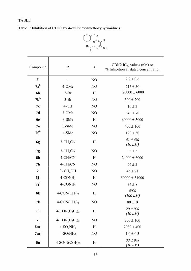
Table 1: Inhibition of CDK2 by 4-cyclohexylmethoxypyrimidines.
N
N
O
NH2N
X
R
H
Compound R X CDK2 IC50 values (nM) or % Inhibition at stated concentration
2a - NO 2.2 ± 0.6
7ab 4-OMe NO 215 ± 50
6b 3-Br H 26000 ± 6000
7bb 3-Br NO 500 ± 200
7c 4-OH NO 16 ± 3
7d 3-OMe NO 340 ± 70
6e 3-SMe H 60000 ± 5000
7e 3-SMe NO 400 ± 100
7f b 4-SMe NO 120 ± 30
6g 3-CH2CN H 41 ± 4% (10 μM)
7g 3-CH2CN NO 33 ± 3
6h 4-CH2CN H 24000 ± 6000
7h 4-CH2CN NO 64 ± 3
7i 3- CH2OH NO 45 ± 21
6jb 4-CONH2 H 59000 ± 31000
7jb 4-CONH2 NO 34 ± 8
6k 4-CON(CH3)2 H 49% (100 μM)
7k 4-CON(CH3)2 NO 80 ±10
6l 4-CON(C2H5)2 H 29 ± 9% (10 μM)
7l 4-CON(C2H5)2 NO 200 ± 100
6mb 4-SO2NH2 H 2930 ± 400
7mb 4-SO2NH2 NO 1.0 ± 0.3
6n 4-SO2N(C2H5)2 H 33 ± 9% (10 μM)
14
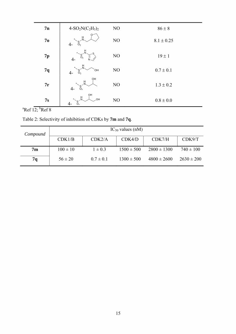
7n 4-SO2N(C2H5)2 NO 86 ± 8
7o 4-
HN
SO2
*
O
NO 8.1 ± 0.25
7p 4- N
SHN
SO2
*
NO 19 ± 1
7q 4- OH
HN
SO2
*
NO 0.7 ± 0.1
7r 4-
HN
SO2
*
OH
NO 1.3 ± 0.2
7s 4-
HN
SO2
*
OH
OH
NO 0.8 ± 0.0
aRef 12; bRef 8
Table 2: Selectivity of inhibition of CDKs by 7m and 7q.
IC50 values (nM) Compound
CDK1/B CDK2/A CDK4/D CDK7/H CDK9/T
7m 100 ± 10 1 ± 0.3 1500 ± 500 2800 ± 1300 740 ± 100
7q 56 ± 20 0.7 ± 0.1 1300 ± 500 4800 ± 2600 2630 ± 200
15
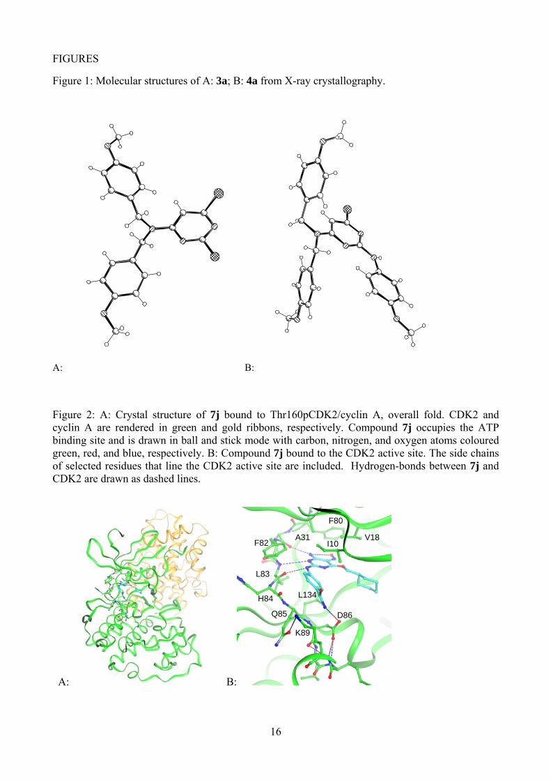
FIGURES
Figure 1: Molecular structures of A: 3a; B: 4a from X-ray crystallography.
A: B:
Figure 2: A: Crystal structure of 7j bound to Thr160pCDK2/cyclin A, overall fold. CDK2 and cyclin A are rendered in green and gold ribbons, respectively. Compound 7j occupies the ATP binding site and is drawn in ball and stick mode with carbon, nitrogen, and oxygen atoms coloured green, red, and blue, respectively. B: Compound 7j bound to the CDK2 active site. The side chains of selected residues that line the CDK2 active site are included. Hydrogen-bonds between 7j and CDK2 are drawn as dashed lines.
A: B:
I10 V18 A31
F80
F82
H84
Q85 D86
K89
L83
L134
16
Related Documents