Contents lists available at ScienceDirect Blood Cells, Molecules and Diseases journal homepage: www.elsevier.com/locate/bcmd Review Structure and function of haemoglobins David A. Gell School of Medicine, University of Tasmania, TAS 7000, Australia ARTICLE INFO Keywords: Haemoglobin Myoglobin Truncated haemoglobin Flavohaemoglobin Haem Model porphyrins Oxygen binding Hexacoordinate haem Nitric oxide Cooperative oxygen binding Allostery ABSTRACT Haemoglobin (Hb) is widely known as the iron-containing protein in blood that is essential for O 2 transport in mammals. Less widely recognised is that erythrocyte Hb belongs to a large family of Hb proteins with members distributed across all three domains of life—bacteria, archaea and eukaryotes. This review, aimed chiefly at researchers new to the field, attempts a broad overview of the diversity, and common features, in Hb structure and function. Topics include structural and functional classification of Hbs; principles of O 2 binding affinity and selectivity between O 2 /NO/CO and other small ligands; hexacoordinate (containing bis-imidazole coordinated haem) Hbs; bacterial truncated Hbs; flavohaemoglobins; enzymatic reactions of Hbs with bioactive gases, par- ticularly NO, and protection from nitrosative stress; and, sensor Hbs. A final section sketches the evolution of work on the structural basis for allosteric O 2 binding by mammalian RBC Hb, including the development of newer kinetic models. Where possible, reference to historical works is included, in order to provide context for current advances in Hb research. 1. Introduction and scope This review attempts a broad overview of selected topics in Hb structure and function. It is necessarily superficial in all areas, but the hope is that there is some benefit in taking a broad view, and perhaps this can be a helpful for researchers new to the field. Throughout, I have tried to acknowledge authoritative work that has had enduring ‘cur- rency’, regardless of publication date. Hb research is highly inter- disciplinary and I have endeavoured to include some of the important contributions from bioinorganic chemistry, spectroscopy and structural biology that have shaped our current understanding of Hb proteins. 2. The Hb superfamily – evolutionary conservation and diversification of function 2.1. Hbs share a common three-dimensional structure and haem cofactor All Hbs have a conserved core topology, comprising 6–8 α-helices (labelled A–H). The very first protein structures to be determi- ned—those of myoglobin (Mb) from muscle of the sperm whale [1,2], and red blood cell (RBC) Hb from horse erythrocytes [3]—revealed the globin structural blueprint, and firmly established the enduring para- digm that structure underlies function. As the founding member of the Hb family, Mb provides the reference against which all other Hb se- quences and structures are compared [4]. For example, in each of the > 200 non-redundant Hb structures in the protein data bank, position F8 refers to the amino acid residue that is structurally equivalent to the eighth residue in helix F of sperm whale Mb—this is the haem-coordinating histidine residue (HisF8), which is the only re- sidue that is 100% conserved across the whole Hb superfamily [5,6]. Residues in non-helical segments are referenced in relation to adjacent helices; thus, CD1 refers to the first residue of the linker joining α- helices C and D, and HC3 refers to the third residue following helix H, on the carboxyl terminus. Two different structural sub-classes of the globin fold are recognised (Table 1 and Fig. 1). The 3-on-3 fold is the canonical Hb fold, ex- emplified by Mb. The ‘3-on-3’ designation refers to the α-helical ‘sandwich’ formed by the A-G-H and B-E-F helices [7]. The C and D helices are supporting structures, and are not always present. The second structural class is the truncated Hb (trHb) class—also called 2- on-2, 2-over-2, or 2/2 Hbs, based on the arrangement of the B–E and G–H helical pairs, [8]. In trHbs, the A, C, D, and F helices are much reduced or absent. Whereas some Hbs function as monomers, other Hbs are assembled from multiple globin subunits. Examples of the latter include mam- malian RBC Hb, which is a tetramer of two Hb α and two Hb β subunits [3], and earthworm Hb (erythrocruorin), which comprises 144 globin chains with four unique sequences together with an additional 36 non- globin subunits [9]. Within these oligomers it is the individual subunits that conform to the conserved globin fold. Allosteric function (see Section 7) and multisubunit organisation of Hbs have arisen a number of times in evolution to solve the problem of O 2 transport (see reviews http://dx.doi.org/10.1016/j.bcmd.2017.10.006 Received 14 May 2017; Received in revised form 29 October 2017; Accepted 30 October 2017 E-mail address: [email protected]. Blood Cells, Molecules and Diseases 70 (2018) 13–42 Available online 31 October 2017 1079-9796/ © 2017 Elsevier Inc. All rights reserved. T

Welcome message from author
This document is posted to help you gain knowledge. Please leave a comment to let me know what you think about it! Share it to your friends and learn new things together.
Transcript
Contents lists available at ScienceDirect
Blood Cells, Molecules and Diseases
journal homepage: www.elsevier.com/locate/bcmd
Review
Structure and function of haemoglobins
David A. GellSchool of Medicine, University of Tasmania, TAS 7000, Australia
A R T I C L E I N F O
Keywords:HaemoglobinMyoglobinTruncated haemoglobinFlavohaemoglobinHaemModel porphyrinsOxygen bindingHexacoordinate haemNitric oxideCooperative oxygen bindingAllostery
A B S T R A C T
Haemoglobin (Hb) is widely known as the iron-containing protein in blood that is essential for O2 transport inmammals. Less widely recognised is that erythrocyte Hb belongs to a large family of Hb proteins with membersdistributed across all three domains of life—bacteria, archaea and eukaryotes. This review, aimed chiefly atresearchers new to the field, attempts a broad overview of the diversity, and common features, in Hb structureand function. Topics include structural and functional classification of Hbs; principles of O2 binding affinity andselectivity between O2/NO/CO and other small ligands; hexacoordinate (containing bis-imidazole coordinatedhaem) Hbs; bacterial truncated Hbs; flavohaemoglobins; enzymatic reactions of Hbs with bioactive gases, par-ticularly NO, and protection from nitrosative stress; and, sensor Hbs. A final section sketches the evolution ofwork on the structural basis for allosteric O2 binding by mammalian RBC Hb, including the development ofnewer kinetic models. Where possible, reference to historical works is included, in order to provide context forcurrent advances in Hb research.
1. Introduction and scope
This review attempts a broad overview of selected topics in Hbstructure and function. It is necessarily superficial in all areas, but thehope is that there is some benefit in taking a broad view, and perhapsthis can be a helpful for researchers new to the field. Throughout, I havetried to acknowledge authoritative work that has had enduring ‘cur-rency’, regardless of publication date. Hb research is highly inter-disciplinary and I have endeavoured to include some of the importantcontributions from bioinorganic chemistry, spectroscopy and structuralbiology that have shaped our current understanding of Hb proteins.
2. The Hb superfamily – evolutionary conservation anddiversification of function
2.1. Hbs share a common three-dimensional structure and haem cofactor
All Hbs have a conserved core topology, comprising 6–8 α-helices(labelled A–H). The very first protein structures to be determi-ned—those of myoglobin (Mb) from muscle of the sperm whale [1,2],and red blood cell (RBC) Hb from horse erythrocytes [3]—revealed theglobin structural blueprint, and firmly established the enduring para-digm that structure underlies function. As the founding member of theHb family, Mb provides the reference against which all other Hb se-quences and structures are compared [4]. For example, in each ofthe> 200 non-redundant Hb structures in the protein data bank,
position F8 refers to the amino acid residue that is structurallyequivalent to the eighth residue in helix F of sperm whale Mb—this isthe haem-coordinating histidine residue (HisF8), which is the only re-sidue that is 100% conserved across the whole Hb superfamily [5,6].Residues in non-helical segments are referenced in relation to adjacenthelices; thus, CD1 refers to the first residue of the linker joining α-helices C and D, and HC3 refers to the third residue following helix H,on the carboxyl terminus.
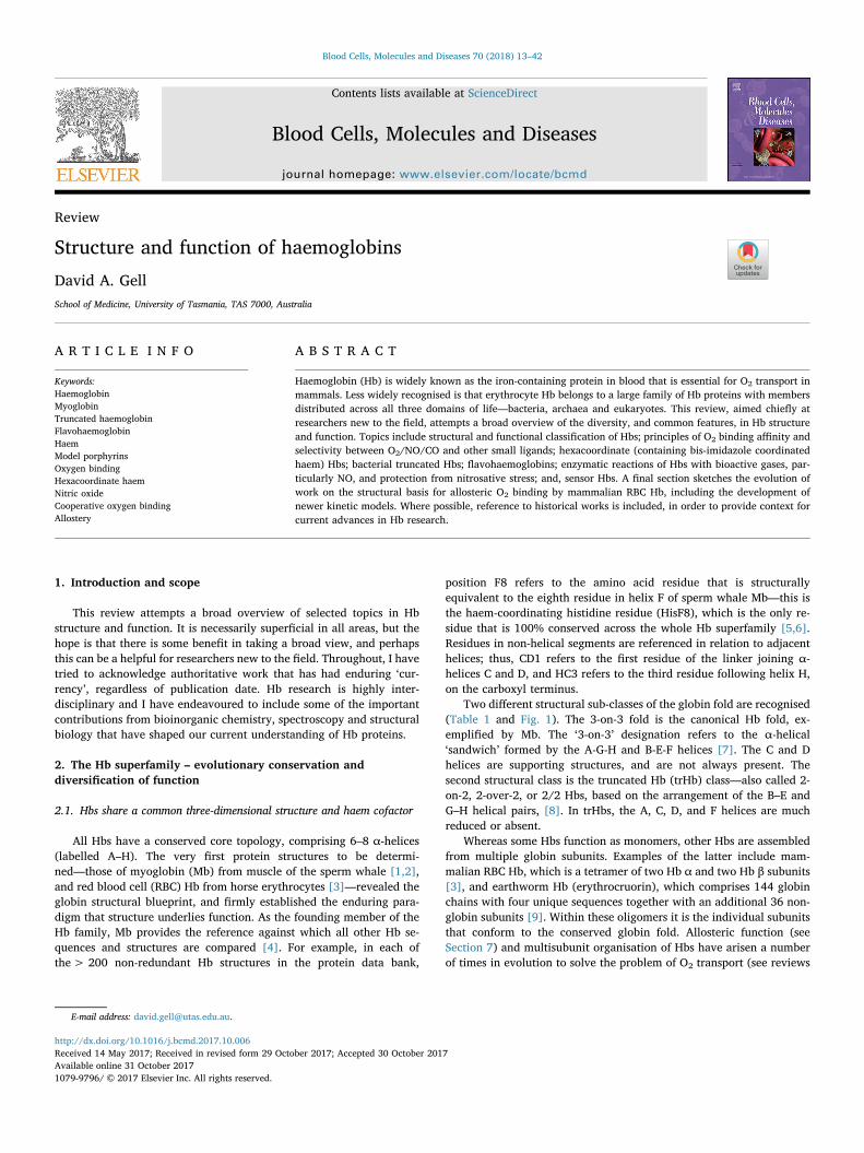
Two different structural sub-classes of the globin fold are recognised(Table 1 and Fig. 1). The 3-on-3 fold is the canonical Hb fold, ex-emplified by Mb. The ‘3-on-3’ designation refers to the α-helical‘sandwich’ formed by the A-G-H and B-E-F helices [7]. The C and Dhelices are supporting structures, and are not always present. Thesecond structural class is the truncated Hb (trHb) class—also called 2-on-2, 2-over-2, or 2/2 Hbs, based on the arrangement of the B–E andG–H helical pairs, [8]. In trHbs, the A, C, D, and F helices are muchreduced or absent.
Whereas some Hbs function as monomers, other Hbs are assembledfrom multiple globin subunits. Examples of the latter include mam-malian RBC Hb, which is a tetramer of two Hb α and two Hb β subunits[3], and earthworm Hb (erythrocruorin), which comprises 144 globinchains with four unique sequences together with an additional 36 non-globin subunits [9]. Within these oligomers it is the individual subunitsthat conform to the conserved globin fold. Allosteric function (seeSection 7) and multisubunit organisation of Hbs have arisen a numberof times in evolution to solve the problem of O2 transport (see reviews
http://dx.doi.org/10.1016/j.bcmd.2017.10.006Received 14 May 2017; Received in revised form 29 October 2017; Accepted 30 October 2017
E-mail address: [email protected].
Blood Cells, Molecules and Diseases 70 (2018) 13–42
Available online 31 October 20171079-9796/ © 2017 Elsevier Inc. All rights reserved.
T
by Royer et al. [10,11]).Each globin polypeptide binds a single molecule of iron-proto-
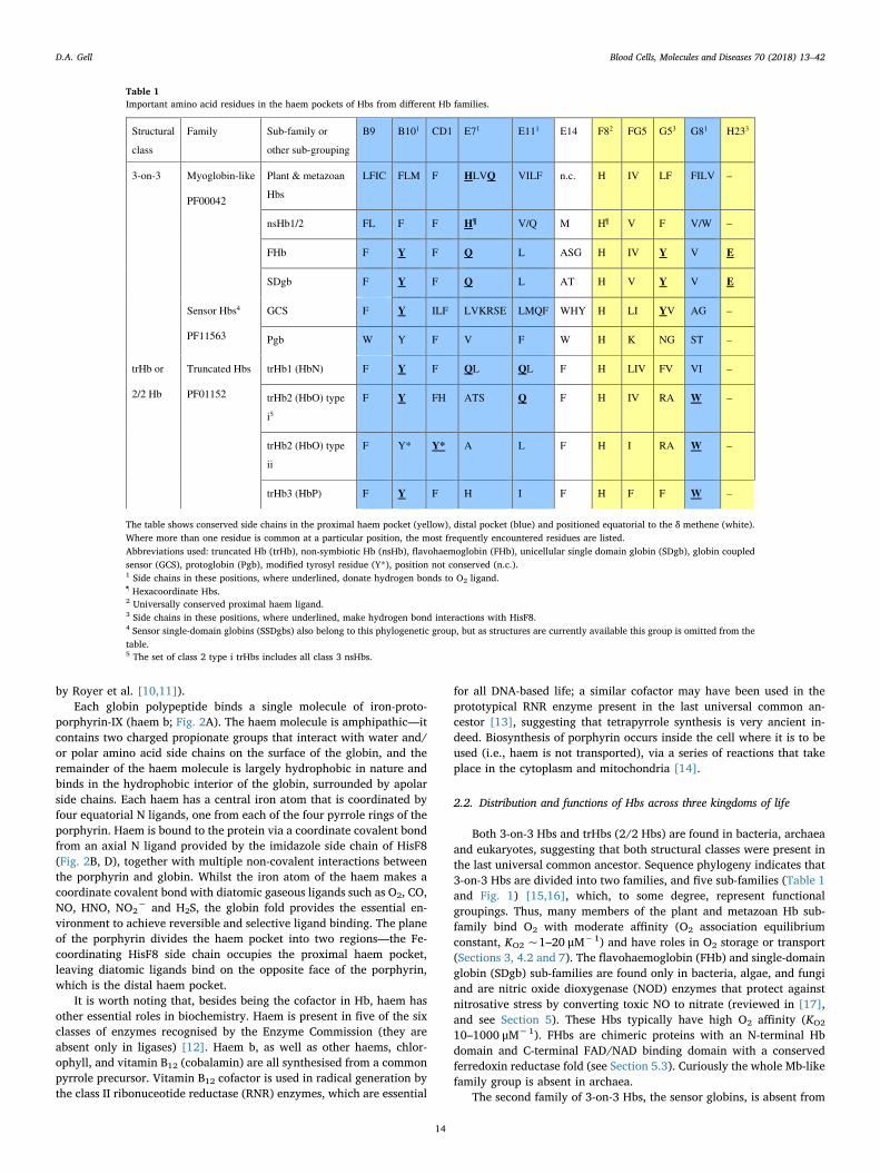
porphyrin-IX (haem b; Fig. 2A). The haem molecule is amphipathic—itcontains two charged propionate groups that interact with water and/or polar amino acid side chains on the surface of the globin, and theremainder of the haem molecule is largely hydrophobic in nature andbinds in the hydrophobic interior of the globin, surrounded by apolarside chains. Each haem has a central iron atom that is coordinated byfour equatorial N ligands, one from each of the four pyrrole rings of theporphyrin. Haem is bound to the protein via a coordinate covalent bondfrom an axial N ligand provided by the imidazole side chain of HisF8(Fig. 2B, D), together with multiple non-covalent interactions betweenthe porphyrin and globin. Whilst the iron atom of the haem makes acoordinate covalent bond with diatomic gaseous ligands such as O2, CO,NO, HNO, NO2
− and H2S, the globin fold provides the essential en-vironment to achieve reversible and selective ligand binding. The planeof the porphyrin divides the haem pocket into two regions—the Fe-coordinating HisF8 side chain occupies the proximal haem pocket,leaving diatomic ligands bind on the opposite face of the porphyrin,which is the distal haem pocket.
It is worth noting that, besides being the cofactor in Hb, haem hasother essential roles in biochemistry. Haem is present in five of the sixclasses of enzymes recognised by the Enzyme Commission (they areabsent only in ligases) [12]. Haem b, as well as other haems, chlor-ophyll, and vitamin B12 (cobalamin) are all synthesised from a commonpyrrole precursor. Vitamin B12 cofactor is used in radical generation bythe class II ribonuceotide reductase (RNR) enzymes, which are essential
for all DNA-based life; a similar cofactor may have been used in theprototypical RNR enzyme present in the last universal common an-cestor [13], suggesting that tetrapyrrole synthesis is very ancient in-deed. Biosynthesis of porphyrin occurs inside the cell where it is to beused (i.e., haem is not transported), via a series of reactions that takeplace in the cytoplasm and mitochondria [14].
2.2. Distribution and functions of Hbs across three kingdoms of life
Both 3-on-3 Hbs and trHbs (2/2 Hbs) are found in bacteria, archaeaand eukaryotes, suggesting that both structural classes were present inthe last universal common ancestor. Sequence phylogeny indicates that3-on-3 Hbs are divided into two families, and five sub-families (Table 1and Fig. 1) [15,16], which, to some degree, represent functionalgroupings. Thus, many members of the plant and metazoan Hb sub-family bind O2 with moderate affinity (O2 association equilibriumconstant, KO2 ~1–20 μM−1) and have roles in O2 storage or transport(Sections 3, 4.2 and 7). The flavohaemoglobin (FHb) and single-domainglobin (SDgb) sub-families are found only in bacteria, algae, and fungiand are nitric oxide dioxygenase (NOD) enzymes that protect againstnitrosative stress by converting toxic NO to nitrate (reviewed in [17],and see Section 5). These Hbs typically have high O2 affinity (KO2
10–1000 μM−1). FHbs are chimeric proteins with an N-terminal Hbdomain and C-terminal FAD/NAD binding domain with a conservedferredoxin reductase fold (see Section 5.3). Curiously the whole Mb-likefamily group is absent in archaea.
The second family of 3-on-3 Hbs, the sensor globins, is absent from
Table 1Important amino acid residues in the haem pockets of Hbs from different Hb families.
Structural
class
Family Sub-family or
other sub-grouping
B9 B101 CD1 E71 E111 E14 F82 FG5 G53 G81 H233
3-on-3 Myoglobin-like
PF00042
Plant & metazoan
Hbs
LFIC FLM F HLVQ VILF n.c. H IV LF FILV –
nsHb1/2 FL F F H¶ V/Q M H¶ V F V/W –
FHb F Y F Q L ASG H IV Y V E
SDgb F Y F Q L AT H V Y V E
Sensor Hbs4
PF11563
GCS F Y ILF LVKRSE LMQF WHY H LI YV AG –
Pgb W Y F V F W H K NG ST –
trHb or
2/2 Hb
Truncated Hbs
PF01152
trHb1 (HbN) F Y F QL QL F H LIV FV VI –
trHb2 (HbO) type
i5
F Y FH ATS Q F H IV RA W –
trHb2 (HbO) type
ii
F Y* Y* A L F H I RA W –
trHb3 (HbP) F Y F H I F H F F W –
The table shows conserved side chains in the proximal haem pocket (yellow), distal pocket (blue) and positioned equatorial to the δ methene (white).Where more than one residue is common at a particular position, the most frequently encountered residues are listed.Abbreviations used: truncated Hb (trHb), non-symbiotic Hb (nsHb), flavohaemoglobin (FHb), unicellular single domain globin (SDgb), globin coupledsensor (GCS), protoglobin (Pgb), modified tyrosyl residue (Y*), position not conserved (n.c.).1 Side chains in these positions, where underlined, donate hydrogen bonds to O2 ligand.¶ Hexacoordinate Hbs.2 Universally conserved proximal haem ligand.3 Side chains in these positions, where underlined, make hydrogen bond interactions with HisF8.4 Sensor single-domain globins (SSDgbs) also belong to this phylogenetic group, but as structures are currently available this group is omitted from thetable.5 The set of class 2 type i trHbs includes all class 3 nsHbs.
D.A. Gell Blood Cells, Molecules and Diseases 70 (2018) 13–42
14

Plant and metazoan Hb subfamily
—sperm whale Mb
—Cerebratulus mini Hb
trHb3 (trHbP)trHb2 (trHbO)trHb1 (trHbN)
SDgb subfamily
—Cgb
FHb subfamily
—YHb
GCS subfamily
—HemAT
Pgb subfamily
—Pgb
A
B
C
D
E
F
G
H
Z
Z
CD
EF
FG
N-term
C-term
FG
N-term
Mb-like family 3-on-3 Hb fold
N-term
Globin-coupled sensor family 3-on-3 Hb fold
Phi (EF)
Truncated Hbs 2-on-2 Hb fold
Haem
B
E
F
G
C
H
A
B
E
F
G
C
HA
C D
FAD
Haem
Ferredoxin
reductase
domain
Hb domain
C-term
C-term
C-term
N-term
C-term
N-term
C-term
N-term
Fig. 1. Conserved and variable features of Hb tertiary structure. The figure shows tertiary structures that are representative of the major Hb families and sub-families (see also Table 1).Most Hbs from the plant and metazoan Hb sub-family are highly similar to sperm whale Mb (pdb 2mgm) [152]; variations include the Cerebratulus lacteus mini Hb (1kr7) [161]. Otherfamilies/sub-families are represented by Cgb from Campylobacter jejuni (2wy4) [107], YHb from Saccharomyces cerevisae (4g1v) [105], HemAT from Bacillus subtilis (1or4) [397], Pgb fromMethanosarcina acetivorans (2veb) [63], trHb1 (trHbN) from Tetrahymena pyriformis (3aq5) [174], trHb2 (trHbO) from Mycobacterium tuberculosis (1ngk) [178], trHb3 (HbP) fromCampylobacter jejuni (2ig3) [180]. Conserved α-helices that comprise the canonical 3-on-3 tertiary structure are colour-coded as follows: A (red-brown), B (pink), E (yellow/tan), F(green), G (cyan), H (blue). Functionally important loops (CD, EF, FG) are also labelled. Conserved elements of the 2-on-2 Hb fold are coloured as for 3-on-3 Hbs. Additional secondarystructure elements present in Mb, but variably present in other Hbs are coloured grey. Additional structural elements that are unique to individual Hb sub-families are coloured magenta.Features of the haem pocket are shown in more detail in Figs. 3 and 4. (For interpretation of the references to colour in this figure legend, the reader is referred to the web version of thisarticle.)
D.A. Gell Blood Cells, Molecules and Diseases 70 (2018) 13–42
15

eukaryotes. Of the sensor globins, the two-domain globin-coupledsensors (GCSs; Section 6) are best understood; these proteins are typi-cally sensors of O2, NO, or CO that regulates aerotactic responses tothese ligands (reviewed [18]). These proteins sometimes show low O2
affinities (KO2 < 1 μM−1; [19]). Other subfamilies of the sensor glo-bins are less well characterised. The third major phylogenetic family ofHbs defines a separate structural class, the trHbs. TrHbs comprise atleast 3 subfamilies (trHb1/2/3, also called trHbN/O/P, or trHb groupsI/II/III) [20,21]. Structure and ligand binding has been characterised
for numerous trHbs, but physiological functions have only been tenta-tively assigned and appear quite diverse. Some trHbs have extremelyhigh O2 affinity (KO2 5000–22,000 μM−1; e.g. [22]) suggesting thatthey are likely to have enzymatic functions. TrHbs exist as single-do-main proteins, or as chimeras with putative oxidoreductase or signal-ling domains [23,24]. Of the chimeric Hbs, only the FHbs have beencrystallized in their multidomain form.
The implication from phylogenetics is that all Hb families arose inbacteria and, today, about half of bacterial genomes contain Hbs [16].
AA
B
C
D
GE
HF 1/2 O2
1/2 C3
CO
FeFe
HisF8 HisF8
1-Me-Im2-Me-Im
O2
Fe
Fe
1/2 C3
1/2
N1
N
N N/C
1/2 C3b
A
BC
D
BD BD
A
BC
D
C(b3)
C(b8)
N(4)
N(2)
Fe)8b(C)3b(C N(4)N(2)
O1
1/2 O2
C5
C6
C6b
Fe
C5
C6
C5b
C6bN2 N2b
N1
N/C
N
N
N
N
3
2
7
8
17
18
13
12
177
Fig. 2. Haem stereochemistry in Mb and modelporphyrins. (A, B) Haem group and proximalHisF8 residue from the crystal structure of un-liganded Mb, a high-spin 5-coordinate Fe(II)complex, determined at a resolution of 1.15 Å(pdb 1bzp) [185]. The 2FO–FC electron densitymap (mesh) is contoured at 2 σ. The atomcolour coding is as follows: iron (orange),oxygen (red), nitrogen (blue), carbon (yellow).Pyrrole rings (Roman letters) and methenebridge positions (Greek letters) are indicated.Substituents at the β-pyrrolic positions (num-bered according to the IUPAC convention) are:methyl (−CH3; positions 2, 7, 12, 18), vinyl(−CH]CH2; positions 3, 8), and propionate(−CH2CH2COOH; positions 13, 17). Viewedalong the plane of the porphyrin ring (panel B),only the B and D pyrrole rings are shown forclarity. The Fe atom is displaced 0.29 Å out ofthe plane defined by the four pyrrole N atoms(horizontal line), towards HisF8. (C, D) Haemgroup and proximal HisF8 residue from Mb(CO), a low-spin 6-coordinate Fe(II) complex,determined at a resolution of 1.15 Å (pbd 1bzr)[185]. The Fe atom is in plane with four pyrroleN atoms (0.015 Å displacement towards CO).Figure panels A–D were prepared from co-ordinates and structure factors deposited in theProtein Data Bank, using the software, PYMOL.(E, F) Crystal structure of the synthetic por-phyrin, Fe(Tp-OCH3PP)(2-MeHIm), a high-spin5-coordinate Fe(II) complex with low O2 affi-nity that serves as a synthetic model of un-liganded T state Hb [36]. In E, atom positionsare shown as thermal vibration ellipsoids (50%probability). (G, H) Crystal structure of Fe(TpivPP)(1-MeIm)(O2), a synthetic model ofMb(O2) [41]. The O2 and 1-methyl imidazoleligand are statistically disordered with 1/2 oc-cupancy at two positions. Panels E–H wereprepared from data deposited at the CambridgeCrystallographic Data Centre (CCDC) using theCCDC software program MERCURY (E, G) or PYMOL
(F, G). Hydrogen atoms are omitted throughoutthis figure. (For interpretation of the referencesto colour in this figure legend, the reader isreferred to the web version of this article.)
D.A. Gell Blood Cells, Molecules and Diseases 70 (2018) 13–42
16

Consequently, functions of the extant bacterial Hbs reflect possiblefunctions of the ancestral Hb. An ancestral Hb would have operated in alargely anoxic, or local microaerophilic, environment, prior to a sig-nificant rise in global O2 2.4 billion years ago [25]. Thus, enzymaticscavenging of toxic O2 and NO [26], sulfur metabolism [27], or sensingand signalling in response to O2, NO, or CO [16], are possible ancestralfunctions. When compared to bacterial Hbs, vertebrate Hbs (such asRBC Hb and Mb) share highest sequence similarity to SDgb proteins,and it has been suggested that lateral transfer of the SDgb gene(s) to theprimitive unicellular eukaryote occurred around the time of the en-dosymbiotic events that generated mitochondria and plastids [28]. Mband erythrocyte Hb still retain physiological roles in NO removal (seeSection 5.4), in addition to O2 storage and transport.
3. Structure-function relationships of the haem pocket
3.1. A 5-coordinate haem with an axial imidazole base is essential for O2
binding
O2 binding is the hallmark feature of Hbs. Pioneering work usingsynthetic porphyrins as models of the Hb biosite established two es-sential conditions for high affinity and reversible O2 binding. Theserequirements are: (1) a 5-coordinate haem with an axial base, such asimidazole (Fig. 2A, B) or pyridine, and (2) a mechanism to preventirreversible haem oxidation. This important work is comprehensivelydescribed in a series of excellent reviews [29,30,31]. The followingsection briefly describes the importance of an axial base, and the non-bonding interactions that protect the haem group from solvent and limitirreversible haem oxidation.
As with other transition metal complexes, haem ligands make co-ordinate, or dative, σ bonds with the metal centre in which both elec-trons are donated from a lone pair on the ligand (O2/CO/NO, etc.).Studies of free haems and synthetic porphyrins show that four-co-ordinate metallo-porphyrins have little interest in O2 binding, whereas5-coordinate Fe(II) prophyrins with an axial imidazole base bind O2
with high affinity. This can be rationalised by considering the energy ofthe dz2 orbital relative to the other four metal d orbitals. By standardconvention, only the dz2 orbital is oriented perpendicular to the por-phyrin plane and has σ-type geometry that is necessary to participate inσ bonding with an incoming O2/CO/NO ligand. In 4-coordinate haem,the dz2 orbital has low energy and so is fully occupied with d-shellelectrons from Fe(II) and, thus, unable to participate in bonding to O2.By contrast, in 5-coordinate haem with an axial base, the energy se-paration between the five metal d orbitals is reduced, allowing the 5 d-shell electrons of Fe(II) to spread out, partially freeing up the dz2 orbital(see [32,33] for illustrated explanations). Magnetic susceptibilitymeasurements, first made by Pauling and Coryell [34,35], establishedthat 5-coordinate deoxy HbFe(II) has 4 unpaired electrons (it is highspin; S = 2), and recent Mössbauer spectroscopy shows that the groundstate electronic configuration for 5-coordinate deoxy Mb, deoxy Hb andsynthetic high spin Fe(II) porphyrins with an axial imidazole base is(dxz)2(dyz)1(dxy)1(dz2)1(dx2−y2)1 [36,37]. It appears that the partlyfilled dz2 orbital is a pre-requisite for FeeO2 bonding. Interestingly,experiment and density function theory (DFT) suggest that the elec-tronic ground state with an axial imidazole is distinct, even comparedto complexes with a similar base such as pyridine [36,37,38], and thismight be important for O2 binding in ways that are not yet fully un-derstood.
3.2. Side chain packing around the haem group prevents irreversible Feoxidation and haem dissociation
Without the protection afforded by the globin fold, free Fe(II) por-phyrins are oxidised in seconds to Fe(III) species, which are unable tobind O2. In contrast, Fe(II) haemoproteins are stable for days. Broadlytwo mechanisms are responsible for oxidation of haems. The first is the
μ-peroxo mechanism, which is initiated upon reaction of a (porphinato)Fe(II)(O2) species with a second unligated (porphinato)Fe(II). The re-sult is the generation of μ-oxo dimers with an Fe(III)–O–Fe(III) centre(see [30]). In the presence of water, and at room temperature, theconversion to μ-oxo dimers occurs on a ms time scale. This reaction isessentially blocked in haem proteins by the bulk of the globin protein,which prevents close approach of two haem moieties. In order forsynthetic model compounds to reversibly bind O2, the pathway to μ-oxodimers must be sterically blocked, as was achieved by James Collmanand co workers with the famous picket fence porphyrins, which are stillthe only synthetic (porphinato)Fe(II)(O2) complexes for which x-raycrystal structures have been reported (Fig. 2G, H) [39,40,41], althougha large number of alternative designs function as O2 binders [29,30,31].
The second pathway for haem oxidation involves the net transfer ofone electron from Fe(II) to O2, generating Fe(III) haem and superoxide.The reaction is termed autooxidation. The rate of autooxidation shows abiphasic response to O2 concentration, suggesting that it occurs in haemproteins by different mechanisms in high [O2] (PO2 > P50; where P50 isO2 partial pressure (PO2) at which 50% of O2 binding sites are satu-rated) or at low [O2] (PO2 < P50) [42]. At high [O2], a process withunimolecular kinetics dominates, which might involve protonation ofbound O2 followed by dissociation of a neutral superoxide radical (HOO%), or reductive displacement of a superoxide anion (O2
−%) by hydroxylor other anionic species ([43] and references therein). Recent combinedquantum mechanics/molecular mechanics (QM/MM) calculations showthat the reductive displacement of O2
−% is the more energetically fa-vourable process [44]. The reductive displacement overcomes the en-ergetic penalty that would be incurred to separate negatively chargedO2
−% from the Fe(III)+ haem in a purely dissociative mechanism. Atlow [O2], a bimolecular reaction takes over, in which an electron istransferred from Fe(II)(H2O) haem (the water molecule is weakly co-ordinated), to an O2 molecule that is in the distal haem pocket but notcoordinated to Fe(II) (an outer sphere electron transfer), generatingsuperoxide anion (O2
−%). Consequently, interactions that stabilisebound O2 (see Section 3.6) or prevent water entry into the haem pocket(below) slow the rate of autooxidation [42,45,46].
Close packing of apolar side chains around the porphyrin, and en-trance to the haem pocket, as well as polar interactions with the haempropionates, are important for high affinity haem binding, and pre-venting ingress of water to the haem pocket. Sequence alignment of Hbs[5], including non-symbiotic plant Hbs [47,48], FHbs and SDgbs[49,50], GCSs and Pgbs [51] and trHbs [8,21,50,52,53], shows thatresidues in Van der Waals contact with the porphyrin (such as residuesat positions CD1, F4, FG5, G5, G8, H15 and H19) have conservedphysicochemical properties, with bulky apolar Met, Ile, Leu, Val andPhe side chains being heavily represented in these positions. Aromaticresidues, particularly Phe, but also Trp, are common in the haem pocketpresumably because π-stacking or edge-to-face (T-stacking) interactionswith the porphyrin macrocycle are highly stabilising [54]. For example,PheCD1, which makes π-stacking interactions with pyrrole ring C, is thesecond-most highly conserved globin residue after HisF8. Only in GCSsis PheCD1 replaced with Leu or Ile [51], and, in trHb2, PheCD1 can bereplaced by polar His or Tyr [50], which play a specialised role inelectrostatic stabilisation of ligands (Section 3.7). In Mb, the disruptionof hydrophobic protein-porphyrin interactions by mutagenesis resultsin a 10–100-fold increase in the rate of haem loss, due to hydration andscission of the Fe–HisF8 bond [46,55], and this translates directly to aloss of overall protein stability [56]. Polar or basic residues (commonlyLys, Arg, His, Ser) are typically found at one or more of the surfacepositions F7, FG3, CD3 (or CE3 when the D helix is absent) and E10,where they make stabilising electrostatic interactions with the haempropionates [45,55].
A number of Hbs have additional, unusual, protein-porphyrin con-tacts. For example, Pgbs and trHb2s (Fig. 1) have loops or additionalhelical elements that partially bury the propionates [57]. The trHb1sfrom cyanobacteria Synechocystis [58,59,60] and Synechococcus [61]
D.A. Gell Blood Cells, Molecules and Diseases 70 (2018) 13–42
17

are highly unusual in having a covalent bond between the side chain ofHisH16 and the vinyl group of pyrrole ring A [59]. The high-resolution(≤1.7 Å) crystal structures of these proteins also show highly ruffledporphyrins [61,62]. A similarly distorted prophyrin occurs in Pgb fromMethanosarcina acetivorans (1.3-Å resolution) [63]), which lacks cova-lent attachment of the porphyrin ring. In-plane (compression of theporphyrin core) and out-of-plane (ruffling) distortions, presumably in-duced by protein-porphyrin contacts, are thought to alter O2 affinitythrough changes in porphyrin electronic structure [64–68], and maycontribute to the high O2 affinities of trHb and Pgb proteins. Finally, asmall number of globins have been discovered that don't bind haem atall [69,70]. In these proteins, the size of the haem-pocket cavity isdramatically reduced, and, in one case, is occupied by a fatty acid [70].
3.3. Steric interactions between the proximal His and the porphyrin ringlower iron reactivity
Reactivity of haem Fe is predominantly determined by the nature ofthe axial ligand (see Section 3.1); however, although all Hbs coordinatehaem Fe through the same axial imidazole (the side chain of HisF8), theO2 affinities of Hbs still vary widely—dissociation rate constants spanseven orders of magnitude, ranging from 10−3 to 104 s−1 [71]. Sidechains in the proximal haem pocket control reactivity of the ironthrough two mechanisms: (1) steric interactions that perturb the geo-metry of the haem–HisF8 interaction (compared to a free haem–imi-dazole) and (2) changes in the basicity of the HisF8 side chain broughtabout by electrostatic (hydrogen bonding) interactions (the topic ofSection 3.4).
Steric effects can influence Hb affinity because ligand binding re-quires a change in the stereochemistry (or ‘shape’) of the haem co-ordination complex (see early reviews by Hoard [72], Perutz [33] andScheidt [32]). Work in synthetic porphyrins [36,37,73–77] has de-monstrated the following general rules (of course, with notable ex-ceptions):
• In general, 5-coordinate porphyrins are high spin (4 or 5 unpairedelectrons in the case of Fe(II) or Fe(III), respectively), square pyr-amidal complexes with the Fe atom displaced 0.39–0.54 Å out of theplane defined by the four pyrrole N atoms, and the porphyrin ske-leton domed towards the axial base (Fig. 2E, F)
• On the other hand, 6-coordinate porphyrins are low spin (0 or 1unpaired electron for Fe(II) or Fe(III), respectively), octahedral co-ordination complexes in which the porphyrin is flat, and Fe lies inthe porphyrin plane (displacement from the pyrrole N plane≤0.11 Å) (Fig. 2G, H)
On this basis, steric interactions that hinder movement of the ironinto the porphyrin plane resist the high spin to low spin transition thatis required by O2 binding, and, consequently, reduce O2 affinity. Suchsteric effects arise largely from interactions between the porphyrin ringand the proximal imidazole base itself, and the magnitude of the effectdepends upon the position and orientation of the imidazole base withrespect to the porphyrin, which, in haem proteins, is controlled by theprotein structure.
Rotation or tilting of the imidazole with respect to porphyrin drawsthe Fe–His bond out of a perfectly axial position and increases stericinteraction between imidazole and porphyrin. In Hb α chains, a stericclash between the HisF8 imidazole and the porphyrin in 6-coordinate(ligated) structure contributes to ~1000-fold lowering of the O2 affinityof the T state (see Section 7.7). Smaller effects have been demonstratedin picket fence porphyrins with a sterically hindered proximal base (2-methyl imidazole, shown in Fig. 2E, F, or 1,2-dimethyl imidazole)which show ~100-fold lower affinity for O2 than do compounds withan unhindered base (1-methyl imidazole, Fig. 2G, H) [78–80].
The azimuthal angle of the HisF8 side chain with respect to thepyrrole nitrogens is another factor that modulates steric interactions
(Fig. 3) and therefore has the potential to modulate O2 affinity. Theeffect of azimuthal angle has been studied in sperm whale myoglobin(Mb) [81–84], mammalian Hb [85,86] and plant leghaemoglobin (LHb)[84,87], using an innovative approach devised by Doug Barrick. In thisapproach, HisF8 was mutated to Gly, and an active haem site was re-constituted by the addition of free imidazole base, which naturallyoccupies the proximal haem pocket and coordinates the Fe(II) haem[81]. A HisF8 → Gly substitution in sperm whale Mb results in ~4-foldincreased O2 and CO binding affinity, largely due to a decreased dis-sociation rate constant [84]. Crystal structures reveal that the exo-genous imidazole adopts an unhindered staggered position [81,83],rather than the hindered eclipsed position seen in the wild-type protein.In Mb, the HisF8 side chain makes a hydrogen bond to the side chain ofSerF7, which appears to stabilise the eclipsed position, based on ob-servations that SerF7→ Ala, Val, Leu mutations increase Fe reactivity[88]. Soybean LHb has O2 affinity that is 20-fold higher than Mb (KO2
23 μM−1) [89,90] and this has been attributed to smaller steric effectsin the plant protein. In the crystal structure of unliganded LHb, theproximal HisF8 imidazole shows ~50% occupancy in two staggeredpositions, separated by ~90°, suggesting that this side chain is moremobile than HisF8 in other globins [91]. In LHb, HisF8→ Gly muta-tions (with exogenous imidazole added) cause a small decrease in ligandaffinity [84], consistent with HisF8 already being fully unhindered inthe high affinity wild-type protein. The role of the proximal pocket incontrolling ligand affinity was confirmed by swapping the F helices ofLHb and Mb: a three-fold decrease in O2/CO affinity occurred for achimera carrying the Mb F-helix, whereas a five-fold increase in affinitywas conferred on a chimera carrying the Lb F-helix [84]. Circumstantialevidence that a staggered imidazole position contributes to affinitycomes from structural studies of Hbs with the highest O2 affinities. Forexample, Hbs from parasitic Paramphistomum epiclitum (KO2 3300 μM−1
[92,93]), rice nsHb1 (KO2 1800 μM−1 [94]), trHb1 from Synechocystissp. (KO2 17,000 μM−1 [95]), trHb2 from Geobacillus stearothermophilus(KO2 13,200 μM−1 [96]) and trHb2 from M. tuberculosis(KO2 ≥ 7900 μM−1 [97]), all have a staggered proximal imidazole withan azimuthal angle close to the ideal 45°.
3.4. Hydrogen bonding of the proximal histidine modulates Fe reactivity
The second major factor influencing iron reactivity in Hbs is hy-drogen bonding between the proximal HisF8 side chain and residues inthe proximal pocket (Fig. 4). Whilst HisF8 coordinates haem Fe througha lone pair on the side chain Nε2 atom, the protonated Nδ1 atom is freeto participate in hydrogen bonding to other chemical groups.
Resonance Raman and density functional theory (DFT) calculationsshow that hydrogen bonding through Nδ1 alters the electronic proper-ties of the proximal imidazole such that it can donate more or lessnegative charge to the haem Fe, and this modulates the FeeXO bondstrength (where X is C, N, or O) [98–101]. Interestingly, DFT calcula-tions suggest that moderate strength hydrogen bond, e.g., to a carbonylacceptor group, results in synergic enhancement of π-backbonding fromFe to O2 and σ-donation from O2 to Fe, thus stabilising FeeO2 [101]. Onthe other hand, a strong hydrogen bond to His Nδ1 increases the imi-dazolate character of the His side chain, causing it to donate morenegative charge to the haem, and resulting in σ-donor competition withthe XO ligand for the Fe dz2 acceptor orbital. This competition weakensthe FeeXO bond. It has been noted that haems with strong electrondonating axial ligands, such as imidazolate or thiolate ligands, fre-quently have similar binding affinities for NO/CO/O2 ligands, com-pared to haems with a neutral axial imidazole (see Section 3.6), sug-gesting that electronic properties of the proximal His have a role inligand discrimination, in addition to overall affinity and reactivity ofthe FeeXO complex [102,103].
In light of the above, it is interesting that hydrogen bonding from aneutral HisF8 imidazole to the carbonyl group of residue F7 is commonin O2 transport Hbs, and that proximal haem pocket interactions are
D.A. Gell Blood Cells, Molecules and Diseases 70 (2018) 13–42
18

quite different in the bacterial FHbs [104,105] and SDgbs [106,107],which function primarily as NOD enzymes (Fig. 4). In FHbs and SDgbs,a carboxylate (GluH23) and phenol (TyrG5) pair is highly conserved inthe proximal haem pocket, and makes strong hydrogen bond interac-tions that stabilise HisF8 as an imidazolate. σ-Donor competition be-tween O2 and the axial imidazolate increases negative charge on O2,which favours NOD enzyme activity [17,105–108]. A similarly posi-tioned carboxylic acid is found in the haem enzymes, horse radishperoxidase (HRP) and cytochrome c peroxidase (CcP), and contributesto the mechanism of OeO bond cleavage [109,110].
Measurements of the Fe–His bond stretching frequency (νFe–His)together with potentiometry have confirmed the chemical similarity ofhaem sites from FHb/SDgb sub-families and peroxidase enzymes. Thus,stretching frequencies for 5-coordinate Campylobacter jejuni Cgb(251 cm−1) [106,107], Vitreoscilla SDgb (252 cm−1), and E. coli Hmp(244 cm−1) [108] are significantly higher than for unliganded Mb(220 cm−1) [111–113] and unliganded Hb (~215 cm−1) [114–117],and similar to values from cytochrome c peroxidase (248 cm−1) andHRP (244 cm−1) enzymes [118,119]. The Fe(III)/Fe(II) midpoint po-tential for Cygb is −134 mV at pH 7 [107], and for E. coli Hmp it is−121 mV [120]; both are considerably lower than Mb (+58 mV) ormammalian Hb (+150 mV) [121], and closer to midpoint potentialsfor HRP (−250 mV) [122] and Cytochrome c peroxidase (−194 mV)[123].
It is worth noting that coordinating haem through lone pairs on S orO atoms (Cys, Met or Tyr side chains) as seen in other non-Hb haemproteins, causes even greater shifts in oxidation potential according tohard-soft acid-base principles [124]. Thus, haem proteins with 5-co-ordinate Tyr/− (−303 mV) or His/Tyr (−550 mV) arrangements havemuch lower redox potentials and strongly favour Fe(III) centres [124].A HisF8 → Tyr mutant of Mb has a midpoint potential ~250 mV lowerthan the wild type [125]. This has important clinical relevance in theform of the methaemoglobinopathies, wherein mutations of proximalHisF8 or distal HisE7 to Tyr (all these natural Hb variants are referredto as HbMs) cause rapid oxidation to Fe(III) methaemoglobin (metHb),which is unable to transport O2 (biochemical and clinical correlates ofnatural Hb variants are reviewed in [126]).
3.5. The FeeO2 bond is polar
Whilst the proximal HisF8 plays a dominant role in controlling Fe
reactivity, residues in the distal haem pocket are important for con-trolling affinity and selectivity through more direct interactions withligands. Because NO, CO and O2 are similar sized apolar molecules,discrimination between these by Mb and other Hbs must occur at thelevel of the FeeXO complexes [127], which have substantially differentelectronic properties [98,99]. The precise electronic structure of theFeeO2 bond has been debated since the 1936 papers on this subjectwere first published by Pauling and Coryell [29,30,34,35,128–134].Infrared spectroscopy of the OeO bond clearly supports formal reduc-tion of bound oxygen to superoxide; thus the νOeO frequency in Hb(O2) (1107 cm−1) [135], Mb(O2) (1103 cm−1) [136] and synthetic(porphinato)Fe(II)(O2) complexes (1150 cm−1) [137] are clearly in arange expected for O2
− (1150–1100 cm−1) [29,137], and not for mo-lecular O2 (1555 cm−1) or peroxide (O2
2−; 842 cm−1) [29]. This isconsistent with the formal superoxo model, Fe(III)+(O2
−), proposed byH. G. Weiss [128,129]. A bonding model involving σ-donation from anelectron pair on O2
− to Fe(III), with minimal contribution from π-backdonation, is generally accepted [99], although computationalstudies and Mössbauer spectroscopy indicate the net charge transfer isless that± 1 e− [131,138]. Whatever the model, there is consensusthat the FeeO2 bond is highly polar. In comparison, smaller decreasesin νCeO (from 2145 to ~1950 cm−1) and νNeO (from 1877 to~1600 cm−1) upon haem binding suggest less transfer of electroniccharge to these ligands and hence less polar Fe(II)(CO) and Fe(II)(NO)complexes [98]. The mechanisms by which Hbs discriminate betweenpolar and non-polar ligand adducts are discussed next.
3.6. Hydrogen bonding with HisE7 stabilises bound O2 in Mb and RBC Hb
Selective stabilisation of bound O2 over CO and NO is physiologi-cally important to prevent poisoning by endogenous CO/NO, which,although present at very low levels, have dramatically stronger bindingto Fe(II) haems. The intrinsic high CO affinity of Fe(II) haems is de-monstrated by binding experiments in apolar solvents, which show theequilibrium constant for CO binding (KCO) is ~104 greater than KO2
([30,31,127] and references therein). Haem proteins with an apolardistal haem pocket have similar preferences for CO over O2—for theseproteins, the ratio KNO:KCO:KO2 is typically in the order of ~106:~103:1[102]. If this were true of Hb or Mb, then CO produced by normal haemcatabolism (porphyrin cleavage at the α methene by haem oxygenase)would result in poisoning. Poisoning does not happen because KCO:KO2
is reduced to 25:1 for Mb [139]. The mechanism is an increase in theaffinity for O2, relative to CO, arising from electrostatic interactionsbetween the partial negative charge on bound O2 and polar side chainsin the distal pocket (Fig. 5) [127]. Anticorrelation of νFeeXO and νXeOstretching frequencies has been used extensively to probe the electro-static potential of the distal haem site in Hbs and other haem proteins,and is reviewed elsewhere [99,140].
Pauling was the first to propose [128] that hydrogen bonding withthe distal HisE7 stabilised partial negative charge on O2 ligand inmammalian RBC Hb and Mb (Fig. 5, top left), and this hydrogenbonding was subsequently confirmed experimentally by Raman[141–143], EPR [144], X-ray crystallography [145,146], neutron dif-fraction [147] and NMR [148]. The theory that electrostatic interac-tions selectively enhance O2 binding has also been borne out by ex-tensive mutagenesis studies in muscle Mb (reviewed by [127,149]) andby computational studies [66,150]. For example, substitution of HisE7with Val, the side chain of which cannot form hydrogen bonds, causesa> 1000-fold increase in the O2 dissociation rate constant (kO2), and a100-fold decrease in the equilibrium association constant, KO2 [151].
In their quantitative review of O2/CO/NO binding, Phillips andOlson [127] show that a polar E7 side chain has two distinct effects onligand binding kinetics. The first effect is that polar HisE7 causes asmall (~10-fold) decrease in the binding rate constant for O2 or CO,compared to model synthetic porphyrins in apolar solvent, or haempocket mutants with an apolar E7 side chain [139]; the reason is that
HisF8HisF8
haemhaem
haemhaem
proximal imidazole ‘eclipsed’ proximal imidazole ‘staggered’
A
B
C
D
Fig. 3. Steric hindrance between HisF8 and the porphyrin ring. (A, B) In sperm whale Mb,the proximal imidazole base is eclipsed by the closest pyrrole nitrogen atoms—azimuthalangle ~0°. (C, D) In LHb, the azimuthal angle (arrow) is ~30°, close to the maximumstaggered position of 45°.
D.A. Gell Blood Cells, Molecules and Diseases 70 (2018) 13–42
19

Mb FHb/SDgb
Horse radish peroxidaseCytochrome c peroxidase
TrpAsp
His
Fe
H2O
Asp
Tyr
His
Fe
H2O
TyrG5
GluH23
HisF8
CN–
Haem
Fe
HisF8
O2
LeuG5
TyrH23
Haem
Fe
LeuF4
SerF7
HaemHaem
Fig. 4. Electrostatic interaction networks in the proximalhaem pocket. Hydrogen bond networks (dashed magentalines) between the proximal HisF8 side chain and residuesin the proximal haem pocket can be broadly divided intotwo groups. In Mb (pdb 2mgm) [152], and most other Hbs,HisF8 is weakly hydrogen bonded to a backbone carbonylin the F4 position (LeuF4, in addition to the hydroxyl ofSerF7, in Mb). A distinctly different electrostatic environ-ment occurs in FHbs and SDgbs, represented here by Cgbfrom C. jejuni (pdb 2wy4) [107]; in these proteins, highlyconserved GluH23 anion and TyrG5 make strong hydrogenbonding interactions that increase the imidazolate char-acter of HisF8 and impart an electron ‘push’ to the haem(arrow). A similar polar environment is seen in the per-oxidase enzymes, horse radish peroxidase (pdb 1hch)[553] and cytochrome c peroxidase (pdb 1zby) [554], inwhich the proximal imidazolate is hydrogen bonded to thecarboxylate side chain of Asp. Protonation states, for polargroups only, are tentative for illustration purposes (apolarCeH groups are not shown).
HisE7
PheCD1
HisF8
Haem
+
–
–
+
Mb
FHb/SDgb
PheCD1
TyrB10
GlnE7
Haem
HisF8
GCS
IleCD1
TyrB10
Haem
HisF8
Pgb
TyrB10
PheCD1
HaemHisF8
trHb1
TyrB10
PheCD1
GlnE7
GlnE11
Haem
HisF8
trHb2 (type ii) trHb3trHb2 (type i)
Tyr*B10Tyr*CD1
TrpG8
Haem
HisF8
TyrB10
PheCD1
GlnE11
TrpG8
HaemHisF8
TyrB10
PheCD1
TrpG8
TrpE14
HisE7
Haem
HisF8
CN– CN
–
CN–
CN– CN
–
O2
O2
O2
Fig. 5. Electrostatic interactions stabilise ligand in the distal haem pocket. Hydrogen bond networks (dashed magenta lines) between Fe-coordinated ligand (either ferrous ligand, O2, orferric ligand, CN−) and side chains in the distal haem pocket are highly varied. Networks are shown for the following representative x-ray crystal structures: sperm whale Mb (pdb 2mgm)[152], with protonation states of HisE7/F8 Nδ1 and Nε2 atoms shown, based on neutron diffraction [555] (apolar CeH groups are not shown); Cgb from C. jejuni (pdb 2wy4) [107];HemAT from B. subtilis (pdb 1or4) [397]; Pgb from M. acetivorans (pdb 2veb) [63]; trHb1 from T. pyriformis (pdb 3aq5) [174]; trHb2 from B. subtilis (pdb 1ux8) [53]; trHb2 from M.tuberculosis with covalently linked TyrB10–TyrCD1 (pdb 1ngk) [178]; and trHb3 from C. jejuni (pdb 2ig3) [180].
D.A. Gell Blood Cells, Molecules and Diseases 70 (2018) 13–42
20

electrostatic interactions with HisE7 stabilise a non-coordinated watermolecule in the haem pocket of unliganded Mb that must dissociate toallow room for diatomic ligands to enter the haem pocket [152,153].More recently the reverse exchange of CO for water has been measured[153] and the role of non-coordinated water molecules in controllingligand binding rates has been shown in other Hbs [154]. Together,these results show that the hydrogen bonding potential of residues inthe distal pocket contributes significantly to the overall kinetic barrierfor O2 binding.
The second effect of the polar E7 side chain, as suggested byPauling, is to stabilise polar Fe–ligand complexes. In the case of O2
binding to Mb, the small negative effect of HisE7 on the ligand asso-ciation is offset by a ~1000-fold decrease in the dissociation rate con-stant of the polar Fe(III)+(O2
−) complex. The net result is ~100-foldincrease in O2 affinity of Mb with HisE7 compared to Mb mutants withapolar side chains at the E7 position [127]. Mutant studies suggest thathydrogen bonding with HisE7 contributes around −3.5 kcal mol−1 toO2 binding affinity in Mb, and around −2 kcal mol−1 in the Hb α andHb β chains [155]. These values are similar to those obtained from QMcomputational approaches, which are in the range −3.7 to−6.8 kcal mol−1 for Mb [66,150,156]. For apolar Fe(II)(CO) com-plexes, the positive contribution from weak hydrogen bonding to HisE7is smaller than the negative contribution from steric interference bywater in the distal pocket, leading to an overall ~5-fold drop in KCO forMb compared to apolar haem pocket mutants of Mb [127]. For NO, itappears that a ~10-fold stabilisation of NO by hydrogen bonding isalmost exactly offset by the 10-fold reduction in KNO due to water in thehaem pocket. In summary, hydrogen bonding interactions in the distalpocket increase Hb affinity and selectivity for O2 ligand by lowering theHb(O2) dissociation rate.
3.7. Hydrogen bonding networks in the distal pocket are highly varied
Taking a broad view of Hb structure, it is striking that the plant andmetazoan Hb sub-family is the only group in which distal HisE7 isconserved and acts as the sole hydrogen bond donor to bound ligand(Table 1). In other families, a more extended hydrogen bond networkinvolving two or more polar side chains is typical (Fig. 5). One of thesepolar residues is almost always TyrB10, which replaces Phe, Ile or Leuat the corresponding position in Mb. The FHbs and SDgbs share highlyconserved TyrB10 and GlnE7 residues in the distal pocket, which hy-drogen bond with bound ligand [105,157,158]. An extended polarnetwork in the distal pocket is typically associated with higher O2
binding affinities and is also a feature in the haem enzymes HRP andCcP [102]. Some typical and atypical features of the hydrogen bondnetwork in each of the three Hb families (Table 1) are briefly discussednext.
Even within the restricted group of plant and metazoan Hbs, O2
affinities vary over at least 3 orders of magnitude (e.g., see [92]). Mb-like Hbs from parasitic nematodes such as Paramphistomum epiclitum(TyrB10/TyrE7; KO2 3300 μM−1) [92,93] and Ascaris suum (TyrB10/GlnE7; KO2 425 μM−1) [159,160] bind O2 with much higher affinitythan Mb (KO2 1.1 μM−1) [139], and stabilise O2 through hydrogenbonding to TyrB10, as seen for the non-plant and metazoan Hbs.However, the identity of side chains in the B10/E7 positions clearlydoes not tell the whole story, as demonstrated by studies of Hb fromnerve tissue of the nemertean worm Cerebratulus lacteus. Althoughclearly clustering with the metazoan Hbs, C. lacteus Hb has an unusualtruncated structure, missing helix A and most of H [161], and, althoughit has a TyrB10/GlnE7 sequence in the distal pocket, it exhibits onlymoderate O2 affinity (KO2 1.3 μM−1) that is very similar to that of Mb.Crystal structures indicate an extended hydrogen bond network in thedistal pocket involving TyrB10, GlnE7, and ThrE11, of which TyrB10makes the most significant interactions directly with the bound O2 orCO [161,162]. The lower-than-expected O2 affinity is attributed toequilibrium between different hydrogen bonding patterns [163,164]: a
high affinity state in which the phenol of TyrB10 hydrogen bonds to theO2 or CO ligand, and a lower affinity in which TyrB10 phenol is hy-drogen bonded to the side chain of ThrE11 and a lone pair on phenolicO of TyrB10 is directed towards the ligand with consequent electro-static repulsion of the ligand [163,164]. Evidence for this explanationcomes from molecular dynamics (MD) and QM/MM calculations[163,165], and by the fact that mutation of ThrE11 → Val causes a1000-fold reduction in the O2 dissociation rate constant and a 130-folddecrease in KO2 [164]. A much smaller 7-fold decrease in CO dis-sociation rate constant for the E11Thr → Val mutant is also consistentwith the proposed model as Fe(II)(CO) is expected to be less sensitive toelectrostatic effects. A similar equilibrium may explain moderate affi-nity (KO2 1.2 μM−1; [166]) in trHb1 from Paramecium caudatum [167].
Unlike the case of Mb (Section 3.6), mutation of distal HisE7 → Val(or Ala or Leu) in soybean LHb causes little change in O2 affinity[87,90], suggesting little electrostatic stabilisation of ligands by HisE7in LHb. The reason appears to be that interactions between HisE7 andTyrB10 constrain the imidazole side chain in a position that reduceshydrogen bonding to O2. For Mb(CO), and a number of other Hbs,multiple νCeO stretching frequencies, measured by infra-red spectro-scopy (IR), indicate the presence of multiple conformational states ofHisE7 [168], some of which correspond to conformers with hydrogenbond interactions between protonated Nε2 of HisE7 and the CO ligand[66]. In the case of LHb, the presence of a single major νCeO peak ledKundu et al. to suggest that HisE7 is restrained by a hydrogen bondfrom TyrB10 to Nδ1 of the imidazole [169]. Mutation of TyrB10 freedHisE7 to adopt other conformations (as evidenced by multiplicity ofνCeO) and increased O2 affinity, suggesting that electrostatic interac-tions between HisE7 and ligand were enhanced [87,167,169]. Althougha hydrogen bond from TyrB10 to HisE7 is not seen in the crystalstructure [90], computational studies indicate that TyrB10-interactingand non-interacting conformers of HisE7 are populated in solution, andthat the HisE7 rotamer with a hydrogen bond to TyrB10 makes weakerelectrostatic interactions with O2 [167].
Structural plasticity appears to be common in the distal pocket ofproteins in the sensor Hb family, suggesting this might be linked tosignal transduction mechanisms. The distal haem pocket in severalbacterial globin-coupled sensors (GCS) undergoes conformationalchange such that TyrB10 can coordinate bound ligand or swing away togenerate a low affinity state [19] [170]. This group of Hbs is notable forlow conservation of PheCD1 (can be Phe, Ile or Leu) and there is con-siderable variation at E7 and E11 that is not seen in other Hb groups. Inone GCS from Geobacter sulfurreducens, HisE11 and HisF8 form hex-acoordinate haem [171] (Section 4). In Pgb from Methanosarcina acet-ivorans, TyrB10 is conserved but the distance from the haem site meansthis side chain only makes a small contribution to ligand affinity[63,172]. M. acetivorans is an obligate anaerobe that lives in marinesediments where it is thought to metabolise CO, hence CO binding in-eractions with Pgb have been of interest. Complex CO binding kinetics[172] and structural plasticity in the distal haem pocket [173] suggestan equilibrium between a high affinity liganded state with hydrogenbonding from TrpB9 to CO, and a lower affinity state in which thisinteraction is absent. These varied properties demonstrate that there isstill much to discover about the structural basis for ligand affinity inthis group.
Finally, members of the bacterial trHb family typically have highligand affinities and display a number of distinctive distal pocket fea-tures. Hydrogen bond networks in the distal pocket of these proteinsfrequently involve multiple polar side chains that interact with eachother, as well as with bound haem ligands (Fig. 5, bottom row). Crys-tallographic studies have identified distal pocket hydrogen bond net-works comprising side chains of TyrB10, GlnE7 and GlnE11 in trHb1proteins, and mutations at any of these positions increase O2 dissocia-tion and autooxidation rates [8,174–176]. A hydrogen bonding to li-gand through the indole N of TrpG8 is a distinctive feature of trHb2[53,96,177–179] and trHb3 proteins [180–182]; in each case, the
D.A. Gell Blood Cells, Molecules and Diseases 70 (2018) 13–42
21

indole ring of TrpG8 lies parallel to the porphyrin ring and makes ad-ditional π-stacking interactions with the porphyrin macrocycle. SometrHb2s (type ii in Table 1) have a distal pocket with an unusual post-translational modification, whereby a pair of tyrosine side chains arelinked via a covalent bond between the Oη atom of TyrB10 and the Cε2
atom of TyrCD1 [178]; the remaining hydroxyl of TyrCD1 donates ahydrogen bond to the O2 ligand. These π-stacking interactions andcovalent modifications are expected to stabilise electrostatic interac-tions with haem ligands.
The trHb3 group is notable as the only group outside the plant andvertebrate Hbs in which HisE7 is conserved. In the crystal structure oftrHb3 from C. jejuni, HisE7 does not make a hydrogen bond with thebound CN− ligand [180]; however, RR, mutagenesis and MD simula-tions indicate that TyrB10, TrpG8 and His E7 all contribute to the hy-drogen bond network [181,182]. Mutation of TyrB10 and HisE7 toapolar residues increases the O2 affinity dramatically, fromKO2 = 222 μM−1 in the wild-type protein, to KO2 = 15,000 μM−1
[181], suggesting that TrpG8 on its own makes a highly stabilisinginteraction that is moderated by the extended hydrogen bond network.MD simulations suggest that multiple hydrogen bond networks maycoexist in C. jejuni trHb3 to lower O2 affinity, as also proposed forCerebratulus lacteus miniHb, LHb, and P. caudatum trHb1 (see above),suggesting this might be a common mechanism to regulate ligand af-finity [182].
3.8. Steric hindrance as a mechanism for discriminating haem ligands
The different geometry of linear FeeCeO (~180°) versus bentFeeOeO (~120°) and moderately bent FeeNeO (~140°) provide forpotential steric selection of ligand binding, as proposed by Collman[137]. Initial reports of a bent FeeCeO conformation in MbCO struc-tures were found to be artifacts of isotropic refinement of anisotropiccrystals, due to limited resolution in the original x-ray diffraction stu-dies [183]. Model porphyrins [31] and crystal structures of Mb de-termined with atomic (1 Å) resolution [184,185], as well as numerousother protein structures, show that CO invariably binds with a lineargeometry. Although steric hindrance is not required for a quantitativeexplanation of CO/O2 selectivity [127], Collman points out [31] that alinear FeeCeO unit does not discount a role for sterics CO/O2 dis-crimination as a mechanism in other Hbs. The rationale is that sterichindrance does alter relative stabilities 1000-fold in synthetic por-phyrin models without substantial FeeCeO distortion; instead distor-tions in the porphyrin core [36,37,77], that may be too small to bedetected in the majority of protein crystals, are implicated.
3.9. The distal haem pocket captures diatomic ligands prior to Fe–ligandbond formation
Whilst ligand dissociation rate constants are predominantly con-trolled by iron reactivity (proximal haem pocket effects) and electro-static stabilisation of FeeXO, rate constants for ligand binding arecontrolled by accessibility of the iron and capture of ligand into thedistal haem pocket. The effect of polar residues and water in the distalpocket on ligand association rate has already been mentioned (Section3.6). Hydrophobic residues in the distal haem pocket have a stronginfluence on ligand association rates by providing a hydrophobic pocketinto which O2/CO/NO can bind prior to reaction with the haem Fe.Residues LeuB10, PheCD1, ValE11, IleG8 determine the size of thedistal haem pocket and have effects on O2 binding rates [186–188]. Asmaller volume increases the number of collisions of O2 with the Fecentre—as illustrated by LeuB10 → Phe mutants of Mb [189,190] andHb [191], which increase the affinity and the rate of geminate CO re-binding following photolysis, whereas mutation to Ala has the oppositeeffect. ValE11 is highly conserved in O2 transport Hbs and has a subtlesteric interaction that is destabilising to the distal pocket water mole-cule in unliganded Mb, but not substantial enough to significantly
hinder haem ligands [187,188]. In the β subunit of RBC Hb, ValE11hinders ligand binding in the T, but not R, quaternary state (see Section7.7). In addition to forming the ligand binding/rebinding cavity, hy-drophobic residues in the distal pocket have important effects on haembinding, lowering autooxidation rates, and making steric interactions toposition polar side chains. For example, PheCD4 helps to position HisE7for hydrogen bonding in Mb [192].
3.10. Diatomic ligands enter the distal haem pocket by defined pathways
The driving ‘force’ that concentrates ligands to the haem pocket isthe hydrophobic effect that causes hydrophobic O2/CO/NO to partitionfrom the solvent water to the apolar cavities in the distal haem pocket[153,193]; the pathway of ligand diffusion from the exterior of proteinto the distal haem site dictates the kinetics. Perutz proposed entry of O2
to the haem pocket of Mb and Hb via a solvent channel created by anoutward rotation of the HisE7 side chain [194,195]. The E7 gatingmechanism is supported by the everted position of the HisE7 side chainin crystal structures of Mb carrying bulky ligands [196–198], and alsoin crystals of MbCO at pH 4 [199], where it was supposed that the lowpH increases the cationic state of HisE7, thus increasing its solventexposure. Even at pH 7.0, atomic-resolution structures of Mb reveal~20% occupancy of the everted HisE7 side chain conformation [184].In solution, the multiplicity of the νCO stretching frequencies can beexplained using MD [200] and QM/MM [66] as arising from a mix ofhydrogen bonded and everted, or open, states of HisE7.
An alternative route for ligand migration is diffusion via a longerpathway of atom-sized hydrophobic cavities in the protein. Xe is a re-latively large and inert atom that binds with low affinity purely throughVan der Waals interactions and has been used extensively as a probe forinternal cavities. Crystal structures of Mb under pressurised Xe atmo-sphere show Xe molecules bound in a number of cavities within theprotein matrix [201,202]. When the Fe–ligand bond is broken, natu-rally by thermal energy or induced by photolysis, the ligand molecule ispositioned close to the haem-Fe from which it can rebind (geminaterebinding with ps–ns time constants), or diffuse to the bulk solvent andbe replaced by ligand diffusing in from the solvent (bimolecular re-binding). The Xe-binding sites in Mb have been proposed as transientsites for O2/CO/NO ligands migrating to the haem pocket [203];however, filling cavities with Xe under pressure had no effect on ge-minate or bimolecular CO binding for Hb [191] and only small effectson the slow (μs) phase of geminate rebinding in Mb (reflecting a smallfraction of geminate rebinding from internal Xe sites) [71,204,205].The results described above suggest that Xe-binding cavities in Hb andMb are not major routes of ligand migration.
Mutational, crystallographic and kinetic studies of laser flash pho-tolysis of Hb(CO) and stopped-flow spectrophotometry by Olson,Gibson and colleagues suggest that the E7 gate is the predominantpathway for ligand entry in Mb [71,204] and Hb α and β chains[155,191,206]. Royer and colleagues [207,208], using similar methods,identified the E7 gate as the major route of ligand entry and exit in HbIfrom the clam Sapharca inaequivalvis, which is representative of a groupof dimeric metazoan Hbs that have a dimer interface formed by the Eand F helices. Interestingly, the MD results of Boechi et al. [193] sug-gest that HisE7 does not provide a physical barrier to O2 diffusion in theclosed state, but rather, in the open state, creates a hydrophobic site,corresponding to a shallow free energy well (−2 kcal mol−1 comparedto O2 in solvent), above the edge of the porphyrin ring and ~5 Å fromthe iron into which ligand would be captured if traversing the E7channel. Depth of energy well was directly related to the bimolecularrate constant for entry into the haem pocket in Mb mutants. MD si-mulations of RBC Hb reveal a similar E7-linked hydrophobic cavity andsuggest that opening of the E7 gate regulates ligand association rate inthe β chain [209]. Together these results suggest HisE7 as a commongas migration pathway in plant and metazoan ‘HisE7’ Hbs; it is clear,however, that other ligand migration pathways operate in different
D.A. Gell Blood Cells, Molecules and Diseases 70 (2018) 13–42
22

groups of Hbs.In particular hydrophobic tunnels have emerged as major routes of
ligand diffusion in the trHb1 and trHb2 families and some other Hbs.Channels or tunnels are formed by contiguous or nearly contiguousseries of cavities leading from the exterior of the protein to the haempocket in the trHb1 proteins from Paramecium caudatum [8,210],Chlamydomonas eugametos [8,210] and Mycobacterium tuberculosis[210–212], and in trHb2 from Mycobacterium tuberculosis[178,213,214] the thermophilic actinobacterium, Thermobifida fusca[215,216], Pgb from Methanosarcina acetivorans [63] and the metazoanC. lacteus Hb [161,165,217,218]. Compared to the cavities in Mb(13–45 Å3), the tunnel cavities are much larger (180–400 Å3), are linedwith hydrophobic residues forming a barrier to water, and can ac-commodate multiple Xe atoms consistent with diffusion pathways forapolar ligands [210]. The locations of cavities are conserved betweenmembers of the trHb1 family, but distinct from those in Pgb and C.lacteus Hb, suggesting separate evolutionary origins. MD simulationshave demonstrated that ligand migration through these systems ofcavities can account for the observed kinetics of ligand binding[154,210,214,219]. MD studies also show that non-coordinated waters,stabilised by the polar residues in the distal pocket of trHbs, create abarrier to the final step of ligand migration and have a strong effect onthe overall ligand binding kinetics [154,220,221]. Bustamante et al.suggest that, although ligand migration occurs through the apolartunnels, water in the distal pocket may still exchange with the bulksolvent through an E7-like gate [154]. Finally, trHb3 proteins lackconserved hydrophobic tunnels seen in trHb1 and trHb2; trHb3 is theonly group outside the plant and metazoan Hb group to conserve HisE7;the distal HisE7 side chain adopts a partially outward facing con-formation in one molecule of the asymmetric unit in crystals of trHb3from Campylobacter jejuni [180], all suggesting that an E7 gate me-chanism may be at play.
4. Hexacoordinate Hbs (HxHbs)
4.1. HxHbs as physiological and pathophysiological states
In free haem, the binding of one axial imidazole generates muchhigher affinity for binding a second base in the remaining sixth metalcoordination site, leading to rapid formation of a bis-imidazole com-plex. Bis-imidazole coordination dramatically reduces affinity for dia-tomic ligands, through a competition effect, unless interaction with thesecond imidazole can be selectively hindered [222]. In Mb, erythrocyteHb, and many other Hbs, the distal HisE7 can potentially coordinate thehaem, but is restrained from doing so by the protein matrix; under milddenaturing/non-physiological conditions, however, formation of a re-versible 6-coordinate bis-histidyl complex can occur in Mb and Hb[223,224]. Bis-histidyl coordination favours Fe(III) over Fe(II) oxida-tion state and so can be coupled with Fe oxidation. Under pathophy-siological conditions, 6-coordinate Fe(III) haems, termed hemichromes,can potentially involve protein groups other than HisE7 as Fe ligands,and are formed as part of an irreversible process of Hb oxidation anddenaturation [225]. It is now known that bis-histidyl haem coordina-tion also has an important physiological role in RBC Hb production(Section 4.3). What is more, there are a large number of Hbs thatconstitutively employ reversible haem coordination by a side chain inthe distal haem pocket as part of normal physiological function, andthese are referred to as hexacoordinate Hbs (HxHbs).
The largest group of HxHbs are the non-symbiotic Hbs (nsHbs) fromplants [226,227]. Plant nsHbs form three phylogenetic groups: class 1and class 2 nsHbs have a 3-on-3 Hb structure, a third class of plantnsHbs has a 2-on-2 trHb fold. All class 1 and 2 nsHbs, for which co-ordination state has been reported, have hexacoordinate haems [226].In mammals, a neural-specific Hb, neuroglobin (Ngb) [228–231], andubiquitously expressed cytoglobin (Cygb) [232,233] are HxHbs. A thirdvertebrate HxHb, globin X, has been found in fish and amphibians
[234,235]. Drosophila melanogaster Hb [236] and a neural Hb from bi-valve mollusc, Spisula solidissima [237], are other examples of HxHbsfrom the metazoan subfamily. In all these cases the distal HisE7 servesas the sixth metal ligand.
Outside the plant and metazoan Hbs, HisE7 is generally not con-served and only a handful of bacterial Hbs are known to be HxHbs.Cases that have been established by crystallography include group ItrHbs from cyanobacteria Synechocystis sp. and Synechococcus sp.,which have HisF8/HisE10 coordination [58,60,61,62,238]; a highlyunusual HisF8/LysE10 coordination in trHb1 from the alga Chlamydo-monas reinhardtii [239]; and, a single GCS, from Geobacter sulfurreducenswith HisF8/HisE11 [171]. The class 3 plant nsHbs have a trHb fold, andcan form pentacoordinate or hexacoordinate structures. The mechan-isms that regulate haem coordination are unclear but, in A. thalianatrHb, a role for an N-terminal extension to the conserved Hb domain isimplicated [47,240]. Multiple hexacoordinate states involving TyrCD1or TyrB10 in trHb2 from cold-adapted Pseudoalteromonas haloplanktishave been suggested [241]. A survey of all these proteins suggests thathexacoordination does not segregate with a particular biological func-tion and several functions, similar to 5-coordinate Hbs, seem likely asdescribed below.
4.2. Some HxHbs may function as O2 transporters
A physiological role in O2 transport or storage typically requires O2
binding with P50 in order of 1 Torr. Because HisE7 competes with O2 forFe binding in HxHbs, the intrinsic O2 affinity of the 5-coordinate speciesmust typically be> 10-fold greater than in Mb to achieve a similaroverall KO2. Consequently, O2 dissociation rates may then be too slowto facilitate O2 diffusion (kO2 in order of 1 s−1 is required)[226,227,242]. The plant class 2 nsHbs have binding parameters con-sistent with O2 transport function—KO2 2.9 ± 3.5 μM−1 (P50~0.2 Torr) and kO2 1.1 ± 1.2 s−1, and may help to maintain cellularmetabolism under conditions of hypoxia, although this is just onetheory (reviewed by [227]). Drosophila melanogaster HxHb binds O2
with a P50 of 0.12 Torr [243] and knock out studies support a role in O2
transport or signalling [244]. Neural specific HxHb from the bivalvemollusc (Spisula solidissima) binds O2 with a P50 of 0.3–0.6 Torr, in asimilar range to 5-coordinate neural globins from the annelid (Aphroditeaculeate; P50 1.1 Torr) and nemertean worm (Cerebratulus lacteus; P500.6 Torr), and a role for these proteins in O2 delivery to nerve cells iswell established [245]. All these proteins are expressed at high con-centration (1–3 mM) as required to facilitate O2 diffusion. On the otherhand, vertebrate Ngb is expressed at μM concentration in most neuraltissues arguing against a transport function, except in retinal rod cells,where Ngb concentrations of 0.1–0.2 mM may facilitate O2 supply tomitochondria during visual activity [246]. Cygb, Ngb and globinX alsodisplay high rates of autooxidation that make a role in O2 transportunlikely [226,246]. Cygb has been shown to regulate NO signalling inthe vascular system [247] and is efficiently reduced by the cytochromeb5/cytochrome b5 reductase system [248], satisfying multiple criteria asa bona fide NOD enzyme. Cygb is ubiquitously expressed and so mighthave a general role in protecting electron transport proteins from NOtoxicity [17]. Knocking down Ngb expression increases hypoxic neu-ronal injury in vitro and ischemic cerebral injury in vivo, suggestingroles in scavenging reactive O2 or NO species, or in signal transduction(reviewed by [249]). Globin X is distantly related to Ngb, but is non-neuronal [235] and has recently been shown to have a nitrite reductaseactivity in fish RBCs [234].
For class 1 nsHbs, O2 transport function is unlikely as the averageKO2 is 410 μM−1 (P50 0.002 Torr) due to very high intrinsic reactivityof the 5-coordinate haem, coupled with weak hexacoordination andstrong electrostatic stabilisation of bound O2 by distal HisE7 [227]. Analternative possibility is that class 1 nsHbs detoxify NO (see Section 5).The reaction with NO involves oxidation of the haem Fe, and so anefficient reduction mechanism is needed for catalytic turnover.
D.A. Gell Blood Cells, Molecules and Diseases 70 (2018) 13–42
23

Interestingly, HxHbs have more rapid reduction kinetics than 5-co-ordinate Hbs in vitro [250], although a cognate reductase for nsHbs invivo has yet to be identified and is an important criterion for assigning abiological NOD function [251].
4.3. Hexacoordination linked to protein conformational change
A number of cases show that conversion between 5-coordinate andhexacoordinate haem can be linked to a ‘signalling’ function, such asprotein-protein interaction or redox regulation, through a coupledconformational change in the globin protein. An example is the inter-action between α-Hb stabilising protein (AHSP) and α subunit of RBCHb (hereafter referred to as simply ‘α’ for simplicity). AHSP performs achaperone function by stabilising nascent free α until the Hb β subunit(hereafter referred to as simply as β) becomes available to form afunctional tetramer. In this way, AHSP protects developing erythroidcells from harmful effects that otherwise arise from α redox activity,haem loss and precipitation [252–254]. AHSP binds to α that has anoxidised Fe(III) haem ~100-fold more tightly (KD 0.17 nM) than itbinds to α with a functional Fe(II) haem centre (KD 17 nM) [255–257],largely due to 100-fold lower dissociation rate constant of the AHSP·αFe(III) complex [257]. Greater stability of the AHSP·αFe(III) complexarises because, upon AHSP binding, αFe(III) undergoes a conforma-tional change coupled to conversion of the 5-coordinate haem to aHisF8/HisE7 hexacoordinate haem [256,258–260]. In contrast, con-version of AHSP·αFe(II) to the hexacoordinate complex is thermo-dynamically unfavourable [260].
AHSP also binds to α(O2), and the AHSP·α(O2) complex converts toa HisF8/HisE7 hexacoordinate complex with a half-life of 14–23 min(pH 7, 37 °C) by an autooxidation mechanism [261,262]. A steric in-teraction between a cis-prolyl residue of AHSP and the G helix of αpromotes autooxidation and α conformational change[256,258,263–265]. The physiological importance is to convert all freeforms of α that are not paired with β to a redox-inert state with lowcytotoxicity.
AHSP binds more tightly to α with an Fe(III) centre, compared to αwith an Fe(II) centre, and, hence, stabilises the Fe(III) oxidation stateover Fe(II). The degree of stabilisation has been measured as a largedrop in the redox midpoint potential of α from+40 mV to−78 mV (in1 M glycine, pH 6.0, 8 °C) [266]. The lower redox potential and thehexacoordinate structure physically blocks ligand access and inhibitsreactions with H2O2 and other species that would otherwise generateharmful radicals through redox cycling [258–260,266]. It is well es-tablished that bis-imidazole coordination favours Fe(III) over Fe(II) inmodel haem systems, and the lower redox potentials for 6-coordinateHbs compared to 5-coordinate Hbs confirms this pattern in haemo-proteins [230,267,268]. This is also clearly demonstrated by Hb mu-tations that switch the native 5-coordinate or 6-coordinate structure.For example, a double HisE7 → Val, ValE11 → His mutation in Mbgenerates a hexacoordinate haem and shifts the redox midpoint po-tential from +76 mV [269] to −128 mV [270]. In another studymutations that converted HxHbs to pentacoordinate structures, in-creased the redox midpoint by +112 mV or +146 mV in two instances[268]. The interesting thing about the AHSP·α complex is that thepresence of Fe(III) or Fe(II) haem is sufficient to tip the balance betweentwo different protein conformations—Fe(III) haem selects hex-acoordination coupled with conformational switch to the high-affinityAHSP·α complex, whereas, Fe(II) haem selects pentacoordination andretains the native-like conformation. The overall effect is to trap oxi-dised (non functional) α, whilst allowing reduced (functional) α tomore rapidly dissociate and rebind β.
To perform its chaperone function, AHSP binds to α with a high onrate (kon ~107 M−1 s−1) [257], which is 20-fold greater than the rateconstant for α binding to β. At the same time, the greater stability of theα·β dimer ensures a net transfer of α from AHSP·α to α·β; the transfer is100-fold faster for 5-coordinate AHSP·αFe(II) compared to 6-coordinate
AHSP·αFe(III). Once formed, bis-histidyl α can be reduced by metHbreductase whilst in complex with AHSP, or after transfer to β, to gen-erate functional Hb [262]. There is evidence that Hb chains are initiallysynthesised with Fe(III) haems, in which case AHSP·α complexes maybe the major pathway for incorporation of native Fe(II) α chains intomature Hb (see [271] and references therein). In this scenario the bis-histidyl intermediate might actually facilitate haem reduction, as fasterreduction kinetics for 6-coordinate haem, compared to 5-coordinatehaem has been shown in other systems [250]. Finally AHSP can alsobind and stabilise α apoprotein in a partially folded state, suggesting itmight facilitate haem insertion into nascent α chains [254,255,272].
A second example of structural change linked to hexacoordinationinvolves allosteric regulation of vertebrate Ngb and Cygb. Ngb andCygb are allosterically regulated by intermolecular disulfide bond for-mation, suggesting they may respond to cellular redox stress[273–276]. In Ngb, reduction of the CysCD7–CysD5 disulfide bondlowers O2 affinity from a P50 of 0.67–1 Torr in the oxidised state[230,274,277] to 7.5–10 Torr in the reduced state (and a similar dropin O2 affinity is seen for Ngb with Cys residues mutated to Gly or Ser)[274,277]. Ngb undergoes considerable conformational change uponligand binding, including conversion from a 6-coordinate unliganded toliganded state, a ‘sliding’ of the haem group, and movement of the F,CD, EF, FG regions [278,279]. High hydrostatic pressure has been usedto shift the equilibrium between the 5-coordinate and 6-coordinateunliganded states, suggesting that the conformation or flexibility of theCD region is an important determinant of 6-coordination in the un-liganded state [280,281], although the role of the disulfide was notaddressed directly in these studies. A strand-to-helix transition in theCD loop upon disulfide formation has been proposed as the mechanismof redox control [282] and gains some support from the only crystalstructure of wild-type human Ngb in the oxidised state [283] comparedwith that of a triple-Cys mutant which cannot form a disulfide bond[284]. Ngb is also allosterically regulated by H+ [277] and by phos-phorylation [285]. The nature of disulfide bonding in Cygb is moreuncertain due to the formation of both intra- and inter-molecular dis-ulfide linked species in vitro. One study by Lechauve et al. found thatreduction of an intramolecular CysB2–CysE9 disulfide bond increasesP50 from 0.2 to 2 Torr, similar to the effect seen in Ngb [276], althoughothers have found the change in affinity to be< 2-fold [274]. There isevidence that Fe coordination in Cygb is also regulated by lipid binding[286].
Finally, HxHb from Synechocystis undergoes a large conformationalchange on binding to the ferric ligands, CN− and N3
− [62], which in-volves a hinging motion of the E helix, rearrangement of hydrogenbonding around the pyrrole D propionate, and the formation of an extraturn in the F helix. Large conformational changes in response to ligandbinding are unusual for Hbs and it is attractive to speculate that theyrepresent novel signalling mechanisms.
5. Hbs and protection from nitrosative stress
5.1. NO production and nitrosative stress
NO is produced in living systems by two general mechanisms: (a) bythe nitric oxide synthase (NOS) class of haem-containing enzymes usingthe amino acid L-arginine as substrate [287], or (b) by the process ofnitrate reduction in bacteria [288,289], fungi [290] and plants [291] toliberate energy for metabolism under conditions of low [O2]. Bacteriaand fungi employ a denitrification reaction, which uses nitrate (NO3
−),instead of O2, as the terminal electron acceptor to liberate energy underhypoxic conditions. In the process, nitrate is reduced in subsequentsteps to nitrite (NO2
−), NO, N2O and finally N2, from which N can beassimilated by nitrogen fixation. A competing process, respiratory am-monification, can convert nitrite directly to ammonia without produc-tion of NO [292]. An alternative nitrate reduction pathway, that alsogenerates NO, operates under hypoxic conditions in plants [291]. As a
D.A. Gell Blood Cells, Molecules and Diseases 70 (2018) 13–42
24

signalling molecule, NO has numerous roles in eukaryotes and pro-karyotes including regulation of smooth muscle by binding to the haem-enzyme guanylate cyclase (generating the second messenger, cyclic-GMP), targeted killing of pathogens by macrophages and other cells inthe immune system [293], transcriptional regulation of bacterial viru-lence [294], and, induction of ethylene production in plants [295]. NOacts on its signalling targets at nM concentration, but is also toxicthrough inhibiting many other haem- or iron-containing enzymes atsimilar concentrations, as well as generating non-specific damagethrough radical chemistry, all of which contribute to nitrosative stress(reviewed in [17]). In particular, NOD activity is likely to be importantin many cell types to prevent potent (nM) inhibition of cytochrome coxidase, the terminal oxidase of respiration [17,296].
5.2. Reactions of Hb with NO and other bioactive gases in the haem pocket
It has been known since 1865, when it was shown by Hermann (see[297]), that NO has extremely high affinity for Hb. Under physiologicalconditions the major Hb/Mb species in mammals are deoxy Hb/MbFe(II) and Hb/Mb(O2), both of which undergo extremely rapid reactionwith NO. Under anaerobic conditions, HbFe(II) and MbFe(II) bind NOreversibly, but with extremely high affinity (KNO ~2 × 10¹¹ M−1) andthe bimolecular association rate is close to diffusion controlled(~108 M−1 s−1) [155,187,297,298]. Upon exposure to O2, the ferrousnitrosyl HbFe(II)(NO) species converts to metHb and nitrate (NO3
−)[299] at a rate governed by the slow dissociation of NO from Fe(II)haems (kNO is 10−4 to 10−5 s−1) [300,301]. On the other hand, thereaction of NO with Mb(O2) or Hb(O2) to form metHb and nitrate isextremely fast (bimolecular rate constant ~1 × 10⁸ M−1 s−1) [302]and similar in speed to bimolecular reaction of NO to unliganded Hb; inboth cases the rate-limiting step is capture of NO into the distal haempocket [187,189]. Reaction products of Mb(O2) or Hb(O2) in excess NOare exclusively metHb and nitrate [303]. Additional reaction products,such as S-nitrosated Hb (see Section 5.4) and nitrite (NO2
−) have beenreported at low (physiological) [NO]:[Hb] mixing ratios [304]; how-ever these products may arise from artificially high local [NO] as amixing artefact of bolus NO addition, as, when NO donors are used,nitrosation products and NO2
− account for< 1% of the NO consumed[305,306].
The NO induced oxidation of Hb(O2) proceeds by an NO dioxy-genation (NOD) mechanism [307]. The NOD reaction involves captureof NO in the distal pocket of HbO2, where it reacts with bound O2 toform an Fe(III) cis-peroxynitrite transition state, Fe(III)+(OONO−), thatimmediately isomerises to an Fe(III)+(NO3
−) complex [308–310]. BothO atoms of the bound O2 are incorporated into the NO3
−, as determinedby 18O labelling, confirming the NOD mechanism [311]. The transientFe(III)+(NO3
−) intermediate dissociates in< 1 ms at neutral pH, butcan be observed spectrophotometrically at high pH [308]. An alter-native pathway proceeding by reaction of non-coordinated O2 in thedistal pocket of HbFe(II)(NO), termed O2 nitrosylation, has been pro-posed [312], but problems with this mechanism have been raised byGardner [17]. The NOD mechanism is consistent with kinetics of NOreactions with vertebrate Hb/Mb and bacterial FHbs, and is likely to bea general mechanism for NO reaction with Hb over a wide range of O2
concentrations in vivo.In an authoritative review of the NOD mechanism, Gardiner [17]
identifies several key features of Hbs that promote NOD activity asfollows. (1) High O2 affinity promotes the NOD reaction, which pro-ceeds through the O2 ligated haem, and mutations that reduce O2 af-finity increase NO-inhibition of the NOD reaction. Hbs for which thephysiological importance of NOD enzyme activity has been demon-strated, have O2 binding affinity orders of magnitude higher than Hbsthat function in O2 transport. (2) Sufficient space is required in thedistal haem pocket to capture both O2 and NO ligands efficiently andallow formation of the Fe(III) cis-peroxynitrite transition state, andthese structural features have been conserved in evolution of FHbs and
vertebrate Hbs. (3) The radical nature of the Fe(III)+(O2−) centre
(Section 3.5) may promote rapid reaction with NO through radicalpairing. (4) A mechanism for univalent reduction of the ferric Hb backto the starting ferrous form is needed for catalytic turnover. Identifyingan effective reduction pathway in vivo is essential before assigning aNOD function to any particular Hb because virtually all Hbs can de-monstrate NOD activity in vitro under suitable assay conditions [251].
In bacterial FHbs, Fe(II) haem is regenerated by the C-terminaloxidoreductase domain in order to maintain NOD enzyme turnover,providing incontrovertible evidence for an enzymatic function in theseproteins. In mammalian Hb, NOD activity (together with autooxida-tion) generates Fe(III) metHb, and this is reduced back to functional Fe(II) Hb by cytochrome b5, which, in turn, receives electrons from cy-tochrome b5 reductase (also called metHb reductase). Both FHb andcytochrome b5 reductase are flavin-containing enzymes that receivereducing equivalents from NADPH. Thus the overall NOD reaction is
+ + → + ++ +2NO 2O NAD(P)H 2NO NAD(P) H2 3
– (1)
The flavin adenine dinucleotide has an important feature, in that itcan exist in an oxidised (FAD), one-electron reduced (semiquinone; %FADH) or fully reduced (FADH2) state, and can therefore act as med-iator between single-electron reduction of Fe(III) haem and the two-electron oxidation/reduction of NADPH/NADP+ [313]; two Fe(III) Hbreduction cycles must occur for every NADPH turned over.
The above paragraphs details reactions of ferrous Hb species withNO. An interesting feature of NO is that, unlike CO and O2, NO can alsobind to Fe(III) haems, albeit with much lower affinity (kon1.5 × 105 M−1 s−1; KD 7 × 10⁻⁵ M for Mb) [314]. The ferric nitrosylFe(III)(NO) complex has some Fe(II)(NO+) character (see [315]). Here,NO+ is isoelectronic with CO and the FeeNeO unit adopts a linearstructure, like FeeCeO (and unlike the bent ferrous Fe(II)(NO%) com-plex). The Fe(II)(NO+) character also confers reactivity towards nu-cleophiles. An example is the reductive nitrosylation of metMb (ormetHb) by excess NO inder anaerobic conditions to form nitrite:
↔ + + → +
+
+ −
+
[Fe(III)(NO) Fe(II)(NO )] OH NO HbFe(II)(NO) NO
H
–2
(2)
In this reaction, the nucleophilic OH− reacts with Fe(II)(NO+) toform an Fe(II)(ONOH) intermediate with a bimolecular rate constant of~3 × 10² M−1 s−1 for Mb and ~3 × 10³ M−1 s−1 for Hb [316].Chemical and biochemical features of reductive nitrosylation are re-viwed by Ford [315]. A ferric nitrosyl haem is also an intermediate inthe nitrite reductase reaction that converts NO2
− to NO in bacterialdenitrification (essentially the reverse of reductive nitrosylation) [317].It has long been known that deoxygenated HbFe(II) can react with ni-trite under anaerobic conditions to form Hb(II)NO and met(III)Hb bythe following linked reactions [318].
+ + → + +− + −HbFe(II) NO H HbFe(III) NO OH2 (3)
+ →HbFe(II) NO HbFe(II)(NO) (4)
The reason for highlighting these reactions is that they may beimportant for the biological function of some Hbs, such as in metabo-lism of nitrogen oxides under anoxic conditions by the nitrate reductasereaction [319], and they are also implicated in serveral putative me-chanisms of NO signalling by mammalian Hb and Mb, as described inSection 5.4.
Several other oxides of nitrogen are of pharmacological interest; oneof these is nitroxyl (HNO, also called azanone). Whether HNO is pro-duced endogenously in mammals is controversial, but conversion ofexogenously supplied HNO to NO is chemically plausible and may ac-count for some of its biological effects. Nevertheless HNO has distinctpharmacology suggesting some unique biological chemistry [320]. LikeNO, HNO is reactive to both Fe(II) and Fe(III) haems, and promotessimilarly fast oxidation of Mb(O2) to metMb [321]. One potentiallyimportant difference is that, unlike NO, HNO reacts directly and rapidly
D.A. Gell Blood Cells, Molecules and Diseases 70 (2018) 13–42
25

with thiols (106 to 107 M−1 s−1) [321].Finally, hydrogen sulfide (H2S) reacts with Hb(O2) and ferryl HbFe
(IV)(O) species to generate sulfhaems, which have a sulfur atom in-corporated into the haem pyrrole B leading to defective O2 binding andclinical cyanosis [322]. Nevertheless some organisms living in sulfurrich environments, such as hydrothermal vents and mangrove swamps,form symbiotic relationships with sulfide-oxidising γ-proteobacteriaand produce globins that transport H2S. The clam, Lucina pectinata, hasthree Hbs, two that transport O2, and a third, HbI, which binds H2S withhigh affinity and releases it by slow dissociation from the Fe(III) haemor by haem reduction [323].
5.3. Bacterial and yeast FHbs, and uni-cellular single-domain globins(SDgbs)
FHbs were the first bacterial Hbs to be described [324,325]. Theycontain three domains—an N-terminal globin domain, a central FAD-binding domain and a C-terminal NADP-binding domain; the FAD andNAD binding domains combine to form a conserved ferredoxin re-ductase fold [104]. Residues of the haem pocket implicated in NODactivity are highly conserved across FHbs, as are residues surroundingthe flavin cofactor that play roles in electron transfer from NADP [326].FHbs from E. coli [251,327], Salmonella enterica [328], Salmonella ty-phimurium [329], Ralstonia entropha [330], Staphylococcus aureus [331],Bacillus subtilis [332], and Saccharomyces cerevisiae (yeast) FHb[333,334] have been shown to protect their host organisms from ni-trosative stress in gene knockout studies, considered the gold standardfor assigning biological function [16]. In E. coli, expression of HMP isregulated by the transcription factor FNR (fumarate and nitrate re-duction) [335], which is a master regulator controlling response toanoxia and oxidative and nitrosative stress [307]. In a number of cases,resistance to nitrosative stress conferred by FHb contributes to pa-thogen virulence [329,331,336].
An unusual feature of some FHbs is the ability to accommodate thealiphatic chain of a fatty acid into the haem pocket. The FHb fromRalstonia eutropha (formerly Alcaligenes eutrophus; a soil and waterbacterium of interest in bioremediation due to its ability to decomposechlorinated aromatics) was isolated with a molecule of phosphatidy-lethanolamine or phosphatidylglycerol in the haem pocket [337]. In-stability of R. eutropha FHb in the absence of lipid suggests a con-stitutive complex [158]. HMP from E. coli has only been crystallized inthe absence of lipid [157], but, nevertheless, binds to unsaturated orcyclopropanated fatty acids in solution with nanomolar affinity [338].Resonance Raman and x-ray absorption fine structure (EXAFS) suggestlipid-bound HMP has a 6-coordinate high-spin iron centre, which theauthors attribute to an unusual coordinate bond donated by π electronsfrom an unsaturated moiety of the lipid [339]. The lipid alters kineticsof CO/O2 binding, although the significance of this is not clear [326]. Ithas been suggested that the lipid interaction serves to colocalise FHbwith the respiratory chain complexes in the membrane [326]. Reduc-tion of membrane lipid hydroperoxides has been demonstrated for HMP[340], suggesting a role in reversing oxidative membrane damage. Incontrast, FHb from the yeast, Saccharomyces cerevisiae, appears not tobind any lipid [105]. Taken together, the data suggest diversification offunction among FHbs from different groups of organisms. FHbs from S.cerevisiae [105] and R. eutropha FHb [158] have been crystallized with avariety of bulky azole ligands (potent FHb inhibitors [341]) in the haempocket, and comparisons of these structures with apo and lipid-boundstructures has led to the hypothesis that the NAD, FAD, and Hb domainsundergo relative motions during the catalytic cycle, perhaps to regulateelectron transfer between the NAD and FAD cofactors [158].
Turning to the single domain Hbs, a NOD function for SDgb fromCampylobacter jejuni (Cgb) has been demonstrated by gene deletion[342], biochemistry [107,181] and expression analyses [342,343]. Thephysiological role of SDgb from Vitreoscilla stercoraria (VHb) has not yetbeen conclusively demonstrated (the organism is not amenable to
genetic modification), but VHb fused with a FHb reductase domain hasbeen extensively used in biotechnology applications to protect againstnitrosative stress [344,345]. Although evidence that SDgs functiongenerally as NOD enzymes is emerging, an important piece of thepuzzle—identifying the reductase for SDgb proteins in vivo—remains tobe solved. Many bacterial trHbs are also likely to be NOD enzymes[346]. A role in nitrosative stress response has been demonstrated ge-netically for trHbs from Synechococcus [347], Chlamydomonas re-inhardtii [348,349], Mycobacterium species [350,351] and the antarcticmarine bacterium Pseudoalteromonas haloplanktis [352].
5.4. Reactions of mammalian Mb and Hb with NO
The finding that Hb(O2) is a potent inhibitor of venous and arterialrelaxant responses provided key evidence in the original discovery thatNO is the endothelium-derived relaxing factor [353]. It is now acceptedthat NO is a central regulator of blood flow, and that Hb inside RBCsplays a central role in destroying NO through the NOD reaction, therebyproviding a sink for NO that is produced by endothelial NO synthase(eNOS) and preventing NO reaching toxic levels [354]. Whilst thelifetime of NO inside the RBC is< 1 μs, physical barriers to NO diffu-sion such as the RBC membrane and the unstirred layer proximal to themembrane, as well as flow effects that draw RBCs away from the en-dothelium towards the centre of the vessel lumen, all combine to in-crease the lifetime of NO to the ms timescale in the lumen wall, andallow NO to accumulate to physiologically active concentrations (see[305,355] and references therein). The importance of this compart-mentalisation is dramatically demonstrated by the rapid increase inblood pressure that occurs, due to consumption of NO in the en-dothelium, when Hb is released into plasma under pathological con-ditions [356]. Several experiments have demonstrated that NOD is theprimary mechanism of NO removal in vivo. First, human subjectsbreathing NO experienced a rise in metHb and nitrate levels in stoi-chiometric amounts, as expected for NOD reaction, with minimal levelsof alternative reaction products [357]. Second, transfusion of re-combinant Hb mutants into rabbits reveals a linear relationship be-tween NOD activity, determined spectrophotometrically in vitro, andthe rise in arterial blood pressure in vivo [309].
In addition to the established paracrine function of NO discussedabove, evidence is emerging for endocrine signalling by nitrogen oxidesin a number of physiological systems, including the cardiovascularsystem. Due to the short half-life of NO, endocrine signalling wouldrequire conversion of NO to another chemical form and back to NO atthe target tissue; two ‘transportable’ nitrogen oxides that have receivedconsiderable attention from the field are nitrite (NO2
−) and S-ni-trosothiols (R–SNO), as outlined briefly in the following paragraphs.
In humans, nitrite comes from diet, from reduction of nitrate bymicroorganisms (e.g., crypts of tongue) or from spontaneous oxidationof NO, which is generated by endogenous NOS enzymes [358]. En-dogenous production accounts for the major fraction of circulating ni-trite under normal circumstances. Nitrite has vasodilatory activity, anda mechanism has been proposed whereby nitrite is converted to NO bynitrite reductase reaction with deoxyHb [359,360]. Enhancement of thereaction under reduced oxygen saturation and acid conditions sug-gested this could operate under allosteric control [360–362]. A majorchallenge to the nitrate reductase concept is that any NO producedwould immediately be consumed by the NOD reaction or remain tightlybound to HbFe(II), and would not escape the RBC. An alternativesuggestion is that nitrite and NO react in some combination with metHbto generate N2O3, which can diffuse out of the RBC and decompose toliberate NO in the vessel wall. The ferric nitrosyl species might formfirst and react with the nucleophilic NO2
− [315,363–365].
↔ + → ++ −[Fe(III)(NO) Fe(II)(NO )] NO HbFe(II) N O2 2 3 (5)
Or, alternatively an [Fe(III)(NO2−) ↔ Fe(II)–NO2%] species forms
first and reacts with %NO in a radical-radical type interaction
D.A. Gell Blood Cells, Molecules and Diseases 70 (2018) 13–42
26

[363,366]. In any case, metHb is a minor (≤1%) fraction of total Hband any reactions involving NO have to compete with the diffusion ratelimited reaction with HbFe(II)(O2) or HbFe(II); thus, the bioactive roleof nitrite in regulating blood flow is still not understood [367].
S-nitrosothiol is a second form of transportable ‘NO’ that has gen-erated a large amount of interest. S-nitrosation is a modification of thiol(R–SH) groups in proteins (cysteine residues), and low molecularweight components (notably glutathione) to form S-nitrosothiol groups(R–SNO; more accurately represented as a combination of three re-sonance structures see [368]). A model proposed by the Stamler groupis that NO binds initially to HbFe(II) and is subsequently transferred toβCys(93)F9 upon oxygenation of tetramers in the lungs; then, as Hbconverts to the T state at lower O2 tension in the microvasculature, thequaternary change in Hb exposes βCys(93)F9 to solvent favouringtransfer of the nitroso group to the thiol of another protein or smallmolecule that can then move out of the RBC, so avoiding the problem ofNO inactivation by the NOD reaction [369–374]. This mechanism,however, is controversial on a number of counts. First, other workersfound no evidence for a direct transfer of NO from haem to βCys(93)F9upon oxygenation [365,375,376]. One of the thorny issues is that≤0.1% of Hb βCys(93)F9 sites are nitrosated leading to difficulty inaccurately measuring changes in this micropopulation [377]. Second,an allosteric mechanism for regulating the migration of NO from thenitrosyl haem to βCys(93)F9 has not been established.
Nevertheless, S-nitrosation is emerging as a general mechanism ofredox based signalling [378] and the biochemistry of S-nitrosation hasbeen extensively studied [321,379]. Early work showed that S-ni-trosation of cysteine and glutathione by solutions of NO required thepresence of O2 [380–382], suggesting that oxidation of NO or the thiolgroup is necessary. In aerobic solution, NO is oxidised to N2O3 whichhas NO+-like activity and reacts readily with thiol, which is a strongnucleophile.
+ → + +− +N O RSH RSNO NO H2 3 2 (6)
The alternative is one-electron oxidation of the thiol to a thiyl ra-dical, which can pair with the %NO radical
→ ∙ + +− +RSH RS e H (7)
∙ + ∙ →RS NO RSNO (8)
A third reaction—direct reaction of %NO with thiol, followed byoxidation of the RSHNO% intermediate—has been proposed [304], buthas been considered by others as less likely under physiological con-ditions [321,379]. Some groups have postulated that intramolecular S-nitrosation in Hb involves coupled oxidation/reduction of the thiol/Fe(II)(NO+) haem [364,383,384]. Others favour nitrosation of βCys(93)F9 via reaction with N2O3 generated via Reaction 5 [315,364,365].
Whatever the mechanism, genetic evidence supports a physiogicalrole for βCys(93)F9, which is notable as one of only three residuesconserved across all mammalian and bird Hbs (the others being HisE7and PheCD1), in cardiovascular function. Experiments on mice withRBCs expressing human Hb showed that a βCys(93)F9 → Ala mutationcaused no gross changes in systemic and pulmonary arteriole bloodpressure [385], but did show defects in peripheral blood flow undernormoxic and hypoxic condutions [386] and increased the severity ofischemic injury and mortality in models of myocardial infarction [387]consistent with a role in NO signalling.
In summary, whilst there is evidence that nitrite and S-nitrosationproducts have biological activity, the physiology of NO and Hb isdominated by the diffusion rate-limited NOD reaction of NO with Hb(O2) and autooxidation of HbFe(II)(NO) species, as described in theopening paragraphs of Section 5.2. To conclude this section, it has re-cently been discovered that Hb and Mb are expressed at low levels in avariety of tissues outside RBCs and muscle, where they may have rolesin NO signalling (see [17,246]). Examples include erythrocyte Hbchains expressed in neurons and endothelial cells of blood vessels
[388–393]. As one example, Hb α subunit is expressed in arterioleendothelial cells, where it regulates NO signalling to smooth muscle inredox sensitive manner consistent with a NOD activity [389].
6. Hbs in O2 sensing
The two-domain globin GCSs, sensor single-domain globins(SSDgbs), and more distantly related Pgbs, form a family of sensor Hbspresent in ~30% of globin-containing bacterial and archaeal genomes[51,394]. Of these, the GCSs have been the most widely studied. GCSscomprise an N-terminal Hb domain and a variable C-terminal signallingdomain [395,396].
The structures of the Hb domains from several GCSs have beendetermined, together with ligand binding and enzymatic activities(reviewed by [18]), yet the precise roles of SDGs in vivo are still poorlyunderstood due to a lack of genetic and physiological studies [16].Homology of the C-terminal GCS domain to proteins of known functionis currently the best guide to likely physiological roles of GCSs. Thus,HemAT from the archaeon Halobacterium salinarum and HemAT fromthe Gram-positive bacterium Bacillus subtilis carry a C-terminal methylaccepting chemotaxis protein (MCP) domain, suggesting they sense O2
in order to trigger an aerotactic response [397,398]; a model for O2
signal transduction has been proposed for these proteins [397]. Aglobin coupled histidine kinase from Anaeromyxobacter sp. shows his-tidine kinase activity in response to O2 or CO binding [399]. A numberof GCSs have a C-terminal diguanylate cyclase domain and produce thebacterial second messenger, bis-(3′-5′)-cyclic diguanosine monopho-sphate (c-di-GMP), to regulate biofilm formation, in response to O2
binding [400,401]. Histidine kinase and c-di-GMP represent two majorsecond-messenger signalling pathways in bacteria. Interestingly a newclass of Hb coupled to adenylate cyclase has recently been discovered inthe eukaryotic parasite Leishmania [402] and Gram-negative bacteriumVibrio brasiliensis [403]; these proteins function in oxidative stress re-sponses and appear to be structurally related to a non-haem globininvolved in stress signalling in Gram-positive B. subtilis [69].
7. The role of human RBC Hb in O2 transport
7.1. Natural genetic variants of RBC Hb
Since Linus Pauling and colleagues [404] described the basis ofsickle cell anaemia as a specific chemical defect in Hb caused by achange in a single allele—the first ‘molecular disease’—insight intoalmost every feature of Hb molecular function has come from study ofnatural Hb variants and their associated clinical consequences (re-viewed [126]). There are over 1000 mutations in Hb genes that havebeen described [405], largely giving rise to single amino acid changes,most of which are catalogued on the Globin Gene Server (http://globin.cse.psu.edu) [406]. Although the clinical correlates of natural Hb se-quence variation are beyond the scope of this review, a brief mention ismade of HbS (βGlu(6)A3 → Val; the sickle cell mutation of Pauling)because it is the most common and medically important Hb variant. Thefrequency of the βS allele is attributed to positive genetic selectionbecause it confers survival advantages against malaria [407]. The βGlu(6)A3 → Val substitution switches a negatively charged side chain for ahydrophobic one, which permits deoxygenated Hb in the T quaternarystructure to form polymers that disrupt the architecture and flexibilityof the RBC, causing multitude of biochemical and physical changes,including the eponymous sickle cell shape. The process of polymerisa-tion and cell deformation is complex; Frank Ferrone has made the majorcontribution to understanding the physical mechanisms (see for ex-ample, [408]). The pathological outcomes of Hb polymerisation are: (1)deformed cells block vessels in the microcirculation causeing ischemiaand re-perfusion injuries, and (2) RBCs undergo lysis, resulting inhaemolytic anaemia. The pathological, clinical and epidemiologicalfactors have recently been succinctly summarised by Gladwin and
D.A. Gell Blood Cells, Molecules and Diseases 70 (2018) 13–42
27

colleagues [407].
7.2. Cooperative binding of O2 to Hb
The solubility of O2 in water (~0.3 mM at 20 °C and 1 atm pressure)is insufficient to achieve full tissue oxygenation in most animals.Consequently, vertebrates have evolved mechanisms to store (Mb) andtransport (Hb) O2. The concentration of Hb tetramers in RBCs is typi-cally ~5.2 mM (equivalent to ~21 mM haem) and RBCs typically ac-count for 45% of blood volume, giving a theoretical O2 concentration of~9 mM. The level of O2 in air is 21% by volume, which corresponds to~8.75 mM at 1 atm pressure and 20 °C (one mole of gas under theseconditions occupies 24.0 l by the ideal gas law). Thus, Hb raises the O2
concentration in blood to the same level as in air. A plot of O2 sa-turation against the partial pressure of O2 (PO2) has the characteristicsigmoidal shape (Fig. 6) that was first described by Christian Bohr in1903 [409] and follows a simple empirical relationship, described by A.V. Hill in 1910 [410]
=
+
Y P(P P )
n
n nO2
50 O2
In this equation, Y is the fraction O2 saturation, and P50 is the valueof PO2 at which 50% of the O2-binding sites are occupied. The factor, n,called the Hill coefficient, measures cooperativity—the tendency forbinding (or dissociation) at one site to promote binding (or dissocia-tion) at all other sites. Positive cooperativity (n > 1) ensures a largerelease of O2 over the relatively small change in PO2 between the lungs(PO2 ~100 Torr) and the venous blood (PO2 ~40 Torr), as shown bydashed lines in Fig. 6A. Although the Hill equation gives no insight intohow cooperatively is achieved, it is still widely used as a convenientdescription of O2 affinity and cooperativity for different Hbs. However,
as shown in Fig. 6B, the Hill equation, with only 2 fitted parameters,fails to accurately capture the O2 saturation profile, particularly at lowPO2.
Quantitative models that describe cooperative O2 binding and giveinsight into the molecular mechanisms started with G. S. Adair, who, in1925, showed that Hb contained four binding sites for O2 [411]. Thebinding model of Adair, with four binding constants, accurately cap-tures the complete O2 saturation curve (Fig. 6B). The development andrigorous testing of models, including the sequential binding models ofAdair [412] and, later, Koshland, Némethy and Filmer (the KNF model)[413], has been the subject of excellent reviews [414–417]. In parti-cular, the Monod, Wyman and Changeux (MWC) ‘two-state concerted’model [418], based on the concept of allostery proposed by Monod andJacob in 1961 [419], has endured as a theoretical basis for under-standing cooperative O2 binding. Key features of the MWC model,drawn on in later sections are:
• Hb adopts two alternative and distinct quaternary structures, termedthe tense (T) and relaxed (R) states that are in equilibrium. In theabsence of O2, the T quaternary conformation is much more stablethan R ([To] / [Ro] ≫ 1, where [To] and [Ro] are the molar con-centrations of the unliganded T and R states)
• Quaternary and tertiary conformations are strictly coupled. In the Tstate, all four subunits have a tertiary (t) conformation with low O2
affinity (association equilibrium constant KT), whereas in the Rstate, all four subunits have a tertiary conformation (r) with high O2
affinity (KR)
Because O2 has higher affinity for empty sites in the R quaternarystate, the T↔ R equilibrium must shift towards R with the addition ofO2, according to Le Châtelier's principle. The basis for cooperativity is
10 100
PO2
(torr)
0
20
40
60
80
100
Oxyg
en
sa
tura
tio
n (
%)
1
10 100
PO2
(torr)
1
0
20
40
60
80
100
Oxyg
en
sa
tura
tio
n (
%)
Hill MWC Adair
10 100
PO2
(torr)
1 10 100
PO2
(torr)
1
A
B
Arte
ria
l
Ve
no
us
Fig. 6. O2 saturation curves of RBC Hb. (A) Curves areshown for purified haemoglobin (black circle) in the ab-sence of organic phosphates (1.2 mM Hb, 0.1 M potassiumphosphate, pH 7.4, 25 °C), data taken from Imai [420], andfor whole fresh blood (red circles) that contained physio-logical levels of 2,3-DPG (0.89 μmol 2,3-DPG per μmole Hbtetramer) and CO2 (PCO2 = 40 Torr; pH 7.4, 37 °C), datataken from Winslow et al. [556]. The saturation function ofthe Monod, Wyman, Changeux (MWC) model (Eq. (2) from[415]) was fit to the data using a standard unweightedLevenberg-Marquardt procedure (solid lines). The Hillequation (main text) was also fit to the data (not shown).The derived MWC and Hill parameters for purified Hb inthe absence of allosteric effectors were: L = 7.66 × 10⁶;c = 2.578 × 10⁻³; 1/KT = 58.5 Torr; P50 = 8.26 Torr;Hill coefficient (n) = 3. The derived parameters for wholeblood in the presence of allosteric effectors were:L = 1.558 × 10⁴; c = 1.66 × 10⁻²; 1/KT = 150.6 Torr;P50 = 28.8 Torr; n = 2.52. Typical arterial and venous PO2is 100 and 40 Torr, respectively. (B) A comparison of fittingthe equations of Hill (two parameters), MWC (three para-meters), and Adair (four parameters) to saturation data forpurified Hb as shown in A. Note that the Hill equationdeviates considerably from the data, particularly at lowPO2. (For interpretation of the references to colour in thisfigure legend, the reader is referred to the web version ofthis article.)
D.A. Gell Blood Cells, Molecules and Diseases 70 (2018) 13–42
28

that the T↔ R quaternary switch requires a concerted switch of allsubunits to low or high affinity. In the MWC model, it is important tonote that O2 binding to an individual site does not increase the O2
binding affinity of other sites within that tetramer, other than to in-crease the probability of a switch to the R quaternary structure; in otherwords, there is no cooperativity within the T or R quaternary states. TheMWC model gives quantitative fit to the O2 saturation curve generatedunder a given set of solution conditions, using just three independentparameters L, KT and KR. Fitted values for the MWC parameters can beinterpreted as follows, with reference to typical data obtained for highlypurified Hb (Fig. 6A, black data points and fitted curve; data from[420]). The value L is the equilibrium constant between the R and Tquaternary states with zero O2 molecules bound (L = [To] / [Ro]). Inthe current example, [To] / [Ro] = 7.66 × 10⁶, showing that [Ro] isnegligible. The values for KT and KR are typically presented as c= KT /KR and 1 / KT. In the current example, c = 2.6 × 10⁻³, showing thataffinity of T is ~103-fold lower than the affinity of R. Also, for every nO2 bound (for 0≤ n ≤ 4 sites) the quaternary equilibrium constantincreases by a factor cn; thus, with all four O2 sites occupied the value of[T4] / [R4] = L·cn = 3.4 × 10⁻⁴, in other words [Ro] is now negli-gible. These parameters are significantly modulated by pH, salt, organicphosphate concentration and temperature [421–423], as demonstratedby displacement of the red and black curves in Fig. 6, and discussed infollowing sections. The parameter values presented in this paragraphare for illustrative purposes, and the reader is encouraged to consult theorginal works (e.g., references above) where more sophisticated fitting,such as weighting based on experimental errors will be employed.
An extension of the MWC model, proposed by Eaton, Henry and co-workers and called the tertiary two-state (TTS) model, successfullypredicts kinetics, as well as thermodynamics, of O2 binding (Section7.9) and also accurately models O2 binding globally across differentconditions of pH, temperature and allosteric effectors[415,416,424,425]. The distinguishing feature of the TTS model is thatthe two tertiary subunit conformations, t and r, exist in equilibriumwithin both T and R quaternary states. In other words, tertiary andquaternary structural changes are not strictly coupled as they are in theoriginal MWC model. Instead, T quaternary and t tertiary structures aremutually stabilising, as are the R and r structures, thus cooperativitystill arises from a shift from T to R in the presence of O2, according to LeChâtelier.
7.3. Hb is allosterically regulated by H+, CO2 and 2,3-diphosphoglycerate
In addition to positive allosteric action of O2, other physiologicallyimportant allosteric effectors lower O2 affinity. In his 1904 study, Bohrfound that CO2 lowered O2 binding affinity [409]; this response, whichis due to decreasing pH, is called the Bohr effect. CO2 arising from re-spiration is converted to carbonic acid (CO2 + H2O ↔ H2CO3) insideerythrocytes by carbonic anhydrase. Carbonic acid dissociates to bi-carbonate (HCO3
−) and H+. Bicarbonate is released back into theplasma in exchange for Cl−, whereas H+ accumulates inside ery-throcytes. This is the primary mechanism for CO2 transport—bicarbo-nate in the serum accounts for 70–80% of CO2 transport, with another15–20% of CO2 being carried as carbamino Hb (by reaction of CO2 withthe terminal amine groups of α and β chains)[426–428], and the re-maining ~7% CO2 is transported by dissolution. In the lungs, the abovereactions are reversed. Details of the physiological interplay betweenO2 and CO2 transport have been reviewed recently [429]; for the pur-pose of the present review, however, we are interested in how H+ (andalso Cl−) act as allosteric inhibitors of Hb through the Bohr effect.
The alkaline Bohr effect describes the decrease in O2 affinity with adecrease in pH in the range pH 7.9–6.3 and is defined as ∂ log(P50)/∂pH.Under physiological conditions (0.1 M Cl−, pH 7.4) the magnitude ofthe Bohr effect is −0.5 [430], which also corresponds to ~0.5H+ re-leased per O2 bound [431]. The Bohr protons come from ionisablegroups in Hb, for which the affinity for H+ is greater in the unligated
state compared to the O2 ligated state; in other words, the pK of theionisable group is significantly increased in the unligated state of Hb.On chemical grounds, His side chains or the terminal amino groups arethe most likely sources of the Bohr protons. Ho, Lukin, and colleaguesmeasured H+ dissociation by NMR to determine the pK of all 26 surfaceHis side chains in T and R. They found that the pK of βHis(146)HC3increases from 6.4 in the ligated R state to 7.9 in the unliganded T state,and accounts for 60% of the Bohr effect [432] consistent with predic-tions of Perutz [433] and Kilmartin and Wootton [434]. The alkalineBohr effect is reduced by approximately 50% in the absence of Cl−, andvirtually all of the chloride-independent contribution of the Bohr effectoccurs through βHis(146)HC3 [432]. The chloride-dependent Bohr ef-fect is still not completely understood; there is no consensus about thebinding sites for Cl− and the effect is more likely to arise from a net-work of electrostatic interactions involving a large number of residues,which together stabilise additional H+ binding in the T state [431]. Amore extreme version of the Bohr effect, called the Root effect [435] isof physiological importance in many species of bony fish, but is stillpoorly understood at the structural level (for example, see [436]).
In addition to CO2 and H+, organic phosphates also regulate (lower)the affinity of vertebrate Hb. In some species, such as human and horse,2,3-diphosphoglycerate (2,3-DPG), is an allosteric inhibitor [437,438];in the absence of 2,3-DPG Hb affinity remains too high to effectivelyrelease O2 in the tissues (Fig. 6A, compare black and red traces). 2,3-DPG is produced in mammalian RBC as a secondary metabolite ofglycolysis and in human RBCs is ~5 mM (approximately equimolarwith Hb tetramers). Other mammalian species, such as cattle and sheep,have naturally ‘low affinity’ Hbs with low sensitivity to 2,3-DPG. Ty-pically, Hbs with high intrinsic oxygen affinity have P50 values in therange 4–6 Torr, whereas low affinity Hbs have P50 values in the range10–20 Torr, as measured in conditions of 0.05 M Bis-Tris, 0.1 M NaCl,pH 6.5–7.5, at 20–25 °C [439]. Inositol pentaphosphate (IPP) is thecorresponding allosteric regulator of Hb in birds, and ATP or GTPperforms this function in reptiles and fish. In humans, 2,3-DPG con-centration increases in response to prolonged hypoxia over hours ordays during acclimatisation to altitude, or anaemia, in order to increaseO2 delivery [429]. In pregnancy 2,3-DPG can increase up to 30%,which, coupled with a relative insensitivity of foetal Hb (α2γ2) to 2,3-DPG [440], facilitates O2 transport to the foetus.
7.4. Quaternary interactions mediate cooperative O2 binding
The first structures of Hb, solved by Max Perutz and colleagues at5.5 Å, showed that Hb is a dimer-of-dimers, with two equivalent α·βdimers arranged with a two-fold rotational symmetry [3,441] (cover,this issue). The dimer-of-dimers is realised in solution as the equili-brium αβ + αβ ↔ (αβ)·(αβ). Within the (αβ)·(αβ) tetramer, α makestwo different contact interfaces with β, one within the permanent αβdimer, designated the α1β1 interface, and a second across the dimer-of-dimers (allosteric) interface, designated α1β2 (there are, of course,symmetry related α2β2 and α2β1 interfaces). The 5.5-Å resolution dataobtained by Perutz showed that the two permanent αβ dimers rotate by15° with respect to one-another in the liganded [3] compared to un-liganded [441] tetramer, and, in doing so, narrow the separation be-tween the β subunits in liganded Hb. In this quaternary transition,Perutz saw a connection with the thermodynamic R (high affinity) andT (low affinity) states from MWC model.
At this point it is important to note that proteins are flexible entitiesand that a crystal structure can only represent a subset of the con-formational ensembles present in solution. Although the majority of Hbstructures determined by crystallography are highly similar to the ori-ginal ‘T’ or ‘R’ structures, a number of liganded structures with differentrotations of the αβ dimers have been crystallized [442,443]. A liganded‘R2’ structure with αβ dimer rotation of ~23° relative to the T structure[442] is represented by at least 14 structures in the Protein Data Bank[444]. It was proposed that the R2 structure represented the true end
D.A. Gell Blood Cells, Molecules and Diseases 70 (2018) 13–42
29

point for the quaternary transition in solution [445], but it was shownlater that R and R2 have the same high O2 affinity [446]. Using otherstructural methods, namely NMR [447–453] and wide-angle X-rayscattering (WAXS) [454], studies by Chein Ho and others indicate thatthe time-averaged solution structure of liganded Hb is unlikely to beidentical to crystallized structures (reviewed in [431]). Most recently,the Cryo-EM structure of Hb(CO) has been determined at a resolution of2.3 Å, and this is highly similar to the R2 crystal structure [455]. Theunifying view is that the thermodynamic R state corresponds to anensemble of structures in a relatively flat energy landscape, rather thana single unique structure [431,456,457]. Unlike dimer rotation angles,specific side chain contacts at the α1β2 interface (described in[458,459]) are largely conserved in liganded Hbs crystallized in dif-ferent R-like states (R, RR etc.) implying that these α1β2 intermolecularcontacts have greater relevance to the thermodynamic R state thandimer rotation angles [457,460].
Early studies showed that quaternary interactions across the α1β2interface lower the intrinsic O2 affinity of haems in the T state. This isclearly demonstrated by the fact that isolated Hb α and β monomers[461] and permanent αβ dimers [462] bind O2 non-cooperatively andwith high affinity similar to that of R state tetramers. In fact, as firstnoted by Wyman [463] and extensively explored by Ackers and co-workers (reviewed [464–467]), the αβ+ αβ↔ (αβ)·(αβ) bindingequilibrium is thermodynamically linked to O2 saturation. Thus, thedimer-tetramer affinity is much higher for deoxy Hb (KA
~4 × 10¹⁰ M−1) than for fully ligated Hb(O2) (KA ~9× 10⁵ M−1)[466,468–470] (KA values given for the condition 0.1 m Tris.HCl, 0.1 MNaCl, 1 mM EDTA, pH 7.4, 21.5 °C). In contrast, the association con-stant for α·β heterodimerisation (KA ~5× 10¹¹ M−1) is independent ofO2 ligation state [471], indicating the absence of allosteric transitionsacross this interface. The free energy change for binding 4O2 to two αβdimers is −33.2 kcal mol−1; for binding 4O2 to an (αβ)·(αβ) tetramerit is −26.9 kcal mol−1; the difference is 6.3 ± 0.2 kcal mol−1
[465,466], which is the (unfavourable) free energy that must be sup-plied from O2 binding in order to switch from T to R conformation andis also called the cooperative free energy of binding. It is worth notingan alternative definition of cooperative free energy, also frequentlyused, as the difference in free energy between binding of the first andfourth O2 molecule; this can be obtained directly from a transformationof the Hill plot (given knowledge of the intrinsic binding affinities of αand β subunits) [472] and has a value of 3.7 kcal mol−1 measuredunder the same conditions as above.
7.5. Structural features that distinguish high affinity R from low affinity T
Based on his structures of liganded [473,474] and unliganded horseHb [475] at 2.8 Å-resolution, together with knowledge of syntheticporphyrins from the work of L. J. Hoard [72–75], Perutz proposed hisstereochemical mechanism for cooperative effects in Hb. In the ste-reochemical mechanism, interaction between the haems (cooperativity)results from a switch between two (coupled tertiary and quaternary)structures, where one structure (tertiary t in T) resists the stereo-chemical changes required (see Section 3.3) by O2 binding (thereforehas low affinity), and one structure (tertiary r in R) does not (thereforeis high affinity) [430,458]. The structural features that characterize theunliganded T [457,475,476] and liganded R states [457,473,474,477]have been reviewed (for example, see [430]) and some key points areoutlined in the following paragraph.
First, structural changes are restricted to the ‘allosteric core’, whichincludes the haem and contiguous structural elements, including HisF8,the F helix and the FG corner (the FG corner sits at the allosteric α1β2interface) [458,459,478–481]. Second, the T and R quaternary struc-tures are characterised by different sets of side chain interactions at theallosteric α1β2 interface (as introduced in the previous section). The FGcorners from Hb α and β subunits both contribute to the allosteric in-terface but behave quite differently. The interfacial contacts made by
αFG corners change very little during the quaternary transition, andhence these are termed ‘hinge’ contacts; whereas, βFG makes differentset of side chain contacts in T and R, and so these are termed ‘switch’contacts [430]. There are no structural differences at the α1β1 interfacedetectible from x-ray diffraction studies, suggesting each αβ dimerbehaves as a single unit during the allosteric transition. Third, ~0.4 Åmovement of the Fe atom into the plane of the porphyrin ring, whichoccurs upon ligand binding, is amplified through a pivoting movementof helix F to a much larger displacement of the FG corners. Although theα1β1 interface does not undergo structural changes, the permanent αβdimer does change shape as the αFG and βFG corners within the sameαβ dimer move ~2.5 Å closer together in R compared to T [459,479];this movement presumably forces dimer rotation and requires one ofthe FG corners (βFG) to switch partners.
7.6. The unliganded T and liganded R structures are unstrained
With all the structural data available, can we understand thestructural basis for O2 affinity in RBC Hb? Perutz proposed that affinityis lowered in quaternary T because the T structure resists stereo-chemical changes at the haem required by O2 ligation [458,482]. Tolook for evidence of strain in the haem, imposed by the protein, thehaem centre of Hb has been compared with model haems (Fig. 2). Thefindings are (1) that model 5-coordinate high spin Fe(II) porphyrins[36,37,77,78,483,484] have similar stereochemistry to the haems ofunliganded T [457,476], and (2) synthetic 6-coordinate, low spin, O2-ligated porphyrins [39,41] have the same stereochemistry as liganded Rstate Hb [457,477]. Other sensitive spectroscopic tests have been ap-plied which conclude that the above porphyrin species are excellentmodels for deoxy Hb and Hb(O2) [484–486]. As well as establishingthat Fe moves ~0.4 Å into the porphyrin plane in the T ↔ R transition,the above studies show that strain in both unliganded T and liganded Rmust be low. A similar conclusion is reached based on energy mini-misation of Hb haems [478]. The essential point is that substantialstrain is only expected when there is a miss-match between the haemstereochemistry and the tertiary or quaternary state, for example, li-ganded tertiary t in quaternary T [478]. Unliganded Hb has been crys-tallized in the T state from solutions containing PEG, and subsequentlyliganded to obtain structures of ligated t in T which do show someporphyrin ring distortions and lengthening of the Fe–Nε, particularly atthe α haems, indicative of a strained conformation (reviewed in [430]).
7.7. Strain in the liganded T state: structural basis for the low affinity of T
As concluded at the end of the preceeding section, ligation of ter-tiary t within quaternary T introduces strain into the structure—-meaning that the energies of certain bonding or non-bonding interac-tions within the Hb tetramer are raised (e.g., bonds are stretched), andthis partially offsets a favourable energy change from FeeO2 bondformation, and so lowers O2 affinity overall. For ligation of tertiary r inquaternary R the restraining interactions—equivalent to the co-operative free energy—have been removed. It is interesting to considertwo aspects of the strain mechanism. Firstly, there are interactions atthe haem that directly alter O2 affinity (reviewed in general terms inSection 3). Based on differences between the liganded and unligandedHb structures available in 1970, Perutz identified strain in the Fe–Hisbond, resisting in-plane movement of the Fe atom, as a key factor [458].At the same time, Perutz also identified steric interference betweenValE11 and O2 in the distal pocket of the β chains in T state Hb. Thesemechanisms (discussed in the next paragraphs) are now understoodquite well. A second aspect of the strain mechanism—identifying thefull network of molecular interactions that quantitatively account forthe cooperative free energy—has been harder to answer. Perutz’ ori-ginal concept was that eight salt bridges present in T, but not R, sta-bilised the T structure, and that the cooperative free energy was re-quired for breaking these bonds [458]. However, considerable evidence
D.A. Gell Blood Cells, Molecules and Diseases 70 (2018) 13–42
30

now indicates that quaternary salt bridges do not make a large con-tribution to the low affinity of T (see discussions in [430,478,487,488]).Structure-based computational approaches suggest that cooperativefree energy comes from multiple steric interactions, mostly within theallosteric core, including steric interactions with the haem and at theα1β2 interface, as well as the C-terminal regions that form T-specific saltbridges, rather than being located in a small number of discrete bondinginteractions [209,489–493].
Returning to proximal and distal interactions at haem. ResonanceRaman spectroscopy (RR) provides a wealth of information about haemstructure, but the stretching frequency of the Fe–His bond (νFe-His)[112,494] is one of the only parameters showing a simple correlationwith O2 binding affinity [495,496] and so has been extensively used totrack allosteric changes in Hb. Although there is no evidence for largestrains in crystal structures of unliganded T state Hb, RR is sensitive tomuch smaller changes bond parameters. A lower νFe–His frequency in Tstate deoxy Hb (215–216 cm−1) [114–116] compared to Mb(220 cm−1) [111–113] or the isolated Hb α (223 cm−1) or β(224 cm−1) chains [494] reflects increased Fe–His tension in the deoxyT state. Similar decreases of 10–16 cm−1 in νFe–His are seen in hin-dered versus unhindered model porphyrins [497] [498]. These differ-ences correspond to strain energies of< 0.3 kcal mol−1 [478,499](less than a tenth of the cooperative free energy), consistent with theconclusions above that strain on the haem in unliganded T state issmall. However, the correlation of νFe–His in the unliganded state withO2 affinity suggests that small strains in the unliganded molecule areconverted to proportionally larger strains in the ligated tertiary t state.Unfortunately, νFe–His is not resonance-enhanced in the 6-coordinateliganded complexes so strains in ligated complexes cannot be measureddirectly in the same way.
Multiple lines of evidence indicate that Fe–His strain is greater in αchains than in β. Early evidence came from studies of NO adducts. NOexerts a strong trans effect on Fe(II) that weakens the proximal Fe–Hisbond. In the background of strong stabilisation of the T state using theallosteric effector, inositol hexaphosphate (IHP), NO binding rupturesthe Fe–His bond in the α chains in solution [500–502] and also in acrystal structure of T state Hb(NO) [503]. Rupture of the α Fe–His hasalso been seen in a crystal of T state Hb(CN) [504]. As α Fe–His remainsintact in R state Hb(NO) and Hb(CN), the above data indicate that in-creased strain in liganded T, compared to liganded R, is sufficient torupture the Fe–His bond.
Using their approach described in Section 3.3, Barrick and collea-gues specifically ablated the Fe–His bond in α or β, or in both chains[85,86]. They showed that Hb retaining only α Fe–His had similar COaffinity to intact Hb, retained substantial cooperativity, and retainedsignatures of the quaternary transition in 1H NMR spectra. On the otherhand, tetramers retaining only β Fe–His had> 10-fold increase in COaffinity, essentially lost cooperativity and showed diminished markersof quaternary transition. These data demonstrate that strain transmittedspecifically through α Fe–His substantially lowers O2 affinity and is amajor determinant of quaternary switching. QM/MM implementationof an in silico version of the Barrick experiment also show that reducedO2 affinity in α is almost entirely due to proximal effects (strain in theFe–His bond), whereas proximal effects account for very little of thereduced affinity of β [209]. Recently νFe–His has been measured se-parately for α and β chains within quaternary T by using mixed pro-tohaem/mesohaem hybrid tetramers, showing νFe–His is lower in αchains (207 cm−1) than in β chains (218 cm−1) [116]. Subsequently,Bringas et al. have used computational methods to produce geometryoptimised structures of liganded T and find that the Fe–His is dramati-cally strained in Hb α (Fe–Nε bond length of 2.20 Å) compared to iso-lated haem (Fe–Nε bond length 2.12 Å), and this difference disappearedin the optimised R quaternary structure [209]. In the same structures, βFe–His was less perturbed, and, in fact, slightly compressed (2.08 Å in Tversus 2.09 Å in R).
Increased Fe–His strain in α is attributed to greater off-axis rotation
of HisF8 imidazole in Hb α [480]. The smaller strains in β Fe–His arisefrom force on the porphyrin from side chains surrounding HisF8, inparticular from PheG5 [480,493,505]. Affinity in β chains is loweredpredominantly, not by Fe–His strain, but by a more distributed set ofsteric effects surrounding the haem [209,480,493], including inter-ference between O2 ligand and ValE11 in the distal haem pocket[458,506–508]. A new finding from recent QM/MM work is that a shiftin HisE7 gate equilibrium towards the open state occurs for β, but notα, upon T → R transition leading to an increased ligand binding rate[209]. The net result, as consistently shown by experiment[155,509,510,511] and computation [209,493], is that α and β chainshave similar O2 affinity in T, probably within a factor of two, and si-milar increase in affinity upon T→ R transition, consistent with theproposal that functional equivalence is important in maintaining highcooperativity [510]. Nevertheless, despite progress over ~60 years ofresearch since the first pioneering work of Perutz, it seems the struc-tural basis for cooperative free energy is still not completely understood[488].
7.8. Allosteric effectors act by shifting the tertiary t ↔ r equilibrium
Early pioneering work described in Section 7.4 provides strongevidence that quaternary interactions underpin cooperative O2 binding(so-called homotypic allosteric interaction). The discovery of allostericmechanisms that operate at the tertiary structural level has emergedfrom efforts to understand how heterotypic allosteric effectors (in-hibitors) change O2 affinity. Co-crystal structures with human T Hbshow one binding site for the allosteric inhibitor, 2,3-DPG, between thetwo β chains at the entrance to the central Hb cavity, with negativecharges on the 2,3-DPG interacting with multiple positively chargedgroups on Hb [512,513]. This site was predicted by Perutz [433], whosuggested that closer positioning of the β chains in the R state wouldblock 2,3-DPG binding to R, and that the action of 2,3-DPG could beexplained by preferential binding to, and stabilisation of, the T state,equivalent to a 15–30-fold increase in the MWC parameter, L (L =[T0] / [R0]). However, precise measures of ligand equilibria and ki-netics over a wide range of conditions show that up to 50% of the de-crease in P50 induced by 2,3-DPG arises from a change in the intrinsicO2 affinities of the T and R states, KT and KR [421,422,514,515]. Thisproposal breaks one of the central postulates of the MWC model—thatthere is no allostery within T or R quaternary states. And, lowering of KT
and KR is even more dramatic with potent synthetic allosteric effectorssuch as IHP, Bezafibrate (BzF) and L35 [421,514,516]. These effectorshave multiple binding sites, in the Hb central cavity and on the surfaceof Hb, and bind to both T and R quaternary structures [481,517–519].Furthermore, IHP and L35 also lower the O2 affinity of semi-haemoglobins, which are dimeric forms containing a haem group inonly one α or β chain, emphasising a mechanism that is independent ofquaternary structure [520].
A series of elegant experiments, using Hb in crystals or in gels, haveshown that allosteric effectors act by shifting the tertiary t↔ r equili-brium. The first set of experiments was stimulated by the discovery thatT state deoxy Hb can be crystallized from solutions containing PEG andsubsequently liganded without the crystals being destroyed [521,522].Using this method, Eaton, Henry and co-workers showed that T state Hbcrystals bound O2 non-cooperatively and with low affinity(P50 = 148 Torr at pH 7.2, 15 °C) and with no Bohr effect [511,523].The O2 affinity in the crystals was equal to the low extreme of affinity insolution, which can only be attained by adding strong allosteric in-hibitors IHP and BzF together (these bind to separate sites to havecombined effect) [421,422]. The same allosteric inhibitors had no effecton the extreme low affinity of the crystals [524]. An explanation, putforward by Rivetti et al. [523], is that the T state Hb in solution con-tains both high affinity r and low affinity t tertiary structures in equi-librium, and allosteric inhibitors shift the tertiary equilibrium in ther → t direction, thus lowering O2 affinity without a change in
D.A. Gell Blood Cells, Molecules and Diseases 70 (2018) 13–42
31

quaternary state. Crystallization freezes out the equilibrium in favour ofthe lower energy t tertiary structure in quaternary T, and crystal forcesresist changes in tertiary or quaternary structure rendering the crystalsinsensitive to allosteric effectors.
The second set of important experiments was inspired by the dis-covery that embedding Hb in silica gel dramatically slows quaternaryswitching, from a timescale of μs in solution to days in the gel state[525]. Deoxy T state Hb embedded in silica gel has a P50 of ~12 Torr atpH 7.0, 15 °C in the absence of allosteric effectors, and a P50 of~139 Torr in the presence of IHP (2 mM) and BzF (2 mM), matchingthe values obtained under similar conditions in solution [526,527].Furthermore, the Bohr effect is of similar magnitude in silica gels and insolution, indicating that increased H+ binding lowers O2 affinity in-dependent of quaternary changes [526,527]. It can also be shown thatHb crystallized in the R state [446], or embedded in silica gels [525],can be deoxygenated and rebinds O2 with high affinity (P500.1–0.3 Torr) [446], similar to the 4th oxygen binding to R in solution.
Results in Hb crystals and Hb in silica gels demonstrate that co-operative O2 binding requires quaternary structural changes. In addi-tion, silica gels permit tertiary structural changes whilst restrainingquaternary changes, and, in doing so, demonstrate that tertiary levelchanges account for most of the effect of IHP/Bzf, and most of thechloride dependent and independent Bohr effect.
7.9. The kinetic pathway from haem-ligand interactions to quaternarychanges
The final sections of this review consider the time scales of struc-tural changes involved in ligand binding to Mb and to Hb, and thecausative sequence of events that underlies tertiary and quaternarytransitions in Hb. The kinetics of ligand binding in Mb was revealed inflash photolysis experiments pioneered by Quentin H. Gibson andothers. Flash photolysis of Hb(CO) generates unliganded Hb in the Rstate, that subsequently relaxes back towards the equilibrium un-liganded T state on a μs–ms time scale; bimolecular rebinding of COoccurs with a fast and a slow rate, corresponding to binding to the Rand T deoxy states [528–531]. Monitoring the fraction of CO rebindingto R and T enabled Sawicki and Gibson to measure the R→ T qua-ternary transition to occur with a time constant of ~20 μs [532]. Bymeasuring on even shorter time scales, Eaton, Henry and colleaguesfound that a fraction of sites switch to lower affinity tertiary states witha time constant< 1 μs, and proposed that a r → t tertiary structuralchange precedes quaternary structural changes [533]. This conclusion isconsistent with later computational kinetic models developed by MartinKarplus and coworkers [414,491,492,534]. Silica gels came to the foreagain due to their capacity to dramatically slow tertiary, and essentiallyfreeze quaternary, structural changes. After laser photolysis of Hb(CO)in silica gels, two bimolecular CO rebinding rates can be separated,with time constants of ~25 ms (slow) and ~1 ms (fast) at 20 °C [488],corresponding to binding to t (low affinity) and r (high affinity) sub-units. The same fast and slow CO rebinding rates can be detected inboth R and T quaternary state, implying that t (or r) is functionallyidentical in T and R [425,488,535], an idea that underpins the TTSmodel of allostery [415,416,424,425].
One of the current frontiers in understanding Hb function is to tracethe kinetic pathway of the tertiary and quaternary transition over theperiod of fs–ms as it occurs in solution. For the much simpler Mbsystem, a full 3D picture of structural changes down to the ps timedomain has been obtained by time-resolved x-ray diffraction methodsand spectroscopy. Following the synchronised severing of the FeeCObond in Mb(CO) with a laser light pulse, stereochemical changes in-cluding doming of the porphyrin and movement of Fe ~0.3 Å out of theporphyrin plane occur with biphasic kinetics with a fast 50–70 fs phaseand slower ~0.4 ps phase [111,536–540]. The sudden change in haemgeometry compresses the Fe–His bond, which can be detected within1 ps by time-resolved RR as the appearance of a νFe–His signal at
222 cm−1—the increased value of νFe–His, compared to the 220 cm−1
value measured for unliganded Hb at equilibrium, indicates bondshortening [111]. The change in haem geometry introduces strain intothe protein, which relaxes with stretched exponential kinetics [541]and a time constant of ~100 ps [111]. Time-resolved Laue crystal-lography has revealed a series of Mb x-ray structures at ~1.8 Å re-solution, with the first data set obtained within 100 ps after photolysis,and a number of subsequent structures obtained out to the μs time point[542,543]. The data shows correlated motions of helix C, F, H awayfrom the haem, and movement of helix A, E towards the haem within100 ps of FeeCO photolysis, suggesting an ‘upward’ force on haem anda ‘downward’ force on proximal His is generated in the initial photo-product [542,543]. Tilting motions of the haem and conformationalchanges in distal haem pocket residues, including HisE7, are larger thandifferences between the static HbCO and Hb structures [542,543]. Theinvention of the x-ray free electron laser has allowed x-ray diffraction[544] and x-ray scattering [545] analysis to be extended down to the pstime domain. The experiment has shown that a global expansion of Mboccurs within 1 ps of FeeCO photolysis, after which the protein un-dergoes oscillatory motions with a ~4 ps period that decay away toequilibrium; the effect has been dubbed a ‘protein quake’ [544,545].Future development of these techniques may see the first complete,atomic level, deterministic model for any protein.
Turning back to the structural transitions of the more complex Hbmolecule. Although the T and R endpoints are related through simplerotation of αβ dimers (together with a small ~1 Å translation move-ment), the process of the T↔ R transition involves two separate andsequential rotations involving different sub domains of Hb, rather thana single rotation [534]. Evidence supporting a two-step transitioncomes from several experimental studies. First, time-resolved magneticCD [546] and RR [547] show that the βTrp(37)C3–αAsp(94)G1 hy-drogen bond in the hinge region rearranges with a time constant of~2 μs following photolysis of HbCO; whereas, the αTyr(42)C7–βAsp(99)G1 hydrogen bond in the switch region rearranges with a timeconstant of 21 μs [547]. Time resolved WAXS, which is a sensitive toglobal protein shape, indicates that the major quaternary reorganisa-tion in the R → T directions takes place in the 2 μs phase [548]. Thiscould correspond to the major quaternary transition in the minimumenergy pathway calculations of Fischer and Karplus (who model thereverse, T → R transition) [534]. According to Spiro, the major R → Trotation proceeds reorganisation of side chains in the switch region intotheir T-specific contacts, including the formation of the αTyr(42)C7–βAsp(99)G1 hydrogen bond, but these switch region contactsare still needed to attain the high-affinity t tertiary structure [549];however, some uncertainty remains in correlating reorganisation of theswitch contacts in the Karplus model with markers of the T-state spe-cific contacts as reported by RR [534].
A further series of experiments by Spiro and co-workers was de-signed to resolve events in the Hb α and β subunits, and to correlatechanges at the haem sites with reorganisation of the hinge and switchregions, as reported by changes in the RR marker bands [116,505,550].As with Mb, compression of the Fe–His bond occurs in< 1 ps followingphotolysis of HbCO—indicated by appearance of νFe–His at 230 cm−1
[115,117,550]—and is followed by relaxation of the protein in a seriesof steps over a μs time scale, compared to sub-ns relaxation in Mb[551,552]. RR measurements obtained from protohaem/mesohaemhybrid Hbs [116,505] show that νFe–His in β evolves earlier than in α,with an initial reduction in β νFe–His at ~3 μs coinciding with theproposed rotation of the αβ dimers to establish the quaternary T hingecontacts. The α chain evolution occurs later, and with a more dramaticreduction in νFe–His at 20 μs [116], coinciding with the proposed ro-tation of the α subunits to establish the T switch contacts [116,505].
8. Concluding remarks
Work on Mb and RBC Hb has ushered in many of the great
D.A. Gell Blood Cells, Molecules and Diseases 70 (2018) 13–42
32

breakthroughs in molecular biology (to use the term in its original andwidest sense), including such conceptual leaps as the theory of allos-tery, as well as driving the development of methods that have re-volutionised biology, like protein crystallography. Now exciting newprospects lie on the horizon, made possible by the application ofpowerful new time-resolved spectroscopic methods, the growing cap-ability of computational chemistry to tackle macromolecular systemsand metal centres, and the x-ray free electron laser, to name a few. Withthese tools it is hoped that the first complete deterministic descriptionof protein function might be achieved for Mb or other robust singlesubunit Hbs. Beyond this, and despite the remarkable progress of~60 years, a complete structural explanation for oxygen affinity in RBCHb remains an unsolved problem. Another promising space for futureresearch comes from the avalanche of genomic data, particularly frombacterial species, which is revealing the true extent of Hb diversity. Thisvast sequence space is sure to throw up new Hb functions and me-chanisms through biochemical and biophysical studies. At the sametime, the biological functions of many Hbs remain elusive despite ex-tensive knowledge of their structure and chemistry; thus genetic studiesin the host organsisms are crucial to soundly attribute function. Theunexpected diversity of Hbs in vertebrates is another new and openfield. Until recently Mb and RBC Hb were the only known vertebrateHbs; now Cygb, Ngb, globin E, globin Y, globin X and androglobin havebeen discovered, and the tissue distribution of even Mb and Hb α/βsubunits is much wider than originally understood. The role of theseproteins in vertebrate physiology is grist to the mill. There is plenty tooccupy the new wave of Hb researchers.
References
[1] J.C. Kendrew, G. Bodo, H.M. Dintzis, R.G. Parrish, H. Wyckoff, D.C. Phillips, Athree-dimensional model of the myoglobin molecule obtained by x-ray analysis,Nature 181 (1958) 662–666.
[2] J.C. Kendrew, R.E. Dickerson, B.E. Strandberg, R.G. Hart, D.R. Davies,D.C. Phillips, V.C. Shore, Structure of myoglobin: a three-dimensional Fouriersynthesis at 2 A. resolution, Nature 185 (1960) 422–427.
[3] M.F. Perutz, M.G. Rossmann, A.F. Cullis, H. Muirhead, G. Will, A.C. North,Structure of haemoglobin: a three-dimensional Fourier synthesis at 5.5-A. re-solution, obtained by X-ray analysis, Nature 185 (1960) 416–422.
[4] A.M. Lesk, C. Chothia, How different amino acid sequences determine similarprotein structures: the structure and evolutionary dynamics of the globins, J. Mol.Biol. 136 (1980) 225–270.
[5] O.H. Kapp, L. Moens, J. Vanfleteren, C.N. Trotman, T. Suzuki, S.N. Vinogradov,Alignment of 700 globin sequences: extent of amino acid substitution and itscorrelation with variation in volume, Protein Sci. 4 (1995) 2179–2190.
[6] T.A. Freitas, S. Hou, E.M. Dioum, J.A. Saito, J. Newhouse, G. Gonzalez,M.A. Gilles-Gonzalez, M. Alam, Ancestral hemoglobins in archaea, Proc. Natl.Acad. Sci. U. S. A. 101 (2004) 6675–6680.
[7] L. Holm, C. Sander, Structural alignment of globins, phycocyanins and colicin A,FEBS Lett. 315 (1993) 301–306.
[8] A. Pesce, M. Couture, S. Dewilde, M. Guertin, K. Yamauchi, P. Ascenzi, L. Moens,M. Bolognesi, A novel two-over-two α-helical sandwich fold is characteristic of thetruncated hemoglobin family, EMBO J. 19 (2000) 2424–2434.
[9] W.E. Royer Jr., H. Sharma, K. Strand, J.E. Knapp, B. Bhyravbhatla, Lumbricuserythrocruorin at 3.5 A resolution: architecture of a megadalton respiratorycomplex, Structure 14 (2006) 1167–1177.
[10] W.E. Royer Jr., J.E. Knapp, K. Strand, H.A. Heaslet, Cooperative hemoglobins:conserved fold, diverse quaternary assemblies and allosteric mechanisms, TrendsBiochem. Sci. 26 (2001) 297–304.
[11] W.E. Royer Jr., H. Zhu, T.A. Gorr, J.F. Flores, J.E. Knapp, Allosteric hemoglobinassembly: diversity and similarity, J. Biol. Chem. 280 (2005) 27477–27480.
[12] L.J. Smith, A. Kahraman, J.M. Thornton, Heme proteins–diversity in structuralcharacteristics, function, and folding, Proteins 78 (2010) 2349–2368.
[13] D. Lundin, G. Berggren, D.T. Logan, B.M. Sjoberg, The origin and evolution ofribonucleotide reduction, Life (Basel) 5 (2015) 604–636.
[14] P. Ponka, Cell biology of heme, Am J Med Sci 318 (1999) 241–256.[15] S.N. Vinogradov, D. Hoogewijs, X. Bailly, R. Arredondo-Peter, M. Guertin,
J. Gough, S. Dewilde, L. Moens, J.R. Vanfleteren, Three globin lineages belongingto two structural classes in genomes from the three kingdoms of life, Proc. Natl.Acad. Sci. U. S. A. 102 (2005) 11385–11389.
[16] S.N. Vinogradov, M. Tinajero-Trejo, R.K. Poole, D. Hoogewijs, Bacterial and ar-chaeal globins - a revised perspective, Biochim. Biophys. Acta 1834 (2013)1789–1800.
[17] P.R. Gardner, Hemoglobin: a nitric-oxide dioxygenase, Scientifica (Cairo) 2012(2012) 683729.
[18] M. Martinkova, K. Kitanishi, T. Shimizu, Heme-based globin-coupled oxygen
sensors: linking oxygen binding to functional regulation of diguanylate cyclase,histidine kinase, and methyl-accepting chemotaxis, J. Biol. Chem. 288 (2013)27702–27711.
[19] W. Zhang, J.S. Olson, G.N. Phillips Jr., Biophysical and kinetic characterization ofHemAT, an aerotaxis receptor from Bacillus subtilis, Biophys. J. 88 (2005)2801–2814.
[20] M. Nardini, A. Pesce, M. Milani, M. Bolognesi, Protein fold and structure in thetruncated (2/2) globin family, Gene 398 (2007) 2–11.
[21] J.P. Bustamante, L. Radusky, L. Boechi, D.A. Estrin, A. Ten Have, M.A. Marti,Evolutionary and functional relationships in the truncated hemoglobin family,PLoS Comput. Biol. 12 (2016) e1004701.
[22] M. Couture, T.K. Das, P.Y. Savard, Y. Ouellet, J.B. Wittenberg, B.A. Wittenberg,D.L. Rousseau, M. Guertin, Structural investigations of the hemoglobin of thecyanobacterium Synechocystis PCC6803 reveal a unique distal heme pocket, Eur. J.Biochem. 267 (2000) 4770–4780.
[23] M.D. Hade, J. Kaur, P.K. Chakraborti, K.L. Dikshit, Multidomain truncated he-moglobins: new members of the globin family exhibiting tandem repeats of globinunits and domain fusion, IUBMB Life 2017 (2017).
[24] A. Bonamore, A. Attili, F. Arenghi, B. Catacchio, E. Chiancone, V. Morea, A. Boffi,A novel chimera: the “truncated hemoglobin-antibiotic monooxygenase” fromStreptomyces avermitilis, Gene 398 (2007) 52–61.
[25] A.H. Knoll, M.A. Nowak, The timetable of evolution, Sci. Adv. 3 (2017) e1603076.[26] M.T. Forrester, M.W. Foster, Protection from nitrosative stress: a central role for
microbial flavohemoglobin, Free Radic. Biol. Med. 52 (2012) 1620–1633.[27] X. Bailly, S. Vinogradov, The sulfide binding function of annelid hemoglobins:
relic of an old biosystem? J. Inorg. Biochem. 99 (2005) 142–150.[28] S.N. Vinogradov, D. Hoogewijs, X. Bailly, K. Mizuguchi, S. Dewilde, L. Moens,
J.R. Vanfleteren, A model of globin evolution, Gene 398 (2007) 132–142.[29] R.D. Jones, D.A. Summerville, F. Basolo, Synthetic oxygen carriers related to
biological systems, Chem. Rev. 79 (1979) (189-179).[30] M. Momenteau, C.A. Reed, Synthetic heme dioxygen complexes, Chem. Rev. 94
(1994) 659–698.[31] J.P. Collman, R. Boulatov, C.J. Sunderland, L. Fu, Functional analogues of cyto-
chrome c oxidase, myoglobin, and hemoglobin, Chem. Rev. 104 (2004) 561–588.[32] W.R. Scheidt, C.A. Reed, Spin-state/stereochemical relationships in iron por-
phyrins: implications for the hemoproteins, Chem. Rev. 81 (1981) 543–555.[33] M.F. Perutz, Regulation of oxygen affinity of hemoglobin: influence of structure of
the globin on the heme iron, Annu. Rev. Biochem. 48 (1979) 327–386.[34] L. Pauling, C.D. Coryell, The magnetic properties and structure of the hemo-
chromogens and related substances, Proc. Natl. Acad. Sci. U. S. A. 22 (1936)159–163.
[35] L. Pauling, C.D. Coryell, The magnetic properties and structure of hemoglobin,oxyhemoglobin and carbonmonoxyhemoglobin, Proc. Natl. Acad. Sci. U. S. A. 22(1936) 210–216.
[36] C. Hu, A. Roth, M.K. Ellison, J. An, C.M. Ellis, C.E. Schulz, W.R. Scheidt, Electronicconfiguration assignment and the importance of low-lying excited states in high-spin imidazole-ligated iron(II) porphyrinates, J. Am. Chem. Soc. 127 (2005)5675–5688.
[37] C. Hu, J. An, B.C. Noll, C.E. Schulz, W.R. Scheidt, Electronic configuration of high-spin imidazole-ligated iron(II) octaethylporphyrinates, Inorg. Chem. 45 (2006)4177–4185.
[38] M.-S. Liao, S. Scheiner, Electronic structure and bonding in unligated and ligatedFeII porphyrins, J. Chem. Phys. 116 (2002) 3635–3645.
[39] J.P. Collman, R.R. Gagne, C.A. Reed, W.T. Robinson, G.A. Rodley, Structure of aniron(II) dioxygen complex; a model for oxygen carrying hemeproteins, Proc. Natl.Acad. Sci. U. S. A. 71 (1974) 1326–1329.
[40] G.B. Jameson, F.S. Molinaro, J.A. Ibers, J.P. Collman, J.I. Brauman, E. Rose,K.S. Suslick, Structural changes upon oxygenation of an iron(II)(porphyrinato)(imidazole) complex, J. Am. Chem. Soc. 100 (1978) 6769–6770.
[41] G.B. Jameson, G.A. Rodley, W.T. Robinson, R.R. Gagne, C. Reed, J.P. Collman,Structure of a dioxygen adduct of (1-methylimidazole)-meso-tetrakis (α,α,α,α-o-pivalamidophenyl) porphinatoiron (II). An iron dioxygen model for the hemecomponent of oxymyoglobin, Inorg. Chem. 17 (1978) 850–857.
[42] R.E. Brantley Jr., S.J. Smerdon, A.J. Wilkinson, E.W. Singleton, J.S. Olson, Themechanism of autooxidation of myoglobin, J. Biol. Chem. 268 (1993) 6995–7010.
[43] K. Shikama, A controversy on the mechanism of autoxidation of oxymyoglobinand oxyhaemoglobin: oxidation, dissociation, or displacement? Biochem. J. 223(1984) 279–280.
[44] J.P. Arcon, P. Rosi, A.A. Petruk, M.A. Marti, D.A. Estrin, Molecular mechanism ofmyoglobin autoxidation: insights from computer simulations, J. Phys. Chem. B119 (2015) 1802–1813.
[45] R.T. Aranda, H. Cai, C.E. Worley, E.J. Levin, R. Li, J.S. Olson, G.N. Phillips Jr.,M.P. Richards, Structural analysis of fish versus mammalian hemoglobins: effect ofthe heme pocket environment on autooxidation and hemin loss, Proteins 75(2009) 217–230.
[46] E.C. Liong, Y. Dou, E.E. Scott, J.S. Olson, G.N. Phillips Jr., Waterproofing the hemepocket. Role of proximal amino acid side chains in preventing hemin loss frommyoglobin, J. Biol. Chem. 276 (2001) 9093–9100.
[47] N. Mukhi, S. Dhindwal, S. Uppal, A. Kapoor, R. Arya, P. Kumar, J. Kaur, S. Kundu,Structural and functional significance of the N- and C-terminal appendages inArabidopsis truncated hemoglobin, Biochemistry 55 (2016) 1724–1740.
[48] X. Bai, J. Long, X. He, J. Yan, X. Chen, Y. Tan, K. Li, L. Chen, H. Xu, Overexpressionof spinach non-symbiotic hemoglobin in Arabidopsis resulted in decreased NOcontent and lowered nitrate and other abiotic stresses tolerance, Sci Rep 6 (2016)26400.
[49] A.D. Frey, P.T. Kallio, Bacterial hemoglobins and flavohemoglobins: versatile
D.A. Gell Blood Cells, Molecules and Diseases 70 (2018) 13–42
33
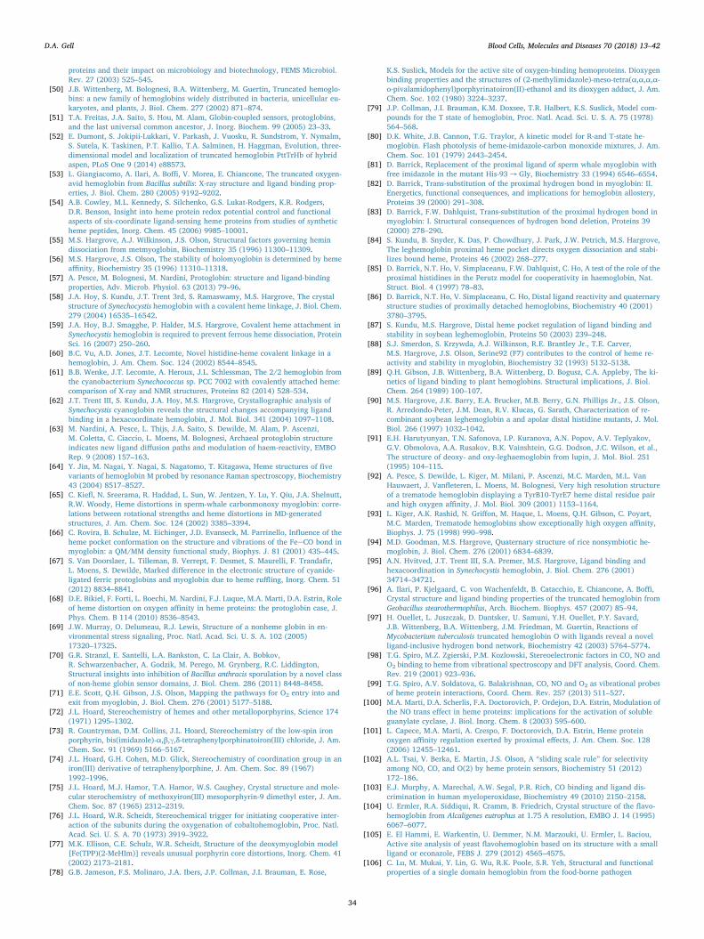
proteins and their impact on microbiology and biotechnology, FEMS Microbiol.Rev. 27 (2003) 525–545.
[50] J.B. Wittenberg, M. Bolognesi, B.A. Wittenberg, M. Guertin, Truncated hemoglo-bins: a new family of hemoglobins widely distributed in bacteria, unicellular eu-karyotes, and plants, J. Biol. Chem. 277 (2002) 871–874.
[51] T.A. Freitas, J.A. Saito, S. Hou, M. Alam, Globin-coupled sensors, protoglobins,and the last universal common ancestor, J. Inorg. Biochem. 99 (2005) 23–33.
[52] E. Dumont, S. Jokipii-Lukkari, V. Parkash, J. Vuosku, R. Sundstrom, Y. Nymalm,S. Sutela, K. Taskinen, P.T. Kallio, T.A. Salminen, H. Haggman, Evolution, three-dimensional model and localization of truncated hemoglobin PttTrHb of hybridaspen, PLoS One 9 (2014) e88573.
[53] L. Giangiacomo, A. Ilari, A. Boffi, V. Morea, E. Chiancone, The truncated oxygen-avid hemoglobin from Bacillus subtilis: X-ray structure and ligand binding prop-erties, J. Biol. Chem. 280 (2005) 9192–9202.
[54] A.B. Cowley, M.L. Kennedy, S. Silchenko, G.S. Lukat-Rodgers, K.R. Rodgers,D.R. Benson, Insight into heme protein redox potential control and functionalaspects of six-coordinate ligand-sensing heme proteins from studies of syntheticheme peptides, Inorg. Chem. 45 (2006) 9985–10001.
[55] M.S. Hargrove, A.J. Wilkinson, J.S. Olson, Structural factors governing hemindissociation from metmyoglobin, Biochemistry 35 (1996) 11300–11309.
[56] M.S. Hargrove, J.S. Olson, The stability of holomyoglobin is determined by hemeaffinity, Biochemistry 35 (1996) 11310–11318.
[57] A. Pesce, M. Bolognesi, M. Nardini, Protoglobin: structure and ligand-bindingproperties, Adv. Microb. Physiol. 63 (2013) 79–96.
[58] J.A. Hoy, S. Kundu, J.T. Trent 3rd, S. Ramaswamy, M.S. Hargrove, The crystalstructure of Synechocystis hemoglobin with a covalent heme linkage, J. Biol. Chem.279 (2004) 16535–16542.
[59] J.A. Hoy, B.J. Smagghe, P. Halder, M.S. Hargrove, Covalent heme attachment inSynechocystis hemoglobin is required to prevent ferrous heme dissociation, ProteinSci. 16 (2007) 250–260.
[60] B.C. Vu, A.D. Jones, J.T. Lecomte, Novel histidine-heme covalent linkage in ahemoglobin, J. Am. Chem. Soc. 124 (2002) 8544–8545.
[61] B.B. Wenke, J.T. Lecomte, A. Heroux, J.L. Schlessman, The 2/2 hemoglobin fromthe cyanobacterium Synechococcus sp. PCC 7002 with covalently attached heme:comparison of X-ray and NMR structures, Proteins 82 (2014) 528–534.
[62] J.T. Trent III, S. Kundu, J.A. Hoy, M.S. Hargrove, Crystallographic analysis ofSynechocystis cyanoglobin reveals the structural changes accompanying ligandbinding in a hexacoordinate hemoglobin, J. Mol. Biol. 341 (2004) 1097–1108.
[63] M. Nardini, A. Pesce, L. Thijs, J.A. Saito, S. Dewilde, M. Alam, P. Ascenzi,M. Coletta, C. Ciaccio, L. Moens, M. Bolognesi, Archaeal protoglobin structureindicates new ligand diffusion paths and modulation of haem-reactivity, EMBORep. 9 (2008) 157–163.
[64] Y. Jin, M. Nagai, Y. Nagai, S. Nagatomo, T. Kitagawa, Heme structures of fivevariants of hemoglobin M probed by resonance Raman spectroscopy, Biochemistry43 (2004) 8517–8527.
[65] C. Kiefl, N. Sreerama, R. Haddad, L. Sun, W. Jentzen, Y. Lu, Y. Qiu, J.A. Shelnutt,R.W. Woody, Heme distortions in sperm-whale carbonmonoxy myoglobin: corre-lations between rotational strengths and heme distortions in MD-generatedstructures, J. Am. Chem. Soc. 124 (2002) 3385–3394.
[66] C. Rovira, B. Schulze, M. Eichinger, J.D. Evanseck, M. Parrinello, Influence of theheme pocket conformation on the structure and vibrations of the FeeCO bond inmyoglobin: a QM/MM density functional study, Biophys. J. 81 (2001) 435–445.
[67] S. Van Doorslaer, L. Tilleman, B. Verrept, F. Desmet, S. Maurelli, F. Trandafir,L. Moens, S. Dewilde, Marked difference in the electronic structure of cyanide-ligated ferric protoglobins and myoglobin due to heme ruffling, Inorg. Chem. 51(2012) 8834–8841.
[68] D.E. Bikiel, F. Forti, L. Boechi, M. Nardini, F.J. Luque, M.A. Marti, D.A. Estrin, Roleof heme distortion on oxygen affinity in heme proteins: the protoglobin case, J.Phys. Chem. B 114 (2010) 8536–8543.
[69] J.W. Murray, O. Delumeau, R.J. Lewis, Structure of a nonheme globin in en-vironmental stress signaling, Proc. Natl. Acad. Sci. U. S. A. 102 (2005)17320–17325.
[70] G.R. Stranzl, E. Santelli, L.A. Bankston, C. La Clair, A. Bobkov,R. Schwarzenbacher, A. Godzik, M. Perego, M. Grynberg, R.C. Liddington,Structural insights into inhibition of Bacillus anthracis sporulation by a novel classof non-heme globin sensor domains, J. Biol. Chem. 286 (2011) 8448–8458.
[71] E.E. Scott, Q.H. Gibson, J.S. Olson, Mapping the pathways for O2 entry into andexit from myoglobin, J. Biol. Chem. 276 (2001) 5177–5188.
[72] J.L. Hoard, Stereochemistry of hemes and other metalloporphyrins, Science 174(1971) 1295–1302.
[73] R. Countryman, D.M. Collins, J.L. Hoard, Stereochemistry of the low-spin ironporphyrin, bis(imidazole)-α,β,γ,δ-tetraphenylporphinatoiron(III) chloride, J. Am.Chem. Soc. 91 (1969) 5166–5167.
[74] J.L. Hoard, G.H. Cohen, M.D. Glick, Stereochemistry of coordination group in aniron(III) derivative of tetraphenylporphine, J. Am. Chem. Soc. 89 (1967)1992–1996.
[75] J.L. Hoard, M.J. Hamor, T.A. Hamor, W.S. Caughey, Crystal structure and mole-cular sterochemistry of methoxyiron(III) mesoporphyrin-9 dimethyl ester, J. Am.Chem. Soc. 87 (1965) 2312–2319.
[76] J.L. Hoard, W.R. Scheidt, Stereochemical trigger for initiating cooperative inter-action of the subunits during the oxygenation of cobaltohemoglobin, Proc. Natl.Acad. Sci. U. S. A. 70 (1973) 3919–3922.
[77] M.K. Ellison, C.E. Schulz, W.R. Scheidt, Structure of the deoxymyoglobin model[Fe(TPP)(2-MeHIm)] reveals unusual porphyrin core distortions, Inorg. Chem. 41(2002) 2173–2181.
[78] G.B. Jameson, F.S. Molinaro, J.A. Ibers, J.P. Collman, J.I. Brauman, E. Rose,
K.S. Suslick, Models for the active site of oxygen-binding hemoproteins. Dioxygenbinding properties and the structures of (2-methylimidazole)-meso-tetra(α,α,α,α-o-pivalamidophenyl)porphyrinatoiron(II)-ethanol and its dioxygen adduct, J. Am.Chem. Soc. 102 (1980) 3224–3237.
[79] J.P. Collman, J.I. Brauman, K.M. Doxsee, T.R. Halbert, K.S. Suslick, Model com-pounds for the T state of hemoglobin, Proc. Natl. Acad. Sci. U. S. A. 75 (1978)564–568.
[80] D.K. White, J.B. Cannon, T.G. Traylor, A kinetic model for R-and T-state he-moglobin. Flash photolysis of heme-imidazole-carbon monoxide mixtures, J. Am.Chem. Soc. 101 (1979) 2443–2454.
[81] D. Barrick, Replacement of the proximal ligand of sperm whale myoglobin withfree imidazole in the mutant His-93 → Gly, Biochemistry 33 (1994) 6546–6554.
[82] D. Barrick, Trans-substitution of the proximal hydrogen bond in myoglobin: II.Energetics, functional consequences, and implications for hemoglobin allostery,Proteins 39 (2000) 291–308.
[83] D. Barrick, F.W. Dahlquist, Trans-substitution of the proximal hydrogen bond inmyoglobin: I. Structural consequences of hydrogen bond deletion, Proteins 39(2000) 278–290.
[84] S. Kundu, B. Snyder, K. Das, P. Chowdhury, J. Park, J.W. Petrich, M.S. Hargrove,The leghemoglobin proximal heme pocket directs oxygen dissociation and stabi-lizes bound heme, Proteins 46 (2002) 268–277.
[85] D. Barrick, N.T. Ho, V. Simplaceanu, F.W. Dahlquist, C. Ho, A test of the role of theproximal histidines in the Perutz model for cooperativity in haemoglobin, Nat.Struct. Biol. 4 (1997) 78–83.
[86] D. Barrick, N.T. Ho, V. Simplaceanu, C. Ho, Distal ligand reactivity and quaternarystructure studies of proximally detached hemoglobins, Biochemistry 40 (2001)3780–3795.
[87] S. Kundu, M.S. Hargrove, Distal heme pocket regulation of ligand binding andstability in soybean leghemoglobin, Proteins 50 (2003) 239–248.
[88] S.J. Smerdon, S. Krzywda, A.J. Wilkinson, R.E. Brantley Jr., T.E. Carver,M.S. Hargrove, J.S. Olson, Serine92 (F7) contributes to the control of heme re-activity and stability in myoglobin, Biochemistry 32 (1993) 5132–5138.
[89] Q.H. Gibson, J.B. Wittenberg, B.A. Wittenberg, D. Bogusz, C.A. Appleby, The ki-netics of ligand binding to plant hemoglobins. Structural implications, J. Biol.Chem. 264 (1989) 100–107.
[90] M.S. Hargrove, J.K. Barry, E.A. Brucker, M.B. Berry, G.N. Phillips Jr., J.S. Olson,R. Arredondo-Peter, J.M. Dean, R.V. Klucas, G. Sarath, Characterization of re-combinant soybean leghemoglobin a and apolar distal histidine mutants, J. Mol.Biol. 266 (1997) 1032–1042.
[91] E.H. Harutyunyan, T.N. Safonova, I.P. Kuranova, A.N. Popov, A.V. Teplyakov,G.V. Obmolova, A.A. Rusakov, B.K. Vainshtein, G.G. Dodson, J.C. Wilson, et al.,The structure of deoxy- and oxy-leghaemoglobin from lupin, J. Mol. Biol. 251(1995) 104–115.
[92] A. Pesce, S. Dewilde, L. Kiger, M. Milani, P. Ascenzi, M.C. Marden, M.L. VanHauwaert, J. Vanfleteren, L. Moens, M. Bolognesi, Very high resolution structureof a trematode hemoglobin displaying a TyrB10-TyrE7 heme distal residue pairand high oxygen affinity, J. Mol. Biol. 309 (2001) 1153–1164.
[93] L. Kiger, A.K. Rashid, N. Griffon, M. Haque, L. Moens, Q.H. Gibson, C. Poyart,M.C. Marden, Trematode hemoglobins show exceptionally high oxygen affinity,Biophys. J. 75 (1998) 990–998.
[94] M.D. Goodman, M.S. Hargrove, Quaternary structure of rice nonsymbiotic he-moglobin, J. Biol. Chem. 276 (2001) 6834–6839.
[95] A.N. Hvitved, J.T. Trent III, S.A. Premer, M.S. Hargrove, Ligand binding andhexacoordination in Synechocystis hemoglobin, J. Biol. Chem. 276 (2001)34714–34721.
[96] A. Ilari, P. Kjelgaard, C. von Wachenfeldt, B. Catacchio, E. Chiancone, A. Boffi,Crystal structure and ligand binding properties of the truncated hemoglobin fromGeobacillus stearothermophilus, Arch. Biochem. Biophys. 457 (2007) 85–94.
[97] H. Ouellet, L. Juszczak, D. Dantsker, U. Samuni, Y.H. Ouellet, P.Y. Savard,J.B. Wittenberg, B.A. Wittenberg, J.M. Friedman, M. Guertin, Reactions ofMycobacterium tuberculosis truncated hemoglobin O with ligands reveal a novelligand-inclusive hydrogen bond network, Biochemistry 42 (2003) 5764–5774.
[98] T.G. Spiro, M.Z. Zgierski, P.M. Kozlowski, Stereoelectronic factors in CO, NO andO2 binding to heme from vibrational spectroscopy and DFT analysis, Coord. Chem.Rev. 219 (2001) 923–936.
[99] T.G. Spiro, A.V. Soldatova, G. Balakrishnan, CO, NO and O2 as vibrational probesof heme protein interactions, Coord. Chem. Rev. 257 (2013) 511–527.
[100] M.A. Marti, D.A. Scherlis, F.A. Doctorovich, P. Ordejon, D.A. Estrin, Modulation ofthe NO trans effect in heme proteins: implications for the activation of solubleguanylate cyclase, J. Biol. Inorg. Chem. 8 (2003) 595–600.
[101] L. Capece, M.A. Marti, A. Crespo, F. Doctorovich, D.A. Estrin, Heme proteinoxygen affinity regulation exerted by proximal effects, J. Am. Chem. Soc. 128(2006) 12455–12461.
[102] A.L. Tsai, V. Berka, E. Martin, J.S. Olson, A “sliding scale rule” for selectivityamong NO, CO, and O(2) by heme protein sensors, Biochemistry 51 (2012)172–186.
[103] E.J. Murphy, A. Marechal, A.W. Segal, P.R. Rich, CO binding and ligand dis-crimination in human myeloperoxidase, Biochemistry 49 (2010) 2150–2158.
[104] U. Ermler, R.A. Siddiqui, R. Cramm, B. Friedrich, Crystal structure of the flavo-hemoglobin from Alcaligenes eutrophus at 1.75 A resolution, EMBO J. 14 (1995)6067–6077.
[105] E. El Hammi, E. Warkentin, U. Demmer, N.M. Marzouki, U. Ermler, L. Baciou,Active site analysis of yeast flavohemoglobin based on its structure with a smallligand or econazole, FEBS J. 279 (2012) 4565–4575.
[106] C. Lu, M. Mukai, Y. Lin, G. Wu, R.K. Poole, S.R. Yeh, Structural and functionalproperties of a single domain hemoglobin from the food-borne pathogen
D.A. Gell Blood Cells, Molecules and Diseases 70 (2018) 13–42
34

Campylobactor jejuni, J. Biol. Chem. 282 (2007) 25917–25928.[107] M. Shepherd, V. Barynin, C. Lu, P.V. Bernhardt, G. Wu, S.R. Yeh, T. Egawa,
S.E. Sedelnikova, D.W. Rice, J.L. Wilson, R.K. Poole, The single-domain globinfrom the pathogenic bacterium Campylobacter jejuni: novel D-helix conformation,proximal hydrogen bonding that influences ligand binding, and peroxidase-likeredox properties, J. Biol. Chem. 285 (2010) 12747–12754.
[108] M. Mukai, C.E. Mills, R.K. Poole, S.R. Yeh, Flavohemoglobin, a globin with aperoxidase-like catalytic site, J. Biol. Chem. 276 (2001) 7272–7277.
[109] T.L. Poulos, The role of the proximal ligand in heme enzymes, JBIC, J. Biol. Inorg.Chem. 1 (1996) 356–359.
[110] J.H. Dawson, Probing structure-function relations in heme-containing oxygenasesand peroxidases, Science 240 (1988) 433–439.
[111] Y. Mizutani, T. Kitagawa, Ultrafast structural relaxation of myoglobin followingphotodissociation of carbon monoxide probed by time-resolved resonance Ramanspectroscopy, J. Phys. Chem. B 105 (2001) 10992–10999.
[112] J. Kincaid, P. Stein, T.G. Spiro, Absence of heme-localized strain in T state he-moglobin: insensitivity of heme-imidazole resonance Raman frequencies to qua-ternary structure, Proc. Natl. Acad. Sci. U. S. A. 76 (1979) 549–552.
[113] O. Bangcharoenpaurpong, K.T. Schomacker, P.M. Champion, Resonance Ramaninvestigation of myoglobin and hemoglobin, J. Am. Chem. Soc. 106 (1984)5688–5698.
[114] M.R. Ondrias, D.L. Rousseau, J.A. Shelnutt, S.R. Simon, Quaternary-transforma-tion-induced changes at the heme in deoxyhemoglobins, Biochemistry 21 (1982)3428–3437.
[115] J.M. Friedman, T.W. Scott, R.A. Stepnoski, M. Ikeda-Saito, T. Yonetani, The iron-proximal histidine linkage and protein control of oxygen binding in hemoglobin. Atransient Raman study, J. Biol. Chem. 258 (1983) 10564–10572.
[116] G. Balakrishnan, M. Ibrahim, P.J. Mak, J. Hata, J.R. Kincaid, T.G. Spiro, Linkingconformation change to hemoglobin activation via chain-selective time-resolvedresonance Raman spectroscopy of protoheme/mesoheme hybrids, J. Biol. Inorg.Chem. 14 (2009) 741–750.
[117] Y. Mizutani, M. Nagai, Ultrafast protein dynamics of hemoglobin as studied bypicosecond time-resolved resonance Raman spectroscopy, Chem. Phys. 396 (2012)45–52.
[118] G. Smulevich, R. Evangelista-Kirkup, A. English, T.G. Spiro, Raman and infraredspectra of cytochrome c peroxidase-carbon monoxide adducts in alternative con-formational states, Biochemistry 25 (1986) 4426–4430.
[119] J. Teraoka, T. Kitagawa, Structural implication of the heme-linked ionization ofhorseradish peroxidase probed by the Fe-histidine stretching Raman line, J. Biol.Chem. 256 (1981) 3969–3977.
[120] C.E. Cooper, N. Ioannidis, R. D'Mello, R.K. Poole, Haem, flavin and oxygen in-teractions in Hmp, a flavohaemoglobin from Escherichia coli, Biochem. Soc. Trans.22 (1994) 709–713.
[121] E. Antonini, J. Wyman, M. Brunori, J.F. Taylor, A. Rossi-Fanelli, A. Caputo, Studieson the oxidation-reduction potentials of heme proteins. I. Human hemoglobin, J.Biol. Chem. 239 (1964) 907–912.
[122] H. Yamada, R. Makino, I. Yamazaki, Effects of 2,4-substituents of deuterophemeupon redox potentials of horseradish peroxidases, Arch. Biochem. Biophys. 169(1975) 344–353.
[123] C.W. Conroy, P. Tyma, P.H. Daum, J.E. Erman, Oxidation-reduction potentialmeasurements of cytochrome c peroxidase and pH dependent spectral transitionsin the ferrous enzyme, Biochim. Biophys. Acta 537 (1978) 62–69.
[124] C.J. Reedy, M.M. Elvekrog, B.R. Gibney, Development of a heme protein structure-electrochemical function database, Nucleic Acids Res. 36 (2008) D307–13.
[125] D.P. Hildebrand, D.L. Burk, R. Maurus, J.C. Ferrer, G.D. Brayer, A.G. Mauk, Theproximal ligand variant His93Tyr of horse heart myoglobin, Biochemistry 34(1995) 1997–2005.
[126] C.S. Thom, C.F. Dickson, D.A. Gell, M.J. Weiss, Hemoglobin variants: biochemicalproperties and clinical correlates, Cold Spring Harb. Perspect. Med. 3 (2013)a011858.
[127] J.S. Olson, G.N. Phillips Jr., Myoglobin discriminates between O2, NO and CO byelectrostatic interactions with the bound ligand, J. Biol. Inorg. Chem. 2 (1997)544–552.
[128] L. Pauling, J.J. Weiss, Nature of the iron-oxygen bond in oxyhaemoglobin, Nature203 (1964) 182–183.
[129] J.J. Weiss, Nature of the iron-oxygen bond in oxyhaemoglobin, Nature 202 (1964)83–84.
[130] J.B. Wittenberg, B.A. Wittenberg, J. Peisach, W.E. Blumberg, On the state of theiron and the nature of the ligand in oxyhemoglobin, Proc. Natl. Acad. Sci. U. S. A.67 (1970) 1846–1853.
[131] H. Chen, M. Ikeda-Saito, S. Shaik, Nature of the FeeO2 bonding in oxy-myoglobin:effect of the protein, J. Am. Chem. Soc. 130 (2008) 14778–14790.
[132] K.L. Bren, R. Eisenberg, H.B. Gray, Discovery of the magnetic behavior of he-moglobin: a beginning of bioinorganic chemistry, Proc. Natl. Acad. Sci. U. S. A.112 (2015) 13123–13127.
[133] S.A. Wilson, E. Green, Mathews II, M. Benfatto, K.O. Hodgson, B. Hedman,R. Sarangi, X-ray absorption spectroscopic investigation of the electronic structuredifferences in solution and crystalline oxyhemoglobin, Proc. Natl. Acad. Sci. U. S.A. 110 (2013) 16333–16338.
[134] S.A. Wilson, T. Kroll, R.A. Decreau, R.K. Hocking, M. Lundberg, B. Hedman,K.O. Hodgson, E.I. Solomon, Iron L-edge X-ray absorption spectroscopy of oxy-picket fence porphyrin: experimental insight into FeeO2 bonding, J. Am. Chem.Soc. 135 (2013) 1124–1136.
[135] C.H. Barlow, J.C. Maxwell, W.J. Wallace, W.S. Caughey, Elucidation of the modeof binding of oxygen to iron in oxyhemoglobin by infrared spectroscopy, Biochem.Biophys. Res. Commun. 55 (1973) 91–96.
[136] J.C. Maxwell, J.A. Volpe, C.H. Barlow, W.S. Caughey, Infrared evidence for themode of binding of oxygen to iron of myoglobin from heart muscle, Biochem.Biophys. Res. Commun. 58 (1974) 166–171.
[137] J.P. Collman, J.I. Brauman, T.R. Halbert, K.S. Suslick, Nature of O2 and CO bindingto metalloporphyrins and heme proteins, Proc. Natl. Acad. Sci. U. S. A. 73 (1976)3333–3337.
[138] K.P. Jensen, U. Ryde, How O2 binds to heme: reasons for rapid binding and spininversion, J. Biol. Chem. 279 (2004) 14561–14569.
[139] R.J. Rohlfs, A.J. Mathews, T.E. Carver, J.S. Olson, B.A. Springer, K.D. Egeberg,S.G. Sligar, The effects of amino acid substitution at position E7 (residue 64) onthe kinetics of ligand binding to sperm whale myoglobin, J. Biol. Chem. 265(1990) 3168–3176.
[140] T.G. Spiro, I.H. Wasbotten, CO as a vibrational probe of heme protein active sites,J. Inorg. Biochem. 99 (2005) 34–44.
[141] T. Kitagawa, M.R. Ondrias, D.L. Rousseau, M. Ikeda-Saito, T. Yonetani, Evidencefor hydrogen bonding of bound dioxygen to the distal histidine of oxycobaltmyoglobin and haemoglobin, Nature 298 (1982) 869–871.
[142] D. Morikis, P.M. Champion, B.A. Springer, S.G. Sligar, Resonance raman in-vestigations of site-directed mutants of myoglobin: effects of distal histidine re-placement, Biochemistry 28 (1989) 4791–4800.
[143] M. Unno, J.F. Christiansen, J.S. Olson, J.T. Sage, P.M. Champion, Evidence forhydrogen bonding effects in the iron ligand vibrations of carbonmonoxy myo-globin, J. Am. Chem. Soc. 120 (1998) 2670–2671.
[144] T. Yonetani, H. Yamamoto, T. Iizuka, Studies on cobalt myoglobins and he-moglobins. 3. Electron paramagnetic resonance studies of reversible oxygenationof cobalt myoglobins and hemoglobins, J. Biol. Chem. 249 (1974) 2168–2174.
[145] S.E. Phillips, Structure and refinement of oxymyoglobin at 1.6 A resolution, J. Mol.Biol. 142 (1980) 531–554.
[146] B. Shaanan, The iron-oxygen bond in human oxyhaemoglobin, Nature 296 (1982)683–684.
[147] S.E. Phillips, B.P. Schoenborn, Neutron diffraction reveals oxygen-histidine hy-drogen bond in oxymyoglobin, Nature 292 (1981) 81–82.
[148] J.A. Lukin, V. Simplaceanu, M. Zou, N.T. Ho, C. Ho, NMR reveals hydrogen bondsbetween oxygen and distal histidines in oxyhemoglobin, Proc. Natl. Acad. Sci. U. S.A. 97 (2000) 10354–10358.
[149] J.S. Olson, G.N. Phillips Jr., Kinetic pathways and barriers for ligand binding tomyoglobin, J. Biol. Chem. 271 (1996) 17593–17596.
[150] D.A. Scherlis, D.A. Estrin, Hydrogen bonding and O2 affinity of hemoglobins, J.Am. Chem. Soc. 123 (2001) 8436–8437.
[151] B.A. Springer, K.D. Egeberg, S.G. Sligar, R.J. Rohlfs, A.J. Mathews, J.S. Olson,Discrimination between oxygen and carbon monoxide and inhibition of auto-oxidation by myoglobin. Site-directed mutagenesis of the distal histidine, J. Biol.Chem. 264 (1989) 3057–3060.
[152] M.L. Quillin, R.M. Arduini, J.S. Olson, G.N. Phillips Jr., High-resolution crystalstructures of distal histidine mutants of sperm whale myoglobin, J. Mol. Biol. 234(1993) 140–155.
[153] R.A. Goldbeck, S. Bhaskaran, C. Ortega, J.L. Mendoza, J.S. Olson, J. Soman,D.S. Kliger, R.M. Esquerra, Water and ligand entry in myoglobin: assessing thespeed and extent of heme pocket hydration after CO photodissociation, Proc. Natl.Acad. Sci. U. S. A. 103 (2006) 1254–1259.
[154] J.P. Bustamante, S. Abbruzzetti, A. Marcelli, D. Gauto, L. Boechi, A. Bonamore,A. Boffi, S. Bruno, A. Feis, P. Foggi, D.A. Estrin, C. Viappiani, Ligand uptakemodulation by internal water molecules and hydrophobic cavities in hemoglobins,J. Phys. Chem. B 118 (2014) 1234–1245.
[155] I. Birukou, R.L. Schweers, J.S. Olson, Distal histidine stabilizes bound O2 and actsas a gate for ligand entry in both subunits of adult human hemoglobin, J. Biol.Chem. 285 (2010) 8840–8854.
[156] I. Degtyarenko, R.M. Nieminen, C. Rovira, Structure and dynamics of dioxygenbound to cobalt and iron heme, Biophys. J. 91 (2006) 2024–2034.
[157] A. Ilari, A. Bonamore, A. Farina, K.A. Johnson, A. Boffi, The X-ray structure offerric Escherichia coli flavohemoglobin reveals an unexpected geometry of thedistal heme pocket, J. Biol. Chem. 277 (2002) 23725–23732.
[158] E. El Hammi, E. Warkentin, U. Demmer, F. Limam, N.M. Marzouki, U. Ermler,L. Baciou, Structure of Ralstonia eutropha flavohemoglobin in complex with threeantibiotic azole compounds, Biochemistry 50 (2011) 1255–1264.
[159] J. Yang, A.P. Kloek, D.E. Goldberg, F.S. Mathews, The structure of Ascaris he-moglobin domain I at 2.2 A resolution: molecular features of oxygen avidity, Proc.Natl. Acad. Sci. U. S. A. 92 (1995) 4224–4228.
[160] Q.H. Gibson, R. Regan, J.S. Olson, T.E. Carver, B. Dixon, B. Pohajdak, P.K. Sharma,S.N. Vinogradov, Kinetics of ligand binding to Pseudoterranova decipiens andAscaris suum hemoglobins and to Leu-29 → Tyr sperm whale myoglobin mutant, J.Biol. Chem. 268 (1993) 16993–16998.
[161] A. Pesce, M. Nardini, S. Dewilde, E. Geuens, K. Yamauchi, P. Ascenzi, A.F. Riggs,L. Moens, M. Bolognesi, The 109 residue nerve tissue minihemoglobin fromCerebratulus lacteus highlights striking structural plasticity of the α-helical globinfold, Structure 10 (2002) 725–735.
[162] F. Germani, A. Pesce, A. Venturini, L. Moens, M. Bolognesi, S. Dewilde, M. Nardini,High resolution crystal structures of the Cerebratulus lacteus mini-Hb in the un-ligated and carbomonoxy states, Int. J. Mol. Sci. 13 (2012) 8025–8037.
[163] M.A. Marti, D.E. Bikiel, A. Crespo, M. Nardini, M. Bolognesi, D.A. Estrin, Twodistinct heme distal site states define Cerebratulus lacteus mini-hemoglobin oxygenaffinity, Proteins 62 (2006) 641–648.
[164] A. Pesce, M. Nardini, P. Ascenzi, E. Geuens, S. Dewilde, L. Moens, M. Bolognesi,A.F. Riggs, A. Hale, P. Deng, G.U. Nienhaus, J.S. Olson, K. Nienhaus, Thr-E11regulates O2 affinity in Cerebratulus lacteus mini-hemoglobin, J. Biol. Chem. 279(2004) 33662–33672.
D.A. Gell Blood Cells, Molecules and Diseases 70 (2018) 13–42
35

[165] M.A. Marti, A. Crespo, L. Capece, L. Boechi, D.E. Bikiel, D.A. Scherlis, D.A. Estrin,Dioxygen affinity in heme proteins investigated by computer simulation, J. Inorg.Biochem. 100 (2006) 761–770.
[166] T.K. Das, R.E. Weber, S. Dewilde, J.B. Wittenberg, B.A. Wittenberg, K. Yamauchi,M.L. Van Hauwaert, L. Moens, D.L. Rousseau, Ligand binding in the ferric andferrous states of Paramecium hemoglobin, Biochemistry 39 (2000) 14330–14340.
[167] M.A. Marti, L. Capece, D.E. Bikiel, B. Falcone, D.A. Estrin, Oxygen affinity con-trolled by dynamical distal conformations: the soybean leghemoglobin and theParamecium caudatum hemoglobin cases, Proteins 68 (2007) 480–487.
[168] G.N. Phillips, M.L. Teodoro, T. Li, B. Smith, J.S. Olson, Bound CO is a molecularprobe of electrostatic potential in the distal pocket of myoglobin, J. Phys. Chem. B103 (1999) 8817–8829.
[169] S. Kundu, G.C. Blouin, S.A. Premer, G. Sarath, J.S. Olson, M.S. Hargrove, TyrosineB10 inhibits stabilization of bound carbon monoxide and oxygen in soybean le-ghemoglobin, Biochemistry 43 (2004) 6241–6252.
[170] M. Tarnawski, T.R. Barends, I. Schlichting, Structural analysis of an oxygen-regulated diguanylate cyclase, Acta Crystallogr. D Biol. Crystallogr. 71 (2015)2158–2177.
[171] A. Pesce, L. Thijs, M. Nardini, F. Desmet, L. Sisinni, L. Gourlay, A. Bolli, M. Coletta,S. Van Doorslaer, X. Wan, M. Alam, P. Ascenzi, L. Moens, M. Bolognesi, S. Dewilde,HisE11 and HisF8 provide bis-histidyl heme hexa-coordination in the globin do-main of Geobacter sulfurreducens globin-coupled sensor, J. Mol. Biol. 386 (2009)246–260.
[172] L. Tilleman, S. Abbruzzetti, C. Ciaccio, G. De Sanctis, M. Nardini, A. Pesce,F. Desmet, L. Moens, S. Van Doorslaer, S. Bruno, M. Bolognesi, P. Ascenzi,M. Coletta, C. Viappiani, S. Dewilde, Structural basis for the regulation of CObinding in the archaeal protoglobin from Methanosarcina acetivorans, PLoS One 10(2015) e0125959.
[173] A. Pesce, L. Tilleman, J. Donne, E. Aste, P. Ascenzi, C. Ciaccio, M. Coletta,L. Moens, C. Viappiani, S. Dewilde, M. Bolognesi, M. Nardini, Structure and haem-distal site plasticity in Methanosarcina acetivorans protoglobin, PLoS One 8 (2013)e66144.
[174] J. Igarashi, K. Kobayashi, A. Matsuoka, A hydrogen-bonding network formed bythe B10-E7-E11 residues of a truncated hemoglobin from Tetrahymena pyriformis iscritical for stability of bound oxygen and nitric oxide detoxification, J. Biol. Inorg.Chem. 16 (2011) 599–609.
[175] M. Couture, S.R. Yeh, B.A. Wittenberg, J.B. Wittenberg, Y. Ouellet, D.L. Rousseau,M. Guertin, A cooperative oxygen-binding hemoglobin from Mycobacterium tu-berculosis, Proc. Natl. Acad. Sci. U. S. A. 96 (1999) 11223–11228.
[176] S.R. Yeh, M. Couture, Y. Ouellet, M. Guertin, D.L. Rousseau, A cooperative oxygenbinding hemoglobin fromMycobacterium tuberculosis. Stabilization of heme ligandsby a distal tyrosine residue, J. Biol. Chem. 275 (2000) 1679–1684.
[177] H. Ouellet, M. Milani, M. LaBarre, M. Bolognesi, M. Couture, M. Guertin, The rolesof Tyr(CD1) and Trp(G8) in Mycobacterium tuberculosis truncated hemoglobin O inligand binding and on the heme distal site architecture, Biochemistry 46 (2007)11440–11450.
[178] M. Milani, P.Y. Savard, H. Ouellet, P. Ascenzi, M. Guertin, M. Bolognesi, ATyrCD1/TrpG8 hydrogen bond network and a TyrB10TyrCD1 covalent link shapethe heme distal site of Mycobacterium tuberculosis hemoglobin O, Proc. Natl. Acad.Sci. U. S. A. 100 (2003) 5766–5771.
[179] M. Mukai, P.Y. Savard, H. Ouellet, M. Guertin, S.R. Yeh, Unique ligand-proteininteractions in a new truncated hemoglobin from Mycobacterium tuberculosis,Biochemistry 41 (2002) 3897–3905.
[180] M. Nardini, A. Pesce, M. Labarre, C. Richard, A. Bolli, P. Ascenzi, M. Guertin,M. Bolognesi, Structural determinants in the group III truncated hemoglobin fromCampylobacter jejuni, J. Biol. Chem. 281 (2006) 37803–37812.
[181] C. Lu, T. Egawa, L.M. Wainwright, R.K. Poole, S.R. Yeh, Structural and functionalproperties of a truncated hemoglobin from a food-borne pathogen Campylobacterjejuni, J. Biol. Chem. 282 (2007) 13627–13636.
[182] P. Arroyo Manez, C. Lu, L. Boechi, M.A. Marti, M. Shepherd, J.L. Wilson,R.K. Poole, F.J. Luque, S.R. Yeh, D.A. Estrin, Role of the distal hydrogen-bondingnetwork in regulating oxygen affinity in the truncated hemoglobin III fromCampylobacter jejuni, Biochemistry 50 (2011) 3946–3956.
[183] B. Stec, G.N. Phillips Jr., How the CO in myoglobin acquired its bend: lessons ininterpretation of crystallographic data, Acta Crystallogr. Sect. D 57 (2001)751–754.
[184] J. Vojtechovsky, K. Chu, J. Berendzen, R.M. Sweet, I. Schlichting, Crystal struc-tures of myoglobin-ligand complexes at near-atomic resolution, Biophys. J. 77(1999) 2153–2174.
[185] G.S. Kachalova, A.N. Popov, H.D. Bartunik, A steric mechanism for inhibition ofCO binding to heme proteins, Science 284 (1999) 473–476.
[186] T.E. Carver, R.E. Brantley Jr., E.W. Singleton, R.M. Arduini, M.L. Quillin,G.N. Phillips Jr., J.S. Olson, A novel site-directed mutant of myoglobin with anunusually high O2 affinity and low autooxidation rate, J. Biol. Chem. 267 (1992)14443–14450.
[187] M.L. Quillin, T. Li, J.S. Olson, G.N. Phillips Jr., Y. Dou, M. Ikeda-Saito, R. Regan,M. Carlson, Q.H. Gibson, H. Li, et al., Structural and functional effects of apolarmutations of the distal valine in myoglobin, J. Mol. Biol. 245 (1995) 416–436.
[188] D.G. Lambright, S. Balasubramanian, S.M. Decatur, S.G. Boxer, Anatomy anddynamics of a ligand-binding pathway in myoglobin: the roles of residues 45, 60,64, and 68, Biochemistry 33 (1994) 5518–5525.
[189] Q.H. Gibson, R. Regan, R. Elber, J.S. Olson, T.E. Carver, Distal pocket residuesaffect picosecond ligand recombination in myoglobin. An experimental and mo-lecular dynamics study of position 29 mutants, J. Biol. Chem. 267 (1992)22022–22034.
[190] F. Schotte, J. Soman, J.S. Olson, M. Wulff, P.A. Anfinrud, Picosecond time-
resolved X-ray crystallography: probing protein function in real time, J. Struct.Biol. 147 (2004) 235–246.
[191] I. Birukou, D.H. Maillett, A. Birukova, J.S. Olson, Modulating distal cavities in theα and β subunits of human HbA reveals the primary ligand migration pathway,Biochemistry 50 (2011) 7361–7374.
[192] H.H. Lai, T. Li, D.S. Lyons, G.N. Phillips Jr., J.S. Olson, Q.H. Gibson, Phe-46(CD4)orients the distal histidine for hydrogen bonding to bound ligands in sperm whalemyoglobin, Proteins 22 (1995) 322–339.
[193] L. Boechi, M. Arrar, M.A. Marti, J.S. Olson, A.E. Roitberg, D.A. Estrin,Hydrophobic effect drives oxygen uptake in myoglobin via histidine E7, J. Biol.Chem. 288 (2013) 6754–6762.
[194] M.F. Perutz, F.S. Mathews, An x-ray study of azide methaemoglobin, J. Mol. Biol.21 (1966) 199–202.
[195] M.F. Perutz, Myoglobin and haemoglobin: role of distal residues in reactions withhaem ligands, Trends Biochem. Sci. 14 (1989) 42–44.
[196] M. Bolognesi, E. Cannillo, P. Ascenzi, G.M. Giacometti, A. Merli, M. Brunori,Reactivity of ferric Aplysia and sperm whale myoglobins towards imidazole: X-rayand binding study, J. Mol. Biol. 158 (1982) 305–315.
[197] K.A. Johnson, J.S. Olson, G.N. Phillips Jr., Structure of myoglobin-ethyl iso-cyanide. Histidine as a swinging door for ligand entry, J. Mol. Biol. 207 (1989)459–463.
[198] D. Ringe, G.A. Petsko, D.E. Kerr, P.R. Ortiz de Montellano, Reaction of myoglobinwith phenylhydrazine: a molecular doorstop, Biochemistry 23 (1984) 2–4.
[199] F. Yang, G.N. Phillips Jr., Crystal structures of CO-, deoxy- and met-myoglobins atvarious pH values, J. Mol. Biol. 256 (1996) 762–774.
[200] P. Jewsbury, T. Kitagawa, The distal residue-CO interaction in carbonmonoxymyoglobins: a molecular dynamics study of two distal histidine tautomers,Biophys. J. 67 (1994) 2236–2250.
[201] B.P. Schoenborn, H.C. Watson, J.C. Kendrew, Binding of xenon to sperm whalemyoglobin, Nature 207 (1965) 28–30.
[202] R.F. Tilton Jr., I.D. Kuntz Jr., G.A. Petsko, Cavities in proteins: structure of ametmyoglobin-xenon complex solved to 1.9 A, Biochemistry 23 (1984)2849–2857.
[203] J. Cohen, A. Arkhipov, R. Braun, K. Schulten, Imaging the migration pathways forO2, CO, NO, and Xe inside myoglobin, Biophys. J. 91 (2006) 1844–1857.
[204] E.E. Scott, Q.H. Gibson, Ligand migration in sperm whale myoglobin,Biochemistry 36 (1997) 11909–11917.
[205] A. Ostermann, R. Waschipky, F.G. Parak, G.U. Nienhaus, Ligand binding andconformational motions in myoglobin, Nature 404 (2000) 205–208.
[206] I. Birukou, J. Soman, J.S. Olson, Blocking the gate to ligand entry in human he-moglobin, J. Biol. Chem. 286 (2011) 10515–10529.
[207] J.E. Knapp, R. Pahl, J. Cohen, J.C. Nichols, K. Schulten, Q.H. Gibson, V. Srajer,W.E. Royer Jr., Ligand migration and cavities within scapharca dimeric HbI:studies by time-resolved crystallo-graphy, Xe binding, and computational analysis,Structure 17 (2009) 1494–1504.
[208] K. Nienhaus, J.E. Knapp, P. Palladino, W.E. Royer Jr., G.U. Nienhaus, Ligandmigration and binding in the dimeric hemoglobin of Scapharca inaequivalvis,Biochemistry 46 (2007) 14018–14031.
[209] M. Bringas, A.A. Petruk, D.A. Estrin, L. Capece, M.A. Marti, Tertiary and qua-ternary structural basis of oxygen affinity in human hemoglobin as revealed bymultiscale simulations, Sci Rep 7 (2017) 10926.
[210] M. Milani, A. Pesce, Y. Ouellet, S. Dewilde, J. Friedman, P. Ascenzi, M. Guertin,M. Bolognesi, Heme-ligand tunneling in group I truncated hemoglobins, J. Biol.Chem. 279 (2004) 21520–21525.
[211] M. Milani, Y. Ouellet, H. Ouellet, M. Guertin, A. Boffi, G. Antonini, A. Bocedi,M. Mattu, M. Bolognesi, P. Ascenzi, Cyanide binding to truncated hemoglobins: acrystallographic and kinetic study, Biochemistry 43 (2004) 5213–5221.
[212] M. Milani, A. Pesce, Y. Ouellet, P. Ascenzi, M. Guertin, M. Bolognesi,Mycobacterium tuberculosis hemoglobin N displays a protein tunnel suited for O2
diffusion to the heme, EMBO J. 20 (2001) 3902–3909.[213] L. Boechi, M.A. Marti, M. Milani, M. Bolognesi, F.J. Luque, D.A. Estrin, Structural
determinants of ligand migration in Mycobacterium tuberculosis truncated he-moglobin O, Proteins 73 (2008) 372–379.
[214] V. Guallar, C. Lu, K. Borrelli, T. Egawa, S.R. Yeh, Ligand migration in the truncatedhemoglobin-II from Mycobacterium tuberculosis: the role of G8 tryptophan, J. Biol.Chem. 284 (2009) 3106–3116.
[215] A. Marcelli, S. Abbruzzetti, J.P. Bustamante, A. Feis, A. Bonamore, A. Boffi,C. Gellini, P.R. Salvi, D.A. Estrin, S. Bruno, C. Viappiani, P. Foggi, Following ligandmigration pathways from picoseconds to milliseconds in type II truncated he-moglobin from Thermobifida fusca, PLoS One 7 (2012) e39884.
[216] A. Bonamore, A. Ilari, L. Giangiacomo, A. Bellelli, V. Morea, A. Boffi, A novelthermostable hemoglobin from the actinobacterium Thermobifida fusca, FEBS J.272 (2005) 4189–4201.
[217] M.D. Salter, K. Nienhaus, G.U. Nienhaus, S. Dewilde, L. Moens, A. Pesce,M. Nardini, M. Bolognesi, J.S. Olson, The apolar channel in Cerebratulus lacteushemoglobin is the route for O2 entry and exit, J. Biol. Chem. 283 (2008)35689–35702.
[218] A. Pesce, M. Nardini, S. Dewilde, L. Capece, M.A. Marti, S. Congia, M.D. Salter,G.C. Blouin, D.A. Estrin, P. Ascenzi, L. Moens, M. Bolognesi, J.S. Olson, Ligandmigration in the apolar tunnel of Cerebratulus lacteus mini-hemoglobin, J. Biol.Chem. 286 (2011) 5347–5358.
[219] J.P. Bustamante, M.E. Szretter, M. Sued, M.A. Marti, D.A. Estrin, L. Boechi, Aquantitative model for oxygen uptake and release in a family of hemeproteins,Bioinformatics 32 (2016) 1805–1813.
[220] Y.H. Ouellet, R. Daigle, P. Lague, D. Dantsker, M. Milani, M. Bolognesi,J.M. Friedman, M. Guertin, Ligand binding to truncated hemoglobin N from
D.A. Gell Blood Cells, Molecules and Diseases 70 (2018) 13–42
36

Mycobacterium tuberculosis is strongly modulated by the interplay between thedistal heme pocket residues and internal water, J. Biol. Chem. 283 (2008)27270–27278.
[221] A. Jasaitis, H. Ouellet, J.-C. Lambry, J.-L. Martin, J.M. Friedman, M. Guertin,M.H. Vos, Ultrafast heme-ligand recombination in truncated hemoglobin HbOfrom Mycobacterium tuberculosis: a ligand cage, Chem. Phys. 396 (2012) 10–16.
[222] M. Rougee, D. Brault, Influence of trans weak or strong field ligands upon theaffinity of deuteroheme for carbon monoxide. Monoimidazoleheme as a referencefor unconstrained five-coordinate hemoproteins, Biochemistry 14 (1975)4100–4106.
[223] V.L. Robinson, B.B. Smith, A. Arnone, A pH-dependent aquomet-to-hemichrometransition in crystalline horse methemoglobin, Biochemistry 42 (2003)10113–10125.
[224] D.A. Svistunenko, M.A. Sharpe, P. Nicholls, C. Blenkinsop, N.A. Davies, J. Dunne,M.T. Wilson, C.E. Cooper, The pH dependence of naturally occurring low-spinforms of methaemoglobin and metmyoglobin: an EPR study, Biochem. J. 351(2000) 595–605.
[225] J.M. Rifkind, O. Abugo, A. Levy, J. Heim, Detection, formation, and relevance ofhemichromes and hemochromes, Methods Enzymol. 231 (1994) 449–480.
[226] S. Kakar, F.G. Hoffman, J.F. Storz, M. Fabian, M.S. Hargrove, Structure and re-activity of hexacoordinate hemoglobins, Biophys. Chem. 152 (2010) 1–14.
[227] B.J. Smagghe, J.A. Hoy, R. Percifield, S. Kundu, M.S. Hargrove, G. Sarath,J.L. Hilbert, R.A. Watts, E.S. Dennis, W.J. Peacock, S. Dewilde, L. Moens,G.C. Blouin, J.S. Olson, C.A. Appleby, Review: correlations between oxygen affi-nity and sequence classifications of plant hemoglobins, Biopolymers 91 (2009)1083–1096.
[228] T. Burmester, B. Weich, S. Reinhardt, T. Hankeln, A vertebrate globin expressed inthe brain, Nature 407 (2000) 520–523.
[229] M. Couture, T. Burmester, T. Hankeln, D.L. Rousseau, The heme environment ofmouse neuroglobin. Evidence for the presence of two conformations of the hemepocket, J. Biol. Chem. 276 (2001) 36377–36382.
[230] S. Dewilde, L. Kiger, T. Burmester, T. Hankeln, V. Baudin-Creuza, T. Aerts,M.C. Marden, R. Caubergs, L. Moens, Biochemical characterization and ligandbinding properties of neuroglobin, a novel member of the globin family, J. Biol.Chem. 276 (2001) 38949–38955.
[231] J.T. Trent 3rd, R.A. Watts, M.S. Hargrove, Human neuroglobin, a hexacoordinatehemoglobin that reversibly binds oxygen, J. Biol. Chem. 276 (2001)30106–30110.
[232] H. Sawai, N. Kawada, K. Yoshizato, H. Nakajima, S. Aono, Y. Shiro,Characterization of the heme environmental structure of cytoglobin, a fourthglobin in humans, Biochemistry 42 (2003) 5133–5142.
[233] N. Kawada, D.B. Kristensen, K. Asahina, K. Nakatani, Y. Minamiyama, S. Seki,K. Yoshizato, Characterization of a stellate cell activation-associated protein(STAP) with peroxidase activity found in rat hepatic stellate cells, J. Biol. Chem.276 (2001) 25318–25323.
[234] P. Corti, J. Xue, J. Tejero, N. Wajih, M. Sun, D.B. Stolz, M. Tsang, D.B. Kim-Shapiro, M.T. Gladwin, Globin X is a six-coordinate globin that reduces nitrite tonitric oxide in fish red blood cells, Proc. Natl. Acad. Sci. U. S. A. 113 (2016)8538–8543.
[235] A. Roesner, C. Fuchs, T. Hankeln, T. Burmester, A globin gene of ancient evolu-tionary origin in lower vertebrates: evidence for two distinct globin families inanimals, Mol. Biol. Evol. 22 (2005) 12–20.
[236] D. de Sanctis, S. Dewilde, C. Vonrhein, A. Pesce, L. Moens, P. Ascenzi, T. Hankeln,T. Burmester, M. Ponassi, M. Nardini, M. Bolognesi, Bishistidyl heme hex-acoordination, a key structural property in Drosophila melanogaster hemoglobin, J.Biol. Chem. 280 (2005) 27222–27229.
[237] S. Dewilde, B. Ebner, E. Vinck, K. Gilany, T. Hankeln, T. Burmester, J. Kreiling,C. Reinisch, J.R. Vanfleteren, L. Kiger, M.C. Marden, C. Hundahl, A. Fago, S. VanDoorslaer, L. Moens, The nerve hemoglobin of the bivalve mollusc Spisula soli-dissima: molecular cloning, ligand binding studies, and phylogenetic analysis, J.Biol. Chem. 281 (2006) 5364–5372.
[238] C.J. Falzone, B. Christie Vu, N.L. Scott, J.T.J. Lecomte, The solution structure ofthe recombinant hemoglobin from the cyanobacterium Synechocystis sp. PCC 6803in its hemichrome state, J. Mol. Biol. 324 (2002) 1015–1029.
[239] S.L. Rice, L.E. Boucher, J.L. Schlessman, M.R. Preimesberger, J. Bosch,J.T. Lecomte, Structure of Chlamydomonas reinhardtii THB1, a group 1 truncatedhemoglobin with a rare histidine-lysine heme ligation, Acta Crystallogr. Sect. F:Struct. Biol. Cryst. Commun. 71 (2015) 718–725.
[240] B.J. Reeder, M.A. Hough, The structure of a class 3 nonsymbiotic plant hae-moglobin from Arabidopsis thaliana reveals a novel N-terminal helical extension,Acta Crystallogr. D Biol. Crystallogr. 70 (2014) 1411–1418.
[241] B.D. Howes, D. Giordano, L. Boechi, R. Russo, S. Mucciacciaro, C. Ciaccio,F. Sinibaldi, M. Fittipaldi, M.A. Marti, D.A. Estrin, G. di Prisco, M. Coletta,C. Verde, G. Smulevich, The peculiar heme pocket of the 2/2 hemoglobin of cold-adapted Pseudoalteromonas haloplanktis TAC125, J. Biol. Inorg. Chem. 13 (2010).
[242] L. Kiger, J. Uzan, S. Dewilde, T. Burmester, T. Hankeln, L. Moens, D. Hamdane,V. Baudin-Creuza, M. Marden, Neuroglobin ligand binding kinetics, IUBMB Life 56(2004) 709–719.
[243] T. Hankeln, V. Jaenicke, L. Kiger, S. Dewilde, G. Ungerechts, M. Schmidt, J. Urban,M.C. Marden, L. Moens, T. Burmester, Characterization of Drosophila hemoglobin.Evidence for hemoglobin-mediated respiration in insects, J. Biol. Chem. 277(2002) 29012–29017.
[244] E. Gleixner, F. Ripp, T.A. Gorr, R. Schuh, C. Wolf, T. Burmester, T. Hankeln,Knockdown of Drosophila hemoglobin suggests a role in O2 homeostasis, InsectBiochem. Mol. Biol. 72 (2016) 20–30.
[245] C. Hundahl, A. Fago, S. Dewilde, L. Moens, T. Hankeln, T. Burmester, R.E. Weber,
Oxygen binding properties of non-mammalian nerve globins, FEBS J. 273 (2006)1323–1329.
[246] P. Ascenzi, S. Gustincich, M. Marino, Mammalian nerve globins in search offunctions, IUBMB Life 66 (2014) 268–276.
[247] X. Liu, M.A. El-Mahdy, J. Boslett, S. Varadharaj, C. Hemann, T.M. Abdelghany,R.S. Ismail, S.C. Little, D. Zhou, L.T. Thuy, N. Kawada, J.L. Zweier, Cytoglobinregulates blood pressure and vascular tone through nitric oxide metabolism in thevascular wall, Nat. Commun. 8 (2017) 14807.
[248] M.B. Amdahl, C.E. Sparacino-Watkins, P. Corti, M.T. Gladwin, J. Tejero, Efficientreduction of vertebrate cytoglobins by the cytochrome b5/cytochrome b5 re-ductase/NADH system, Biochemistry 56 (2017) 3993–4004.
[249] D.A. Greenberg, K. Jin, A.A. Khan, Neuroglobin: an endogenous neuroprotectant,Curr. Opin. Pharmacol. 8 (2008) 20–24.
[250] T.R. Weiland, S. Kundu, J.T. Trent 3rd, J.A. Hoy, M.S. Hargrove, Bis-histidylhexacoordination in hemoglobins facilitates heme reduction kinetics, J. Am.Chem. Soc. 126 (2004) 11930–11935.
[251] B.J. Smagghe, J.T. Trent 3rd, M.S. Hargrove, NO dioxygenase activity in he-moglobins is ubiquitous in vitro, but limited by reduction in vivo, PLoS One 3(2008) e2039.
[252] A.J. Kihm, Y. Kong, W. Hong, J.E. Russell, S. Rouda, K. Adachi, M.C. Simon,G.A. Blobel, M.J. Weiss, An abundant erythroid protein that stabilizes free α-haemoglobin, Nature 417 (2002) 758–763.
[253] Y. Kong, S. Zhou, A.J. Kihm, A.M. Katein, X. Yu, D.A. Gell, J.P. Mackay, K. Adachi,L. Foster-Brown, C.S. Louden, A.J. Gow, M.J. Weiss, Loss of α-hemoglobin-stabi-lizing protein impairs erythropoiesis and exacerbates β-thalassemia, J. Clin.Invest. 114 (2004) 1457–1466.
[254] X. Yu, Y. Kong, L.C. Dore, O. Abdulmalik, A.M. Katein, S. Zhou, J.K. Choi, D. Gell,J.P. Mackay, A.J. Gow, M.J. Weiss, An erythroid chaperone that facilitates foldingof α-globin subunits for hemoglobin synthesis, J. Clin. Invest. 117 (2007)1856–1865.
[255] D. Gell, Y. Kong, S.A. Eaton, M.J. Weiss, J.P. Mackay, Biophysical characterizationof the α-globin binding protein α-hemoglobin stabilizing protein, J. Biol. Chem.277 (2002) 40602–40609.
[256] D.A. Gell, L. Feng, S. Zhou, P.D. Jeffrey, K. Bendak, A. Gow, M.J. Weiss, Y. Shi,J.P. Mackay, A cis-proline in α-hemoglobin stabilizing protein directs the struc-tural reorganization of α-hemoglobin, J. Biol. Chem. 284 (2009) 29462–29469.
[257] T.L. Mollan, E. Khandros, M.J. Weiss, J.S. Olson, The kinetics of α-globin bindingto α hemoglobin stabilizing protein (AHSP) indicate preferential stabilization of ahemichrome folding intermediate, J. Biol. Chem. 287 (2012) 11338–11350.
[258] L. Feng, D.A. Gell, S. Zhou, L. Gu, Y. Kong, J. Li, M. Hu, N. Yan, C. Lee, A.M. Rich,R.S. Armstrong, P.A. Lay, A.J. Gow, M.J. Weiss, J.P. Mackay, Y. Shi, Molecularmechanism of AHSP-mediated stabilization of α-hemoglobin, Cell 119 (2004)629–640.
[259] L. Feng, S. Zhou, L. Gu, D.A. Gell, J.P. Mackay, M.J. Weiss, A.J. Gow, Y. Shi,Structure of oxidized α-haemoglobin bound to AHSP reveals a protective me-chanism for haem, Nature 435 (2005) 697–701.
[260] D. Hamdane, C. Vasseur-Godbillon, V. Baudin-Creuza, G.H. Hoa, M.C. Marden,Reversible hexacoordination of α-hemoglobin-stabilizing protein (AHSP)/α-he-moglobin versus pressure. Evidence for protection of the α-chains by their cha-perone, J. Biol. Chem. 282 (2007) 6398–6404.
[261] C. Vasseur-Godbillon, D. Hamdane, M.C. Marden, V. Baudin-Creuza, High-yieldexpression in Escherichia coli of soluble human α-hemoglobin complexed with itsmolecular chaperone, Protein Eng. Des. Sel. 19 (2006) 91–97.
[262] S. Zhou, J.S. Olson, M. Fabian, M.J. Weiss, A.J. Gow, Biochemical fates of α he-moglobin bound to α hemoglobin-stabilizing protein AHSP, J. Biol. Chem. 281(2006) 32611–32618.
[263] C.M. Santiveri, J.M. Perez-Canadillas, M.K. Vadivelu, M.D. Allen, T.J. Rutherford,N.A. Watkins, M. Bycroft, NMR structure of the α-hemoglobin stabilizing protein:insights into conformational heterogeneity and binding, J. Biol. Chem. 279 (2004)34963–34970.
[264] V. Baudin-Creuza, C. Vasseur-Godbillon, C. Pato, C. Prehu, H. Wajcman,M.C. Marden, Transfer of human α- to β-hemoglobin via its chaperone protein:evidence for a new state, J. Biol. Chem. 279 (2004) 36530–36533.
[265] C.F. Dickson, A.M. Rich, W.M. D'Avigdor, D.A. Collins, J.A. Lowry, T.L. Mollan,E. Khandros, J.S. Olson, M.J. Weiss, J.P. Mackay, P.A. Lay, D.A. Gell, α-Hemoglobin-stabilizing protein (ahsp) perturbs the proximal heme pocket of oxy-α-hemoglobin and weakens the iron-oxygen bond, J. Biol. Chem. 288 (2013)19986–20001.
[266] T.L. Mollan, S. Banerjee, G. Wu, C.J. Parker Siburt, A.L. Tsai, J.S. Olson,M.J. Weiss, A.L. Crumbliss, A.I. Alayash, α-Hemoglobin stabilizing protein (AHSP)markedly decreases the redox potential and reactivity of α subunits of human HbAwith hydrogen peroxide, J. Biol. Chem. 288 (2013) 4288–4298.
[267] J.T. Lecomte, N.L. Scott, B.C. Vu, C.J. Falzone, Binding of ferric heme by the re-combinant globin from the cyanobacterium Synechocystis sp. PCC 6803,Biochemistry 40 (2001) 6541–6552.
[268] P. Halder, J.T. Trent 3rd, M.S. Hargrove, Influence of the protein matrix on in-tramolecular histidine ligation in ferric and ferrous hexacoordinate hemoglobins,Proteins 66 (2007) 172–182.
[269] B.R. Van Dyke, P. Saltman, F.A. Armstrong, Control of myoglobin electron-transferrates by the distal (nonbound) histidine residue, J. Am. Chem. Soc. 118 (1996)3490–3492.
[270] Y. Dou, S.J. Admiraal, M. Ikeda-Saito, S. Krzywda, A.J. Wilkinson, T. Li, J.S. Olson,R.C. Prince, I.J. Pickering, G.N. George, Alteration of axial coordination by proteinengineering in myoglobin. Bisimidazole ligation in the His64 → Val/Val68→ Hisdouble mutant, J. Biol. Chem. 270 (1995) 15993–16001.
[271] E. Khandros, T.L. Mollan, X. Yu, X. Wang, Y. Yao, J. D'Souza, D.A. Gell, J.S. Olson,
D.A. Gell Blood Cells, Molecules and Diseases 70 (2018) 13–42
37

M.J. Weiss, Insights into hemoglobin assembly through in vivo mutagenesis of α-hemoglobin stabilizing protein, J. Biol. Chem. 287 (2012) 11325–11337.
[272] K. Krishna Kumar, C.F. Dickson, M.J. Weiss, J.P. Mackay, D.A. Gell, AHSP (α-haemoglobin stabilizing protein) stabilizes apo-α-haemoglobin in a partiallyfolded state, Biochem. J. 432 (2010) 275–282.
[273] L. Astudillo, S. Bernad, V. Derrien, P. Sebban, J. Miksovska, Reduction of the in-ternal disulfide bond between Cys 38 and 83 switches the ligand migrationpathway in cytoglobin, J. Inorg. Biochem. 129 (2013) 23–29.
[274] D. Hamdane, L. Kiger, S. Dewilde, B.N. Green, A. Pesce, J. Uzan, T. Burmester,T. Hankeln, M. Bolognesi, L. Moens, M.C. Marden, The redox state of the cellregulates the ligand binding affinity of human neuroglobin and cytoglobin, J. Biol.Chem. 278 (2003) 51713–51721.
[275] P. Ascenzi, M. Marino, F. Polticelli, M. Coletta, M. Gioia, S. Marini, A. Pesce,M. Nardini, M. Bolognesi, B.J. Reeder, M.T. Wilson, Non-covalent and covalentmodifications modulate the reactivity of monomeric mammalian globins, Biochim.Biophys. Acta 1834 (2013) 1750–1756.
[276] C. Lechauve, C. Chauvierre, S. Dewilde, L. Moens, B.N. Green, M.C. Marden,C. Celier, L. Kiger, Cytoglobin conformations and disulfide bond formation, FEBSJ. 277 (2010) 2696–2704.
[277] A. Fago, C. Hundahl, S. Dewilde, K. Gilany, L. Moens, R.E. Weber, Allostericregulation and temperature dependence of oxygen binding in human neuroglobinand cytoglobin. Molecular mechanisms and physiological significance, J. Biol.Chem. 279 (2004) 44417–44426.
[278] G. Avella, C. Ardiccioni, A. Scaglione, T. Moschetti, C. Rondinelli,L.C. Montemiglio, C. Savino, A. Giuffre, M. Brunori, B. Vallone, Engineering theinternal cavity of neuroglobin demonstrates the role of the haem-sliding me-chanism, Acta Crystallogr. D Biol. Crystallogr. 70 (2014) 1640–1648.
[279] B. Vallone, K. Nienhaus, A. Matthes, M. Brunori, G.U. Nienhaus, The structure ofcarbonmonoxy neuroglobin reveals a heme-sliding mechanism for control of li-gand affinity, Proc. Natl. Acad. Sci. U. S. A. 101 (2004) 17351–17356.
[280] D. Hamdane, L. Kiger, G.H. Hoa, S. Dewilde, J. Uzan, T. Burmester, T. Hankeln,L. Moens, M.C. Marden, High pressure enhances hexacoordination in neuroglobinand other globins, J. Biol. Chem. 280 (2005) 36809–36814.
[281] L. Capece, M.A. Marti, A. Bidon-Chanal, A. Nadra, F.J. Luque, D.A. Estrin, Highpressure reveals structural determinants for globin hexacoordination: neuroglobinand myoglobin cases, Proteins 75 (2009) 885–894.
[282] A.N. Morozov, J.P. Roach, M. Kotzer, D.C. Chatfield, A possible mechanism forredox control of human neuroglobin activity, J. Chem. Inf. Model. 54 (2014)1997–2003.
[283] B.G. Guimaraes, D. Hamdane, C. Lechauve, M.C. Marden, B. Golinelli-Pimpaneau,The crystal structure of wild-type human brain neuroglobin reveals flexibility ofthe disulfide bond that regulates oxygen affinity, Acta Crystallogr. D Biol.Crystallogr. 70 (2014) 1005–1014.
[284] A. Pesce, S. Dewilde, M. Nardini, L. Moens, P. Ascenzi, T. Hankeln, T. Burmester,M. Bolognesi, Human brain neuroglobin structure reveals a distinct mode ofcontrolling oxygen affinity, Structure 11 (2003) 1087–1095.
[285] T. Jayaraman, J. Tejero, B.B. Chen, A.B. Blood, S. Frizzell, C. Shapiro, M. Tiso,B.L. Hood, X. Wang, X. Zhao, T.P. Conrads, R.K. Mallampalli, M.T. Gladwin, 14-3-3 binding and phosphorylation of neuroglobin during hypoxia modulate six-to-fiveheme pocket coordination and rate of nitrite reduction to nitric oxide, J. Biol.Chem. 286 (2011) 42679–42689.
[286] B.J. Reeder, D.A. Svistunenko, M.T. Wilson, Lipid binding to cytoglobin leads to achange in haem co-ordination: a role for cytoglobin in lipid signalling of oxidativestress, Biochem. J. 434 (2011) 483–492.
[287] A.G. Tennyson, S.J. Lippard, Generation, translocation, and action of nitric oxidein living systems, Chem. Biol. 18 (2011) 1211–1220.
[288] A.L. Ducluzeau, R. van Lis, S. Duval, B. Schoepp-Cothenet, M.J. Russell,W. Nitschke, Was nitric oxide the first deep electron sink? Trends Biochem. Sci. 34(2009) 9–15.
[289] W.G. Zumft, Cell biology and molecular basis of denitrification, Microbiol. Mol.Biol. Rev. 61 (1997) 533–616.
[290] H. Shoun, S. Fushinobu, L. Jiang, S.W. Kim, T. Wakagi, Fungal denitrification andnitric oxide reductase cytochrome P450nor, Philos. Trans. R. Soc. Lond. Ser. BBiol. Sci. 367 (2012) 1186–1194.
[291] A.U. Igamberdiev, R.D. Hill, Nitrate, NO and haemoglobin in plant adaptation tohypoxia: an alternative to classic fermentation pathways, J. Exp. Bot. 55 (2004)2473–2482.
[292] J. Simon, Enzymology and bioenergetics of respiratory nitrite ammonification,FEMS Microbiol. Rev. 26 (2002) 285–309.
[293] C. Bogdan, Nitric oxide and the immune response, Nat. Immunol. 2 (2001)907–916.
[294] J. Green, M.D. Rolfe, L.J. Smith, Transcriptional regulation of bacterial virulencegene expression by molecular oxygen and nitric oxide, Virulence 5 (2014)794–809.
[295] L.A. del Rio, F.J. Corpas, J.B. Barroso, Nitric oxide and nitric oxide synthase ac-tivity in plants, Phytochemistry 65 (2004) 783–792.
[296] M. Brunori, A. Giuffre, E. Forte, D. Mastronicola, M.C. Barone, P. Sarti, Control ofcytochrome c oxidase activity by nitric oxide, Biochim. Biophys. Acta 1655 (2004)365–371.
[297] W.H. Gibson, F.J. Roughton, The kinetics and equilibria of the reactions of nitricoxide with sheep haemoglobin, J. Physiol. 136 (1957) 507–524.
[298] Q.H. Gibson, F.J. Roughton, Further studies on the kinetics and equilibria of thereaction of nitric oxide with haemoproteins, Proc. R. Soc. Lond. B Biol. Sci. 163(1965) 197–205.
[299] E.V. Arnold, D.S. Bohle, Isolation and oxygenation reactions of ni-trosylmyoglobins, Methods Enzymol. 269 (1996) 41–55.
[300] E.G. Moore, Q.H. Gibson, Cooperativity in the dissociation of nitric oxide fromhemoglobin, J. Biol. Chem. 251 (1976) 2788–2794.
[301] S. Herold, G. Rock, Mechanistic studies of the oxygen-mediated oxidation of ni-trosylhemoglobin, Biochemistry 44 (2005) 6223–6231.
[302] R.F. Eich, T. Li, D.D. Lemon, D.H. Doherty, S.R. Curry, J.F. Aitken, A.J. Mathews,K.A. Johnson, R.D. Smith, G.N. Phillips Jr., J.S. Olson, Mechanism of NO-inducedoxidation of myoglobin and hemoglobin, Biochemistry 35 (1996) 6976–6983.
[303] M.P. Doyle, J.W. Hoekstra, Oxidation of nitrogen oxides by bound dioxygen inhemoproteins, J. Inorg. Biochem. 14 (1981) 351–358.
[304] A.J. Gow, B.P. Luchsinger, J.R. Pawloski, D.J. Singel, J.S. Stamler, The oxyhe-moglobin reaction of nitric oxide, Proc. Natl. Acad. Sci. U. S. A. 96 (1999)9027–9032.
[305] M.S. Joshi, T.B. Ferguson Jr., T.H. Han, D.R. Hyduke, J.C. Liao, T. Rassaf,N. Bryan, M. Feelisch, J.R. Lancaster Jr., Nitric oxide is consumed, rather thanconserved, by reaction with oxyhemoglobin under physiological conditions, Proc.Natl. Acad. Sci. U. S. A. 99 (2002) 10341–10346.
[306] Y. Zhang, N. Hogg, Mixing artifacts from the bolus addition of nitric oxide tooxymyoglobin: implications for S-nitrosothiol formation, Free Radic. Biol. Med. 32(2002) 1212–1219.
[307] P.R. Gardner, A.M. Gardner, L.A. Martin, A.L. Salzman, Nitric oxide dioxygenase:an enzymic function for flavohemoglobin, Proc. Natl. Acad. Sci. U. S. A. 95 (1998)10378–10383.
[308] E.T. Yukl, S. de Vries, P. Moenne-Loccoz, The millisecond intermediate in thereaction of nitric oxide with oxymyoglobin is an iron(III)–nitrato complex, not aperoxynitrite, J. Am. Chem. Soc. 131 (2009) 7234–7235.
[309] J.S. Olson, E.W. Foley, C. Rogge, A.L. Tsai, M.P. Doyle, D.D. Lemon, No scavengingand the hypertensive effect of hemoglobin-based blood substitutes, Free Radic.Biol. Med. 36 (2004) 685–697.
[310] S. Herold, M. Exner, T. Nauser, Kinetic and mechanistic studies of the NO*-mediated oxidation of oxymyoglobin and oxyhemoglobin, Biochemistry 40 (2001)3385–3395.
[311] P.R. Gardner, A.M. Gardner, W.T. Brashear, T. Suzuki, A.N. Hvitved, K.D. Setchell,J.S. Olson, Hemoglobins dioxygenate nitric oxide with high fidelity, J. Inorg.Biochem. 100 (2006) 542–550.
[312] A. Hausladen, A.J. Gow, J.S. Stamler, Nitrosative stress: metabolic pathway in-volving the flavohemoglobin, Proc. Natl. Acad. Sci. U. S. A. 95 (1998)14100–14105.
[313] N. Carrillo, E.A. Ceccarelli, Open questions in ferredoxin-NADP+ reductase cat-alytic mechanism, Eur. J. Biochem. 270 (2003) 1900–1915.
[314] M. Hoshino, K. Ozawa, H. Seki, P.C. Ford, Photochemistry of nitric oxide adductsof water-soluble iron (III) porphyrin and ferrihemoproteins studied by nanosecondlaser photolysis, J. Am. Chem. Soc. 115 (1993) 9568–9575.
[315] P.C. Ford, B.O. Fernandez, M.D. Lim, Mechanisms of reductive nitrosylation iniron and copper models relevant to biological systems, Chem. Rev. 105 (2005)2439–2456.
[316] M. Hoshino, M. Maeda, R. Konishi, H. Seki, P.C. Ford, Studies on the reactionmechanism for reductive nitrosylation of ferrihemoproteins in buffer solutions, J.Am. Chem. Soc. 118 (1996) 5702–5707.
[317] F. Cutruzzola, K. Brown, E.K. Wilson, A. Bellelli, M. Arese, M. Tegoni,C. Cambillau, M. Brunori, The nitrite reductase from Pseudomonas aeruginosa: es-sential role of two active-site histidines in the catalytic and structural properties,Proc. Natl. Acad. Sci. U. S. A. 98 (2001) 2232–2237.
[318] M.P. Doyle, R.A. Pickering, T.M. DeWeert, J.W. Hoekstra, D. Pater, Kinetics andmechanism of the oxidation of human deoxyhemoglobin by nitrites, J. Biol. Chem.256 (1981) 12393–12398.
[319] R. Sturms, A.A. DiSpirito, M.S. Hargrove, Plant and cyanobacterial hemoglobinsreduce nitrite to nitric oxide under anoxic conditions, Biochemistry 50 (2011)3873–3878.
[320] C.H. Switzer, W. Flores-Santana, D. Mancardi, S. Donzelli, D. Basudhar,L.A. Ridnour, K.M. Miranda, J.M. Fukuto, N. Paolocci, D.A. Wink, The emergenceof nitroxyl (HNO) as a pharmacological agent, Biochim. Biophys. Acta 1787(2009) 835–840.
[321] J.M. Fukuto, C.C. J., R.L. Kinkade, A comparison of the chemistry associated withthe biological signaling and actions of nitroxyl (HNO) and nitric oxide (NO), J.Inorg. Biochem. 118 (2012) 201–208.
[322] B.B. Rios-Gonzalez, E.M. Roman-Morales, R. Pietri, J. Lopez-Garriga, Hydrogensulfide activation in hemeproteins: the sulfheme scenario, J. Inorg. Biochem. 133(2014) 78–86.
[323] D.W. Kraus, J.B. Wittenberg, Hemoglobins of the Lucina pectinata/bacteria sym-biosis. I. Molecular properties, kinetics and equilibria of reactions with ligands, J.Biol. Chem. 265 (1990) 16043–16053.
[324] S.N. Vinogradov, D.A. Walz, B. Pohajdak, L. Moens, O.H. Kapp, T. Suzuki,C.N. Trotman, Adventitious variability? The amino acid sequences of non-vertebrate globins, Comp. Biochem. Physiol. B 106 (1993) 1–26.
[325] C. Khosla, J.E. Bailey, The Vitreoscilla hemoglobin gene: molecular cloning, nu-cleotide sequence and genetic expression in Escherichia coli, Mol Gen Genet 214(1988) 158–161.
[326] A. Bonamore, A. Boffi, Flavohemoglobin: structure and reactivity, IUBMB Life 60(2008) 19–28.
[327] S.G. Vasudevan, W.L. Armarego, D.C. Shaw, P.E. Lilley, N.E. Dixon, R.K. Poole,Isolation and nucleotide sequence of the hmp gene that encodes a haemoglobin-like protein in Escherichia coli K-12, Mol Gen Genet 226 (1991) 49–58.
[328] S. McLean, L.A. Bowman, R.K. Poole, Peroxynitrite stress is exacerbated by fla-vohaemoglobin-derived oxidative stress in Salmonella typhimurium and is relievedby nitric oxide, Microbiology 156 (1099) (2010) 3556–3565.
[329] I.S. Bang, L. Liu, A. Vazquez-Torres, M.L. Crouch, J.S. Stamler, F.C. Fang,
D.A. Gell Blood Cells, Molecules and Diseases 70 (2018) 13–42
38

Maintenance of nitric oxide and redox homeostasis by the salmonella flavohe-moglobin hmp, J. Biol. Chem. 281 (2006) 28039–28047.
[330] R. Cramm, R.A. Siddiqui, B. Friedrich, Primary sequence and evidence for aphysiological function of the flavohemoprotein of Alcaligenes eutrophus, J. Biol.Chem. 269 (1994) 7349–7354.
[331] A.R. Richardson, P.M. Dunman, F.C. Fang, The nitrosative stress response ofStaphylococcus aureus is required for resistance to innate immunity, Mol.Microbiol. 61 (2006) 927–939.
[332] M.M. Nakano, Essential role of flavohemoglobin in long-term anaerobic survival ofBacillus subtilis, J. Bacteriol. 188 (2006) 6415–6418.
[333] A. Lewinska, G. Bartosz, Yeast flavohemoglobin protects against nitrosative stressand controls ferric reductase activity, Redox Rep. 11 (2006) 231–239.
[334] L. Liu, M. Zeng, A. Hausladen, J. Heitman, J.S. Stamler, Protection from ni-trosative stress by yeast flavohemoglobin, Proc. Natl. Acad. Sci. U. S. A. 97 (2000)4672–4676.
[335] R.K. Poole, M.F. Anjum, J. Membrillo-Hernandez, S.O. Kim, M.N. Hughes,V. Stewart, Nitric oxide, nitrite, and Fnr regulation of hmp (flavohemoglobin) geneexpression in Escherichia coli K-12, J. Bacteriol. 178 (1996) 5487–5492.
[336] L. Svensson, B.I. Marklund, M. Poljakovic, K. Persson, Uropathogenic Escherichiacoli and tolerance to nitric oxide: the role of flavohemoglobin, J. Urol. 175 (2006)749–753.
[337] G. Ollesch, A. Kaunzinger, D. Juchelka, M. Schubert-Zsilavecz, U. Ermler,Phospholipid bound to the flavohemoprotein from Alcaligenes eutrophus, Eur. J.Biochem. 262 (1999) 396–405.
[338] A. Bonamore, A. Farina, M. Gattoni, M.E. Schinina, A. Bellelli, A. Boffi, Interactionwith membrane lipids and heme ligand binding properties of Escherichia coli fla-vohemoglobin, Biochemistry 42 (2003) 5792–5801.
[339] P. D'Angelo, D. Lucarelli, S. della Longa, M. Benfatto, J.L. Hazemann, A. Feis,G. Smulevich, A. Ilari, A. Bonamore, A. Boffi, Unusual heme iron-lipid acyl chaincoordination in Escherichia coli flavohemoglobin, Biophys. J. 86 (2004)3882–3892.
[340] A. Bonamore, P. Gentili, A. Ilari, M.E. Schinina, A. Boffi, Escherichia coli flavohe-moglobin is an efficient alkylhydroperoxide reductase, J. Biol. Chem. 278 (2003)22272–22277.
[341] R.A. Helmick, A.E. Fletcher, A.M. Gardner, C.R. Gessner, A.N. Hvitved,M.C. Gustin, P.R. Gardner, Imidazole antibiotics inhibit the nitric oxide dioxy-genase function of microbial flavohemoglobin, Antimicrob. Agents Chemother. 49(2005) 1837–1843.
[342] K.T. Elvers, G. Wu, N.J. Gilberthorpe, R.K. Poole, S.F. Park, Role of an induciblesingle-domain hemoglobin in mediating resistance to nitric oxide and nitrosativestress in Campylobacter jejuni and Campylobacter coli, J. Bacteriol. 186 (2004)5332–5341.
[343] C.E. Monk, B.M. Pearson, F. Mulholland, H.K. Smith, R.K. Poole, Oxygen- andNssR-dependent globin expression and enhanced iron acquisition in the responseof campylobacter to nitrosative stress, J. Biol. Chem. 283 (2008) 28413–28425.
[344] A.D. Frey, P.T. Kallio, Nitric oxide detoxification–a new era for bacterial globins inbiotechnology? Trends Biotechnol. 23 (2005) 69–73.
[345] R. Kaur, R. Pathania, V. Sharma, S.C. Mande, K.L. Dikshit, Chimeric Vitreoscillahemoglobin (VHb) carrying a flavoreductase domain relieves nitrosative stress inEscherichia coli: new insight into the functional role of VHb, Appl. Environ.Microbiol. 68 (2002) 152–160.
[346] M. Milani, A. Pesce, H. Ouellet, M. Guertin, M. Bolognesi, Truncated hemoglobinsand nitric oxide action, IUBMB Life 55 (2003) 623–627.
[347] N.L. Scott, Y. Xu, G. Shen, D.A. Vuletich, C.J. Falzone, Z. Li, M. Ludwig, M.P. Pond,M.R. Preimesberger, D.A. Bryant, J.T. Lecomte, Functional and structural char-acterization of the 2/2 hemoglobin from Synechococcus sp. PCC 7002,Biochemistry 49 (2010) 7000–7011.
[348] E.A. Johnson, S.L. Rice, M.R. Preimesberger, D.B. Nye, L. Gilevicius, B.B. Wenke,J.M. Brown, G.B. Witman, J.T. Lecomte, Characterization of THB1, aChlamydomonas reinhardtii truncated hemoglobin: linkage to nitrogen metabolismand identification of lysine as the distal heme ligand, Biochemistry 53 (2014)4573–4589.
[349] A. Hemschemeier, M. Duner, D. Casero, S.S. Merchant, M. Winkler, T. Happe,Hypoxic survival requires a 2-on-2 hemoglobin in a process involving nitric oxide,Proc. Natl. Acad. Sci. U. S. A. 110 (2013) 10854–10859.
[350] H. Ouellet, Y. Ouellet, C. Richard, M. Labarre, B. Wittenberg, J. Wittenberg,M. Guertin, Truncated hemoglobin HbN protects Mycobacterium bovis from nitricoxide, Proc. Natl. Acad. Sci. U. S. A. 99 (2002) 5902–5907.
[351] S. Arya, D. Sethi, S. Singh, M.D. Hade, V. Singh, P. Raju, S.B. Chodisetti, D. Verma,G.C. Varshney, J.N. Agrewala, K.L. Dikshit, Truncated hemoglobin, HbN, is post-translationally modified in Mycobacterium tuberculosis and modulates host-pa-thogen interactions during intracellular infection, J. Biol. Chem. 288 (2013)29987–29999.
[352] E. Parrilli, M. Giuliani, D. Giordano, R. Russo, G. Marino, C. Verde, M.L. Tutino,The role of a 2-on-2 haemoglobin in oxidative and nitrosative stress resistance ofAntarctic Pseudoalteromonas haloplanktis TAC125, Biochimie 92 (2010)1003–1009.
[353] L.J. Ignarro, G.M. Byrns Re Fau-Buga, K.S. Buga Gm Fau-Wood, K.S. Wood,Endothelium-derived relaxing factor from pulmonary artery and vein possessespharmacologic and chemical properties identical to those of nitric oxide radical,Circ. Res. 61 (1987) 866–879.
[354] J.H. Crawford, B.K. Chacko, C.G. Kevil, R.P. Patel, The red blood cell and vascularfunction in health and disease, Antioxid. Redox Signal. 6 (2004) 992–999.
[355] D.B. Kim-Shapiro, A.N. Schechter, M.T. Gladwin, Unraveling the reactions of nitricoxide, nitrite, and hemoglobin in physiology and therapeutics, Arterioscler.Thromb. Vasc. Biol. 26 (2006) 697–705.
[356] R.P. Rother, L. Bell, P. Hillmen, M.T. Gladwin, The clinical sequelae of in-travascular hemolysis and extracellular plasma hemoglobin: a novel mechanism ofhuman disease, JAMA 293 (2005) 1653–1662.
[357] M.T. Gladwin, F.P. Ognibene, L.K. Pannell, J.S. Nichols, M.E. Pease-Fye,J.H. Shelhamer, A.N. Schechter, Relative role of heme nitrosylation and beta-cy-steine 93 nitrosation in the transport and metabolism of nitric oxide by he-moglobin in the human circulation, Proc. Natl. Acad. Sci. U. S. A. 97 (2000)9943–9948.
[358] J.O. Lundberg, M.T. Weitzberg E Fau-Gladwin, M.T. Gladwin, The nitrate-nitrite-nitric oxide pathway in physiology and therapeutics, Nat. Rev. Drug Discov. 7(2008) 156–167 (LID - 10 1038/nrd2466 [doi]).
[359] M.T. Gladwin, J.H. Shelhamer, A.N. Schechter, M.E. Pease-Fye, M.A. Waclawiw,J.A. Panza, F.P. Ognibene, R.O. Cannon III, Role of circulating nitrite and S-ni-trosohemoglobin in the regulation of regional blood flow in humans, Proc. Natl.Acad. Sci. U. S. A. 97 (2000) 11482–11487.
[360] K. Cosby, J.H. Partovi Ks Fau-Crawford, R.P. Crawford Jh Fau-Patel, C.D. Patel RpFau-Reiter, S. Reiter Cd Fau-Martyr, B.K. Martyr S Fau-Yang, M.A. Yang Bk Fau-Waclawiw, G. Waclawiw Ma Fau-Zalos, X. Zalos G Fau-Xu, K.T. Xu X Fau-Huang,H. Huang Kt Fau-Shields, D.B. Shields H Fau-Kim-Shapiro, A.N. Kim-Shapiro DbFau-Schechter, R.O. Schechter An Fau-Cannon III, M.T. Cannon Ro Fau-GladwinIII, M.T. Gladwin, Nitrite reduction to nitric oxide by deoxyhemoglobin vasodi-lates the human circulation, Nat. Med. 9 (2003) 1498–1505.
[361] K.T. Huang, N. Keszler A Fau-Patel, R.P. Patel N Fau-Patel, M.T. Patel Rp Fau-Gladwin, D.B. Gladwin Mt Fau-Kim-Shapiro, N. Kim-Shapiro Db Fau-Hogg,N. Hogg, The reaction between nitrite and deoxyhemoglobin. Reassessment ofreaction kinetics and stoichiometry, J. Biol. Chem. 280 (2005) 31126–31131.
[362] Z. Huang, S. Shiva, D.B. Kim-Shapiro, R.P. Patel, L.A. Ringwood, C.E. Irby,K.T. Huang, C. Ho, N. Hogg, A.N. Schechter, M.T. Gladwin, Enzymatic function ofhemoglobin as a nitrite reductase that produces NO under allosteric control, J.Clin. Invest. 115 (2005) 2099–2107.
[363] K.H. Hopmann, B. Cardey, M.T. Gladwin, D.B. Kim-Shapiro, A. Ghosh,Hemoglobin as a nitrite anhydrase: modeling methemoglobin-mediated N2O3
formation, Chemistry 17 (2011) 6348–6358.[364] E. Nagababu, S. Ramasamy, J.M. Rifkind, S-nitrosohemoglobin: a mechanism for
its formation in conjunction with nitrite reduction by deoxyhemoglobin, NitricOxide 15 (2006) 20–29.
[365] J. Tejero, S. Basu, C. Helms, N. Hogg, S.B. King, D.B. Kim-Shapiro, M.T. Gladwin,Low NO concentration dependence of reductive nitrosylation reaction of he-moglobin, J. Biol. Chem. 287 (2012) 18262–18274.
[366] S. Basu, R. Grubina, J. Huang, J. Conradie, Z. Huang, A. Jeffers, A. Jiang, X. He,I. Azarov, R. Seibert, A. Mehta, R. Patel, S.B. King, N. Hogg, A. Ghosh,M.T. Gladwin, D.B. Kim-Shapiro, Catalytic generation of N2O3 by the concertednitrite reductase and anhydrase activity of hemoglobin, Nat. Chem. Biol. 3 (2007)785–794.
[367] D.B. Kim-Shapiro, M.T. Gladwin, Mechanisms of nitrite bioactivation, Nitric Oxide38 (2014) 58–68.
[368] Q.K. Timerghazin, G.H. Peslherbe, A.M. English, Resonance description of S-ni-trosothiols: insights into reactivity, Org. Lett. 9 (2007) 3049–3052.
[369] L. Jia, C. Bonaventura, J. Bonaventura, J.S. Stamler, S-nitrosohaemoglobin: adynamic activity of blood involved in vascular control, Nature 380 (1996)221–226.
[370] J.S. Stamler, L. Jia, J.P. Eu, T.J. McMahon, I.T. Demchenko, J. Bonaventura,K. Gernert, C.A. Piantadosi, Blood flow regulation by S-nitrosohemoglobin in thephysiological oxygen gradient, Science 276 (1997) 2034–20347.
[371] A.J. Gow, J.S. Stamler, Reactions between nitric oxide and haemoglobin underphysiological conditions, Nature 391 (1998) 169–173.
[372] T.J. McMahon, R.E. Moon, B.P. Luschinger, M.S. Carraway, A.E. Stone, B.W. Stolp,A.J. Gow, J.R. Pawloski, P. Watke, D.J. Singel, C.A. Piantadosi, J.S. Stamler, Nitricoxide in the human respiratory cycle, Nat. Med. 8 (2002) 711–717.
[373] A. Doctor, R. Platt, M.L. Sheram, A. Eischeid, T. McMahon, T. Maxey, J. Doherty,M. Axelrod, J. Kline, M. Gurka, A. Gow, B. Gaston, Hemoglobin conformationcouples erythrocyte S-nitrosothiol content to O2 gradients, Proc. Natl. Acad. Sci. U.S. A. 102 (2005) 5709–5714.
[374] J.R. Pawloski, D.T. Hess, J.S. Stamler, Export by red blood cells of nitric oxidebioactivity, Nature 409 (2001) 622–626.
[375] X. Xu, M. Cho, N.Y. Spencer, N. Patel, Z. Huang, H. Shields, S.B. King,M.T. Gladwin, N. Hogg, D.B. Kim-Shapiro, Measurements of nitric oxide on theheme iron and beta-93 thiol of human hemoglobin during cycles of oxygenationand deoxygenation, Proc. Natl. Acad. Sci. U. S. A. 100 (2003) 11303–11308.
[376] K.T. Huang, I. Azarov, S. Basu, J. Huang, D.B. Kim-Shapiro, Lack of allostericallycontrolled intramolecular transfer of nitric oxide from the heme to cysteine in thebeta subunit of hemoglobin, Blood 107 (2006) 2602–2604.
[377] A. Doctor, B. Gaston, D.B. Kim-Shapiro, Detecting physiologic fluctuations in theS-nitrosohemoglobin micropopulation: triiodide versus 3C, Blood 108 (2006)3225–3227.
[378] B.C. Smith, M.A. Marletta, Mechanisms of S-nitrosothiol formation and selectivityin nitric oxide signaling, Curr. Opin. Chem. Biol. 16 (2012) 498–506.
[379] K.A. Broniowska, N. Hogg, The chemical biology of S-nitrosothiols, Antioxid.Redox Signal. 17 (2012) 969–980.
[380] D.A. Wink, R.W. Nims, J.F. Darbyshire, D. Christodoulou, I. Hanbauer, G.W. Cox,F. Laval, J. Laval, J.A. Cook, M.C. Krishna, et al., Reaction kinetics for nitrosationof cysteine and glutathione in aerobic nitric oxide solutions at neutral pH. Insightsinto the fate and physiological effects of intermediates generated in the NO/O2
reaction, Chem. Res. Toxicol. 7 (1994) 519–525.[381] V.G. Kharitonov, A.R. Sundquist, V.S. Sharma, Kinetics of nitrosation of thiols by
nitric oxide in the presence of oxygen, J. Biol. Chem. 270 (1995) 28158–28164.
D.A. Gell Blood Cells, Molecules and Diseases 70 (2018) 13–42
39

[382] S. Goldstein, G. Czapski, Mechanism of the nitrosation of thiols and amines byoxygenated NO solutions: the nature of the nitrosating intermediates, J. Am.Chem. Soc. 118 (1996) 3419–3425.
[383] M. Angelo, J.S. Singel Dj Fau-Stamler, J.S. Stamler, An S-nitrosothiol (SNO) syn-thase function of hemoglobin that utilizes nitrite as a substrate, Proc. Natl. Acad.Sci. U. S. A. 103 (2006) 8366–8371.
[384] B.P. Luchsinger, A.J. Rich En Fau-Gow, E.M. Gow Aj Fau-Williams, J.S. WilliamsEm Fau-Stamler, D.J. Stamler Js Fau-Singel, D.J. Singel, Routes to S-nitroso-he-moglobin formation with heme redox and preferential reactivity in the beta sub-units, Proc. Natl. Acad. Sci. U. S. A. 100 (2003) 461–466.
[385] T.S. Isbell, C.W. Sun, L.C. Wu, X. Teng, D.A. Vitturi, B.G. Branch, C.G. Kevil,N. Peng, J.M. Wyss, N. Ambalavanan, L. Schwiebert, J. Ren, K.M. Pawlik,M.B. Renfrow, R.P. Patel, T.M. Townes, SNO-hemoglobin is not essential for redblood cell-dependent hypoxic vasodilation, Nat. Med. 14 (2008) 773–777.
[386] R. Zhang, D.T. Hess, Z. Qian, A. Hausladen, F. Fonseca, R. Chaube, J.D. Reynolds,J.S. Stamler, Hemoglobin betaCys93 is essential for cardiovascular function andintegrated response to hypoxia, Proc. Natl. Acad. Sci. U. S. A. 112 (2015)6425–6430.
[387] R. Zhang, D.T. Hess, J.D. Reynolds, J.S. Stamler, Hemoglobin S-nitrosylation playsan essential role in cardioprotection, J. Clin. Invest. 126 (2016) 4654–4658.
[388] F. Richter, B.H. Meurers, C. Zhu, V.P. Medvedeva, M.F. Chesselet, Neurons expresshemoglobin α- and β-chains in rat and human brains, J. Comp. Neurol. 515 (2009)538–547.
[389] A.C. Straub, A.W. Lohman, M. Billaud, S.R. Johnstone, S.T. Dwyer, M.Y. Lee,P.S. Bortz, A.K. Best, L. Columbus, B. Gaston, B.E. Isakson, Endothelial cell ex-pression of haemoglobin α regulates nitric oxide signalling, Nature 491 (2012)473–477.
[390] L.A. Biwer, E.P. Taddeo, B.M. Kenwood, K.L. Hoehn, A.C. Straub, B.E. Isakson,Two functionally distinct pools of eNOS in endothelium are facilitated bymyoendothelial junction lipid composition, Biochim. Biophys. Acta 1861 (2016)671–679.
[391] N. Brown, K. Alkhayer, R. Clements, N. Singhal, R. Gregory, S. Azzam, S. Li,E. Freeman, J. McDonough, Neuronal hemoglobin expression and its relevance tomultiple sclerosis neuropathology, J. Mol. Neurosci. 59 (2016) 1–17.
[392] T.C.T. Keller, J.T. Butcher, G.B. Broseghini-Filho, C. Marziano, L.J. DeLalio,S. Rogers, B. Ning, J.N. Martin, S. Chechova, M. Cabot, X. Shu, A.K. Best,M.E. Good, A. Simao Padilha, M. Purdy, M. Yeager, S.M. Peirce, S. Hu, A. Doctor,E. Barrett, T.H. Le, L. Columbus, B.E. Isakson, Modulating vascular hemodynamicswith an alpha globin mimetic peptide (HbαX), Hypertension 68 (2016)1494–1503.
[393] X. Liu, J. Tong, J.R. Zweier, D. Follmer, C. Hemann, R.S. Ismail, J.L. Zweier,Differences in oxygen-dependent nitric oxide metabolism by cytoglobin andmyoglobin account for their differing functional roles, FEBS J. 280 (2013)3621–3631.
[394] S.N. Vinogradov, L. Moens, Diversity of globin function: enzymatic, transport,storage, and sensing, J. Biol. Chem. 283 (2008) 8773–8777.
[395] S. Hou, R.W. Larsen, D. Boudko, C.W. Riley, E. Karatan, M. Zimmer, G.W. Ordal,M. Alam, Myoglobin-like aerotaxis transducers in archaea and bacteria, Nature403 (2000) 540–544.
[396] S. Hou, T. Freitas, R.W. Larsen, M. Piatibratov, V. Sivozhelezov, A. Yamamoto,E.A. Meleshkevitch, M. Zimmer, G.W. Ordal, M. Alam, Globin-coupled sensors: aclass of heme-containing sensors in archaea and bacteria, Proc. Natl. Acad. Sci. U.S. A. 98 (2001) 9353–9358.
[397] W. Zhang, G.N. Phillips Jr., Structure of the oxygen sensor in Bacillus subtilis: signaltransduction of chemotaxis by control of symmetry, Structure 11 (2003)1097–1110.
[398] S. Aono, T. Kato, M. Matsuki, H. Nakajima, T. Ohta, T. Uchida, T. Kitagawa,Resonance Raman and ligand binding studies of the oxygen-sensing signal trans-ducer protein HemAT from Bacillus subtilis, J. Biol. Chem. 277 (2002)13528–13538.
[399] K. Kitanishi, K. Kobayashi, T. Uchida, K. Ishimori, J. Igarashi, T. Shimizu,Identification and functional and spectral characterization of a globin-coupledhistidine kinase from Anaeromyxobacter sp. Fw109-5, J. Biol. Chem. 286 (2011)35522–35534.
[400] X. Wan, J.R. Tuckerman, J.A. Saito, T.A. Freitas, J.S. Newhouse, J.R. Denery,M.Y. Galperin, G. Gonzalez, M.A. Gilles-Gonzalez, M. Alam, Globins synthesize thesecond messenger bis-(3′-5′)-cyclic diguanosine monophosphate in bacteria, J.Mol. Biol. 388 (2009) 262–270.
[401] J.R. Tuckerman, G. Gonzalez, E.H. Sousa, X. Wan, J.A. Saito, M. Alam, M.A. Gilles-Gonzalez, An oxygen-sensing diguanylate cyclase and phosphodiesterase couplefor c-di-GMP control, Biochemistry 48 (2009) 9764–9774.
[402] S. Sen Santara, J. Roy, S. Mukherjee, M. Bose, R. Saha, S. Adak, Globin-coupledheme containing oxygen sensor soluble adenylate cyclase in Leishmania preventscell death during hypoxia, Proc. Natl. Acad. Sci. U. S. A. 110 (2013) 16790–16795.
[403] X. Jia, J.B. Wang, S. Rivera, D. Duong, E.E. Weinert, An O2-sensing stressosomefrom a Gram-negative bacterium, Nat. Commun. 7 (2016) 12381.
[404] L. Pauling, H.A. Itano, et al., Sickle cell anemia a molecular disease, Science 110(1949) 543–548.
[405] B. Giardine, J. Borg, D.R. Higgs, K.R. Peterson, S. Philipsen, D. Maglott,B.K. Singleton, D.J. Anstee, A.N. Basak, B. Clark, F.C. Costa, P. Faustino,H. Fedosyuk, A.E. Felice, A. Francina, R. Galanello, M.V. Gallivan, M. Georgitsi,R.J. Gibbons, P.C. Giordano, C.L. Harteveld, J.D. Hoyer, M. Jarvis, P. Joly,E. Kanavakis, P. Kollia, S. Menzel, W. Miller, K. Moradkhani, J. Old,A. Papachatzopoulou, M.N. Papadakis, P. Papadopoulos, S. Pavlovic, L. Perseu,M. Radmilovic, C. Riemer, S. Satta, I. Schrijver, M. Stojiljkovic, S.L. Thein,J. Traeger-Synodinos, R. Tully, T. Wada, J.S. Waye, C. Wiemann, B. Zukic,
D.H. Chui, H. Wajcman, R.C. Hardison, G.P. Patrinos, Systematic documentationand analysis of human genetic variation in hemoglobinopathies using the micro-attribution approach, Nat. Genet. 43 (2011) 295–301.
[406] R.C. Hardison, D.H. Chui, B. Giardine, C. Riemer, G.P. Patrinos, N. Anagnou,W. Miller, H. Wajcman, HbVar: a relational database of human hemoglobin var-iants and thalassemia mutations at the globin gene server, Hum. Mutat. 19 (2002)225–233.
[407] D.C. Rees, T.N. Williams, M.T. Gladwin, Sickle-cell disease, Lancet 376 (2010)2018–2031.
[408] F.A. Ferrone, Polymerization and sickle cell disease: a molecular view,Microcirculation 11 (2004) 115–128.
[409] C. Bohr, K. Hasselbalch, A. Krogh, Uber einen in biologischer Beziehung wichtigenEinfluss, den die Kohlensaurespannung des Blutes auf dessen Sauerstoffbindungubt, Acta Physiol. 16 (1904) 402–412.
[410] A.V. Hill, Proceedings of the physiological society: January 22, 1910, J. Physiol.40 (1910) iv–vii.
[411] G.S. Adair, A critical study of the direct method of measuring osmotic pressure ofhemoglobin, Proc. R. Soc. London, Ser. A 108A (1925) 627–637.
[412] G.S. Adair, The hemoglobin system: VI. The oxygen disssociation curve of he-moglobin, J. Biol. Chem. 63 (1925) 529–545.
[413] D.E. Koshland Jr., G. Nemethy, D. Filmer, Comparison of experimental bindingdata and theoretical models in proteins containing subunits, Biochemistry 5(1966) 365–385.
[414] Q. Cui, M. Karplus, Allostery and cooperativity revisited, Protein Sci. 17 (2008)1295–1307.
[415] W.A. Eaton, E.R. Henry, J. Hofrichter, S. Bettati, C. Viappiani, A. Mozzarelli,Evolution of allosteric models for hemoglobin, IUBMB Life 59 (2007) 586–599.
[416] W.A. Eaton, E.R. Henry, J. Hofrichter, A. Mozzarelli, Is cooperative oxygenbinding by hemoglobin really understood? Nat. Struct. Biol. 6 (1999) 351–358.
[417] E.R. Henry, A. Mozzarelli, C. Viappiani, S. Abbruzzetti, S. Bettati, L. Ronda,S. Bruno, W.A. Eaton, Experiments on hemoglobin in single crystals and silica gelsdistinguish among allosteric models, Biophys. J. 109 (2015) 1264–1272.
[418] J. Monod, J. Wyman, J.P. Changeux, On the nature of allosteric transitions: aplausible model, J. Mol. Biol. 12 (1965) 88–118.
[419] J. Monod, F. Jacob, Teleonomic mechanisms in cellular metabolism, growth, anddifferentiation, Cold Spring Harb. Symp. Quant. Biol. 26 (1961) 389–401.
[420] K. Imai, Precision determination and Adair scheme analysis of oxygen equilibriumcurves of concentrated hemoglobin solution. A strict examination of Adair con-stant evaluation methods, Biophys. Chem. 37 (1990) 197–210.
[421] M.C. Marden, B. Bohn, J. Kister, C. Poyart, Effectors of hemoglobin. Separation ofallosteric and affinity factors, Biophys. J. 57 (1990) 397–403.
[422] K. Imai, The Monod-Wyman-Changeux allosteric model describes haemoglobinoxygenation with only one adjustable parameter, J. Mol. Biol. 167 (1983)741–749.
[423] M.L. Doyle, J.M. Holt, G.K. Ackers, Effects of NaCl on the linkages between O2
binding and subunit assembly in human hemoglobin: titration of the quaternaryenhancement effect, Biophys. Chem. 64 (1997) 271–287.
[424] E.R. Henry, C.M. Jones, J. Hofrichter, W.A. Eaton, Can a two-state MWC allostericmodel explain hemoglobin kinetics? Biochemistry 36 (1997) 6511–6528.
[425] E.R. Henry, S. Bettati, J. Hofrichter, W.A. Eaton, A tertiary two-state allostericmodel for hemoglobin, Biophys. Chem. 98 (2002) 149–164.
[426] C. Bauer, E. Schroder, Carbamino compounds of haemoglobin in human adult andfoetal blood, J. Physiol. 227 (1972) 457–471.
[427] J.V. Kilmartin, L. Rossi-Bernardi, Inhibition of CO2 combination and reduction ofthe Bohr effect in haemoglobin chemically modified at its α-amino groups, Nature222 (1969) 1243–1246.
[428] T.C. Lee, Q.H. Gibson, Allosteric properties of carbamylated hemoglobins, J. Biol.Chem. 256 (1981) 4570–4577.
[429] F.B. Jensen, Red blood cell pH, the Bohr effect, and other oxygenation-linkedphenomena in blood O2 and CO2 transport, Acta Physiol. Scand. 182 (2004)215–227.
[430] M.F. Perutz, A.J. Wilkinson, M. Paoli, G.G. Dodson, The stereochemical me-chanism of the cooperative effects in hemoglobin revisited, Annu. Rev. Biophys.Biomol. Struct. 27 (1998) 1–34.
[431] Y. Yuan, M.F. Tam, V. Simplaceanu, C. Ho, New look at hemoglobin allostery,Chem. Rev. 115 (2015) 1702–1724.
[432] J.A. Lukin, C. Ho, The structure–function relationship of hemoglobin in solution atatomic resolution, Chem. Rev. 104 (2004) 1219–1230.
[433] M.F. Perutz, The Bohr effect and combination with organic phosphates, Nature228 (1970) 734–739.
[434] J.V. Kilmartin, J.F. Wootton, Inhibition of Bohr effect after removal of C-terminalhistidines from haemoglobin beta-chains, Nature 228 (1970) 766–767.
[435] R.W. Root, The respiratory function of the blood of marine fishes, Biol. Bull. 61(1931).
[436] E. Dodson, G. Dodson, Movements at the hemoglobin A-hemes and their role inligand binding, analyzed by X-ray crystallography, Biopolymers 91 (2009)1056–1063.
[437] R. Benesch, R.E. Benesch, The effect of organic phosphates from the human ery-throcyte on the allosteric properties of hemoglobin, Biochem. Biophys. Res.Commun. 26 (1967) 162–167.
[438] R. Benesch, R.E. Benesch, Intracellular organic phosphates as regulators of oxygenrelease by haemoglobin, Nature 221 (1969) 618–622.
[439] M.F. Perutz, K. Imai, Regulation of oxygen affinity of mammalian haemoglobins, J.Mol. Biol. 136 (1980) 183–191.
[440] I. Tyuma, K. Shimizu, Different response to organic phosphates of human fetal andadult hemoglobins, Arch. Biochem. Biophys. 129 (1969) 404–405.
D.A. Gell Blood Cells, Molecules and Diseases 70 (2018) 13–42
40

[441] H. Muirhead, M.F. Perutz, Structure of haemoglobin. A three-dimensional fouriersynthesis of reduced human haemoglobin at 5.5 A resolution, Nature 199 (1963)633–638.
[442] M.M. Silva, P.H. Rogers, A. Arnone, A third quaternary structure of human he-moglobin A at 1.7-A resolution, J. Biol. Chem. 267 (1992) 17248–17256.
[443] M.K. Safo, D.J. Abraham, The enigma of the liganded hemoglobin end state: anovel quaternary structure of human carbonmonoxy hemoglobin, Biochemistry 44(2005) 8347–8359.
[444] S. Dey, P. Chakrabarti, J. Janin, A survey of hemoglobin quaternary structures,Proteins 79 (2011) 2861–2870.
[445] R. Srinivasan, G.D. Rose, The T-to-R transformation in hemoglobin: a reevaluation,Proc. Natl. Acad. Sci. U. S. A. 91 (1994) 11113–11117.
[446] N. Shibayama, K. Sugiyama, S.Y. Park, Structures and oxygen affinities of crys-talline human hemoglobin C (beta6 Glu-> Lys) in the R and R2 quaternarystructures, J. Biol. Chem. 286 (2011) 33661–33668.
[447] J.S. Fan, Y. Zheng, W.Y. Choy, V. Simplaceanu, N.T. Ho, C. Ho, D. Yang, Solutionstructure and dynamics of human hemoglobin in the carbonmonoxy form,Biochemistry 52 (2013) 5809–5820.
[448] Q. Gong, V. Simplaceanu, J.A. Lukin, J.L. Giovannelli, N.T. Ho, C. Ho, Quaternarystructure of carbonmonoxyhemoglobins in solution: structural changes induced bythe allosteric effector inositol hexaphosphate, Biochemistry 45 (2006) 5140–5148.
[449] J.A. Lukin, G. Kontaxis, V. Simplaceanu, Y. Yuan, A. Bax, C. Ho, Quaternarystructure of hemoglobin in solution, Proc. Natl. Acad. Sci. U. S. A. 100 (2003)517–520.
[450] S.C. Sahu, V. Simplaceanu, Q. Gong, N.T. Ho, F. Tian, J.H. Prestegard, C. Ho,Insights into the solution structure of human deoxyhemoglobin in the absence andpresence of an allosteric effector, Biochemistry 46 (2007) 9973–9980.
[451] X.J. Song, V. Simplaceanu, N.T. Ho, C. Ho, Effector-induced structural fluctuationregulates the ligand affinity of an allosteric protein: binding of inositol hexapho-sphate has distinct dynamic consequences for the T and R states of hemoglobin,Biochemistry 47 (2008) 4907–4915.
[452] J.A. Lukin, G. Kontaxis, V. Simplaceanu, Y. Yuan, A. Bax, C. Ho, Backbone re-sonance assignments of human adult hemoglobin in the carbonmonoxy form, J.Biomol. NMR 28 (2004) 203–204.
[453] Y. Xu, Y. Zheng, J.S. Fan, D. Yang, A new strategy for structure determination oflarge proteins in solution without deuteration, Nat. Methods 3 (2006) 931–937.
[454] L. Makowski, J. Bardhan, D. Gore, J. Lal, S. Mandava, S. Park, D.J. Rodi, N.T. Ho,C. Ho, R.F. Fischetti, WAXS studies of the structural diversity of hemoglobin insolution, J. Mol. Biol. 408 (2011) 909–921.
[455] M. Khoshouei, M. Radjainia, W. Baumeister, R. Danev, Cryo-EM structure ofhaemoglobin at 3.2 Å determined with the Volta phase plate, Nat. Commun. 8(2017).
[456] N. Shibayama, K. Sugiyama, J.R. Tame, S.Y. Park, Capturing the hemoglobin al-losteric transition in a single crystal form, J. Am. Chem. Soc. 136 (2014)5097–5105.
[457] S.Y. Park, T. Yokoyama, N. Shibayama, Y. Shiro, J.R. Tame, 1.25 A resolutioncrystal structures of human haemoglobin in the oxy, deoxy and carbonmonoxyforms, J. Mol. Biol. 360 (2006) 690–701.
[458] M.F. Perutz, Stereochemistry of cooperative effects in haemoglobin, Nature 228(1970) 726–739.
[459] J. Baldwin, C. Chothia, Haemoglobin: the structural changes related to ligandbinding and its allosteric mechanism, J. Mol. Biol. 129 (1979) 175–220.
[460] T.C. Mueser, P.H. Rogers, A. Arnone, Interface sliding as illustrated by the multiplequaternary structures of liganded hemoglobin, Biochemistry 39 (2000)15353–15364.
[461] M. Brunori, R.W. Noble, E. Antonini, J. Wyman, The reactions of the isolated α andβ chains of human hemoglobin with oxygen and carbon monoxide, J. Biol. Chem.241 (1966) 5238–5243.
[462] J.A. Hewitt, J.V. Kilmartin, L.F. Eyck, M.F. Perutz, Noncooperativity of the dimerin the reaction of hemoglobin with oxygen, Proc. Natl. Acad. Sci. U. S. A. 69(1972) 203–207.
[463] J. Wyman Jr., Linked functions and reciprocal effects in hemoglobin: a secondlook, Adv. Protein Chem. 19 (1964) 223–286.
[464] G.K. Ackers, Energetics of subunit assembly and ligand binding in human he-moglobin, Biophys. J. 32 (1980) 331–346.
[465] G.K. Ackers, Deciphering the molecular code of hemoglobin allostery, Adv. ProteinChem. 51 (1998) 185–253.
[466] G.K. Ackers, M.L. Doyle, D. Myers, M.A. Daugherty, Molecular code for co-operativity in hemoglobin, Science 255 (1992) 54–63.
[467] J.M. Holt, G.K. Ackers, The Hill coefficient: inadequate resolution of cooperativityin human hemoglobin, Methods Enzymol. 455 (2009) 193–212.
[468] F.C. Mills, M.L. Johnson, G.K. Ackers, Oxygenation-linked subunit interactions inhuman hemoglobin: experimental studies on the concentration dependence ofoxygenation curves, Biochemistry 15 (1976) 5350–5362.
[469] A.H. Chu, G.K. Ackers, Mutual effects of protons, NaCl, and oxygen on the dimer-tetramer assembly of human hemoglobin. The dimer Bohr effect, J. Biol. Chem.256 (1981) 1199–1205.
[470] S.H. Ip, G.K. Ackers, Thermodynamic studies on subunit assembly in human he-moglobin. Temperature dependence of the dimer-tetramer association constantsfor oxygenated and unliganded hemoglobins, J. Biol. Chem. 252 (1977) 82–87.
[471] R. Valdes Jr., G.K. Ackers, Thermodynamic studies on subunit assembly in humanhemoglobin. Calorimetric measurements on the reconstitution of oxyhemoglobinfrom isolated chains, J. Biol. Chem. 252 (1977) 88–91.
[472] H.A. Saroff, A.P. Minton, The Hill plot and the energy of interaction in he-moglobin, Science 175 (1972) 1253–1255.
[473] M.F. Perutz, H. Muirhead, J.M. Cox, L.C. Goaman, Three-dimensional Fourier
synthesis of horse oxyhaemoglobin at 2.8 A resolution: the atomic model, Nature219 (1968) 131–139.
[474] M.F. Perutz, H. Miurhead, J.M. Cox, L.C. Goaman, F.S. Mathews, E.L. McGandy,L.E. Webb, Three-dimensional Fourier synthesis of horse oxyhaemoglobin at 2.8 Aresolution: (1) x-ray analysis, Nature 219 (1968) 29–32.
[475] W. Bolton, M.F. Perutz, Three dimensional fourier synthesis of horse deox-yhaemoglobin at 2.8 Angstrom units resolution, Nature 228 (1970) 551–552.
[476] G. Fermi, M.F. Perutz, B. Shaanan, R. Fourme, The crystal structure of humandeoxyhaemoglobin at 1.74 A resolution, J. Mol. Biol. 175 (1984) 159–174.
[477] B. Shaanan, Structure of human oxyhaemoglobin at 2.1 A resolution, J. Mol. Biol.171 (1983) 31–59.
[478] B.R. Gelin, A.W. Lee, M. Karplus, Hemoglobin tertiary structural change on ligandbinding. Its role in the co-operative mechanism, J. Mol. Biol. 171 (1983) 489–559.
[479] J. Janin, S.J. Wodak, Reaction pathway for the quaternary structure change inhemoglobin, Biopolymers 24 (1985) 509–526.
[480] B.R. Gelin, M. Karplus, Mechanism of tertiary structural change in hemoglobin,Proc. Natl. Acad. Sci. U. S. A. 74 (1977) 801–805.
[481] D.J. Abraham, R.A. Peascoe, R.S. Randad, J. Panikker, X-ray diffraction study of diand tetra-ligated T-state hemoglobin from high salt crystals, J. Mol. Biol. 227(1992) 480–492.
[482] M.F. Perutz, Nature of haem-haem interaction, Nature 237 (1972) 495–499.[483] J.L. Hoard, K.M. Smith (Ed.), Porphyrins and Metalloporphyrins, Elsevier,
Amsterdam, 1985, pp. 317–380.[484] M.F. Perutz, S.S. Hasnain, P.J. Duke, J.L. Sessler, J.E. Hahn, Stereochemistry of
iron in deoxyhaemoglobin, Nature 295 (1982) 535–538.[485] P. Eisenberger, R.G. Shulman, B.M. Kincaid, G.S. Brown, S. Ogawa, Extended X-ray
absorption fine structure determination of iron nitrogen distances in haemoglobin,Nature 274 (1978) 30–34.
[486] K. Spartalian, G. Lang, J.P. Collman, R.R. Gagne, C.A. Reed, Mössbauer spectro-scopy of hemoglobin model compounds: evidence for conformational excitation, J.Chem. Phys. 63 (1975) 5375–5382.
[487] R.G. Shulman, Spectroscopic contributions to the understanding of hemoglobinfunction: implications for structural biology, IUBMB Life 51 (2001) 351–357.
[488] C. Viappiani, S. Abbruzzetti, L. Ronda, S. Bettati, E.R. Henry, A. Mozzarelli,W.A. Eaton, Experimental basis for a new allosteric model for multisubunit pro-teins, Proc. Natl. Acad. Sci. U. S. A. 111 (2014) 12758–12763.
[489] A. Szabo, M. Karplus, A mathematical model for structure-function relations inhemoglobin, J. Mol. Biol. 72 (1972) 163–197.
[490] A. Szabo, M. Karplus, Analysis of cooperativity in hemoglobin. Valency hybrids,oxidation, and methemoglobin replacement reactions, Biochemistry 14 (1975)931–940.
[491] A.W. Lee, M. Karplus, Structure-specific model of hemoglobin cooperativity, Proc.Natl. Acad. Sci. U. S. A. 80 (1983) 7055–7059.
[492] A.W. Lee, M. Karplus, C. Poyart, E. Bursaux, Analysis of proton release in oxygenbinding by hemoglobin: implications for the cooperative mechanism, Biochemistry27 (1988) 1285–1301.
[493] R.E. Alcantara, C. Xu, T.G. Spiro, V. Guallar, A quantum-chemical picture of he-moglobin affinity, Proc. Natl. Acad. Sci. U. S. A. 104 (2007) 18451–18455.
[494] K. Nagai, T. Kitagawa, H. Morimoto, Quaternary structures and low frequencymolecular vibrations of haems of deoxy and oxyhaemoglobin studied by resonanceraman scattering, J. Mol. Biol. 136 (1980) 271–289.
[495] S. Matsukawa, K. Mawatari, Y. Yoneyama, T.J. Kitagawa, Correlation between theiron-histidine stretching frequencies and oxygen affinity of hemoglobins. A con-tinuous strain model, J. Am. Chem. Soc. 107 (1985) 1108–1113.
[496] J.M. Friedman, Structure, dynamics, and reactivity in hemoglobin, Science 228(1985) 1273–1280.
[497] H. Hori, T. Kitagawa, Iron-ligand stretching band in the resonance Raman spectraof ferrous iron porphyrin derivatives. Importance as a probe band for quaternarystructure of hemoglobin, J. Am. Chem. Soc. 102 (1980) 3608–3613.
[498] J.P. Collman, J.I. Brauman, T.J. Collins, B.L. Iverson, G. Lang, R.B. Pettman,J.L. Sessler, M.A. Walters, Synthesis and characterization of the “pocket” por-phyrins, J. Am. Chem. Soc. 105 (1983) 3038–3052.
[499] M.F. Perutz, Structure and mechanism of haemoglobin, Br. Med. Bull. 32 (1976)195–208.
[500] R. Hille, J.S. Olson, G. Palmer, Spectral transitions of nitrosyl hemes during ligandbinding to hemoglobin, J. Biol. Chem. 254 (1979) 12110–12120.
[501] J.C. Maxwell, W.S. Caughey, An infrared study of NO bonding to heme B andhemoglobin A. Evidence for inositol hexaphosphate induced cleavage of proximalhistidine to iron bonds, Biochemistry 15 (1976) 388–396.
[502] A. Szabo, M.F. Perutz, Equilibrium between six- and five-coordinated hemes innitrosylhemoglobin: interpretation of electron spin resonance spectra,Biochemistry 15 (1976) 4427–4428.
[503] N.L. Chan, J.S. Kavanaugh, P.H. Rogers, A. Arnone, Crystallographic analysis ofthe interaction of nitric oxide with quaternary-T human hemoglobin, Biochemistry43 (2004) 118–132.
[504] M. Paoli, G. Dodson, R.C. Liddington, A.J. Wilkinson, Tension in haemoglobinrevealed by Fe-His(F8) bond rupture in the fully liganded T-state, J. Mol. Biol. 271(1997) 161–167.
[505] E.M. Jones, E. Monza, G. Balakrishnan, G.C. Blouin, P.J. Mak, Q. Zhu, J.R. Kincaid,V. Guallar, T.G. Spiro, Differential control of heme reactivity in α and β subunits ofhemoglobin: a combined Raman spectroscopic and computational study, J. Am.Chem. Soc. 136 (2014) 10325–10339.
[506] A.J. Mathews, J.S. Olson, J.P. Renaud, J. Tame, K. Nagai, The assignment ofcarbon monoxide association rate constants to the α and β subunits in native andmutant human deoxyhemoglobin tetramers, J. Biol. Chem. 266 (1991)21631–21639.
D.A. Gell Blood Cells, Molecules and Diseases 70 (2018) 13–42
41

[507] K. Nagai, B. Luisi, D. Shih, G. Miyazaki, K. Imai, C. Poyart, A. De Young,L. Kwiatkowsky, R.W. Noble, S.H. Lin, et al., Distal residues in the oxygen bindingsite of haemoglobin studied by protein engineering, Nature 329 (1987) 858–860.
[508] J. Tame, D.T. Shih, J. Pagnier, G. Fermi, K. Nagai, Functional role of the distalvaline (E11) residue of α subunits in human haemoglobin, J. Mol. Biol. 218 (1991)761–767.
[509] S. Bruno, S. Bettati, M. Manfredini, A. Mozzarelli, M. Bolognesi, D. Deriu,C. Rosano, A. Tsuneshige, T. Yonetani, E.R. Henry, Oxygen binding byα(Fe2+)2β(Ni2+)2 hemoglobin crystals, Protein Sci. 9 (2000) 683–692.
[510] S. Unzai, R. Eich, N. Shibayama, J.S. Olson, H. Morimoto, Rate constants for O2
and CO binding to the α and beta subunits within the R and T states of humanhemoglobin, J. Biol. Chem. 273 (1998) 23150–23159.
[511] A. Mozzarelli, C. Rivetti, G.L. Rossi, E.R. Henry, W.A. Eaton, Crystals of hae-moglobin with the T quaternary structure bind oxygen noncooperatively with noBohr effect, Nature 351 (1991) 416–419.
[512] A. Arnone, X-ray diffraction study of binding of 2,3-diphosphoglycerate to humandeoxyhaemoglobin, Nature 237 (1972) 146–149.
[513] V. Richard, G.G. Dodson, Y. Mauguen, Human deoxyhaemoglobin-2,3-dipho-sphoglycerate complex low-salt structure at 2.5 A resolution, J. Mol. Biol. 233(1993) 270–274.
[514] T. Yonetani, S.I. Park, A. Tsuneshige, K. Imai, K. Kanaori, Global allostery model ofhemoglobin. Modulation of O(2) affinity, cooperativity, and Bohr effect by het-erotropic allosteric effectors, J. Biol. Chem. 277 (2002) 34508–34520.
[515] J. Kister, C. Poyart, S.J. Edelstein, An expanded two-state allosteric model forinteractions of human hemoglobin A with nonsaturating concentrations of 2,3-diphosphoglycerate, J. Biol. Chem. 262 (1987) 12085–12091.
[516] T. Yonetani, M. Laberge, Protein dynamics explain the allosteric behaviors ofhemoglobin, Biochim. Biophys. Acta 1784 (2008) 1146–1158.
[517] N. Shibayama, S. Miura, J.R. Tame, T. Yonetani, S.Y. Park, Crystal structure ofhorse carbonmonoxyhemoglobin-bezafibrate complex at 1.55-A resolution. Anovel allosteric binding site in R-state hemoglobin, J. Biol. Chem. 277 (2002)38791–38796.
[518] T. Yokoyama, S. Neya, A. Tsuneshige, T. Yonetani, S.Y. Park, J.R. Tame, R-statehaemoglobin with low oxygen affinity: crystal structures of deoxy human andcarbonmonoxy horse haemoglobin bound to the effector molecule L35, J. Mol.Biol. 356 (2006) 790–801.
[519] I. Lalezari, P. Lalezari, C. Poyart, M. Marden, J. Kister, B. Bohn, G. Fermi,M.F. Perutz, New effectors of human hemoglobin: structure and function,Biochemistry 29 (1990) 1515–1523.
[520] A. Tsuneshige, K. Kanaori, U. Samuni, D. Danstker, J.M. Friedman, S. Neya,L. Giangiacomo, T. Yonetani, Semihemoglobins, high oxygen affinity dimericforms of human hemoglobin respond efficiently to allosteric effectors withoutforming tetramers, J. Biol. Chem. 279 (2004) 48959–48967.
[521] M.J. Grabowski, A.M. Brzozowski, Z.S. Derewenda, T. Skarznnski, M. Cygler,A. Stepien, A.E. Derewenda, Crystallization of human oxyhaemoglobin from poly(ethylene glycol) solutions, Biochem. J. 171 (1978) 277–279.
[522] K.B. Ward, B.C. Wishner, E.E. Lattman, W.E. Love, Structure of deoxyhemoglobinA crystals grown from polyethylene glycol solutions, J. Mol. Biol. 98 (1975)161–177.
[523] C. Rivetti, A. Mozzarelli, G.L. Rossi, E.R. Henry, W.A. Eaton, Oxygen binding bysingle crystals of hemoglobin, Biochemistry 32 (1993) 2888–2906.
[524] A. Mozzarelli, C. Rivetti, G.L. Rossi, W.A. Eaton, E.R. Henry, Allosteric effectors donot alter the oxygen affinity of hemoglobin crystals, Protein Sci. 6 (1997)484–489.
[525] N. Shibayama, S. Saigo, Fixation of the quaternary structures of human adulthaemoglobin by encapsulation in transparent porous silica gels, J. Mol. Biol. 251(1995) 203–209.
[526] S. Bettati, A. Mozzarelli, T state hemoglobin binds oxygen noncooperatively withallosteric effects of protons, inositol hexaphosphate, and chloride, J. Biol. Chem.272 (1997) 32050–32055.
[527] S. Bruno, M. Bonaccio, S. Bettati, C. Rivetti, C. Viappiani, S. Abbruzzetti,A. Mozzarelli, High and low oxygen affinity conformations of T state hemoglobin,Protein Sci. 10 (2001) 2401–2407.
[528] Q.H. Gibson, The photochemical formation of a quickly reacting form of hae-moglobin, Biochem. J. 71 (1959) 293–303.
[529] R. Cassoly, Q.H. Gibson, S. Ogawa, R.G. Shulman, Effects of phosphate upon CObinding kinetics and NMR spectra of hemoglobin valency hybrids, Biochem.Biophys. Res. Commun. 44 (1971) 1015–1021.
[530] E. Antonini, E. Chiancone, M. Brunori, Studies on the relations between molecularand functional properties of hemoglobin. VI. Observations on the kinetics of he-moglobin reactions in concentrated salt solutions, J. Biol. Chem. 242 (1967)4360–4366.
[531] J.J. Hopfield, R.G. Shulman, S. Ogawa, An allosteric model of hemoglobin. I.Kinetics, J. Mol. Biol. 61 (1971) 425–443.
[532] C.A. Sawicki, Q.H. Gibson, Quaternary conformational changes in human he-moglobin studied by laser photolysis of carboxyhemoglobin, J. Biol. Chem. 251(1976) 1533–1542.
[533] J. Hofrichter, J.H. Sommer, E.R. Henry, W.A. Eaton, Nanosecond absorptionspectroscopy of hemoglobin: elementary processes in kinetic cooperativity, Proc.Natl. Acad. Sci. U. S. A. 80 (1983) 2235–2239.
[534] S. Fischer, K.W. Olsen, K. Nam, M. Karplus, Unsuspected pathway of the allosterictransition in hemoglobin, Proc. Natl. Acad. Sci. U. S. A. 108 (2011) 5608–5613.
[535] C. Viappiani, S. Bettati, S. Bruno, L. Ronda, S. Abbruzzetti, A. Mozzarelli,W.A. Eaton, New insights into allosteric mechanisms from trapping unstableprotein conformations in silica gels, Proc. Natl. Acad. Sci. U. S. A. 101 (2004)14414–14419.
[536] S. Franzen, B. Bohn, C. Poyart, G. DePillis, S.G. Boxer, J.L. Martin, Functionalaspects of ultra-rapid heme doming in hemoglobin, myoglobin, and the myoglobinmutant H93G, J. Biol. Chem. 270 (1995) 1718–1720.
[537] S. Franzen, B. Bohn, C. Poyart, J.L. Martin, Evidence for sub-picosecond hemedoming in hemoglobin and myoglobin: a time-resolved resonance Raman com-parison of carbonmonoxy and deoxy species, Biochemistry 34 (1995) 1224–1237.
[538] M. Levantino, H.T. Lemke, G. Schiro, M. Glownia, A. Cupane, M. Cammarata,Observing heme doming in myoglobin with femtosecond X-ray absorption spec-troscopy, Struct. Dyn. 2 (2015) 041713.
[539] J.L. Martin, A. Migus, C. Poyart, Y. Lecarpentier, R. Astier, A. Antonetti,Femtosecond photolysis of CO-ligated protoheme and hemoproteins: appearanceof deoxy species with a 350-fsec time constant, Proc. Natl. Acad. Sci. U. S. A. 80(1983) 173–177.
[540] J.W. Petrich, C. Poyart, J.L. Martin, Photophysics and reactivity of heme proteins:a femtosecond absorption study of hemoglobin, myoglobin, and protoheme,Biochemistry 27 (1988) 4049–4060.
[541] M. Lim, T.A. Jackson, P.A. Anfinrud, Nonexponential protein relaxation: dynamicsof conformational change in myoglobin, Proc. Natl. Acad. Sci. U. S. A. 90 (1993)5801–5804.
[542] F. Schotte, M. Lim, T.A. Jackson, A.V. Smirnov, J. Soman, J.S. Olson, G.N. PhillipsJr., M. Wulff, P.A. Anfinrud, Watching a protein as it functions with 150-ps time-resolved x-ray crystallography, Science 300 (2003) 1944–1947.
[543] D. Bourgeois, B. Vallone, A. Arcovito, G. Sciara, F. Schotte, P.A. Anfinrud,M. Brunori, Extended subnanosecond structural dynamics of myoglobin revealedby Laue crystallography, Proc. Natl. Acad. Sci. U. S. A. 103 (2006) 4924–4929.
[544] T.R. Barends, L. Foucar, A. Ardevol, K. Nass, A. Aquila, S. Botha, R.B. Doak,K. Falahati, E. Hartmann, M. Hilpert, M. Heinz, M.C. Hoffmann, J. Kofinger,J.E. Koglin, G. Kovacsova, M. Liang, D. Milathianaki, H.T. Lemke, J. Reinstein,C.M. Roome, R.L. Shoeman, G.J. Williams, I. Burghardt, G. Hummer, S. Boutet,I. Schlichting, Direct observation of ultrafast collective motions in CO myoglobinupon ligand dissociation, Science 350 (2015) 445–450.
[545] M. Levantino, G. Schiro, H.T. Lemke, G. Cottone, J.M. Glownia, D. Zhu, M. Chollet,H. Ihee, A. Cupane, M. Cammarata, Ultrafast myoglobin structural dynamics ob-served with an X-ray free-electron laser, Nat. Commun. 6 (2015) 6772.
[546] R.A. Goldbeck, R.M. Esquerra, D.S. Kliger, Hydrogen bonding to Trp beta37 is thefirst step in a compound pathway for hemoglobin allostery, J. Am. Chem. Soc. 124(2002) 7646–7647.
[547] G. Balakrishnan, M.A. Case, A. Pevsner, X. Zhao, C. Tengroth, G.L. McLendon,T.G. Spiro, Time-resolved absorption and UV resonance Raman spectra revealstepwise formation of T quaternary contacts in the allosteric pathway of he-moglobin, J. Mol. Biol. 340 (2004) 843–856.
[548] M. Cammarata, M. Levantino, M. Wulff, A. Cupane, Unveiling the timescale of theR-T transition in human hemoglobin, J. Mol. Biol. 400 (2010) 951–962.
[549] T.G. Spiro, G. Balakrishnan, Quaternary speeding in hemoglobin, J. Mol. Biol. 400(2010) 949–950.
[550] E.M. Jones, G. Balakrishnan, T.G. Spiro, Heme reactivity is uncoupled from qua-ternary structure in gel-encapsulated hemoglobin: a resonance Raman spectro-scopic study, J. Am. Chem. Soc. 134 (2012) 3461–3471.
[551] V. Jayaraman, K.R. Rodgers, I. Mukerji, T.G. Spiro, Hemoglobin allostery: re-sonance Raman spectroscopy of kinetic intermediates, Science 269 (1995)1843–1848.
[552] J.M. Friedman, D.L. Rousseau, M.R. Ondrias, R.A. Stepnoski, Transient Ramanstudy of hemoglobin: structural dependence of the iron-histidine linkage, Science218 (1982) 1244–1246.
[553] G.I. Berglund, G.H. Carlsson, A.T. Smith, H. Szoke, A. Henriksen, J. Hajdu, Thecatalytic pathway of horseradish peroxidase at high resolution, Nature 417 (2002)463–468.
[554] C.A. Bonagura, B. Bhaskar, H. Shimizu, H. Li, M. Sundaramoorthy, D.E. McRee,D.B. Goodin, T.L. Poulos, High-resolution crystal structures and spectroscopy ofnative and compound I cytochrome c peroxidase, Biochemistry 42 (2003)5600–5608.
[555] T. Chatake, N. Shibayama, S.Y. Park, K. Kurihara, T. Tamada, I. Tanaka,N. Niimura, R. Kuroki, Y. Morimoto, Protonation states of buried histidine residuesin human deoxyhemoglobin revealed by neutron crystallography, J. Am. Chem.Soc. 129 (2007) 14840–14841.
[556] R.M. Winslow, M.L. Swenberg, R.L. Berger, R.I. Shrager, M. Luzzana, M. Samaja,L. Rossi-Bernardi, Oxygen equilibrium curve of normal human blood and itsevaluation by Adair's equation, J. Biol. Chem. 252 (1977) 2331–2337.
D.A. Gell Blood Cells, Molecules and Diseases 70 (2018) 13–42
42
Related Documents