Retina Structural and Genetic Assessment of the ABCA4- Associated Optical Gap Phenotype Kalev N˜ oupuu, 1,2 Winston Lee, 1 Jana Zernant, 1 Stephen H. Tsang, 1,3 and Rando Allikmets 1,3 1 Department of Ophthalmology, Columbia University, New York, New York, United States 2 Eye Clinic, Tartu University Hospital, Tartu, Estonia 3 Department of Pathology & Cell Biology, Columbia University, New York, New York, United States Correspondence: Rando Allikmets, Department of Ophthalmology, Eye Research Annex Room 202, 160 Fort Washington Avenue, New York, NY 10032, USA; [email protected]. Submitted: April 28, 2014 Accepted: September 29, 2014 Citation: N˜ oupuu N, LeeW, Zernant J, Tsang SH, Allikmets R. Structural and genetic assessment of the ABCA4- associated optical gap phenotype. Invest Ophthalmol Vis Sci. 2014;55:7217–7226. DOI:10.1167/ iovs.14-14674 PURPOSE. To investigate the developmental stages and genetic etiology of the optical gap phenotype in recessive Stargardt disease (STGD1). METHODS. Single and longitudinal data points from 15 patients, including four sibling pairs, exhibiting an optical gap phenotype on spectral-domain optical coherence tomography (SD- OCT) with confirmed disease-causing ABCA4 alleles were retrospectively analyzed. Fundus images with corresponding SD-OCT scans were collected with a confocal scanning laser ophthalmoscope. Structural phenotypes were assigned to three developmental stages according to SD-OCT. The ABCA4 gene was screened in all patients. RESULTS. At least two disease-causing ABCA4 variants where identified in each patient; all except one (91%) were compound heterozygous for the p.G1961E mutation. All patients exhibited structural findings on SD-OCT that grouped into three progressive developmental stages over several years. Stage 1 was characterized by mild disruptions of the ellipsoid zone (EZ) band over the fovea. Stage 2 was a progressive expansion of the EZ band loss resulting in an empty lesion devoid of photoreceptors. Stage 3 observed a structural collapse of the inner retinal layers into the optical gap space leading to involvement and atrophy of the RPE thereafter. CONCLUSIONS. The optical gap phenotype in STGD1 can be structurally divided into three progressive stages spanning several years. This particular phenotype also appears to be highly associated with the p.G1961E mutation of ABCA4. Taken together, it appears that a focal loss of photoreceptors sequentially precedes RPE dysfunction in the early development of ABCA4- associated optical gap lesions. Keywords: ABCA4, Stargardt disease, optical gap, p.G1961E mutation, optical coherence tomography S targardt disease (STGD1) is an early-onset autosomal recessive macular dystrophy with a reported prevalence between 1:8000 to 1:10,000, making it the most common form of juvenile macular disease. 1 Stargardt disease is caused by mutations in the ABCA4 gene, which encodes an adenosine triphosphatase (ATP)-binding cassette transporter located in the outer segments of photoreceptors. 2 ABCA4 performs an important function in the visual cycle being responsible for flipping of all-trans- and 11-cis-retinoids from the intradiscal space to the cytoplasm. 3 Mutations in ABCA4 result in the accumulation of protonated N-retinylidene-PE (N-ral-PE) in the photoreceptor outer segments along with a secondary accumulation of N-retinylidene-N-retinyl-ethanolamine (A2E) in the RPE cells during the process of disc shedding and subsequent phagocytosis. 4,5 The excess of A2E has been associated with a toxic effect on RPE cells resulting in cell death. 6,7 In addition to phenotypic heterogeneity within the clinical spectrum of STGD1, 8 mutations in ABCA4 have been reported in other retinal degenerative diseases such as cone– rod dystrophy, 9 autosomal recessive retinitis pigmentosa, 10,11 and AMD. 12 Stargardt disease often initially presents with early atrophic changes in the macula and white-yellow pisciform flecks, but can vary in time from the appearance of bull’s eye maculopathy 13 to extensive chorioretinal atrophy. 14,15 Several grading systems have been established to characterize the overall progression of STGD1 phenotype. 14,16 Functionally, STGD1 can be staged into three groups with respect to electrophysiological findings of the outer retina: Group 1 exhibits pattern electroretinography (pERG) abnormalities, but normal full-field photopic and scotopic responses, group 2 exhibits changes in isolated photopic function and group 3 exhibits significant dysfunction in both the scotopic and photopic systems. 16 A less common previously documented phenotype within the STGD1 clinical spectrum is the optical gap, also referred to as an optical empty lesion or foveal cavitation. 13,17,18 In addition to STGD1, optical gaps have been described in solar retinopathy, rod monochromatism, and maculopathies associ- ated with RP1L1 mutations. 19–23 The optical gap is exclusively detectable by spectral-domain optical coherence tomography (SD-OCT) and appears to represent a focal loss of ellipsoid zone (EZ) reflectance in the outer fovea. 17 The aim of this study was to characterize the optical gap phenotype according to its developmental stages and to investigate its association with specific ABCA4 mutations. Copyright 2014 The Association for Research in Vision and Ophthalmology, Inc. www.iovs.org j ISSN: 1552-5783 7217

Welcome message from author
This document is posted to help you gain knowledge. Please leave a comment to let me know what you think about it! Share it to your friends and learn new things together.
Transcript
Retina
Structural and Genetic Assessment of the ABCA4-Associated Optical Gap Phenotype
Kalev Noupuu,1,2 Winston Lee,1 Jana Zernant,1 Stephen H. Tsang,1,3 and Rando Allikmets1,3
1Department of Ophthalmology, Columbia University, New York, New York, United States2Eye Clinic, Tartu University Hospital, Tartu, Estonia3Department of Pathology & Cell Biology, Columbia University, New York, New York, United States
Correspondence: Rando Allikmets,Department of Ophthalmology, EyeResearch Annex Room 202, 160 FortWashington Avenue, New York, NY10032, USA;[email protected].
Submitted: April 28, 2014Accepted: September 29, 2014
Citation: Noupuu N, Lee W, Zernant J,Tsang SH, Allikmets R. Structural andgenetic assessment of the ABCA4-associated optical gap phenotype.Invest Ophthalmol Vis Sci.2014;55:7217–7226. DOI:10.1167/iovs.14-14674
PURPOSE. To investigate the developmental stages and genetic etiology of the optical gapphenotype in recessive Stargardt disease (STGD1).
METHODS. Single and longitudinal data points from 15 patients, including four sibling pairs,exhibiting an optical gap phenotype on spectral-domain optical coherence tomography (SD-OCT) with confirmed disease-causing ABCA4 alleles were retrospectively analyzed. Fundusimages with corresponding SD-OCT scans were collected with a confocal scanning laserophthalmoscope. Structural phenotypes were assigned to three developmental stagesaccording to SD-OCT. The ABCA4 gene was screened in all patients.
RESULTS. At least two disease-causing ABCA4 variants where identified in each patient; allexcept one (91%) were compound heterozygous for the p.G1961E mutation. All patientsexhibited structural findings on SD-OCT that grouped into three progressive developmentalstages over several years. Stage 1 was characterized by mild disruptions of the ellipsoid zone(EZ) band over the fovea. Stage 2 was a progressive expansion of the EZ band loss resulting inan empty lesion devoid of photoreceptors. Stage 3 observed a structural collapse of the innerretinal layers into the optical gap space leading to involvement and atrophy of the RPEthereafter.
CONCLUSIONS. The optical gap phenotype in STGD1 can be structurally divided into threeprogressive stages spanning several years. This particular phenotype also appears to be highlyassociated with the p.G1961E mutation of ABCA4. Taken together, it appears that a focal lossof photoreceptors sequentially precedes RPE dysfunction in the early development of ABCA4-associated optical gap lesions.
Keywords: ABCA4, Stargardt disease, optical gap, p.G1961E mutation, optical coherencetomography
Stargardt disease (STGD1) is an early-onset autosomalrecessive macular dystrophy with a reported prevalence
between 1:8000 to 1:10,000, making it the most commonform of juvenile macular disease.1 Stargardt disease is causedby mutations in the ABCA4 gene, which encodes an adenosinetriphosphatase (ATP)-binding cassette transporter located inthe outer segments of photoreceptors.2 ABCA4 performs animportant function in the visual cycle being responsible forflipping of all-trans- and 11-cis-retinoids from the intradiscalspace to the cytoplasm.3 Mutations in ABCA4 result in theaccumulation of protonated N-retinylidene-PE (N-ral-PE) in thephotoreceptor outer segments along with a secondaryaccumulation of N-retinylidene-N-retinyl-ethanolamine (A2E)in the RPE cells during the process of disc shedding andsubsequent phagocytosis.4,5 The excess of A2E has beenassociated with a toxic effect on RPE cells resulting in celldeath.6,7 In addition to phenotypic heterogeneity within theclinical spectrum of STGD1,8 mutations in ABCA4 have beenreported in other retinal degenerative diseases such as cone–rod dystrophy,9 autosomal recessive retinitis pigmentosa,10,11
and AMD.12 Stargardt disease often initially presents with earlyatrophic changes in the macula and white-yellow pisciformflecks, but can vary in time from the appearance of bull’s eye
maculopathy13 to extensive chorioretinal atrophy.14,15 Several
grading systems have been established to characterize the
overall progression of STGD1 phenotype.14,16 Functionally,
STGD1 can be staged into three groups with respect to
electrophysiological findings of the outer retina: Group 1
exhibits pattern electroretinography (pERG) abnormalities,
but normal full-field photopic and scotopic responses, group 2
exhibits changes in isolated photopic function and group 3
exhibits significant dysfunction in both the scotopic and
photopic systems.16
A less common previously documented phenotype within
the STGD1 clinical spectrum is the optical gap, also referred
to as an optical empty lesion or foveal cavitation.13,17,18 In
addition to STGD1, optical gaps have been described in solar
retinopathy, rod monochromatism, and maculopathies associ-
ated with RP1L1 mutations.19–23 The optical gap is exclusively
detectable by spectral-domain optical coherence tomography
(SD-OCT) and appears to represent a focal loss of ellipsoid
zone (EZ) reflectance in the outer fovea.17 The aim of this
study was to characterize the optical gap phenotype
according to its developmental stages and to investigate its
association with specific ABCA4 mutations.
Copyright 2014 The Association for Research in Vision and Ophthalmology, Inc.
www.iovs.org j ISSN: 1552-5783 7217

MATERIALS AND METHODS
Patients and Clinical Evaluation
A retrospective review of 437 patients with a clinical diagnosisand genetic confirmation of STGD1 was conducted at theDepartment of Ophthalmology, Columbia University (NewYork, NY, USA). Of this cohort, color fundus photos,autofluorescence (AF) imaging, and SD-OCT were available in179 patients. A single reader (KN) identified the presentedoptical gap phenotype on SD-OCT within this set of clinicaldata and confirmed the findings with the referring retinaphysician (SHT). Fifteen patients with the optical gapphenotype were identified and included in the study.Phenotypic staging of each patient was defined and carriedout by two independent graders (KN and WL).
All patients were enrolled in the study after consentingunder the protocol #AAAI9906. The protocol was approved bythe institutional review board at Columbia University andadhered to tenets set out in the Declaration of Helsinki. Eachpatient underwent a complete ophthalmic examination by aretinal specialist (SHT), including slit-lamp examination anddilated fundus examination. The function of the retina wasassessed with full-field and multifocal ERG, best correctedvisual acuity (BCVA) was detected, while the structure wasexamined using color fundus photography, AF imaging, and SD-OCT. Fixation was assessed using fundus photos with fixationneedle. Patients were grouped according to the developmentalstages of the optical gap phenotype, which were generatedbased on SD-OCT images.
Retinal Imaging
Color fundus photos were obtained with a FF 450plus FundusCamera (Carl Zeiss Meditec AG, Jena, Germany). Spectral-domain OCT scans and corresponding fundus images (488-nmAF and/or near-infrared [IF] reflectance) were acquired using aSpectralis HRAþOCT (Heidelberg Engineering, Heidelberg,Germany). Data from one patient (P14) was acquired withthe Cirrus HD-OCT (Carl Zeiss Meditec, Dublin, CA, USA).Autofluorescence images were acquired by illuminating thefundus with an argon laser source (488 nm) and viewing theresultant fluorescence through a bandpass filter with a shortwavelength cutoff at 495 nm.
Full-Field and Multifocal Electroretinography
Ganzfeld full-field ERGs for each patient were recorded usingthe Diagnosys Espion Electrophysiology System (DiagnosysLLC, Littleton, MA, USA). For each recording, the pupils weremaximally dilated and measured before testing using guttateTropicamide (1%) and Phenylephrine Hydrochloride (2.5%);and the corneas were anesthetized with guttate Proparacaine0.5%. Silver impregnated fiber electrodes (DTL; Diagnosys LLC,Littleton, MA, USA) were used with a ground electrode on theforehead. Full-field ERGs to test generalized retinal functionwere performed using extended testing protocols incorporat-ing the International Society for Clinical Electrophysiology ofVision standard.24 Multifocal (mf) ERG was recorded andanalyzed with the VERIS system (VERIS EDI, San Mateo, CA,USA) using a Burian-Allen contact electrode. Test wasperformed according to International Society for ClinicalElectrophysiology of Vision standards and guidelines.25
ABCA4 Screening and Analysis
All patients were screened for ABCA4 variants by completesequencing of all coding and intron/exon boundaries of theT
AB
LE
1.
Sum
mar
yo
fD
em
ogra
ph
ican
dC
lin
ical
Dat
ao
fA
BC
A4
-Ass
ocia
ted
Op
tical
Gap
Pat
ien
ts
Pati
en
t#,
SE
XA
ge,
y
BC
VA
Sn
ell
en
(lo
gM
AR
)A
ge
of
On
set,
y
OC
TScan
s
An
aly
zed
Op
tical
Gap
Sta
gin
g
ER
G
Gro
up
Lesi
on
Sh
ap
e
on
AF
Fix
ati
on
Init
ial
Cu
rren
t
OD
OS
OD
OS
OD
OS
Ell
ipti
cal
Cir
cu
lar
OD
OS
P1
,F
22
20
/40
(0.3
)2
0/5
0(0
.40
)U
nkn
ow
n1
11
11
N/A
-þ
N/A
N/A
P2
,F
25
20
/40
(0.3
)2
0/3
0(0
.18
)2
41
11
11
N/A
-þ
Eccen
tric
Eccen
tric
P3
,M
30
20
/30
(0.1
8)
20
/30
(0.1
8)
25
11
11
1G
1-
þN
/AN
/A
P4
,M
30
20
/25
(0.1
0)
20
/25
(0.1
0)
25
11
11
1G
1-
þN
/AN
/A
P5
,F
18
20
/60
(0.4
8)
20
/60
(0.4
8)
14
30
01
1G
1þ
-E
ccen
tric
Eccen
tric
P6
,F
24
20
/15
0(0
.88
)2
0/1
50
(0.8
8)
14
22
22
2G
1Sp
eckle
dN
/AN
/A
P7
,F
22
20
/10
0(0
.70
)2
0/1
00
(0.7
0)
17
22
22
2G
1þ
-E
ccen
tric
Eccen
tric
P8
,F
19
20
/80
(0.6
0)
20
/80
(0.6
0)
15
22
22
2G
1þ
-E
ccen
tric
Eccen
tric
P9
,F
25
20
/10
0(0
.70
)2
0/1
50
(0.8
8)
15
12
22
2G
1þ
-E
ccen
tric
Eccen
tric
P1
0,
F2
32
0/4
0(0
.3)
20
/30
(0.1
8)
18
12
22
2G
1þ
-Fo
veal
Eccen
tric
P1
1,
M2
32
0/4
0(0
.3)
20
/30
(0.1
8)
22
12
22
2G
1-
þFo
veal
Eccen
tric
P1
2,
F2
22
0/5
0(0
.40
)2
0/7
0(0
.54
)2
13
22
22
G1
þ-
N/A
N/A
P1
3,
M1
22
0/5
0(0
.40
)2
0/5
0(0
.40
)1
02
22
22
G1
þ-
Eccen
tric
Eccen
tric
P1
4,
F3
02
0/1
00
(0.7
0)
20
/10
0(0
.70
)2
52
Atr
op
hy
2A
tro
ph
y3
G1
þ*
-E
ccen
tric
Eccen
tric
P1
5,
F2
82
0/3
0(0
.18
)2
0/3
0(0
.18
)2
63
33
Atr
op
hy
Atr
op
hy
G1
þ-
Eccen
tric
Eccen
tric
F,fe
mal
e;
M,
mal
e;
N/A
,n
ot
avai
lab
le.
*Left
eye
on
ly.
Structural and Genetic Assessment of the ABCA4 Optical Gap IOVS j November 2014 j Vol. 55 j No. 11 j 7218
gene by either Sanger sequencing or by next-generationsequencing (NGS) as described before,26 or with the IlluminaTruSeq Custom Amplicon protocol (Illumina, San Diego, CA,USA), followed by sequencing on Illumina MiSeq platform. TheNGS reads were analyzed and compared with the referencegenome GRCh37/hg19, using the variant discovery softwareNextGENe (SoftGenetics LLC, State College, PA, USA). Alldetected possibly disease-associated variants were confirmedby Sanger sequencing and analyzed with Alamut software (inthe public domain at http://www.interactive-biosoftware.com). Segregation of the variants with the disease was analyzedif family members were available. The allele frequencies of allvariants were compared to the Exome Variant Server (EVS)dataset, NHLBI Exome Sequencing Project, Seattle, Washing-ton, United States (in the public domain at http://snp.gs.washington.edu/EVS/; accessed March 2014).
RESULTS
Clinical and Genetic Evaluation
A comprehensive review of clinically diagnosed and geneticallyconfirmed STGD1 patients with well-documented SD-OCT andcorresponding AF images was undertaken. In this cohort, 15 (8of whom represent sibling pairs) patients were observed toexhibit the optical gap phenotype. A summary of the clinicaland demographic findings in each of these patients ispresented in Table 1. The mean age of the patients was 23.5years (range, 12–30 years) and predominantly consisted ofwomen (73.3%). The reported age of the disease onset was inthe second and third decades of life (mean age of onset of 19.4years) corresponding to a mean symptomatic disease durationof 4years. One patient (P1) was reportedly asymptomatic at thetime of presentation.
All except two patients (P1, P2) had undergone full-fieldERG at an initial visit and were categorized to group 1 inSTGD1 disease as described earlier.16 Scotopic and maximalresponses were within age-matched normal limits and 30-Hzflicker responses were relatively spared with minor implicitdelays in P6 and P10. Multifocal ERG data were collected in sixpatients, while one-half of them had data available only for theright eye. Each had decreased responses in the central 58 to 158
on mfERG, showing much larger affected area than SD-OCTand AF images predicted.
Thirteen patients exhibited a bull’s eye–like phenotype oncolor fundus photos. Parafoveal flecks were observed in 6patients (12 eyes). Fixation was assessed at first visit in 10patients. Eighteen eyes from 10 patients had extrafovealfixation, except for P10 and P11, who had preserved centralfixation in the right eye only.
Results of the ABCA4 screening are summarized in Table 2.The cohort of 15 patients included 11 unrelated cases and foursibling pairs. Segregation analysis was available for 11 patients.At least two (expected) disease-causing variants were identifiedin each patient. Interestingly, 91% of unrelated cases werecompound heterozygous for the p.G1961E variant. Therefore,the allele frequency of p.G1961E in patients with the opticalgap phenotype in this study was 46.7% and in our entireSTGD1 cohort it is 13.4% (see below and SupplementaryTables S1, S2), which results in a highly statistically significantdifference (v2 ¼ 20.9; P ¼ 5 3 10�6).
Structural Staging and Assessment of the OpticalGap Phenotype With SD-OCT and AF Imaging
A thorough analysis of each patient indicated that the opticalgap phenotype may be structurally divided into threedevelopmental stages on SD-OCT over several years.
Stage 1: Moderate Ellipsoid Zone Disruptions. Fivepatients (10 eyes) from the cohort presented at the initial stageof optical gap. An apparent thinning of the outer nuclear layer(ONL) of the macula was present; however, a relative sparingof laminar structure was noted. Distinctive structural breaks inthe EZ band and degradation of photoreceptors in the fovealouter retina were observed on SD-OCT. Photoreceptor debrisand remnants of the ellipsoid were present in the gap while theRPE and external limiting membrane were intact (Fig. 1, P2 andP3). A small, dark roundish lesion with reduced AF signal wasseen on AF images of four patients (Fig. 1, P2 and P3), while P5exhibited a more horizontally-elongated lesion. Small parafo-veal flecks within close proximity to the lesion were apparentin P3 and P4. Best corrected visual acuity was relatively mildlyaffected in patients from this group, ranging from 20/25 to 20/60 (mean 20/40) and appeared to be dependent on the degreeof EZ band loss.
TABLE 2. Summary of Genetic Data of ABCA4-Associated Optical Gap Patients
Patient #, Sex
ABCA4 Mutations
DNA Level Protein Level
1, F*† c.[286A > G];[5882G > A] p.[(N96D)];[(G1961E)]P2, F*† c.[286A > G];[5882G > A] p.[(N96D)];[(G1961E)]P3, M* c.[1622T > C(;)3113C > T(;)5882G > A] p.[(L541P(;)A1038V(;)G1961E)]P4, M* c.[1622T > C(;)3113C > T(;)5882G > A] p.[(L541P(;)A1038V(;)G1961E)]P5, F*† c.[1622T > C;3113C > T];[5882G > A] p.[(L541P;A1038V)];[(G1961E)]P6, F*† c.[1622T > C;3113C > T];[5882G > A] p.[(L541P;A1038V)];[(G1961E)]P7, F*† c.[1622T > C];[5882G > A] p.[(L541P)];[(G1961E)]P8, F*† c.[1622T > C];[5882G > A] p.[(L541P)];[(G1961E)]P9, F† c.[5882G > A];[6448T > C] p.[(G1961E)];[(C2150R)]P10, F† c.[4139C > T];[5882G > A] p.[(P1380L)];[(G1961E)]P11, M c.[5318C > T(;)5882G > A] p.[(A1773V(;)G1961E)]P12, F† c.[5196þ1056A > G];[5882G > A] p.[?];[(G1961E)]P13, M† c.[2461T > A];[6449G > A] p.[(W821R)];[(C2150Y)]P14, F c.[5882G > A(;)6229C > T] p.[(G1961E(;)R2077W)]P15, F† c.[1622T > C;4328G > A];[5882G > A] p.[(L541P;R1443H)];[(G1961E)]
* Sibling pairs: P1 and P2, P3 and P4, P5 and P6, P7 and P8.† The variants are confirmed on different chromosomes.
Structural and Genetic Assessment of the ABCA4 Optical Gap IOVS j November 2014 j Vol. 55 j No. 11 j 7219

FIGURE 1. Structural staging of the ABCA4-associated optical gap phenotype. Stage 1: Early subfoveal ellipsoid zone (EZ) band disruption and lysis.Note photoreceptor debris in the optical gap (P2 and P3 SD-OCT, white arrows) and early flecks in the parafovea (P3, AF and SD-OCT, red arrows)with apparent thinning of the outer nuclear layer. Stage 2: Gap expansion and total absence of the EZ in the subfoveal area (P12 and P7, SD-OCT).Note residual granular deposits attached to the ELM (P7, SD-OCT, small arrow). A central, ellipsoidal lesion with a hyperautofluorescent border isdetected on AF imaging of the macula. Stage 3: A structural collapse of the neurosensory retina into the cavity with residual gaps along the edge ofthe lesion (P14 and P15, SD-OCT, white arrows) correlated with darkening of the lesion on AF imaging. Note the small area of relative outer retinalayer sparing in the central fovea in P15 (P15, SD-OCT).
Structural and Genetic Assessment of the ABCA4 Optical Gap IOVS j November 2014 j Vol. 55 j No. 11 j 7220
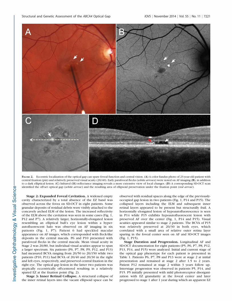
Stage 2: Expanded Foveal Cavitation. A widened emptycavity characterized by a total absence of the EZ band wasobserved across the fovea on SD-OCT in eight patients. Somegranular deposits of residual debris were visibly attached to theconcavely arched ELM of the lesion. The increased reflectivityof the ELM above the cavitation was seen in some cases (Fig. 1,P12 and P7). A relatively larger, horizontally-elongated lesionresembling an elliptical bull’s eye lesion within a hyper-autofluorescent halo was observed on AF imaging in sixpatients (Fig. 1, P7). Patient 6 had speckled macularappearance on AF images, which corresponded with fleck-likedeposits in the central macula. P6 and P10 presented withparafoveal flecks in the central macula. Mean visual acuity inStage 2 was 20/80, but individual visual acuities appear to spana larger spectrum. Six patients (P6, P7, P8, P9, P12, and P13)had measured BCVA ranging from 20/50 to 20/150 while twopatients (P10, P11) had BCVA of 20/40 and 20/30 in the rightand left eyes, respectively, and preserved central fixation in theright eye. The optical gap lesion in the latter two patients wasatypically eccentrically off-centered resulting in a relativelyspared EZ at the fixation point (Fig. 2).
Stage 3: Inner Retinal Collapse. A structural collapse ofthe inner retinal layers into the vacant ellipsoid space can be
observed with residual spaces along the edge of the previously-occupied gap lesion in two patients (Fig. 1, P14 and P15). Thecollapsed layers including the ELM and subsequent innerretinal layers appeared to be present but structurally frail. Ahorizontally elongated lesion of hypoautofluorescence is seenin P14 while P15 exhibits hypoautofluorescent lesion withpreserved AF over the center (Fig. 1, P14 and P15). Visualacuities appeared similar to stage 2 patients. The BCVA of P15was relatively preserved at 20/30 in both eyes, whichcorrelated with a small area of relative outer retina layersparing in the foveal center seen on AF and SD-OCT images(Fig. 1, P15).
Stage Duration and Progression. Longitudinal AF andSD-OCT documentation for eight patients (P5, P6, P7, P8, P12,P13, P14, and P15) were analyzed. Initial and current stage ofthe optical gap phenotype for each patient is presented inTable 1. Patients P6, P7, P8 and P13 were at stage 2 at initialpresentation and remained at stage 2 after 1.5 to 2 years.Patient P12 remained at stage 2 within 3 years follow up.Interstage progression was observed in patients P5, P14, andP15. P5 initially presented with mild photoreceptor disorgani-zation with EZ granularity at the foveal center and laterprogressed to stage 1 after 1 year during which an apparent EZ
FIGURE 2. Eccentric localization of the optical gap can spare foveal function and central vision. (A) A color fundus photo of 23-year-old patient withcentral fixation (pin) and relatively preserved visual acuity (20/40). Early parafoveal flecks (white arrows) were noted on AF imaging (B), in additionto a dark elliptical lesion. (C) Infrared (IR) reflectance imaging reveals a more extensive view of local changes. (D) A corresponding SD-OCT scanidentified the off-set optical gap (white arrow) and the resulting area of ellipsoid preservation under the fixation point (red arrow).
Structural and Genetic Assessment of the ABCA4 Optical Gap IOVS j November 2014 j Vol. 55 j No. 11 j 7221

break and gap had occurred (Fig. 3, P5a and P5b). Her visualacuity progressed from 20/30 and 20/25 to 20/60 in both eyes.Interestingly, P6, the older sibling of P5, exhibited a moreadvanced stage 2 optical gap (Fig. 3, P6). P14 progressed fromstage 2 to stage 3 within 2 years; however, retained a stableBCVA of 20/100 in both eyes (Fig. 4, P14a and P14b).Progression within stage 2 prior to converting into stage 3 isseen in P8 after 1 year follow-up (Fig. 4, P8a and P8b).Progression of stage 3 gap to progressive atrophy within 1 yearis seen in P15. Best corrected visual acuity progressivelydecreased from 20/30 to 20/50 in both eyes within 2 years (Fig.5, P15a and P15b). Summary of stage duration and transition isschematically presented in Figure 6.
DISCUSSION
The optical gap phenotype has been indiscriminately associ-ated with several hereditary retinal dystrophies including rodmonochromatism,19,20,23 maculopathies caused by mutationsin KCVN227 and RP1L122 genes, and recessive Stargardtdisease.13,17,18 Sporadic cases and nonhereditary occurrences,such as solar retinopathy or isolated occurrences of laser-pointer injury or drug-induced (tamoxifen) toxicity, have also
been reported with optical gap–like lesions.21,28,29 Althougheach condition is etiologically distinct, these conditionscomprise a group of disorders that predominately affect fovealcones. STGD1 is a predominantly juvenile-onset conditioncharacterized by early RPE changes leading to macular atrophy.
Phenotypic staging can provide descriptive insight into thenatural history of a particular disease. Greenberg et al.20
recently reported such a system in assessing the structurallyanalogous optical gap phenotype in patients with rodmonochromatism. One of the aims of assessment of theABCA4-associated phenotype in this study was to understandits systematic progression to possibly decipher the early effectsof ABCA4 dysfunction in this phenotypic subgroup of STGD1patients. The analysis of this patient cohort suggests that thedevelopment of optical gap can be subdivided into threestructural stages over several years. Stage 1 patients presentwith disorganization of photoreceptors and intermittent breaksin EZ band forming gap lesion in the subfoveal region. Thevisibility of the EZ band (also known as the inner/outersegment junction) seen on the SD-OCT, has been attributed tothe scattering of light by mitochondria in the ellipsoid zone ofphotoreceptor inner segments.30 Its disappearance has thusbeen an indicator of photoreceptor loss and an explanation fordeclines in visual acuity. Central vision was relatively spared in
FIGURE 3. Optical gap development from early structural changes to stage 1. P5a: Fundus images of the left macula with corresponding AF image tothe right. A small focal loss of AF (white arrow) corresponds to area of photoreceptor disruption and debris accumulation seen on an SD-OCT scanover the fovea. Best corrected visual acuity was 20/25. P5b: After 1 year, palpable disruptions (white arrow) of the EZ and optical gap formationwith residual debris were seen in the foveal area. Progression was accompanied by a decrease in vision (20/60) and darkening of the central lesionon AF. P6: The sibling of P5, 6 years older, presented with a more progressed disease stage (stage 2). Note an expanded empty cavity devoid ofphotoreceptors (EZ band) on SD-OCT (white arrow). The structural loss corresponded to a larger area of hypoautofluorescence (white arrow) andthe presence of nascent parafoveal flecks.
Structural and Genetic Assessment of the ABCA4 Optical Gap IOVS j November 2014 j Vol. 55 j No. 11 j 7222
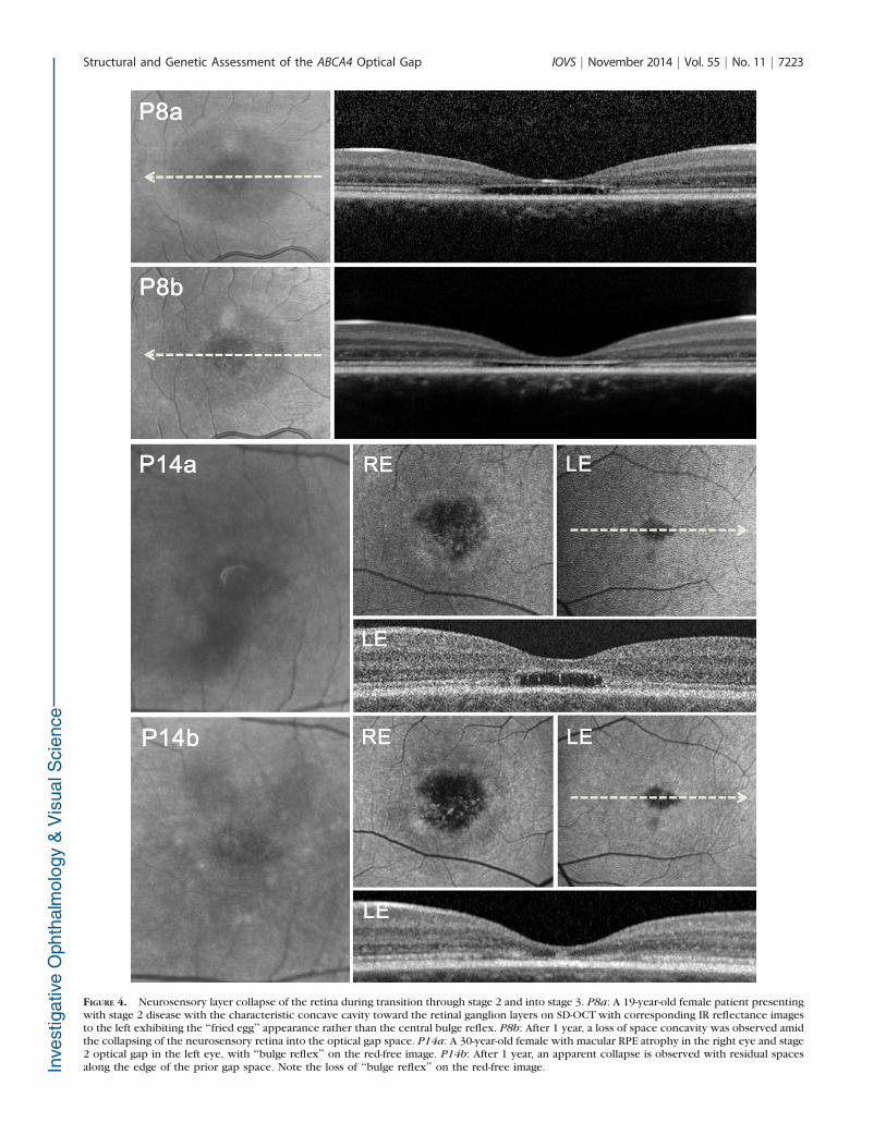
FIGURE 4. Neurosensory layer collapse of the retina during transition through stage 2 and into stage 3. P8a: A 19-year-old female patient presentingwith stage 2 disease with the characteristic concave cavity toward the retinal ganglion layers on SD-OCT with corresponding IR reflectance imagesto the left exhibiting the ‘‘fried egg’’ appearance rather than the central bulge reflex. P8b: After 1 year, a loss of space concavity was observed amidthe collapsing of the neurosensory retina into the optical gap space. P14a: A 30-year-old female with macular RPE atrophy in the right eye and stage2 optical gap in the left eye, with ‘‘bulge reflex’’ on the red-free image. P14b: After 1 year, an apparent collapse is observed with residual spacesalong the edge of the prior gap space. Note the loss of ‘‘bulge reflex’’ on the red-free image.
Structural and Genetic Assessment of the ABCA4 Optical Gap IOVS j November 2014 j Vol. 55 j No. 11 j 7223

stage 1 patients in our cohort and was also largely dependenton EZ band disruption. A further spatial depletion of the EZband is seen in stage 2 patients where an expansive subfovealempty cavity is apparent with an accompanying decline invisual acuity. In some patients residual debris attached to ELMwas present, which is hypothesized to represent the degener-ative remains of photoreceptor outer segments secondary toloss of direct apposition and contact with RPE cells disturbingtheir phagocytosis by RPE cells.17,20 Optical gaps within thesetwo stages are distinct in that structural changes appear to berestricted to losses of the EZ (photoreceptors) within thesubfoveal region, while the RPE seems to stay relatively intact.Stage 3 depicts what appears to be a structural collapse of theinner retina into the gap lesion, which then sets forth theinevitable involvement of other retinal layers. Events followingthis stage appear to lead to generalized RPE atrophythroughout the macula, which is indistinguishable from STGD1
cases that present with early centralized atrophy, suggestingthat optical gaps are likely more prevalent among STGD1patients than currently estimated, especially if patients areinitially examined at a later stage of the disease.
The measured visual acuity of stage 2 and 3 patientsappeared, at times, inconsistent with their structural diseasestage. A plausible explanation may suggest somewhat pre-served photoreceptors under the fixation point. Two patients(P10 and P11) at stage 2 had minimally eccentric gap lesionwith relatively preserved ellipsoid under the fixation point(Fig. 2), while P15 at stage 3 possibly had some foveal sparingsuggested by SD-OCT images and relatively spared visual acuity.Full-field ERG findings with normal retinal scotopic andphotopic mass responses suggest a localized disease, whilemfERG from nine eyes showed decreased responses in the 58
to 158 of retina in the posterior pole, showing mainly conedysfunction. Similar to other mfERG studies in STGD1,31,32 the
FIGURE 5. Advanced stage 3 disease and subsequent atrophy in a 28-year-old female patient. P15a: Color fundus image of the foveal lesion of theright eye with corresponding AF images of the right and left eyes to the right. A dark area of hypoautofluorescence surrounding a central patch ofpreserved AF was noted. Spectral-domain OCT shows neurosensory collapse, filling the previous empty gap space. P15b: Ensuing concentric RPEatrophy 1 year later eliminated residual patterns of optical gap pathology such EZ band spaces (white arrows show previous location of EZ bandspace). Photoreceptor atrophy and RPE thinning progressed concurrently.
Structural and Genetic Assessment of the ABCA4 Optical Gap IOVS j November 2014 j Vol. 55 j No. 11 j 7224

functionally affected areas were much larger than structuralchanges on SD-OCT or AF, suggesting that functional lossprecedes the structural changes in these patients. Additionally,given that decreases in mfERG response have been attributedto the influence of the EZ band, a case can be made for earlyphotoreceptor dysfunction preceding structural RPE loss.33
Genetic screening confirmed that all patients were com-pound heterozygous for ABCA4 mutations. Interestingly, thep.G1961E variant was present in 10 of 11 unrelated cases(91%). The p.G1961E mutation is the most frequent disease-associated ABCA4 allele seen in approximately 10% of STGD1patients of European origin.34 This fraction was almost thesame in our cohort of 179 patients, including 157 unrelatedindividuals (42/157; 13.4%), but strikingly higher in patientswith the optical gap phenotype (46.7% vs. 13.4%, P < 0.0001).It has to be noted, however, that while the optical gapphenotype is definitely associated with the p.G1961E variant,the reverse is not the case since a larger fraction (32 unrelatedindividuals) who harbored the p.G1961E allele did not presentwith optical gap. Fourteen of these individuals were clinicallycharacterized at the same age after onset as the optical gapgroup. Of the other disease-associated ABCA4 alleles com-pound heterozygous with p.G1961E, the p.L541P mutation,presenting alone or as a complex allele with the p.A1038Vvariant, was observed in seven cases (four unrelated) with
optical gap (Table 2 and Supplementary Table S1) while onlyonce in patients without the phenotype (Supplementary TableS1). However, due to a relatively small size of the optical gapcohort we cannot make an unequivocal conclusion about theassociation of this allele with the optical gap phenotype.
With the exception of P13, the optical gap was notobserved in the SD-OCT scans of any other non-p.G1961Epatients (n ¼ 131) whose age at time of examination, age ofonset and estimated disease duration were not statisticallydifferent from those of p.G1961E (n ¼ 48) patients (Supple-mentary Table S2). Therefore, we can conclude that thep.G1961E variant, maybe sometimes together with the p.L541Por p. (L541P; A1038V) allele, is currently the only ABCA4
mutation associated with the optical gap phenotype.In summary, the ABCA4-associated optical gap phenotype
may be a more prevalent occurrence preceding central macularatrophy in patients with STGD1 than previously thought. Astriking association of the optical gap phenotype with thep.G1961E mutant allele in the ABCA4 gene was observed;however, further studies on larger patient cohorts are needed tovalidate this phenotype–genotype correlation and determine thefunctional association. STGD1 patients with the optical gapphenotype present with a localized disease which can bestructurally divided into three developmental stages initiated byphotoreceptor loss and subsequent RPE involvement.
FIGURE 6. Gantt chart summarizing ABCA4-associated optical gap stage progression and duration. Longitudinal OCT imaging was available for eightpatients in the study cohort. Patient 5 presented with minor EZ changes (stage 0) and progressed bilaterally to stage 1 optical gap a year after initialexamination. Patients 6, 7, 8, and 13 initially presented and remained in stage 2 over a range of 1.5 to 2 years; P12 remained in this stage over a 3-year period. Patients 15 progressed to an atrophic stage following stage 3, while P14 exhibited unilateral progression from stage 2 to stage 3 in theleft eye.
Structural and Genetic Assessment of the ABCA4 Optical Gap IOVS j November 2014 j Vol. 55 j No. 11 j 7225

Acknowledgments
Supported by grants from the National Eye Institute/NationalInstitutes of Health (Bethesda, MD, USA) EY021163, EY019861,and EY019007 (Core Support for Vision Research), FoundationFighting Blindness (Owings Mills, MD, USA), and an unrestrictedfunds from Research to Prevent Blindness (New York, NY, USA) tothe Department of Ophthalmology, Columbia University.
Disclosure: K. Noupuu, None; W. Lee, None; J. Zernant, None;S.H. Tsang, None; R. Allikmets, None
References
1. Blacharski P. Retinal Dystrophies and Degenerations. SanDiego: Raven Press; 1988.
2. Allikmets R, Singh N, Sun H, et al. A photoreceptor cell-specific ATP-binding transporter gene (ABCR) is mutated inrecessive Stargardt macular dystrophy. Nat Genet. 1997;15:236–246.
3. Quazi F, Molday RS. ATP-binding cassette transporter ABCA4and chemical isomerization protect photoreceptor cells fromthe toxic accumulation of excess 11-cis-retinal. Proc Natl AcadSci U S A. 2014;111:5024–5029.
4. Cideciyan AV, Aleman TS, Swider M, et al. Mutations in ABCA4result in accumulation of lipofuscin before slowing of theretinoid cycle: a reappraisal of the human disease sequence.Hum Mol Genet. 2004;13:525–534.
5. Weng J, Mata NL, Azarian SM, Tzekov RT, Birch DG, Travis GH.Insights into the function of Rim protein in photoreceptorsand etiology of Stargardt’s disease from the phenotype in abcrknockout mice. Cell. 1999;98:13–23.
6. Sparrow JR, Boulton M. RPE lipofuscin and its role in retinalpathobiology. Exp Eye Res. 2005;80:595–606.
7. Sparrow JR, Nakanishi K, Parish CA. The lipofuscin fluoro-phore A2E mediates blue light-induced damage to retinalpigmented epithelial cells. Invest Ophthalmol Vis Sci. 2000;41:1981–1989.
8. Fishman GA, Stone EM, Grover S, Derlacki DJ, Haines HL,Hockey RR. Variation of clinical expression in patients withStargardt dystrophy and sequence variations in the ABCR gene.Arch Ophthalmol. 1999;117:504–510.
9. Maugeri A, Klevering BJ, Rohrschneider K, et al. Mutations in theABCA4 (ABCR) gene are the major cause of autosomal recessivecone-rod dystrophy. Am J Hum Genet. 2000;67:960–966.
10. Cremers FP, van de Pol DJ, van Driel M, et al. Autosomalrecessive retinitis pigmentosa and cone-rod dystrophy causedby splice site mutations in the Stargardt’s disease gene ABCR.Hum Mol Genet. 1998;7:355–362.
11. Martinez-Mir A, Paloma E, Allikmets R, et al. Retinitispigmentosa caused by a homozygous mutation in the Stargardtdisease gene ABCR. Nat Genet. 1998;18:11–12.
12. Allikmets R, Shroyer NF, Singh N, et al. Mutation of theStargardt disease gene (ABCR) in age-related macular degen-eration. Science. 1997;277:1805–1807.
13. Cella W, Greenstein VC, Zernant-Rajang J, et al. G1961Emutant allele in the Stargardt disease gene ABCA4 causes bull’seye maculopathy. Exp Eye Res. 2009;89:16–24.
14. Fishman GA. Fundus flavimaculatus. A clinical classification.Arch Ophthalmol. 1976;94:2061–2067.
15. Westeneng-van Haaften SC, Boon CJ, Cremers FP, HoefslootLH, den Hollander AI, Hoyng CB. Clinical and geneticcharacteristics of late-onset Stargardt’s disease. Ophthalmolo-gy. 2012;119:1199–1210.
16. Lois N, Holder GE, Bunce C, Fitzke FW, Bird AC. Phenotypicsubtypes of Stargardt macular dystrophy-fundus flavimacula-tus. Arch Ophthalmol. 2001;119:359–369.
17. Leng T, Marmor MF, Kellner U, et al. Foveal cavitation as anoptical coherence tomography finding in central conedysfunction. Retina. 2012;32:1411–1419.
18. Ritter M, Zotter S, Schmidt WM, et al. Characterization ofstargardt disease using polarization-sensitive optical coher-ence tomography and fundus autofluorescence imaging.Invest Ophthalmol Vis Sci. 2013;54:6416–6425.
19. Fahim AT, Khan NW, Zahid S, et al. Diagnostic fundusautofluorescence patterns in achromatopsia. Am J Ophthal-
mol. 2013;156:1211–1219, e1212.
20. Greenberg JP, Sherman J, Zweifel SA, et al. Spectral-domainoptical coherence tomography staging and autofluorescenceimaging in achromatopsia. JAMA Ophthalmol. 2014;132:437–445.
21. Jain A, Desai RU, Charalel RA, Quiram P, Yannuzzi L, Sarraf D.Solar retinopathy: comparison of optical coherence tomogra-phy (OCT) and fluorescein angiography (FA). Retina. 2009;29:1340–1345.
22. Park SJ, Woo SJ, Park KH, Hwang JM, Chung H. Morphologicphotoreceptor abnormality in occult macular dystrophy onspectral-domain optical coherence tomography. Invest Oph-
thalmol Vis Sci. 2010;51:3673–3679.
23. Thiadens AA, Somervuo V, van den Born LI, et al. Progressiveloss of cones in achromatopsia: an imaging study usingspectral-domain optical coherence tomography. Invest Oph-
thalmol Vis Sci. 2010;51:5952–5957.
24. Marmor MF, Fulton AB, Holder GE, et al. ISCEV standard forfull-field clinical electroretinography (2008 update). Doc
Ophthalmol. 2009;118:69–77.
25. Hood DC, Bach M, Brigell M, et al. ISCEV guidelines for clinicalmultifocal electroretinography (2007 edition). Doc Ophthal-
mol. 2008;116:1–11.
26. Zernant J, Schubert C, Im KM, et al. Analysis of the ABCA4gene by next-generation sequencing. Invest Ophthalmol Vis
Sci. 2011;52:8479–8487.
27. Sergouniotis PI, Holder GE, Robson AG, Michaelides M,Webster AR, Moore AT. High-resolution optical coherencetomography imaging in KCNV2 retinopathy. Br J Ophthalmol.2012;96:213–217.
28. Dirani A, Chelala E, Fadlallah A, Antonios R, Cherfan G.Bilateral macular injury from a green laser pointer. Clin
Ophthalmol. 2013;7:2127–2130.
29. Doshi RR, Fortun JA, Kim BT, Dubovy SR, Rosenfeld PJ.Pseudocystic foveal cavitation in tamoxifen retinopathy. Am J
Ophthalmol. 2014;157:1291–1298, e1293.
30. Spaide RF, Curcio CA. Anatomical correlates to the bands seenin the outer retina by optical coherence tomography:literature review and model. Retina. 2011;31:1609–1619.
31. Kretschmann U, Seeliger MW, Ruether K, Usui T, Apfelstedt-Sylla E, Zrenner E. Multifocal electroretinography in patientswith Stargardt’s macular dystrophy. Br J Ophthalmol. 1998;82:267–275.
32. Maia-Lopes S, Silva ED, Silva MF, Reis A, Faria P, Castelo-Branco M. Evidence of widespread retinal dysfunction inpatients with stargardt disease and morphologically unaffect-ed carrier relatives. Invest Ophthalmol Vis Sci. 2008;49:1191–1199.
33. Testa F, Rossi S, Sodi A, et al. Correlation betweenphotoreceptor layer integrity and visual function in patientswith Stargardt disease: implications for gene therapy. Invest
Ophthalmol Vis Sci. 2012;53:4409–4415.
34. Burke TR, Fishman GA, Zernant J, et al. Retinal phenotypes inpatients homozygous for the G1961E mutation in the ABCA4gene. Invest Ophthalmol Vis Sci. 2012;53:4458–4467.
Structural and Genetic Assessment of the ABCA4 Optical Gap IOVS j November 2014 j Vol. 55 j No. 11 j 7226
Related Documents