Full length article Structural analysis of the tongue and hyoid apparatus in a woodpecker Jae-Young Jung a,⇑ , Steven E. Naleway a , Nicholas A. Yaraghi b , Steven Herrera b , Vincent R. Sherman a , Eric A. Bushong c , Mark H. Ellisman c , David Kisailus b , Joanna McKittrick a,d a Materials Science and Engineering Program, University of California, San Diego, La Jolla, CA 92093, USA b Department of Chemical and Environmental Engineering, University of California, Riverside, Riverside, CA 92521, USA c National Center for Microscopy and Imaging Research and Department of Neurosciences, University of California, San Diego, La Jolla, CA 92093, USA d Department of Mechanical and Aerospace Engineering, University of California, San Diego, La Jolla, CA 92093, USA article info Article history: Received 12 November 2015 Received in revised form 21 February 2016 Accepted 17 March 2016 Available online 18 March 2016 Keywords: Woodpecker Tongue Hyoid bone X-ray micro-computed tomography Nanoindentation abstract Woodpeckers avoid brain injury while they peck at trees up to 20 Hz with speeds up to 7 m/s, undergoing decelerations up to 1200 g. Along with the head, beak and neck, the hyoid apparatus (tongue bone and associated soft tissues) is subjected to these high impact forces. The shape of the hyoid apparatus is unu- sual in woodpeckers and its structure and mechanical properties have not been reported in detail. High- resolution X-ray micro-computed tomography and scanning electron microscopy with energy dispersive X-ray spectroscopy were performed and correlated with nanoindentation mapping. The hyoid apparatus has four distinct bone sections, with three joints between these sections. Nanoindentation results on cross-sectional regions of each bone reveal a previously unreported structure consisting of a stiff core and outer, more compliant shell with moduli of up to 27.4 GPa and 8.5 GPa, respectively. The bending resistance is low at the posterior section of the hyoid bones, indicating that this region has a high degree of flexibility to absorb impact. These new structural findings can be applied to further studies on the energy dissipation of the woodpecker during its drumming behavior, and may have implications for the design of engineered impact-absorbing structures. Statement of Significance Woodpeckers avoid brain injury while they peck at trees, which results in extreme impact conditions. One common adaptation in woodpeckers is the unusual shape of the elongated tongue, also called the hyoid apparatus. The relationship between the structure and mechanical properties of the bony part of the hyoid apparatus has not been previously reported. A three dimensional model of the bony tongue was developed, and the hardness and stiffness were evaluated. A new type of bone structure, which is opposite of typical skeletal bone structure was found. The combined microstructural and mechanical property analysis indicate possible energy absorption routes for the hyoid apparatus and are applicable to the design of engineered structures. Ó 2016 Acta Materialia Inc. Published by Elsevier Ltd. All rights reserved. 1. Introduction Woodpeckers (family Picidae) are found in forested areas worldwide, except in Australia and surrounding areas. They feed by pecking (tapping or drumming) into wood and using their ton- gue to extract insects or sap. They have strong tail feathers, which are used as a lever during pecking and zygodactyl feet that help them balance. The extreme conditions during pecking include head speeds up to 7 m/s, a deceleration up to 1200 g and pecking rates of 20 times per second, which occur without sustaining concussions or brain damage [1]. One adaptation in woodpeckers is the unusual structure of its hyoid apparatus [2]. The hyoid apparatus in birds consists of the tongue bones along with associated connective tissues (cartilage, and soft tissues such as muscles, dermis and epidermis). The primary function of the hyoid apparatus is to anchor and allow for the extension of the tongue [3]. However, considering the extreme conditions experienced by the entire head during pecking, this structure must be capable of effectively dissipating energy to avoid failure. http://dx.doi.org/10.1016/j.actbio.2016.03.030 1742-7061/Ó 2016 Acta Materialia Inc. Published by Elsevier Ltd. All rights reserved. ⇑ Corresponding author at: 9500 Gilman Drive, La Jolla, CA 92093-0418, USA. E-mail address: [email protected] (J.-Y. Jung). Acta Biomaterialia 37 (2016) 1–13 Contents lists available at ScienceDirect Acta Biomaterialia journal homepage: www.elsevier.com/locate/actabiomat

Welcome message from author
This document is posted to help you gain knowledge. Please leave a comment to let me know what you think about it! Share it to your friends and learn new things together.
Transcript
Full length article
Structural analysis of the tongue and hyoid apparatus in a woodpecker
Jae-Young Jung a,⇑, Steven E. Naleway a, Nicholas A. Yaraghi b, Steven Herrera b, Vincent R. Sherman a,Eric A. Bushong c, Mark H. Ellisman c, David Kisailus b, Joanna McKittrick a,d
aMaterials Science and Engineering Program, University of California, San Diego, La Jolla, CA 92093, USAbDepartment of Chemical and Environmental Engineering, University of California, Riverside, Riverside, CA 92521, USAcNational Center for Microscopy and Imaging Research and Department of Neurosciences, University of California, San Diego, La Jolla, CA 92093, USAdDepartment of Mechanical and Aerospace Engineering, University of California, San Diego, La Jolla, CA 92093, USA
a r t i c l e i n f o
Article history:Received 12 November 2015Received in revised form 21 February 2016Accepted 17 March 2016Available online 18 March 2016
Keywords:WoodpeckerTongueHyoid boneX-ray micro-computed tomographyNanoindentation
a b s t r a c t
Woodpeckers avoid brain injury while they peck at trees up to 20 Hz with speeds up to 7 m/s, undergoingdecelerations up to 1200 g. Along with the head, beak and neck, the hyoid apparatus (tongue bone andassociated soft tissues) is subjected to these high impact forces. The shape of the hyoid apparatus is unu-sual in woodpeckers and its structure and mechanical properties have not been reported in detail. High-resolution X-ray micro-computed tomography and scanning electron microscopy with energy dispersiveX-ray spectroscopy were performed and correlated with nanoindentation mapping. The hyoid apparatushas four distinct bone sections, with three joints between these sections. Nanoindentation results oncross-sectional regions of each bone reveal a previously unreported structure consisting of a stiff coreand outer, more compliant shell with moduli of up to 27.4 GPa and 8.5 GPa, respectively. The bendingresistance is low at the posterior section of the hyoid bones, indicating that this region has a high degreeof flexibility to absorb impact. These new structural findings can be applied to further studies on theenergy dissipation of the woodpecker during its drumming behavior, and may have implications forthe design of engineered impact-absorbing structures.
Statement of Significance
Woodpeckers avoid brain injury while they peck at trees, which results in extreme impact conditions.One common adaptation in woodpeckers is the unusual shape of the elongated tongue, also called thehyoid apparatus. The relationship between the structure and mechanical properties of the bony part ofthe hyoid apparatus has not been previously reported. A three dimensional model of the bony tonguewas developed, and the hardness and stiffness were evaluated. A new type of bone structure, which isopposite of typical skeletal bone structure was found. The combined microstructural and mechanicalproperty analysis indicate possible energy absorption routes for the hyoid apparatus and are applicableto the design of engineered structures.
� 2016 Acta Materialia Inc. Published by Elsevier Ltd. All rights reserved.
1. Introduction
Woodpeckers (family Picidae) are found in forested areasworldwide, except in Australia and surrounding areas. They feedby pecking (tapping or drumming) into wood and using their ton-gue to extract insects or sap. They have strong tail feathers, whichare used as a lever during pecking and zygodactyl feet that helpthem balance. The extreme conditions during pecking include head
speeds up to 7 m/s, a deceleration up to 1200 g and pecking rates of20 times per second, which occur without sustaining concussionsor brain damage [1].
One adaptation in woodpeckers is the unusual structure of itshyoid apparatus [2]. The hyoid apparatus in birds consists of thetongue bones along with associated connective tissues (cartilage,and soft tissues such as muscles, dermis and epidermis). Theprimary function of the hyoid apparatus is to anchor and allowfor the extension of the tongue [3]. However, considering theextreme conditions experienced by the entire head during pecking,this structure must be capable of effectively dissipating energy toavoid failure.
http://dx.doi.org/10.1016/j.actbio.2016.03.0301742-7061/� 2016 Acta Materialia Inc. Published by Elsevier Ltd. All rights reserved.
⇑ Corresponding author at: 9500 Gilman Drive, La Jolla, CA 92093-0418, USA.E-mail address: [email protected] (J.-Y. Jung).
Acta Biomaterialia 37 (2016) 1–13
Contents lists available at ScienceDirect
Acta Biomaterialia
journal homepage: www.elsevier .com/locate /actabiomat
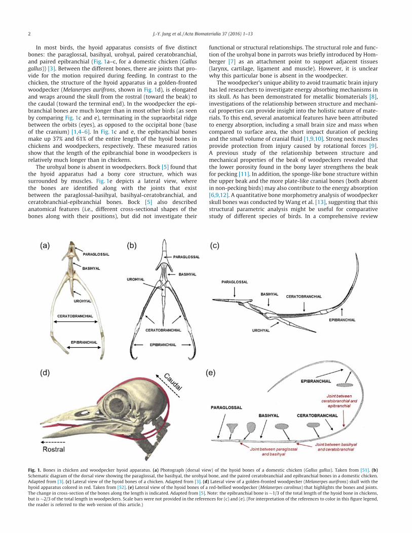
In most birds, the hyoid apparatus consists of five distinctbones: the paraglossal, basihyal, urohyal, paired ceratobranchial,and paired epibranchial (Fig. 1a–c, for a domestic chicken (Gallusgallus)) [3]. Between the different bones, there are joints that pro-vide for the motion required during feeding. In contrast to thechicken, the structure of the hyoid apparatus in a golden-frontedwoodpecker (Melanerpes aurifrons, shown in Fig. 1d), is elongatedand wraps around the skull from the rostral (toward the beak) tothe caudal (toward the terminal end). In the woodpecker the epi-branchial bones are much longer than in most other birds (as seenby comparing Fig. 1c and e), terminating in the supraorbital ridgebetween the orbits (eyes), as opposed to the occipital bone (baseof the cranium) [1,4–6]. In Fig. 1c and e, the epibranchial bonesmake up 37% and 61% of the entire length of the hyoid bones inchickens and woodpeckers, respectively. These measured ratiosshow that the length of the epibranchial bone in woodpeckers isrelatively much longer than in chickens.
The urohyal bone is absent in woodpeckers. Bock [5] found thatthe hyoid apparatus had a bony core structure, which wassurrounded by muscles. Fig. 1e depicts a lateral view, wherethe bones are identified along with the joints that existbetween the paraglossal-basihyal, basihyal-ceratobranchial, andceratobranchial-epibranchial bones. Bock [5] also describedanatomical features (i.e., different cross-sectional shapes of thebones along with their positions), but did not investigate their
functional or structural relationships. The structural role and func-tion of the urohyal bone in parrots was briefly introduced by Hom-berger [7] as an attachment point to support adjacent tissues(larynx, cartilage, ligament and muscle). However, it is unclearwhy this particular bone is absent in the woodpecker.
The woodpecker’s unique ability to avoid traumatic brain injuryhas led researchers to investigate energy absorbing mechanisms inits skull. As has been demonstrated for metallic biomaterials [8],investigations of the relationship between structure and mechani-cal properties can provide insight into the holistic nature of mate-rials. To this end, several anatomical features have been attributedto energy absorption, including a small brain size and mass whencompared to surface area, the short impact duration of peckingand the small volume of cranial fluid [1,9,10]. Strong neck musclesprovide protection from injury caused by rotational forces [9].A previous study of the relationship between structure andmechanical properties of the beak of woodpeckers revealed thatthe lower porosity found in the bony layer strengthens the beakfor pecking [11]. In addition, the sponge-like bone structure withinthe upper beak and the more plate-like cranial bones (both absentin non-pecking birds) may also contribute to the energy absorption[6,9,12]. A quantitative bone morphometry analysis of woodpeckerskull bones was conducted by Wang et al. [13], suggesting that thisstructural parametric analysis might be useful for comparativestudy of different species of birds. In a comprehensive review
Fig. 1. Bones in chicken and woodpecker hyoid apparatus. (a) Photograph (dorsal view) of the hyoid bones of a domestic chicken (Gallus gallus). Taken from [51]. (b)Schematic diagram of the dorsal view showing the paraglossal, the basihyal, the urohyal bone, and the paired ceratobranchial and epibranchial bones in a domestic chicken.Adapted from [3]. (c) Lateral view of the hyoid bones of a chicken. Adapted from [3]. (d) Lateral view of a golden-fronted woodpecker (Melanerpes aurifrons) skull with thehyoid apparatus colored in red. Taken from [52]. (e) Lateral view of the hyoid bones of a red-bellied woodpecker (Melanerpes carolinus) that highlights the bones and joints.The change in cross-section of the bones along the length is indicated. Adapted from [5]. Note: the epibranchial bone is �1/3 of the total length of the hyoid bone in chickens,but is �2/3 of the total length in woodpeckers. Scale bars were not provided in the references for (c) and (e). (For interpretation of the references to color in this figure legend,the reader is referred to the web version of this article.)
2 J.-Y. Jung et al. / Acta Biomaterialia 37 (2016) 1–13
paper [14], the mechanisms that provide impact resistance to thewoodpecker’s head were discussed, that include woodpecker-inspired shock-absorbing applications. However, information onthe hierarchical structure of the hyoid apparatus is not yetunderstood.
Reports on the potential ability of the hyoid apparatus to with-stand impact have focused upon its potential to aid the skull inavoiding brain injuries and have relied upon numerical and finiteelement analyses [6,12,15–18]. Oda et al. [15] determined thatthe presence of the hyoid bones may lower the compressive andtensile stresses in the brain up to 40% during pecking. Wanget al. [6] suggested that the three most important factors for shockabsorption are: 1) the macro/micro structures of the head, includ-ing the hyoid apparatus, 2) the uneven plate-like trabecular bonesin the cranium, and 3) the unequal lengths of the upper and lowerbeak. The hyoid bone was also suggested to play the role of a ‘seatbelt’ after impact [6]. Yoon et al. [16] mimicked the head for appli-cation in devices that experience high-g and high-frequencymechanical forces by using a simplified mechanical vibrationmodel that simulated the head as a damper and spring. Theyreported that the woodpecker-inspired shock-absorbing systemshowed a 1% failure rate at 60,000 g, compared to a hard resinshock-absorbing system, which showed 26% of failure. Zhu et al.[17] performed a numerical study for the impact response of thehead and reported that stress waves propagated from the upperbill to the posterior of the skull. It was concluded that the stresswave was decreased by two structural features: 1) having the skullwrapped with the hyoid apparatus and 2) the viscoelastic energyabsorption of the biological tissues. In addition, the function ofthe hyoid bone was computationally assessed by Liu et al. [18]who found that there was 30% less deformation of the whole headwith the hyoid bones than without them. They concluded that thehyoid bone and muscle contributed together to increase the rigid-ity of the whole head, reducing the deformation and oscillation ofthe skull.
When solely considering the hyoid apparatus, initial mechani-cal properties were reported and based on tensile tests includingboth bone and soft tissues [19]. It was determined that the elasticmodulus and tensile strength were 1.3 GPa and 76.0 MPa, respec-tively, at the joint between the paraglossal and basihyal bone[19]. In the epibranchial bone, the elastic modulus and tensilestrength were 3.7 GPa and 92.0 MPa at the rostral position and1.7 GPa and 131.0 MPa in the midsection [19]. The higher modulusand lower strength of the rostral end of the epibranchial bone com-pared to the midsection were attributed to the amount of soft tis-sue surrounding the bone. Because the diameter of the hyoidapparatus is relatively constant along the epibranchial bone, it ispossible that there is a thinner sheath of soft tissue surroundingthe bone at the rostral end and a thicker sheath at the midsection,which would result in an increased bone diameter at the rostralend compared to the midsection.
The interpretation of the results of the previous studies[6,15–19] on the hyoid apparatus used the term ‘‘hyoid bone”instead of ‘‘hyoid apparatus” (or sometimes ‘‘lingual apparatus”[3]). As a result, the previously acquired mechanical propertiesand numerical simulation results on the ‘‘hyoid bone” were basedon the properties of bulk hyoid apparatus. Here, we distinguish thedifference between the individual hyoid bones and the hyoidapparatus.
The objective of this study is to specifically analyze the morpho-logical and structural features and associated mechanical proper-ties of the hyoid bones of acorn woodpeckers (Melanerpesformicivorus) to examine the structure/mechanical property rela-tionships that allow it to avoid failure during the extreme condi-tions of pecking. Livingston [2] observed that the woodpeckertongue showed fascinating aspects of adaptable design in nature
by linking structure and function. By using sophisticated analyticaltechniques, the structure of the woodpecker hyoid apparatus canbe more accurately assessed. The results of this study have impli-cations for the design of engineered structures, such as impact-absorbing protective headgear for athletes and the military.
2. Experimental
2.1. Sample collection and preparation
This study was conducted under the approval of animal careand use program by Institutional Animal Care and Use Committee(IACUC) at University of California, San Diego (Tissue Permit Num-ber: T14068).
Three adult acorn woodpeckers were donated soon after deathfrom a northern California ranch. These were immediately frozenand kept in a frozen state during transportation to the lab. All sam-ples were stored in a freezer at �20 �C. The woodpeckers weregradually thawed at room temperature for 30 min before perform-ing other tests. All tests were performed under ambient conditions(25 �C, 60% relative humidity)
2.2. X-ray Micro-computed tomography (l-CT) and three-dimensionalreconstruction
The whole birds were scanned by X-ray micro-computedtomography (l-CT, SkyScan 1076, Bruker microCT, Kontich, Bel-gium) with a rotation step of 0.7�, an exposure time of 1600 ms,a 100 kV acceleration voltage, and an isotropic voxel size of36.00 lm. The heads were scanned with an isotropic voxel sizeof 9.06 lm, while the other scan parameters were the same asfor the whole birds. In addition, high-resolution l-CT (HR-lCT,MicroXCT-200, Xradia, Pleasanton, CA) was used for imaging smallpieces (5 mm � 5 mm � 2 mm) of each hyoid bone with a0.916 lm voxel size at a 40 kV acceleration voltage. The rotationangle and tilt increment were 360� and 0.2�, respectively. Theimages and three-dimensional reconstructed models were devel-oped using the software programs CTvox and Dataviewer (BrukermicroCT, Kontich, Belgium) and XMReconstructor (Xradia, Pleasan-ton, CA). ImageJ software (National Institutes of Health, Bethesda,MD) was used upon orthoslice images of the heads to calculatethe cross-sectional area and dimensions of the hyoid bone at vary-ing positions along its length. Each dimension was measured atleast six times and the mean values were calculated.
Each hyoid bone was selected for visualization and analyzedusing Amira software (FEI Visualization Sciences Group,Burlington, MA). After the reconstruction, cross-sectional dimen-sions were determined by creating triangle mesh models savingthem into the extension format of the virtual reality modelinglanguage.
2.3. Microscopic evaluation and chemical composition
Two birds were thawed for dissection and subsequent excisionof the hyoid apparatus. Each was submerged immediately into a2.5 vol.% glutaraldehyde solution for 24 h for tissue fixation. Afterthat, they were dehydrated in increasing concentrations of etha-nol: 30%, 50%, 70%, 90%, and 100% (vol.%) for 10 min each and thendried by a critical point dryer (Autosamdri 815A, Tousimis, Rock-ville, MD). After the dehydration process, the hyoid apparatuswas cut along the transverse and longitudinal planes and dividedinto several pieces. Two separate sets of samples were preparedfor the microstructural analysis and the nanoindentation test fromeach piece. Samples for the nanoindentation test were embeddedin an epoxy resin and cured overnight at room temperature.
J.-Y. Jung et al. / Acta Biomaterialia 37 (2016) 1–13 3

Embedded samples were cut into smaller pieces using a jeweler’ssaw and both sides were polished using a series of SiC paper withaverage particle sizes of 35 lm and 15.3 lm, followed by fine pol-ishing with an alumina powder media down to 0.05 lm to providea mirror finish. Optical micrographs were obtained by a lightmicroscope (VHX1000, Keyence, Osaka, Japan) to visualize thecolor and shape of each tissue without any staining. These sampleswere used for nanoindentation tests first and then sputter coatedwith iridum (K575X, Emitech, Fall River, MA) at 85 mA for 10 s.Samples for the microstructural analysis were also coated withiridium without epoxy. Scanning electron microscopy (SEM) andenergy dispersive X-ray spectroscopy (EDS) were performed(XL30 UHR-SEM, FEI Company, Hillsboro, OR and Inca, Oxford,Abingdon, Oxfordshire, UK) at 15 keV for the prepared samplesindividually. Another SEM device (XL30 ESEM, FEI Company, Hills-boro, OR) with a back-scattered electron (BSE) detector was uti-lized to highlight atomic mass contrast within the tissues. Inaddition, the elemental composition was acquired from thecross-sections near the nanoindentation sites. The EDS spectra ateach indent were obtained at least three times to determine theaverage and standard deviation.
A dried sample from the lingual apex and body were tested sep-arately as a whole piece by thermogravimetric analysis (TGA) (SDTQ600 TGA, TA Instruments, New Castle, DE) at a ramp rate of 10 �C/min and a range of 20 � 600 �C, following a previously reportedprocedure [20] to determine the amount of water, mineral, andprotein. A polished cross-sectional sample of the lingual apexwas analyzed by Fourier transform infrared (FTIR) spectroscopy(Equinox 55, Bruker Optics, Billerica, MA). The spectral range was400–4000 cm�1 with a 4 cm�1 resolution. The scan number was1024 in reflection mode. The background signal was collected intransmission mode through air.
2.4. Nanoindentation
The hardness and elastic modulus were acquired by nanoinden-tation (TI 950 TriboIndenter, Hysitron, Minneapolis, MN) with adiamond cube corner tip on polished cross-sectional pieces of fourdifferent hyoid bones. Indentation mapping was carried out withdisplacement controlled indents to a maximum depth of 500 nm.A trapezoidal load function consisting of a 5 s loading segment, a2 s hold, and a 5 s unloading segment was used. Indents werearranged in square arrays, spaced either 20 or 30 lm apart. A fusedquartz standard sample from Hysitron was used to calibrate the tiparea function for the diamond cube corner tip. The tests wereperformed at least ten times for each sample and the averagevalue and standard deviation were calculated. The hardness (H)is given by:
H ¼ Pmax
Ac; ð1Þ
where Pmax is the maximum load (in N) and the Ac is the projectedarea of the indenter at peak load computed from the area function,Ac ¼ FðhcÞ, where hc is the contact depth [21]. For a cube corner tip
indenter, the area function Ac ¼ 2:598h2c . The slope of the initial
unloading load–displacement curve, S = dP/dh, was used to calcu-late hc:
hc ¼ hmax � 3Pmax
4S; ð2Þ
where hmax is the maximum displacement. The reduced elasticmodulus, Er, is given by:
Er ¼ffiffiffi
2p
2Sffiffiffiffiffi
Acp : ð3Þ
The Young’s modulus, E, was obtained from:
1Er
¼ 1� v2
Eþ 1� v2
i
Ei; ð4Þ
where Ei and v i are the Young’s modulus (1,140 GPa) and Poisson’sratio (0.07) of the indenter [22] and v is the Poisson’s ratio of thetested sample (taken as 0.3) [23]. The test sites (basihyal, cerato-branchial and epibranchial) for the indentation mapping weredetermined after confirming the macro/micro structure of thesebones and all indents were performed on the transverse cross-sections. Details of nanoindentation test specimens are listed inTable 1.
2.5. Statistical analysis
One-way ANOVA analyses were used to identify statisticallysignificant differences of the Young’s moduli and hardnessamong the different hyoid bones [24]. In addition, a multiplecomparison test was conducted by Tukey’s least significant dif-ference procedure, which is valid if the preliminary test (theone-way ANOVA F-test) shows a significant difference [25]. Apaired sample t-test was used to compare statistically signifi-cant differences of the Young’s moduli and hardness betweenthe two different bone regions for each hyoid bone. The crite-rion for statistical significance was p < 0.05 for both the ANOVAand the t-test.
3. Results and discussion
3.1. Macroscale structure
Fig. 2a shows a photograph of an acorn woodpecker with anoverall length and width of �216 � 51 mm and a head length,width and height of 57.8 � 15.4 � 27.9 mm. Fig. 2b shows a pho-tograph of the dissected hyoid apparatus. The hyoid apparatus iscomposed of four different regions: the lingual apex, lingual body,lingual root, and hyoid horns, which are connected along its sagit-tal plane from rostral to caudal positions [26]. The paraglossal andbasihyal bones are located inside the lingual apex and body,respectively. The caudal end of basihyal bone, the rostral end ofceratobranchial bones, and their connecting joint are located atthe lingual root. The hyoid horns contain the ceratobranchial andepibranchial bones along with their joint. Fig. 2c shows dried andsectioned pieces that were used for further analysis. The barbedtips at the lingual apex are clearly visible in Fig. 2b and c (black).The lingual body has the largest diameter. Fig. 2d–f show the mag-nified images of the interfaces between the lingual apex and body(Fig. 2d), the lingual body and root (Fig. 2e), and the lingual rootand hyoid horn (Fig. 2f), which surround the three joints in thehyoid apparatus.
Fig. 3 shows l-CT reconstructed images of the head. The hyoidbone wraps around the head, from the rostral end at the beak tothe supraorbital ridge between the two orbits. Some woodpeckerswith long probing tongues have even more elongated hyoid hornsthat can pass through the right nasal cavity and upper jaw (as isthe case for the European woodpecker (Picus viridis)) or can circlethe right orbital bone (as is the case for the North American hairywoodpecker (Picoides villosus)) [5]. The acorn woodpecker’s hyoidhorns are less elongated than these two species, which is likelydue to a specialization for either drilling or probing functions [5].
The color distribution in Fig. 3 reflects the X-ray intensity pro-file based on the normalized material density, ranging from thehighest density (red), middle-range density (higher? lower: yel-low? green? light blue) to the lowest density (dark blue). Thedensity is correlated with the amount of mineral: red having the
4 J.-Y. Jung et al. / Acta Biomaterialia 37 (2016) 1–13
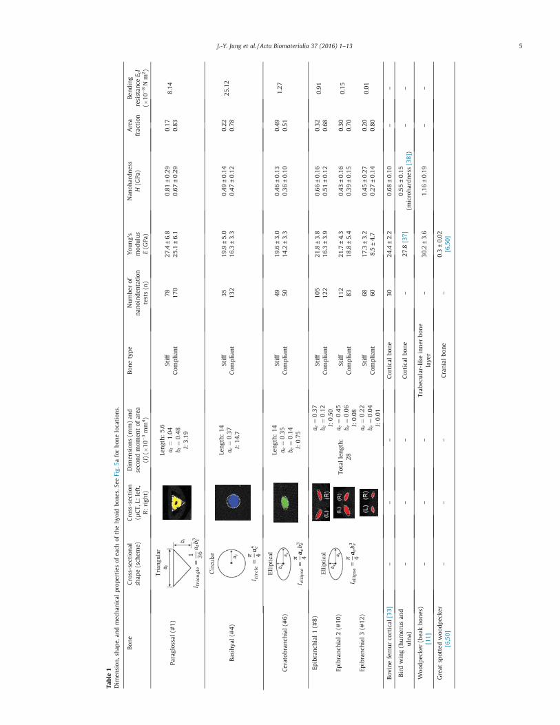
Table1
Dim
ension
,sha
pe,a
ndmecha
nicalprop
erties
ofea
chof
thehy
oidbo
nes.
SeeFig.
5aforbo
nelocation
s.
Bon
eCross-section
alsh
ape(sch
eme)
Cross-section
(lCT,
L:left,
R:righ
t)
Dim
ension
s(m
m)an
dseco
ndmom
entof
area
(I)(�
10�3mm
4)
Bon
etype
Numbe
rof
nan
oinde
ntation
tests(n)
You
ng’s
mod
ulus
E(G
Pa)
Nan
ohardn
ess
H(G
Pa)
Area
fraction
Ben
ding
resistan
ceE cI
(�10
�8Nm
2)
Paraglos
sal(#
1)
Tria
ngul
a
1 36
rLe
ngth:5.6
a t¼
1:04
b t¼
0:48
I:3.19
Stiff
7827
.4±6.8
0.81
±0.29
0.17
8.14
Com
pliant
170
25.1
±6.1
0.67
±0.29
0.83
Basihya
l(#
4)
Circ
ular
4
Length:14
a c¼
0:37
I:14
.7
Stiff
3519
.9±5.0
0.49
±0.14
0.22
25.12
Com
pliant
132
16.3
±3.3
0.47
±0.12
0.78
Ceratob
ranch
ial(#
6)
Ellip
tical
4
Length:14
a e¼
0:35
b e¼
0:14
I:0.75
Stiff
4919
.6±3.0
0.46
±0.13
0.49
1.27
Com
pliant
5014
.2±3.3
0.36
±0.10
0.51
Epibranch
ial1(#
8)El
liptic
al
4
Totallength:
28
a e¼
0:37
b e¼
0:12
I:0.50
Stiff
105
21.8
±3.8
0.66
±0.16
0.32
0.91
Com
pliant
122
16.3
±3.9
0.51
±0.12
0.68
Epibranch
ial2(#
10)
a e¼
0:45
b e¼
0:06
I:0.08
Stiff
112
21.7
±4.3
0.43
±0.16
0.30
0.15
Com
pliant
8318
.8±5.4
0.39
±0.15
0.70
Epibranch
ial3(#
12)
a e¼
0:22
b e¼
0:04
I:0.01
Stiff
6817
.3±3.2
0.45
±0.27
0.20
0.01
Com
pliant
608.5±4.7
0.27
±0.14
0.80
Bov
inefemurco
rtical
[33]
––
–Corticalbo
ne
3024
.4±2.2
0.68
±0.10
––
Birdwing(humerusan
dulna)
––
–Corticalbo
ne
–27
.8[37]
0.55
±0.15
(microhardn
ess[38])
––
Woo
dpecke
r(bea
kbo
nes)
[11]
––
–Trab
ecular-like
inner
bone
laye
r–
30.2
±3.6
1.16
±0.19
––
Great
spottedwoo
dpecke
r[6,50]
––
–Cranialbo
ne
–0.3±0.02
[6,50]
J.-Y. Jung et al. / Acta Biomaterialia 37 (2016) 1–13 5
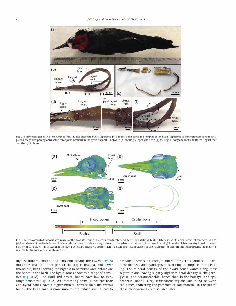
highest mineral content and dark blue having the lowest. Fig. 3aillustrates that the inner part of the upper (maxilla) and lower(mandible) beak showing the highest mineralized area, which arethe bones in the beak. The hyoid bones show mid-range of densi-ties (Fig. 3a–d). The skull and orbital bones have low to mid-range densities (Fig. 3a–c). An interesting point is that the beakand hyoid bones have a higher mineral density than the cranialbones. The beak bone is more mineralized, which should lead to
a relative increase in strength and stiffness. This could be to rein-force the beak and hyoid apparatus during the impacts from peck-ing. The mineral density of the hyoid bones varies along theirsagittal plane, having slightly higher mineral density in the para-glossal and ceratobranchial bones than in the basihyal and epi-branchial bones. X-ray transparent regions are found betweenthe bones, indicating the presence of soft material in the joints;these observations are discussed later.
Fig. 2. (a) Photograph of an acorn woodpecker. (b) The dissected hyoid apparatus. (c) The dried and sectioned samples of the hyoid apparatus in transverse and longitudinalplanes. Magnified photographs of the three joint locations in the hyoid apparatus between (d) the lingual apex and body, (e) the lingual body and root, and (f) the lingual rootand the hyoid horn.
Fig. 3. Micro-computed tomography images of the head structure of an acorn woodpecker at different orientations. (a) Left lateral view, (b) dorsal view, (c) ventral view, and(d) lateral view of the hyoid bones. A color scale is shown to indicate the gradient in color (that is associated with mineral density) from the highest density in red to lowestdensity in dark blue. This shows that the hyoid bones are relatively denser than the skull. (For interpretation of the references to color in this figure legend, the reader isreferred to the web version of this article.)
6 J.-Y. Jung et al. / Acta Biomaterialia 37 (2016) 1–13
3.2. Microscale structure
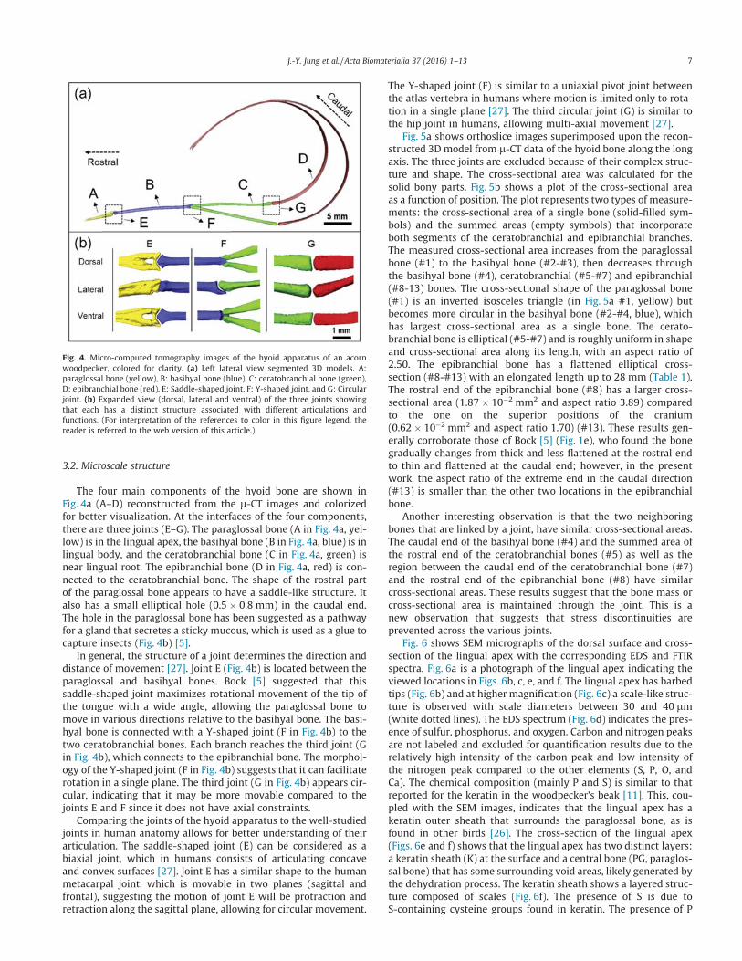
The four main components of the hyoid bone are shown inFig. 4a (A–D) reconstructed from the l-CT images and colorizedfor better visualization. At the interfaces of the four components,there are three joints (E–G). The paraglossal bone (A in Fig. 4a, yel-low) is in the lingual apex, the basihyal bone (B in Fig. 4a, blue) is inlingual body, and the ceratobranchial bone (C in Fig. 4a, green) isnear lingual root. The epibranchial bone (D in Fig. 4a, red) is con-nected to the ceratobranchial bone. The shape of the rostral partof the paraglossal bone appears to have a saddle-like structure. Italso has a small elliptical hole (0.5 � 0.8 mm) in the caudal end.The hole in the paraglossal bone has been suggested as a pathwayfor a gland that secretes a sticky mucous, which is used as a glue tocapture insects (Fig. 4b) [5].
In general, the structure of a joint determines the direction anddistance of movement [27]. Joint E (Fig. 4b) is located between theparaglossal and basihyal bones. Bock [5] suggested that thissaddle-shaped joint maximizes rotational movement of the tip ofthe tongue with a wide angle, allowing the paraglossal bone tomove in various directions relative to the basihyal bone. The basi-hyal bone is connected with a Y-shaped joint (F in Fig. 4b) to thetwo ceratobranchial bones. Each branch reaches the third joint (Gin Fig. 4b), which connects to the epibranchial bone. The morphol-ogy of the Y-shaped joint (F in Fig. 4b) suggests that it can facilitaterotation in a single plane. The third joint (G in Fig. 4b) appears cir-cular, indicating that it may be more movable compared to thejoints E and F since it does not have axial constraints.
Comparing the joints of the hyoid apparatus to the well-studiedjoints in human anatomy allows for better understanding of theirarticulation. The saddle-shaped joint (E) can be considered as abiaxial joint, which in humans consists of articulating concaveand convex surfaces [27]. Joint E has a similar shape to the humanmetacarpal joint, which is movable in two planes (sagittal andfrontal), suggesting the motion of joint E will be protraction andretraction along the sagittal plane, allowing for circular movement.
The Y-shaped joint (F) is similar to a uniaxial pivot joint betweenthe atlas vertebra in humans where motion is limited only to rota-tion in a single plane [27]. The third circular joint (G) is similar tothe hip joint in humans, allowing multi-axial movement [27].
Fig. 5a shows orthoslice images superimposed upon the recon-structed 3D model from l-CT data of the hyoid bone along the longaxis. The three joints are excluded because of their complex struc-ture and shape. The cross-sectional area was calculated for thesolid bony parts. Fig. 5b shows a plot of the cross-sectional areaas a function of position. The plot represents two types of measure-ments: the cross-sectional area of a single bone (solid-filled sym-bols) and the summed areas (empty symbols) that incorporateboth segments of the ceratobranchial and epibranchial branches.The measured cross-sectional area increases from the paraglossalbone (#1) to the basihyal bone (#2-#3), then decreases throughthe basihyal bone (#4), ceratobranchial (#5-#7) and epibranchial(#8-13) bones. The cross-sectional shape of the paraglossal bone(#1) is an inverted isosceles triangle (in Fig. 5a #1, yellow) butbecomes more circular in the basihyal bone (#2-#4, blue), whichhas largest cross-sectional area as a single bone. The cerato-branchial bone is elliptical (#5-#7) and is roughly uniform in shapeand cross-sectional area along its length, with an aspect ratio of2.50. The epibranchial bone has a flattened elliptical cross-section (#8-#13) with an elongated length up to 28 mm (Table 1).The rostral end of the epibranchial bone (#8) has a larger cross-sectional area (1.87 � 10�2 mm2 and aspect ratio 3.89) comparedto the one on the superior positions of the cranium(0.62 � 10�2 mm2 and aspect ratio 1.70) (#13). These results gen-erally corroborate those of Bock [5] (Fig. 1e), who found the bonegradually changes from thick and less flattened at the rostral endto thin and flattened at the caudal end; however, in the presentwork, the aspect ratio of the extreme end in the caudal direction(#13) is smaller than the other two locations in the epibranchialbone.
Another interesting observation is that the two neighboringbones that are linked by a joint, have similar cross-sectional areas.The caudal end of the basihyal bone (#4) and the summed area ofthe rostral end of the ceratobranchial bones (#5) as well as theregion between the caudal end of the ceratobranchial bone (#7)and the rostral end of the epibranchial bone (#8) have similarcross-sectional areas. These results suggest that the bone mass orcross-sectional area is maintained through the joint. This is anew observation that suggests that stress discontinuities areprevented across the various joints.
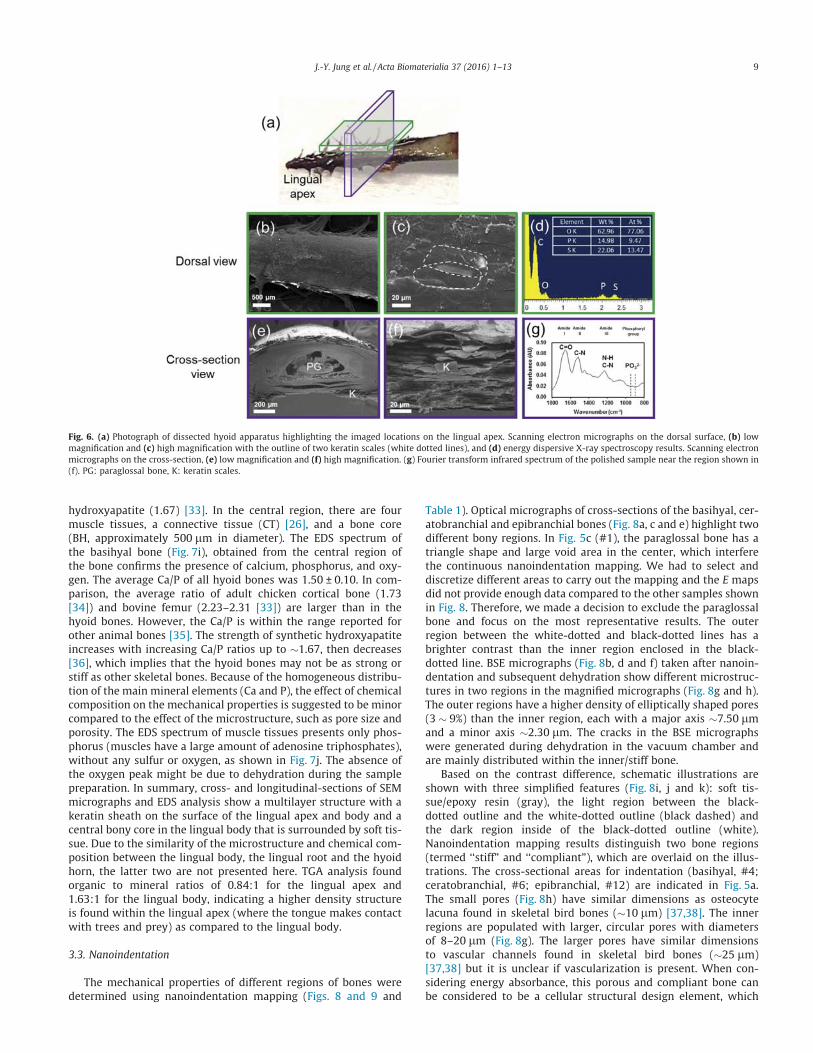
Fig. 6 shows SEM micrographs of the dorsal surface and cross-section of the lingual apex with the corresponding EDS and FTIRspectra. Fig. 6a is a photograph of the lingual apex indicating theviewed locations in Figs. 6b, c, e, and f. The lingual apex has barbedtips (Fig. 6b) and at higher magnification (Fig. 6c) a scale-like struc-ture is observed with scale diameters between 30 and 40 lm(white dotted lines). The EDS spectrum (Fig. 6d) indicates the pres-ence of sulfur, phosphorus, and oxygen. Carbon and nitrogen peaksare not labeled and excluded for quantification results due to therelatively high intensity of the carbon peak and low intensity ofthe nitrogen peak compared to the other elements (S, P, O, andCa). The chemical composition (mainly P and S) is similar to thatreported for the keratin in the woodpecker’s beak [11]. This, cou-pled with the SEM images, indicates that the lingual apex has akeratin outer sheath that surrounds the paraglossal bone, as isfound in other birds [26]. The cross-section of the lingual apex(Figs. 6e and f) shows that the lingual apex has two distinct layers:a keratin sheath (K) at the surface and a central bone (PG, paraglos-sal bone) that has some surrounding void areas, likely generated bythe dehydration process. The keratin sheath shows a layered struc-ture composed of scales (Fig. 6f). The presence of S is due toS-containing cysteine groups found in keratin. The presence of P
Fig. 4. Micro-computed tomography images of the hyoid apparatus of an acornwoodpecker, colored for clarity. (a) Left lateral view segmented 3D models. A:paraglossal bone (yellow), B: basihyal bone (blue), C: ceratobranchial bone (green),D: epibranchial bone (red), E: Saddle-shaped joint, F: Y-shaped joint, and G: Circularjoint. (b) Expanded view (dorsal, lateral and ventral) of the three joints showingthat each has a distinct structure associated with different articulations andfunctions. (For interpretation of the references to color in this figure legend, thereader is referred to the web version of this article.)
J.-Y. Jung et al. / Acta Biomaterialia 37 (2016) 1–13 7

in the keratin might be related to a strengthening effect, known asphosphorylation, of the keratin filaments at the cellular level [28].As shown in Fig. 6g, FTIR spectrum of the cross-section of the lin-gual apex exhibits a peak near 950 cm�1, a known band for phos-phorylated proteins [29]. Also present are the typical peaks ofkeratin near 1600–1700 cm�1 (amide I), 1550 cm�1 (amide II),and 1200–1300 cm�1 (amide III) [29].
Fig. 7a provides an overview of observed regions in Fig. 7b–j. InFig. 7b and c, a similar scale-like structure is observed on the lin-gual body. The EDS spectrum of the cross-section of the lingualbody (Fig. 7d) is similar to the lingual apex (Fig. 6d), indicating thatit is keratin. As shown in Fig. 7e, the longitudinal-section image ofthe lingual body illustrates a similar structure to the cross-sectional structure of the lingual apex. Some bundles of fibers are
observed near the center part of the basihyal bone (Fig. 7f and g).The fibers are �194 nm in diameter and >10 lm in length, compa-rable to mineralized collagen fibril bundles found in the bony coreof woodpecker’s beak [11]. Overall, the multilayered structure ofthe lingual body is similar to the lingual apex. In the dorsal view,the shape and dimension of the keratin scales in both the lingualapex and body have isotropic shapes, which are comparable tothe keratin scales on beaks of other birds [30–32], in contrast tothe elongated scales found on woodpecker beaks [11]. Fig. 7hshows a SEM image and Fig. 7i (from BH, basihyal bone) and 7j(from M, muscle tissues) show EDS spectra of the cross-sectionsof the lingual body. In Fig. 7h, the outer region is covered withthe dermis (D) and epidermis (ED). The ratio of calcium tophosphorus (Ca/P) is 1.56, which is lower than stoichiometric
Fig. 5. (a) Orthoslice images of the hyoid bone with reconstructed 3D models from micro-computed tomography data. (b) Plot of the measured cross-sectional area at eachlocation. (c) Transverse cross-sectional images at each location showing the shape of hyoid bone along its length. Scale bar: 500 lm.
8 J.-Y. Jung et al. / Acta Biomaterialia 37 (2016) 1–13
hydroxyapatite (1.67) [33]. In the central region, there are fourmuscle tissues, a connective tissue (CT) [26], and a bone core(BH, approximately 500 lm in diameter). The EDS spectrum ofthe basihyal bone (Fig. 7i), obtained from the central region ofthe bone confirms the presence of calcium, phosphorus, and oxy-gen. The average Ca/P of all hyoid bones was 1.50 ± 0.10. In com-parison, the average ratio of adult chicken cortical bone (1.73[34]) and bovine femur (2.23–2.31 [33]) are larger than in thehyoid bones. However, the Ca/P is within the range reported forother animal bones [35]. The strength of synthetic hydroxyapatiteincreases with increasing Ca/P ratios up to �1.67, then decreases[36], which implies that the hyoid bones may not be as strong orstiff as other skeletal bones. Because of the homogeneous distribu-tion of the main mineral elements (Ca and P), the effect of chemicalcomposition on the mechanical properties is suggested to be minorcompared to the effect of the microstructure, such as pore size andporosity. The EDS spectrum of muscle tissues presents only phos-phorus (muscles have a large amount of adenosine triphosphates),without any sulfur or oxygen, as shown in Fig. 7j. The absence ofthe oxygen peak might be due to dehydration during the samplepreparation. In summary, cross- and longitudinal-sections of SEMmicrographs and EDS analysis show a multilayer structure with akeratin sheath on the surface of the lingual apex and body and acentral bony core in the lingual body that is surrounded by soft tis-sue. Due to the similarity of the microstructure and chemical com-position between the lingual body, the lingual root and the hyoidhorn, the latter two are not presented here. TGA analysis foundorganic to mineral ratios of 0.84:1 for the lingual apex and1.63:1 for the lingual body, indicating a higher density structureis found within the lingual apex (where the tongue makes contactwith trees and prey) as compared to the lingual body.
3.3. Nanoindentation
The mechanical properties of different regions of bones weredetermined using nanoindentation mapping (Figs. 8 and 9 and
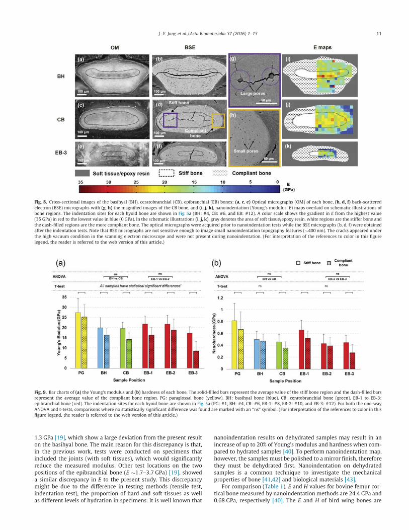
Table 1). Optical micrographs of cross-sections of the basihyal, cer-atobranchial and epibranchial bones (Fig. 8a, c and e) highlight twodifferent bony regions. In Fig. 5c (#1), the paraglossal bone has atriangle shape and large void area in the center, which interferethe continuous nanoindentation mapping. We had to select anddiscretize different areas to carry out the mapping and the E mapsdid not provide enough data compared to the other samples shownin Fig. 8. Therefore, we made a decision to exclude the paraglossalbone and focus on the most representative results. The outerregion between the white-dotted and black-dotted lines has abrighter contrast than the inner region enclosed in the black-dotted line. BSE micrographs (Fig. 8b, d and f) taken after nanoin-dentation and subsequent dehydration show different microstruc-tures in two regions in the magnified micrographs (Fig. 8g and h).The outer regions have a higher density of elliptically shaped pores(3 � 9%) than the inner region, each with a major axis �7.50 lmand a minor axis �2.30 lm. The cracks in the BSE micrographswere generated during dehydration in the vacuum chamber andare mainly distributed within the inner/stiff bone.
Based on the contrast difference, schematic illustrations areshown with three simplified features (Fig. 8i, j and k): soft tis-sue/epoxy resin (gray), the light region between the black-dotted outline and the white-dotted outline (black dashed) andthe dark region inside of the black-dotted outline (white).Nanoindentation mapping results distinguish two bone regions(termed ‘‘stiff” and ‘‘compliant”), which are overlaid on the illus-trations. The cross-sectional areas for indentation (basihyal, #4;ceratobranchial, #6; epibranchial, #12) are indicated in Fig. 5a.The small pores (Fig. 8h) have similar dimensions as osteocytelacuna found in skeletal bird bones (�10 lm) [37,38]. The innerregions are populated with larger, circular pores with diametersof 8–20 lm (Fig. 8g). The larger pores have similar dimensionsto vascular channels found in skeletal bird bones (�25 lm)[37,38] but it is unclear if vascularization is present. When con-sidering energy absorbance, this porous and compliant bone canbe considered to be a cellular structural design element, which
Fig. 6. (a) Photograph of dissected hyoid apparatus highlighting the imaged locations on the lingual apex. Scanning electron micrographs on the dorsal surface, (b) lowmagnification and (c) high magnification with the outline of two keratin scales (white dotted lines), and (d) energy dispersive X-ray spectroscopy results. Scanning electronmicrographs on the cross-section, (e) low magnification and (f) high magnification. (g) Fourier transform infrared spectrum of the polished sample near the region shown in(f). PG: paraglossal bone, K: keratin scales.
J.-Y. Jung et al. / Acta Biomaterialia 37 (2016) 1–13 9

are found throughout nature and are known to be effective atincreasing energy absorbance [39].
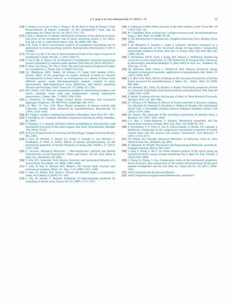
The average E and H values, the standard deviation, and thenumber of indentation tests for each specimen are listed in Table 1.The average E (and H) of each hyoid bone ranged from �17 to27 GPa (0.4–0.8 GPa) for the inner bone and from �9 to 25 GPa(0.3–0.7 GPa) for the outer bone (Fig. 9a and b), indicating the bonesconsist of an outer, more compliant region and an inner, more stiffregion. According to statistical analyses using a t-test between thestiff and compliant bones, the E values of the inner region are signif-icantly higher (p < 0.05, Fig. 9a) than the outer region in all of thebones. The stiffer bone always has a higher average E when com-pared to the compliant bone, ranging from a slight increase in theparaglossal bone to a two-fold increase in the epibranchial bone.Unlike E, a few of the comparisons between the stiff and compliantbone for H were found to exhibit no statistically significant differ-ences. However, these cases (BH and EB-2) can be explained bythe fact that H is dependent upon not only elastic deformation (asis the case for E), but also plastic deformation, which is generallyconsidered to be more variable. These bone structures are unlikemost other bone, where a dense outer sheath surrounds a less dense
core, such as with mammalian skeletal bones. It is unclear what, ifany, mechanical advantage this arrangement has in the hyoidbones. However, it can be speculated that a more compliant shellcould protect the stiffer, more brittle core.
From Fig. 8b, d and f, the average pore size (major axis, a, andminor axis, b) in the compliant bone is larger in the epibranchial(a � 7.50 lm and b � 2.30 lm) compared to the basihyal(a � 6.30 lm and b � 2.90 lm) or ceratobranchial (a � 5.50 lmand b � 2.00 lm) bones, which reduces the average E. Among thehyoid bones, the paraglossal bone had the highest E and H values,up to three times greater than the epibranchial bone for both thestiff and compliant regions, which corroborates the l-CT resultsin Fig. 3 that show a higher mineral density at the rostral position.The E values of the basihyal and ceratobranchial bones as well asthe rostral end (EB-1) and midsection (EB-2) of the epibranchialbones are not significantly different (labeled as ‘ns’ in Fig. 9a) forboth stiff and compliant regions. In the epibranchial bone, EB-3has a significantly lower E value than EB-1 and EB-2, as well asthe other bones.
Previously reported values of Young’s modulus obtained fromtensile tests of the hyoid apparatus near the first joint were
Fig. 7. (a) Photograph of dissected hyoid apparatus highlighting the imaged locations on the lingual body. (b–c) Scanning electron micrographs and (d) energy dispersive X-ray spectroscopy result of the dorsal surface. Scanning electron micrographs (e–g) on the longitudinal-section and (h) on the cross-section displaying four different muscles(M) surrounding the basihyal bone (BH). (i–j) Energy dispersive X-ray spectroscopy results of the center of the BH bone and the muscles, respectively. CT: connective tissue,D: dermis.
10 J.-Y. Jung et al. / Acta Biomaterialia 37 (2016) 1–13
1.3 GPa [19], which show a large deviation from the present resulton the basihyal bone. The main reason for this discrepancy is that,in the previous work, tests were conducted on specimens thatincluded the joints (with soft tissues), which would significantlyreduce the measured modulus. Other test locations on the twopositions of the epibranchial bone (E �1.7–3.7 GPa) [19], showeda similar discrepancy in E to the present study. This discrepancymight be due to the difference in testing methods (tensile test,indentation test), the proportion of hard and soft tissues as wellas different levels of hydration in specimens. It is well known that
nanoindentation results on dehydrated samples may result in anincrease of up to 20% of Young’s modulus and hardness when com-pared to hydrated samples [40]. To perform nanoindentation map,however, the samples must be polished to a mirror finish, thereforethey must be dehydrated first. Nanoindentation on dehydratedsamples is a common technique to investigate the mechanicalproperties of bone [41,42] and biological materials [43].
For comparison (Table 1), E and H values for bovine femur cor-tical bone measured by nanoindentation methods are 24.4 GPa and0.68 GPa, respectively [40]. The E and H of bird wing bones are
Fig. 8. Cross-sectional images of the basihyal (BH), ceratobranchial (CB), epibranchial (EB) bones: (a, c, e) Optical micrographs (OM) of each bone, (b, d, f) back-scatteredelectron (BSE) micrographs with (g, h) the magnified images of the CB bone, and (i, j, k), nanoindentation (Young’s modulus, E) maps overlaid on schematic illustrations ofbone regions. The indentation sites for each hyoid bone are shown in Fig. 5a (BH: #4, CB: #6, and EB: #12). A color scale shows the gradient in E from the highest value(35 GPa) in red to the lowest value in blue (0 GPa). In the schematic illustrations (i, j, k), gray denotes the area of soft tissue/epoxy resin, white regions are the stiffer bone andthe dash-filled regions are the more compliant bone. The optical micrographs were acquired prior to nanoindentation tests while the BSE micrographs (b, d, f) were obtainedafter the indentation tests. Note that BSE micrographs are not sensitive enough to image small nanoindentation topography features (�400 nm). The cracks appeared underthe high vacuum condition in the scanning electron microscope and were not present during nanoindentation. (For interpretation of the references to color in this figurelegend, the reader is referred to the web version of this article.)
Fig. 9. Bar charts of (a) the Young’s modulus and (b) hardness of each bone. The solid-filled bars represent the average value of the stiff bone region and the dash-filled barsrepresent the average value of the compliant bone region. PG: paraglossal bone (yellow). BH: basihyal bone (blue). CB: ceratobranchial bone (green). EB-1 to EB-3:epibranchial bone (red). The indentation sites for each hyoid bone are shown in Fig. 5a (PG: #1, BH: #4, CB: #6, EB-1: #8, EB-2: #10, and EB-3: #12). For both the one-wayANOVA and t-tests, comparisons where no statistically significant difference was found are marked with an ‘‘ns” symbol. (For interpretation of the references to color in thisfigure legend, the reader is referred to the web version of this article.)
J.-Y. Jung et al. / Acta Biomaterialia 37 (2016) 1–13 11

27.8 GPa (bending and tension) [44] and 0.55 GPa (Vickers hard-ness) [38], respectively. The E and H of beak bones from a wood-pecker are 30.2 GPa and 1.16 GPa (nanoindentation), respectively[11]. In comparison with the above, the E and H of the hyoid bonesare close to the values of the bird skeletal bones. The E and H valuesdepend on structural features, such as porosity and proportion oforganic materials [45,46]. Since there are no reported mechanicalproperties for the other bird hyoid bones, further comparativeanalysis cannot be made.
3.4. Structure–mechanical properties relationship
Structural shape is an important factor in the bending resis-tance, EI, where I is the second moment of area [11,47]. Alongthe length of the hyoid bones, the term (I) changes along withthe observed shapes: triangular, to circular, and finally to elliptical:
Itriangle ¼ 136
atb3t ð5Þ
Icircle ¼ p4a4c ð6Þ
Iellipse ¼ p4aeb
3e ; ð7Þ
where at = base and bt = height for the triangle, ac = radius for thecircle and ae = major axis and be = minor axis for the ellipse. FromEqs. (5)–(7), I varies along the length, from the triangular paraglossal(3.19 � 10�3 mm4) through the circular basihyal (14.7 � 10�3 mm4)to the elliptical ceratobranchial (0.75 � 10�3 mm4) and epibranchial(0.01–0.50 � 10�3 mm4) bones (Table 1).
Because the bones consist of two regions with different Young’smoduli, a volume fraction rule-of-mixtures was used to calculatethe bending resistance of each bone (stiff and compliant regions)[48]. For simplicity, it was assumed that the diameter of each bonewas the same along its length, so the area fraction (A) was usedinstead of the volume fraction. The area fractions of the stiff (Astiff)and compliant (Acompliant) bone regions were measured from thecross-sectional images of optical micrographs (Table 1). Astiff is�0.2 in the paraglossal and basihyal bones, �0.5 in theceratobranchial bone and 0.2–0.3 in the epibranchial bones. Thecomposite Young’s modulus (Ec) is given as:
Ec ¼ EstiffAstiff þ EcompliantAcompliant ð8ÞAs shown in Table 1, the bending resistance of the composite
bone (EcI) increases from the paraglossal bone to a maximum inthe basihyal bone (25.12 � 10�8 N m2). It then greatly decreasesthrough the ceratobranchial to a minimum at the caudal end ofthe epibranchial bones (0.01 � 10�8 N m2). The hyoid bones canbe modeled as a flat spiral spring, where the angular deflection isproportional to (EcI)�1. This indicates that the epibranchial bonescan undergo substantial deflection compared to the paraglossaland basihyal bones; the latter need sufficient stiffness forexcavating insects from trees. Since the flat spiral spring is usedto store elastic energy [49], this shape is possibly related toprevious statements of Oda et al. [15] and Zhou et al. [19] thatthe hyoid bones play a role as a shock-absorber or a damperduring pecking.
4. Conclusions
Macro- and micro-structural analysis of acorn woodpecker’s(Melanerpes formicivorus) hyoid apparatus and hyoid bones wereinvestigated by a multiscale structural analysis and mechanicalproperty evaluation. The main findings are:
� The first hyoid bone/joint 3D model was successfully developedusing micro-computed tomography image analysis and used forquantitative analysis of cross-sectional area and shape changeof the hyoid bones along their lengths.
� Scanning electron microscopy and energy dispersive X-rayspectroscopy on the lingual apex and body reveal a multilay-ered structure with a keratin sheath on the lingual apex andbody and a central bony core in the lingual body.
� Electron energy dispersive X-ray analysis showed that Ca/Pratio of all hyoid bones averaged 1.5, a slightly lower ratio thanfor skeletal bird bones (1.7) [34].
� Nanoindentation results show that all hyoid bone cross-sections consist of a dense/stiff interior region surrounded bya porous/compliant region. This compliant region may be effec-tive at dissipating energy in the hyoid bones during pecking.
� For the Young’s modulus, the paraglossal bone has the highestvalue, comparable to the inner bony part of the beak. The basi-hyal and ceratobranchial bones have similar values, but arelower compared to the paraglossal bone. The epibranchial boneexhibited properties that varied from the highest at the rostralto the lowest at the caudal.
Acknowledgments
The authors thank to the Mr. and Ms. Sherman for providing thewoodpecker specimens and Ms. Esther Cory and Dr. Robert L. Sahfor l-CT scans. This work is supported by an Academic SenateBridge Grant (2013–2014) and a National Science Foundation, Bio-materials Grant 1507978 (J.M.) and a Multi-University ResearchInitiative through the Air Force Office of Scientific Research(AFOSR-FA9550-15-1-0009) (J.J., D.K., J.M., S.E.N., V.R.S., N.Y., S.H.). Some of the work described here was carried out using sharedresearch resources at the National Center for Microscopy andImaging Research (NCMIR) at UCSD supported by the NIH underaward number P41 GM103412 (M.H.E.).
References
[1] P.A. May, J.M. Fuster, J. Haber, A. Hirschman, Woodpecker drilling behavior: Anendorsement of the rotational theory of impact brain injury, Arch. Neurol. 36(1979) 370–373.
[2] R. Livingston, Some observations on the natural history of the tongue, Ann. R.Coll. Surg. Engl. 19 (1956) 185.
[3] D.G. Homberger, R.A. Meyers, Morphology of the lingual apparatus of thedomestic chicken, Gallus gallus, with special attention to the structure of thefasciae, Am. J. Anat. 186 (1989) 217–257.
[4] R.A. Wallace, Aberrations in tongue structure of some malanerpinewoodpeckers, Wilson Bull. 86 (1974) 79–82.
[5] W.J. Bock, Functional and evolutionary morphology of woodpeckers, Ostrich 70(1999) 23–31.
[6] L. Wang, J.T.-M. Cheung, F. Pu, D. Li, M. Zhang, Y. Fan, Why do woodpeckersresist head impact injury: a biomechanical Investigation, PLoS One 6 (2011)e26490.
[7] D. Homberger, The lingual apparatus of the African grey parrot, Psittacuserithacus Linne (Aves: Psittacidae): Description and Theoretical MechanicalAnalysis, Wilson Bull. 99 (1987) 738–740.
[8] S. Nag, R. Banerjee, H.L. Fraser, A novel combinatorial approach forunderstanding microstructural evolution and its relationship to mechanicalproperties in metallic biomaterials, Acta Biomater. 3 (2007) 369–376.
[9] P.A. May, J. Fuster, P. Newman, A. Hirschman, Woodpeckers and head injury,Lancet 307 (1976) 1347–1348.
[10] L.J. Gibson, Woodpecker pecking: how woodpeckers avoid brain injury, J. Zool.270 (2006) 462–465.
[11] N. Lee, M.F. Horstemeyer, H. Rhee, B. Nabors, J. Liao, L.N. Williams, Hierarchicalmultiscale structure-property relationships of the red-bellied woodpecker(Melanerpes carolinus) beak, J. R. Soc. Interface 11 (2014) 20140274.
[12] L. Wang, Y. Fan, Role of mechanical performance of cranial bone in impactprotection of woodpecker brain-a finite element study, in: World Congress onMedical Physics and Biomedical Engineering, IFMBE Proceedings, vol. 39, 2013,pp. 165–167.
[13] L. Wang, X. Niu, Y. Ni, P. Xu, X. Liu, S. Lu, M. Zhang, Y. Fan, Effect ofmicrostructure of spongy bone in different parts of woodpecker’s skull onresistance to impact injury, J. Nanomater. 2013 (2013) 17.
12 J.-Y. Jung et al. / Acta Biomaterialia 37 (2016) 1–13
[14] L. Wang, S. Lu, X. Liu, X. Niu, C. Wang, Y. Ni, M. Zhao, C. Feng, M. Zhang, Y. Fan,Biomechanism of impact resistance in the woodpecker’s head and itsapplication, Sci. China Life Sci. 56 (2013) 715–719.
[15] J. Oda, J. Sakamoto, K. Sakano, Mechanical evaluation of the skeletal structureand tissue of the woodpecker and its shock absorbing system, J. Soc. Mech.Eng. Int. J. Ser. A: Solid Mech. Mater. Eng. 49 (2006) 390–396.
[16] S.-H. Yoon, S. Park, A mechanical analysis of woodpecker drumming and itsapplication to shock-absorbing systems, Bioinspiration Biomimetics 6 (2011)016003.
[17] Z.D. Zhu, G.J. Ma, C.W. Wu, Z. Chen, Numerical study of the impact response ofwoodpecker’s head, AIP Adv. 2 (2012) 042173.
[18] Y. Liu, X. Qiu, X. Zhang, T.X. Yu, Response of woodpecker’s head during peckingprocess simulated by material point method, PLoS One 10 (2015) e0122677.
[19] P. Zhou, X.Q. Kong, C.W. Wu, Z. Chen, The novel mechanical property of tongueof a woodpecker, J. Bionic Eng. 6 (2009) 214–218.
[20] L.D. Mkukuma, J.M.S. Skakle, I.R. Gibson, C.T. Imrie, R.M. Aspden, D.W.L.Hukins, Effect of the proportion of organic material in bone on thermaldecomposition of bone mineral: an investigation of a variety of bones fromdifferent species using thermogravimetric analysis coupled to massspectrometry, high-temperature x-ray diffraction, and fourier transforminfrared spectroscopy, Calcif. Tissue Int. 75 (2004) 321–328.
[21] W.C. Oliver, G.M. Pharr, An improved technique for determining hardness andelastic modulus using load and displacement sensing indentationexperiments, J. Mater. Res. 7 (1992) 1564–1583.
[22] G. Simmons, H. Wang, Single Crystal Elastic Constants and CalculatedAggregate Properties, The MIT Press, Cambridge, MA, 1971.
[23] J.-Y. Rho, T.Y. Tsui, G.M. Pharr, Elastic properties of human cortical andtrabecular lamellar bone measured by nanoindentation, Biomaterials 18(1997) 1325–1330.
[24] R.V. Hogg, J. Ledolter, Engineering Statistics, Macmillan, New York, NY, 1987.[25] Y. Hochberg, A.C. Tamhane, Multiple Comparison Procedures, Wiley, Hoboken,
NJ, 2009.[26] S. Erdogan, S.-I. Iwasaki, Function-related morphological characteristics and
specialized structures of the avian tongue, Ann. Anat. Anatomischer Anzeiger196 (2014) 75–87.
[27] D. Rizzo, Fundamentals of Anatomy and Physiology, Cengage Learning, Boston,MA, 2009.
[28] G. Fois, M. Weimer, T. Busch, E.T. Felder, F. Oswald, G. von Wichert, T.Seufferlein, P. Dietl, E. Felder, Effects of keratin phosphorylation on themechanical properties of keratin filaments in living cells, FASEB J. 27 (2013)1322–1329.
[29] G. Socrates, Biological Molecules – Macromolecules. Infrared and RamanCharacteristic Group Frequencies: Tables and Charts, 3rd ed., John Wiley &Sons, Inc., Chichester, UK, 2001.
[30] Y. Seki, M.S. Schneider, M.A. Meyers, Structure and mechanical behavior of atoucan beak, Acta Mater. 53 (2005) 5281–5296.
[31] Y. Seki, B. Kad, D. Benson, M.A. Meyers, The toucan beak: sructure andmechanical response, Mater. Sci. Eng., C 26 (2006) 1412–1420.
[32] Y. Seki, S.G. Bodde, M.A. Meyers, Toucan and hornbill beaks: a comparativestudy, Acta Mater. 6 (2010) 331–343.
[33] C. Ooi, M. Hamdi, S. Ramesh, Properties of hydroxyapatite produced byannealing of bovine bone, Ceram. Int. 33 (2007) 1171–1177.
[34] E. Pellegrino, R. Biltz, Mineralization in the chick embryo, Calcif. Tissue Res. 10(1972) 128–135.
[35] M. Tzaphlidou, Bone architecture: collagen structure and calcium/phosphorusmaps, J. Biol. Phys. 34 (2008) 39–49.
[36] D. Shi, Introduction to Biomaterials, Tsinghua University Press, Beijing, China,2006.
[37] E. de Margerie, S. Sanchez, J. Cubo, J. Castanet, Torsional resistance as aprincipal component of the structural design of long bones: comparativemultivariate evidence in birds, Anat. Rec. A: Discov. Mol. Cell. Evol. Biol. 282(2005) 49–66.
[38] E. Novitskaya, M.S.R. Vairo, J. Kiang, M.A. Meyers, J. McKittrick, Reinforcingstructures in avian wing bones, in: J.M. McKittrick, R. Narayan (Eds.), Advancesin Bioceramics and Biotechnologies II, John Wiley & Sons, Inc., Hoboken, NJ,2014, pp. 47–56.
[39] S.E. Naleway, M.M. Porter, J. McKittrick, M.A. Meyers, Structural designelements in biological materials: application to bioinspiration, Adv. Mater. 27(2015) 5455–5476.
[40] J.-Y. Rho, G.M. Pharr, Effects of drying on the mechanical properties of bovinefemur measured by nanoindentation, J. Mater. Sci. - Mater. Med. 10 (1999)485–488.
[41] A.K. Bembey, M.L. Oyen, A.J. Bushby, A. Boyde, Viscoelastic properties of boneas a function of hydration state determined by nanoindentation, Phil. Mag. 86(2006) 5691–5703.
[42] A. Boyde, Scanning electron microscopy of bone, in: Bone Research Protocols,Springer, 2012, pp. 365–400.
[43] J.C. Weaver, G.W. Milliron, A. Miserez, K. Evans-Lutterodt, S. Herrera, I. Gallana,W.J. Mershon, B. Swanson, P. Zavattieri, E. DiMasi, D. Kisailus, The stomatopoddactyl club: a formidable damage-tolerant biological hammer, Science 336(2012) 1275–1280.
[44] J.D. Currey, The evolution of the mechanical properties of amniote bone, J.Biomech. 20 (1987) 1035–1044.
[45] J.-Y. Rho, L. Kuhn-Spearing, P. Zioupos, Mechanical properties and thehierarchical structure of bone, Med. Eng. Phys. 20 (1998) 92–102.
[46] E. Novitskaya, P.-Y. Chen, S. Lee, A. Castro-Ceseña, G. Hirata, V.A. Lubarda, J.McKittrick, Anisotropy in the compressive mechanical properties of bovinecortical bone and the mineral and protein constituents, Acta Biomater. 7(2011) 3170–3177.
[47] A.P. Boresi, R.J. Schmidt, Advanced Mechanics of Materials, sixth ed., JohnWiley & Sons, Inc., Hoboken, NJ, 2003.
[48] D. Askeland, W. Wright, The Science and Engineering of Materials, seventh ed.,Cengage Learning, Boston, MA, 2015.
[49] J. Tang, Z. Wang, Z. Mi, Y. Yu, Finite element analysis of flat spiral spring onmechanical elastic energy storage technology, Res. J. Appl. Sci. Eng. Technol. 7(2014) 993–1000.
[50] L. Wang, H. Zhang, Y. Fan, Comparative study of the mechanical properties,micro-structure, and composition of the cranial and beak bones of the greatspotted woodpecker and the lark bird, Sci. China Life Sci. 54 (2011) 1036–1041.
[51] <http://bantams.the-kozaks.com/Bones>.[52] <http://digimorph.org/specimens/Melanerpes_aurifrons/>.
J.-Y. Jung et al. / Acta Biomaterialia 37 (2016) 1–13 13
Related Documents