1 STROKE & COVID19 Cases from the Frontlines Dr. Bay Leslie- Mazwi, Massachusetts General Hospital Dr. Steven Feske, Brigham and Women’s Hospital Dr. Brett Cucchiara, The Hospital of the University of Pennsylvania

Welcome message from author
This document is posted to help you gain knowledge. Please leave a comment to let me know what you think about it! Share it to your friends and learn new things together.
Transcript
1
STROKE & COVID19
Cases from the Frontlines
Dr. Bay Leslie- Mazwi, Massachusetts General Hospital
Dr. Steven Feske, Brigham and Women’s Hospital
Dr. Brett Cucchiara, The Hospital of the University of Pennsylvania
2
COVID-19 and Stroke
AHA Webinar
May 21, 2020
BRIGHAM AND WOMEN’S HOSPITAL
Steven Feske, M.D.
Chief, Stroke Division
Brigham and Women’s Hospital
3
Drs. Cucchiara, Feske, and Leslie-Mazwi have no relevant disclosures.
Disclosures
4
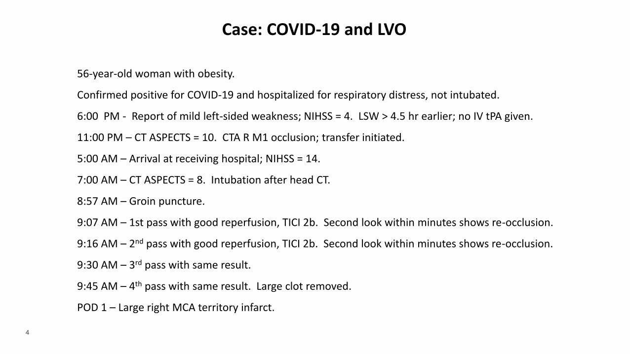
56-year-old woman with obesity.
Confirmed positive for COVID-19 and hospitalized for respiratory distress, not intubated.
6:00 PM - Report of mild left-sided weakness; NIHSS = 4. LSW > 4.5 hr earlier; no IV tPA given.
11:00 PM – CT ASPECTS = 10. CTA R M1 occlusion; transfer initiated.
5:00 AM – Arrival at receiving hospital; NIHSS = 14.
7:00 AM – CT ASPECTS = 8. Intubation after head CT.
8:57 AM – Groin puncture.
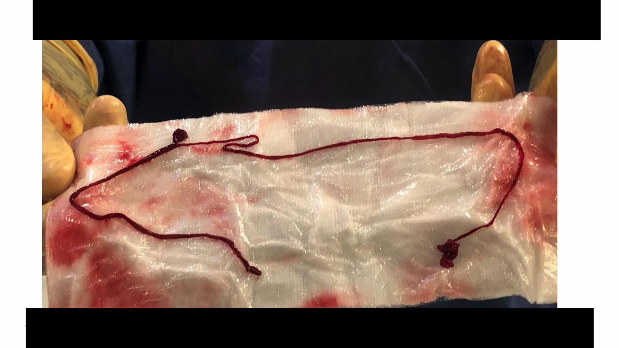
9:07 AM – 1st pass with good reperfusion, TICI 2b. Second look within minutes shows re-occlusion.
9:16 AM – 2nd pass with good reperfusion, TICI 2b. Second look within minutes shows re-occlusion.
9:30 AM – 3rd pass with same result.
9:45 AM – 4th pass with same result. Large clot removed.
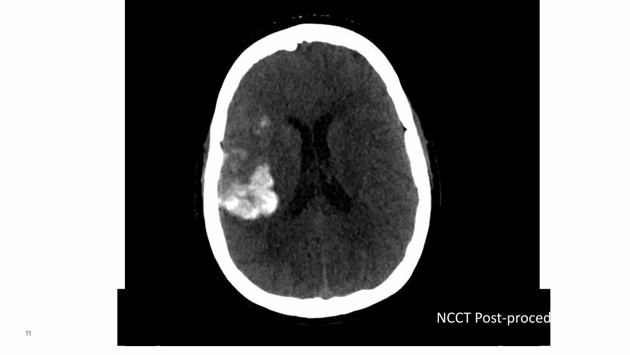
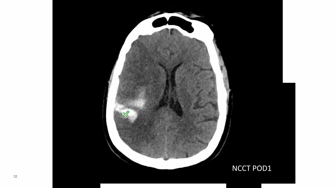
POD 1 – Large right MCA territory infarct.
Case: COVID-19 and LVO
5
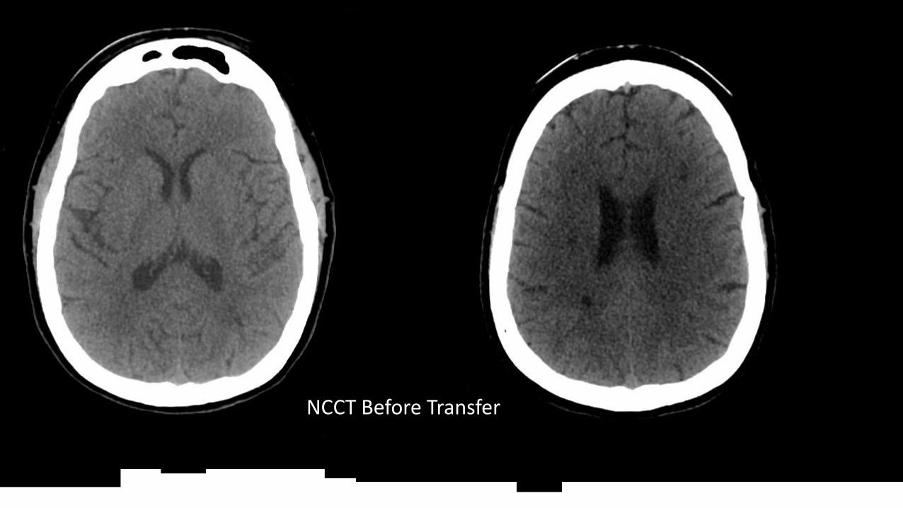
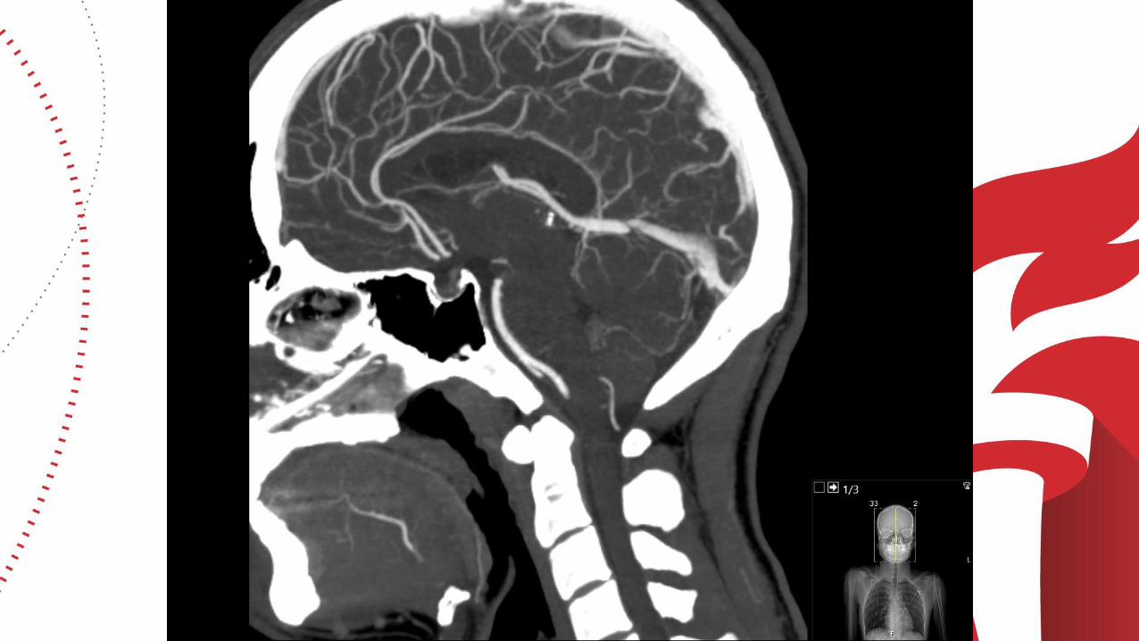
NCCT Before Transfer
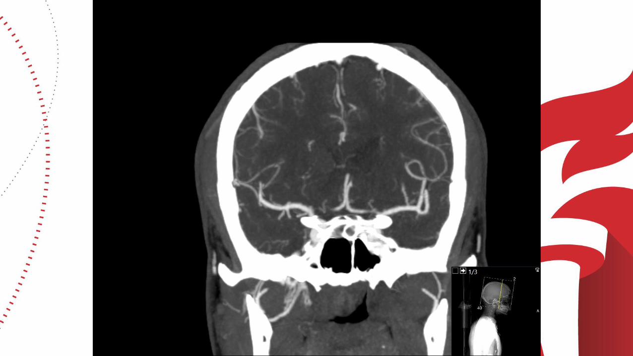
6
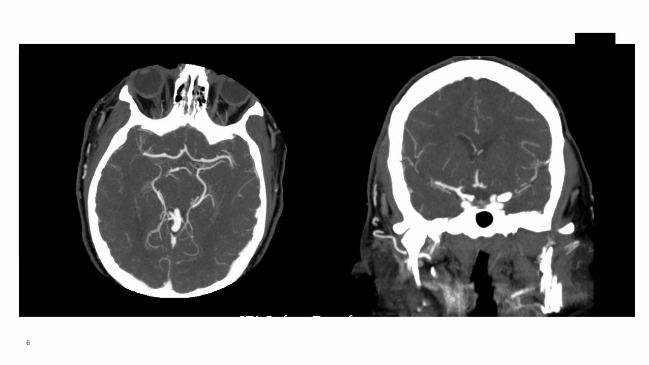
CTA Before Transfer
7
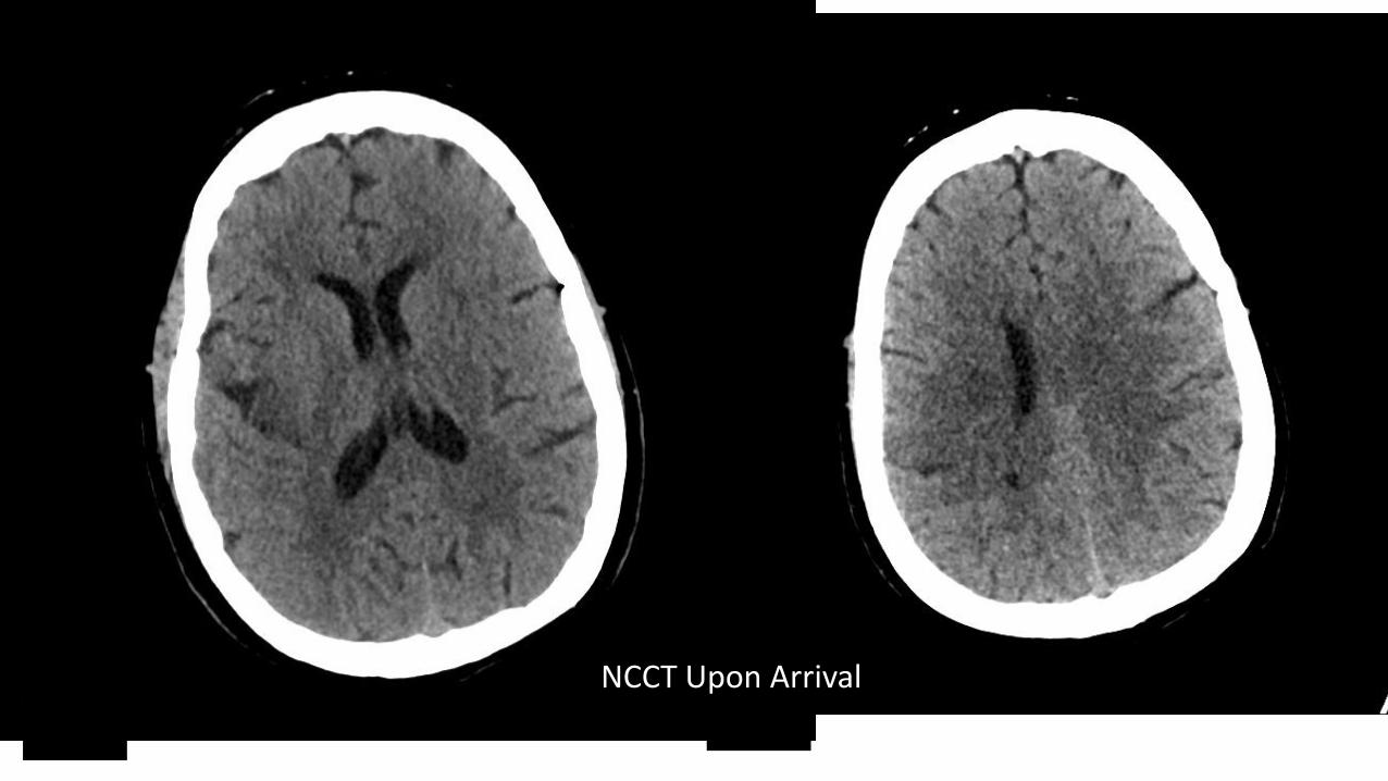
NCCT Upon Arrival
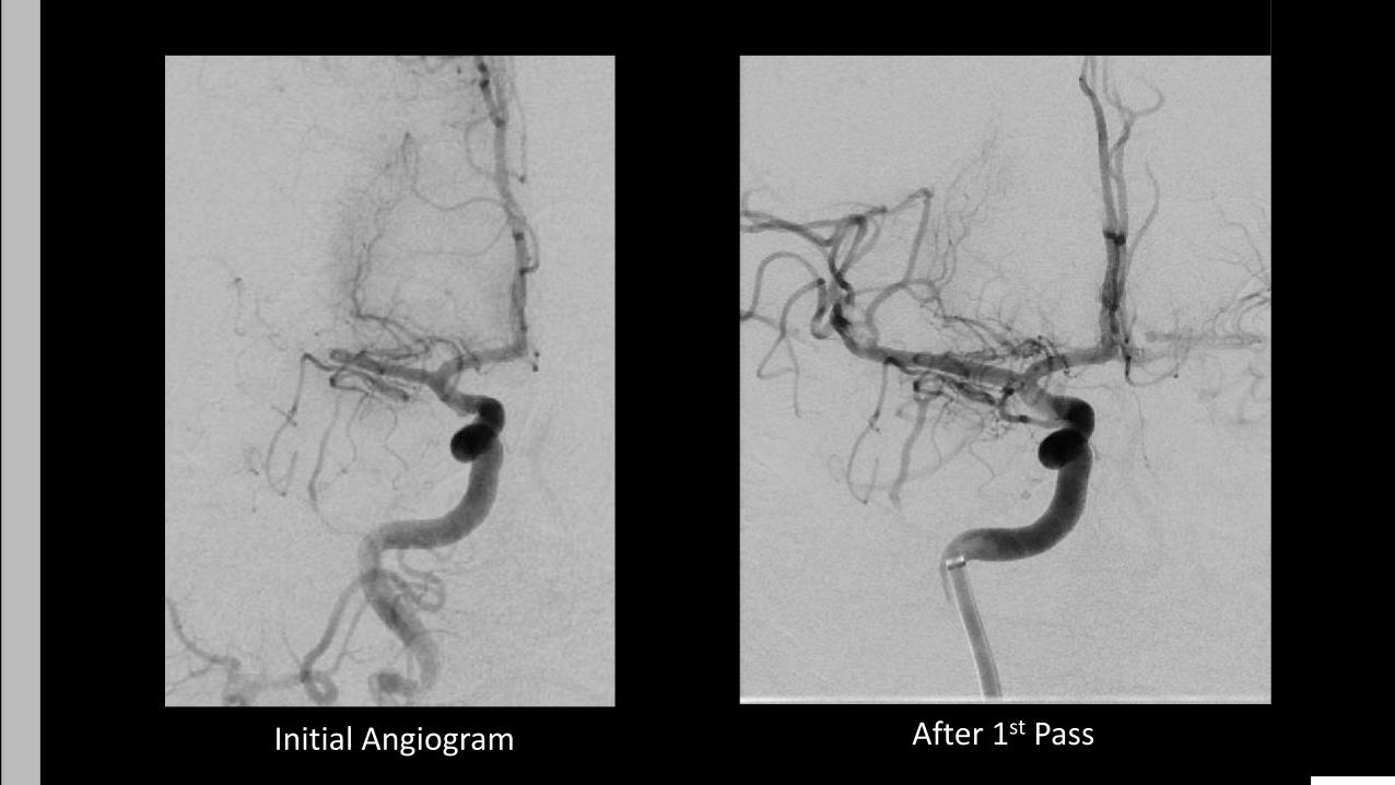
8 Initial Angiogram After 1st Pass
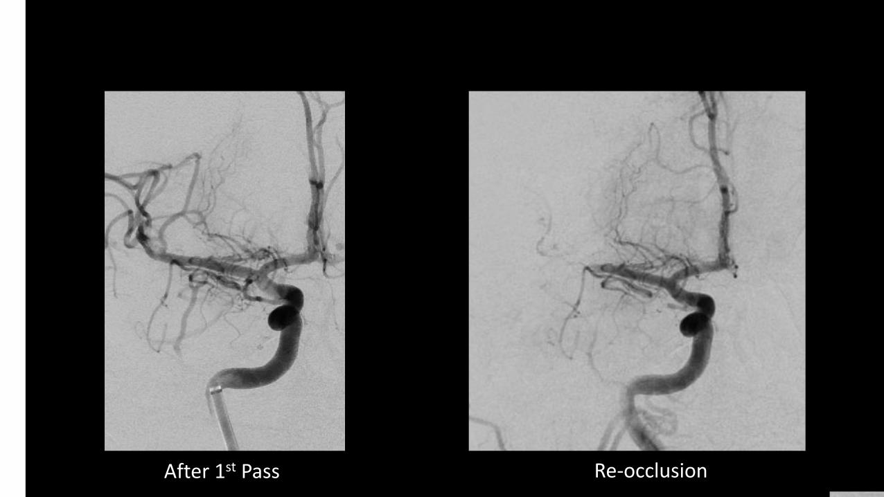
9 After 1st Pass Re-occlusion
10
11
NCCT Post-procedure
12
NCCT POD1
13
Delays – What are the causes of delays? Can we shorten them?
• PPE donning and doffing, transport, imaging, intubation
Are patients hypercoagulable? Why? Should we treat with ACs?
• Klok Thromb Res 2020; Zhang NEJM 2020
Do patient with COVID-19 have increased risk of stroke? If so, what
might be the mechanisms?
• Oxley TJ et al. NEJM 2020;382:e60.
Considerations
14
CASE 1
24 YO M WITH NO PMH NOTED ABRUPT ONSET L LEG WEAKNESS IN THE EVENING
NEXT AM ON WAKING NOTED L ARM WEAKNESS AND DYSARTHRIA – CALLED 911
PARAMEDICS NOTED LOW GRADE FEVER; HE REPORTED SORE THROAT FOR PRIOR 2 WEEKS
NO RESPIRATORY SYMPTOMS, VITALS NORMAL ON HOSPITAL ARRIVAL
15
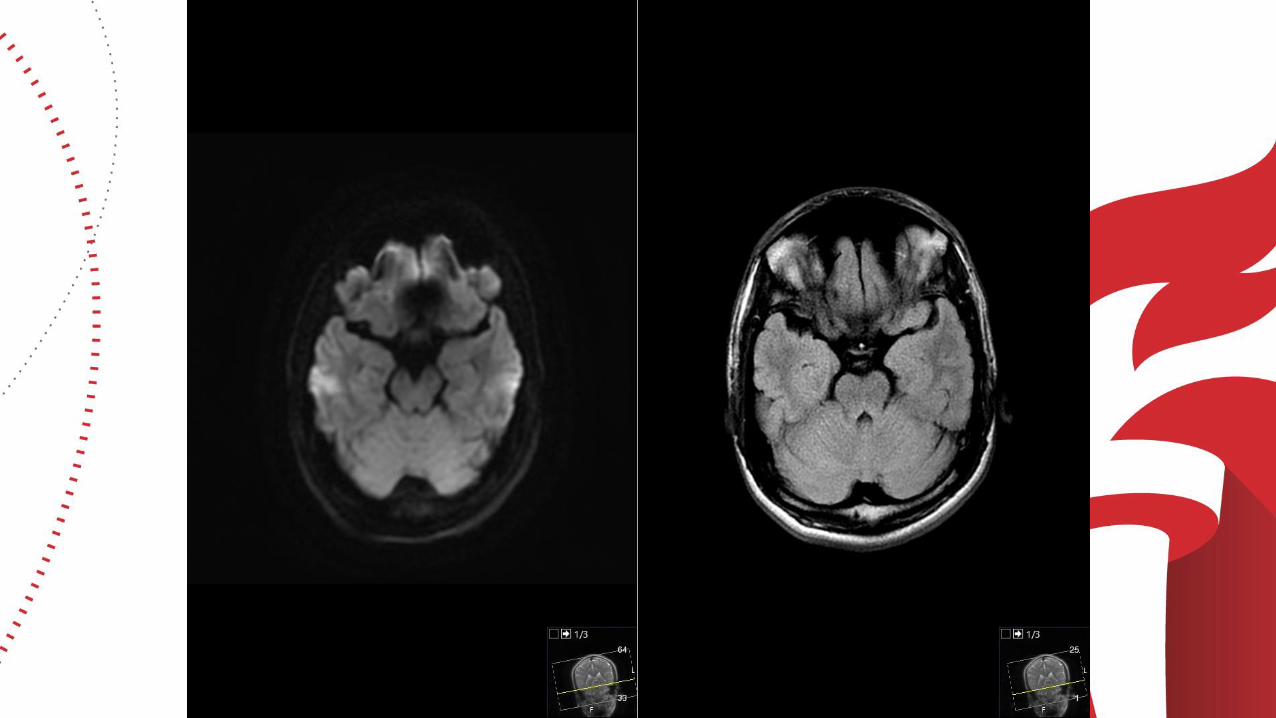
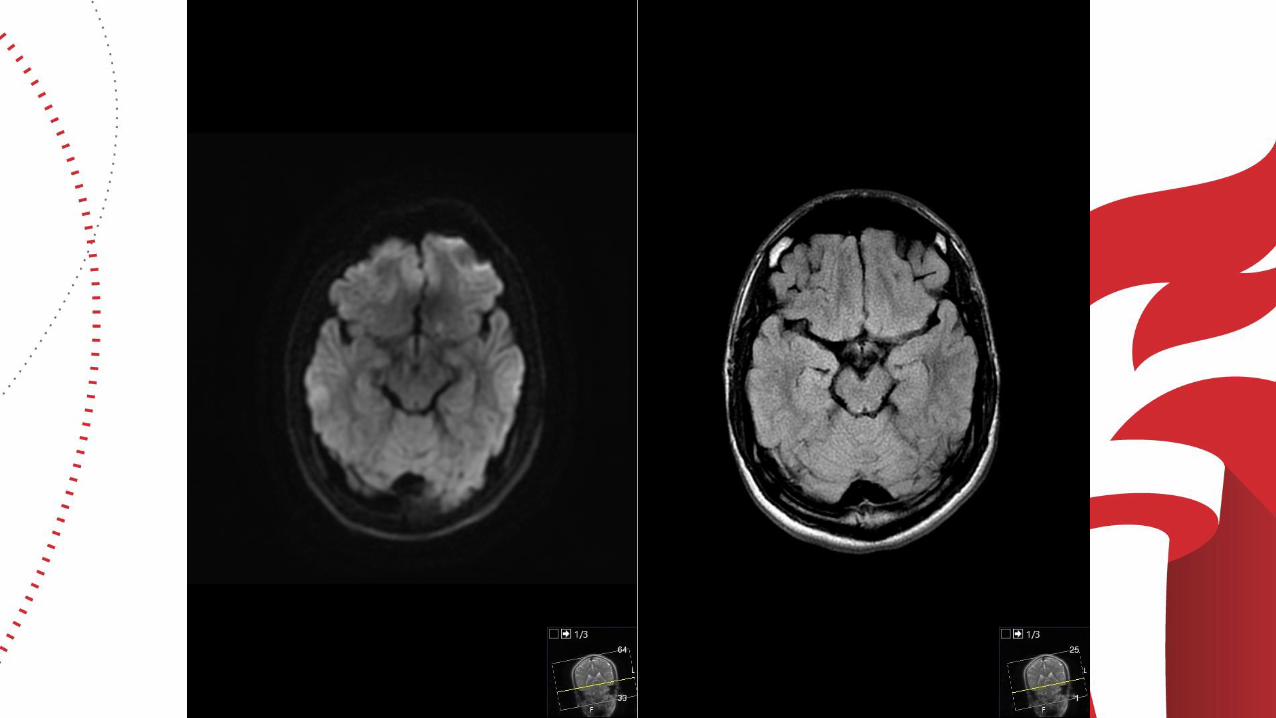
CASE 1
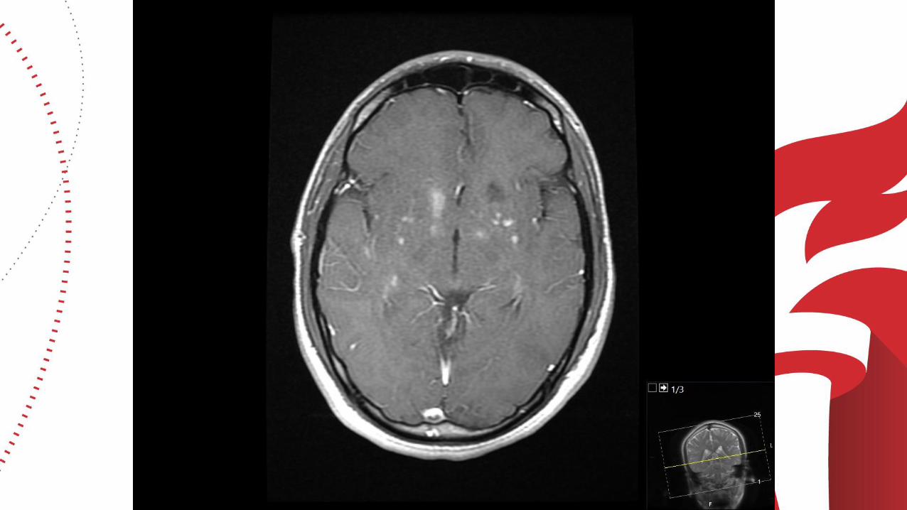
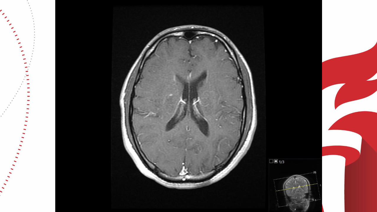
CTA AND MRI DONE
NO LVO OR VASCULAR ABNORMALITIES ON CTA
MRI ABNORMAL (SEE FILMS)
16
17
18
19
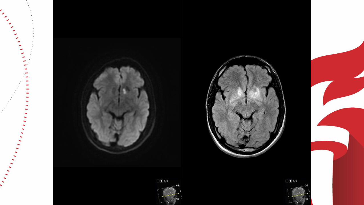
20
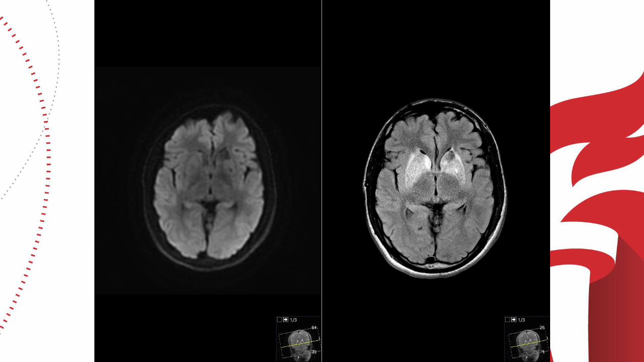
21

22

23

24

25

26

27

28
29
30
31

32

33
34

35
36
CASE 1
GIVEN SORE THROAT, COVID SWAB DONE AND PCR+
LP DONE AND CSF UNREMARKABLE
BROAD SEROLOGIC TESTING FOR OTHER INFECTIOUS/INFLAMMATORY DISEASE NEGATIVE
EMPIRICALLY TREATED WITH PREDNISONE 60 MG DAILY
AT FOLLOW-UP 2 WEEKS POST DISCHARGE, MINIMAL RESIDUAL L LEG WEAKNESS BUT OTHERWISE ASYMPTOMATIC
37
CASE 1
WHAT IS THIS????
PERIVASCULAR ENHANCEMENT ON MRI AND SYMMETRIC BASAL GANGLIA SIGNAL ABNORMALITY SUGGEST VASCULAR INFECTIOUS/INFLAMMATORY PROCESS, ? COVID ENDOTHELIAL-ITIS?
ACUTE INFARCTIONS LIKELY RELATED TO THIS
F/U IMAGING PENDING
38
39
COVID-19 AND STROKECASES AND DISCUSSIONAHA WEBINAR, 2020T.M. (Bay) Leslie-Mazwi, M.D.
Director of Endovascular Stroke Services
Neuroendovascular | Neurologic Critical Care
Massachusetts General Hospital
Harvard Medical School
Boston, MA
40
CASE PRESENTATION
69 Y.O. FEMALE WITH COPD, DM TYPE II
PRESENTING TO OSH WITH FEVER AND CHILLS
NO KNOWN COVID-19 EXPOSURE
COVID-19 POSITIVE (NASAL SWAB X 1)
ADMITTED FOR 10 DAYS OF HYDROXYCHLOROQINE AND AZITHROMYCIN
WORSENED RESPIRATORY STATUS LED TO ICU TRANSFER DAY 8
ON ICU TRANSFER HEAD CT FOR DELIRIUM
41
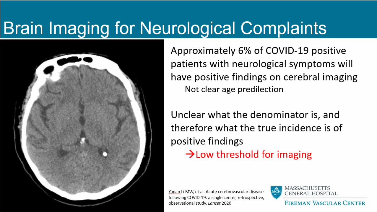
BRAIN IMAGING FOR NEUROLOGICAL COMPLAINTS
Approximately 6% of COVID-19 positive patients with neurological symptoms will have positive findings on cerebral imaging
Not clear age predilection
Unclear what the denominator is, and therefore what the true incidence is of positive findings→Low threshold for imaging
Yanan Li MW, et al. Acute cerebrovascular disease following COVID-19: a single center, retrospective, observational study. Lancet 2020
42
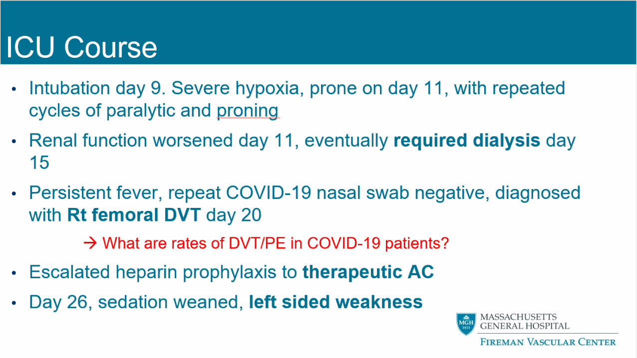
ICU COURSE
INTUBATION DAY 9. SEVERE HYPOXIA, PRONE ON DAY 11, WITH REPEATED CYCLES OF PARALYTIC AND PRONING
RENAL FUNCTION WORSENED DAY 11, EVENTUALLY REQUIRED DIALYSIS DAY 15
PERSISTENT FEVER, REPEAT COVID-19 NASAL SWAB NEGATIVE, DIAGNOSED WITH RT FEMORAL DVT DAY 20
→ What are rates of DVT/PE in COVID-19 patients?
ESCALATED HEPARIN PROPHYLAXIS TO THERAPEUTIC AC
DAY 26, SEDATION WEANED, LEFT SIDED WEAKNESS
43
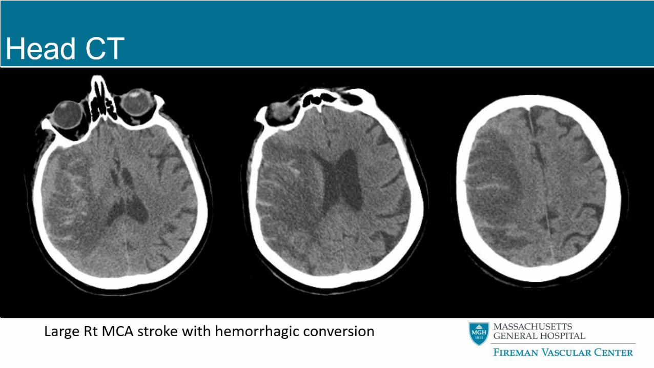
HEAD CT
Large Rt MCA stroke with hemorrhagic conversion
44
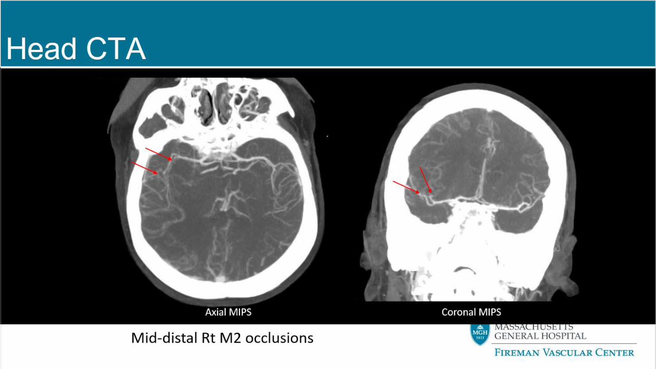
HEAD CTA
Mid-distal Rt M2 occlusions
Coronal MIPSAxial MIPS
45
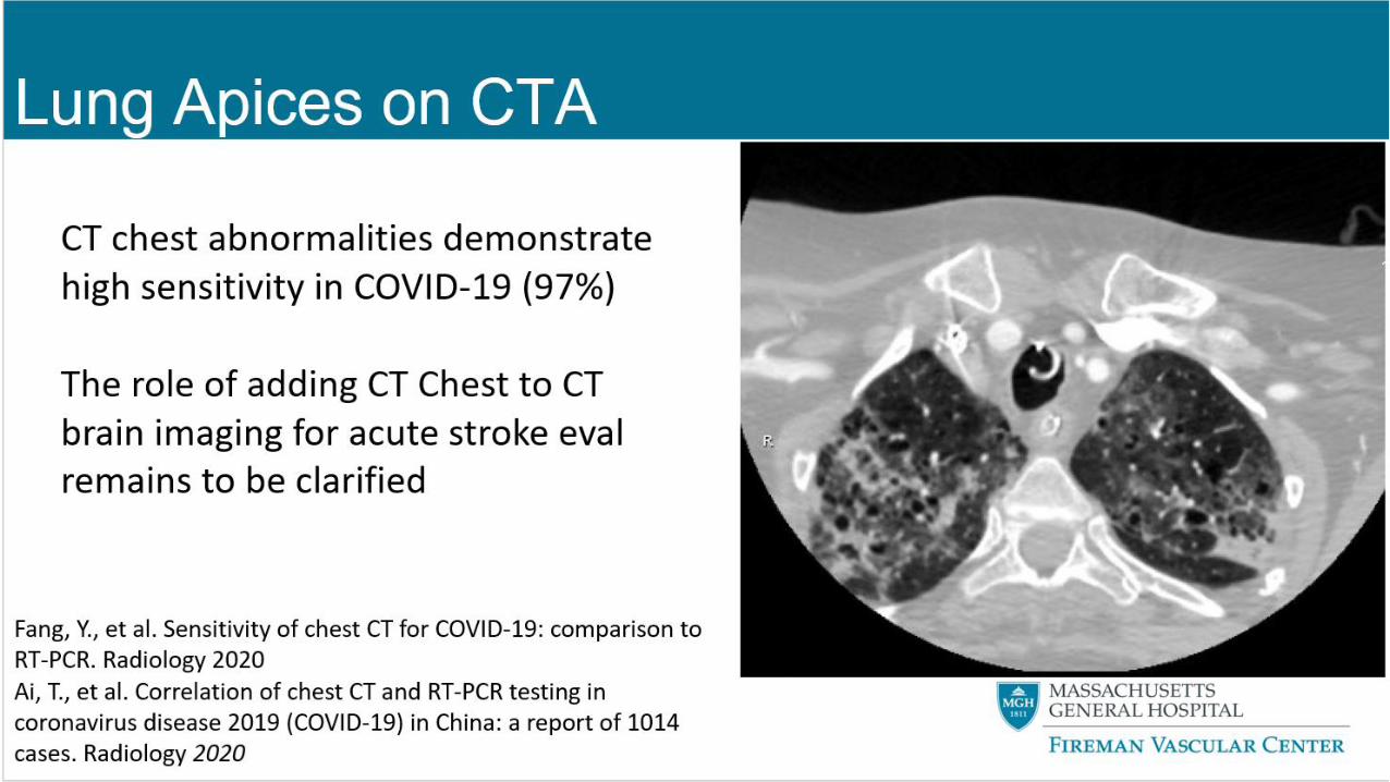
LUNG APICES ON CTA
CT chest abnormalities demonstrate high sensitivity in COVID-19 (97%)
The role of adding CT Chest to CT brain imaging for acute stroke eval remains to be clarified
Fang, Y., et al. Sensitivity of chest CT for COVID-19: comparison to RT-PCR. Radiology 2020Ai, T., et al. Correlation of chest CT and RT-PCR testing in coronavirus disease 2019 (COVID-19) in China: a report of 1014 cases. Radiology 2020
46
ICU COURSE
NOT A CANDIDATE FOR ACUTE INTERVENTION (THROMBOLYSIS OR THROMBECTOMY)
MANNITOL BOLUSES INITIATED (5MM MIDLINE SHIFT)
HEPARIN STOPPED, IVC FILTER PLACED
TRANSFER TO CSC INITIATED
HEAD CT REPEATED AT 4 HOURS GIVEN HEMORRHAGIC TRANSFORMATION
47
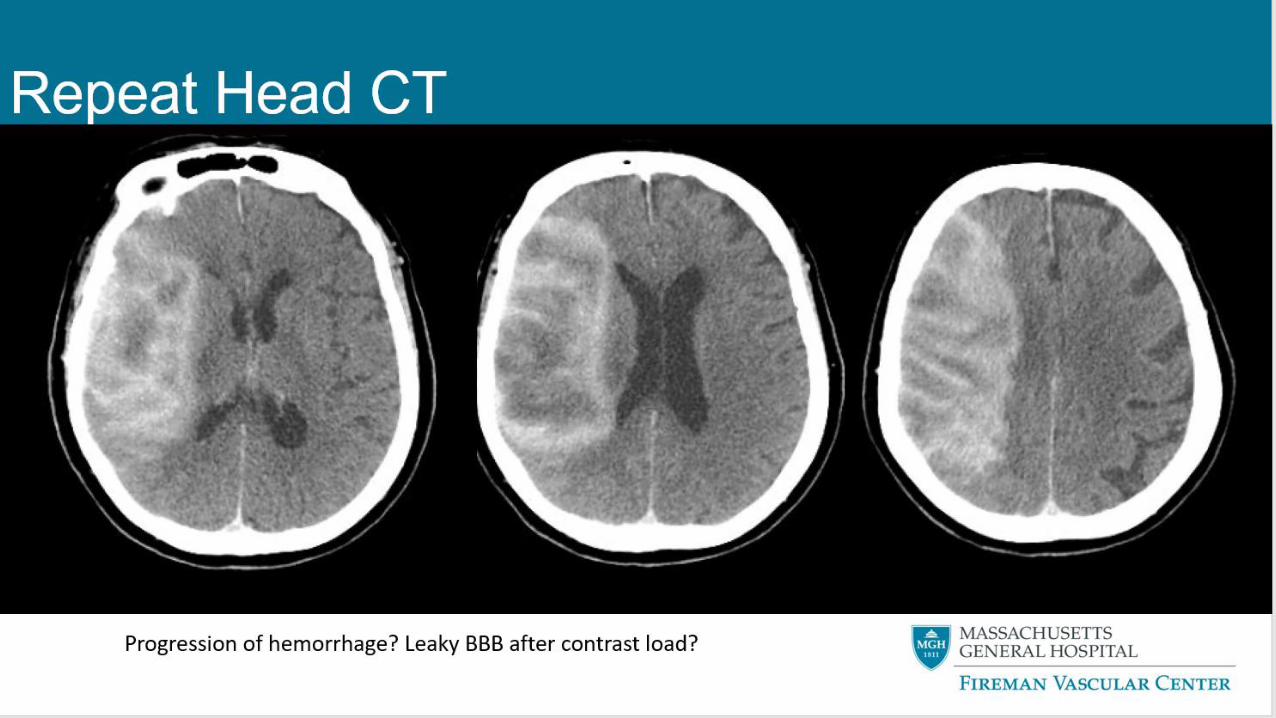
REPEAT HEAD CT
Progression of hemorrhage? Leaky BBB after contrast load?
48
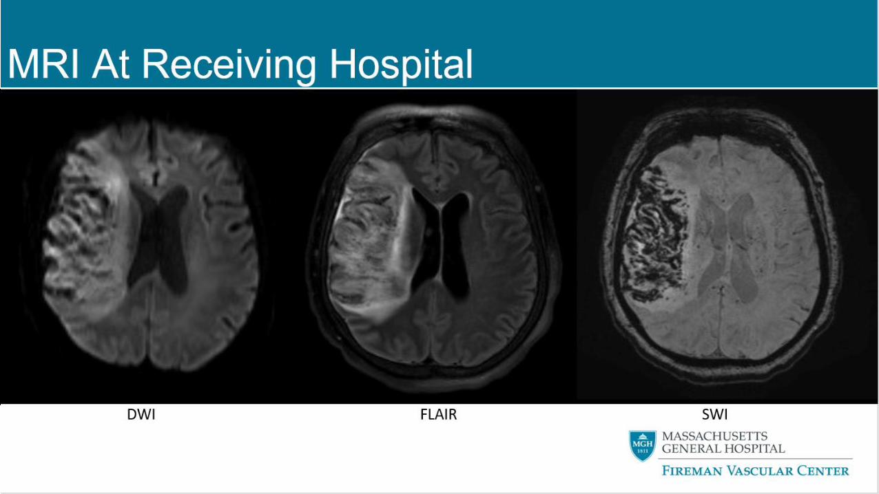
MRI AT RECEIVING HOSPITAL
DWI FLAIR SWI
49
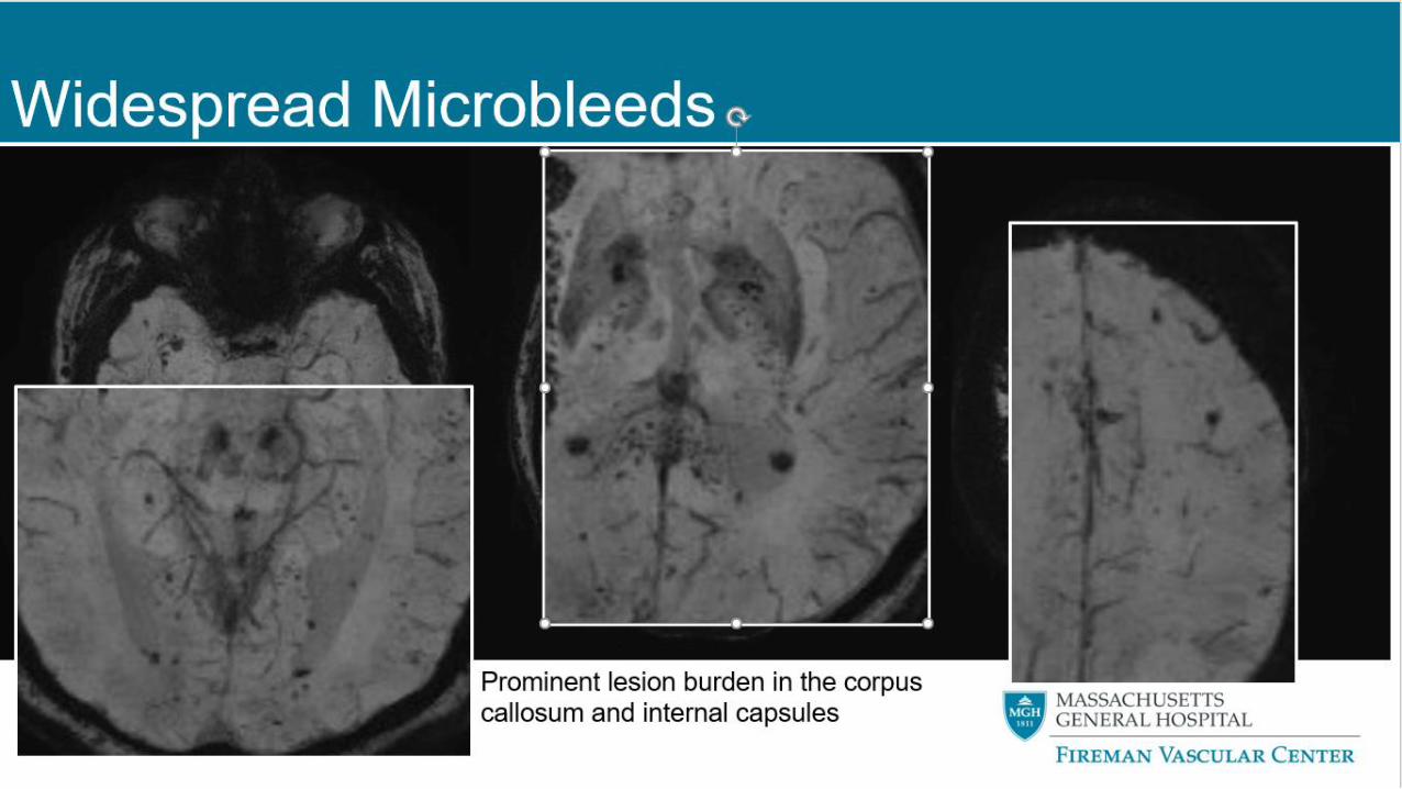
WIDESPREAD MICROBLEEDS
Prominent lesion burden in the corpus callosum and
internal capsules
50
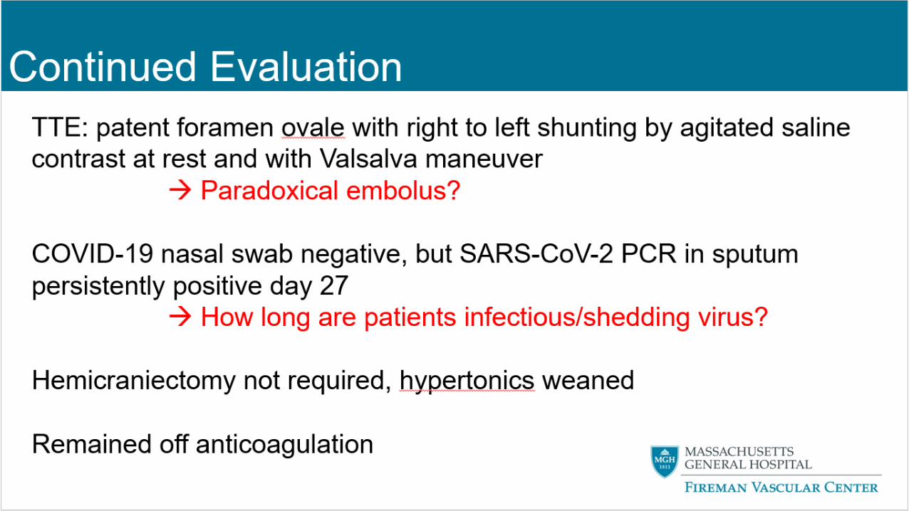
CONTINUED EVALUATION
TTE: patent foramen ovale with right to left shunting by agitated saline
contrast at rest and with Valsalva maneuver
→ Paradoxical embolus?
COVID-19 nasal swab negative, but SARS-CoV-2 PCR in sputum
persistently positive day 27
→ How long are patients infectious/shedding virus?
Hemicraniectomy not required, hypertonics weaned
Remained off anticoagulation
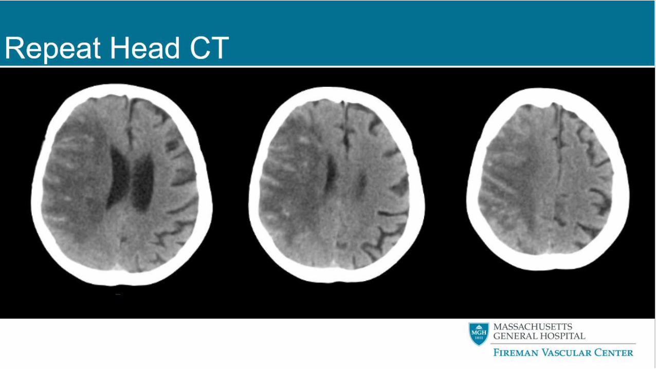
51
REPEAT HEAD CT
52
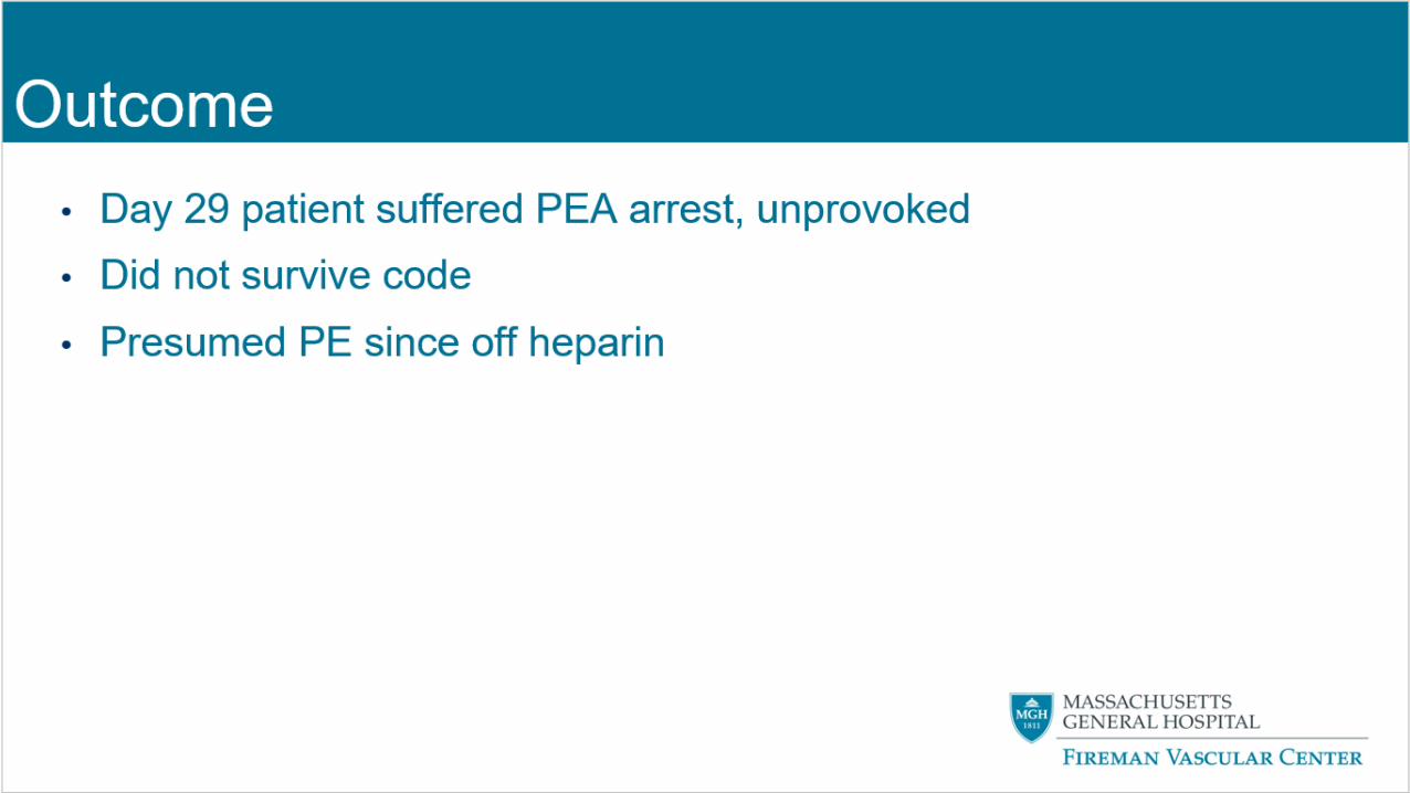
OUTCOME
DAY 29 PATIENT SUFFERED PEA ARREST, UNPROVOKED
DID NOT SURVIVE CODE
PRESUMED PE SINCE OFF HEPARIN
53
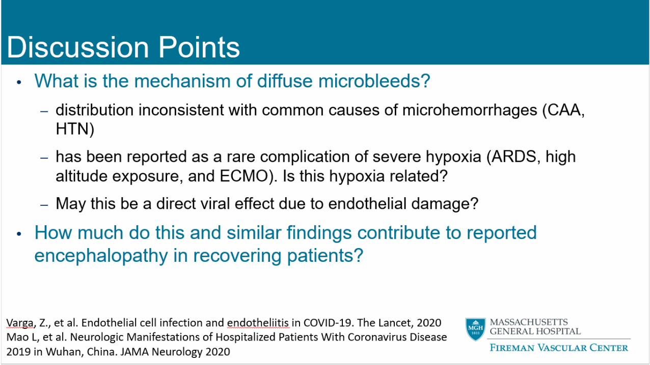
DISCUSSION POINTS
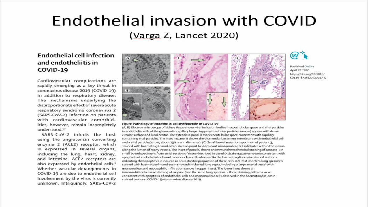
WHAT IS THE MECHANISM OF DIFFUSE MICROBLEEDS?
• distribution inconsistent with common causes of microhemorrhages (CAA, HTN)
• has been reported as a rare complication of severe hypoxia (ARDS, high altitude exposure, and ECMO). Is this hypoxia related?
• May this be a direct viral effect due to endothelial damage?
HOW MUCH DO THIS AND SIMILAR FINDINGS CONTRIBUTE TO REPORTED ENCEPHALOPATHY IN RECOVERING PATIENTS?
Varga, Z., et al. Endothelial cell infection and endotheliitis in COVID-19. The Lancet, 2020Mao L, et al. Neurologic Manifestations of Hospitalized Patients With Coronavirus Disease 2019 in Wuhan, China. JAMA Neurology 2020
5454
DR. FESKE- CASE #2
55
30-year-old man with history of polysubstance abuse found down unresponsive without signs of trauma; glu 300.
Woke after naloxone to agitated confusion; intubated for persistent hypoxia and increased WOB.
Trauma evaluation, including NCCT was negative.
NP swab for SARS-CoV-2 positive; severe bilateral pneumonia requiring proning and consideration of ECMO,
although he ultimately improved without ECMO.
Course complicated by aspiration pneumonia, sepsis with metabolic acidosis, thrombocytopenia (63K), left IJ DVT.
DRVVT confirmed positive; lupus anticoagulant and anti-cardiolipin and anti-β-2- glycoprotein negative.
Case: COVID-19 and ICH
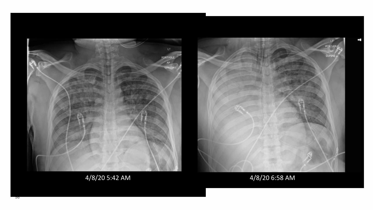
56
4/8/20 5:42 AM 4/8/20 6:58 AM
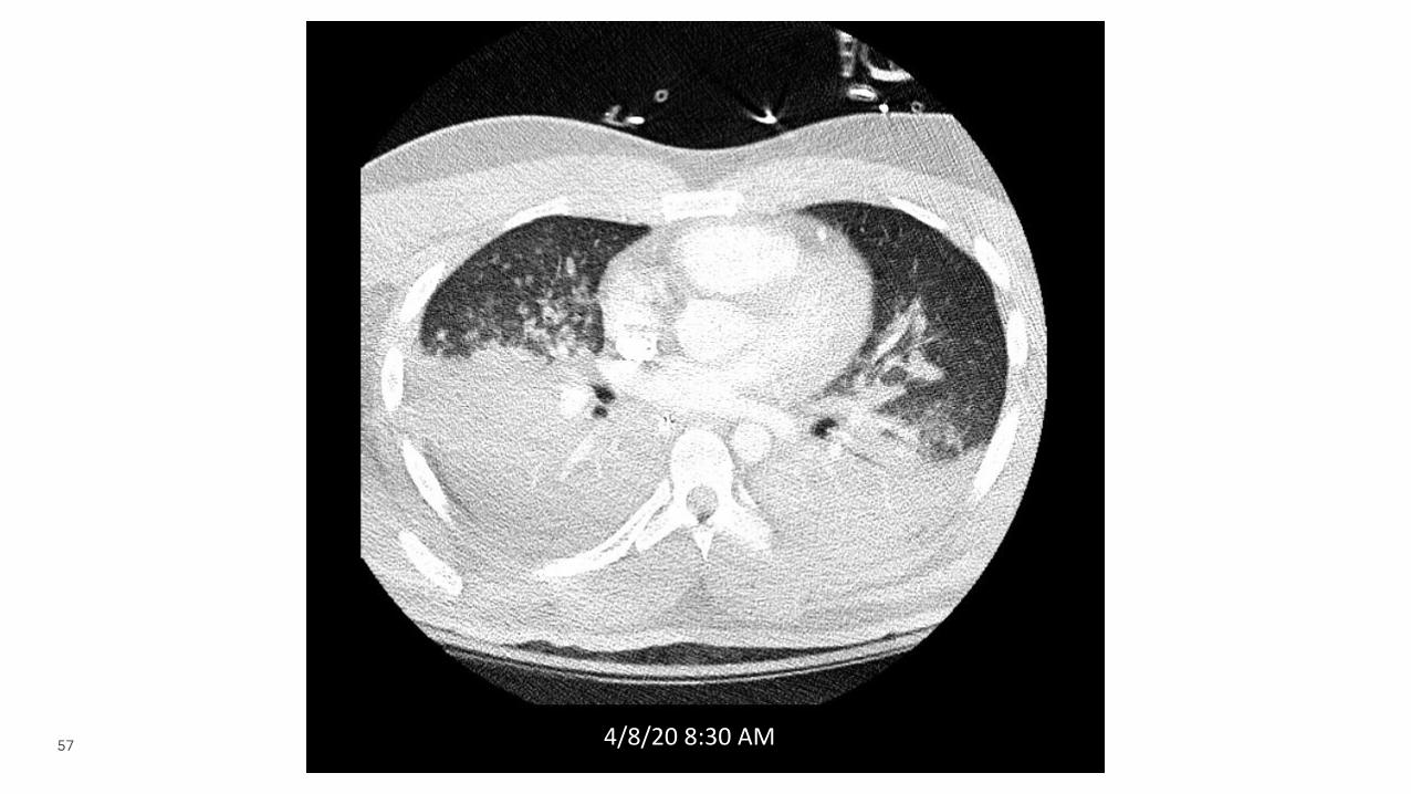
57 4/8/20 8:30 AM
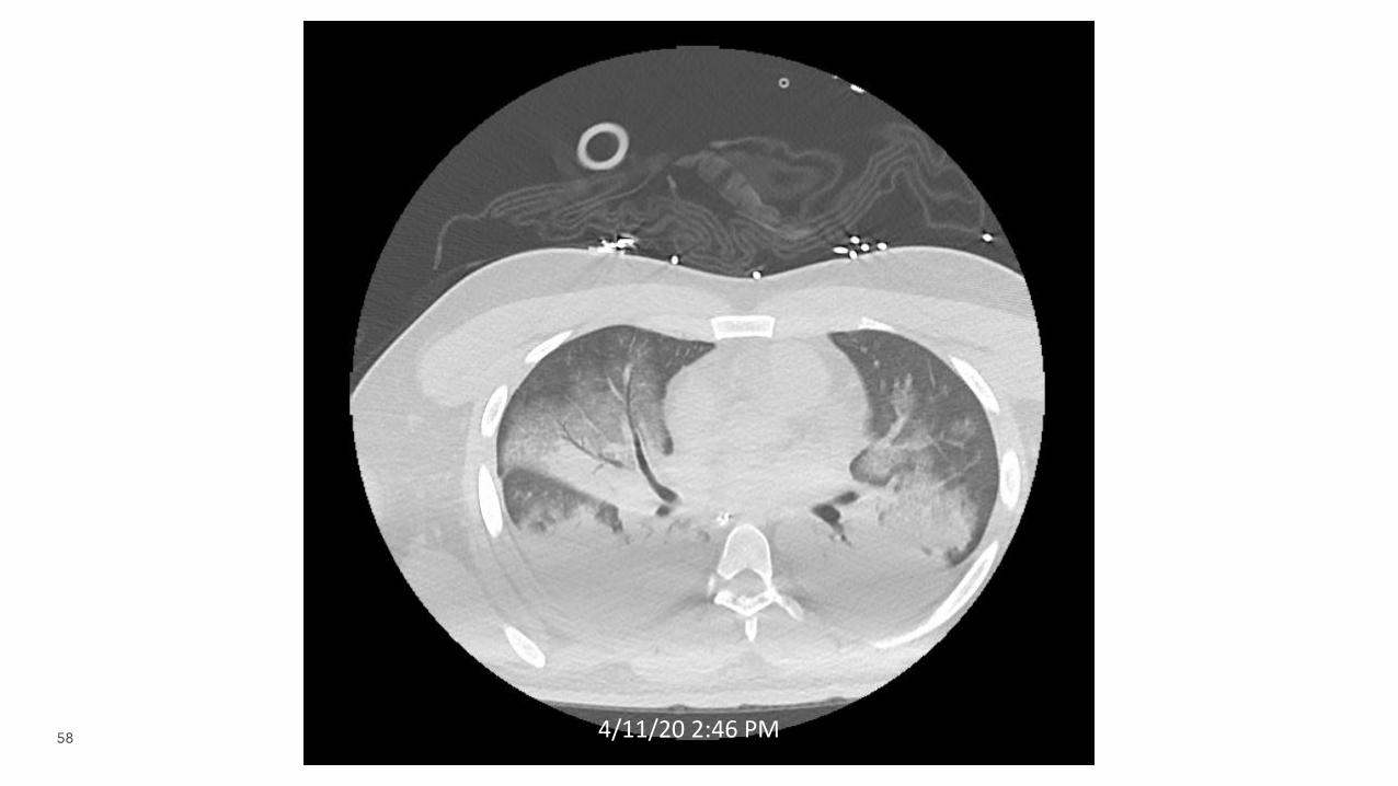
58 4/11/20 2:46 PM
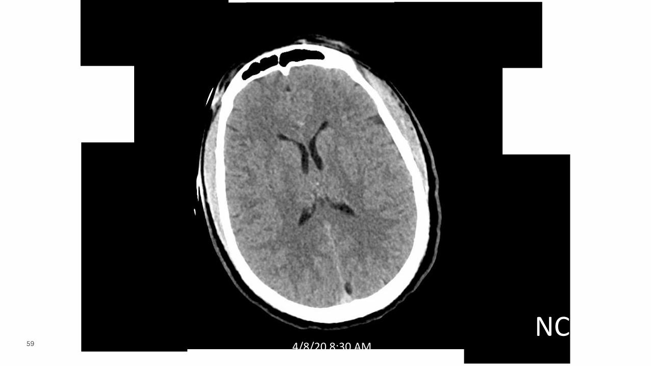
59 4/8/20 8:30 AMNCCT
60
30-year-old man with history of polysubstance abuse found down unresponsive without signs of trauma; glu 300.
Woke after naloxone to agitated confusion; intubated for persistent hypoxia and increased WOB.
NP swab for SARS-CoV-2 positive; severe bilateral pneumonia requiring proning and consideration of ECMO.
Course complicated by aspiration pneumonia, sepsis with metabolic acidosis, thrombocytopenia (63K), left IJ DVT.
DRVVT confirmed positive; lupus anticoagulant and anti-cardiolipin and anti-β-2- glycoprotein negative.
He was anticoagulated with UFH and then LMWH for the DVT when he developed right arm and leg weakness.
Case: COVID-19 and ICH
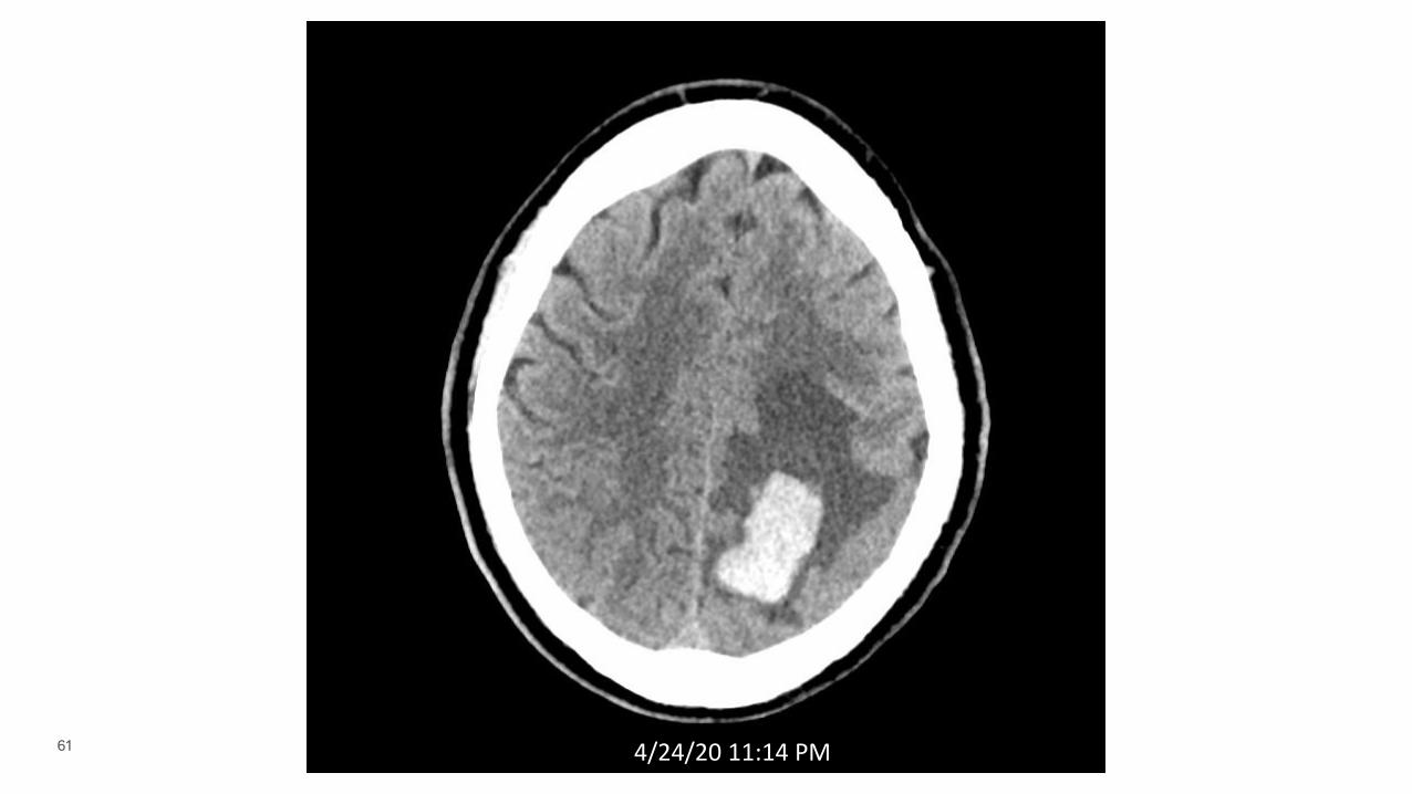
61 4/24/20 11:14 PMNCCT
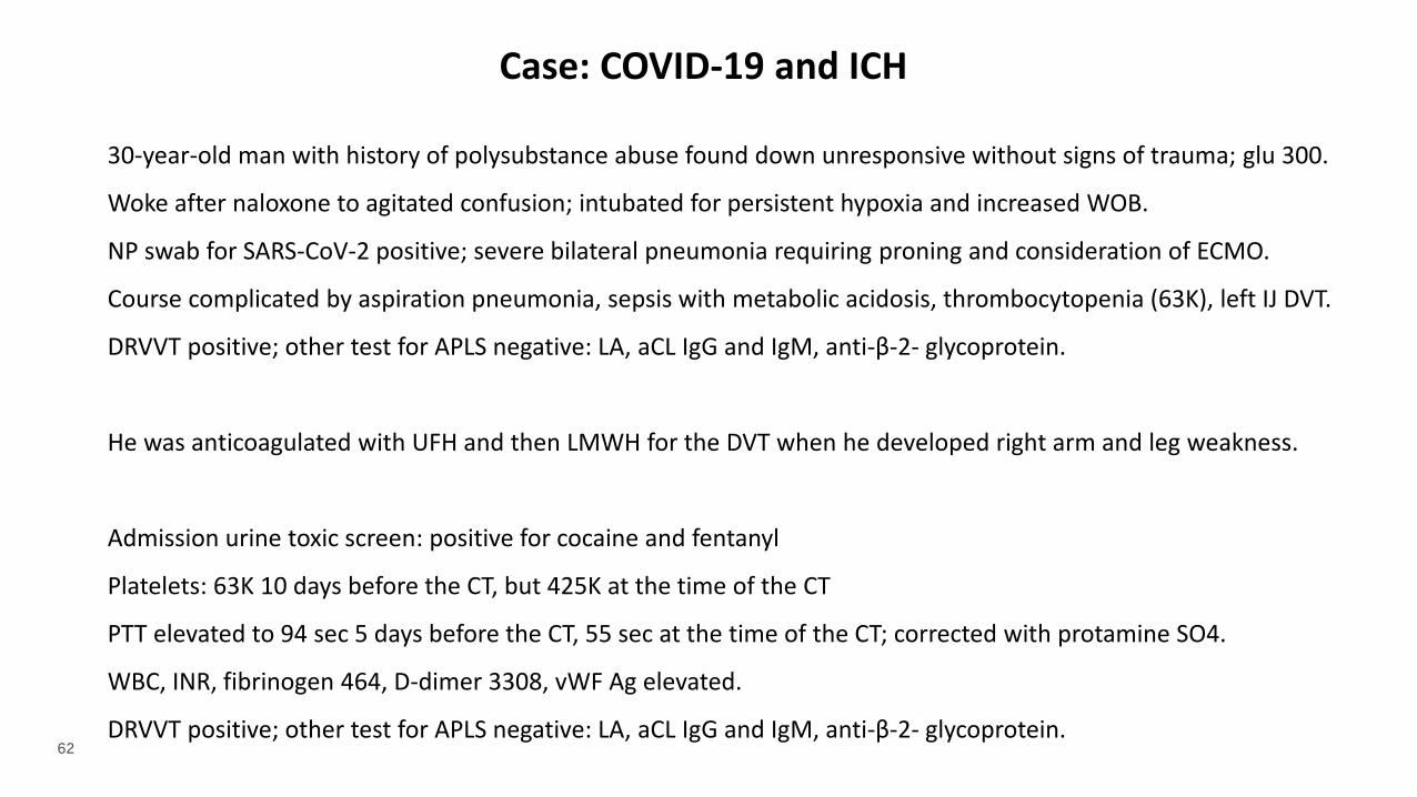
62
30-year-old man with history of polysubstance abuse found down unresponsive without signs of trauma; glu 300.
Woke after naloxone to agitated confusion; intubated for persistent hypoxia and increased WOB.
NP swab for SARS-CoV-2 positive; severe bilateral pneumonia requiring proning and consideration of ECMO.
Course complicated by aspiration pneumonia, sepsis with metabolic acidosis, thrombocytopenia (63K), left IJ DVT.
DRVVT positive; other test for APLS negative: LA, aCL IgG and IgM, anti-β-2- glycoprotein.
He was anticoagulated with UFH and then LMWH for the DVT when he developed right arm and leg weakness.
Admission urine toxic screen: positive for cocaine and fentanyl
Platelets: 63K 10 days before the CT, but 425K at the time of the CT
PTT elevated to 94 sec 5 days before the CT, 55 sec at the time of the CT; corrected with protamine SO4.
WBC, INR, fibrinogen 464, D-dimer 3308, vWF Ag elevated.
DRVVT positive; other test for APLS negative: LA, aCL IgG and IgM, anti-β-2- glycoprotein.
Case: COVID-19 and ICH
63 4/24/20 11:14 PM CTA
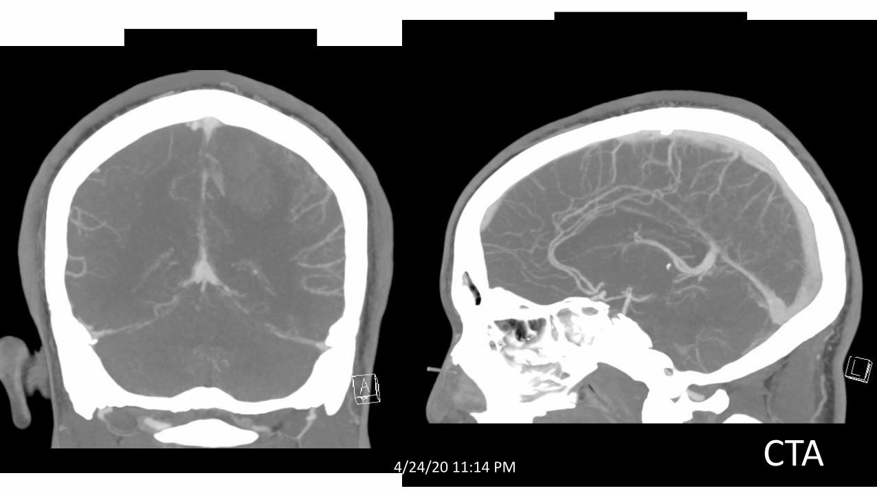
64 4/25/20 2:53 PM
MRVDWI
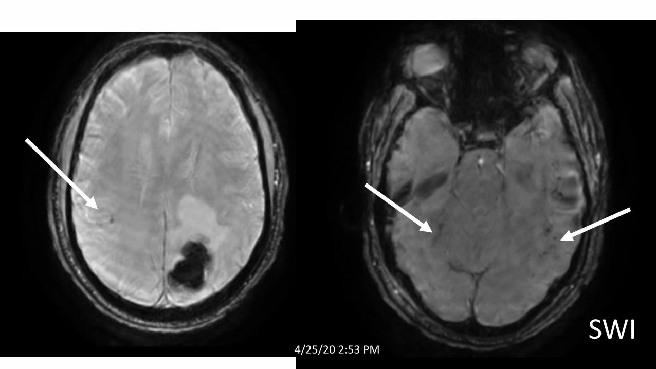
65 4/25/20 2:53 PMSWI
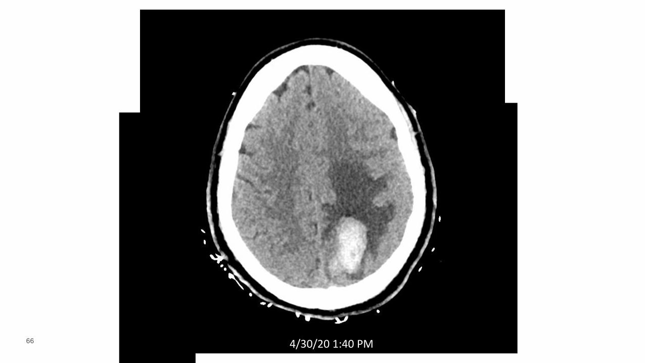
66 4/30/20 1:40 PMNCCT
67
Are patients hypercoagulable? What are the possible mechanisms? Did
this patient have APLS? What is the best approach to treatment?
What are the possible causes of cerebral hemorrhage in COVID-19?
What was the likely cause in this case? Should COVID-19 influence the
treatment?
What the causes of seizures in COVID-19 patients. Of encephalopathy?
Considerations
68
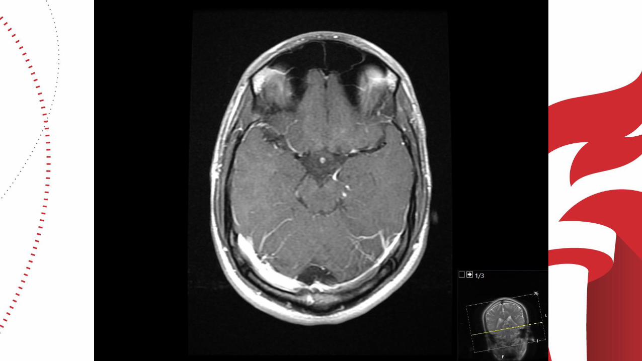
CUCCHIAR-CASE 252 YO F WITH HTN AND NEWLY DIAGNOSED, POORLY CONTROLLED DM2 (HEMOGLOBIN A1C OF 17.4)
DEVELOPED BILATERAL HAND PARESTHESIAS THE WEEK PRIOR TO PRESENTATION, FOLLOWED BY DYSPNEA, COUGH, HEADACHE AND CONFUSION.
SHE WALKED IN TO THE ED AFEBRILE, TACHYCARDIC (115 BEATS PER MINUTE), HYPERTENSIVE (220/118 MMHG), AND HYPOXEMIC (OXYGEN SATURATION 49% ON ROOM AIR). SHE HAD NO FOCAL NEUROLOGICAL DEFICITS ON EXAMINATION.
RAPIDLY DEVELOPED REFRACTORY HYPOXEMIA DESPITE 20 LITERS PER MINUTE OF SUPPLEMENTAL OXYGEN AND WAS INTUBATED; COVID PCR+ ON NASAL SWAB
HEAD CT DONE WITHIN 24 HOURS OF ADMISSION
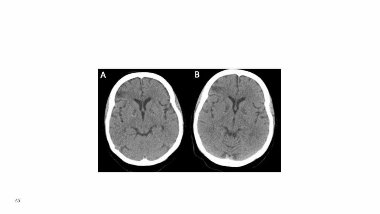
69
70
CUCCHIAR-CASE 2
CARBOXYHEMOGLOBIN WAS NOT ELEVATED AND URINE TOXICOLOGY SCREEN WAS NEGATIVE.
SEVERELY HYPERGLYCEMIC (GLUCOSE 1114 MG/DL) AND SUBSEQUENTLY IN SHOCK WITH LACTATE OF 7.7 MMOL/L, LOW CENTRAL VENOUS SATURATION (52%), AND ELEVATED TROPONIN AND NT-PROBNP. ULTRASOUND SHOWED SIGNS OF RIGHT VENTRICULAR FAILURE AND A SUBCLAVIAN VEIN THROMBUS, AND SHE WAS DIAGNOSED WITH OBSTRUCTIVE SHOCK DUE TO PRESUMED PULMONARY EMBOLISM.
ELEVATED D-DIMER (>128 UG/ML FIBRINOGEN EQUIVALENT UNITS, NORMAL <0.5), LOW FIBRINOGEN (NADIR OF 66 MG/DL), AND LOW PLATELETS (52 THOUSAND CELLS/UL).
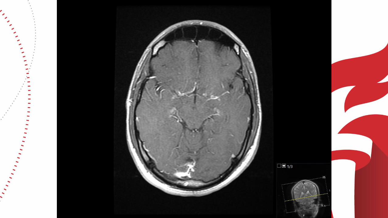
SHE WAS STARTED ON A HEPARIN INFUSION, AND REPEAT HEAD CT ON HOSPITAL DAY 3 WAS STABLE. HER COURSE WAS COMPLICATED BY ACUTE RENAL FAILURE REQUIRING CONTINUOUS RENAL REPLACEMENT THERAPY, SHOCK LIVER, AND VENTILATOR-ASSOCIATED PNEUMONIA.
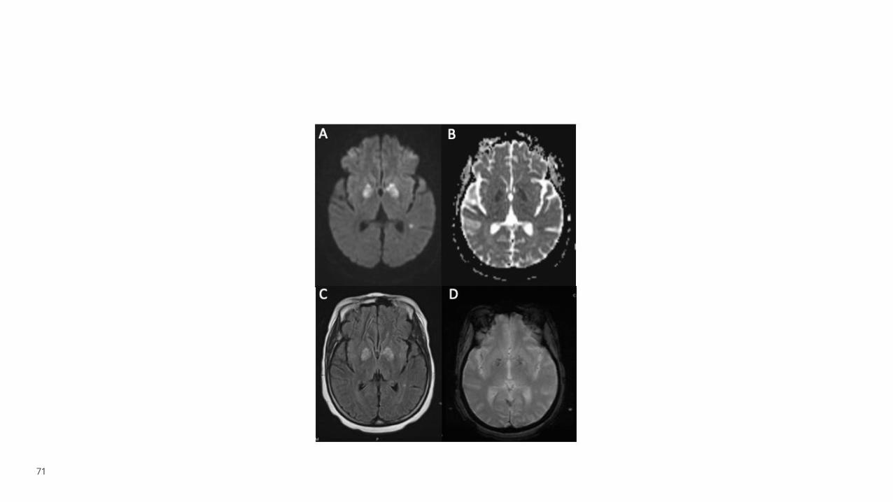
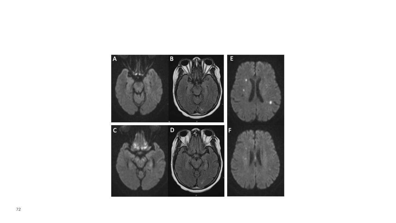
HER MENTAL STATUS REMAINED POOR ON HOSPITAL DAY 8. THEREFORE, A BRAIN MRI WAS OBTAINED
71
72
73
CUCCHIAR-CASE 2
IMAGING C/W BILATERAL GLOBUS PALLIDUS INFARCTIONS; SIMILAR INFARCTION PATTERN REPORTED IN PATIENTS WITH CARDIAC ARREST/SEVERE HYPOXIC-ISCHEMIC INJURY, OFTEN DUE TO DRUG OVERDOSE
MULTIFOCAL OTHER STROKES WITH POSSIBLE IBZ WATERSHED TOPOGRAPHY
74
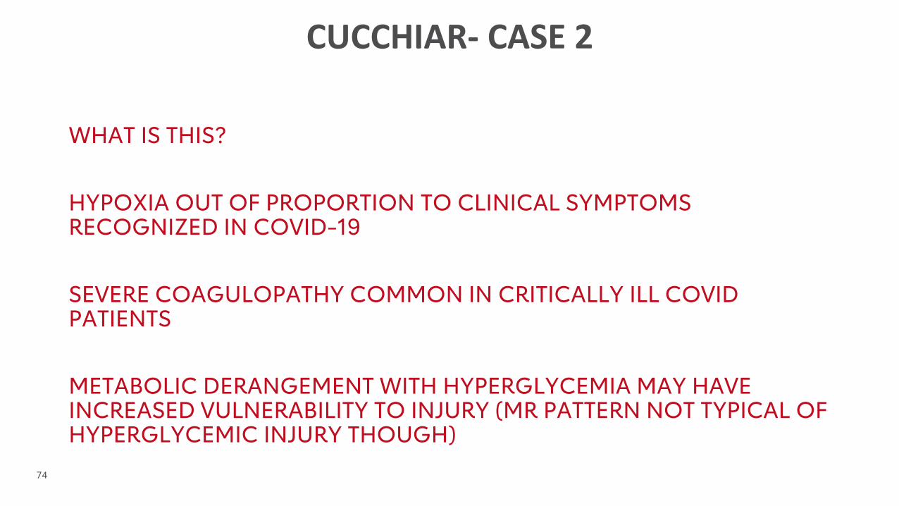
CUCCHIAR- CASE 2
WHAT IS THIS?
HYPOXIA OUT OF PROPORTION TO CLINICAL SYMPTOMS RECOGNIZED IN COVID-19
SEVERE COAGULOPATHY COMMON IN CRITICALLY ILL COVID PATIENTS
METABOLIC DERANGEMENT WITH HYPERGLYCEMIA MAY HAVE INCREASED VULNERABILITY TO INJURY (MR PATTERN NOT TYPICAL OF HYPERGLYCEMIC INJURY THOUGH)
75
76
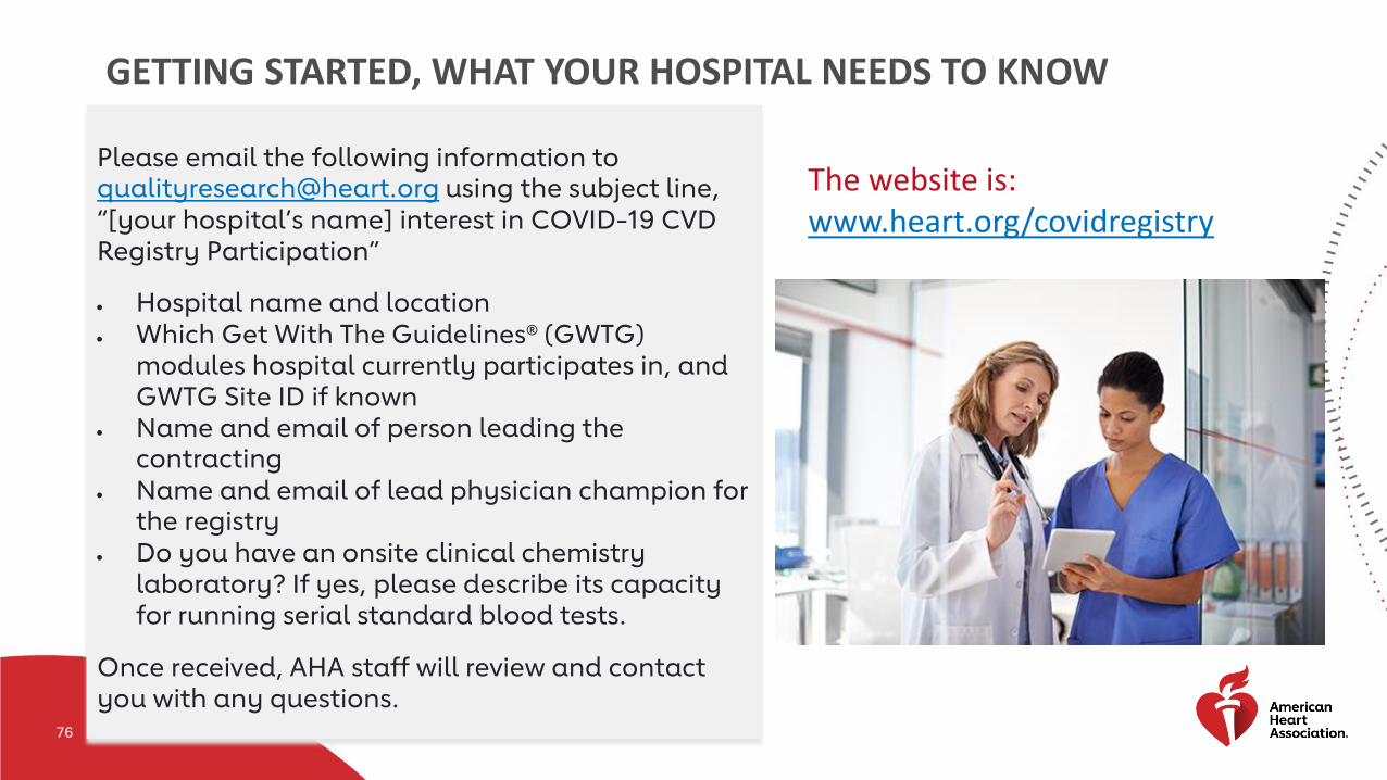
GETTING STARTED, WHAT YOUR HOSPITAL NEEDS TO KNOW
Please email the following information to [email protected] using the subject line,
“[your hospital’s name] interest in COVID-19 CVD Registry Participation”
• Hospital name and location
• Which Get With The Guidelines® (GWTG) modules hospital currently participates in, and GWTG Site ID if known
• Name and email of person leading the contracting
• Name and email of lead physician champion for the registry
• Do you have an onsite clinical chemistry laboratory? If yes, please describe its capacity for running serial standard blood tests.
Once received, AHA staff will review and contact you with any questions.
The website is: www.heart.org/covidregistry
77
78
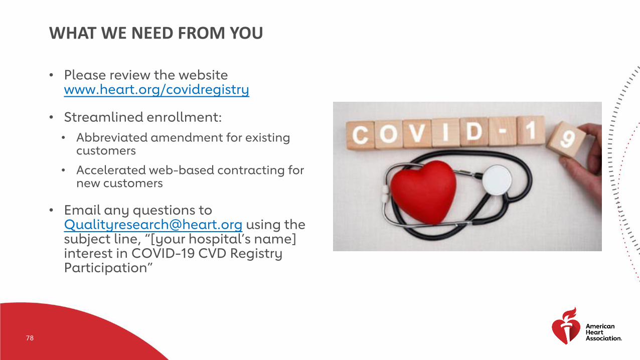
WHAT WE NEED FROM YOU
• Please review the websitewww.heart.org/covidregistry
• Streamlined enrollment:
• Abbreviated amendment for existing customers
• Accelerated web-based contracting for new customers
• Email any questions to [email protected] using the subject line, “[your hospital’s name] interest in COVID-19 CVD Registry Participation”
79
8080
STROKE AND DIABETES
May 27th
11am CSTMissy Moreda MSN APRN ACCNS-AG CCRN CNRN SCRN, will be presenting. Missy has more than 20 years of experience as a neuro ICU nurse and is currently a Diabetes Educator Clinical Nurse Specialist for Duke Raleigh Hospital.

8181
SECTION HEADER
Related Documents