In today’s busy practice it’s not uncommon to have artifact on ECGs. As you know, the electrical activity of the heart is sensed by electrodes that are placed on the skin’s surface. The heart’s electrical signal is very small (normally 0.0001 to 0.003 volt) and unfortunately this can be combined with artifactual signals of similar frequency and often larger amplitude. Each patient’s ECG is unique. The patient’s physical size, skin type, age, and pathology as well as where the electrodes are placed, influence the results of the tracing. Poor signal quality can cause noise or artifact on the ECG, which in turn leads to inaccurate analysis of the ECG. 1 This document will guide you through all the elements that can affect ECG tracings and provide you with tips on how to reduce artifact in your practice. REDUCING ARTIFACT ON ELECTROCARDIOGRAPHS STEPS

Welcome message from author
This document is posted to help you gain knowledge. Please leave a comment to let me know what you think about it! Share it to your friends and learn new things together.
Transcript

In today’s busy practice it’s not uncommon to have artifact on ECGs. As you know, the electrical activity of the heart is sensed by electrodes that are placed on the skin’s surface. The heart’s electrical signal is very small (normally 0.0001 to 0.003 volt) and unfortunately this can be combined with artifactual signals of similar frequency and often larger amplitude.
Each patient’s ECG is unique. The patient’s physical size, skin type, age, and pathology as well as where the electrodes are placed, influence the results of the tracing. Poor signal quality can cause noise or artifact on the ECG, which in turn leads to inaccurate analysis of the ECG.1
This document will guide you through all the elements that can affect ECG tracings and provide you with tips on how to reduce artifact in your practice.
REDUCING ARTIFACT ON E L E C T R O C A R D I O G R A P H SSTEPS
1 Perform good skin preparation The build up of skin oils and residues increase the resistance to the conduction of the electrical signal, therefore, it is crucial to prepare the skin properly. Ideal skin preparation should follow the steps below:
• Shave or clip hair if present. This allows for better electrode contact and reduces painful removal of patches. Shaving may increase skin irritation under the electrode.
• Rub vigorously with a gauze pad. Five to ten gentle strokes can effectively decrease the electrical resistance with minimal reddening of the skin. This will ensure that the heart’s electrical signals can travel to the electrodes.
• Rub the skin with either isopropyl alcohol or soap and water to remove skin oils. Soap and water can minimize skin irritation. Let skin dry thoroughly. This will allow for better electrode-to-skin contact as the adhesive base on the electrode will then grip the skin. Let dry thoroughly.
2 Check the electrodes Check the electrode gel to make sure it is fresh and moist. The electrode gel greatly affects the transmission of signals from the skin to the electrode. A dry electrode with inadequate conducting gel will not work properly since it reduces the conduction of the ECG signal. In most instances, electrode gel dry-out is simply a result of incorrect storage. Electrodes are packaged in metal foil pouches to prevent gel evaporation, therefore it is important to remember the following:
• Use electrodes prior to their expiration date
• Do not remove the electrodes from their pouch until they are ready to be used.
• Do not place electrodes in open bins or drawers. If a pouch is already open, place it in a zip lock bag to preserve moisture and prevent evaporation.
• Avoid warm storage areas.
Do not use different brands of electrodes during a test. Resistance varies from brand to brand, and the signal is best when an equal signal comes from all of the electrodes. In fact, using electrodes of dissimilar composition can actually prevent an ECG trace. Pre-gelled silver/silver chloride electrodes are recommended.
3 Make sure the patient is warm, relaxed and does not move or speak • It is important to make sure the patient is warm and relaxed during
the test because we want to reduce muscle tremors and movement as much as possible since they can impair the quality of recordings. When a patient is cold, he/she may tense or shiver which can cause muscle artifact on the tracings.
• The patient’s arms should lay flat along the side of the body. If the patient does not fit comfortably on the exam table, due to size, have them cross their arms on their stomach to reduce muscle tension. Large patients might tense their arms if they do not fit on the table.
• Patients should not read or talk when an ECG test is being performed.
• Make sure the patient is not sweating nor has moist skin because it causes poor electrode contact with the skin.
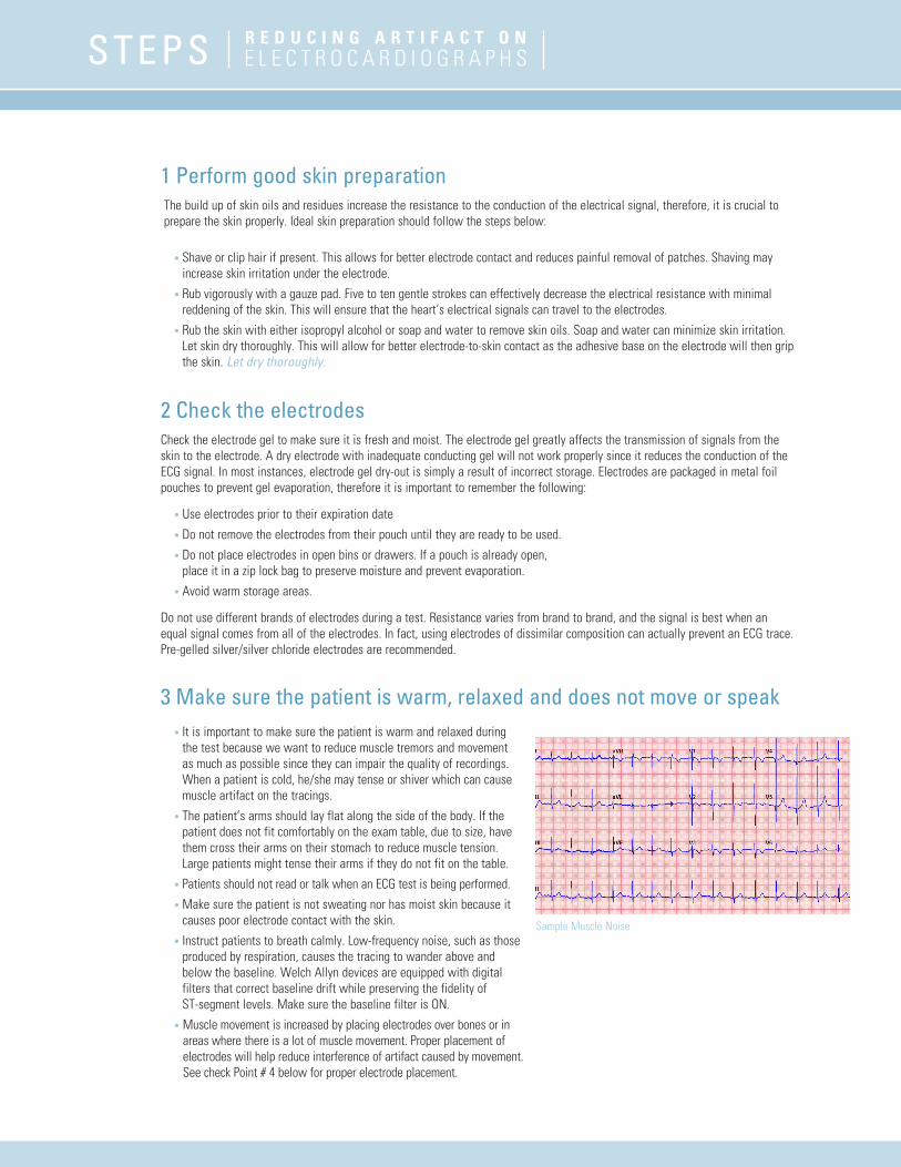
• Instruct patients to breath calmly. Low-frequency noise, such as those produced by respiration, causes the tracing to wander above and below the baseline. Welch Allyn devices are equipped with digital filters that correct baseline drift while preserving the fidelity of ST-segment levels. Make sure the baseline filter is ON.
• Muscle movement is increased by placing electrodes over bones or in areas where there is a lot of muscle movement. Proper placement of electrodes will help reduce interference of artifact caused by movement. See check Point # 4 below for proper electrode placement.
Sample Muscle Noise
R E D U C I N G A R T I F A C T o N E L E C T R O C A R D I O G R A P H SSTEPS
4 Apply electrodes When you are recording ECGs, you should ensure that chest leads are placed in the proper position and the electrodes have good skin contact to minimize artifacts. Incorrect placement of precordial leads may lead to a false diagnosis of infarction. The reversal of limb leads and the switching of precordial leads have been well-documented as causes of alterations in ECGs.2
Do NoT place electrodes over:
• Bones • Irritated skin • Areas where there is a lot of muscle movement • Incisions
If you are using snap electrodes, attach the electrodes to the snap connectors first to increase patient comfort. Do not squeeze the gel out by pressing in the center of the electrode.
Do not use different brands of electrodes during a test. Resistance varies from brand to brand, and the signal is best when an equal signal comes from all of the electrodes. In fact, using electrodes of dissimilar metals can actually prevent an ECG trace. Pre-gelled silver/silver chloride electrodes are recommended.
5 Check alligator clipsAlligator clip connections should be cleaned prior to use. Gel build up on the electrode alligator clips can occur over time and could affect conductivity. Alligator clips should rest flat on the patient’s skin. For good conduction, make sure the metallic side is in contact with the skin.
6 Check lead wires • Periodically inspect cables and lead wires for breaks and cracks and replace as needed. Breaks in the wires and connections
between the electrode and the ECG device will always be a source of problems. Poor contact at any snap connection, loose pins at the cable end of the lead wire, and breaks in the conductors of the lead wire or patient cable can cause intermittent loss of the ECG tracing, 50/60 Hz. pickup, or trace instability.3
• Lead wires should not be pulling on the electrodes as it will result in poor contact with the skin. Untangle wires or reposition electrodes if necessary.
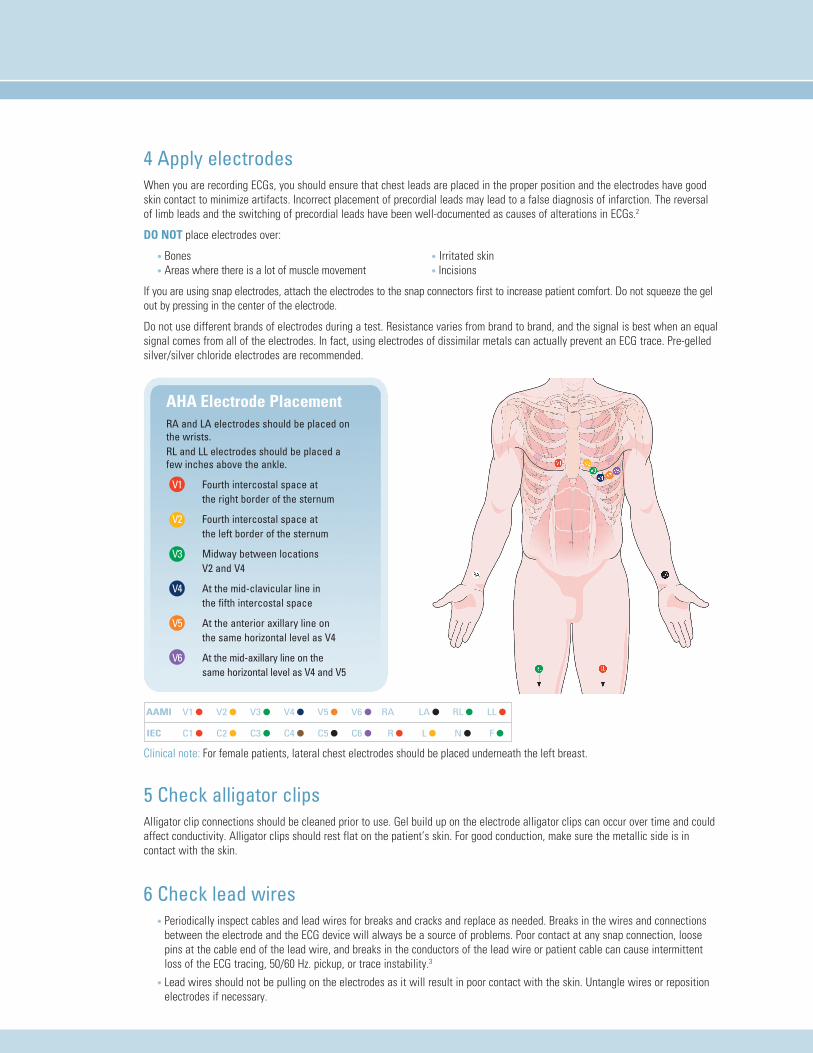
AHA Electrode PlacementRA and LA electrodes should be placed on the wrists.RL and LL electrodes should be placed a few inches above the ankle.
V1 Fourth intercostal space at the right border of the sternum
V2 Fourth intercostal space at the left border of the sternum
V3 Midway between locations V2 and V4
V4 At the mid-clavicular line in the fifth intercostal space
V5 At the anterior axillary line on the same horizontal level as V4
V6 At the mid-axillary line on the same horizontal level as V4 and V5
Clinical note: For female patients, lateral chest electrodes should be placed underneath the left breast.
AAMI
IEC
V1 l
C1 l
V2 l
C2 l
V3 l
C3 l
V4 l
C4 l
V5 l
C5 l
V6 l
C6 l
RA
R l
LA l
L l
RL l
N l
LL l
F l

4341 State Street Road, PO Box 220, Skaneateles Falls, NY 13153-0220 USA(p) 800.535.6663 (f) 315.685.2174 www.welchallyn.com
© 2008 Welch Allyn MC5539 SM2401 Rev A
7 Check the Patient cable connection to the ECG deviceVerify that the patient cable is securely connected to the ECG device and that no gaps exist between connectors.
8 Check for AC interferences 50/60 Hz. interference looks like small regular peaks and produces a wide, fuzzy baseline. It is traced to the 50/60 Hz. current which supplies power to the electrical wall outlets. The 50/60Hz. energy “radiates” from the electrical wiring in the patient’s room and can be picked up by the patient and/or the lead wires. This will result in AC interference on the ECG tracing. There are several causes of AC interference:
• Electrical wires in the walls, ceiling or floor. Suggestion: Move the examination table away from walls.
• Presence of other electrical equipment in the room. Suggestion 1: Unplug any equipment which could interfere with the ECG signal:
– Electrical beds and/or examination table
– Surgical head lights and chokes in fluorescent lamps
– Autoclaves
– Chargers for PC, Otoscopes etc.
Suggestion 2: Move the ECG device, patient cable and patient away from equipment emitting electrical magnetic emission.
• Improper grounding of the ECG device. Suggestion 1: Verify that the outlet the ECG device is plugged into is properly grounded.
Suggestion 2: Operate the ECG device on battery.
9 Verify filter settings in the deviceFinally, if the tracings are still showing artifact, make sure that the following filters are turned on:
• Muscle filter
• AC filter
• Wandering baseline filter
Note for CardioPerfect users: Filters can be applied during the recording but might not appear on the report based on your settings. We recommend turning filters OFF prior to recording to make sure you have eliminated the artifact as much as possible by performing good skin preparation and electrode positioning. Once the signal has stabilized you can turn the filter back ON in the recording window.
References:1. Knight BP, Pelosi F, Michaud GF, Strickberger SA, Morady F. Clinical consequences of electrocardiographic artifact mimicking ventricular tachycardia. N Engl J Med 1999;341:1270–1274.
2. Hurst JW. Images in cardiovascular medicine: “switched” precordial leads. Circulation 2000;101:2870–2871.
3. Michael Smith M.S., B.S.E.E. Rx FOR ECG MONITORING ARTIFACT. Critical Care Nurse 1984
4. The Society for Cardiological Science and Technology, Clinical Guidelines by Consensus, Number 1, Recording a standard 12-lead electrocardiogram, April 2005, Review Date: April 2006
Sample AC Interference
R E D U C I N G A R T I F A C T o N E L E C T R O C A R D I O G R A P H SSTEPS
Related Documents