1 Korean J Physiol Pharmacol Vol 16: 1-9, February, 2012 http://dx.doi.org/10.4196/kjpp.2012.16.1.1 ABBREVIATIONS: ESCs, embryonic stem cells; NSCs, neural stem cells; iPSCs, induced pluripotent stem cells; HDAC, histone deacety- lase; BDNF, brain-derived neurotrophic factor; SVZ, subventricular zone; HC, hippocampus; SAR, structure-activity relationship; SSRIs, selective serotonin reuptake inhibitors; DG, dentate gyrus; HRP, horse radish peroxidise; SMA, spinal muscular atrophy. Received December 8, 2011, Revised January 20, 2012, Accepted January 25, 2012 Corresponding to: Hyun-Jung Kim, Laboratory of Molecular and Stem Cell Pharmacology, College of Pharmacy, Chung-Ang Univer- sity, Seoul 156-756, Korea. (Tel) 82-2-820-5619, (Fax) 82-2-815-5619, (E-mail) [email protected] This is an Open Access article distributed under the terms of the Creative Commons Attribution Non-Commercial License (http:// creativecommons.org/licenses/by-nc/3.0) which permits unrestricted non-commercial use, distribution, and reproduction in any medium, provided the original work is properly cited. Stem Cells in Drug Screening for Neurodegenerative Disease Hyun-Jung Kim, and Chang Yun Jin Laboratory of Stem Cell and Molecular Pharmacology, College of Pharmacy, Chung-Ang University, Seoul 156-756, Korea Because the average human life span has recently increased, the number of patients who are diagnosed with neurodegenerative diseases has escalated. Recent advances in stem cell research have given us access to unlimited numbers of multi-potent or pluripotent cells for screening for new drugs for neurodegenerative diseases. Neural stem cells (NSCs) are a good model with which to screen effective drugs that increase neurogenesis. Recent technologies for human embryonic stem cells (ESCs) or induced pluripotent stem cells (iPSCs) can provide human cells that harbour specific neurode- generative disease. This article discusses the use of NSCs, ESCs and iPSCs for neurodegenerative drug screening and toxicity evaluation. In addition, we introduce drugs or natural products that are recently identified to affect the stem cell fate to generate neurons or glia. Key Words: Stem cells, Neurodegeneration, Drug screening, IPS, ES cells Stem cells have been considered as a good source for po- tential treatment of neurodegenerative diseases (reviewed in [1-7]). Stem cells have the ability to proliferate and dif- ferentiate into various cell types (reviewed in [8-21]). Human embryonic stem cells (ESCs) can be derived from the inner cell mass of blastocysts and be differentiated into all types of cells composing the human body [22-26]. However, using ESCs create ethical problems of destroying embryos and problems after transplantation such that cells derived from ESCs may be rejected from the recipient pa- tients and immunosuppressants are required to be ad- ministered after transplantation [27-30]. Stem cells derived from further-developed embryos have relatively limited ability to differentiate and proliferate [31-38]. For example, neural stem cells (NSCs) can only be differentiated into cells comprising the nervous system such as neurons and glia [38-44]. Recent breakthroughs in the generation of in- duced pluripotent stem cells (iPSCs) showed that cells from patients can be converted into ESCs like pluripotent cells and if used in patients in the future, immunosuppressants may not be needed after transplantation [45-49]. Another advantage of iPSCs is that they can be induced to form dif- ferentiated types of cells including neurons and glia and the mechanisms involved in neurodegeneration of humans can be explored [50-53]. In addition, drug screening can be performed in the disease bearing cells that are differenti- ated from human iPSCs [53-57]. Therefore human stem cells including ESCs, adult stem cells and iPSCs provide a strategy in which these cells can be used for new drug screening or evaluating drug efficacies. In addition, poten- tial toxicity can be predicted using human stem cells [58]. In this review, we focus on recent advances that deal with the concept that stem cells provide a good platform for drug screening in neurodegenerative diseases and evaluation of drug toxicities. Degeneration of the nervous system results in diseases including Parkinson’s disease, Alzheimer’s disease, multi- ple sclerosis, Huntington’s disease and so on. Since drugs or therapies that cure neurodegenerative diseases have not been developed yet, there is an enormous need for new drugs and better therapies. Until recently, emphasis has been on the potential use of stem cells in cell replace- ment/transplantation [59-62]. However, using stem cells as a model system to develop new drugs and evaluate toxicity has begun to receive increased attention [63,64]. This re- view first introduces recent findings identifying chemicals and natural products that induce differentiation of stem cells into neurons or glia. We also discuss advances in drug screening and in evaluating toxicities using human stem cells. NSCs for Screening of Chemicals or Natural Products that Induce Neuronal or Glial Differentiation An essential characteristic of NSCs is that, although

Welcome message from author
This document is posted to help you gain knowledge. Please leave a comment to let me know what you think about it! Share it to your friends and learn new things together.
Transcript
1
Korean J Physiol PharmacolVol 16: 1-9, February, 2012http://dx.doi.org/10.4196/kjpp.2012.16.1.1
ABBREVIATIONS: ESCs, embryonic stem cells; NSCs, neural stem cells; iPSCs, induced pluripotent stem cells; HDAC, histone deacety-lase; BDNF, brain-derived neurotrophic factor; SVZ, subventricular zone; HC, hippocampus; SAR, structure-activity relationship; SSRIs, selective serotonin reuptake inhibitors; DG, dentate gyrus; HRP, horse radish peroxidise; SMA, spinal muscular atrophy.
Received December 8, 2011, Revised January 20, 2012, Accepted January 25, 2012
Corresponding to: Hyun-Jung Kim, Laboratory of Molecular and Stem Cell Pharmacology, College of Pharmacy, Chung-Ang Univer-sity, Seoul 156-756, Korea. (Tel) 82-2-820-5619, (Fax) 82-2-815-5619, (E-mail) [email protected]
This is an Open Access article distributed under the terms of the Creative Commons Attribution Non-Commercial License (http://
creativecommons.org/licenses/by-nc/3.0) which permits unrestricted non-commercial use, distribution, and reproduction in any medium, provided the original work is properly cited.
Stem Cells in Drug Screening for Neurodegenerative Disease
Hyun-Jung Kim, and Chang Yun Jin
Laboratory of Stem Cell and Molecular Pharmacology, College of Pharmacy, Chung-Ang University, Seoul 156-756, Korea
Because the average human life span has recently increased, the number of patients who are diagnosed with neurodegenerative diseases has escalated. Recent advances in stem cell research have given us access to unlimited numbers of multi-potent or pluripotent cells for screening for new drugs for neurodegenerative diseases. Neural stem cells (NSCs) are a good model with which to screen effective drugs that increase neurogenesis. Recent technologies for human embryonic stem cells (ESCs) or induced pluripotent stem cells (iPSCs) can provide human cells that harbour specific neurode-generative disease. This article discusses the use of NSCs, ESCs and iPSCs for neurodegenerative drug screening and toxicity evaluation. In addition, we introduce drugs or natural products that are recently identified to affect the stem cell fate to generate neurons or glia.
Key Words: Stem cells, Neurodegeneration, Drug screening, IPS, ES cells
Stem cells have been considered as a good source for po-tential treatment of neurodegenerative diseases (reviewed in [1-7]). Stem cells have the ability to proliferate and dif-ferentiate into various cell types (reviewed in [8-21]). Human embryonic stem cells (ESCs) can be derived from the inner cell mass of blastocysts and be differentiated into all types of cells composing the human body [22-26]. However, using ESCs create ethical problems of destroying embryos and problems after transplantation such that cells derived from ESCs may be rejected from the recipient pa-tients and immunosuppressants are required to be ad-ministered after transplantation [27-30]. Stem cells derived from further-developed embryos have relatively limited ability to differentiate and proliferate [31-38]. For example, neural stem cells (NSCs) can only be differentiated into cells comprising the nervous system such as neurons and glia [38-44]. Recent breakthroughs in the generation of in-duced pluripotent stem cells (iPSCs) showed that cells from patients can be converted into ESCs like pluripotent cells and if used in patients in the future, immunosuppressants may not be needed after transplantation [45-49]. Another advantage of iPSCs is that they can be induced to form dif-ferentiated types of cells including neurons and glia and the mechanisms involved in neurodegeneration of humans can be explored [50-53]. In addition, drug screening can be performed in the disease bearing cells that are differenti-
ated from human iPSCs [53-57]. Therefore human stem cells including ESCs, adult stem cells and iPSCs provide a strategy in which these cells can be used for new drug screening or evaluating drug efficacies. In addition, poten-tial toxicity can be predicted using human stem cells [58]. In this review, we focus on recent advances that deal with the concept that stem cells provide a good platform for drug screening in neurodegenerative diseases and evaluation of drug toxicities. Degeneration of the nervous system results in diseases including Parkinson’s disease, Alzheimer’s disease, multi-ple sclerosis, Huntington’s disease and so on. Since drugs or therapies that cure neurodegenerative diseases have not been developed yet, there is an enormous need for new drugs and better therapies. Until recently, emphasis has been on the potential use of stem cells in cell replace-ment/transplantation [59-62]. However, using stem cells as a model system to develop new drugs and evaluate toxicity has begun to receive increased attention [63,64]. This re-view first introduces recent findings identifying chemicals and natural products that induce differentiation of stem cells into neurons or glia. We also discuss advances in drug screening and in evaluating toxicities using human stem cells.
NSCs for Screening of Chemicals or Natural Products that Induce Neuronal or Glial
Differentiation
An essential characteristic of NSCs is that, although
2 HJ Kim and CY Jin
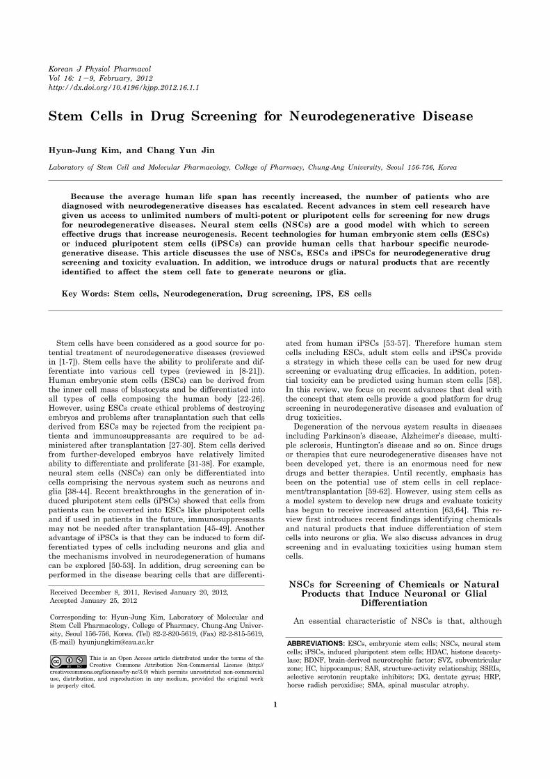
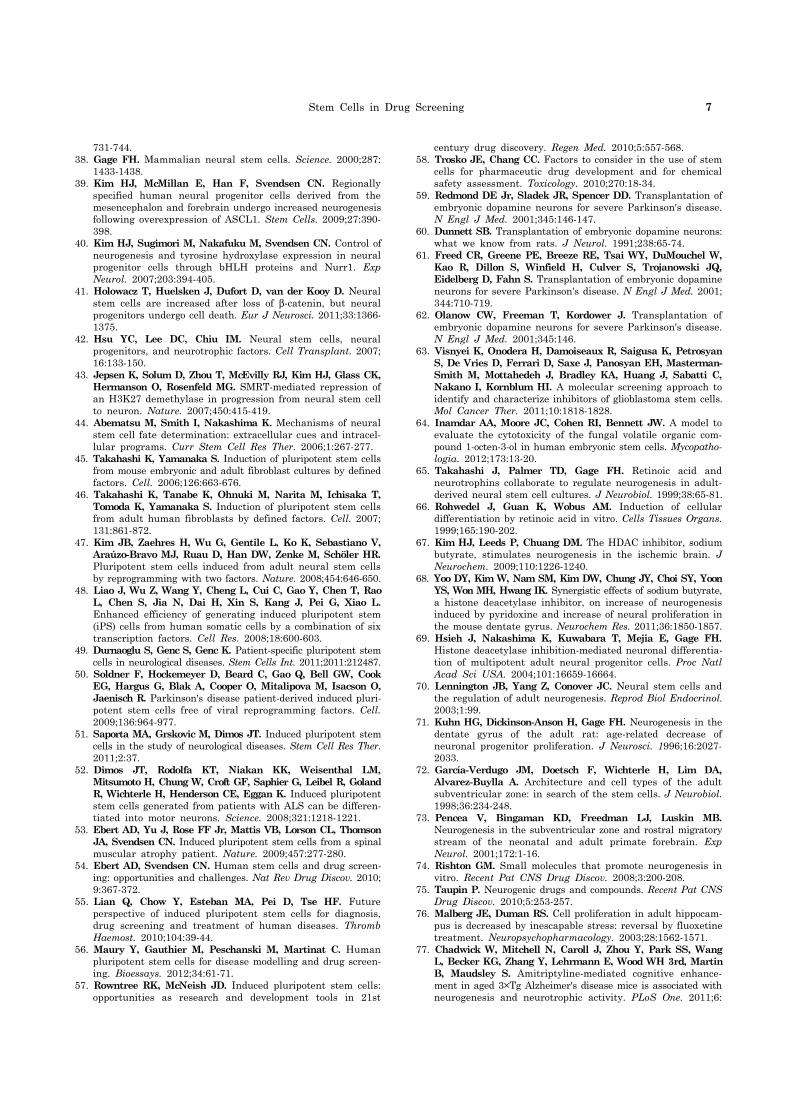
Fig. 1. High-throughput screening for the development of new drugs that are effective for the treatment of neurodegenerative diseases. NSC can be derived from adult cells, fetus cells, ESCs or iPSCs. After NSC plating, chemical or natural product libraries are treated. The effect of each drugs are detected by immuno-cytochemistry using cell type specific antibodies. If fluorescence conjugatedsecondary antibodies are used, cells can be visualized by fluorescence microscopy and the numbers of detec-ted/differentiated cells are counted. If HRP conjugated secondary anti-bodies are used, cells can be treated with substrates and lysed to be mea-sured by microplate reader. Once thechemicals or natural products that are effective are found, and struc-tural activity relationship studies, animal studies and toxicity evalua-tions are done, promising agents can go on to clinical trials and may fur-ther be developed as new drugs.
somewhat restricted, they respond to environmental cues. For example, NSC differentiation into neurons can be in-duced by treatment of retinoic acid [65,66]. Similarly, his-tone deacetylase (HDAC) inhibitors can also cause NSCs to differentiate into astrocytes [67-69]. Retinoic acid treat-ment of NSC induced immediate up-regulation of a proneu-ral gene, NeuroD, and increased p21 expression. Retinoic acid also affected the expression of trkA, trkB, trkC and p75NGFR, causing better responses to neurotrophic factors and neuron maturation [65]. Modulation of ES cell differ-entiation was influenced by retinoic acid [66]. However, it was dependent on the concentration of retinoic acid and the developmental stage of the cells. An HDAC inhibitor, so-dium butyrate, is reported to increase proliferation of NSCs and levels of brain derived neurotrophic factor (BDNF) in the ischemic brain of adult rodents [67]. Increases in new born neurons in ischemic regions of the brain by sodium butyrate appear to be mediated by BDNF, because BDNF receptor antagonists markedly reduced NSCs proliferation and attenuated behavioural benefits. Identification of new cell-fate modulators in NSCs provides several advantages. First, the chemicals or natural products that induce neuro-genesis have the potential to be used in neurodegenerative diseases. In the adult human brain, it is known that NSCs
exist in certain areas such as the subventricular zone (SVZ) and the hippocampus (HC) [38,70-73]. It would be beneficial for patients who suffer from neurodegeneration to take drugs or natural products that increase neurogenesis from endogenous NSCs (Fig. 1). In addition to potential use in the clinic, identification of new cell fate regulators may in-duce a homogeneous population, and provide a good model for drug screening. The underlying mechanisms of differ-entiation would also be useful to help understand stem cell biology and facilitate new drug development. A homoge-neous population produced using chemicals that induce a certain type of cell may also be useful for transplantation in future potential cell replacement therapy. As illustrated in Fig. 1, NSCs can be generated from either the fetus, the adult, ESCs or iPSCs. After treatment of chemical or natural product libraries, the levels of differ-entiation of NSC can be determined by image-based im-munocytochemistry or immunostaining-based microplate reading quantitation methods. The chemicals or natural products that induce high levels of neurogenesis can further be used for studying structure-activity relationships (SAR) to generate more efficient but less toxic molecules as new drug candidates. Several laboratories have recognized the importance of identifying small molecules for controlling
Stem Cells in Drug Screening 3
NSCs fate [74,75]. Some widely used drugs such as anti-depressants and anticonvulsants have been shown to regu-late stem cell proliferation and differentiation [69,76-81]. Interesting clinical effects of selective serotonin reuptake inhibitors (SSRIs) in ameliorating cognition in Alzheimer’s disease have been demonstrated [82]. Alzheimer’s disease patients with depression have been treated with SSRIs in combination with cholinesterase inhibitors (donepezil, rav-astigmine and galantamine) and showed better cognitive function than patients who were treated only with chol-inesterase inhibitors [82]. Fluoxetine also increased neuro-genesis by increasing NSCs proliferation and cell survival [79,80,83]. Administration of fluoxetine for 28 days sig-nificantly improved depression when measured in animals by a novelty-suppressed feeding test and an increase in neurogenesis in the HC was observed [81]. However, the use of fluoxetine to induce neurogenesis was challenged by recent data that chronic exposure to fluoxetine actually de-creased neurogenesis in the adult SVZ [84]. The anti-depressant sertraline increased neuronal differentiation through glucocorticoid receptors and increased both im-mature neuroblasts (double-cortin positive), and mature neurons (Map2 positive) [85]. Administration of tricyclic an-tidepressants such as amitriptyline caused cognitive bene-fits in patients suffering from Alzheimer’s disease [86]. Amitriptyline increased neurotrophic factor levels in pa-tients’ serum. In cognitively impaired, aged, transgenic mice, amitriptyline treatment improved both short and long term memory retention and increased neurogenesis in the dentate gyrus (DG) [77]. It is also reported that a mood stabilizer, lithium, and carbamazepine increased neuro-genesis but decreased astrocytogenesis [87]. Lithium and carbamazepine increased proliferation and decreased apop-tosis of NSCs that are derived from HC [87,88]. When 3 month old double transgenic CRND8 mice (overexpressing the Swedish and Indiana mutations in the human amyloid precursor protein) were treated with lithium for 5 weeks, lithium induced proliferation of cells in the HC and induced neuronal fate specification [89]. However, when lithium was used to treat 7 month old transgenic CRND8 mice, the proliferative effects on NSCs and neurogenic effects of lith-ium were abolished, suggesting that lithium-induced facili-tation of neurogenesis declines with Alzheimer disease progression. The anticonvulsant valproate has effects on NSCs. Intere-stingly, the effects of valproate on neuronal differentiation appear to depend on the origin of the NSCs. Valproate en-hanced neurogenesis in NSCs derived from either entire adult HC or forebrain [90-92]. However, in NSCs from DG of the HC, valproate induces astrocytogenesis while redu-cing neuronal differentiation [87]. A recent article sugges-ted that valproate protected NSCs by reducing NSCs death by upregulating the antiapototic gene Bcl-XL and activat-ing NF-kB signalling pathways [93]. In the early 2000s, VPA was known to function as a HDAC inhibitor [69]. As mentio-ned above, HDAC activity has an important role in enhanc-ing neurogenesis by upregulation of the proneural gene Ne-uroD while inhibiting astrocytogenesis. Activation of ERK signalling has been implicated in VPA-induced neurogene-sis [90,92]. Through the beta-catenin-Ras-ERK-p21Cip/WAF1 pathway, NSC proliferation was inhibited while differentia-tion into neurons was increased [92]. Schultz and colleagues described several synthetic mole-cules (for example KHS101) that induce neuronal differen-tiation of adult hippocampal NSCs by image-based screen-
ing [94]. KHS101 increased neurogenesis while reducing astrocytogenesis. In a search for its target, the authors found that KHS101 specifically interacts with the TACC3 protein and knockdown of TACC3 increased neuronal differentiation. TACC3 regulates progenitor cell expansion and terminal cell differentiation in hematopoietic and neu-ral stem cells and appears to mediate the functioning of KHS101. We also identified oxadiazol compounds as in-ducers of astrocytogenesis by image based screening [95]. In a study of NSCs derived from developing rat (embryonic day 14), we found that oxadiazol derivatives specifically in-creased numbers of astrocytes while not affecting those of neurons. For high throughput screening, Saxe and his colleagues used a chemiluminescence-based method on primary neuro-spheres and identified phosphoserine as an enhancer of neurogenesis [96]. Since cell counting after immunostaining requires skills and time, the authors used horse radish per-oxidise (HRP)-conjugated secondary antibody and chem-iluminescence detection was performed by microplate read-er after HRP substrate treatment. Phosphoserine inhibited NSC proliferation, enhanced neurogenesis, and increased cell survival. It was suggested that the metabotropic gluta-mate receptor 4 mediated such effects. In addition to in vi-tro assays, in vivo screening was done in search of chem-icals that enhance neurogenesis in the HC of adult mice [97]. The authors identified 8 chemicals out of 1000 tested that induce neurogenesis. An aminopropyl carbazole named P7C3 showed proneurogenic activity by protecting newborn neurons from apoptosis and by enhancing neurogenesis in the DG. Considering that research has not been done for very long to develop drugs that modulate NSC fate or stem cell fate, it is amazing to find quite a lot of synthetic chemicals that have effects on neurogenesis (Table 1). This may be due to the ability of NSCs to respond to the environment and to differentiate into multiple cell types. Besides the chem-icals mentioned above, recent patent applications report drugs that modulate melanocortin receptors, PPAR-γ, an-giotensin, and 5-HT, and HMG coenzyme A reductase in-hibitors were also neurogenic {reviewed in [74] and patents and references therein}. For example, in rats with trau-matic brain injury, when atorvastatin and simvastatin were given for 14 days, these statins improved spatial learning measured by Morris water maze tests [98,99]. Interestingly, newly generated neurons and vessels were detected in statin-treated brain-injured rats [99]. With fur-ther screening, we should find efficient chemicals that en-hance neuronal differentiation from NSCs. It will be benefi-cial to develop chemicals that have both neurogenic activity and neuron protecting effects for treatment of neuro-degenerative diseases. In addition to synthetic chemicals, recent results show that some natural products also affect cell fate determi-nation of NSCs (Table 2). Until recently, neuroprotective effects of natural products have been intensely studied [100-102]. Methanol extracts of Jeju Native plants pro-tected apoptosis induced by hydrogen peroxides [100]. Visnagin, an active component extracted from the fruits of Ammi visnaga, which has been used as a treatment for low blood-pressure, showed protective effects on kainic acid in-duced mouse hippocampal cell death by reducing in-flammation [101]. BF-7 extracted from a sericultural prod-uct has significant protective effects on amyloid β peptide induced apoptosis through reduction of ROS generation and

4 HJ Kim and CY Jin
Table 1. Synthetic compounds that are known to regulate stem cell fate
Name Structural formula Effects Cells/system Refs
Retinoic acid -Increase neurogenesis NSCs 65, 66
Sodium butyrate -Increase neurogenesis -Increase neural proliferation
In vivo 67, 68
Amitriptyline -Increase neurotrophic factor levels in DG NSCs 77
Fluoxetine -Increase neurogenesis In vivo NSCs
79-81, 83
Sertraline -Increase neurogenesis -Attenuate cellular damage
NSCs 85
Carbamazepine -Increase neurogenesis -Decrease astrocytogenesis
NSCs 87
Valproate -Increase neurogenesis -Reduce NSCs death -Neuroprotection
NSCs 90-93
KHS101 -Increase neurogenesis NSCs 94
Oxadiazol compounds -Enhance astrocyte differentiation NSCs 95
Phosphoserine -Inhibit NSCs proliferation -Enhance neurogenesis -Increase cell survival
hESCs NSCs
96
P7C3 -Protect newborn neurons from apoptosis -Enhance neurogenesis
NSCs 97
Atorvastatin -Increase neurogenesis -Reduce neuronal death
In vivo 98, 99
diminished caspase activity [102]. Glycyrrhizae radix is re-ported to cause improvements in spatial learning, memory and stress-induced anxiety [103]. Garcinol, a polyisopreny-lated benzophenone derivative in Garcinia indica fruit rind, is known to increase the numbers of neurons in EGF re-sponsive neurospheres by increasing survival [104]. The
survival enhancing effects of Garcinol were mediated by ERK activation and ERK activation modulated neurite outgrowth. Ginsenosides that are derived from Panax noto-ginseng were also identified as enhancers of neurogenesis in EGF-responsive NSCs [105]. Interestingly, ginsenosides induced neurogenesis at the expense of astrogliogenesis.

Stem Cells in Drug Screening 5
Table 2. Natural products that are known to affect stem cell survival, proliferation and differentiation
Name Plant origin Effects Cells Refs
Saururus chinesis extract Saururus chinesis -Protective effect on apoptotic cell death SH-SY5Y cells 100Smilax china extract Smilax china -Protective effect on apoptotic cell death SH-SY5Y cells 100Visnagin Ammi visnaga -Protect neuronal cell In vivo 101BF-7 Silkworm -Neuroprotection
-Enhance cognitive functionSKN-SH cells 102
Glycyrrhizae radix Glycyrrhiza Uralensis -Anti-stress effects In vivo 103Garcinol Garcinia indica -Promote proliferation
-Increase neurogenesisNSCs 104
Ginsenoside Rg5 Panax notoginseng -Increase neurogenesis -Decrease astrocytogenesis
NSCs 105
Casticin Croton betulaster -Increase neurogenesis -Decrease neuronal cell death
NSCs 106
Curcumin Indian spice turmeric -Increase neurogenesis -Decrease neuronal cell death and glial cell activation
NSCs 107, 108
Nelumbo nucifera rhizome extract
Nelumbo nucifera -Increase neurogenesis In vivo 109, 110
The neurogenic effect of the ginsenosides was abolished completely by treatment with the Ca2+ channel antagonist nifedipine. A flavonoid, casticin, extracted from Croton be-tulaster also increased neuronal differentiation and decrea-sed neuronal cell death [106]. Casticin increased neuronal transcription factor Tbr2 and did not affect gliogenesis when detected by immunocytochemistry with GFAP, S100β, Olig2 and NG2. NSCs cultured on top of astrocytes that were treated with casticin induced neurogenesis and con-ditioned media from casticin-treated astrocytes reproduced such effects. Curcumin, a natural phenolic component of yellow curry spice attenuates astroglial and microglial acti-vation in kainic acid induced seizure [107]. In NSCs, curcu-min has proliferation-promoting effects [108]. It was re-ported that administration of curcumin to adult mice in-creased HC neurogenesis. Methanol extracts of Nelumbo nucifera, a rhizome, increased NSC proliferation and in-creased neurogenesis in vivo [109,110].
Human Stem Cells for Drug Screening
Recent advances in screening technologies have enabled scientists to identify effective small molecules that induce neurogenesis. However, many studies were done using ro-dent NSCs as mentioned above or with highly proliferative immortalized or cancerous cell lines that do not accurately reflect the human pathophysiological condition. It is thus desirable to test or screen drugs with human cells to ob-serve the effects and mechanisms of drugs. However, until very recently, it was almost impossible to obtain enough human tissues or cells that represent human neurodegene-rative conditions. A recent breakthrough made in the stem cell research field is the generation of iPSCs from human fibroblasts or other somatic cells [45-48]. Using numerous combinations of stemness genes, Takahashi and his col-leagues found that Oct4, Sox2, Klf4 and c-myc could repro-gram mice fibroblasts into ES like cells [45]. Human so-matic cells could also be converted into ES like cells by in-troduction of a few stemness genes [46,111,112]. Further-more iPSCs were generated from fibroblasts taken from pa-tients suffering from neurodegenerative diseases [52,53,113].
Thus the disease mechanism can be studied in these cells and drug screening for specific diseases can be done. Recent advances in gene editing such as zinc finger nuclease medi-ated and helper-dependent adenoviral vector approaches were able to cause insertion or deletion of specific target genes and cause iPSCs to produce isogenic lines [114,115]. Thus disease bearing iPSCs and appropriate control cells could be used for the study of pathological mechanisms of diseases and drug effects can also be more accurately tested in these cells. Since neurodegeneration occurs late in adulthood, it is likely that iPSCs generated from patients would not repre-sent true pathological conditions. Svendsen and his col-leagues reported that iPSCs can be generated from spinal muscular atrophy (SMA) patients [53]. Although early pro-duced motor neuron numbers were not affected, long term culture showed degeneration of motor neurons that had dif-ferentiated from iPSCs generated from an SMA patient. Interestingly, when iPSCs were generated from a patient with Parkinson’s disease, there was not much loss of dop-amine neurons [50]. The cells probably needed more time to develop Parkinson’s disease that they are harbouring. Although much more research is needed to develop a sys-tem for drug screening, human iPSCs that are generated from patients with specific disease are a good model with which to test and screen drugs. In addition to screening drugs that are effective in treat-ment, it seems apparent that human stem cells are an ex-cellent model to evaluate drug toxicity. Before moving on to phase I clinical trials, it would be safer to test toxicity on human ESCs derived cardiomyocytes or other sources to predict adverse effects (Maybe this step could be called clinical trial phase 0.5). Since cellular contents of human cells are different from those of rodent or other animal cells, toxicities that are not identified in animal models could be detected in human stem cell derived differentiated cells.
CONCLUSION
Screening for drugs that modulate stem cell self-renewal and differentiation, or protect cell death, can be performed

6 HJ Kim and CY Jin
to develop new drugs to treat human neurodegenerative disease. Stem cells provide a good platform with which to perform drug screening and evaluation of toxicity. In this review, we have introduced drugs and natural products that modulate stem cell fate to neurons or glia. With the ability of stem cells to respond to the environment, we ex-pect to see, in the near future, more progress in identifying new drugs that regulate stem cell proliferation and differ-entiation and are used in neurodegenerative diseases.
ACKNOWLEDGEMENTS
This research was supported by the Chung-Ang University Research Scholarship Grants in 2010-2012.
REFERENCES
1. Kim HJ. Stem cell potential in Parkinson's disease and molecular factors for the generation of dopamine neurons. Biochim Biophys Acta. 2011;1812:1-11.
2. Jones JM, Thomson JA. Human embryonic stem cell technology. Semin Reprod Med. 2000;18:219-223.
3. Studer L, Tabar V, McKay RD. Transplantation of expanded mesencephalic precursors leads to recovery in parkinsonian rats. Nat Neurosci. 1998;1:290-295.
4. Sánchez-Pernaute R, Studer L, Bankiewicz KS, Major EO, McKay RD. In vitro generation and transplantation of precur-sor-derived human dopamine neurons. J Neurosci Res. 2001; 65:284-288.
5. Levy YS, Stroomza M, Melamed E, Offen D. Embryonic and adult stem cells as a source for cell therapy in Parkinson's disease. J Mol Neurosci. 2004;24:353-386.
6. Chen LW, Kuang F, Wei LC, Ding YX, Yung KK, Chan YS. Potential application of induced pluripotent stem cells in cell replacement therapy for Parkinson's disease. CNS Neurol Disord Drug Targets. 2011;10:449-458.
7. Lindvall O, Kokaia Z. Stem cell therapy for human brain disorders. Kidney Int. 2005;68:1937-1939.
8. Germain N, Banda E, Grabel L. Embryonic stem cell neuro-genesis and neural specification. J Cell Biochem. 2010;111: 535-542.
9. Fraichard A, Chassande O, Bilbaut G, Dehay C, Savatier P, Samarut J. In vitro differentiation of embryonic stem cells into glial cells and functional neurons. J Cell Sci. 1995;108:3181- 3188.
10. Kehat I, Kenyagin-Karsenti D, Snir M, Segev H, Amit M, Gepstein A, Livne E, Binah O, Itskovitz-Eldor J, Gepstein L. Human embryonic stem cells can differentiate into myocytes with structural and functional properties of cardiomyocytes. J Clin Invest. 2001;108:407-414.
11. Xu C, Police S, Rao N, Carpenter MK. Characterization and enrichment of cardiomyocytes derived from human embryonic stem cells. Circ Res. 2002;91:501-508.
12. Mummery C, Ward-van Oostwaard D, Doevendans P, Spijker R, van den Brink S, Hassink R, van der Heyden M, Opthof T, Pera M, de la Riviere AB, Passier R, Tertoolen L. Differentiation of human embryonic stem cells to cardio-myocytes: role of coculture with visceral endoderm-like cells. Circulation. 2003;107:2733-2740.
13. Noguchi H. Production of pancreatic beta-cells from stem cells. Curr Diabetes Rev. 2010;6:184-190.
14. Shi Y. Generation of functional insulin-producing cells from human embryonic stem cells in vitro. Methods Mol Biol. 2010;636:79-85.
15. Chen C, Zhang Y, Sheng X, Huang C, Zang YQ. Differentiation of embryonic stem cells towards pancreatic progenitor cells and their transplantation into streptozotocin-induced diabetic mice. Cell Biol Int. 2008;32:456-461.
16. Yamamoto H, Quinn G, Asari A, Yamanokuchi H, Teratani T, Terada M, Ochiya T. Differentiation of embryonic stem cells into hepatocytes: biological functions and therapeutic appli-cation. Hepatology. 2003;37:983-993.
17. Teratani T, Yamamoto H, Aoyagi K, Sasaki H, Asari A, Quinn G, Sasaki H, Terada M, Ochiya T. Direct hepatic fate specifi-cation from mouse embryonic stem cells. Hepatology. 2005;41: 836-846.
18. Van Haute L, De Block G, Liebaers I, Sermon K, De Rycke M. Generation of lung epithelial-like tissue from human embryonic stem cells. Respir Res. 2009;10:105.
19. Wang D, Haviland DL, Burns AR, Zsigmond E, Wetsel RA. A pure population of lung alveolar epithelial type II cells derived from human embryonic stem cells. Proc Natl Acad Sci USA. 2007;104:4449-4454.
20. Yamamoto M, Tachibana T, Hashimoto H, Ishiwata I, Ishikawa H. The differentiation of early embryonic stem cells into adipocytes-like cells. Hum Cell. 2003;16:117-122.
21. Kim HJ, Rosenfeld MG. Epigenetic control of stem cell fate to neurons and glia. Arch Pharm Res. 2010;33:1467-1473.
22. Thomson JA, Itskovitz-Eldor J, Shapiro SS, Waknitz MA, Swiergiel JJ, Marshall VS, Jones JM. Embryonic stem cell lines derived from human blastocysts. Science. 1998;282:1145- 1147.
23. Suemori H. Establishment and therapeutic use of human embryonic stem cell lines. Hum Cell. 2006;19:65-70.
24. Gepstein L. Derivation and potential applications of human embryonic stem cells. Circ Res. 2002;91:866-876.
25. Reubinoff BE, Pera MF, Fong CY, Trounson A, Bongso A. Embryonic stem cell lines from human blastocysts: somatic differentiation in vitro. Nat Biotechnol. 2000;18:399-404.
26. Pera MF, Reubinoff B, Trounson A. Human embryonic stem cells. J Cell Sci. 2000;113:5-10.
27. Lim JM, Lee M, Lee EJ, Gong SP, Lee ST. Stem cell engi-neering: limitation, alternatives, and insight. Ann NY Acad Sci. 2011;1229:89-98.
28. Hyun I. The bioethics of stem cell research and therapy. J Clin Invest. 2010;120:71-75.
29. Tasso R, Pennesi G. When stem cells meet immunoregulation. Int Immunopharmacol. 2009;9:596-598.
30. Cabrera CM, Cobo F, Nieto A, Concha A. Strategies for preventing immunologic rejection of transplanted human embryonic stem cells. Cytotherapy. 2006;8:517-518.
31. Usas A, Mačiulaitis J, Mačiulaitis R, Jakubonienė N, Milašius A, Huard J. Skeletal muscle-derived stem cells: Implications for cell-mediated therapies. Medicina (Kaunas). 2011;47:469- 479.
32. Frati C, Savi M, Graiani G, Lagrasta C, Cavalli S, Prezioso L, Rossetti P, Mangiaracina C, Ferraro F, Madeddu D, Musso E, Stilli D, Rossini A, Falco A, Angelis AD, Rossi F, Urbanek K, Leri A, Kajstura J, Anversa P, Quaini E, Quaini F. Resident cardiac stem cells. Curr Pharm Des. 2011;17:3252-3257.
33. Fuh E, Brinton TJ. Bone marrow stem cells for the treatment of ischemic heart disease: a clinical trial review. J Cardiovasc Transl Res. 2009;2:202-218.
34. Chugh AR, Zuba-Surma EK, Dawn B. Bone marrow-derived mesenchymal stems cells and cardiac repair. Minerva Cardio-angiol. 2009;57:185-202.
35. Woo DH, Kim SK, Lim HJ, Heo J, Park HS, Kang GY, Kim SE, You HJ, Hoeppner DJ, Kim Y, Kwon H, Choi TH, Lee JH, Hong SH, Song KW, Ahn EK, Chenoweth JG, Tesar PJ, McKay RD, Kim JH. Direct and indirect contribution of human embryonic stem cell-derived hepatocyte-like cells to liver repair in mice. Gastroenterology. http://dx.doi.org/10.1053/j.gastro.2011. 11.030.
36. Turner R, Lozoya O, Wang Y, Cardinale V, Gaudio E, Alpini G, Mendel G, Wauthier E, Barbier C, Alvaro D, Reid LM. Human hepatic stem cell and maturational liver lineage biology. Hepatology. 2011;53:1035-1045.
37. Grisar JC, Haddad F, Gomari FA, Wu JC. Endothelial proge-nitor cells in cardiovascular disease and chronic inflammation: from biomarker to therapeutic agent. Biomark Med. 2011;5:
Stem Cells in Drug Screening 7
731-744.38. Gage FH. Mammalian neural stem cells. Science. 2000;287:
1433-1438.39. Kim HJ, McMillan E, Han F, Svendsen CN. Regionally
specified human neural progenitor cells derived from the mesencephalon and forebrain undergo increased neurogenesis following overexpression of ASCL1. Stem Cells. 2009;27:390- 398.
40. Kim HJ, Sugimori M, Nakafuku M, Svendsen CN. Control of neurogenesis and tyrosine hydroxylase expression in neural progenitor cells through bHLH proteins and Nurr1. Exp Neurol. 2007;203:394-405.
41. Holowacz T, Huelsken J, Dufort D, van der Kooy D. Neural stem cells are increased after loss of β-catenin, but neural progenitors undergo cell death. Eur J Neurosci. 2011;33:1366- 1375.
42. Hsu YC, Lee DC, Chiu IM. Neural stem cells, neural progenitors, and neurotrophic factors. Cell Transplant. 2007; 16:133-150.
43. Jepsen K, Solum D, Zhou T, McEvilly RJ, Kim HJ, Glass CK, Hermanson O, Rosenfeld MG. SMRT-mediated repression of an H3K27 demethylase in progression from neural stem cell to neuron. Nature. 2007;450:415-419.
44. Abematsu M, Smith I, Nakashima K. Mechanisms of neural stem cell fate determination: extracellular cues and intracel-lular programs. Curr Stem Cell Res Ther. 2006;1:267-277.
45. Takahashi K, Yamanaka S. Induction of pluripotent stem cells from mouse embryonic and adult fibroblast cultures by defined factors. Cell. 2006;126:663-676.
46. Takahashi K, Tanabe K, Ohnuki M, Narita M, Ichisaka T, Tomoda K, Yamanaka S. Induction of pluripotent stem cells from adult human fibroblasts by defined factors. Cell. 2007; 131:861-872.
47. Kim JB, Zaehres H, Wu G, Gentile L, Ko K, Sebastiano V, Araúzo-Bravo MJ, Ruau D, Han DW, Zenke M, Schöler HR. Pluripotent stem cells induced from adult neural stem cells by reprogramming with two factors. Nature. 2008;454:646-650.
48. Liao J, Wu Z, Wang Y, Cheng L, Cui C, Gao Y, Chen T, Rao L, Chen S, Jia N, Dai H, Xin S, Kang J, Pei G, Xiao L. Enhanced efficiency of generating induced pluripotent stem (iPS) cells from human somatic cells by a combination of six transcription factors. Cell Res. 2008;18:600-603.
49. Durnaoglu S, Genc S, Genc K. Patient-specific pluripotent stem cells in neurological diseases. Stem Cells Int. 2011;2011:212487.
50. Soldner F, Hockemeyer D, Beard C, Gao Q, Bell GW, Cook EG, Hargus G, Blak A, Cooper O, Mitalipova M, Isacson O, Jaenisch R. Parkinson's disease patient-derived induced pluri-potent stem cells free of viral reprogramming factors. Cell. 2009;136:964-977.
51. Saporta MA, Grskovic M, Dimos JT. Induced pluripotent stem cells in the study of neurological diseases. Stem Cell Res Ther. 2011;2:37.
52. Dimos JT, Rodolfa KT, Niakan KK, Weisenthal LM, Mitsumoto H, Chung W, Croft GF, Saphier G, Leibel R, Goland R, Wichterle H, Henderson CE, Eggan K. Induced pluripotent stem cells generated from patients with ALS can be differen-tiated into motor neurons. Science. 2008;321:1218-1221.
53. Ebert AD, Yu J, Rose FF Jr, Mattis VB, Lorson CL, Thomson JA, Svendsen CN. Induced pluripotent stem cells from a spinal muscular atrophy patient. Nature. 2009;457:277-280.
54. Ebert AD, Svendsen CN. Human stem cells and drug screen-ing: opportunities and challenges. Nat Rev Drug Discov. 2010; 9:367-372.
55. Lian Q, Chow Y, Esteban MA, Pei D, Tse HF. Future perspective of induced pluripotent stem cells for diagnosis, drug screening and treatment of human diseases. Thromb Haemost. 2010;104:39-44.
56. Maury Y, Gauthier M, Peschanski M, Martinat C. Human pluripotent stem cells for disease modelling and drug screen-ing. Bioessays. 2012;34:61-71.
57. Rowntree RK, McNeish JD. Induced pluripotent stem cells: opportunities as research and development tools in 21st
century drug discovery. Regen Med. 2010;5:557-568.58. Trosko JE, Chang CC. Factors to consider in the use of stem
cells for pharmaceutic drug development and for chemical safety assessment. Toxicology. 2010;270:18-34.
59. Redmond DE Jr, Sladek JR, Spencer DD. Transplantation of embryonic dopamine neurons for severe Parkinson's disease. N Engl J Med. 2001;345:146-147.
60. Dunnett SB. Transplantation of embryonic dopamine neurons: what we know from rats. J Neurol. 1991;238:65-74.
61. Freed CR, Greene PE, Breeze RE, Tsai WY, DuMouchel W, Kao R, Dillon S, Winfield H, Culver S, Trojanowski JQ, Eidelberg D, Fahn S. Transplantation of embryonic dopamine neurons for severe Parkinson's disease. N Engl J Med. 2001; 344:710-719.
62. Olanow CW, Freeman T, Kordower J. Transplantation of embryonic dopamine neurons for severe Parkinson's disease. N Engl J Med. 2001;345:146.
63. Visnyei K, Onodera H, Damoiseaux R, Saigusa K, Petrosyan S, De Vries D, Ferrari D, Saxe J, Panosyan EH, Masterman- Smith M, Mottahedeh J, Bradley KA, Huang J, Sabatti C, Nakano I, Kornblum HI. A molecular screening approach to identify and characterize inhibitors of glioblastoma stem cells. Mol Cancer Ther. 2011;10:1818-1828.
64. Inamdar AA, Moore JC, Cohen RI, Bennett JW. A model to evaluate the cytotoxicity of the fungal volatile organic com-pound 1-octen-3-ol in human embryonic stem cells. Mycopatho-logia. 2012;173:13-20.
65. Takahashi J, Palmer TD, Gage FH. Retinoic acid and neurotrophins collaborate to regulate neurogenesis in adult- derived neural stem cell cultures. J Neurobiol. 1999;38:65-81.
66. Rohwedel J, Guan K, Wobus AM. Induction of cellular differentiation by retinoic acid in vitro. Cells Tissues Organs. 1999;165:190-202.
67. Kim HJ, Leeds P, Chuang DM. The HDAC inhibitor, sodium butyrate, stimulates neurogenesis in the ischemic brain. J Neurochem. 2009;110:1226-1240.
68. Yoo DY, Kim W, Nam SM, Kim DW, Chung JY, Choi SY, Yoon YS, Won MH, Hwang IK. Synergistic effects of sodium butyrate, a histone deacetylase inhibitor, on increase of neurogenesis induced by pyridoxine and increase of neural proliferation in the mouse dentate gyrus. Neurochem Res. 2011;36:1850-1857.
69. Hsieh J, Nakashima K, Kuwabara T, Mejia E, Gage FH. Histone deacetylase inhibition-mediated neuronal differentia-tion of multipotent adult neural progenitor cells. Proc Natl Acad Sci USA. 2004;101:16659-16664.
70. Lennington JB, Yang Z, Conover JC. Neural stem cells and the regulation of adult neurogenesis. Reprod Biol Endocrinol. 2003;1:99.
71. Kuhn HG, Dickinson-Anson H, Gage FH. Neurogenesis in the dentate gyrus of the adult rat: age-related decrease of neuronal progenitor proliferation. J Neurosci. 1996;16:2027- 2033.
72. García-Verdugo JM, Doetsch F, Wichterle H, Lim DA, Alvarez-Buylla A. Architecture and cell types of the adult subventricular zone: in search of the stem cells. J Neurobiol. 1998;36:234-248.
73. Pencea V, Bingaman KD, Freedman LJ, Luskin MB. Neurogenesis in the subventricular zone and rostral migratory stream of the neonatal and adult primate forebrain. Exp Neurol. 2001;172:1-16.
74. Rishton GM. Small molecules that promote neurogenesis in vitro. Recent Pat CNS Drug Discov. 2008;3:200-208.
75. Taupin P. Neurogenic drugs and compounds. Recent Pat CNS Drug Discov. 2010;5:253-257.
76. Malberg JE, Duman RS. Cell proliferation in adult hippocam-pus is decreased by inescapable stress: reversal by fluoxetine treatment. Neuropsychopharmacology. 2003;28:1562-1571.
77. Chadwick W, Mitchell N, Caroll J, Zhou Y, Park SS, Wang L, Becker KG, Zhang Y, Lehrmann E, Wood WH 3rd, Martin B, Maudsley S. Amitriptyline-mediated cognitive enhance-ment in aged 3×Tg Alzheimer's disease mice is associated with neurogenesis and neurotrophic activity. PLoS One. 2011;6:

8 HJ Kim and CY Jin
e21660. 78. Malberg JE, Eisch AJ, Nestler EJ, Duman RS. Chronic anti-
depressant treatment increases neurogenesis in adult rat hippocampus. J Neurosci. 2000;20:9104-9110.
79. Boldrini M, Underwood MD, Hen R, Rosoklija GB, Dwork AJ, John Mann J, Arango V. Antidepressants increase neural progenitor cells in the human hippocampus. Neuropsycho-pharmacology. 2009;34:2376-2389.
80. Anacker C, Zunszain PA, Cattaneo A, Carvalho LA, Gara-bedian MJ, Thuret S, Price J, Pariante CM. Antidepressants increase human hippocampal neurogenesis by activating the glucocorticoid receptor. Mol Psychiatry. 2011;16:738-750.
81. Marcussen AB, Flagstad P, Kristjansen PE, Johansen FF, Englund U. Increase in neurogenesis and behavioural benefit after chronic fluoxetine treatment in Wistar rats. Acta Neurol Scand. 2008;117:94-100.
82. Rozzini L, Chilovi BV, Conti M, Bertoletti E, Zanetti M, Trabucchi M, Padovani A. Efficacy of SSRIs on cognition of Alzheimer's disease patients treated with cholinesterase inhibitors. Int Psychogeriatr. 2010;22:114-119.
83. Encinas JM, Vaahtokari A, Enikolopov G. Fluoxetine targets early progenitor cells in the adult brain. Proc Natl Acad Sci USA. 2006;103:8233-8238.
84. Ohira K, Miyakawa T. Chronic treatment with fluoxetine for more than 6 weeks decreases neurogenesis in the subven-tricular zone of adult mice. Mol Brain. 2011;4:10.
85. Peng ZW, Xue YY, Wang HN, Wang HH, Xue F, Kuang F, Wang BR, Chen YC, Zhang LY, Tan QR. Sertraline promotes hippocampus-derived neural stem cells differentiating into neurons but not glia and attenuates LPS-induced cellular damage. Prog Neuropsychopharmacol Biol Psychiatry. 2012; 36:183-188.
86. Hellweg R, Ziegenhorn A, Heuser I, Deuschle M. Serum concentrations of nerve growth factor and brain-derived neurotrophic factor in depressed patients before and after antidepressant treatment. Pharmacopsychiatry. 2008;41:66- 71.
87. Boku S, Nakagawa S, Masuda T, Nishikawa H, Kato A, Toda H, Song N, Kitaichi Y, Inoue T, Koyama T. Effects of mood stabilizers on adult dentate gyrus-derived neural precursor cells. Prog Neuropsychopharmacol Biol Psychiatry. 2011;35: 111-117.
88. Hanson ND, Nemeroff CB, Owens MJ. Lithium, but not fluoxetine or the corticotropin-releasing factor receptor 1 receptor antagonist R121919, increases cell proliferation in the adult dentate gyrus. J Pharmacol Exp Ther. 2011;337:180-186.
89. Fiorentini A, Rosi MC, Grossi C, Luccarini I, Casamenti F. Lithium improves hippocampal neurogenesis, neuropathology and cognitive functions in APP mutant mice. PLoS One. 2010;5:e14382.
90. Hao Y, Creson T, Zhang L, Li P, Du F, Yuan P, Gould TD, Manji HK, Chen G. Mood stabilizer valproate promotes ERK pathway-dependent cortical neuronal growth and neurogene-sis. J Neurosci. 2004;24:6590-6599.
91. Laeng P, Pitts RL, Lemire AL, Drabik CE, Weiner A, Tang H, Thyagarajan R, Mallon BS, Altar CA. The mood stabilizer valproic acid stimulates GABA neurogenesis from rat fore-brain stem cells. J Neurochem. 2004;91:238-251.
92. Jung GA, Yoon JY, Moon BS, Yang DH, Kim HY, Lee SH, Bryja V, Arenas E, Choi KY. Valproic acid induces differenti-ation and inhibition of proliferation in neural progenitor cells via the beta-catenin-Ras-ERK-p21Cip/WAF1 pathway. BMC Cell Biol. 2008;9:66.
93. Go HS, Seo JE, Kim KC, Han SM, Kim P, Kang YS, Han SH, Shin CY, Ko KH. Valproic acid inhibits neural progenitor cell death by activation of NF-κB signaling pathway and up- regulation of Bcl-XL. J Biomed Sci. 2011;18:48.
94. Wurdak H, Zhu S, Min KH, Aimone L, Lairson LL, Watson J, Chopiuk G, Demas J, Charette B, Halder R, Weerapana E, Cravatt BF, Cline HT, Peters EC, Zhang J, Walker JR, Wu C, Chang J, Tuntland T, Cho CY, Schultz PG. A small molecule accelerates neuronal differentiation in the adult rat.
Proc Natl Acad Sci USA. 2010;107:16542-16547.95. Chang DJ, Jeong MY, Song J, Jin CY, Suh YG, Kim HJ, Min
KH. Discovery of small molecules that enhance astrocyte differentiation in rat fetal neural stem cells. Bioorg Med Chem Lett. 2011;21:7050-7053.
96. Saxe JP, Wu H, Kelly TK, Phelps ME, Sun YE, Kornblum HI, Huang J. A phenotypic small-molecule screen identifies an orphan ligand-receptor pair that regulates neural stem cell differentiation. Chem Biol. 2007;14:1019-1030.
97. Pieper AA, Xie S, Capota E, Estill SJ, Zhong J, Long JM, Becker GL, Huntington P, Goldman SE, Shen CH, Capota M, Britt JK, Kotti T, Ure K, Brat DJ, Williams NS, MacMillan KS, Naidoo J, Melito L, Hsieh J, De Brabander J, Ready JM, McKnight SL. Discovery of a proneurogenic, neuroprotective chemical. Cell. 2010;142:39-51.
98. Lu D, Qu C, Goussev A, Jiang H, Lu C, Schallert T, Mahmood A, Chen J, Li Y, Chopp M. Statins increase neurogenesis in the dentate gyrus, reduce delayed neuronal death in the hippocampal CA3 region, and improve spatial learning in rat after traumatic brain injury. J Neurotrauma. 2007;24:1132- 1146.
99. Lu D, Goussev A, Chen J, Pannu P, Li Y, Mahmood A, Chopp M. Atorvastatin reduces neurological deficit and increases synaptogenesis, angiogenesis, and neuronal survival in rats subjected to traumatic brain injury. J Neurotrauma. 2004;21: 21-32.
100. Kong PJ, Kim YM, Lee HJ, Kim SS, Yoo ES, Chun W. Neuroprotective effects of methanol extracts of Jeju native plants on hydrogen peroxide-induced cytotoxicity in SH-SY5Y human neuroblastoma Cells. Korean J Physiol Pharmacol. 2007;11:170-174.
101. Kwon MS, Lee JK, Park SH, Sim YB, Jung JS, Won MH, Kim SM, Suh HW. Neuroprotective effect of visnagin on kainic acid-induced neuronal cell death in the mice hippocampus. Korean J Physiol Pharmacol. 2010;14:257-263.
102. Chae HS, Kang YK, Shin YK, Lee HJ, Yu JI, Lee KG, Yeo JH, Kim YS, Sohn DS, Kim KY, Lee WB, Lee SH, Kim SS. The role of BF-7 on neuroprotection and enhancement of cognitive function. Korean J Physiol Pharmacol. 2004;8:173- 179.
103. Park HJ, Shim HS, Kim H, Kim KS, Lee H, Hahm DH, Shim I. Effects of glycyrrhizae radix on repeated restraint stress- induced neurochemical and behavioral responses. Korean J Physiol Pharmacol. 2010;14:371-376.
104. Weng MS, Liao CH, Yu SY, Lin JK. Garcinol promotes neurogenesis in rat cortical progenitor cells through the duration of extracellular signal-regulated kinase signaling. J Agric Food Chem. 2011;59:1031-1040.
105. Liu JW, Tian SJ, de Barry J, Luu B. Panaxadiol glycosides that induce neuronal differentiation in neurosphere stem cells. J Nat Prod. 2007;70:1329-1334.
106. de Sampaio e Spohr TC, Stipursky J, Sasaki AC, Barbosa PR, Martins V, Benjamim CF, Roque NF, Costa SL, Gomes FC. Effects of the flavonoid casticin from Brazilian Croton betulaster in cerebral cortical progenitors in vitro: direct and indirect action through astrocytes. J Neurosci Res. 2010;88:530-541.
107. Cho JY, Kon PJ, Chun W, Moon YO, Park YT, Lim SY, Kim SS. Curcumin attenuates glial cell activation but cannot suppress hippocampal CA3 neuronal cell death in i.c.v. kanic acid injection model. Korean J Physiol Pharmacol. 2003;7: 307-310.
108. Kim SJ, Son TG, Park HR, Park M, Kim MS, Kim HS, Chung HY, Mattson MP, Lee J. Curcumin stimulates proliferation of embryonic neural progenitor cells and neurogenesis in the adult hippocampus. J Biol Chem. 2008;283:14497-14505.
109. Yoo DY, Kim W, Yoo KY, Lee CH, Choi JH, Kang IJ, Yoon YS, Kim DW, Won MH, Hwang IK. Effects of Nelumbo nucifera rhizome extract on cell proliferation and neuroblast differentiation in the hippocampal dentate gyrus in a scopo-lamine-induced amnesia animal model. Phytother Res. 2011; 25:809-815.
110. Yang WM, Shim KJ, Choi MJ, Park SY, Choi BJ, Chang MS,

Stem Cells in Drug Screening 9
Park SK. Novel effects of Nelumbo nucifera rhizome extract on memory and neurogenesis in the dentate gyrus of the rat hippocampus. Neurosci Lett. 2008;443:104-107.
111. Park IH, Zhao R, West JA, Yabuuchi A, Huo H, Ince TA, Lerou PH, Lensch MW, Daley GQ. Reprogramming of human somatic cells to pluripotency with defined factors. Nature. 2008;451:141-146.
112. Yu J, Vodyanik MA, Smuga-Otto K, Antosiewicz-Bourget J, Frane JL, Tian S, Nie J, Jonsdottir GA, Ruotti V, Stewart R, Slukvin II, Thomson JA. Induced pluripotent stem cell lines derived from human somatic cells. Science. 2007;318:1917-1920.
113. Park IH, Arora N, Huo H, Maherali N, Ahfeldt T, Shimamura A, Lensch MW, Cowan C, Hochedlinger K, Daley GQ. Disease-specific induced pluripotent stem cells. Cell. 2008;
134:877-886.114. Soldner F, Laganière J, Cheng AW, Hockemeyer D, Gao Q,
Alagappan R, Khurana V, Golbe LI, Myers RH, Lindquist S, Zhang L, Guschin D, Fong LK, Vu BJ, Meng X, Urnov FD, Rebar EJ, Gregory PD, Zhang HS, Jaenisch R. Generation of isogenic pluripotent stem cells differing exclusively at two early onset Parkinson point mutations. Cell. 2011;146:318-331.
115. Liu GH, Suzuki K, Qu J, Sancho-Martinez I, Yi F, Li M, Kumar S, Nivet E, Kim J, Soligalla RD, Dubova I, Goebl A, Plongthongkum N, Fung HL, Zhang K, Loring JF, Laurent LC, Izpisua Belmonte JC. Targeted gene correction of laminopathy-associated LMNA mutations in patient-specific iPSCs. Cell Stem Cell. 2011;8:688-694.
Related Documents