1 Genetic Characterisation of Neurodegenerative disorders Thesis submitted in fulfillment of the degree of Doctor of Philosophy Reta Lila Weston Institute of Neurological Studies Institute of Neurology University College London University of London October 2007 Hon Chung, Fung

Welcome message from author
This document is posted to help you gain knowledge. Please leave a comment to let me know what you think about it! Share it to your friends and learn new things together.
Transcript

1
Genetic Characterisation of Neurodegenerative disorders
Thesis submitted in fulfillment of the degreeof Doctor of Philosophy
Reta Lila Weston Institute of Neurological StudiesInstitute of Neurology
University College LondonUniversity of London
October 2007
Hon Chung, Fung

2
I, Hon Chung, Fung, confirm that the work presented in this thesis is my own. Where
information has been derived from other sources, I confirm that this has been indicated
in the thesis.

3
Acknowledgements
Firstly I would like to express my gratitude to all the patients, healthy controls and
their families for their understanding and their participation in this research,
without their generous support we would not have been to make progress in the
research of neurodegenerative diseases.
I give my deepest thanks to my supervisor and director of my Institute, Andrew
Lees, for providing an inspiring and enjoyable environment for my research, and
my principal supervisor Rohan de Silva at the Reta Lila Weston Institute of
Neurological Studies, UCL for his encouragement and for providing an exciting
research project. I must also express my special thanks to my supervisor and
friend, John Hardy, for giving me the confidence and support to begin my
doctoral program. I also give thanks to Alan Pittman, Andrew Singleton and
Amanda Myers for help with genetics throughout this journey.
I am grateful to all the staff and lab colleagues at the Reta Lila Weston Institute
and Laboratory of Neurogenetics, NIA at the National Institutes of Health for
guidance and assistance: Rina Bandopadhyay, Yvonne Mwelwa, Joan Ward,
Kimberly Singleton, Katrina Gwinn-Hardy, Fabienne Wavrant-DeVrieze, Jaime
Duckworth, Sampath Arepalli, Whitney Evans, Dena Hernandez, Sonja Scholz,
Mar Matarin, and Bryan Traynor. Thanks are given to my colleagues of
Department of Neurology, Chang Gung Memorial Hospital: Wen-Chuin Hsu,
Sien-Tsong Chen, Chuin-Mei Chen, Rong-Kuo Lyu, Hong-Shiu Chang, Long-Sun
Ro, Lok-Ming Tang, Tsong-Hai Lee. In particular I would like to thank Yih-Ru
Wu for her great support and encouragement. I also express my thanks to Guey-
Jen Lee-Chen for her support in the early days of my research.
I would also like to gratefully acknowledge the support of some very special
individuals, Roman Cheung, Matthew Chu, Stephen Chow and Denise Chan.
They helped me immensely by giving me encouragement and friendship.
Thanks are given to the Chang Gung Memorial Hospital and College of Medicine,
Chang Gung University, Taiwan and the UCL Bogue fellowship for financial
support.
Finally, I express my warmest gratitude to my Dad and Mom, my family Thomas
and Sarah. Your support and prayer is the key of my success.
4
Collaborations
This thesis has involved a number of important collaborations including the
supply of samples, data and the exchange of scientific thoughts and ideas:
Chapter 3
The cases and control samples from Guam in this study were obtained from
Micronesian Health Study II, 515 Alupang Cove, Tamuning, Guam 96913.
The control samples from Finland were obtained from Department of Neurology,
University of Helsinki, Biomedicum-Helsinki, Haarmaninkatu 8, FIN-02900,
Helsinki, Finland with thanks to Dr. Johanna Eerola.
Chapter 4
The pathologically confirmed PSP cases from the UK in this study were all
obtained from the Sara Koe PSP Research Centre, Queen Square Brain Bank,
Institute of Neurology, University College London, 1 Wakefield Street, London
WC1N 1PJ. The pathologically confirmed control samples from the UK were
obtained from Dr. Chris Morris, Institute for Ageing and Health, MRC Building,
Newcastle General Hospital, Newcastle-upon-Tyne.
The pathologically confirmed PSP cases from the US were obtained from the
Mayo Clinic, Jacksonville, Florida from Dr. Dennis W. Dickson and Ms. Natalie
Thomas.
5
The pathologically confirmed control samples from the US were obtained from
the Laboratory of Neurogenetics NIA, NIH, Bethesda from Dr. Amanda Myers.
Chapter 5
The Taiwanese PD cases and controls samples were from the Department of
Neurology, Chang Gung Memorial Hospital and College of Medicine, Chang
Gung University, Taiwan with thanks to Drs. Yih-Ru, Wu and Chiung-Mei, Chen.
The Finnish PD cases and control were obtained from Department of Neurology,
Helsinki University Central Hospital and University of Helsinki, Biomedicum-
Helsinki, Haarmaninkatu 8, FIN-02900, Helsinki, Finland and Department of
Neurology, Seinäjoki Central Hospital, Seinäjoki.
The Greek PD case and control samples were obtained from Neurogenetic Unit,
Department of Neurology, Medical School, University of Thessaly, Larissa,
Greece with special thanks to Dr. Georgia Xiromerisiou.
Chapter 6
The pathologically confirmed control samples from the US were obtained from
the Laboratory of Neurogenetics NIA, NIH, Bethesda from Dr. Amanda Myers.
Many data and biomaterials were collected from several NIA-NACC funded
institutes with thanks to Drs. Marcelle Morrison-Bogorad, Tony Phelps and
Walter Kukull for helping to coordinate this collection.

6
The pathologically confirmed control samples from the UK were obtained from
Dr. Chris Morris, Institute for Ageing and Health, MRC Building, Newcastle
General Hospital, Newcastle-upon-Tyne.
The Taiwanese AD cases and controls samples were from the Department of
Neurology, Chang Gung Memorial Hospital and College of Medicine, Chang
Gung University, Taiwan with thanks to Drs Wen-Chuin Hsu and Yih-Ru Wu for
collecting the samples, and special thanks to Dr. Guey-Jen Lee-Chen from
Department of Life Science, National Taiwan Normal University for preparation
of the biomaterial samples.

7
Publications
Published papers either as a direct result from (Appendix 10.4) or through
collaborative work during this thesis:
Fung HC, Scholz S, Matarin M, Simon-Sanchez J, Hernandez D, Britton A,
Gibbs JR, Langefeld C, Stiegert ML, Schymick J, Okun MS, Mandel RJ,
Fernandez HH, Foote KD, Rodriguez RL, Peckham E, De Vrieze FW, Gwinn-
Hardy K, Hardy JA, Singleton A. Genome-wide genotyping in Parkinson's disease
and neurologically normal controls: first stage analysis and public release of data.
Lancet Neurol. 2006 Nov;5(11):911-6.
Fung HC, Xiromerisiou G, Gibbs JR, Wu YR, Eerola J, Gourbali V, Hellstrom O,
Chen CM, Duckworth J, Papadimitriou A, Tienari PJ, Hadjigeorgiou GM, Hardy J,
Singleton AB. Association of tau haplotype-tagging polymorphisms with
Parkinson's disease in diverse ethnic Parkinson's disease cohorts. Neurodegener
Dis. 2006;3(6):327-33.
Fung HC, Evans J, Evans W, Duckworth J, Pittman A, de Silva R, Myers A,
Hardy J. The architecture of the tau haplotype block in different ethnicities.
Neurosci Lett. 2005 Mar 29;377(2):81-4.

8
Evans W, Fung HC, Steele J, Eerola J, Tienari P, Pittman A, de Silva R, Myers
A, Vrieze FW, Singleton A, Hardy J. The tau H2 haplotype is almost exclusively
Caucasian in origin. Neurosci Lett. 2004 Oct 21;369(3):183-5.
Pittman AM, Fung HC, de Silva R. Untangling the tau gene association with
neurodegenerative disorders. Hum Mol Genet. 2006 Oct 15;15 Special No 2:
R188-95.
Simon-Sanchez J, Scholz S, Fung HC, Matarin M, Hernandez D, Gibbs JR,
Britton A, de Vrieze FW, Peckham E, Gwinn-Hardy K, Crawley A, Keen JC,
Nash J, Borgaonkar D, Hardy J, Singleton A. Genome-wide SNP assay reveals
structural genomic variation, extended homozygosity and cell-line induced
alterations in normal individuals. Hum Mol Genet. 2007 Jan 1;16(1):1-14.
Scholz SW, Xiromerisiou G, Fung HC, Eerola J, Hellstrom O, Papadimitriou A,
Hadjigeorgiou GM, Tienari PJ, Fernandez HH, Mandel R, Okun MS, Gwinn-
Hardy K, Singleton AB. The human prion gene M129V polymorphism is not
associated with idiopathic Parkinson's disease in three distinct populations.
Neurosci Lett. 2006 Mar 13;395(3):227-9.
9
Hardy J, Pittman A, Myers A, Fung HC, de Silva R, Duckworth J. Tangle
diseases and the tau haplotypes. Alzheimer Dis Assoc Disord. 2006 Jan-
Mar;20(1):60-2.
Pittman AM, Myers AJ, Abou-Sleiman P, Fung HC, Kaleem M, Marlowe L,
Duckworth J, Leung D, Williams D, Kilford L, Thomas N, Morris CM, Dickson
D, Wood NW, Hardy J, Lees AJ, de Silva R. Linkage disequilibrium fine
mapping and haplotype association analysis of the tau gene in progressive
supranuclear palsy and corticobasal degeneration. J Med Genet. 2005
Nov;42(11):837-46.
Hardy J, Pittman A, Myers A, Gwinn-Hardy K, Fung HC, de Silva R, Hutton M,
Duckworth J. Evidence suggesting that Homo neanderthalensis contributed the
H2 MAPT haplotype to Homo sapiens. Biochem Soc Trans. 2005 Aug;33(Pt
4):582-5.

10
Myers AJ, Kaleem M, Marlowe L, Pittman AM, Lees AJ, Fung HC, Duckworth J,
Leung D, Gibson A, Morris CM, de Silva R, Hardy J. The H1c haplotype at the
MAPT locus is associated with Alzheimer's disease. Hum Mol Genet. 2005 Aug
15;14(16):2399-404.

11
Abbreviations
Aβ β-amyloid
AD Alzheimer’s disease
AGD argyrophilic grain disease
ALS amyotrophic lateral sclerosis
CEPH Centre d’Etude du Polymorphisme Humain
CI confidence interval
del-in9 238 bp haplotyping-defining insertion/deletion polymorphism
df degree of freedom
EM expectation-maximization
EOFAD early-onset familial AD
FTD frontotemporal dementia
FTDP-17 FTD with parkinsonism linked to chromosome 17
HapMap The International Haplotype Map Project
HD Huntington’s disease
htSNPs haplotype tagging single nucleotide polymorphisms
HWE Hardy-Weinberg equilibrium
LCR low-copy repeat
LD linkage disequilibrium
LOAD late-onset AD
LRT likelihood ratio test
MT microtubule
NFTs neurofibrillary tangles
OR odds ratios
PD Parkinson’s disease
PDC Parkinson dementia complex of Guam
PiD Pick’s disease
PL-EM partition ligation-expextation maximization
PSP progressive supranuclear palsy
SNP single nucleotide polymorphism
3R 3 MT-binding repeats
4R 4 MT-binding repeats
12
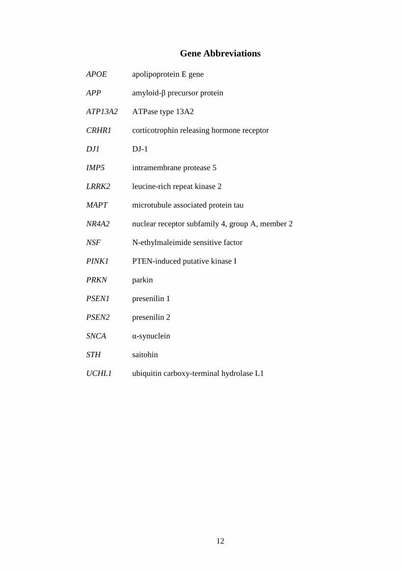
Gene Abbreviations
APOE apolipoprotein E gene
APP amyloid-β precursor protein
ATP13A2 ATPase type 13A2
CRHR1 corticotrophin releasing hormone receptor
DJ1 DJ-1
IMP5 intramembrane protease 5
LRRK2 leucine-rich repeat kinase 2
MAPT microtubule associated protein tau
NR4A2 nuclear receptor subfamily 4, group A, member 2
NSF N-ethylmaleimide sensitive factor
PINK1 PTEN-induced putative kinase I
PRKN parkin
PSEN1 presenilin 1
PSEN2 presenilin 2
SNCA α-synuclein
STH saitohin
UCHL1 ubiquitin carboxy-terminal hydrolase L1

13
Abstract
Our global population is ageing and an ever increasing number of elderly are
affected with neurodegenerative diseases, including the subjects of the studies in
this work, Alzheimer's disease (AD), Parkinson's disease (PD), progressive
supranuclear palsy (PSP) and corticobasal degeneration (CBD).
On strong evidence that several genes may influence the development of sporadic
neurodegenerative diseases, the genetic association approach was used in the
work of this thesis to identify the multiple variants of small effect that may
modulate susceptibility to common, complex neurodegenerative diseases. It has
been shown that the common genetic variation of one of these susceptibility genes,
MAPT, that of the microtubule associated protein, tau, is an important genetic risk
factor for neurodegenerative diseases. There are two major MAPT haplotypes at
17q21.31 designated as H1 and H2.
In order to dissect the relationship between MAPT variants and the pathogenesis
of neurodegenerative diseases, the architecture and distribution the major
haplotypes of MAPT have been assessed. The distribution of H2 haplotype is
almost exclusively in the Caucasian population, with other populations having H2
allele frequencies of essentially zero.
A series of association studies of common variation of MAPT in PSP, CBD, AD
and PD in different populations were performed in this work with the hypothesis
that common molecular pathways are involved in these disorders. Multiple
common variants of the H1 haplotypes were identified and one common
haplotype, H1c, showed preferential association with PSP and AD.
A whole-genome association study of PD was also undertaken in this study in
order to detect if common genetic variability exerts a large effect in risk for
disease in idiopathic PD. Twenty six candidate loci have been found in this
whole-genome association study and they provide the basis for our investigation
of disease causing genetic variants in idiopathic PD.

14
Contents
Chapter 1 Introduction….………………………………………………….24
1.1 Overview…………….……………………………………………….…..24
1.1.1 Impact of Neurodegenerative diseases in the ageing global population.24
1.1.2 The environmental risk factors of neurodegenerative diseases............25
1.1.3 The genetics of neurodegenerative diseases........................................26
1.2 Neurodegenerative Diseases......................................................................28
1.2.1 The genetics of Alzheimer’s disease...................................................28
1.2.2 The genetics of Parkinson’s disease....................................................31
1.2.3 The genetics of progressive supranuclear palsy ..................................34
1.2.4 The genetics of corticobasal degeneration .........................................36
1.2.5 The tauopathies....................................................................................37
1.2.5.1 Microtubule associated protein tau................................................37
1.2.5.2 The tauopathies..............................................................................39
1.3 Genetic Approach to Study Neurodegenerative Diseases.........................42
1.3.1 Genetic epidemiology..........................................................................42
1.3.2 Genetic Mapping of common complex disease genes.........................43
1.3.2.1 The rationale of the population-based genetic association studies...43
1.3.2.2 The design of population genetic association study.......................45
1.3.2.3 Bias due to population stratification..............................................47
1.3.3 Whole-genome association approach for neurodegenerative diseases....48
1.3.3.1 Whole-genome association study...................................................48
1.3.3.2 The International HapMap project.................................................49
1.3.3.3 Markers for genome-wide association studies...............................50

15
1.4 Thesis aims and objectives........................................................................52
Chapter 2 Methods and Materials................................................................55
2.1 Methods......................................................................................................55
2.1.1 DNA sample extraction from tissue.....................................................55
2.1.2 DNA quantification..............................................................................56
2.1.3 Polymerase Chain Reaction.................................................................57
2.1.4 Agarose gel electrophoresis.................................................................59
2.1.5 Genotyping...........................................................................................59
2.1.5.1 Restriction fragment length polymorphism...................................59
2.1.5.2 Pyrosequencing..............................................................................61
2.1.5.3 PCR genotyping.............................................................................62
2.1.6 DNA sequencing..................................................................................62
2.1.7 Whole Genome Scanning....................................................................64
2.1.8 Statistical analysis in population genetic association studies..............66
2.1.8.1 Hardy-Weinberg equilibrium.........................................................66
2.1.8.2 Genetic association studies............................................................66
2.1.8.2.1 Single-locus analysis...............................................................66
2.1.8.2.2 The odds ratio..........................................................................67
2.1.8.2.3 Haplotype Analysis..................................................................68
2.1.8.3 Tagging single nucleotide polymorphisms....................................69
2.1.9 Bioinformatics/Web resources.............................................................70
2.1.9.1 The National Centre for Biotechnology Information (NCBI)........70
2.1.9.2 The University of California Santa Cruz genome browser............70
2.1.9.3 HapMap..........................................................................................71

16
2.1.9.4 Ensembl genome browser..............................................................71
2.1.9.5 SHEsis............................................................................................72
2.2 Materials....................................................................................................72
2.2.1 PCR reagents.......................................................................................72
2.2.2 DNA/genotyping/Sequencing reagents...............................................72
2.2.2.1 DNA extraction reagents...............................................................72
2.2.2.2 Genotyping Reagents.....................................................................73
2.2.2.3 Pyrosequencing reagents................................................................73
2.2.2.4 Sequencing reagents.......................................................................73
2.2.3 Whole Genome Scanning Reagents (Illumina)....................................74
2.2.4 Molecular biology reagents..................................................................74
2.3 Suppliers.....................................................................................................74
Chapter 3 The architecture of the tau haplotype........................................76
3.1 Overview....................................................................................................76
3.2 Background................................................................................................77
3.3 Methods and Materials...............................................................................79
3.3.1. Samples...............................................................................................79
3.3.2. Methods..............................................................................................79
3.3.2.1 Genot yp ing of haplo type-def in ing inse r t ion /dele t i on
polymorphism................................................................................79
3.3.2.2 Genotyping of the flanking SNPs of the tau haplotype block and
the differentiating SNPs of the tau haplotype H1/H2....................80
3.3.2.2.1 Pyrosequencing........................................................................80
3.3.2.2.2 Restriction fragment length polymorphism.............................81
17
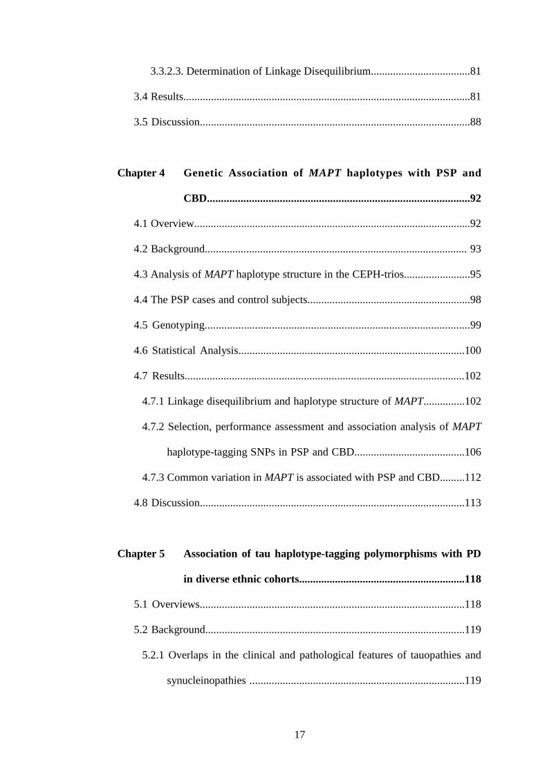
3.3.2.3. Determination of Linkage Disequilibrium....................................81
3.4 Results........................................................................................................81
3.5 Discussion..................................................................................................88
Chapter 4 Genetic Association of MAPT haplotypes with PSP and
CBD..............................................................................................92
4.1 Overview....................................................................................................92
4.2 Background............................................................................................... 93
4.3 Analysis of MAPT haplotype structure in the CEPH-trios........................95
4.4 The PSP cases and control subjects...........................................................98
4.5 Genotyping...............................................................................................99
4.6 Statistical Analysis..................................................................................100
4.7 Results.....................................................................................................102
4.7.1 Linkage disequilibrium and haplotype structure of MAPT...............102
4.7.2 Selection, performance assessment and association analysis of MAPT
haplotype-tagging SNPs in PSP and CBD........................................106
4.7.3 Common variation in MAPT is associated with PSP and CBD.........112
4.8 Discussion................................................................................................113
Chapter 5 Association of tau haplotype-tagging polymorphisms with PD
in diverse ethnic cohorts............................................................118
5.1 Overviews................................................................................................118
5.2 Background..............................................................................................119
5.2.1 Overlaps in the clinical and pathological features of tauopathies and
synucleinopathies ..............................................................................119

18
5.2.2 Genetic risk factors of Parkinson’s disease......................................119
5.3 Case-control samples...............................................................................121
5.3.1 Taiwanese Series...............................................................................122
5.3.2 Greek Series.......................................................................................122
5.3.3 Finnish Series.....................................................................................123
5.4 Genotyping of htSNPs.............................................................................123
5.4.1 Selection of haplotype-tagging SNP markers....................................123
5.4.2 Pyrosequencing Assay.......................................................................124
5.5 Results......................................................................................................125
5.6 Discussion................................................................................................126
Chapter 6 Association Study of MAPT gene haplotypes with AD...........131
6.1 Overview..................................................................................................131
6.2 Background..............................................................................................131
6.3 Case-control samples...............................................................................132
6.3.1 US and UK series...............................................................................132
6.3.2 Taiwanese Series...............................................................................133
6.4 Selection of haplotype-tagging SNPs......................................................134
6.4.1 Markers for the US and UK series....................................................134
6.4.2 Markers for the Taiwanese series......................................................135
6.4.2.1 SNP Selection..............................................................................135
6.4.2.2 Selection, performance, assessment, and association analysis of
MAPT haplotype tagging SNPs in Taiwanese cohort..................136
6.4.2.3 LD and statistical analysis...........................................................138
6.4.3 SNP amplification and genotyping....................................................139

19
6.4.3.1 ApoE genotyping.........................................................................139
6.5 Results......................................................................................................140
6.5.1 The association of the H1c haplotype at the MAPT locus with AD in
UK and US series...............................................................................140
6.5.1.1 Single locus analysis....................................................................140
6.5.1.1.1 Single locus analysis..............................................................140
6.5.1.1.2 Single locus analysis: APOE sub-analysis............................141
6.5.1.2 Haplotype analysis.......................................................................142
6.5.1.2.1 Haplotype analysis.................................................................142
6.5.1.2.2 Haplotype analysis: APOE sub-analysis................................144
6.5.2 The association of the H1 haplotype at the MAPT locus with
Taiwanese series.............................................................................145
6.5.2.1 Linkage disequilibrium pattern of MAPT in Taiwanese.............145
6.5.2.2 Selection, performance, assessment, and association analysis of
MAPT haplotype tagging SNPs..................................................145
6.6 Discussion...............................................................................................146
Chapter 7 Whole Genome Association Study of Neurodegenerative
diseases; First stage PD.............................................................150
7.1 Overview..................................................................................................150
7.2 Background..............................................................................................150
7.3 Case-Control Samples.............................................................................152
7.3.1 Subject Collection.............................................................................152
7.3.2 Sample Preparation...........................................................................153
7.4 Genotyping..............................................................................................154

20
7.5 Data Analysis...........................................................................................154
7.6 Results......................................................................................................155
7.7 Discussion................................................................................................159
Chapter 8 Discussion....................................................................................162
8.1 Summary..................................................................................................162
8.2 General Discussion..................................................................................163
8.2.1 A general association between the MAPT Haplotype and
neurodegenerative diseases..........................................................163
8.3 Future Research.......................................................................................166
8.3.1 Population-based genetic Association studies...................................166
8.3.2 Whole genome association studies in Neurodegenerative diseases...167
9. References.......................................................................................................168
10. Appendices....................................................................................................199
10.1 Chapter 3 Genotyping assays.................................................................199
10.2 Genotyping assays for Association Studies...........................................200
10.3 Genotyping assays for the SNP in LD study of the Taiwanese
Population.............................................................................................200
10.4 Selected reprints of publications arising from the work in this thesis...202
21
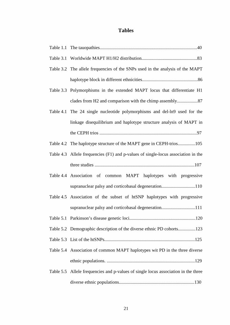
Tables
Table 1.1 The tauopathies....................................................................................40
Table 3.1 Worldwide MAPT H1/H2 distribution................................................83
Table 3.2 The allele frequencies of the SNPs used in the analysis of the MAPT
haplotype block in different ethnicities................................................86
Table 3.3 Polymorphisms in the extended MAPT locus that differentiate H1
clades from H2 and comparison with the chimp assembly..................87
Table 4.1 The 24 single nucleotide polymorphisms and del-ln9 used for the
linkage disequilibrium and haplotype structure analysis of MAPT in
the CEPH trios ....................................................................................97
Table 4.2 The haplotype structure of the MAPT gene in CEPH-trios...............105
Table 4.3 Allele frequencies (F1) and p-values of single-locus association in the
three studies ......................................................................................107
Table 4.4 Association of common MAPT haplotypes with progressive
supranuclear palsy and corticobasal degeneration.............................110
Table 4.5 Association of the subset of htSNP haplotypes with progressive
supranuclear palsy and corticobasal degeneration.............................111
Table 5.1 Parkinson’s disease genetic loci.........................................................120
Table 5.2 Demographic description of the diverse ethnic PD cohorts...............123
Table 5.3 List of the htSNPs..............................................................................125
Table 5.4 Association of common MAPT haplotypes wit PD in the three diverse
ethnic populations. ............................................................................129
Table 5.5 Allele frequencies and p-values of single locus association in the three
diverse ethnic populations.................................................................130

22
Table 6.1 Summary of studies on MAPT H1/H1 diplotype as risk factor for
Alzheimer’s disease...........................................................................131
Table 6.2 The 21 single nucleotide polymorphisms used for the linkage
disequilibrium and haplotype structure analysis of MAPT in the
Taiwanese cohort...............................................................................136
Table 6.3 Single locus association in UK series and US series.........................141
Table 6.4 Single locus associations within APOE-ε4 positive and negative
subsets of the combined US and UK sample.....................................142
Table 6.5 Single locus association analysis of MAPT with Taiwanese AD
cohort.................................................................................................146
Table 7.1 Summary of the most significant associated SNPs in Genome-wide
genotyping .........................................................................................157
Table 10.1 Sequences of PCR primer pairs used for genotying in Chapter 3......199
Table 10.2 Genotyping assays for the SNP in Chapter 3.....................................199
Table 10.3 Haplotyping tagging SNPs for the MAPT association study (PSP, CBD
(ch.4), PD (ch.5) and AD (ch.6)).......................................................200
Table 10.4 PCR Primer pairs of linkage equilibrium structure study in the
Taiwanese population.......................................................................200
Table 10.5 Genotyping assays for the SNP in LD study of the Taiwanese
Population.........................................................................................201

23
Figures
Figure 1.1 MAPT structure and the FTDP-17 mutation spectrum.......................38
Figure 1.2 Testing SNPs for association by direct and indirect methods.............51
Figure 3.1 The extended haplotype block at 17q21.31.........................................78
Figure 3.2 Worldwide Tau H1/H2 Distribution....................................................82
Figure 3.3 The architecture of the tau haplotype block in different ethnicities.....85
Figure 3.4 Pair-wise D’ LD analysis of the different ethnic populations..............87
Figure 3.5 Parsimony Tree of Relationships between Chimp MAPT Locus and
H1 and H2 Haplotypes.........................................................................91
Figure 4.1 Linkage Disequilibrium (LD) across the MAPT gene.......................103
Figure 6.1 MAPT tagging markers capture the diversity of MAPT....................135
Figure 6.2 Analysis of performance of different number of htSNPs in Taiwanese
Cohort.................................................................................................137
Figure 6.3 Linkage disequilibrium (LD) across the MAPT in Taiwanese
population..........................................................................................139
Figure 6.4 MAPT haplotypes..............................................................................143
Figure 7.1 Bar and triangle plots from STRUCTURE using 267 random
autosomal SNPs................................................................................158
24
Chapter 1 Introduction
1.1. Overview
1.1.1 The impact of neurodegenerative diseases in the ageing global population.
The population of our world is ageing and an ever-increasing number of elderly
are affected by neurodegenerative diseases. In the developed world, about 2% of
the population is afflicted at any time [1]. These neurodegenerative diseases
include Alzheimer's disease (AD), Parkinson's disease (PD), amyotrophic lateral
sclerosis (ALS), progressive supranuclear palsy (PSP), corticobasal degeneration
(CBD), Huntington's disease (HD), frontotemporal dementia (FTD), Pick's
Disease (PiD) and prion diseases. Recently, the "Global Market and Future
Outlook for Neurodegenerative Disorder Therapies 2007" forecasts that the
overall neurodegenerative disease drug market will grow from around US$9
billion in 2005 to more than US$17 billion by 2010 (Piribo.com). The overall
neurodegenerative diseases market is growing at over 12% each year. With an
increase in life expectancy and the number of old people, along with advances in
treatments of neurodegenerative diseases, the increases look set to continue.
Neurodegenerative diseases cost the U.S. economy billions of dollars each year in
direct health care costs and lost opportunities; it is estimated that $100 billion per
years is spent on AD alone. The market for AD therapy is expected to rise from
US$4 billion in 2005 to US$7 billion in 2010 with the US market continuing to
dominate. In addition to the financial costs, there is an immense emotional burden
on patients and their caregivers. As the number of elderly citizens increases, these
costs to society will also increase [2].

25
1.1.2 The environmental risk factors of neurodegenerative diseases
Neurodegenerative diseases are characterized by progressive nervous system
dysfunction. These disorders result from the gradual and progressive loss of
neural cells, leading to nervous system dysfunction. Neurodegenerative diseases
can affect abstract thinking, skilled movements, emotional feelings, cognition,
memory and other abilities [3].
Known risk factors for neurodegenerative diseases include certain genetic
polymorphisms and increasing age. Other possible causes may include gender,
poor education, endocrine conditions, oxidative stress, inflammation, stroke,
hypertension, diabetes, smoking, head trauma, depression, infection, tumors,
vitamin deficiencies, immune and metabolic conditions, and chemical exposure.
Because the pathogenesis of many of these diseases remains unknown, the role of
environmental factors in these diseases has to be considered [2]. Although there is
a growing body of evidence suggesting that a large proportion of the non-familial
cases are significantly influenced by genetic factors, it is likely like the
environmental risk factors play a part in the etiology of some of these conditions.
MPTP (1-methyl-4-phenyl-1,2,3,6-tetrahydropyridine) is a good though rare
example of an environmental agent that caused an epidemic of Parkinsonism
among drug misusers [4]. MPTP is a simple chemical, and a viable hypothesis is
that autointoxication by similar molecules may cause sporadic diseases [5]. A
good example is the epidemic of a variety of degenerative diseases on Guam,
where a possible environmental agent has not been discovered [6].
26
Despite the enormous effort put into searching for the environmental factors for
these diseases, epidemiological evidence for an association between
environmental agents and neurodegenerative diseases remains inconclusive [2].
On the other hand, evidence from the familial, twin and association studies for
neurodegenerative diseases, such as AD, PD, FTD, suggesting it is promising to
apply molecular genetics to reveal the pathogenesis of those diseases with
understanding their underlying genetics. The genetics of various
neurodegenerative diseases are briefly reviewed in the following sections.
1.1.3 The genetics of neurodegenerative diseases
Until recently, it was considered impossible to find a common molecular
mechanism for neurodegeneration. However, despite their diverse clinical
manifestations and disease progression, these disorders share some common
characteristics: all these diseases (except Huntington disease) have both sporadic
and inherited types, the onset of all these diseases is usually after the fourth or
fifth decade, and their pathology involves neuronal loss and protein aggregation.
For instance, a normal soluble cellular protein is converted into an abnormal
insoluble aggregated protein rich in β-sheets that is toxic such as β-amyloid in AD.
Emerging evidence for a causal role of the conformational changes of proteins in
neurodegenerative diseases has become clearer recently from genetic studies [7].
Mutations in the genes that encode the protein components of fibrillar aggregates
are genetically associated with the inherited form of neurodegenerative diseases

27
[3]. These include mutations in genes for the β-amyloid (Aβ) precursor protein,
causing AD; α-synuclein, causing PD; or in microtubule-associated protein tau,
causing FTD with parkinsonism. Though these mutations cause mainly the
familial type of neurodegenerative diseases, the pathology of both familial and
sporadic forms is either identical or very similar [8]. Investigation of the
molecular genetics of the familial forms has provided a window into the
molecular biology of both the familial and the sporadic forms of
neurodegenerative diseases. Indeed, there is a growing body of evidence
suggesting that a large proportion of the sporadic cases are also significantly
influenced by genetic factors. These risk genes are likely to be numerous,
displaying intricate patterns of interaction with each other as well as with non-
genetic variables, and — unlike classical Mendelian (“simplex”) disorders —
exhibit no simple or single mode of inheritance. Hence, the genetics of these
diseases has been labelled “complex” [9].
A conception regarding the genetic makeup of complex diseases is the “common
disease/common variant” (CD/CV) hypothesis [10]. According to this theory
common disorders are also governed by common DNA variants, such as single
nucleotide polymorphisms. These variants significantly influence disease risk but
are insufficient to actually cause a specific disorder. Current empirical and
theoretical data support this hypothesis, although there remains great uncertainty
as to the number of the underlying risk factors and their specific effect sizes. It is
believed that there is a risk spectrum predisposing to common disease, such as AD,
as one continuum [10]. The continuum extends from the most clearly defined
genetic forms to cases influenced by genetic susceptibility factors, and further
extending to a less well defined area of cases that may be caused by genes of low
28
penetrance and/or non-genetic factors. In AD, for instance, rare, fully penetrant
autosomal dominant mutations in 3 genes (i.e. APP, PSEN1, and PSEN2) have
been shown to cause the disease, while a common, incompletely penetrant
susceptibility variant, ε4 in the apolipoprotein E gene (APOE) significantly
increases the risk for AD.
Similar genetic susceptibility traits such as APOE in AD are also found in other
neurodegenerative diseases, such as MAPT in PSP. Therefore, genetic analysis is
having a substantial impact in the attempts to dissect the etiology and
pathogenesis of the neurodegenerative diseases, making it increasingly clearer
how disease may be initiated. In the following sections, the genetics of various
neurodegenerative diseases and the genetic approach for studying of these
diseases will be reviewed.
1.2 Neurodegenerative Diseases
1.2.1 The genetics of Alzheimer’s disease
AD is the most common neurodegenerative disease and afflicts ~5% of those over
65 years. It is one of the most serious health problems in the industrialized world.
It is an insidious and progressive neurodegenerative disorder that accounts for the
vast majority of age-related dementia and is characterized by global cognitive
decline and the accumulation of Aβ deposits and neurofibrillary tangles in the
brain. Family history is the second-greatest risk factor for the disease after age,
and the growing understanding of AD genetics has been central to the knowledge
of the pathogenic mechanisms leading to the disease. Genetically, AD is complex

29
and heterogenous and appears to follow an age-related dichotomy: rare and highly
penetrant early-onset familial AD (EOFAD) mutations in different genes are
transmitted in an autosomal dominant fashion, while late-onset AD (LOAD)
without obvious familial segregation is thought to be explained by the CD/CV
hypothesis [11].
The EOFAD represents only a small fraction of all AD cases (≤ 5%) and typically
presents with onset ages younger than 65 years, showing autosomal dominant
transmission within affected families. These forms of the disease are caused by
mutations in the amyloid-β precursor protein (APP) on chromosome 21 [12] and
the presenilin 1 gene (PSEN1) on chromosome 14 [13] and presenilin 2 gene
(PSEN2) on chromosome 1 [14-16]. To date, more than 160 mutations in these 3
genes have been reported to cause EOFAD. An up-to-date overview of disease-
causing mutations in these genes can be found at the Alzheimer Disease &
Frontotemporal Dementia Mutation Database [17]. The most frequently mutated
gene, PSEN1, accounts for the majority of AD cases with onset prior to age 50.
While these AD-causing mutations occur in 3 different genes located on 3
different chromosomes, they all share a common biochemical pathway, i.e., the
altered production of Aβ leading to an overabundance of the Aβ42 species relative
to the Aβ40 species, which eventually results in neuronal cell death and dementia.
The mutations in the APP gene occur at the A cleavage sites, thereby altering
APP processing such that more Aβ42 is produced [18]. The presenilins are a
central component of γ-secretase, the enzyme responsible for liberating the C-
terminal fragment of APP, and mutations in the presenilins also alter APP
processing such that more Aβ42 is produced [19]. These genetic data are the

30
intellectual basis for the amyloid hypothesis of the disorder, suggests that Aβ42 is
the initiating molecule in Alzheimer’s disease.
LOAD, on the other hand, is classically defined as AD with onset at age 65 years
or older and represents the vast majority of all AD cases. While segregation and
twin studies conclusively suggest a major role of genetic factors in this form of
AD [20], to date, only one such factor has been established, the ε4 allele of the
APOE gene on chromosome 19q13 [21;22]. In contrast to all other association-
based findings in AD, the risk effect of APOE-ε4 has been consistently replicated
in a large number of studies across many ethnic groups, yielding odds ratios (OR)
between approximately 3 and approximately 15 for heterozygous and
homozygous carriers, respectively, of the ε4 allele in white individuals [23]. In
addition to the increased risk exerted by the ε4-allele, a weak, albeit significant,
protective effect for the minor allele, ε2, has also been reported in several studies
[9]. Unlike the mutations in the known EOFAD genes, APOE-ε4 is neither
necessary nor sufficient to cause AD but instead operates as a genetic risk
modifier by decreasing the age of onset in a dose-dependent manner. Despite its
long-known and well-established genetic association, the biochemical
consequences of APOE-ε4 in AD pathogenesis are not yet fully understood but
likely encompass Aβ-aggregation/clearance and/or cholesterol homeostasis.
Several lines of evidence suggest that numerous additional LOAD loci [24] —
and probably also EOFAD loci [25;26] — remain to be identified, since the 4
known genes together account for probably less than 50% of the genetic variance
of AD. As outlined above, it is currently unclear how many of these loci will be
proved to be risk factors as opposed to causative variants. As candidates for the

31
former, more than 3 dozen genes have been significantly associated with AD in
the past [27;28]. Despite the more than 500 independent association studies,
however, no single gene has been shown to be a risk factor with the same degree
of replication or consistency that has been shown for APOE-ε4. One of the
conclusions to be drawn from currently available data, as well as from the few
independently performed meta-analyses on putative AD risk factors is that even if
some of the published associations were genuine, their overall effect size is likely
to be only minor, i.e. with OR not exceeding 2.
1.2.2 The genetics of Parkinson’s disease
PD is the second most common neurodegenerative disease of adult onset.
Histopathologically, it is characterized by a severe loss of dopaminergic neurons
in the substantia nigra and cytoplasmic inclusions consisting of insoluble protein
aggregates, which lead to a progressive movement disorder including the classic
triad of tremor, bradykinesia, and rigidity, with an average onset age between 50
and 60 years. As in AD, there appears to be an age-dependent dichotomy: the
majority of individuals with an early or even juvenile onset show typical
Mendelian inheritance. However, unlike in AD, these cases show a predominantly
autosomal-recessive mode of inheritance, and there is an ongoing debate as to
whether genetic factors play any substantial role in contributing to disease risk in
cases with onset beyond approximately 50 years [29;30]. Notwithstanding these
uncertainties, there are a plethora of genetic studies on both forms of the disease,
and mutations in at least 6 genes have now been shown to cause familial early-
onset parkinsonism (α-synuclein, SNCA) [31]; parkin (PRKN) [32]; DJ-1 (DJ1)

32
[33]; PTEN-induced putative kinase I (PINK1) [34]; and leucine-rich repeat
kinase 2 or dardarin (LRRK2) [35] and ATP13A2 [36], with several other linkage
regions pending characterization and/or replication. As was the case in the study
of AD, the first locus to be characterized –SNCA, on chromosome 4q21 –
which codes for α-synuclein the protein that is the major constituent of the Lewy
body (LB), one of the classic neuropathological hallmarks of the disease [31],
which can be found at the core of LBs. While the exact mechanisms underlying α-
synuclein toxicity currently remains only incompletely understood, recent
evidence suggests that some SNCA mutations may change normal protein function
quantitatively rather than qualitatively, via duplication or triplication of the SNCA
gene [37;38]. Mutations in a second gene, LRRK2 with dominant inheritance have
been identified by several different laboratories [35]. While the functional
consequences of LRRK2 mutations are still unknown, it was suggested that at least
some mutations could interfere with the protein’s kinase activity [39]. While
changes in SNCA and LRRK2 are the leading causes of autosomal-dominant forms
of PD, the majority of affected pedigrees actually show a recessive mode of
inheritance. The most frequently involved gene in recessive parkinsonism is
parkin (PRKN) on chromosome 6q25 [32;40], which causes nearly half of all
early-onset PD cases. Parkin is an ubiquitin ligase that is involved in the
ubiquitination of proteins targeted for degradation by the proteasomal system. The
spectrum of parkin mutations ranges from amino acid-changing single base
mutations to complex genomic rearrangements and exon deletions, which
probably result in a loss of protein function. It has been speculated that this may
trigger cell death by rendering neurons more vulnerable to cytotoxic insults, e.g.,
the accumulation of glycosylated α-synuclein [41]. In addition to parkin mutations,
33
genetic analyses of two non-parkin early-onset, autosomal-recessive PD pedigrees
revealed two independent, homozygous mutations in DJ1[33] on chromosome
1p36 [42]. Both mutations result in a loss-of-function of DJ-1, a protein that is
suggested to be involved in oxidative stress response. While several studies have
independently confirmed the presence of DJ1 mutations in other PD cases, the
frequency of disease-causing variants in this gene is estimated to be low (∼1%)
[43]. Less than 13 Mb toward the long arm of the same chromosome, additional
PD-causing mutations were subsequently discovered in PINK1 [34] following
positive linkage evidence to this region [44]. PINK1 codes an enzyme that is
expressed at particularly high levels in brain, and the first two mutations identified
(G309D and W437ter) were predicted to lead to a loss-of-function that may render
neurons more vulnerable to cellular stress, similar to the effects of PRKN
mutations. While Lewy bodies are typically not found in brains of patients bearing
PRKN mutations, it is currently unclear whether these are present in PD cases
with mutations in DJ1 and PINK1. At least six additional candidate PD loci have
been described, including putative disease-causing mutations in the ubiquitin
carboxy-terminal hydrolase L1 (UCHL1) on chromosome 4p14 [45], and in a
nuclear receptor of subfamily 4 (NR4A2, or NURR1) [46] located on 2q22.
However, and unlike the previously outlined PD genes, neither of these maps to
known PD linkage regions, nor were they independently confirmed beyond the
initial reports. However, polymorphisms in both genes have been, albeit
inconsistently, associated with PD in some case-control studies. A recent meta-
analysis of the S18Y polymorphism in UCHL1 showed a modest but significant
protective effect of the Y allele [47], which suggests that this gene may actually
be a susceptibility factor rather than a causal PD gene. Unlike early-onset PD, the

34
heritability of late-onset PD is probably low [29]. Despite this caveat, while a
number of whole genome screens across several late-onset PD family samples
have been performed, only a few overlapping genomic intervals have been
identified. One of the more extensively studied regions is 17q21, containing the
gene encoding the microtubule-associated protein tau (MAPT) [48]. Previously, it
had been shown that rare missense mutations in MAPT lead to a syndrome of FTD
with parkinsonism linked to chromosome 17 (FTDP-17), but to date no mutation
has been identified as causing parkinsonism without frontotemporal degeneration.
However, haplotype analyses of the tau gene have revealed evidence of genetic
association of the H1 haplotype with both PD [49;50] and PSP [51]. Despite the
lack of evidence for genetic linkage to chromosome 19q13, variants in APOE
have also been tested for a role in PD. Across the nearly three dozen different
studies available to date, some authors report a significant risk effect of APOE-ε4
for PD, while others only see association with certain PD phenotypes or even a
risk effect of the ε2 allele, which is protective in AD (see above). A recent meta-
analysis on the effects of APOE in PD concluded that only the ε2-related increase
in PD risk remains significant when all published studies are considered jointly
[52]. Finally, and in addition to the findings in autosomal-dominant familial PD,
there is also some support for a potential role of SNCA variants in the risk for late-
onset PD [53].
1.2.3 The genetics of progressive supranuclear palsy (PSP)
PSP is the second most frequent cause of degenerative parkinsonism after PD [54].
In addition to parkinsonism, the clinical symptoms include early postural

35
instability and supranuclear gaze palsy [55]. Neuropathologically, PSP is
characterized by abundant neurofibrillary tangles and neurophil threads consisting
largely of four repeat tau [56]. Robust genetic association of PSP with MAPT and
rare reports of families with more than one affected member indicated that genetic
factors could play a role in PSP [57;58].
In Europeans, the MAPT gene has an unusual genetic structure, two distinct and
inverted haplotypes have been found and designated as H1 and H2. The H2
haplotype has an allele frequency of approximately 25% [59;60]. This has made
the genetic analysis of PSP and CBD easy in this population and has shown a
robust association between the H1 haplotype of the MAPT locus and both PSP and
CBD [61]. It is likely that there will be a MAPT association with these diseases in
other populations, but the relevant analyses are more difficult to perform in these
other populations because of the absence of the H2 haplotype. A haplotypic
association, in the absence of coding changes, implies that the biological effect
could be mediated either by differences in expression or differences in splicing
between haplotypes. The fact that the disturbances in the splicing of the MAPT
gene is one of the causes of FTDP-17, and the fact that the tangle deposits consist
almost exclusively, of four-repeat tau, suggests that either or both of the above are
equally likely explanations. A more detailed analysis of the structure of the H1
haplotype revealed that it has considerable complexity, and that in fact, the
haplotypic association between H1 and PSP and CBD is driven by a variant of the
haplotype named H1c, which defines a region from the promoter to intron 10 of
the gene. Analysis of this haplotype has not yet led to the determination of
whether it is a particularly high-expressing haplotype, one that particularly
expresses the exon-10 containing transcript, or a mixture of both. (see section

36
1.2.5.1 and Figure 1.1) This haplotypic association is an example of the general
principle that genetic variability at the loci causing autosomal dominant disease
(in this case FTDP-17) is part of the genetic contribution to the sporadic diseases
(in this case PSP and CBD) [62].
1.2.4 The genetics of corticobasal degeneration (CBD)
Corticobasal degeneration (CBD) is a progressive neurological disorder
characterised by atrophy of multiple brain areas including the cerebral cortex and
the basal ganglia [63]. Initial symptoms, such as poor coordination, akinesia,
rigidity, impaired balance and limb dystonia which typically appear at the age of
around 60, are similar to those found in Parkinson’s disease. Other symptoms
such as cognitive and visuo-spatial impairments, apraxia, hesitant and halting
speech, myoclonus (muscular jerks), and dysphagia may also occur.
Neuropathologically, CBD is distinguished from PSP and other dementias by
several important features. Most pathology in CBD is in the cerebrum, whereas
the basal ganglia, diencephalon, and brainstem are mainly the targets of PSP.
Histologically, there are ballooned neurons, astrocytosis, and four repeat tau-
positive neuronal and glial inclusions. The most characteristic neuronal tau
pathology in CBD is numerous and widespread wispy, fine filamentous inclusions
within neuronal cell bodies, whereas affected neurons in PSP have compact, dense
filamentous aggregates [64;65].

37
The genetics of CBD had not been widely studied until now because the disease is
rare and usually sporadic in occurrence. However, the extended H1 haplotype has
also shown to be a genetic risk factor for CBD [66] that was subsequently
independently replicated [67].
1.2.5 The tauopathies
The above neurodegenerative diseases AD, PSP and CBD are collectively belong
to a group of disorders known as the tauopathies, as they all have pathological
fibrillar aggregates of tau in the brain. The characteristics of tau protein and the
tauopathies are reviewed below.
1.2.5.1 Microtubule associated protein tau
The microtubule associated protein, tau was first identified as a “factor essential
for microtubule (MT) assembly”, a heat stable protein that induced the assembly
of MTs from purified tubulin and belonging to the family of MT-associated
proteins [68]. Tau is abundantly expressed in the both the peripheral and central
nervous system [69], where it is enriched in the axons of mature and growing
neurones and, low levels of tau are also present in oligodendrocytes and astrocytes
[70;71]. Tau is a phosphoprotein with developmentally regulated phosphorylation
profiles at up to 38 phosphorylation sites [72]. The level of protein
phosphorylation is highly elevated in foetal tau and pathological tau found within
the insoluble, fibrillar inclusions that define tauopathies, when compared to

38
normal adult brain tau [73]. The human tau gene, MAPT (MIM 157140), spanning
~150 kb of nucleotide sequence on chromosome 17q21.3, consists of one non-
coding- and 14 coding exons [74-76] (Figure 1.1)
Figure 1.1 MAPT structure and the FTDP-17 mutation spectrum
Left: Tau in the central nervous system (CNS) exists as six isoforms due to the alternative splicingof exons 2, 3 and 10 (yellow boxes). Exons 4A, 7 and 8 (red boxes) are absent in the CNS, exon4A is included in peripheral nervous system tau. Exons 2 and 3 code for 29 residue amino-terminalinserts, alternative splicing leads to tau isoforms with 2, 1 or no amino-terminal inserts (2N, 1N or0N). Exon 10 codes for one of four microtubule binding domains – alternative splicing results intau with 3 or 4 microtubule binding repeat domains (3R, 4R). FTDP-17 missense and silentmutations and deletions are indicated with numbering relating to the longest 441 residue 2N,4Risoform. Mutations in red affect the alternative splicing of exon 10. Right: FTDP-17 mutationsaffecting the 3’ splice donor site of exon 10. The majority of these mutations disrupt a predictedpre-mRNA stem-loop structure, inducing increase incorporation of exon 10. Partial sequence of3’-end of exon 10 in red. intronic sequence in black. Proportions are not to scale. (Modified fromGoedert [77])
In the healthy adult human brain, tau protein exists as six major isoforms
produced by the alternative splicing of exons 2, 3 and 10 [78]. (Figure 1.1) The
alternative splicing of exon 10 produces tau isoforms with either three MT-
binding repeats (3R-tau) due to exclusion of exon 10, or four repeats (4R-tau) due
to exon 10 inclusion. It is now widely recognised that several tauopathies are
associated with aberrant splicing of exon 10, causing imbalances in the 3R-

39
tau:4R-tau ratios. For example, the insoluble tau deposits in the different
tauopathies have different tau-isoform compositions; in Pick’s disease (PiD), the
classical Pick bodies consist mainly of 3R-tau isoforms [79;80], whereas in PSP,
CBD and argyrophilic grain disease (AGD), both neuronal and glial inclusions
contain mostly 4R-tau isoforms [79;81-84], and roughly equal amounts of 3R- and
4R-tau make up the paired helical filaments and straight filaments observed in AD
[84;85].
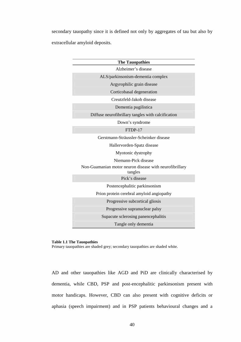
1.2.5.2 The tauopathies
The tauopathies are a group of neurodegenerative disorders that are characterized
pathologically by the presence of fibrillar aggregates of tau in the brain [76;86]
(Table1.1). The most common tauopathy, AD, is characterized clinically by a
progressive loss of verbal and visual memory and intellectual function, resulting
in severe dementia. The cognitive decline in AD has been correlated with various
biomarkers that include the loss of choline acetyl transferase and synaptophysin
reactivity. In addition to abundant extracellular amyloid Aβ deposits, the senile
plaques, hyperphosphorylated tau neurofibrillary tangles (NFTs) constitute the
pathological lesions [87]. Aβ and NFTs also coexist in some other tauopathies like
Down’s syndrome [88;89] however NFTs occur alone in argyrophilic grain
disease (AGD) [82], PiD [90], CBD, PSP [91], FTDP-17 [92], some ALS [93],
Niemann-Pick disease type C [94] and subacute sclerosing panencephalitis. These
disorders are classified as primary tauopathies, since pathological aggregates of
neurofibrillary tau are their main defining characteristic (Table 1.1). AD is a
40
secondary tauopathy since it is defined not only by aggregates of tau but also by
extracellular amyloid deposits.
The Tauopathies
Alzheimer’s disease
ALS/parkinsonism-dementia complex
Argyrophilic grain disease
Corticobasal degeneration
Creutzfeld-Jakob disease
Dementia pugilistica
Diffuse neurofibrillary tangles with calcification
Down’s syndrome
FTDP-17
Gerstmann-Sträussler-Scheinker disease
Hallervorden-Spatz disease
Myotonic dystrophy
Niemann-Pick disease
Non-Guamanian motor neuron disease with neurofibrillarytangles
Pick’s disease
Postencephalitic parkinsonism
Prion protein cerebral amyloid angiopathy
Progressive subcortical gliosis
Progressive supranuclear palsy
Supacute sclerosing panencephalitis
Tangle only dementia
Table 1.1 The TauopathiesPrimary tauopathies are shaded grey; secondary tauopathies are shaded white.
AD and other tauopathies like AGD and PiD are clinically characterised by
dementia, while CBD, PSP and post-encephalitic parkinsonism present with
motor handicaps. However, CBD can also present with cognitive deficits or
aphasia (speech impairment) and in PSP patients behavioural changes and a

41
dysexecution syndrome may be the most prominent symptoms. Owing to the
substantial clinical overlap among various neurodegenerative disorders with tau
pathology, definite diagnosis still requires neuropathological examination.
Neurofibrillary lesions of filamentous tau form within nerve cells that eventually
degenerate and it appears that they die. These lesions are found in nerve cell
bodies and apical dendrites as NFTs and in distal dendrites as neuropil threads.
Ultrastructurally, these lesions consist of paired helical filaments and straight
filaments [76;94]. The tau inclusions in the different tauopathies have
characteristic morphologies and distributions.
The pathological tau filaments are insoluble but can be isolated for biochemical
analysis as the detergent sarkosyl-insoluble fractions of brain homogenates [95].
Thus, in addition to distinct distribution and morphology, tauopathies can also be
classified according to the biochemical composition of tau in the respective
inclusions. The electrophoretic analysis of the insoluble tau from the different
tauopathies shows a banding pattern reflecting the different compositions of the
hyperphosphorylated tau isoforms present in the inclusions. This banding pattern
can be divided into three general categories depending on the presence of four
bands at 60, 64, 68 and 72 kDa that represent hyperphosphorylated tau isoforms
[84], these being predominantly 4R tau pathology (e.g. PSP), mixed 3R/4R tau
pathology (e.g.AD) and predominantly 3R tau pathology (e.g PiD).
42
1.3 Genetic Approach to Study Neurodegenerative Diseases
In the following sections, the different genetic approaches, which have been
employed in the work of this thesis, for studying of neurodegenerative diseases
will be reviewed.
1.3.1 Genetic epidemiology
Genetic epidemiology is a discipline closely related to traditional epidemiology
that focuses on the familial and the population, towards identification of genetic
determinants of disease and the joint effects of genes and non-genetic
determinants such as the environment [96]. Importantly, genetic epidemiology is a
fusion of traditional epidemiological principles with the biology of genes and their
mode of inheritance.
The vast majority of success so far in genetic epidemiology has been related to the
identification of disease causing genes in monogenic disorders, relying heavily on
linkage studies and positional cloning, where familial recurrence appears to obey
the laws of Mendelian inheritance. However, genetic epidemiology today is
increasingly focused on complex diseases such as most neurodegenerative
diseases, diabetes mellitus and cancer. These diseases are thought to be caused by
several interacting genetic and environmental determinants [97] and require quite
different genetic epidemiological study design and interpretation compared to the
traditional genetic linkage studies in monogenic Mendelian disorders [96].

43
1.3.2 Genetic Mapping of common complex disease genes
1.3.2.1 The rationale of the population-based genetic association studies
The rationale of genetic association studies is to detect association between one or
more genetic polymorphism(s) and a trait, which could either be a quantitative
characteristic or a discrete attribute or disease. Association differs from linkage in
that the same allele(s) is associated with the trait in the same manner across the
whole population, whereas linkage allows different alleles to be associated with
the trait in different families. Association studies identify polymorphisms in
which an allele occurring in the general population occurs at a different frequency
in the disease group. In these instances, the disease associated allele does not
cause the disease in the same way that a Mendelian mutation does but increases
susceptibility to the disease as a genetic risk factor, most likely in conjunction
with other genetic and environmental risk factors. Such identified variants have
relatively low penetrance compared to variants causing monogenic Mendelian
disease. Association studies can either be direct or indirect. In direct association
studies target polymorphisms which are themselves putative functional variants
(for example a SNP variant in a gene at a codon that changes an amino acid) are
genotyped in both the general (control) and also trait (disease) population. A
statistically different frequency of the alleles and/or genotypes in the control
population versus the disease group would suggest that the polymorphism in
question has a direct effect on disease pathogenesis. However, it is likely that
many causal variants contributing to complex disorders will be non-coding. These
variants could include those that affect gene regulation, expression or alternative
splicing and such functional variants are difficult to predict. For this reason, most
association studies are indirect; where the polymorphisms genotyped in the

44
control populations and trait populations are surrogates for the unknown causal
locus.
Identifying susceptibility genes for complex disorders by the indirect method
depends on the existence of an association between the causal variants and
surrounding polymorphisms nearby. This association is termed linkage
disequilibrium (LD) and is defined as the non-random association of alleles at two
or more loci and describes a situation in which correlation between nearby
variants such that the alleles at neighbouring markers (observed on the same
chromosome) are associated within a population more than if they were expected
by chance.
Various methods of marker pairwise LD measures have been proposed [98] that
are usually based upon Lewontin’s D’ [99]; this is the association probability. A
probability D’ value of 0.0 between two markers suggests independent allele
assortment, whereas 1.0 means that all copies of the rarer allele occurs exclusively
with one of the alleles at the other marker. D’ is an important measure for the
identification of regions of the genome in which there has been little
recombination thus having the potential for mapping causal loci by indirect
association studies.
This LD measure, however, cannot determine the power of tests for indirect
association studies. The latter depends on the LD measure of r2, the square of the
correlation coefficient. Even when loci are in complete linkage disequilibrium (D’
= 1.0), the pair-wise r2 values can vary widely because the allele frequency of
each locus is also taken to account. For perfect r2 LD (r2=1.0), the allele
45
frequencies at each locus must be the same. The nature between r2 and the power
to detect association is such that, if locus A is causal then a proportional sample
size increase of 1/ r2 would be required to detect the genetic association of locus A
by the indirect association of locus B, with r2 being the pairwise LD value
between locus A and locus B [100].
1.3.2.2 The design of population genetic association studies
The first step in a case-control association study is to find a plausible candidate
gene or genomic interval to test for variants associated with the trait of interest.
Good candidate genes can be identified when prior genetic data exists, for
example genes residing in proximity to a region of a chromosome that has been
previously identified thorough linkage studies. Alternatively a link between a trait
and gene can be established through biological data, for example the genes
encoding ion channels may influence sporadic epilepsy because ion-channel
mutations cause familial epilepsy and antiepileptic drugs target such ion channels
[101] or a link between a pathological trait and a gene.
The second step in the study design is to select appropriate case and control
samples to test for association variants in the gene or genomic interval of interest.
The control samples should consist of random, unrelated individual
representatives of the population under study. The controls should be drawn from
the same population as the cases with the particular biological trait or disease and
the two groups should be age and gender matched as closely as possible [102]. In
terms of sample size, the more the better; larger sample sizes provide greater
statistical power. The key determinant of quality in an association study is sample
size [103]. Sample sizes can vary widely from study to study depending on the

46
availability of samples but typically range from upwards of 50 samples per study
group to more than a thousand.
An important measure of sample size in any association study is power. The
power of a study is the statistical probability that the study can detect a true
association if one is present. Power calculations are based upon the variables of
sample size, the prevalence and effect of the risk variant and the threshold of
significance. For example, 500 cases and 500 controls would be required to detect
an effect of an odds ratio of 1.5 of a susceptibility variant at a frequency of 0.2 (in
the control population) at 80% power. Susceptibility variants of low frequency
(<10%) and that also have low relative risks are the most difficult to identify
because sample sizes in the thousands are required for sufficient study power and
as such rare variants with low relative risks are largely beyond the reach of
genetic epidemiology. Susceptibility variants that are most easy to find with a
modest number of cases and controls are those with a frequency in the general
population close to 0.5, which have a high relative risk.
The third step is to genotype markers (typically SNPs) from the gene or region of
interest in the case and control samples. Statistical methods for analyzing the
population data are described in detail in Chapter 2, and the relevant chapters.
Briefly, this involves statistical tests (usually in a chi-squared distribution) of
association by comparing the allele/genotype/haplotype frequencies between the
case and control populations.

47
1.3.2.3 Bias due to population stratification
In a population-based association study involving hundreds of thousand markers,
minimizing false positives is essential. Sources of false-positives association can
be divided into three main categories: statistical fluctuations that arise by chance
and result in low p-values; technical artefacts; and underlying systematic bias due
to study design. The issue from multiple hypothesis testing is best addressed using
robust criteria for declaring significant associations. While technical artefacts
would probably be avoided if cases and controls are genotyped in an identical
manner, because genotyping errors or missing data should affect cases and
controls equally.
The population stratification remains the major bias which the researchers have to
consider from the beginning of the sample collection. Population stratification
bias is a systematic bias which occurs in the studies of genoytype-disease
associations if the component populations have different genotypic distribution.
Population stratification is the presence of multiple sub-groups within a
population that differ in disease prevalence (or average trait value, for quantitative
traits). This is most commonly due to ethnic admixture, which is defined as
combining two or more populations into a single group and can result in false
positive study results. The false positive (or indeed false negative) claims could
arise if one particular ethnic group is over-represented in the disease group and
has a higher incidence of the variant. Thus the variant could be found to be
associated with the disease even if it does not influence it [104] and so care should
be taken to select ethnically matched samples to protect against population
stratification. There are formal methods to measure covert population
stratification, one such method is to genotype multiple unlinked marker
48
polymorphisms across the genome under the presumption that these are
independent of the disease state and therefore can detect and correct for potential
differences in the genetic make-up of the case and control groups [105].
1.3.3 Whole-genome association approach for neurodegenerative diseases
1.3.3.1 Whole-genome association study
A genome-wide association approach is an association study that surveys most of
the genome for predisposing genetic variants. Because no assumption is made
about the genomic location of the causal variants, this approach could exploit the
strengths of association studies without having to guess the identity of the causal
genes. The genome-wide association approach therefore represents an unbiased
yet fairly comprehensive option that can be attempted even in the absence of
convincing evidence regarding the function or location of the genes [106].
Genome-wide association studies require knowledge about common genetic
variation and the ability to genotype a sufficiently comprehensive set of variants
in a large patient sample. The dbSNP database now contains nearly 9 million
SNPs, including most of the ~11million SNPs with minor allele frequencies of 1%
or greater that are estimated to exist in the human genome [107]. Importantly,
genotyping technology has considerably improved and become cheaper in recent
years. One recent review of SNP genotyping technology cited ‘large-scale’ studies
that involved nearly a hundred thousand genotypes [108]. By contrast, the
HapMap project (discussed in more detail below) plans to include information on
~300 million genotypes.

49
Another crucial advance towards enabling efficient genome-wide studies is the
determination of LD patterns on a genome-wide scale through the HapMap
project, which will be particularly useful for methods that use markers selected on
the basis of LD.
1.3.3.2The International HapMap project
The HapMap is a catalog of common genetic variants that occur in human beings.
It describes what these variants are, where they occur in our DNA, and how they
are distributed among people within populations and among populations in
different parts of the world. The International HapMap Project is not using the
information in the HapMap to establish connections between particular genetic
variants and diseases. Rather, the project is designed to provide information that
other researchers can use to link genetic variants to the risk for specific illnesses,
which will lead to new methods of preventing, diagnosing, and treating disease.
This large and ambitious project (http://www.hapmap.org/) aims to construct
genome-wide maps of LD patterns, at a density of at least 1 SNP per kb, in
samples collected in the USA (Caucasians of western European origin), Nigeria,
China and Japan for public release. The main aims of this project are to facilitate
genetic mapping studies across a broad array of complex phenotypes for use in
candidate gene case-control studies, and to identify sets of SNPs that take
advantage of the LD patterns of the genome and allow more economical
genotyping though indirect association studies [109]. For the purposes of
candidate gene association studies, HapMap project data can be analysed to

50
identify haplotype-tagging SNPs for more efficient and economical genotyping in
case-control cohorts.
1.3.3.3 Markers for genome-wide association studies
Useful markers for a genome-wide association studies must either be the causal
allele or highly correlated (in LD) with the causal allele [110;111]. Most of the
genome falls into segments of strong LD, within which variants are strongly
correlated with each other, and most chromosomes carry one of only a few
common haplotypes [112-114]. Recently, several large genomic regions (of ~500
kb) have been comprehensively examined as part of the “Encyclopedia of DNA
Elements” (ENCODE) project. This project involved the resequencing of 96
chromosomes to ascertain all common variants, and the genotyping of all SNPs
that are either in the dbSNP database or that were identified by resequencing.
(www.genome.gov). These studies have shown that most of the roughly 11
million common SNPs in the genome have groups of neighbours that are all
nearly perfectly correlated with each other — the genotype of one SNP perfectly
predicts those of correlated neighbouring SNPs. One SNP can thereby serve as a
proxy for many others in an association screen. Once the patterns of LD are
known for a given region, a few such tag SNPs can be chosen such that,
individually or in multimarker combinations (haplotypes), they capture most of
the common variation within the region [114;115] (Figure1.2). A proportionally
higher density of variants must be typed to comprehensively survey the fraction of
the genome that shows low LD.

51
Figure 1.2 Testing SNPs for association by direct and indirect methods.
(a) The candidate SNP (red) which to be genotyped is located within the causal gene. A directassociation with the disease/phenotype is tested. (b) The SNPs to be genotyped (red) are chosen onthe basis of linkage disequilibrium (LD) patterns to provide information about as many other SNPsas possible. In this case, the SNP shown in green is tested for association indirectly, as it is in LDwith the other three SNPs [9].
On the basis of previous studies [112-114;116] and initial HapMap data, a few
hundred thousand well-chosen SNPs should be adequate to provide information
about most of the common variation in the genome; a larger number of tag SNPs
is likely to be required in African populations (and those with very recent origins
in Africa), because these populations generally contain more variation and less
LD [114;117]. The precise number of tag SNPs needed is yet to be determined,
and will depend on the methods used to select SNPs, the degree of long-range LD
between blocks and the efficiency with which SNPs in regions of low LD can be
tagged [118;119]. Various algorithms have been proposed for selecting tag SNPs
[115;120-125]; the optimal method will depend partly on which of the many
methods for searching for associations is employed (using haplotypes, single
markers, multiple markers and so on).
a b

52
1.4 Thesis aims and objectives
In this chapter, the rationale and strategies behind the genetic association
approach for neurodegenerative diseases were reviewed. As a common
pathological finding, tau protein inclusions have long been recognized to define
one of the diverse categories of neurodegenerative diseases, that is, tauopathies.
Tau protein dysfunction in those neurodegenerative diseases has been firmly
established as there is growing evidence from two independent lines of research.
First, the biochemical study of the neuropathological lesions that defines these
diseases led to the identification of their molecular components. Second, the study
of familial forms of disease led to the identification of gene defects that cause the
inherited variants of the different diseases. For example, the association of the H1
MAPT haplotype implies tau dysfunction is related to the pathogenesis of PSP and
this is also supported by the pathognomic tau pathology found in the disease post-
mortem. Though the exact mechanism of the formation of pathological tau
inclusion may vary in different neurodegenerative diseases, the abnormal
expression of MAPT gene still remains as the possible culprit causing neuronal
death among various neurodegenerative diseases. Most of the previous association
studies were performed on a single population from a solitary ethnicity. The
impact of this plausible disease candidate gene among different ethnic groups has
not yet been studied.
At the onset of this work, though two distinct MAPT H1, H2 non-recombinant
haplotype blocks had been defined, the characteristics of these unusual haplotype
blocks in different populations had not been established. The first task of the work
in this thesis was to investigate the distribution of these MAPT haplotypes and the
variation, if any, of these blocks among different populations worldwide. With
53
these results, we could gain an understanding of the origins of these haplotypes
and their possible effects on neurodegeneration in different populations.
This work is based on a general hypothesis that the genetic variations at 17q21.31
affecting MAPT gene expression, splicing or mRNA stability modulate pathways
that lead to the death of neurons in different neurodegenerative diseases.
Identification of mutations in the MAPT that directly led to neurodegeneration in
FTDP-17 confirm that tau dysfunction is an important part of the
neurodegenerative sequence [126;127]. Furthermore, the overlap in pathological
findings of deposition of tau tangles in various neurodegenerative diseases suggest
that understanding tangle formation in other diseases besides FTDP-17 would be
critical to understanding the pathogenesis of cell loss. The main aim of the work
in this thesis was to investigate the genetic association of the MAPT haplotype in
various types of neurodegenerative diseases, including PSP, AD and PD.
At the onset of this study, the genetic diversity of the MAPT gene was defined in
terms of the H1 and H2 haplotypes but there was evidence of much greater
diversity.
Using population genetic methods, the underlying LD structure and haplotype
diversity of the MAPT gene was examined. Expanding on this, a Taiwanese
control group was also examined in order to obtain insight into the MAPT LD and
haplotype diversity in a population carrying only the H1 haplotype.
The establishment of the detailed architecture of the MAPT gene gave the basis for
selection of tagged SNPs (htSNPs) for more streamlined and efficient genotyping

54
of the different cohorts of neurodegenerative diseases and healthy controls from
various populations. These htSNPs were genotyped in those cohorts to determine
if the MAPT association, if any, is the same in different cohorts. The allele,
genotype and haplotype frequencies of the htSNPs were statistically assessed for
differences between cases and controls among different populations.
On strong evidence that several genes may influence the development of sporadic
neurodegenerative diseases, an effective and powerful approach to identify the
multiple variants of small effect that modulate susceptibility to common, complex
diseases is the key to detect the small genetic effects on diseases susceptibility
[128]. As neurodegenerative diseases, which studied in the context of this thesis
are a collection of common complex diseases under the influence of genetic risk
factors, they would be good candidates for whole-genome association study. The
emerging high-throughput whole genome genotyping gave us a feasible technique
to detect the genetic variants and susceptibility loci that affect the risk of
developing the neurodegenerative diseases. The first whole-genome association
study of a Parkinson’s disease cohort has been included in the work of this thesis.
This was carried out in order to determine if there is any common genetic
variability exerting a large effect in those risks for Parkinson’s disease in a
population cohort.

55
Chapter 2 Methods and Materials
2.1 Methods
2.1.1 DNA sample extraction from tissue
DNA was routinely extracted by hand from fresh frozen brain material or blood as
required. In the laboratory, two methods were used, the DNeasy Tissue Kit
(Qiagen) or by Proteinase K/phenol-chloroform extraction.
For the the DNeasy Tissue Kit, 100μl anticoagulated blood was mixed with 20 μl
proteinase K in a 2 ml microcentrifuge tube. The volume was adjusted to 220 μl
with PBS. The solution was incubated at 56oC for 10 minutes after 200μl Buffer
AL (Qiagen) was added. 200 μl ethanol (96%-100%) was added to the sample
followed by vortexing to mix the solution thoroughly. The mixture was pipeted
into the DNeasy Mini spin column place in a collection tube. The whole set of
column was centrifuged at 8000 rpm for 1 minutes. After adding 500 μl of Buffer
AW1 (Qiagen), the column was centrifuged again at 8000 rpm. The content in the
DNeasy mini spin column was washed with 500 μl Buffer AW2 (Qiagen) and was
centrifuged at 14,000 rpm for 3 minutes to dry the DNeasy membrane. The
elution of the DNA sample was carried out as adding 200 μl Buffer AE (Qiagen)
directly onto the DNeasy membrane, followed by incubation for 1 minute at room
temperature. Finally, the eluate with DNA sample was collected with centrifuged
for 1 minute at 8000 rpm.
In the latter protocol the blood cells or the frozen brain tissue was first proteolysed
with 100 μl of Proteinase K (10mg/ml) and 240 μl of 10% SDS and 2.06 ml of

56
DNA (TE) buffer incubated overnight at 45oC. The following morning, 2.4 ml of
phenol was added to the lysate, vigorously shaken by hand for 5 minutes and then
centrifuged at 3000 rpm for 5 minutes at 10oC. The supernatant was then removed,
placed in a new tube and 1.2 ml phenol and 1.2 ml of chloroform/isoamyl alcohol
(24:1) added and the mixture was shaken again for 5 minutes. The step was
repeated for a third time, though this time 2.4 ml of chloroform/isoamyl alcohol
was added to the supernatant. The DNA contained within the supernatant fraction
was precipitated by addition of 25 μl of 3 M sodium acetate (pH 5.2) and 5 ml of
100 % Ethanol. Upon precipitation, the DNA thread is removed from the solution
using a glass hook, washed in 70% ethanol, dried, and re-suspended in 0.5 ml
sterile water overnight at 4oC.
2.1.2 DNA quantification
DNA extraction quantity and quality was monitored by UV spectrophotometer
absorption (ND-1000, Nanodrop). The absorption at 260 nm indicated the
concentration of DNA in the sample; the absorption measurement is multiplied by
any dilution factor then by 50 (the absorption coefficient for double-stranded
DNA) for the final concentration value in ng/μl. The ratio of absorption values at
260 nm and 280 nm provides an indication of DNA purity of the sample. Ratios of
>1.5 indicate a pure DNA sample, ratios <1.5 indicate protein contamination.
57
2.1.3 Polymerase Chain Reaction
Polymerase chain reaction (PCR) is a common method of creating and amplifying
copies of specific fragments of DNA. PCR rapidly amplifies a single DNA
molecule into many billions of molecules. Usually, the method is designed to
permit selective amplification of a specific target DNA sequence within a
heterogeneous collection of DNA sequences (e.g. total genomic DNA or a cDNA
population). To selectively PCR-amplify a specific DNA fragment, suitable
primers need to be designed and synthesized. Some prior DNA sequence
information from the target sequence is required for designing two
oligonucleotide primers which are specific for the target (designed in the
computer program Primer Express version 2.0 (Applied Biosystems) and Primer3
[http://fokker.wi.mit.edu/cgi-bin/primer3/primer3_www.cgi]). Primers are short
oligonucleotides, that is, chemically synthesized, single-stranded DNA
fragments — often not more than 50 and usually only 18 to 25 base pairs long —
containing nucleotides that are complementary to the nucleotides at both ends of
the DNA fragment to be amplified. These complementary bases in primer and
DNA template facilitate annealing of the primer to the DNA template to which the
DNA polymerase can bind and begin with the synthesis of a new DNA strand that
is complementary to the DNA template. The primers bind specifically to
complementary DNA sequences at the target site to denatured template DNA. In
the presence of suitably heat-stable DNA polymerase and DNA precursors (the
four deoxynucleoside triphosphates, dATP, dCTP, dGTP and dTTP), they initiate
the synthesis of new DNA strands which are complementary to the individual
DNA strands of the target DNA segment. The PCR is a chain reaction because
newly synthesized DNA strands will act as templates for further DNA synthesis in

58
subsequent cycles. After about 25 cycles of DNA synthesis, the products of the
PCR will include about 105 copies of the specific target sequence.
PCR was performed using the Taq DNA polymerase core kit (Qiagen) and
FastStart PCR Master (Roche). For the former protocol, typical 50 μl reactions
contained 10x reaction buffer, 0.1-0.5 uM of each of forward and reverse primers,
1 unit of Taq DNA polymerase, 2 μl of each 10mM dNTPs and distilled water and
25 ng of genomic DNA template. Some PCR reactions required the addition of 5x
Q solution (Qiagen) for optimal performance and specificity. While in the latter
protocol, 20ul reactions contained 2x reaction reagent (containing the dNTP and
Taq DNA polymerase), 0.1-0.5 uM of each forward and reverse primers, and 25
ng of genomic DNA template. For purposes of DNA sequencing and genotyping
PCR reactions were routinely carried out in volumes of 10 -20 μl.
PCR temperature cycling was achieved by using an automatic Eppendorf
Mastercycler or a Hybaid Multiblock System. The PCR reaction involves an
initial denaturing step at 95oC for 5 minutes, followed by 25-35, 30 second long
cycles of denaturation (95oC), primer annealing (variable, depending on the
annealing temperature of the primers) and extension (72oC) followed by a final
extension of 7 minutes at 72oC and a refrigeration hold at 4oC until sample
collection.

59
2.1.4 Agarose gel electrophoresis
Agarose gels, used for analysing DNA fragment sizes and quality were made by
melting agarose powder (American Bioanalytical) in either TAE or TBE buffer in
a microwave oven. Gels were made routinely between 0.8 and 4% w/vol. The gels
were cast with the addition of 50 ng/mL ethidium bromide and using plastic
combs for wells into which samples could be loaded. Once set, gels were
submerged in either TAE or TBE buffer in the electrophoresis tank. Samples were
pre-mixed with 5x or 6x loading dye/buffer and loaded into the wells of the gel.
Samples were subjected to electrophoresis for approximately 30 minutes to 1 hour
at 80 to 200 mV depending on the size and percentage of agarose of the gel. The
DNA was visualized with AlphaEase FC software version 3.2.1 (Alphainnotech)
under UV illumination.
2.1.5 Genotyping
2.1.5.1 Restriction fragment length polymorphism
Most of the nucleotide variations within the genome of a specific species are not
associated with a disease; they often occur within non-coding DNA sequences. As
a large number of recognition sequences are known for type II restriction
endonucleases, many point mutation polymorphisms will be characterised by
alleles which possess or lack a recognition site for a specific restriction
endonuclease and therefore display a restriction site polymorphism. The
generation or destruction of restriction sites after the point mutation allows the
rapid detection of point mutations after the genomic sequences are amplified by
60
the PCR. Accordingly, individual polymorphisms normally have two detectable
alleles (one lacking and one possessing the specific restriction site).
For SNP analysis by restriction fragment length polymorphism (RFLP), firstly
PCR primers were designed to amplify the region of genomic DNA surrounding
the SNP. Genotyping assays were designed by identifying restriction
endonuclease enzymes whereby the cleavage of PCR product was unique to a
single nucleotide change of the polymorphism in question, by the program Gene
Runner version 3.05.
Restriction endonucleases are enzymes that cleave DNA molecules at specific
nucleotide sequences depending on the particular enzyme used. Enzyme
recognition sites are usually 4 to 6 base pairs in length. If molecules differ in
nucleotide sequence, fragments of different sizes may be generated. The
fragments can be separated by gel electrophoresis.
Typically, 15 μl of raw PCR products were incubated with 1 unit of the
corresponding restriction endonuclease (New England Biolabs and Fermentas) in
a reaction volume of 20 μl at the recommended temperature for at least four hours.
Digests were separated on a 3~4% agarose gel, depending on the sizes of the
predicted fragmented PCR products. The fragments were visualized with ethidium
bromide staining. The images were captured with AlphaEase FC software version
3.2.1 (Alphainnotech) under ultraviolet illumination for genotype scoring.
61
2.1.5.2 Pyrosequencing
Pyrosequencing is a DNA sequencing technique that is based on the detection of
released pyrophosphate (PPi) during DNA synthesis. In a cascade of enzymatic
reactions, visible light is generated that is proportional to the number of
incorporated nucleotides. The cascade starts with a nucleic acid polymerization
reaction in which inorganic PPi is released as a result of nucleotide incorporation
by polymerase. The released PPi is subsequently converted to ATP by ATP
sulphurylase, which provides the energy to luciferase to oxidize luciferin and
generate light. Addition of dNTPs is performed one at a time and because the
added nucleotide is known, as the process continues the complementary DNA
strand is built up and the nucleotide sequence is determined from the signal peaks
in the pyrogram. For SNP analysis by Pyrosequencing, firstly PCR primers were
designed (by primer3 program) to amplify the region of genomic DNA
surrounding the SNP. Then the third primer for the Pyrosequencing assay was
designed by the manufacturer’s internet Pyrosequencing Primer design program
(http://techsupport.pyrosequencing.com/). For performing the assay itself, 15 μl of
the PCR product was first immobilised on Streptavidin-SepharoseTM HP
(Amersham Pharmacia Biotech): 2 μl of Streptavidin-SepharoseTM HP is re-
suspended in 38 μl of binding buffer (10 mM Tris-HCl, 2 M NaCl, 1mM EDTA,
0.1 % Tween-20) and 20 μl of water. Template and beads were mixed
continuously for >5 min at room temperature. The immobilised DNA template
was then transferred to a 96-well filter plate attached to a vacuum manifold
(Millipore), then immersed for 10 seconds in ethanol followed by denaturing
buffer (0.2 M NaOH) and finally wash buffer (20 mM Tris-acetate, pH 7.6, 2 mM
magnesium acetate). The sequencing primer (15 pmoles) was then annealed to the

62
single-stranded template in 12 μl of annealing buffer (20 mM Tris-acetate, 2mM
magnesium acetate, pH 7.6) at 80°C for 2 min before cooling to room temperature.
Samples were analysed using a PSQ 96 System together with SNP Software
(Biotage) following the manufacturer’s instructions. Genotype scoring was carried
out by the SNP software though each individual read (pyrogram) is also visually
inspected for quality control purposes.
2.1.5.3 PCR genotyping
For polymorphisms involving large insertions or deletions (>50 bp) of nucleotide
sequence, genotyping was carried out by running PCR products on an agarose gel,
visualized with ethidium bromide staining. The images were captured with
AlphaEase FC software version 3.2.1 (Alphainnotech) under UV illumination for
genotype scoring.
2.1.6 DNA sequencing
The DNA sequencing method routinely employed is a variation of the Sanger
sequencing method and typically PCR products amplified from human genomic
DNA were used as templates for sequencing. In the sequencing reaction, the PCR
product is subjected to linear amplification with a single primer and dNTPs;
containing a proportion of ddNTPs that are dye terminators not permitting further
extension and are labelled with fluorescent dyes; a different colour for each base.
The result of the linear amplification is to produce different product lengths each
coloured differently according to the terminating nucleotide. Automated capillary
63
electrophoresis is then used to resolve the sequence products at a resolution of one
base pair, generating a readable trace (chromatogram) of the DNA sequence.
Briefly, PCR primers were first designed to amplify the DNA sequence of interest
of the target template DNA, routinely whole genomic DNA. PCR products were
‘cleaned’ by use of Multiscreen PCR μ96 filter plates (Millipore). Typically, 85 μl
of TE buffer was added to the PCR product (15 μl). The mixture was transferred
to the filter plate. The filter plate was placed on the vacuum system (Millipore)
and applied vacuum at 20 inch Hg for 7-12 minutes. The PCR samples were
washed with using 25 μl of water. Then the samples were dissolved in 20 μl of
water by vigorously mixing before these mixtures were used to set up the
sequencing reaction. For the sequencing reaction, 1.5 μl of treated PCR product
was made up to a volume of 10 μl with molecular biology grade H2O. The
mixture including 1 μl (3.2 pmol) of primer (forward or reverse), 2.0 μl of 5x
sequencing buffer (Applied Biosystems) and 0.5 μl of BigDye Mix. The
sequencing reaction involved an initial denaturing step at 96oC for 3 minutes,
followed by 50 cycles of denaturation (95oC, 30 seconds), primer annealing
(variable, depending on the annealing temperature of the primers, 15 seconds) and
extension (60oC, 4 minutes) and a refrigeration hold at 4oC until sample collection.
The resulting reactions were purified using the Montage SEQ Sequencing
Reaction Cleanup Kit (Millipore). The resulting products were subjected to
capillary electrophoresis on an ABI 3100 capillary sequencer (Applied
Biosystems). Sequence traces (ABI chromatograms) were viewed and analysed
using Sequencher software.
64
2.1.7 Whole Genome Scanning
Genetic association studies offer a potentially powerful approach for mapping
causal genes with modest effects, but are limited because only a small number of
genes can be studied at a time. With the completion of phases I and II of the
International Haplotype Map (HapMap) project, together with the development of
efficient, affordable, high-density SNP genotyping technology, the whole genome
scanning is an increasingly used to identify genetic risk factors for complex
diseases. The Infinium II Whole-Genome Genotyping Assay is designed to
interrogate a large number of SNPs at unlimited levels of loci multiplexing. Using
a single bead type and dual colour channel approach, this panel genotypes from
240,000 to 550,000 SNPs per sample.
The infinium II whole genome genotyping consists of four modular steps: (1)
whole genome amplification, (2) target capture to 50-mer probe array, the probes
have been immobilized on beads which have been plated on the BeadChips, (3)
array-based primer extension SNP scoring and (4) signal amplification and
staining.
Typically, the 750 ng of each of DNA samples (Coriell institute for Medical
Research, Philadelphia, PA, USA) was denatured with 0.1 M NaOH and then
neutralized by 270μl of WG#-MP1 reagent (Illumina). The amplification was
proceeded with 300 μl of WG#AMM (Amplification Master Mix) at 37oC for 20
hours. The amplified samples were enzymatically fragmented with WG#FRG
(Illumina) for 1 hour at 37oC. The DNA samples were precipitated with 300 μl of
2-propanol and 100 μl WG#PA1 (Illumina). The mixtures were incubated at 4oC
for 30 minutes and centrifuged at 3000x g for 20 minutes at the same temperature.

65
42 μl of WG#RA1 (Illumina) was added to resuspend the precipitated DNA
samples in hybridization buffer.
The fragmented, resuspended DNA samples were loaded on the humanhap 240,
330 or 550 BeadChips, which contains 25,789,740 tagged SNPs, 33,847,060
tagged SNPs or 59,341,039 tagged SNPs respectively for hybridization (16 hours,
48oC). Following hybridization, WG#RA1 (Illumina) reagent was used to wash
away unhybridized and non-specifically hybridized DNA sample. While
WG#XB1 (Illumina) and WG#XB2 (Illumina) were added for preparing the
Beadchips for the extension reaction. WG#EMM (Illumina) reagent was
dispensed to extend primers hybridized to DNA on the BeadChips, incorporating
labelled nucleotides. NaOH was used to remove the unhybridized DNA. After
neutralization using the WG#XB3 (Illumina) reagent, the labelled extended
primers underwent a multi-layer staining process. Finally, the Beadchips were
washed in the WG#PB1 reagent (Illumina), then dried for 1 hours before they
were transferred to the BeadArray Reader (Illumina).
The BeadArray Reader used a laser to excite the fluorescence of the allele-
specifically extended product on the beads of the BeadChip sections. Light
emissions from these fluorescent products were then recorded in high-resolution
images of the BeadChip sections. Data from these images were analyzed to
determined SNP genotypes using Beadstudio, genotyping software package
(Illumina).

66
2.1.8 Statistical analysis in population genetic association studies
2.1.8.1 Hardy-Weinberg equilibrium
In population genetics, the Hardy–Weinberg equilibrium (HWE), states that,
under certain conditions, after one generation of random mating, the genotype
frequencies at a single gene locus will become fixed at a particular equilibrium
value. It also specifies that those equilibrium frequencies can be represented as a
simple function of the allele frequencies at that locus. In the simplest case of a
single locus with two alleles A and a with allele frequencies of p and q,
respectively, the HWE predicts that the genotypic frequencies for the AA
homozygote to be p2, the Aa heterozygote to be 2pq and the other aa homozygote
to be q2. The expected numbers for HWE can be calculated and be compared to
observed genotypes of the population in question and deviations can be identified
through a chi-squared test. Determination of HWE deviations in the case and
control populations was made in the genetics software program TagIT and
statistical significance was set at p<0.05 for significant deviations.
2.1.8.2 Genetic association studies
2.1.8.2.1 Single-locus analysis
Statistical comparison of the allele and genotype distributions of single loci
between cases and control in the cohorts under study was achieved by chi-square
test, a non-parametric test of statistical significance for bivariate tabular analysis.
For bi-allelic markers, a 2x2 table is used with 2 degrees of freedom for
comparison of allele counts between two groups/populations and a 2x3 table with
3 degrees of freedom (df) for comparison of genotype counts between the two
67
groups. Statistical significance was set at p<0.05, for these tests and all tests
unless otherwise stated.
CLUMP software was routinely used for chi-squared analysis, however using this
approach significance is assessed using a Monte Carlo approach; repeated
simulations are performed to generate tables having the same marginal totals as
the one under consideration; and counting the number of times that a chi-squared
value associated (either greater than or equal to) with the real table is achieved, by
the randomly simulated data. Typically 1000-10000 simulations were performed
or increased further until a satisfactory accurate estimate of the true significance
was achieved.
2.1.8.2.2 The odds ratio
The odds ratio (OR) is a way of comparing whether the probability of a certain
event is the same for two groups. In terms of case-control genetic studies, this is
the ratio of odds of having a particular allele or genotype in the case group
divided by the odds of having the allele or genotype in the control group. An OR
of 1 implies that the allele or genotype is equal in both groups; an OR greater than
1 implies risk in the case group; an OR less than 1 implies protection of the allele
or genotype to the case group. As the calculation is based purely on a sample of
the population in question, it is essentially only an estimate, thus the accuracy is
determined by the size of the sample. For this reason, it is conventional to
calculate the 95% confidence interval (CI) for the OR. For interpretation, a
proposed allele or genotype acts as a significant risk to disease if the OR is grater

68
than 1 and the lower bound of the CI lies not below 1 and vice versa for a
protective allele or genotype in the case group. An OR and 95% CI calculator
(http://www.hutchon.net/ConfidOR.htm) was used for all case-control studies
unless stated otherwise.
2.1.8.2.3 Haplotype Analysis
A “haplotype” is a DNA sequence that has been inherited from one parent. Each
person possesses two haplotypes for most regions of the genome. The most
common type of variation among haplotypes possessed by individuals in a
population is the SNP, in which different alleles are present at a given locus.
Almost always, there are only two alleles at a SNP site among the individuals in a
population. Given the likely complexity of trait determination, it is widely
assumed that the genetic basis (if any) of important traits (e.g. diseases) can be
best understood by assessing the association between the occurrence of particular
haplotypes and particular traits.
Haplotypes and their respective frequencies in the unrelated populations were
calculated by use of the expectation-maximization (EM) algorithm. It predicts
haplotype phase from genotype data from multiple genetic markers, usually SNPs.
A routine algorithm is implemented in SNPHAP software (http://www-
gene.cimr.cam.ac.uk/clayton/software/snphap.txt).
There are also several other forms of EM algorithms available. In TagIT program,
this particular EM algorithms developed to handle sets of population data with
large numbers of SNP loci that are largely uncorrelated with on another (that have

69
little LD between the individual loci). One such algorithm is the partition ligation-
expextation maximization (PL-EM) algorithm. This algorithm first breaks up the
SNP loci into ‘windows’ of smaller subsets of loci, calculates the haplotypes and
their respective frequencies within each ‘window’ by EM and then by ‘ligation’
assembles the sub-sets of haplotype together for final output.
Distributions of multi-locus haplotypes defined by single loci were compared
between case and control groups using WHAP or SHEsis software. This SNP
haplotype analysis suite performs a regression based haplotype association test
through a likelihood ratio test (LRT), which is a χ2 test with n-1 df (degree of
freedom) to derive the associated p value, where n is number of haplotypes
observed for the data set. This test was used for omnibus testing of haplotype
frequencies and also used for individual haplotype specific tests (df =1) of
association.
2.1.8.3 Tagging single nucleotide polymorphisms
The efficiency of genetic association studies can be increased by typing
informative SNP – haplotype tagging SNPs (htSNPs) that are in linkage
disequilibrium with several other SNPs thus a small fraction or subset of SNPs at
the locus or gene of interest are sufficient to ‘capture’ the vast majority of the
genetic variation. The programs TagIt (version 1.19) and Haploview were
routinely used to select htSNPs for genetic association studies. Both programs use
the correlation of r2 (typically haplotype r2) between loci to determine which
70
SNPs (or indeed combinations of SNPs) can predict the allele state of the other
SNPs.
2.1.9 Bioinformatics/Web resources
2.1.9.1 The National Centre for Biotechnology Information (NCBI)
The National Centre for Biotechnology Information (NCBI) is a resource for
molecular biology and genetics and consists of publicly available databases
(http://www.ncbi.nlm.nih.gov/) invaluable for retrieving information such as
nucleotide sequence data and polymorphism frequency data (for example the db
SNP database). The resource also contains web based bioinformatics programs
such as basic local alignment search tool (BLAST), that is used to search and to
retrieve sequences homologous to the one of interest and this program was used
routinely during the work in this thesis.
2.1.9.2 The University of California Santa Cruz genome browser (UCSC)
The University of California Santa Cruz (UCSC, http://genome.ucsc.edu/)
genome browser is a particularly useful web resource that allows for visualization
of an assembled reference human genome (and indeed other organism such as
chimpanzee) annotated with such information as the position of genes,
polymorphic variation, repeats, cross-species conservation and structural variation.
The web resource also contains some useful programs that allow for identifying
the location of nucleotide sequences on the genome (BLAT) and in-silico PCR,

71
for identifying the PCR products generated of primer-pairs when using genomic
DNA as a template.
2.1.9.3 HapMap
The International HapMap project (HapMap) is a web based resource that allows
the retrieval of high-density SNP genotype data in a total of 270 individuals in
four populations: 30 CEPH (Centre d'Etude du Polymorphisme Humain)-trios
(families from Utah, US of Western European origin), 45 unrelated Chinese
individuals from Beijing, thirty trios from the Yoruba people of Ibadan, Nigeria
and 45 unrelated individuals from Tokyo, Japan.
Downloaded population genotype data can be used to analyse the haplotype
diversity of the population in question and one application of such data is to
identify htSNPs for candidate gene genetic association studies.
2.1.9.4 Ensembl genome browser
Ensembl is a joint project between European Molecular Biology Laboratory -
European Bioinformatics Institute and the Wellcome Trust Sanger Institute to
develop a software system which produces and maintains automatic annotation on
selected eukaryotic genomes. This project maintains a shared web-based program
Ensembl 'BLASTView' which provides access to the WU-BLAST and SSAHA
sequence similarity search algorithms via a single interface. It allows for

72
simultaneous searches with up to 30 query sequences against multiple target
species. Throughout the work in this thesis, we retrieve sequences homologous to
the one of interest and this program was used routinely.
2.1.9.5 SHEsis
SHEsis (www.nhgg.org/analysis/) is a software plateform for analyses of linkage
disequilibrium, haplotype construction, and genetic association at polymorphism
loci. In haplotype analysis, this platform uses a Full-Precise Iteration algorithm,
which could reconstruct ambiguous haplotypes and estimate haplotype
frequencies in the given random sample set. For estimation of linkage
disequilibrium (LD): Lewontin’s D’ (|D’|) and r2 were calculated between each
pair of genetic markers.
The SHEsis platform estimates haplotype frequency individually in controls and
in cases to give the results of both single haplotype and a global data
automatically.
2.2 Materials
2.2.1 PCR reagents
Taq DNA polymerase kit (Qiagen)
FastStart PCR Master (Roche)
2.2.2 DNA/genotyping/Sequencing reagents
2.2.2.1 DNA extraction reagents

73
DNA (TE) buffer (Tris-EDTA):
10 mM Tris-Cl, pH 8.0
1mM EDTA
Chloroform
Ethanol
Isoamyl alcohol
Phenol
Phosphate buffered saline
Proteinase K
RNase A
Sodium dodecyl sulphate (SDS) solution, 10%
2.2.2.2 Genotyping Reagents
Restriction fragment length polymorphisms:
All Restriction endonuclease enzymes were either obtained from New
England Biolabs and Fermentas.
2.2.2.3 Pyrosequencing reagents
Pyro Gold reagent (Biotage)
Binding Buffer
10mM Tris-HCl
2M NaCl
1mM EDTA
0.1% Tween20
1 X Annealing Buffer
20 mM Tris-Acetate
2mM MgAc2
Denaturation Solution
0.2 M NaOH
Washing Buffer
10 mM Tris-Acetate
2.2.2.4 Sequencing reagents
Big Dye Mix (Applied Biosystems)

74
BigDye® v1.1/3.1 Sequencing Buffer (5X) (Applied Biosystems)
2.2.3 Whole Genome Scanning Reagents (Illumina)
0.1 N NaOH
WG#MP1 (Neutralizing Reagent)
WG#AMM (Amplification Master Mix)
WG#FRG (Fragmentation Reagent)
WG#PA1 (Precipitation Reagent)
WG#RA1 (Resuspension Reagent)
100% Ethanol (American Bioanalytical)
100% Formamide (American Bioanalytical)
Iso-Propanol (American Bioanalytical)
Staining Reagents:
WG#XC1
WG#XC2
WG#XC3
WG#XC4
WG#TEM
WG#ATM
WG#LTM
2.2.4 Molecular biology reagents
TBE Buffer
Tris-borate pH 8.0 (Mediatech)
TE Buffer:
Tris-HCl pH 7.4
2.3 Suppliers
Suppliers of materials and services used throughout this study:
Alpha Innotech Corporation, 2401 Merced Street, San Leandro, CA 94577,
USA.
American Bioanalytical, 15 Erie Drive, Natick MA 01760, USA.
75
Amersham Pharmacia Biotech, 800 Centennial Avenue, Piscataway, NJ 08855
USA.
Applied Biosystems, 850 Lincoln Centre Drive, Foster City, CA 94404, USA.
Biotage, 1725 Discovery Drive, Charlottesville, VA 22911, USA.
Fermentas, 7520 Connelley Drive, Hanover, MD 21076, USA.
Illumina, 9885 Towne Centre Drive, San Diego, CA 92121, USA
Mediatech, 13884 Park Center Road, Herndon, VA 20717, USA
Millipore, 290 Concord Road, Billerica, MA 01821, USA.
Nanodrop, 3411 Silverside Rd, Bancroft Building, Wilmington, DE 19810, USA.
New England Biolabs, 240 County Road, Ipswich, MA 01938, USA.
Qiagen, 27220 Turnberry Lane, Valencia, CA 91355, USA.
Roche, 9115 Hague Road, Indianapolis, IN 46250, USA.

76
Chapter 3 The architecture of the tau haplotype
3.1 Overview
The microtubule associated protein, tau is the major component of the fibrillar
aggregates which are found in a number of neurodegenerative disorders, including
AD, PSP, CBD and FTDP-17 [129]. That tau dysfunction plays an important role
in neurodegeneration is affirmed by the discovery of mutations in the MAPT gene
causing autosomal dominant disease [127].
In our study, the MAPT locus is found to be very unusual. It appears as two
distinct haplotype clades, H1 and H2, over a region of approximately 1.8Mb.
These two haplotype clades H1 and H2 were found only in European/Caucasian
populations [130;131]. In other populations, only the H1 occurs and shows a
normal pattern of recombination [59;132]. The H2 haplotype shows remarkably
little genetic variation and differs from the H1 haplotype in both sequence and in
terms of the orientation of several elements of the locus. Presumably, these
differences prevent recombination between the heterologous clades [60;133].
Understanding the architecture and distribution of these haplotypes is important,
both for an understanding of population genetics and history and to develop an
understanding of the pathogenesis of neurodegenerative disorders, such as PD and
AD. Therefore we have assessed the distribution of the MAPT H1/H2 haplotype in
different racial groups worldwide and the pattern of the extended haplotype block
over the MAPT gene in different ethnicities.

77
3.2 Background
The MAPT locus is unusual in that there appear to be two distinct haplotype
clades covering the tau gene, MAPT, and the surrounding genetic material. This
locus contains several other genes besides MAPT [131] (Figure 3.1). The H1
haplotype clade is the most common, having an allele frequency of about >70% in
European populations [130]. There appears to be no recombination between H1
and H2 haplotype clades over a region of ~1.8Mb although it is likely that
recombination occurs between different H1 haplotypes and possibly also between
different H2 haplotypes [131]. The 238 bp deletion between exons 9 and 10 was
found in the H2 haplotype exclusively; this insertion/deletion polymorphism was
denoted as del-In9 and used as a haplotype-defining marker. [130].
We have been interested in the MAPT locus because it is a susceptibility locus for
diseases with tau pathology such as neurofibrillary tangles, including PSP [130]
and CBD [67], and possibly also including Parkinson dementia complex of Guam
(PDC) [134], a devastating epidemic tangle disease which, at one time, was the
major cause of death in South Guam, but has now virtually disappeared [135-137].

78
Figure 3.1 The extended haplotype block at 17q21.31The region of chromosome 17q21.31 containing the extended MAPT haplotype block. Thechromosomal coordinates (Mb; million base pairs) are indicated on the left hand axis. They arebased upon the July 2003 draft of the human genome sequence. Relative positions of the SNPs andconfirmed genes are indicated. Arrowheads on genes indicate the direction of transcription.CEN,centromeric; TEL,telomeric.
With this background, we genotyped MAPT for the haplotype-defining
insertion/deletion (del-In9) polymorphism in the MAPT region, five SNPs
flanking this particular MAPT haplotype block in the CEPH diversity panel and
eleven SNPs which differentiate the MAPT haplotype H1 from that of H2 in the
primate panel. The aim of this study was the understanding of the architecture and
distribution of these haplotypes and an understanding of population genetics and
evolution to reveal the pathogenesis of these neurodegenerative diseases.
79
3.3 Methods and Materials
3.3.1. Samples
In this study, the DNAs used were from the CEPH panels that have been
previously described [138], a panel of 30 controls from Guam who were age-
matched for our Guam Parkinson Dementia Complex study (mean age 75 years)
and 150 controls from Finland who were age-matched for the PD study and
genotype described in Chapter 5. (mean age 70 years) [139;140]
A set of primate DNAs including Chimpanzee, Gorilla, Gibbon, Marmoset,
Orangutang, Owl Monkey, Cynomolous Monkey and African Green Monkey
from European Collection of Cell Cultures (http://www.ecacc.org.uk) were also
used in this part of the study.
3.3.2. Methods
3.3.2.1 Genotyping of the H1/H2 haplotype-defining insertion/deletion
polymorphism in MAPT intron 9 (del-In9)
For the insertion/deletion polymorphism, genotyping was carried out by running
PCR products using primers flanking the deletion, on an agarose gel, visualized
with ethidium bromide staining and photographed with a Polaroid camera for
genotype scoring.

80
3.3.2.2 Genotyping of the SNPs flanking the MAPT haplotype block and the
differentiating SNPs of the MAPT haplotype H1/H2
Five SNPs, rs758391, rs1662577, rs70602, rs2668643 and rs894685 were chosen
to flank the telomeric and centromeric ends of the MAPT haplotype block. Seven
SNPs, including rs1801353, rs1047833, rs393152, rs1052553, rs7687, rs2240758
and rs199533 were selected to differentiate the MAPT H1 and H2 haplotypes in
comparison between human and primate genomes. The genotyping of these SNPs
were analyzed by restriction enzyme digestion or Pyrosquencing as described in
the Methods section (Chapter 2). Furthermore, eleven SNPs, including rs1864325,
rs1560310, rs1984937, rs767058, rs754512, rs733966, rs1078830, rs2055794,
rs2217394, STH Q7R [158] and rs9468 were chosen to compare the MAPT H1
and H2 haplotypes between human and primate genomes from MIT Chimp
Assembly (November 2003) (www.ncbi.nlm.nih.gov).
3.3.2.2.1 Pyrosequencing
The SNPs rs70602, rs1801353, rs1047833, rs393152, rs1052553, rs7687,
rs2240758 and rs199533 were analysed by Pyrosequencing. Samples were
analysed using a PSQ 96 System together with SNP Software and SNP reagent
kits (Pyrosequencing Inc., Biotage, Charlottesville, VA) following the
manufacturer’s instructions (Section 2.1.5.2).
81
3.3.2.2.2 Restriction fragment length polymorphism
For SNP analysis by RFLP, 15 μl of PCR product was digested by 1 unit of the
corresponding restriction endonuclease (rs758391 [Hph I (A)]; rs1662577 [BsrG I
(C)]; rs2668643 [Apo I (A)] and rs894685 [Acc I (T)]). The PCR products are
cleaved by the corresponding enzyme once at the indicated (N) allele. Digests
were run out on a 4% agarose gel for analysis.
3.3.2.3. Determination of Linkage Disequilibrium
Linkage disequilibrium (LD) was determined by the “UNPHASED” program. The
LD was calculated pair-wise, using the statistical LD calculations for D’ and r2
from the expectation-maximization (EM)-derived haplotypes.
3.4 Results
The geographical distribution of the MAPT haplotyoes are described and
illustrated graphically in Table 3.1 and Figure 3.2. It was surprising to note that
the H2 haplotype is found almost exclusively in populations with
European/Caucasian ancestry, with middle eastern and European populations
having H2 allele frequencies of ~25%, central Asian (including Finnish)
populations ~5% and other populations (African, East Asian and native American)
having H2 allele frequencies of essentially zero.

82
Figure 3.2 Worldwide Tau H1/H2 Distribution
Diagram showing geographic distribution of the tau locus. H1 is black and H2 is white segment.

83
No. Population (number of subjects) H1 H2
1 Adygei(17) 79 21
2 Balochi(25),Brahui(36),Makrani(25),Sindhi(21) 90 10
3 Bantu(20) 100 0
4 Basque(24) 73 27
5 Bedouin(49),Druze(10),Palestinian(51) 76 24
6 Bergamo(29) 71 29
7 BiakaPygmy(14) 97 3
8 Burusho(25),Hazara(25),Kalash(25),Pathan(25) 93 7
9 Cambodian(25) 100 0
10 Colombian(11) 100 0
11 Chamorro(85) 94 6
12 Dai(13),Lahu(10),Naxi(10) 100 0
13 Daur(10) 100 0
14 French(48) 83 17
15 Finnish(138) 92 8
16 Han(45) 94 6
17 Hezhen(10),Oroqen(10),Tu(10) 100 0
18 Japanese(31) 100 0
19 Karitiana(24) 100 0
20 Mandenka(24) 96 4
21 Maya(25) 96 4
22 Mbuti Pygmy(15) 97 3
23 Miaozu(10),Uygur(9),Xibo(25) 100 0
24 Mongola(10) 95 5
25 Mozabite(30) 87 13
26 NAN Melanesian(22) 100 0
27 Orcadian(16) 72 28
28 Papuan(17) 100 0
29 Pima(25) 100 0
30 Russian(7) 90 10
31 San(28) 100 0
32 Sardinian(10) 71 29
33 She(25),Tujia(8),Yizu(25) 100 0
34 Surui(10) 100 0
35 Tuscan(10) 69 31
36 Yakut(10) 96 4
37 Yoruba(25) 100 0
Table 3.1 Worldwide MAPT H1/H2 distributionThe MAPT H1/H2 haplotype distribution in different populations worldwide is shown. Thenumber shown is the percentage of the total number of samples tested in each population.
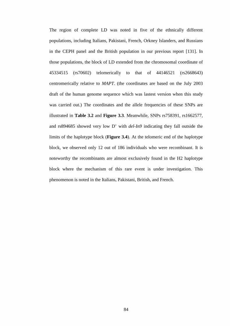
84
The region of complete LD was noted in five of the ethnically different
populations, including Italians, Pakistani, French, Orkney Islanders, and Russians
in the CEPH panel and the British population in our previous report [131]. In
those populations, the block of LD extended from the chromosomal coordinate of
45334515 (rs70602) telomerically to that of 44146521 (rs2668643)
centromerically relative to MAPT. (the coordinates are based on the July 2003
draft of the human genome sequence which was lastest version when this study
was carried out.) The coordinates and the allele frequencies of these SNPs are
illustrated in Table 3.2 and Figure 3.3. Meanwhile, SNPs rs758391, rs1662577,
and rs894685 showed very low D’ with del-In9 indicating they fall outside the
limits of the haplotype block (Figure 3.4). At the telomeric end of the haplotype
block, we observed only 12 out of 186 individuals who were recombinant. It is
noteworthy the recombinants are almost exclusively found in the H2 haplotype
block where the mechanism of this rare event is under investigation. This
phenomenon is noted in the Italians, Pakistani, British, and French.

85
Figure 3.3 The architecture of the tau haplotype block in different ethnicitiesThe haplotype block of MAPT region from rs2668643 (centromeric) to rs70602(telomeric) in six different ethnic groups. The solid bar (red and blue) shows thecomplete LD throughout the region. The red dashed bar shows the region thatrecombination may have occurred in given populations. The chromosomalcoordinations (Mb: million base pairs) are indicated on the left-hand axis.
86
Minor Allele Frequency of SNP
Ethnicity
Popula
tion
Num
ber
ofS
ubje
cts
rs894685
rs2668643
Tau
Ex3
SN
P
del_
In9
rs70602
rs1662577
rs758391
Italy Sardinian 27 0.19 0.30 0.28 0.28 0.24 0.33 0.39
OrkneyIsland
Orcadian 16 0.31 0.28 0.28 0.28 0.28 0.44 0.41
Russia Russian 24 0.19 0.10 0.10 0.10 0.10 0.40 0.35
Pakistan Makrani 27 0.57 0.15 0.11 0.11 0.07 0.35 0.37
France French 29 0.17 0.25 0.21 0.21 0.17 0.48 0.50
UnitedKingdom
British 63 0.19 0.19 0.20 0.20 0.17 0.42 0.50
Table 3.2 The allele frequencies of the SNPs used in the analysis of the MAPT haplotype block indifferent ethnicities.
Comparison between primate and human genomes over the MAPT region revealed
that according to the haplotype-defining insertion/deletion polymorphism marker
(del-In9), and SNPs rs1801353, rs1047833, rs1864325, rs1560310, rs767058,
rs754512, rs733966, rs2240758 and rs199533, the chimpanzee has the H1 variant.
On the other hand, with the rs393152, rs1078830, rs2055794, rs2217394,
rs1052553, STH Q7R, rs9468 and rs7687, the primate sequence corresponds to the
human H2 sequence (November 2003 MIT Chimp Assembly) suggesting that the
evolution of this locus has been complex.

87
Figure 3.4 Pair-wise D’ LD analysis of the different ethnic populations.The blocks are shaded corresponding to the values which were obtained from the LD analysisprogram UNPHASED.
Position dbSNP ID Chimp H1 H2
43795192 rs1801353 C C T
43817563 rs1047833 G G C
44194565 rs393152 G A G
44421540 rs1078830 C T C
44427146 rs2055794 A G A
44453262 rs1864325 C C T
44453969 rs1560310 G G A
44455965 rs1984937 C T G
44474234 rs767058 C C G
44528923 rs2217394 G A G
44531122 rs754512 T T A
44549365 rs1052553 G A G44552141 STH Q7R G A G
44562127 del-In9 + + -
44565039 rs733966 C C T
44577047 rs9468 C T C
44578781 rs7687 C T C
44723915 rs2240758 C C G
45103737 rs199533 C C T
Table 3.3 Polymorphisms in the extended MAPT locus that differentiate H1 clades from H2and comparison with the chimp assembly.
del-In9 is the H1/H2 haplotype-defining insertion deletion polymorphism in intron 9 of the MAPTgene and STH Q7R is the saitohin gene polymorphism that is in complete LD with del-In9. Allpositions are given relative to Build 34 (July 2003) of the Human Genome.

88
3.5 Discussion
The distribution of H2 haplotype exclusively in Caucasians is of interest for three
reasons; first, it raises the question of the origin of the H2 haplotype, since it
appears to be so divergent from the H1 haplotype and that recombination does not
occur with the H1 haplotype over ~1.8Mb [82;131]; second, it suggests that the
MAPT haplotype can be used as an approximate population marker for Caucasian
ancestry in admixed populations and third, it leaves the possibility open that
populations with more H1 individuals could have a higher incidence of H1-
associated diseases, especially, PSP and CBD.
The relative constancy of the H2 allele frequency in Caucasian populations from
the Middle East to the Orkneys suggests that its origin in European populations is
ancient and coincides with the colonization of Europe. The lower frequency in the
Finnish population is consistent with previous genetic data showing that this
group has a substantial genetic contribution from Asian populations [141]. It is
difficult to envisage the origin of the H2 haplotype, either as a mutational event or
as a result of admixture with an earlier human population: the divergence of the
two haplotypes is extensive and suggests considerable genetic separation. This
could reflect either a mutation preventing recombination, and thus maintaining the
separate integrity of the haplotype, or the ancient genetic isolation of a group of
humans in which this haplotype occurred.
Determining whether the difference in H2 haplotype frequency might lead to a
difference in the incidence of neurodegenerative disease is fraught with difficulty
at two levels: first cross-cultural comparisons of incidence of late-onset
neurodegenerative diseases is notoriously difficult and second, it is not yet clear

89
whether the H1 haplotype in toto, or some variant of it, is responsible for the
association; recent data would suggest the latter [142] and, if this were the case it
would make straightforward predictions concerning incidence, difficult until the
pathogenic variant(s) are precisely resolved. Nevertheless, a direct prediction of
disease incidence for these tauopathies based on H1 allele frequencies would be
that non-Caucasians would have an incidence approximately double that of
Caucasians since those populations would have nearly twice as many H1
homozygotes.
In the study of the MAPT haplotype block in different ethnicities, the complete
LD region was noted in five of the ethnically different populations, including
Italians, Pakistanis, French, Orkney Islanders, and Russians, in the CEPH panel
and the British population. The same LD pattern over MAPT region is shown by
different ethnic groups in the diversity panel confirms that this particular LD
block is shared between populations indicating that haplotype structure in human
is ancient, predating the separation of Caucasians.
This pattern of LD strongly suggests that the formation of the H2 haplotype was a
single event either indicating a chromosomal rearrangement [59;131;133] or
limited intermixing with a predating population [143]. Given this, the high
prevalence (~25%) of the H2 haplotype in Caucasians is surprising and may
suggest a strong selection for the H2 haplotype. Of course, the H2 haplotype is
protective against both PSP and CBD [67;130], but these diseases are far too rare,
and too late in lifespan to have had significant impact on allele frequencies.
However, there are occasions when tauopathies could become major causes of
mortality: on Guam, in Umatac, Parkinson–dementia complex was the major
90
cause of death in the 1930 to 1950s [129;135], the Spanish flu epidemic in 1919
led to an epidemic of postencephalitic Parkinsonism: von Economo’s syndrome
[129;144;145] and subacute sclerosing panencephalitis is a, now rare, but
frequently fatal complication of measles infection [129;146;147]. In the latter two
diseases, there has been no study of the MAPT haplotype, and in Guam the H2
haplotype is so rare that there is not sufficient evidence to show any association
between PDC and the H2 haplotype of MAPT. [59;134;140].
Analysis of the sequences on the H1 and H2 backgrounds, and comparison of
these sequences with those of the chimpanzee (Pan troglodytes) sequence show
that, while both H1 and H2 sequences are more similar to each other than to the
chimp sequence, they do not follow a predictable relationship: at some sequences,
the chimp sequence is similar to H1 and at others, it is similar toH2 (Table 3.3,
and also [148;149]). Thus the H1 and H2 sequences do not follow a precursor–
product relationship and one cannot be derived directly from the other, rather both
must have been derived independently from a more distant precursor. Logically,
therefore their relationship could be as illustrated in Figure 3.5.
During the time of the writing of this thesis, Stefansson et al, from the deCODE
group, has found that a ~900 kb inversion polymorphism at the region 17q21.31 in
H2 chromosomes with respect to the H1 haplotype [60]. Jaime Duckworth from
our group also used the bioinformatic data to show similar findings on the
structures in these distinct haplotypes. Chromosomes with the inverted segment in
different orientations represent two distinct lineages, H1 and H2, that have
diverged for as much as 3 million years and show no evidence of having
recombined [60]. This size of this inversion, 900-kb, is smaller than the linkage
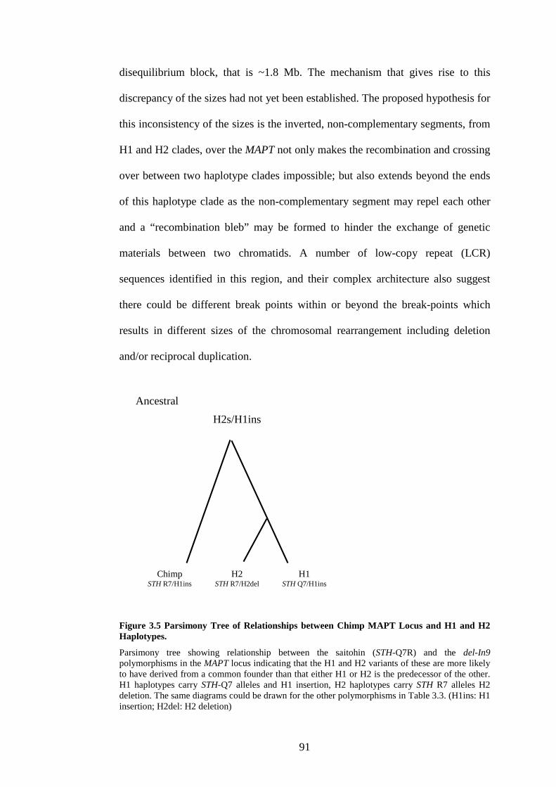
91
H2STH R7/H2del
H1STH Q7/H1ins
ChimpSTH R7/H1ins
Ancestral
H2s/H1ins
disequilibrium block, that is ~1.8 Mb. The mechanism that gives rise to this
discrepancy of the sizes had not yet been established. The proposed hypothesis for
this inconsistency of the sizes is the inverted, non-complementary segments, from
H1 and H2 clades, over the MAPT not only makes the recombination and crossing
over between two haplotype clades impossible; but also extends beyond the ends
of this haplotype clade as the non-complementary segment may repel each other
and a “recombination bleb” may be formed to hinder the exchange of genetic
materials between two chromatids. A number of low-copy repeat (LCR)
sequences identified in this region, and their complex architecture also suggest
there could be different break points within or beyond the break-points which
results in different sizes of the chromosomal rearrangement including deletion
and/or reciprocal duplication.
Figure 3.5 Parsimony Tree of Relationships between Chimp MAPT Locus and H1 and H2Haplotypes.
Parsimony tree showing relationship between the saitohin (STH-Q7R) and the del-In9polymorphisms in the MAPT locus indicating that the H1 and H2 variants of these are more likelyto have derived from a common founder than that either H1 or H2 is the predecessor of the other.H1 haplotypes carry STH-Q7 alleles and H1 insertion, H2 haplotypes carry STH R7 alleles H2deletion. The same diagrams could be drawn for the other polymorphisms in Table 3.3. (H1ins: H1insertion; H2del: H2 deletion)
92
Chapter 4 Genetic Association of MAPT haplotypes withprogressive supranuclear palsy and corticobasaldegeneration
4.1 Overview
The haplotype H1 of the tau gene, MAPT, was found to be highly associated with
progressive supranuclear palsy (PSP) and corticobasal degeneration (CBD) [130].
In order to investigate the pathogenic basis of this association, the association of
MAPT with PSP and CBD based on the underlying haplotype architecture of
MAPT was refined. The common haplotype structure of MAPT and associations
with these related tauopathies were also explored.
Detailed linkage disequilibrium (LD) architecture and common haplotype
structure of MAPT were examined in 27 CEPH-trio individuals. Based on this, 5
htSNPs were identified that capture 95% of the common haplotype diversity of
the region. These, together with the del-In9 polymorphism to define the H1/H2
division, were used to genotype well characterised PSP and CBD case-control
cohorts.
Two common haplotypes defined by the htSNPs and del-In9 were identified to be
associated with PSP, defining a candidate region of ~56 kb spanning sequences
from upstream of MAPT exon 1 to intron 9 on the H1 haplotype background, thus
supporting pathological evidence that underlying variations in MAPT could
contribute to disease pathogenesis possibly by subtle effects on gene expression,
mRNA stability and/or splicing. The sole H2-derived haplotype is under-
represented and, one of the common H1-derived haplotypes is highly associated,
93
with a similar trend observed in CBD. We also observed particularly powerful and
highly significant associations with PSP and CBD of haplotypes formed by 3 H1-
specific SNPs. These findings also form the basis for the investigation of the
possible genetic role of MAPT in Parkinson’s disease and other tauopathies,
including Alzheimer’s disease.
4.2 Background
PSP is usually a sporadic disorder of late adult life. It is the second most common
form of degenerative parkinsonism and is characterised clinically by an akinetic-
rigid syndrome, supranuclear gaze palsy, pseudobulbar signs and cognitive
decline of frontal lobe type [55;150;151]. CBD is an atypical parkinsonian
condition occurring much less frequently than PSP and classically presents with
unilateral cortical sensory loss, alien hand, jerky dystonia, rigidity, bradykinesia
and dementia. PSP is sporadic, with no familial history or MAPT mutations in the
large majority of cases. However, robust genetic association of PSP with MAPT
and reports of the rare families with more than one affected member [57;58]
indicated that genetic factors could play a role. Conrad and colleagues were the
first of many groups to show that variation at the MAPT locus could be an
important genetic influence in sporadic PSP by demonstrating allelic association
with PSP of a dinucleotide polymorphism in MAPT intron 9 [51]. The
overrepresentation of the commoner allele (a0) in PSP and also later in CBD was
then confirmed by a number of groups [66;67]. This suggests that either this
polymorphism itself could contribute to increased risk or that it is in linkage
disequilibrium (LD) with the actual causative variant. Although some MAPT

94
mutations in FTDP-17 cause a clinical picture closely resembling PSP [152-154],
no pathogenic variations of MAPT have yet been identified in clinically and
pathologically diagnosed sporadic and familial PSP [155].
The allelic association of MAPT with PSP and CBD was subsequently extended to
a series of polymorphisms extending over the entire MAPT coding region
spanning nearly 62 kilobases (kb). In approximately 200 unrelated Caucasians,
these polymorphisms were in complete LD, forming two extended haplotypes H1
and H2. The study demonstrated that the more common haplotype, H1, with
which the a0 allele segregated, was significantly over-represented in PSP [130].
Follow up studies extended the MAPT haplotype a further 68kb to the promoter
region of MAPT where three SNPs, highly associated with PSP, were in complete
LD with the rest of the MAPT haplotype [156;157]. This was then extended
extended to a ~1.8Mb haplotype which is in near complete LD [131]. This region
associated with PSP includes several other genes in addition to MAPT, including
saitohin [158;159] (STH; situated within intron 9 of MAPT), NSF (N-
ethylmaleimide sensitive factor), IMP5 (intramembrane protease 5, a presenilin
homologue) [160], CRHR1 (corticotrophin releasing hormone receptor) and
LOC284058, an unknown gene just adjacent to MAPT. (Figure 3.1) Identifying
the functional basis of the H1 haplotype association will be important in providing
an insight into the aetiopathogenesis of PSP and CBD. Although all the genes
within this multi-gene haplotype block are associated with PSP and CBD, the
hallmark tau pathology of these disorders strongly implicates MAPT itself.
The objective of the work in this chapter was therefore to exhaustively analyse the
MAPT haplotype association with PSP and CBD in order to identify non-coding
95
variants that could affect MAPT gene expression, splicing or processing, leading
to tau pathology and selective neuronal loss.
The findings in this section of the study were based on the results from a close
collaborative work with Alan Pittman. The findings have been published in
Journal of Medical Genetics (2005) [177]. With the framework of the association
study between H1c clade of MAPT and PSP and CBD, we went further to
investigate whether these genetic traits also associated with other
neurodegenerative diseases. The important findings in this section are an
inevitably important foundation of the whole project. Therefore, the details of the
study have been put in here for a complete picture of the study.
4.3 Analysis of MAPT haplotype structure in the CEPH-trios
SNP data for the region of the MAPT locus in 27 CEPH trios (Coriell Institute for
Medical Research; http://locus.umdnj.edu/nigms/) from the International HapMap
project (HapMap) web site (http://www.hapmap.org/), was downloaded for
genetic analysis of the MAPT. The raw SNP genotype data was analysed in TagIT,
a software package for identifying and evaluating tagging SNPs applied to
haplotype data, which also contains routines for inferring haplotypes from trio
material and LD analysis (http://popgen.biol.ucl.ac.uk/software) [123].
Initially, any SNPs that had a minor allele frequency of less than 5% were
removed from the HapMap data. The inconsistencies in the data through the
parental-offspring relationship in the CEPH-trios were also checked. A resulting

96
set of 24 SNPs and the del-In9 (Table 4.1) was used, they cover the entire MAPT
gene from upstream of the promoter to beyond exon 13, to infer haplotypes and
their respective frequencies by an Expectation – Maximisation (EM) (= 1x10⁻⁶)
algorithm specifically for CEPH-trio material (EM-trio) [123]. The average
density of the markers was one SNP every 6.7 kb. For convenience, the bi-allelic
(+/-) intron 9 deletion-insertion polymorphism (del-In9) was designated as a SNP.
A total of 34 haplotypes were resolved from parental chromosomes. The pair-wise
LD across MAPT for each SNP was then evaluated by both the measures of D’
and the square of the correlation coefficient (r2). Both measures were calculated
firstly by estimating pair-wise haplotype frequencies through EM-trio, then
assessing the statistical strength of association via a likelihood ratio test (LRT) by
comparing the EM frequencies with haplotype frequencies estimated assuming no
LD. Both measures of LD are based upon D, the basic pairwise-disequilibrium
coefficient, the difference between the probabilities of observing the alleles
independently in the population: D = f(A1B1) – f(A1)f(B1) [99]. A and B refer to
two genetic markers and f is their frequency. D’ is obtained from D/DMAX and a
value of 0.0 suggests independent assortment, whereas 1.0 means that all copies
of the rarer allele occur exclusively with one of the possible alleles at the other
marker. The measure of r2 has a more strict interpretation than that of D’, r2 = 1.0
only when the marker loci also have identical allele frequencies. The allele at the
one locus can always be predicted by the allele at the second locus. Recent work
suggests that r2 is viewed to be the preferred measure of LD for association based
studies [123].

97
SNP Positiona dbSNP ID Alleles Ancestral F1b F2b p-valuec
1 41291420 rs962885 C/T T 0.639 0.361 0.572
2 41301910 rs1078830 C/T C 0.189 0.811 0.426
3 41307507 rs2055794 A/G A 0.185 0.815 0.442
4 41324209 rs7210728 A/G A 0.259 0.741 0.248
5 41333623 rs1864325 C/T C 0.811 0.189 0.426
6 41334330 rs1560310 A/G G 0.185 0.815 0.442
7 41336326 rs3885796 G/T C 0.189 0.811 0.426
8 41342006 rs1467967 A/G A 0.648 0.352 0.851
9 41349204 rs3785880 G/T T 0.462 0.538 0.709
10 41354402 rs1467970 G/T T 0.185 0.815 0.442
11 41354620 rs767058 A/G C 0.815 0.185 0.442
12 41361649 rs1001945 C/G G 0.546 0.454 0.301
13 41374593 rs2435205 A/G A 0.593 0.407 0.251
14 41375573 rs242557 A/G G 0.396 0.604 0.854
15 41382599 rs242562 A/G G 0.375 0.625 0.684
16 41409284 rs2217394 A/G G 0.815 0.185 0.442
17 41410268 rs3785883 A/G G 0.204 0.796 0.524
18 41411483 rs754512 A/T T 0.185 0.815 0.442
19 41419081 rs2435211 C/T C 0.632 0.368 0.061
20 41429726 rs1052553 A/G G 0.815 0.185 0.442
21 41431900 rs2471738 C/T C 0.713 0.287 0.335
22 41442488 del-In9 +/- + 0.823 0.177 0.617
23 41445400 rs733966 C/T C 0.815 0.185 0.442
24 41457408 rs9468 C/T C 0.185 0.815 0.442
25 41461242 rs7521 A/G G 0.434 0.566 0.569
Table 4.1 The 24 single nucleotide polymorphisms and del-ln9 used for the linkagedisequilibrium and haplotype structure analysis of MAPT in the CEPH trios
The 24 SNPs and del-In9 used for the LD and haplotype structure analysis of MAPT in the CEPH-trios. The analysis was performed on the available genotype data for these SNPs from HapMap(http://www.hapmap.org/). In addition, we genotyped the del-In9 in the same CEPH-trios. Alleleand genotype frequencies and p-values for test to fit Hardy-Weinberg equilibrium were calculatedin the program TagIt. The ancestral allele (Chimpanzee) is also indicated. Position on chromosome(in bp) is based on May 2004 build of Human Genome Sequence (http://genome.ucsc.edu).aSNP position on chromosome ballelic frequencies in the CEPH-trioscp values for test to fit Hardy-Weinberg equilibrium
Allelic and genotype frequencies followed by statistical assessment of Hardy-
Weinberg equilibrium (HWE) were made at each locus in the CEPH-trios as
implemented by TagIT. From the LD and haplotype structure of MAPT, htSNPs
were selected to capture the diversity of known MAPT HapMap SNPs in the
CEPH trios. Six tagging SNPs (del-In9, rs1467967, rs242557, rs3785883,
98
rs2471738 and 7521) were selected, then, using TagIT, their performance was
assessed on the CEPH-trios. The tagging approach was focused on the coefficient
of determination (i.e. haplotype, r2) in a linear regression, which uses the
haplotypes defined by the htSNPs to predict the state of the tagged-SNPs. The
basis of this design is that even when individual haplotypes defined by the htSNPs
do not correlate perfectly with tagged-SNPs, haplotype combinations might do so,
and these combinations are identified by selection of the appropriate coefficients
in the linear regression. Haplotype r2: The coefficient of determination from an
analysis of variance of locus i (coding alleles at locus i as “0” or “1”) among the
G groups (number of haplotypes, or groups, defined in the data set in question by
the htSNP set); r2 [hap]i = 1 – R’i/D i, where R’i=2Σp’ig(1- p’ig)/xg which can be
interpreted as the sum of the within-group variances weighted by their frequency.
4.4 The PSP cases and control subjects
The unrelated PSP cases (n= 83) from the Queen Square Brain Bank for
Neurological Disorders, were all white and of western European origin and all
pathologically confirmed. The majority of these cases have been used in previous
studies [131;155;157;158;161]. Pathological confirmation of the diagnosis of PSP
was made following standardized criteria [161]. The unrelated British control
population (n=169), all white, were taken from brain bank tissue with no clinical
evidence of neurodegenerative disease and no abnormal histopathology, from the
MRC Building, Newcastle, UK. The samples were age matched, where the
average age at death was 73.5 years for the PSP cases (63% male) and 76 years
for the controls (51% male). All patients and controls were collected under

99
approved protocols followed by informed consent and this work was approved by
the Joint Research Ethics Committee of the Institute of Neurology and the
National Hospital for Neurology and Neurosurgery.
The unrelated US control population consisted of individuals (n=131; 50% males)
free of abnormal histopathology, an average age at death of 79.9 years. The
unrelated PSP cases (n=238; 50% males) consisted of pathologically confirmed
individuals by standard criteria with an average age at death of 75.3 years. The
unrelated CBD cases (n=44; 50% males) consisted of pathologically confirmed
individuals following standard criteria with an average age at death of 71.3 years.
4.5 Genotyping
The htSNPs (dbSNP numbers: rs1467967, rs242557, rs3785883, rs2471738 and
rs7521 and del-In9) were genotyped in the PSP case-control cohorts as follows:
The 238bp MAPT del-In9 was genotyped as in previous chapter (Chapter 3). PCR
primer pairs were designed by the Primer3 program (http://frodo.wi.mit.edu/cgi-
bin/primer3/primer3_www.cgi) and used to amplify each SNP of interest. PCR
reactions were as follows: 10μl reactions, which contained one unit of DNA
polymerase (Qiagen, Crawley, West Sussex), 10x PCR reaction buffer, 5x Q
solution (Qiagen), 10 pmoles of each oligonucleotide primer pair and 25ng of
sample template genomic DNA.
Genotyping of the SNPs, rs1467967, rs242557, rs3785883, rs2471738 and rs7521
were conducted by Pyrosequencing (Biotage AB) or by restriction digest (RFLP);

100
the following restriction endonucleases cutting the PCR product once at the (N)
allele, respectively: Dra I (A), ApaL I (A), BsaH I (G), BstE II (T) and Pst I (A)
(New England Biolabs and Fermentas). PCR products were incubated overnight
with 2 units of the corresponding restriction enzyme at the recommended
temperature. Digests were separated on 4% agarose gels and visualized with
ethidium bromide staining.
Genotyping accuracy was assessed by re-typing 20% of all genotypes, whole sets
of htSNPs, genotyping by alternative methods and by direct automated DNA
sequencing of random samples.
The ancestral allele at each locus was determined by direct sequence comparison
of the 24 SNP loci in human and chimpanzee MAPT and in addition by searching
for the ancestral allele in NCBI (http://www.ncbi.nlm.nih.gov/).
4.6 Statistical Analysis
For each htSNP, the allele and genotype distribution in the PSP cases were
compared with those in the control group. Statistical assessments for the allele and
genotype frequencies and HWE were made using TagIT. Case-control single-
locus htSNP allelic and genotypic association was calculated statistically in
CLUMP software [162]. The p-values were derived by standard Pearson’s 2 tests
except in cases where cell counts in the contingency tables were less than 5. When
cell counts were less than 5, p-values were determined empirically by 100,000
simulations; the program uses a Monte-Carlo approach that performs repeated

101
simulations to generate random tables having the same marginal totals as the one
under consideration and counting the number of times that a χ2 value associated
with the actual table is achieved by the randomly generated tables. The
heterogeneity between the H1/H1 homozygote populations versus the whole
population was tested using a standard Pearson’s 2 test.
Distribution of haplotypes defined by the htSNPs were compared in the PSP cases
and controls using WHAP software
(http://www.broad.mit.edu/personal/shaun/whap/). This is a SNP haplotype
analysis suite that performs a regression-based haplotype association test through
a LRT, which is χ2 and n-1 degrees of freedom to derive the associated p-value,
where n is the number of haplotypes observed for the data. This test was used to
give an initial assessment of haplotype association (an omnibus test) and then
individual haplotype tests (haplotype-specific tests) of association were performed
again through a LRT (d.f = 1) and by also obtaining empirical p-values by Monte-
Carlo methods (20,000 simulations used). To test the effect of the H1-specific
htSNPs whilst controlling for the extended H1/H2 haplotype, a set of equality
constraints was imposed under the null across the haplotypes identical at the del-
In9 and single-locus and haplotype analysis was performed. The p-values were
corrected according to the number of tests performed where appropriate by the
Bonferroni correction, the significance of which is discussed throughout the text.
102
4.7 Results
4.7.1 Linkage disequilibrium and haplotype structure of MAPT
The average density of the markers is one SNP every 6.7 kilobases (kb). None of
the polymorphisms deviated from Hardy-Weinberg equilibrium (HWE). The
details of all SNPs analysed in the CEPH-trios was summarized in Table 4.1.
Pairwise LD was evaluated across MAPT for all 24 selected SNPs and del-In9 in
the 27 CEPH-trios both by D’ and r2, calculated from the Expectation–
Maximisation-trio (EM-trio) inferred haplotypes. By pairwise LD analysis of the
25 SNPs in CEPH-trios, a greater diversity was identified than reflected by the
description of the two extended H1 and H2 haplotypes alone. The entire MAPT
gene is featured by significant LD as is particularly evident by the measure of D’
(Figure 4.1). However, when LD was assessed by the more stringent measure of
r2 (that accounts for differences in allele frequencies), it appeared more
fragmented, with SNPs that were in high r2 LD with each another, but in moderate
to low r2 LD with the extended H1 and H2 haplotype (defined by the del-In9 and
other SNP loci), suggesting that they are correlated with either the H1 or H2
haplotypes, but with differing frequency. This supports evidence of variability on
the background of these extended haplotypes. In fact, our analyses in the CEPH-
trios show that these underlying blocks of LD were variable exclusively on the
background of the extended H1 haplotype and therefore define haplotypes within
the H1 clade. LD correlation by D’ between many of the described H1-specific
SNPs is relatively low, suggesting a degree of linkage equilibrium between them;
this indicates that, unlike the H1 and H2 haplotypes, there are no constraints to
recombination between variants of the extended H1-haplotypes. This pattern of
LD across the extended H1 haplotype is essentially similar with smaller blocks in

103
the Taiwanese population, in which the extended H2 haplotype is absent (result is
shown in Chapter 6).
Figure 4.1 Linkage Disequilibrium (LD) across the MAPT gene. Numerical LD is presented bygrey-scale, pair-wise between each SNP by both D’ (upper right) and and the more stringentmeasure r2 (bottom left). The darker the shading indicates a higher extent of LD between the SNPs.SNPs are numbered as in Table 4.1.
The EM-inferred MAPT haplotypes and their respective frequencies were obtained
by using the EM estimation algorithm specifically tailored to deal with trio data
(EM trio) as structured in the CEPH-trios [123]. The phased haplotypes were also
obtained (n = 34, representing 42% of the total number of haplotypes in the
CEPH-trios) by resolving parental chromosomes in the CEPH-trios. EM-
predictions depict a total of 14 different MAPT haplotypes of frequency greater
than 1%. Three of these haplotypes are common, having a frequency greater than
10%, with the remaining 21 haplotypes having frequencies of less than 5%. Only

104
one of the common predicted haplotypes (haplotype A, frequency=18.1%) is
representative of H2 (Table 4.2).
It is noteworthy that in addition to the resolved H2 haplotype A, a single resolved
haplotype (haplotype G; frequency 2.9% in resolved) based on variation of H2
haplotype A was resolved that differed from haplotype A by SNP 13 (Table 4.2).
However, this haplotype was not predicted by EM-trio for output as a significant
frequency in the population and represents only ~5% (estimated by EM prediction)
of all H2 haplotypes in the CEPH-trios. It is thought that haplotype prediction
through EM is a more accurate representation of the relative haplotype
frequencies in a population than simply resolving ‘known’ haplotypes because of
a far greater utilisation of the data. The ancestral (chimpanzee) haplotype was also
constructed based upon the alleles of the 24 SNPs and the del-In9. This appears
not to resemble any haplotype present in the CEPH-trios, though its closest
relative (but different by ten loci) would appear to be that of the extended H2
(CEPH-trio haplotype A, from Table 4.2). The other ancestral SNP loci are either
consistent with the H1 haplotype family (SNPs rs962885, rs1864325, rs1560310,
rs1467970, rs1001945, rs754512 and rs733966), including the presence of the 238
bp insertion sequence (del-In9), or the allele is not observed in Homo sapiens
(dbSNPs rs3885796 and rs 767058).

105
Frequency (%)
IDa Haplotypeb EMc Rd
A 0 0 0 0 1 0 0 0 1 0 1 0 0 1 1 1 1 0 0 1 0 1 1 0 1 18.1 17.6
B 1 1 1 1 0 1 1 1 1 1 0 1 1 1 1 0 1 1 0 0 0 0 0 1 0 17.2 23.5
C 0 1 1 1 0 1 1 0 0 1 0 0 0 0 0 0 1 1 1 0 1 0 0 1 1 14.3 23.5
D 0 1 1 0 0 1 1 0 0 1 0 0 0 0 0 0 1 1 0 0 0 0 0 1 0 3.8 …
E 0 1 1 1 0 1 1 0 0 1 0 0 1 1 1 0 1 1 1 0 1 0 0 1 1 1.9 2.9
F 0 1 1 1 0 1 1 0 0 1 0 0 0 0 0 0 1 1 1 0 1 0 0 1 0 1.9 2.9
G 0 0 0 0 1 0 0 0 1 0 1 0 1 1 1 1 1 0 0 1 0 1 1 0 1 … 2.9
H 0 1 1 0 0 1 1 0 0 1 0 0 0 0 0 0 0 1 0 0 0 0 0 1 1 … 2.9
I 0 1 1 1 0 1 1 0 0 1 0 1 1 1 1 0 0 1 0 0 0 0 0 1 1 … 2.9
J 0 1 1 1 0 1 1 0 0 1 0 1 1 1 1 0 1 1 0 0 0 0 0 1 1 … 2.9
K 0 1 1 1 0 1 1 1 1 1 0 1 1 0 0 0 0 1 1 0 1 0 0 1 1 … 2.9
L 0 1 1 1 0 1 1 1 1 1 0 1 1 1 1 0 0 1 0 0 0 0 0 1 1 … 2.9
M 1 1 1 1 0 1 1 0 0 1 0 0 0 1 1 0 1 1 0 0 0 0 0 1 0 … 2.9
N 1 1 1 1 0 1 1 0 0 1 0 1 0 0 0 0 1 1 0 0 0 0 0 1 1 … 2.9
O 1 1 1 1 0 1 1 1 1 1 0 1 0 0 0 0 0 1 1 0 1 0 0 1 1 … 2.9
P 1 1 1 1 0 1 1 1 1 1 0 1 0 1 1 0 1 1 0 0 0 0 0 1 0 … 2.9
Q 1 1 1 1 0 1 1 1 1 1 0 1 1 1 1 0 0 1 1 0 1 0 0 1 1 1.9 …
R 1 1 1 1 0 1 1 1 1 1 0 1 0 0 0 0 1 1 0 0 0 0 0 1 0 1.9 …
S 0 1 1 1 0 1 1 0 0 1 0 0 0 1 1 0 1 1 0 0 0 0 0 1 0 1.9 …
T 0 1 1 1 0 1 1 0 0 1 0 1 1 1 1 0 1 1 0 0 0 0 0 1 0 1.9 …
U 0 1 1 1 0 1 1 0 0 1 0 1 1 1 1 0 0 1 0 0 0 0 0 1 1 1.9 …
V 0 1 1 1 0 1 1 0 0 1 0 0 1 1 1 0 0 1 0 0 0 0 0 1 0 1.9 …
W 0 1 1 1 0 1 1 0 0 1 0 0 1 0 0 0 1 1 1 0 1 0 0 1 1 1.9 …
X 1 1 1 1 0 1 1 1 1 1 0 1 0 0 0 0 1 1 0 0 0 0 0 1 1 1.9 …
1 0 0 0 0 1 - 0 1 1 - 1 0 1 1 1 1 1 0 1 0 0 0 0 1 Ancestral
Table 4.2 The haplotype structure of the MAPT gene in CEPH-trios.The haplotype structure is based upon the 25 markers in Table 4.1. Alleles represented in binary (1=highest letter in alphabet of SNP allele). Haplotypes shown if observed inresolved chromosomes (parental chromosomes, n = 34) or if Expectation-Maximisation (EM-trio) inferred haplotype frequency exceeded 1%. Additionally presented is thebuild of the ancestral genotype (Chimpanzee). a:haplotype identity. b: binary representation. c: infered frequency by Expectation-Maximization (all data). d: resolved haplotype frequency

106
4.7.2 Selection, performance assessment and association analysis of MAPT
haplotype-tagging SNPs in PSP and CBD
Using an association-based criterion (criterion 5 in TagIT, haplotype r2), the
htSNPs were selected [123]. Six htSNPs (rs1467967, rs242557, rs3785883,
rs2471738 and rs7521 and the del-In9) are sufficient to represent all the HapMap
SNPs in the 27 CEPH-trios with a high coefficient of determination. Five of these
htSNPs are H1-specific i.e. they vary only on the H1 background. In addition the
bi-allelic del-In9 marker is used to unambiguously distinguish the extended H1
and H2 haplotypes [130].
In CEPH-trios, the performance value for the 6 htSNPs and del-In9 in the CEPH-
trios was interpreted at an average haplotype r2 value of 0.95 (95%) and a
minimum r2, interpreted as the minimum locus value of 0.68. Excluding the del-
In9 from the set of htSNPs results in a loss of performance of only of 3%, with
performance down to 92% with the five remaining H1-specific htSNPs. This is
because a particular allelic combination of these 5 H1-specific SNPs is
representative of the extended H2 haplotype. The performance value of just the
del-In9 against the known SNPs in the CEPH-trios is just 50% [123] .
The MAPT htSNPs were genotyped in two separate PSP case-control cohorts from
the UK and USA and CBD cases from USA. Single locus association results are
summarised in Table 4.3. In all the groups, there were no significant deviations
from HWE at any of the htSNPs. The strong association of the del-In9 with PSP
was again verified in both the UK and US cohorts (p=1.14x10⁻⁵, 4.021x10⁻⁸,
respectively; Table 4.3). The same trend was observed in CBD but the difference

107
was not significant, possibly due to a small sample size. No evidence of
association was found for htSNPs rs1467967, rs3785883 and rs7521 in the studies,
except in the US CBD study where htSNP rs3785883 is moderately associated
(p=0.019, allelic). The OR and their 95% confidence intervals were calculated.
The values for all 6 htSNPs by comparison of each minor allele verses each major
allele was present (Table 4.3).
Frequency (F1%) Association (p) Odds Ratio (MA)
dbSNP ID Cases Controls Allelic Genotypic OR 95% CI
US PSP
rs1467967 62.8 62.6 0.963 1 0.965 0.703~1.325
rs242557 54.4 31 2.91ex-9 2.29ex-8 2.356 1.706~3.255
rs3785883 17 22.4 0.072 *0.168 0.713 0.487~1.044
rs2471738 67 81.5 1.87ex-5 *1.15ex-4 2.224 1.535~3.222
del-In9 91.6 77.1 4.02ex-8 *1.00ex-5 0.298 0.193~0.462
rs7521 43.2 44.5 0.456 0.671 1.124 0.827~1.526
UK PSP
rs1467967 67.9 64.6 0.993 0.77 0.998 0.639~1.56
rs242557 47.9 35.7 0.012 0.016 1.815 1.209~2.726
rs3785883 25.5 20.6 0.365 0.68 1.227 0.762~1.974
rs2471738 66 80.1 0.001 0.005 2.142 1.368~3.355
del-In9 93.2 76.6 1.14ex-5 5.31ex-5 0.215 0.099~0.466
rs7521 51.2 45.7 0.546 0.814 0.773 0.505~1.183
US CBD
rs1467967 61.9 62.6 0.909 *0.870 1.03 0.619~1.713
rs242557 50 31 0.002 0.01 2.231 1.322~3.764
rs3785883 33.3 22.4 0.019 0.022 1.047 0.586~1.872
rs2471738 67 81.5 0.005 0.011 2.165 1.254~3.736
del-In9 86.4 77.1 0.063 ** 0.532 0.271~1.043
rs7521 43.2 44.5 0.826 0.464 0.807 0.494~1.32
Table 4.3 Allele frequencies (F1) and p-values of single-locus association in the three studies.
Allele frequencies (F1) and p-values of single-locus association in the three studies. The p-valueswere derived by standard Pearson’s 2 tests except in cases where cell counts in the contingencytables were less than 5. When cell counts were less than 5 (*), p-values were determinedempirically by 100,000 simulations (CLUMP software). **A genotypic test was not performed forthe del-In9 in intron 9 in the CBD series, since there were no rare homozygotes in the CBD cases,thus preventing us from performing a valid test. Significant single-locus association of htSNPs areindicated in bold. Odds ratios and their 95% confidence interval are presented for the minor allele(MA) verses the major allele for all htSNPs.
108
The H2 haplotype as defined by del-In9 is a significant protective factor. The H1-
specific SNPs rs242557 and rs2471738 are highly associated with these diseases
and are arguably as important for risk as the association of the extended H1
haplotype. This could particularly be the case in CBD in light of the lack of
association of del-In9 in this particular study.
There is potentially the greater power to detect the contribution to association of
causal variants by performing tests of association for the htSNP-defined
haplotypes rather than individual htSNPs themselves. The six htSNPs were
identified to capture 95% of the common haplotypic diversity of MAPT. An
omnibus test of haplotype frequency differences estimated by EM between cases
and controls in both the UK and US PSP groups was performed. The haplotype
distribution (all haplotypes >1.0%) was found to be highly significant in the UK
PSP cohort (p = 9.75x10⁻⁵, d.f = 19) and in the US PSP cohort (p= 7.40x10⁻¹², d.f
= 20) but not in CBD (p=0.120, d.f = 17). In addition to the global significance of
the haplotype-wide comparison, individual haplotype tests (d.f = 1) were
undertaken for significance through LRT and empirical p-values were derived
through Monte-Carlo methods (20,000 simulations, data not shown). Two
common haplotypes, A and C, which were strongly associated with both UK and
US PSP were identified (Table 4.4). Haplotype A, which derives from the del-
In9-defined H2 haplotype was the most common haplotype in the controls and
was significantly under-represented in both PSP groups. Haplotype C, a variant of
the H1 clade, was highly overrepresented in PSP. It was the commonest haplotype
in PSP but not in the control groups. The most common H1 derived haplotype in
the control population was not associated with either PSP or CBD. These trends
were observed in CBD, though on correction for multiple comparisons, no
109
haplotype was significantly associated. In both PSP cohorts, after strict correction
according to the number of tests performed, only associations of haplotypes A and
C remained significant. Associated haplotypes A and C, derived from the H2 and
H1 haplotypes, respectively, differ by only two H1-specific htSNPs rs242557 and
rs2471738 which, in addition the del-In9, also show powerful single-locus effects.
Haplotypes A and C do not differ by htSNPs rs1467967 and rs7521, and these
SNPs are not associated. The reduction in haplotype A (H2) appears almost
entirely accounted for by the increase in the H1 haplotype C.

110
htSNPhaplotypes
UKPSP
USPSP
USCBD
Frequency (%) Association (LRT) Frequency (%) Association (LRT) Frequency (%) Association (LRT)
ID rs1467967
rs242557
rs3785883
rs2471738
del-In
9
rs7521
Control Case p(pcorrected) Control Case p(pcorrected) Control Case p(pcorrected)
A A G G C H2 G 20.7 6.3 1.46ex-5 (2.77ex-4) 22 6.3 9.55ex-9 (2.01ex-7) 22 8.2 0.02 (0.367)
B G G G C H1 A 16.5 13.9 0.378 (1.000) 12.2 15.8 0.562 (1.000) 12.2 15.4 0.914 (1.000)
C A A G T H1 G 11.3 24.3 0.001 (0.022) 7.8 24 6.42ex-9 (1.35ex-7) 7.8 17.7 0.066 (1.000)
D A A G C H1 A 8.9 3.7 0.110 (1.000) 4 7.9 0.077 (1.000) 4 7.5 0.489 (1.000)
E A G G C H1 A 6.4 8.4 0.949 (1.000) 15.7 6.5 0.014 (0.294) 15.7 4.6 0.148 (1.000)
F G G A C H1 A 4 1 0.291 (1.000) 1.4 0 … 1.400 4.6 0.588 (1.000)
G G A A C H1 A 3.9 5.1 0.691 (1.000) 2.6 3.5 0.937 (1.000) 2.6 3.4 0.834 (1.000)
H A G A C H1 A 2.6 6.5 0.010 (0.173) 0 3.8 0.404 (1.000) 0 0 …
I G A G C H1 A 2.6 3.8 0.960 (1.000) 4.4 5.2 0.376 (1.000) 4.4 3.3 0.61 (1.000)
J A G G C H1 G 2.4 0 0.033 (0.621) 0 3 0.055 (1.000) 0 3.4 0.237 (1.000)
K A A A C H1 G 2.2 0.9 0.378 (1.000) 0 0 … 0.000 0 …
L A G A C H1 G 2.2 4.1 0.496 (1.000) 3.8 3.4 0.338 (1.000) 3.8 0 0.759 (1.000)
M G A G C H1 G 2 2.6 0.744 (1.000) 3.5 3.4 0.930 (1.000) 3.5 5 0.319 (1.000)
N G G A C H1 G 0.9 3.7 0.331 (1.000) 4.3 0.6 0.005 (0.105) 4.3 0 0.018 (0.322)
O A A A C H1 A 0 3.6 0.070 (1.000) 3.4 1.3 0.350 (1.000) 3.4 5 0.386 (1.000)
P G G G T H1 G 1.2 3.4 0.509 (1.000) 0.4 1.4 0.628 (1.000) 0.4 0 …
Q A A G T H1 A 0.7 2.8 0.040 (0.760) 0 1.6 0.003 (0.073) 0 1.2 …
R A G G T H1 G 0.7 2.7 0.114 (1.000) 2.4 1.6 0.386 (1.000) 2.4 1.5 0.493 (1.000)
S G G G C H1 G 1.4 2.4 0.599 (1.000) 2.6 2 0.920 (1.000) 2.6 0 0.621 (1.000)
T A G A T H1 G 0.3 0 … 1.100 0 … 1.100 7 0.713 (1.000)
U A A G C H1 G 1.1 0 … 1.100 1.7 0.270 (1.000) 1.1 3.5 0.17 (1.000)
v G G A T H1 G 1.3 0 … 1.900 1 0.207 (1.000) 1.9 2.8 0.699 (1.000)
w G G G C H2 G 0 0 … 0.000 0 … 0.000 2.9 0.326 (1.000)
x G A A T H1 G 0 0 … 2.700 0.5 0.205 (1.000) 2.7 0 0.174 (1.000)
Table 4.4 Association of common MAPT haplotypes with progressive supranuclear palsy and corticobasal degenerationThe above analysis was based on the output of all haplotypes (>90%), but only those with a frequency >2% were tested for association through thelikelihood ratio test (LRT). After adjustment of p-values, in parentheses, for correction of multiple testing, only haplotypes A and C in both PSP studiesremain significant. No haplotype is significantly associated with CBD after correction for multiple testing.

111
Haplotype Frequency (%) and association (LRT) of haplotype
UK PSP US PSP US CBD
ID
rs24
25
57
rs37
85
88
3
rs24
71
73
8
Contr
ol
PS
P
p-v
alu
e
p(c
orr
ecte
d)
Contr
ol
PS
P
p-v
alu
e
p(c
orr
ecte
d)
Contr
ol
CB
D
p-v
alu
e
p(c
orr
ecte
d)
I G G C 50 30.7 3.14ex-4 (2.51ex-3) 51.3 34.7 1.65ex-5 (1.32ex-4) 51.3 32.6 0.002 (0.019)
II A G T 12 28.3 2.16ex-4 (1.73ex-3) 8.3 27.6 2.31ex-9 (1.85ex-8) 8.3 17.8 0.009 (0.070)
III A G C 13.2 10.2 0.349 (1.000) 13.9 17.7 0.091 (0.730) 13.90 22.1 0.145 (1.000)
IV G A C 10 16.6 0.316 (1.000) 10.2 7.1 0.008 (0.064) 10.2 0 0.034 (0.275)
V A A C 6.9 9 0.454 (1.000) 6.1 6.8 0.728 (1.000) 6.1 12.4 0.619 (1.000)
VI G G T 2.2 5.2 0.087 (0.700) 4 3 0.611 (1.000) 4 4.4 0.603 (1.000)
VII A A T 3.2 0 0.907 (1.000) 2.9 1.6 0.751 (1.000) 2.9 0 0.321 (1.000)
VIII G A T 2.4 0 0.045 (0.356) 3.4 1.4 0.103 (1.000) 3.4 10.7 0.186 (1.000)
Table 4.5 Association of the subset of htSNP haplotypes with progressive supranuclear palsy and corticobasal degenerationThis haplotype analysis was based on a subset of H1 specific htSNP defined haplotypes that show evidence of association after consideration of the del-in9. After correction of p values for multiple testing (bracketed p values), haplotypes I and II in both PSP studies and haplotype I in the CBD studey aresignificant. CBD, corticobasal degeneration; LRT, likelihood ratio test; PSP, progressive supranuclear palsy.

112
4.7.3 Common variation in MAPT is associated with PSP and CBD
To assess whether the significant association with PSP of any of the H1-specific
htSNPs, are independent to that of del-In9, each htSNP was incorporated as
additional explanatory factors to the logistic regression model of the del-In9 that
serves to define the extended H1 and H2 haplotype status. Significant associations
of single locus htSNPs rs242557, rs3785883 and rs2471738 were found (p =
9.00x10⁻⁶, 2.87x10⁻³ and 2.73x10⁻³ respectively) for the US PSP cases, htSNP 21
(p = 0.0421) for the UK PSP cases and htSNPs 14 and 21 (p = 0.0183 and 0.0436,
respectively) for the CBD cases. The effects of haplotypes were probed on sub-
sets of htSNPs again entering the extended haplotype (H1 and H2 status, defined
by the del-In9) as an explanatory factor. Highly significant differences were found
in the distribution of haplotypes defined by three htSNPs rs242557, rs3785883
and rs2471738 in the UK and US PSP and to a lesser extent, the CBD cases (p=
9.34x10⁻⁴, 9.31x10⁻⁵, 0.0292, respectively). This was significant (p =2.49x10⁻⁵,
1.44x10⁻⁸, 0.006) in UK and US PSP and CBD, respectively, when the extended
haplotype was excluded as an explanatory factor (Table 4.4). The haplotypes
those SNPs define are associated with PSP and CBD after consideration of the
del-in9, suggestive that variability of MAPT within the extended H1 clade is a risk
factor in PSP and CBD. Haplotype II (A-G-T) was greatly overrepresented in
each group and the Haplotype I (G-G-C) under-represented (Table 4.5). The
SNPs rs242557, rs3785883 and rs2471738 are H1-specific SNPs in MAPT, i.e.
variable only on the H1 background though the haplotype I allelic combination is
fixed and representative of H2 in addition to H1-derived variants.

113
The htSNP data were re-analysed, after removing all individuals with a H2
chromosome, thus leaving us with a biased H1H1 homozygote population. A
significant (p <0.05) heterogeneity was found in both the control groups after the
removal of the H2 chromosomes, namely at rs1467967 and rs7521 in the US
group and at rs242557, rs2471738 and rs7521 in the UK controls. Removal of the
H2 chromosomes would therefore prevent us from performing valid ‘H1-only’
haplotype analyses in our Caucasian cohorts. For this purpose, it would be
important to extend this study in an H1-only population such as the Japanese and
Taiwanese [59].
4.8 Discussion
To date, genetic association studies have involved the study of one or a few
random polymorphisms in a gene, an approach that bears the risk of missing
adjacent regions of LD within the gene that harbour variants associated with
phenotype. It is therefore important that the haplotype architecture of the entire
gene is considered in order to determine its association with a particular complex
phenotype. In our attempt to provide insight into the basis of the well-established
association of MAPT with PSP and CBD, the haplotype tagging approach was
applied in this study. This provides a substantially streamlined and economical
protocol by using a minimal set of tagging SNPs to study the LD and common
haplotypic diversity of the entire gene or locus.
The underlying LD and haplotype structure of MAPT were first assessed using a
high density map of genotype data from the HapMap project

114
(http://www.hapmap.org). This involved LD analysis using genotype data for 24
SNPs that had been validated in CEPH-trios. In addition, the del-In9 status at the
MAPT locus were included to define the H1 and H2 haplotypes [130]. This
revealed multiple distinct haplotypes based upon the H1 and H2, as defined by
del-In9 with no evidence of recombination between the multiple H1 haplotypes
and the H2 in the CEPH-trios. The presence of multiple H1 haplotypes, inferred
both by EM and resolved to phase, shows a considerable diversity within this
extended haplotype. This H1 haplotype-specific diversity was first suggested by
Golbe and colleagues, based on microsatellite variability [163]. The strict H1/H2
dichotomy and H1 diversity across MAPT and beyond has also been demonstrated
in other studies [49;164]. In a more recent study [60], the lack of recombination
between H1 and H2 has been shown to be due to inversion of the chromosomal
region on 17q21.31 corresponding to the extended MAPT H1/H2 haplotype block
which described in previous chapter [131].
Then, an association-based criterion was used to assign a set of five haplotype-
tagging SNPs (htSNPs) that, together with del-In9 as a sixth bi-allelic tagging
polymorphism, capture 95% of the common haplotype diversity in MAPT. The six
htSNPs were genotyped in two PSP and one CBD case-control cohorts in order to
determine if any particular haplotype had greater association with disease with the
extended H1. In PSP, very strong associations of two common haplotypes were
clearly demonstrated. Firstly, the significant under-representation of the ‘classical’
H2 (haplotype A, Table 4.4) and secondly, strong over-representation of an H1-
derived haplotype (haplotype C, Table 4.4). The other htSNP-derived common
H1 haplotype (haplotype B) showed no association in any of the groups. Some
115
weaker associations of rare haplotypes were detected but were not consistent in
both the British and American cohorts in PSP and significance did not remain
after correction for multiple comparisons. Furthermore, it is difficult to assess the
association of such low-frequency haplotypes in populations of our sample size.
Similar trends were observed in the small number of CBD cases (n=44) with
under-representation of H2 (Haplotype A) and overrepresentation of the H1-
derived haplotype C (Table 4.4). However, they were not significant, possibly
due to the smaller number of CBD cases. Assuming that these findings can be
confirmed in a larger CBD cohort, they suggest that causative variant(s) in PSP
and CBD may affect the same region of MAPT or perhaps even be the same
variant.
Pastor and colleagues defined an extended region in LD of 1.14Mb around MAPT
that is associated with PSP and CBD. Within this haplotype, they similarly
defined a “protective” H2 haplotype that has a significant negative association
with PSP and CBD and an H1-derived haplotype that is associated with PSP and
CBD [165]. The haplotype structure and its associations of the MAPT gene alone
are refined in this study. A particular H1-derived haplotype in MAPT has been
demonstrated to be highly associated with PSP.
In an attempt to further minimise the candidate pathogenic domain of MAPT, a
strong association with PSP and CBD of three-locus haplotypes were identified
based on the sub-set of H1-specific htSNPs, rs242557, rs3785883 and rs2471738.
These associations are independent of the extended H1 and H2 haplotypes,
defined by del-In9. Haplotypes derived from these SNPs span a minimal region
from dbSNPs rs242557 to rs2471738 on the H1 haplotype background in MAPT.
116
This minimal region incorporates ~56.3kb of sequence, from upstream of exon 1
downstream to intron 9 that could harbour potential causal variant(s) that are in
LD with these SNPs. Skipper and colleagues defined a similar associated
candidate region in the 5’-half of MAPT in Norwegian PD cases, thereby
proposing genetic variability that could influence the alternative splicing of MAPT
exons 2 and 3 or, expression levels of MAPT. However, they carried out their
analysis only on H1 homozygous individuals, having removed all H2 carriers [49].
For this reason, we cannot compare findings from both studies. As explained
previously in section 4.7.3, unbiased inclusion of the entire study cohort,
irrespective of H1/H2, status is essential in order to obtain an accurate
representation of haplotype diversity in the population in question. Another study
implicated a MAPT promoter haplotype in PD based not only on allelic
association of the previously defined extended H1 haplotype but also on
differences in transcriptional activity [142]. In future studies, it would be
important to compare LD and association of the MAPT locus in PSP, CBD and
PD using standardised procedures in order to determine if they share the same risk
variants of the MAPT locus that contribute to disease.
The haplotypes we identified that confer protection, risk or are neutral in PSP and
CBD pathogenesis, provide us with the basis for targeted direct sequencing
strategies for MAPT. It is now clear that there are no obvious pathogenic missense
or splice site mutations in MAPT in the large majority of sporadic PSP cases [130].
It is more plausible that the associated SNPs in our study that confer greatest risk
(SNPs rs242557 and rs2471738, Table 4.3), or protection (del-In9 and associated
SNPs through LD; Table 4.1 and Figure 4.1) are in LD with variants that could
117
cause subtle changes either in the alternative splicing or overall expression levels.
It is possible that each neuronal sub-group is dependent on a particular tau
isoform profile and expression level. Aberrations in this homeostasis, could affect
one neuronal sub-group more than another and lead to the selective and disease-
specific neuronal death and tau pathology [166].

118
Chapter 5 Association of tau haplotype-tagging polymorphisms
with Parkinson disease in diverse ethnic cohorts.
5.1 Overviews
The genetic variation of MAPT, has not only been found to be associated with
tauopathies, including Alzheimer’s disease[167], progressive supranuclear palsy
and corticobasal degeneration[67;130;131;165] as described in Chapters 4 and 6.
Several MAPT polymorphisms that define the tau H1 haplotype have been
investigated for an association with PD with conflicting results[133;142;168]. In
order to demonstrate the association of MAPT with PD, a systematic framework
of genetic analysis was devised to examine the possible genetic variations for
genetic study in PD case-control cohorts from three ethnically diverse
populations: Taiwanese, Greek and Finnish.
A moderate association at SNP rs3785883 in the MAPT region in the Greek
cohort as well as for SNP rs7521 and rs242557 (p=0.01 genotypic p=0.04 allelic)
in the Finnish population were found. There were no significant differences in
genotype or allele distribution between cases and controls in the Taiwanese
cohort. There is therefore no consistent association between the MAPT H1
haplotype and PD in three ethnically diverse populations; however, the sub-
haplotypes of MAPT H1 may confer susceptibility to PD.

119
5.2 Background
5.2.1 Overlaps in the clinical and pathological features of tauopathies and
synucleinopathies
Parkinson disease (PD) is the second most common chronic neurodegenerative
disease, characterized by tremor, rigidity, postural instability and bradykinesia.
Epidemiological studies have estimated a cumulative prevalence of PD of greater
than 1 per thousand. PD belongs to a group of diseases termed ‘synucleinopathies’
based on the strong immunostaining of its pathological hallmark, Lewy bodies,
for α-synuclein.[169] Increasing evidence indicates that there is an overlap of the
clinical and pathological features of tauopathies and synucleinopathies, thereby
re-enforcing the notion that these disorders might be linked mechanistically. This
observation raises the possibility that tau protein may be important in PD
pathogenesis.[170;171]
5.2.2 Genetic risk factors of Parkinson’s disease
One of the strongest risk factors of PD is a positive family history. The estimated
genetic risk ratio for PD is approximately 1.7 (70% increase risk for PD if a
sibling has PD) for all ages, and increases over 7-fold for those under age 66
years. Growing evidence shows that genetic abnormalities play a major role in the
aetiopathogenesis of PD. Several loci for familial PD have been reported,
including α-synuclein (SNCA), parkin, PTEN-induced kinase 1 (PINK1),
ubiquitin-C terminal hydrolase-L1 (UCH-L1), DJ-1 and leucine-rich repeat kinase
2 (LRRK2). (Table 5.1) SNCA was the first genetic factor linked to familial PD. In
1996, PD within the Contursi kindred was linked to chromosome 4q21-23 and in
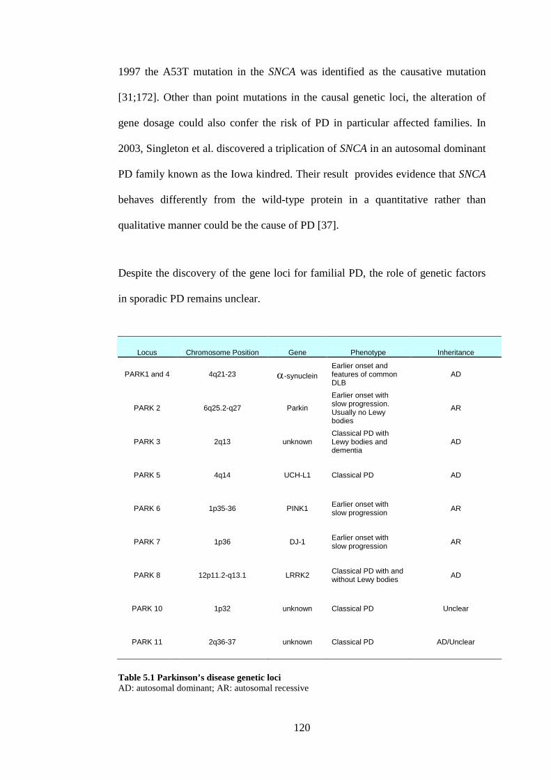
120
1997 the A53T mutation in the SNCA was identified as the causative mutation
[31;172]. Other than point mutations in the causal genetic loci, the alteration of
gene dosage could also confer the risk of PD in particular affected families. In
2003, Singleton et al. discovered a triplication of SNCA in an autosomal dominant
PD family known as the Iowa kindred. Their result provides evidence that SNCA
behaves differently from the wild-type protein in a quantitative rather than
qualitative manner could be the cause of PD [37].
Despite the discovery of the gene loci for familial PD, the role of genetic factors
in sporadic PD remains unclear.
Locus Chromosome Position Gene Phenotype Inheritance
PARK1 and 4 4q21-23 -synuclein
Earlier onset andfeatures of commonDLB
AD
PARK 2 6q25.2-q27 Parkin
Earlier onset withslow progression.Usually no Lewybodies
AR
PARK 3 2q13 unknownClassical PD withLewy bodies anddementia
AD
PARK 5 4q14 UCH-L1 Classical PD AD
PARK 6 1p35-36 PINK1Earlier onset withslow progression
AR
PARK 7 1p36 DJ-1Earlier onset withslow progression
AR
PARK 8 12p11.2-q13.1 LRRK2Classical PD with andwithout Lewy bodies
AD
PARK 10 1p32 unknown Classical PD Unclear
PARK 11 2q36-37 unknown Classical PD AD/Unclear
Table 5.1 Parkinson’s disease genetic lociAD: autosomal dominant; AR: autosomal recessive

121
One of the genetic susceptibility factors that have been found to be associated
with sporadic PD is MAPT. In spite of two meta-analyses supporting an
association between MAPT haplotype H1 and PD[49;173], several MAPT
polymorphisms that define the H1 haplotype have been investigated for an
association with PD with conflicting results.
The aim of the work described in this chapter was to determine the possible
genetic role of MAPT in PD in three ethnically different PD populations, using
htSNPs and to demonstrate if there is a consistent association between the MAPT
H1 haplotype (delineated by del-In9) and PD in different populations worldwide.
5.3 Case-control samples
In this study, 508 patients and 611 healthy controls were recruited from Greek,
Finnish and Taiwanese populations (Table 5.2). All patients were sporadic, based
on pedigree analysis. The PD diagnoses were verified according to PD Society
Brain Bank criteria[174]. Patients with dementia or patients who reported first-
degree relatives with parkinsonism were excluded. Patients with evidence of
secondary parkinsonism or with atypical features such as early dementia,
ophthalmoplegia, early autonomic failure, and pyramidal signs were not included
in this study. Age-at-onset was defined as the age at which the patient noticed the
first symptom indicative of PD. Controls were age-matched normal subjects living
in the same geographical area as the patients who visited our outpatient clinic and
finally were found free of any neurological disease (PD included). After approval
from the hospital internal ethics and scientific boards and written informed
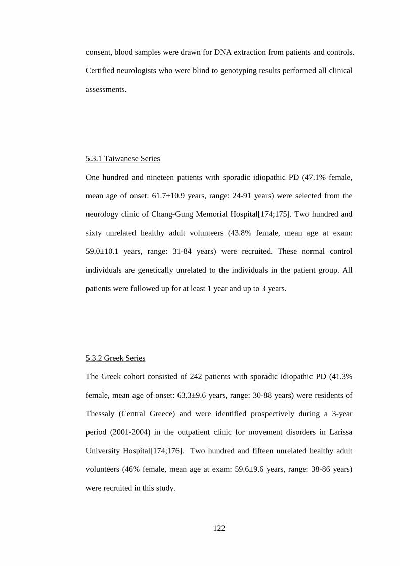
122
consent, blood samples were drawn for DNA extraction from patients and controls.
Certified neurologists who were blind to genotyping results performed all clinical
assessments.
5.3.1 Taiwanese Series
One hundred and nineteen patients with sporadic idiopathic PD (47.1% female,
mean age of onset: 61.7±10.9 years, range: 24-91 years) were selected from the
neurology clinic of Chang-Gung Memorial Hospital[174;175]. Two hundred and
sixty unrelated healthy adult volunteers (43.8% female, mean age at exam:
59.0±10.1 years, range: 31-84 years) were recruited. These normal control
individuals are genetically unrelated to the individuals in the patient group. All
patients were followed up for at least 1 year and up to 3 years.
5.3.2 Greek Series
The Greek cohort consisted of 242 patients with sporadic idiopathic PD (41.3%
female, mean age of onset: 63.3±9.6 years, range: 30-88 years) were residents of
Thessaly (Central Greece) and were identified prospectively during a 3-year
period (2001-2004) in the outpatient clinic for movement disorders in Larissa
University Hospital[174;176]. Two hundred and fifteen unrelated healthy adult
volunteers (46% female, mean age at exam: 59.6±9.6 years, range: 38-86 years)
were recruited in this study.
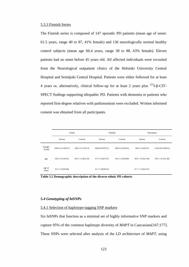
123
5.3.3 Finnish Series
The Finnish series is composed of 147 sporadic PD patients (mean age of onset:
61.5 years, range 40 to 87, 41% female) and 136 neurologically normal healthy
control subjects (mean age 66.4 years, range 38 to 88, 63% female). Eleven
patients had an onset before 45 years old. All affected individuals were recruited
from the Neurological outpatient clinics of the Helsinki University Central
Hospital and Seinäjoki Central Hospital. Patients were either followed for at least
4 years or, alternatively, clinical follow-up for at least 2 years plus 123I-β-CIT-
SPECT findings supporting idiopathic PD. Patients with dementia or patients who
reported first-degree relatives with parkinsonism were excluded. Written informed
consent was obtained from all participants.
Greek Finnish Taiwanese
Patients Controls Patients Controls Patients Controls
Gender(F:M)
100(41.3):142(58.7) 94(43.7):121(56.3) 60(40.8):87(59.2) 86(63.2):50(36.8) 56(47.1):63(52.9) 114(43.8):146(56.2)
age 69.8+/-8.7(44-95) 68.3+/-12.8(32-93) 67.2+/-8.4(37-87) 66.4+/-9.2(38-88) 68.9+/-10.3(41-96) 59.0+/-10.1(31-84)
age atonset
63.3+/-9.6(30-88) 61.5+/-8.8(40-87) 61.7+/-10.9(24-91)
Table 5.2 Demographic description of the diverse ethnic PD cohorts
5.4 Genotyping of htSNPs
5.4.1 Selection of haplotype-tagging SNP markers
Six htSNPs that function as a minimal set of highly informative SNP markers and
capture 95% of the common haplotype diversity of MAPT in Caucasians[167;177].
These SNPs were selected after analysis of the LD architecture of MAPT, using
124
validated HapMap genotype data (http://www.hapmap.org/) for 24 SNPs spanning
MAPT in 27 CEPH-trios individuals. (http://locus.umdnj.edu/nigms/)[177]. The 6
htSNPs were previously analysed and found a good coverage of this region with
capturing > 95% of the diversity of the MAPT haplotype in European populations.
(See section 4.3 and section 6.4.1) However, the performance of these htSNPs in
Asian populations was unknown. A further analysis of MAPT on Taiwanese
healthy controls was later performed and described in Chapter 6.
5.4.2 Pyrosequencing Assay
The htSNPs (db SNPs numbers: rs1467967, rs242557, rs3785883, rs2471738,
rs7521 and the intron 9 deletion-insertion (del-In9); (Table 5.3) were genotyped
in the Parkinson’s disease case-control cohorts as described in previous chapter.
For each SNP, the allele and genotype distributions were compared between cases
and controls in each population. Case-control single locus htSNP allelic and
genotypic association was calculated statistically in De Finetti software
(http://ihg.gsf.de/cgi-bin/hw/hwa1.pl). The square of the correlation coefficient (r²)
and D’ for LD was calculated pair-wise between each using the genetics software
program TagIT (http://popgen.biol.ucl.ac.uk/software.html). Haplotype
predictions were made using an Expectation–Maximisation (EM) algorithm using
TagIT. Haplotypes with frequencies <1% were included in the analysis only when
they were observed in one of the three populations with higher frequency for
comparison reasons. SHEsis software (www.nhgg.org/analysis) was used to
compare the distribution of haplotypes defined by the htSNPs in the Parkinson’s

125
disease cases and controls [178]. This is a SNP haplotype analysis based on a
Full-Precise-Iteration algorithm, which could reconstruct ambiguous haplotypes
and estimate haplotype frequencies in the given random sample set [178].
SNP Positiona dbSNPID Alleles
1 41342006 rs1467967 A/G
2 41375573 rs242557 A/G
3 41410268 rs3785883 A/G
4 41431900 rs2471738 C/T
5 41442488 del-In9 +/-
6 41461242 rs7521 A/G
Table 5.3 List of the htSNPs
aSNP position on chromosome. (Position on chromosome (in bp) is based on May 2004 build ofHuman Genome Sequence (http://genome.ucsc.edu).)
5.5 Results
The single locus associations are illustrated in Table 5.5. In all groups, there were
no deviations from Hardy-Weinberg Equilibrium (HWE) at any of the htSNPs.
Testing disease association with the del-In9 polymorphism, which delineates the
H1 from H2 haplotype were negative either looking at contingency tables or by
examining the odds ratios. However rs242557 is moderately associated with the
disease in the Finnish population (p=0.01 genotypic p=0.04 allelic). A moderate
association was identified at SNP rs3785883 in the Greek cohort for both allele
and genotype frequency (p=0.01, p=0.05 respectively) as well as for SNP rs7521
in the Finnish cohort (genotype p=0.02). There were no significant differences in
genotype or allele distribution between cases and controls in the Taiwanese cohort
(Table 5.5).
126
A global test of haplotype frequency was performed and the differences were
estimated by EM between cases and controls in Greek, Finnish and Taiwanese
series but no difference was found at a p of <0.05 (Table 5.4). Individual
haplotype tests (df = 1) identified haplotype associations in all three cohorts
(Greek cohort haplotype I; Finnish cohort haplotypes H, I, M and U; Taiwanese
cohort haplotypes Q and S). Although an association of the haplotype clade I was
identified in two cohorts the direction of effect was opposite, being protective in
the Greek cohort and a risk factor in the Finnish cohort.
The H2 allele frequency in Greek population (21%) is consistent with other
Caucasian populations but the frequency is significantly lower in the Finnish
population (8%)[59]. There is no Haplotype A (H2) found in the Taiwanese
cohort, in accordance with the reported low frequency of H2 allele in Asian
populations[59]. As expected, no difference was noted in the frequency of
haplotype A (H2) between cases and controls in either the Greek or Finnish series.
These results were consistent with the single-locus analysis that showed that the
del-In9 polymorphism of MAPT is not associated with Parkinson’s disease.
5.6 Discussion
In the analysis of a positional and/or functional candidate gene or region, the
optimal experimental design would be to contiguously screen the entire genomic
sequence to detect all the common variants that exist. This would ensure that all
the common haplotypes are defined. However the approach that we followed
succeeds to scan the common variation of a gene sensitively and comprehensively

127
and provides key fine-mapping data within regions of strong LD particularly in
the Greek and Finish population. In the Taiwanese population, the 6 htSNPs only
capture 87% of common genetic diversity at this locus. Therefore, the haplotype
association of the Taiwanese cohort lacks power to represent all those haplotypes
over the MAPT region. We have found that in the later association study of MAPT
and Taiwanese Alzheimer’s disease, 9 htSNPs to capture 95% of the diversity of
the known CEPH variants (see section 5.5.2). Unfortunately, in this Taiwanese
PD series, there is a lack of material for further investigation with those additional
4 htSNPs.
The association of the polymorphic variants, and constructed haplotypes, with
susceptibility to PD in this study does not reflect a universal or consistent effect in
the populations studied here. These data suggest several possibilities; first, that
genetic variation in MAPT has a different degree of influence on disease in
populations with different ethnic backgrounds, perhaps modulated by other
genetic factors or environmental influences; second, that the variants assessed
here are not directly causal or protective but are in linkage disequilibrium with
other variability and that this variability is different or the LD structure is different,
between populations; third, the positive results identified here are simply type I
errors. The application of a correction for multiple testing, such as Bonferroni’s
would remove the statistical significance of the observed results, however this
particularly stringent procedure would mask association in these series for all
effects except those with particularly high odds ratios. In an attempt to avoid this
type of correction we have utilized independent cohorts to perform the analysis,
this study design is optimal for identifying a risk gene, or for analyzing a risk
128
variant, but it will often give converse associations for alleles in LD with the risk
allele. We aim to replicate the individual associations here in larger series from
these ethnic groups.
In summary, our study failed to demonstrate a consistent association between the
MAPT H1 and H2 haplotypes (delineated by intron 9 del-In9) and PD in three
ethically diverse populations. However, the data presented here suggests that sub-
haplotypes of haplotype H1 may confer susceptibility to PD, and that either allelic
heterogeneity or divergent haplotype composition explain the divergent haplotype
results.
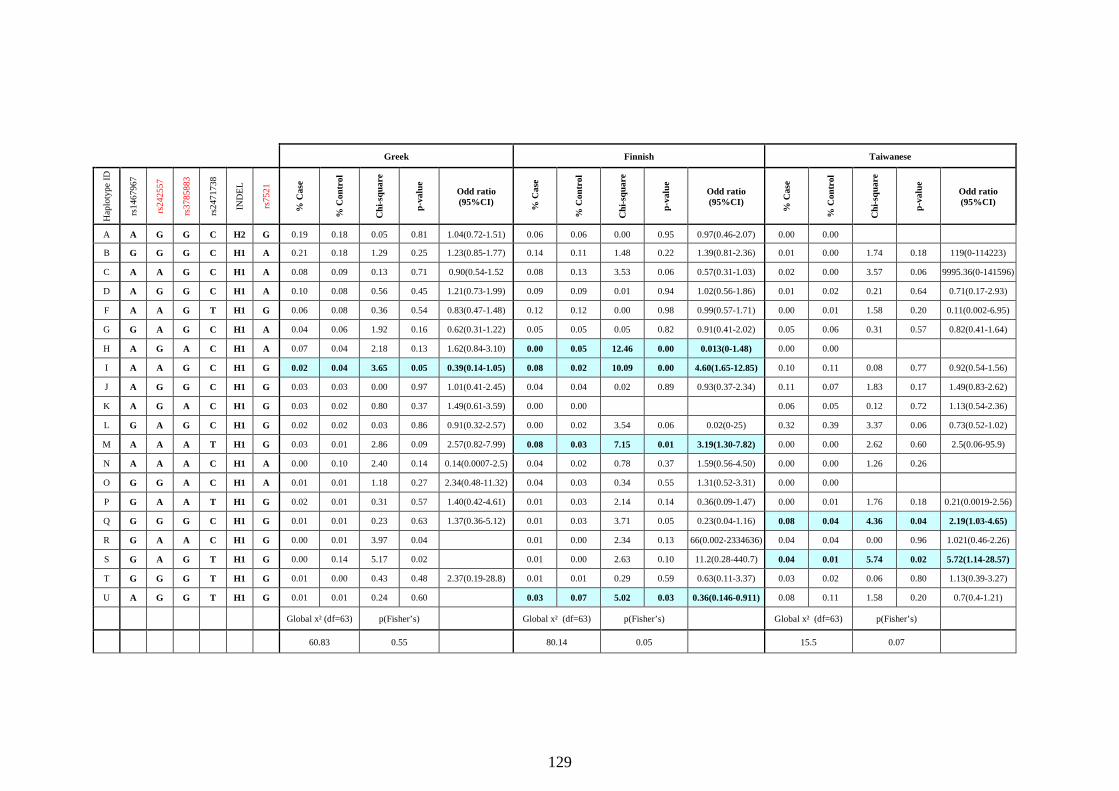
129
Greek Finnish Taiwanese
Hap
loty
pe
ID
rs1
46
79
67
rs2
425
57
rs3
78
58
83
rs2
47
17
38
IND
EL
rs7
52
1
%C
ase
%C
on
tro
l
Ch
i-sq
ua
re
p-v
alu
e
Odd ratio(95%CI)
%C
ase
%C
on
tro
l
Ch
i-sq
ua
re
p-v
alu
e
Odd ratio(95%CI)
%C
ase
%C
on
tro
l
Ch
i-sq
ua
re
p-v
alu
e
Odd ratio(95%CI)
A A G G C H2 G 0.19 0.18 0.05 0.81 1.04(0.72-1.51) 0.06 0.06 0.00 0.95 0.97(0.46-2.07) 0.00 0.00
B G G G C H1 A 0.21 0.18 1.29 0.25 1.23(0.85-1.77) 0.14 0.11 1.48 0.22 1.39(0.81-2.36) 0.01 0.00 1.74 0.18 119(0-114223)
C A A G C H1 A 0.08 0.09 0.13 0.71 0.90(0.54-1.52 0.08 0.13 3.53 0.06 0.57(0.31-1.03) 0.02 0.00 3.57 0.06 9995.36(0-141596)
D A G G C H1 A 0.10 0.08 0.56 0.45 1.21(0.73-1.99) 0.09 0.09 0.01 0.94 1.02(0.56-1.86) 0.01 0.02 0.21 0.64 0.71(0.17-2.93)
F A A G T H1 G 0.06 0.08 0.36 0.54 0.83(0.47-1.48) 0.12 0.12 0.00 0.98 0.99(0.57-1.71) 0.00 0.01 1.58 0.20 0.11(0.002-6.95)
G G A G C H1 A 0.04 0.06 1.92 0.16 0.62(0.31-1.22) 0.05 0.05 0.05 0.82 0.91(0.41-2.02) 0.05 0.06 0.31 0.57 0.82(0.41-1.64)
H A G A C H1 A 0.07 0.04 2.18 0.13 1.62(0.84-3.10) 0.00 0.05 12.46 0.00 0.013(0-1.48) 0.00 0.00
I A A G C H1 G 0.02 0.04 3.65 0.05 0.39(0.14-1.05) 0.08 0.02 10.09 0.00 4.60(1.65-12.85) 0.10 0.11 0.08 0.77 0.92(0.54-1.56)
J A G G C H1 G 0.03 0.03 0.00 0.97 1.01(0.41-2.45) 0.04 0.04 0.02 0.89 0.93(0.37-2.34) 0.11 0.07 1.83 0.17 1.49(0.83-2.62)
K A G A C H1 G 0.03 0.02 0.80 0.37 1.49(0.61-3.59) 0.00 0.00 0.06 0.05 0.12 0.72 1.13(0.54-2.36)
L G A G C H1 G 0.02 0.02 0.03 0.86 0.91(0.32-2.57) 0.00 0.02 3.54 0.06 0.02(0-25) 0.32 0.39 3.37 0.06 0.73(0.52-1.02)
M A A A T H1 G 0.03 0.01 2.86 0.09 2.57(0.82-7.99) 0.08 0.03 7.15 0.01 3.19(1.30-7.82) 0.00 0.00 2.62 0.60 2.5(0.06-95.9)
N A A A C H1 A 0.00 0.10 2.40 0.14 0.14(0.0007-2.5) 0.04 0.02 0.78 0.37 1.59(0.56-4.50) 0.00 0.00 1.26 0.26
O G G A C H1 A 0.01 0.01 1.18 0.27 2.34(0.48-11.32) 0.04 0.03 0.34 0.55 1.31(0.52-3.31) 0.00 0.00
P G A A T H1 G 0.02 0.01 0.31 0.57 1.40(0.42-4.61) 0.01 0.03 2.14 0.14 0.36(0.09-1.47) 0.00 0.01 1.76 0.18 0.21(0.0019-2.56)
Q G G G C H1 G 0.01 0.01 0.23 0.63 1.37(0.36-5.12) 0.01 0.03 3.71 0.05 0.23(0.04-1.16) 0.08 0.04 4.36 0.04 2.19(1.03-4.65)
R G A A C H1 G 0.00 0.01 3.97 0.04 0.01 0.00 2.34 0.13 66(0.002-2334636) 0.04 0.04 0.00 0.96 1.021(0.46-2.26)
S G A G T H1 G 0.00 0.14 5.17 0.02 0.01 0.00 2.63 0.10 11.2(0.28-440.7) 0.04 0.01 5.74 0.02 5.72(1.14-28.57)
T G G G T H1 G 0.01 0.00 0.43 0.48 2.37(0.19-28.8) 0.01 0.01 0.29 0.59 0.63(0.11-3.37) 0.03 0.02 0.06 0.80 1.13(0.39-3.27)
U A G G T H1 G 0.01 0.01 0.24 0.60 0.03 0.07 5.02 0.03 0.36(0.146-0.911) 0.08 0.11 1.58 0.20 0.7(0.4-1.21)
Global x² (df=63) p(Fisher’s) Global x² (df=63) p(Fisher’s) Global x² (df=63) p(Fisher’s)
60.83 0.55 80.14 0.05 15.5 0.07

130
Table 5.4 Association of common MAPT haplotypes with PD in the three diverse ethnic populations.
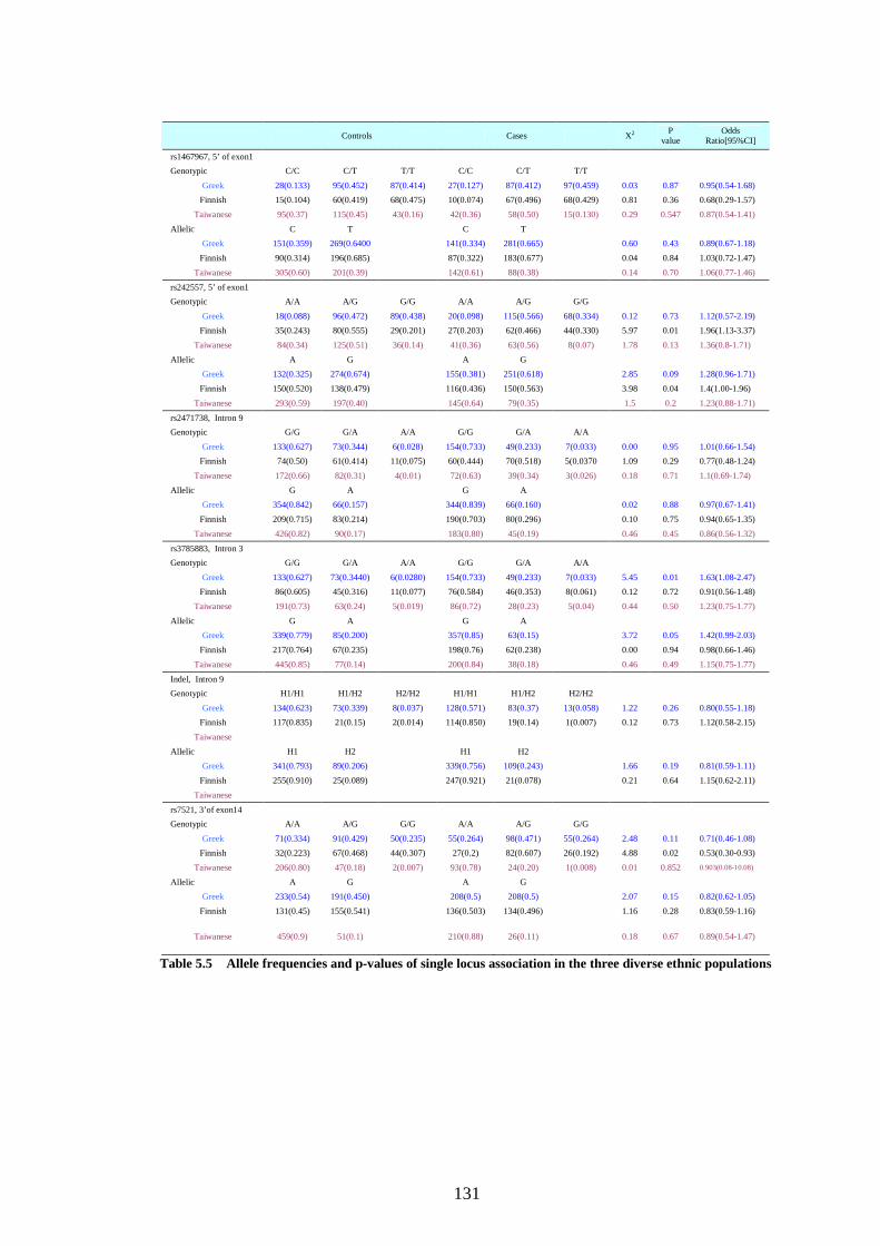
131
Table 5.5 Allele frequencies and p-values of single locus association in the three diverse ethnic populations
Controls Cases X2 Pvalue
OddsRatio[95%CI]
rs1467967, 5’ of exon1
Genotypic C/C C/T T/T C/C C/T T/T
Greek 28(0.133) 95(0.452) 87(0.414) 27(0.127) 87(0.412) 97(0.459) 0.03 0.87 0.95(0.54-1.68)
Finnish 15(0.104) 60(0.419) 68(0.475) 10(0.074) 67(0.496) 68(0.429) 0.81 0.36 0.68(0.29-1.57)
Taiwanese 95(0.37) 115(0.45) 43(0.16) 42(0.36) 58(0.50) 15(0.130) 0.29 0.547 0.87(0.54-1.41)
Allelic C T C T
Greek 151(0.359) 269(0.6400 141(0.334) 281(0.665) 0.60 0.43 0.89(0.67-1.18)
Finnish 90(0.314) 196(0.685) 87(0.322) 183(0.677) 0.04 0.84 1.03(0.72-1.47)
Taiwanese 305(0.60) 201(0.39) 142(0.61) 88(0.38) 0.14 0.70 1.06(0.77-1.46)
rs242557, 5’ of exon1
Genotypic A/A A/G G/G A/A A/G G/G
Greek 18(0.088) 96(0.472) 89(0.438) 20(0.098) 115(0.566) 68(0.334) 0.12 0.73 1.12(0.57-2.19)
Finnish 35(0.243) 80(0.555) 29(0.201) 27(0.203) 62(0.466) 44(0.330) 5.97 0.01 1.96(1.13-3.37)
Taiwanese 84(0.34) 125(0.51) 36(0.14) 41(0.36) 63(0.56) 8(0.07) 1.78 0.13 1.36(0.8-1.71)
Allelic A G A G
Greek 132(0.325) 274(0.674) 155(0.381) 251(0.618) 2.85 0.09 1.28(0.96-1.71)
Finnish 150(0.520) 138(0.479) 116(0.436) 150(0.563) 3.98 0.04 1.4(1.00-1.96)
Taiwanese 293(0.59) 197(0.40) 145(0.64) 79(0.35) 1.5 0.2 1.23(0.88-1.71)
rs2471738, Intron 9
Genotypic G/G G/A A/A G/G G/A A/A
Greek 133(0.627) 73(0.344) 6(0.028) 154(0.733) 49(0.233) 7(0.033) 0.00 0.95 1.01(0.66-1.54)
Finnish 74(0.50) 61(0.414) 11(0.075) 60(0.444) 70(0.518) 5(0.0370 1.09 0.29 0.77(0.48-1.24)
Taiwanese 172(0.66) 82(0.31) 4(0.01) 72(0.63) 39(0.34) 3(0.026) 0.18 0.71 1.1(0.69-1.74)
Allelic G A G A
Greek 354(0.842) 66(0.157) 344(0.839) 66(0.160) 0.02 0.88 0.97(0.67-1.41)
Finnish 209(0.715) 83(0.214) 190(0.703) 80(0.296) 0.10 0.75 0.94(0.65-1.35)
Taiwanese 426(0.82) 90(0.17) 183(0.80) 45(0.19) 0.46 0.45 0.86(0.56-1.32)
rs3785883, Intron 3
Genotypic G/G G/A A/A G/G G/A A/A
Greek 133(0.627) 73(0.3440) 6(0.0280) 154(0.733) 49(0.233) 7(0.033) 5.45 0.01 1.63(1.08-2.47)
Finnish 86(0.605) 45(0.316) 11(0.077) 76(0.584) 46(0.353) 8(0.061) 0.12 0.72 0.91(0.56-1.48)
Taiwanese 191(0.73) 63(0.24) 5(0.019) 86(0.72) 28(0.23) 5(0.04) 0.44 0.50 1.23(0.75-1.77)
Allelic G A G A
Greek 339(0.779) 85(0.200) 357(0.85) 63(0.15) 3.72 0.05 1.42(0.99-2.03)
Finnish 217(0.764) 67(0.235) 198(0.76) 62(0.238) 0.00 0.94 0.98(0.66-1.46)
Taiwanese 445(0.85) 77(0.14) 200(0.84) 38(0.18) 0.46 0.49 1.15(0.75-1.77)
Indel, Intron 9
Genotypic H1/H1 H1/H2 H2/H2 H1/H1 H1/H2 H2/H2
Greek 134(0.623) 73(0.339) 8(0.037) 128(0.571) 83(0.37) 13(0.058) 1.22 0.26 0.80(0.55-1.18)
Finnish 117(0.835) 21(0.15) 2(0.014) 114(0.850) 19(0.14) 1(0.007) 0.12 0.73 1.12(0.58-2.15)
Taiwanese
Allelic H1 H2 H1 H2
Greek 341(0.793) 89(0.206) 339(0.756) 109(0.243) 1.66 0.19 0.81(0.59-1.11)
Finnish 255(0.910) 25(0.089) 247(0.921) 21(0.078) 0.21 0.64 1.15(0.62-2.11)
Taiwanese
rs7521, 3’of exon14
Genotypic A/A A/G G/G A/A A/G G/G
Greek 71(0.334) 91(0.429) 50(0.235) 55(0.264) 98(0.471) 55(0.264) 2.48 0.11 0.71(0.46-1.08)
Finnish 32(0.223) 67(0.468) 44(0.307) 27(0.2) 82(0.607) 26(0.192) 4.88 0.02 0.53(0.30-0.93)
Taiwanese 206(0.80) 47(0.18) 2(0.007) 93(0.78) 24(0.20) 1(0.008) 0.01 0.852 0.903(0.08-10.08)
Allelic A G A G
Greek 233(0.54) 191(0.450) 208(0.5) 208(0.5) 2.07 0.15 0.82(0.62-1.05)
Finnish 131(0.45) 155(0.541) 136(0.503) 134(0.496) 1.16 0.28 0.83(0.59-1.16)
Taiwanese 459(0.9) 51(0.1) 210(0.88) 26(0.11) 0.18 0.67 0.89(0.54-1.47)
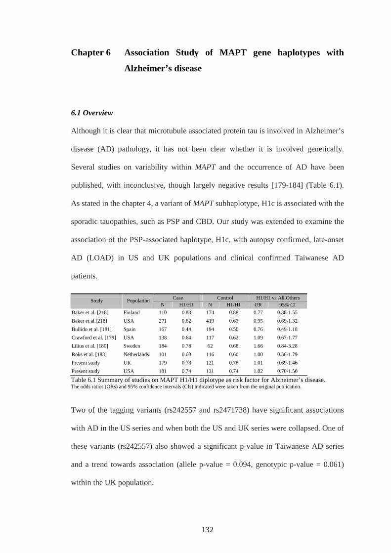
132
Chapter 6 Association Study of MAPT gene haplotypes with
Alzheimer’s disease
6.1 Overview
Although it is clear that microtubule associated protein tau is involved in Alzheimer’s
disease (AD) pathology, it has not been clear whether it is involved genetically.
Several studies on variability within MAPT and the occurrence of AD have been
published, with inconclusive, though largely negative results [179-184] (Table 6.1).
As stated in the chapter 4, a variant of MAPT subhaplotype, H1c is associated with the
sporadic tauopathies, such as PSP and CBD. Our study was extended to examine the
association of the PSP-associated haplotype, H1c, with autopsy confirmed, late-onset
AD (LOAD) in US and UK populations and clinical confirmed Taiwanese AD
patients.
Case Control H1/H1 vs All OthersStudy Population
N H1/H1 N H1/H1 OR 95% CI
Baker et al. [218] Finland 110 0.83 174 0.88 0.77 0.38-1.55
Baker et al.[218] USA 271 0.62 419 0.63 0.95 0.69-1.32
Bullido et al. [181] Spain 167 0.44 194 0.50 0.76 0.49-1.18
Crawford et al. [179] USA 138 0.64 117 0.62 1.09 0.67-1.77
Lilius et al. [180] Sweden 184 0.78 62 0.68 1.66 0.84-3.28
Roks et al. [183] Netherlands 101 0.60 116 0.60 1.00 0.56-1.79
Present study UK 179 0.78 121 0.78 1.01 0.69-1.46
Present study USA 181 0.74 131 0.74 1.02 0.70-1.50
Table 6.1 Summary of studies on MAPT H1/H1 diplotype as risk factor for Alzheimer’s disease.The odds ratios (ORs) and 95% confidence intervals (CIs) indicated were taken from the original publication.
Two of the tagging variants (rs242557 and rs2471738) have significant associations
with AD in the US series and when both the US and UK series were collapsed. One of
these variants (rs242557) also showed a significant p-value in Taiwanese AD series
and a trend towards association (allele p-value = 0.094, genotypic p-value = 0.061)
within the UK population.

133
6.2 Background
AD is the most common cause of dementia in the elderly. It is characterized clinically
by a gradual onset and progression of memory loss, and characterized postmortem by
the presence of two types of neuropathological inclusions: neurofibrillary tangles and
senile plaques [185].
The neurofibrillary tangles, comprised of paired helical filaments of phosphorylated
tau protein are a pathognomic feature of AD. Using immunohistochemical and
biochemical means, it has been demonstrated that tau is modified in AD. One of the
diversified MAPT H1 subhaplotypes, H1c has been shown to be largely responsible
for the association between the H1 clade and the sporadic tauopathies
[131;133;164;165;177]. The inconclusive association between AD and the variants of
MAPT has been studied by several groups [179-184]. These reports have compared
alleles that discriminated between the H1 and H2 clades, but did not assess whether
variability on the H1 background showed association with disease.
The objective of this work was to examine whether the PSP-associated MAPT
haplotype is responsible for the disease pathogenesis with late-onset AD in autopsy
confirmed, late-onset AD (LOAD) and clinical confirmed Taiwanese AD patients.
6.3 Case-control samples
6.3.1 US and UK series
In UK series, there were 179 cases (66% female, mean age of death: 81 years, range:
65-96 years) and 121 controls (51% female, mean age of death: 78 years, range: 65-

134
100 years). While in US series, there were 181 cases (55% female, mean age of death:
81 years, range: 66-97 years) and 131 controls (51% female, mean age of death: 81
years, range: 65-99 years). All samples were pathologically confirmed. Controls were
free of neuropathology at autopsy.
All samples were of Caucasian origin obtained from either the Newcastle Brain Bank,
Newcastle-upon-Tyne, UK (UK series) or various brain banks throughout the United
States, including National institute on Aging, Johns Hopkins Alzheimer’s Disease
Research Center, University of California, the Kathleen Price Bryan Brain Bank,
Duke University Medical Center, Stanford University, New York Brain Bank, Taub
Institute, Columbia University, Massachusetts General Hospital, University of
Michigan, University of Kentucky, Mayo Clinic Jacksonville, University Southern
California, Washington University, St Louis Alzheimer’s Disease Research Center,
University of Washington, Seattle.
6.3.2 Taiwanese Series
Patients were recruited from the dementia outpatient clinic of Chang Gung Medical
Center, Linkou, Taiwan. The diagnosis of AD (n = 110; 60% female, 76.9±6.5 years)
was made by consensus according to the criteria of the National Institute of
Neurological and Communicative Disorders and Stroke and the Alzheimer's Disease
and Related Disorders Association (NINCDS-ADRDA) for probable AD [186].
Subjects without stroke and cognitive impairment represented the control group (n =
117; 46.2% female, aged 59.0±10.1 years). Each subject was informed of the aim of
the study, and all gave their consents to study. Patients with previous clinical history

135
of neurological, psychiatric, somatic, or toxic causes for dementia were excluded.
Evaluation included general physical and neurological assessment, the Mini-Mental
State Examination (MMSE) [187], and Hachinski ischaemia score [188]. Laboratory
studies included complete blood cell count, biochemistry analysis, erythrocyte
sedimentation rate, vitamin B12 and folic acid levels, thyroid stimulating hormone
level and syphilis serological testing. Each patient underwent brain computerized
tomography (CT) scan. All the patients were examined by at least 2 neurologists and
confirmed to fulfil the DSM-IV criteria for dementia.
6.4 Selection of haplotype-tagging SNPs
6.4.1 Markers for the US and UK series
The haplotype structure of MAPT was previously analysed using markers from the
CEPH database (http://www.hapmap.org) (see section 4.3). Using the program TagIT
(http://popgen.biol.ucl.ac.uk/software), a minimum of five SNPs were found in order
to capture the haplotype diversity at the MAPT locus. The performance of our five
tagging SNPs and the del-In9 against each individual SNP typed were tested within
the CEPH-trios, using criterion 5 (an association based criterion using haplotype r2) in
the program TagIT. On average, this set of tag SNPs captures 95% of the diversity of
the known CEPH variants and scores 90% for most individual loci. Performance plots
of the tagging variants as well as the del-In9 polymorphism on its own are shown in
Figure 6.1.

136
Figure 6.1 MAPT tagging markers capture the diversity of MAPT. Solid line: Performance plot ofthe six MAPT SNP tag markers using the data available from CEPH in the hapmap project(http://www.hapmap.org. The plot is a row of vector performance values (as measured by haplotype r2
using criterion 5 from TagIt) for each of our tag SNPs against each of the SNP loci typed in the CEPHtrios. High r2 values indicate good performance, because r2 is a measure of linkage disequilibrium. Ifthere is perfect linkage disequilibrium between two markers, r2 will approach 1, indicating that the twomarkers are segregating together and thus are genetically equivalent. On average, this set of taggingSNPs captures 95% (average r2 = 0.95) of the diversity of the known SNPs as a whole and
predominantly scores > 90% against individual SNP loci. Broken line: In contrast, examining just thedel-In9 variant’s performance demonstrates that it performs well for some loci (r2 > 80%), whereas itperforms poorly (r2 < 50%) for several loci. This is because there are many variants of the H1 clade. Asthe del-In9 variant only distinguishes H1 and H2, it is a reasonable marker to tag the variants that occuron the H1 and H2 backgrounds; however, it will perform poorly if used to tag loci that define sub-haplotypes of the H1 clade. This is important for the current study, as all previous studies examiningAD risk and MAPT genotypes only looked at variants that defined the H1 or H2 clades and variants ofthe H1 clade.
6.4.2 Markers for the Taiwanese series
6.4.2.1 SNP Selection
For the Taiwanese population, there was no genotyping information available on the
known SNPs in the extended MAPT region that covers 45-kb upstream and
downstream of the gene. We selected all the Japanese SNPs with frequency
information published on the JSNP database at http://snp.ims.u-tokyo.ac.jp in the
region. We then tried to use any SNP(s) from dbSNP to fill gaps, when two adjacent
JSNPs were more than 14kb apart. This led us to the SNPs: rs962885, rs2301689,
rs3744457, rs2280004, rs1467967, rs3785880, rs1001945, rs242557, rs242562,
rs2303867, rs3785882, rs3785883, rs3785885, rs2258689, rs2471738, rs916896,
rs7521, rs2074432, rs2277613, rs876944 and rs2301732. (Table 6.2)

137
SNP Positiona dbSNP ID Alleles Ancestral
1 41291420 rs962885 C/T C
2 41291627 rs2301689 A/G G
3 41328711 rs3744457 C/T C
4 41340959 rs2280004 C/T T
5 41342006 rs1467967 A/G G
6 41349204 rs3785880 G/T T
7 41361649 rs1001945 C/G G
8 41375573 rs242557 A/G A
9 41382599 rs242562 A/G A
10 41395691 rs2303867 A/G A
11 41397030 rs3785882 C/G G
12 41410269 rs3785883 A/G G
13 41414477 rs3785885 A/G G
14 41423219 rs2258689 C/T C
15 41431900 rs2471738 C/T C
16 41454642 rs916896 A/G G
17 41461242 rs7521 A/G G
18 41465690 rs2074432b A/G A
19 41472982 rs2277613 C/T C
20 41490227 rs876944 G/T G
21 41500036 rs2301732 A/G A
Table 6.2 The 21 single nucleotide polymorphisms used for the linkage disequilibrium andhaplotype structure analysis of MAPT in the Taiwanese cohortaSNP position on chromosome. (Position on chromosome (in bp) is based on May 2004 build of Human Genome Sequence(http://genome.ucsc.edu).) bThe dbSNP ID rs2074432 was merged into rs1078997 (in May 2004 Human Assembly).
6.4.2.2 Selection, performance, assessment, and association analysis of MAPT
haplotype tagging SNPs in Taiwanese cohort
An association based criterion (haplotype r2) as previous described was selected for
the criterion to tagging SNPs. Nine haplotype tagged SNPs (rs2277613, rs962885,
rs3785880, rs3744457, rs1467967, rs242557, rs3785883, rs2471738, rs7521) were
required to represent all the SNPs in our Taiwanese cohort. The bi-allelic del-In9
marker was used to unambiguously confirm all the Taiwaneses in our study are of the
H1 clade [130]. The performance value for the 9 htSNPs was interpreted at an average
haplotype r2 value of 0.95 (95%). (Figure 6.2)
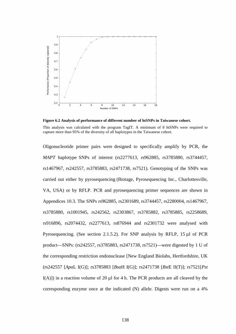
138
Figure 6.2 Analysis of performance of different number of htSNPs in Taiwanese cohort.
This analysis was calculated with the program TagIT. A minimum of 8 htSNPs were required tocapture more than 95% of the diversity of all haplotypes in the Taiwanese cohort.
Oligonucleotide primer pairs were designed to specifically amplify by PCR, the
MAPT haplotype SNPs of interest (rs2277613, rs962885, rs3785880, rs3744457,
rs1467967, rs242557, rs3785883, rs2471738, rs7521). Genotyping of the SNPs was
carried out either by pyrosequencing (Biotage, Pyrosequencing Inc., Charlottesville,
VA, USA) or by RFLP. PCR and pyrosequencing primer sequences are shown in
Appendices 10.3. The SNPs rs962885, rs2301689, rs3744457, rs2280004, rs1467967,
rs3785880, rs1001945, rs242562, rs2303867, rs3785882, rs3785885, rs2258689,
rs916896, rs2074432, rs2277613, rs876944 and rs2301732 were analysed with
Pyrosequencing. (See section 2.1.5.2). For SNP analysis by RFLP, 15 µl of PCR
product—SNPs: (rs242557, rs3785883, rs2471738, rs7521)—were digested by 1 U of
the corresponding restriction endonuclease [New England Biolabs, Hertfordshire, UK
(rs242557 [ApaL I(G)]; rs3785883 [BsaH I(G)]; rs2471738 [BstE II(T)]; rs7521[Pst
I(A)]) in a reaction volume of 20 µl for 4 h. The PCR products are all cleaved by the
corresponding enzyme once at the indicated (N) allele. Digests were run on a 4%
0 2 4 6 8 10 12 14 16 180.2
0.3
0.4
0.5
0.6
0.7
0.8
0.9
1
Number of SNPs
Pe
rfo
rma
nce
(Pro
po
rtio
no
fd
ive
rsity
ca
ptu
red
)

139
agarose gel for analysis. Genotype scoring was carried out blindly by two individuals.
Any discrepancies between the two were resolved by repeating the assay.
6.4.2.3LD and statistical analysis
For each SNP, the allele and genotype distributions in the group of patients were
compared with those in the control group. Statistical assessments for the allele and
genotype frequencies and Hardy-Weinberg equilibrium (HWE) were made using the
genetics software program TagIT (http://popgen.biol.ucl.ac.uk/software.html) [123].
The square of the correlation coefficient (r2) and D' for LD was calculated pair-wise
between each pair of SNP. Haplotype predictions were made using an EM algorithm
using TagIT. Case–control locus-by-locus association was calculated statistically
using a 2 distribution and the significance was calculated using a Monte–Carlo
approach as implemented by CLUMP software [162]. The LD pattern of Taiwanese
normal control population was illustrated with the software package, Graphical
Overview of Linkage Disequilibrium [189]. (Figure 6.3)
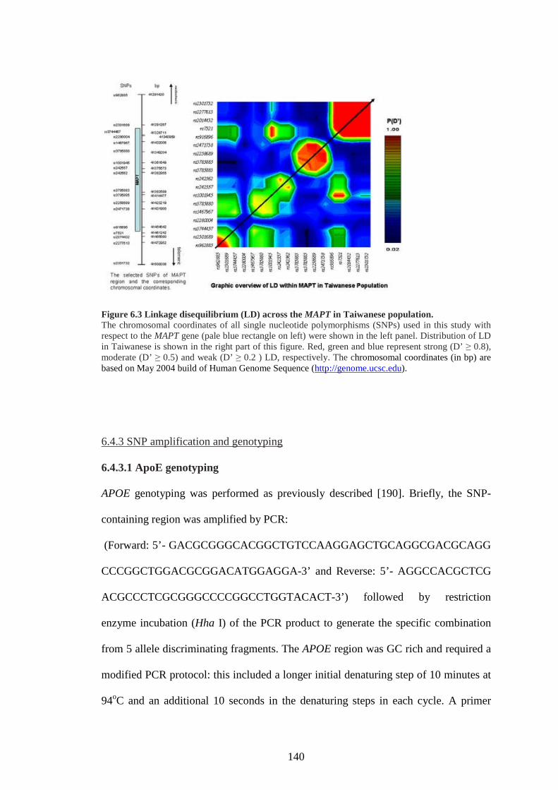
140
Figure 6.3 Linkage disequilibrium (LD) across the MAPT in Taiwanese population.The chromosomal coordinates of all single nucleotide polymorphisms (SNPs) used in this study withrespect to the MAPT gene (pale blue rectangle on left) were shown in the left panel. Distribution of LDin Taiwanese is shown in the right part of this figure. Red, green and blue represent strong (D’ ≥ 0.8),moderate (D’ ≥ 0.5) and weak (D’ ≥ 0.2 ) LD, respectively. The chromosomal coordinates (in bp) arebased on May 2004 build of Human Genome Sequence (http://genome.ucsc.edu).
6.4.3 SNP amplification and genotyping
6.4.3.1 ApoE genotyping
APOE genotyping was performed as previously described [190]. Briefly, the SNP-
containing region was amplified by PCR:
(Forward: 5’- GACGCGGGCACGGCTGTCCAAGGAGCTGCAGGCGACGCAGG
CCCGGCTGGACGCGGACATGGAGGA-3’ and Reverse: 5’- AGGCCACGCTCG
ACGCCCTCGCGGGCCCCGGCCTGGTACACT-3’) followed by restriction
enzyme incubation (Hha I) of the PCR product to generate the specific combination
from 5 allele discriminating fragments. The APOE region was GC rich and required a
modified PCR protocol: this included a longer initial denaturing step of 10 minutes at
94oC and an additional 10 seconds in the denaturing steps in each cycle. A primer

141
annealing temperature of 56oC is used. Digested PCR products were run on a 4% low-
melting agarose gel and stained with ethidium bromide to visualize the allele
discriminating bands.
6.5 Results
6.5.1 The association of the H1c haplotype at the MAPT locus with AD in UK and US
series
6.5.1.1 Single locus analysis
6.5.1.1.1 Single locus analysis
We genotyped six polymorphisms which we have shown previously tag the haplotype
diversity in MAPT in Caucasians in two series of autopsy-confirmed controls and
autopsy confirmed LOAD cases obtained from brain banks in the US and UK. (see
Figure 6.1 for information on the usefulness of these SNPs to tag diversity in these
populations). The single locus associations with AD are shown in Table 6.3. Tests of
association of the intron 9 insertion–deletion (del-In9) polymorphism, which has been
used to define the H1/H2 clades, were negative as we and others have previously
reported [179-184]. Two of the tagging variants (rs242557 and rs2471738) had
significant P-values in both the US series and when both the US and UK series were
collapsed (Table 6.3). One of these tag variants (rs242557) also showed a trend
towards association (allelic P-value = 0.094, genotypic P-value = 0.061) within the
UK population.

142
Variants Major Allele CO AD Risk Allele Odds Ratio
APOE N/A ε4a
12 45 3.47E-18 [2.09E-06] 1.36E-17 [3.46E-14] ε4 8.313
rs1467967 5' of ex 1 A 62 68 0.199 [0.763] 0.334 [0.077] A 1.261
rs242557 5' of ex 1 G 70 62 0.038 [0.043] 0.107 [0.753] A 1.456
rs3785883 Intron 3 G 78 83 0.1 [0.013] 0.195 [0.137] G 1.403
rs2471738 Intron 9 C 82 74 0.024 [0.032] 0.02 [0.075] T 1.598
del-In9 Intron 9 H1 78 78 1 [0.119] 0.248 [0.059] H1 1.020
rs7521 3' of ex 14 G 56 55 0.87 [0.930] 0.315 [0.025] A 1.035
APOE N/A ε4a
13 38 1.28E-11 [7.66E-6] 3.77E-10 [1.24E-8] ε4 4.375
rs1467967 5' of ex 1 A 67 68 0.856 [0.848] 0.821 [0.053] A 1.048
rs242557 5' of ex 1 G 65 58 0.094 [0.616] 0.061 [0.931] A 1.351
rs3785883 Intron 3 G 85 87 0.465 [0.575] 0.293 [0.659] G 1.222
rs2471738 Intron 9 C 82 76 0.081 [0.217] 0.229 [0.942] T 1.449
del-In9 Intron 9 H1 74 74 1 [0.478] 0.763 [0.361] H1 1.005
rs7521 3' of ex 14 G 57 57 1 [0.557] 0.515 [0.490] A 1.006
APOE N/A ε4a
13 41 2.45E-28 [7.07E-11] 5.13E-27 [1.46E-22] ε4 5.938
rs1467967 5' of ex 1 A 65 68 0.262 [0.003] 0.469 [0.7] A 1.157
rs242557 5' of ex 1 G 68 60 0.007 [2.49E-07] 0.013 [0.057] A 1.408
rs3785883 Intron 3 G 81 85 0.071 [0.988] 0.114 [0.046] G 1.325
rs2471738 Intron 9 C 82 75 0.004 [0.046] 0.009 [0.021] T 1.524
del-In9 Intron 9 H1 76 76 0.946 [0.018] 0.224 [0.106] H1 1.016
rs7521 3' of ex 14 G 57 56 0.906 [0.002] 0.168 [0.676] A 1.020
UK Series
UK and US series
US series
Location in
MAPT
Major allele frequency P-value
Allelelic Genotypic
Table 6.3 Single locus association in UK series and US seriesex, exon; CO, control; AD, Alzheimer’s disease; CI, confidence interval. The relative locations, majorallele frequencies, P-values, odds ratios and confidence intervals for all tagging loci tested in eachseries are shown. All values were calculated using SPSS software v.11. Significant values arehighlighted in bold. P-values corrected for age, gender and APOE effects are given in brackets.aFor APOE, the ε 4 allele frequencies are listed, since this is the risk allele for LOAD, even though ε 4is not the major allele.
6.5.1.1.2 Single locus analysis: APOE sub-analysis
We noted that when the single locus P-values were adjusted for age, sex and APOE
status, many of the single locus P-values became more significant (see Table 6.4 for
both series and after collapsed; the P-value of rs242557 prior to age, sex, APOE
adjustment = 0.007, after adjustment = 2.49E-07). We examined whether this effect
was mainly due to age differences, gender differences or differences due to APOE
background and found that the most robust interaction for each marker was with
APOE status. To further examine this interaction, we divided the entire sample
including both US and UK samples into two sub-series on the basis of APOE-ε4
genotype; cases and controls that possessed at least one ε4 allele were analysed
separately from cases and controls that had no ε4 alleles. We found significant single
locus P-values only in the sub-series where neither cases nor controls had any APOE-

143
ε4 alleles, suggesting that the single tag variant association is driven by those
individuals who do not possess APOE-ε4 alleles.
Variants Major Allele CO AD Allelelic Genotypic Risk Allele
Odds
Ratio CI (95%)
rs1467967 5' of ex 1 A 63 67 0.355 0.533 A 1.177 0.833-1.667
rs242557 5' of ex 1 G 68 58 0.01 0.029 A 1.1577 1.112–2.096
rs3785883 Intron 3 G 79 83 0.249 0.159 G 1.288 0.837–1.980
rs2471738 Intron 9 C 83 75 0.017 0.048 T 1.637 1.092–2.454
del-In9 Intron 9 H1 75 78 0.541 0.006 H1 1.129 0.765–1.667
rs7521 3' of ex 14 G 57 54 0.594 0.66 A 1.094 0.787–1.520
rs1467967 5' of ex 1 A 71 68 0.649 0.751 Ga 1.103 0.723–1.681
rs242557 5' of ex 1 G 67 61 0.206 0.182 Aa 1.307 0.863–1.980
rs3785883 Intron 3 G 86 86 0.83 0.653 Ga 1.064 0.607–1.862
rs2471738 Intron 9 C 81 75 0.207 0.308 Ta 1.365 0.841–2.217
del-In9 Intron 9 H1 78 75 0.592 0.842 H2a 1.135 0.714–1.803
rs7521 3' of ex 14 G 56 57 0.845 0.119 Ga 1.04 0.702–1.539
APOE-ε4 negative US and UK series collapsed
APOE-ε4 positive series US and UK series collapsed
Location in MAPT
Major allele frequency P-value
Table 6.4 Single locus associations within APOE-ε4 positive and negative subsets of the combined US and UK sampleThe major allele frequencies, P-values, odds ratios and confidence intervals for all tagging loci tested in the subset of the entiresample that was APOE-ε4 negative and the subset of the series that was APOE-ε4 positive are shown. The APOE-ε4 negativesubset contained all individuals from both the US and UK series that did not possess any APOE-ε4 alleles (genotypes 23 and 33).The APOE-ε4 positive subset contained all individuals from both the US and UK series that possessed at least one ε4 allele(genotypes 24, 34, 44). Abbreviations are the same as given in Table 6.3.aNote that in the APOE-ε4 positive series the risk alleles are flipped for loci rs1467967, del-In9 and rs7521. This probablyreflects the smaller sample size of the controls in this series (n=66).
6.5.1.2 Haplotype analysis
6.5.1.2.1 Haplotype analysis
The location of all tag SNPs with respect to MAPT exon structure as well as major
haplotype frequencies and description of the alleles at each tag SNP for the major
MAPT haplotypes is shown in Figure 6.4. Haplotype frequencies were obtained from
the program SNPHAP (Clayton, D: http://www-gene.cimr.cam.ac.uk/clayton/
software/snphap). As expected, we found no difference in the frequency of haplotype
A (H2) between cases and controls in the UK or US series or when both samples are
combined (UK LOAD frequency = 24.41%, control frequency = 25.44%, US LOAD
frequency = 22.10%, US control frequency = 22.52%, combined series LOAD
frequency = 23.39%, control frequency = 23.48%). This is in contrast to the clear
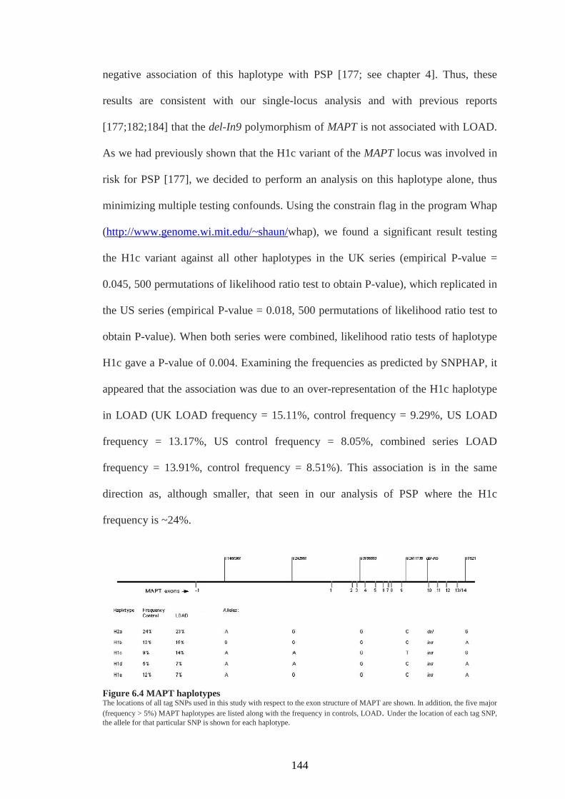
144
negative association of this haplotype with PSP [177; see chapter 4]. Thus, these
results are consistent with our single-locus analysis and with previous reports
[177;182;184] that the del-In9 polymorphism of MAPT is not associated with LOAD.
As we had previously shown that the H1c variant of the MAPT locus was involved in
risk for PSP [177], we decided to perform an analysis on this haplotype alone, thus
minimizing multiple testing confounds. Using the constrain flag in the program Whap
(http://www.genome.wi.mit.edu/~shaun/whap), we found a significant result testing
the H1c variant against all other haplotypes in the UK series (empirical P-value =
0.045, 500 permutations of likelihood ratio test to obtain P-value), which replicated in
the US series (empirical P-value = 0.018, 500 permutations of likelihood ratio test to
obtain P-value). When both series were combined, likelihood ratio tests of haplotype
H1c gave a P-value of 0.004. Examining the frequencies as predicted by SNPHAP, it
appeared that the association was due to an over-representation of the H1c haplotype
in LOAD (UK LOAD frequency = 15.11%, control frequency = 9.29%, US LOAD
frequency = 13.17%, US control frequency = 8.05%, combined series LOAD
frequency = 13.91%, control frequency = 8.51%). This association is in the same
direction as, although smaller, that seen in our analysis of PSP where the H1c
frequency is ~24%.
Figure 6.4 MAPT haplotypesThe locations of all tag SNPs used in this study with respect to the exon structure of MAPT are shown. In addition, the five major
(frequency > 5%) MAPT haplotypes are listed along with the frequency in controls, LOAD. Under the location of each tag SNP,the allele for that particular SNP is shown for each haplotype.

145
6.5.1.2.2 Haplotype analysis: APOE sub-analysis
In the light of the putative interaction with APOE genotype that we found examining
single locus associations, we decided to examine whether APOE status had any
influence on H1c haplotype association that we observed. We first tested whether
there was an APOE interaction with MAPT using the full dataset and the ‘–gxe’ and ‘–
alt-gxe’ flags in Whap. These two flags test whether there is a significant haplotype
effect while adjusting for epistatic effects of another locus (–gxe) and whether there is
a significant interaction between the genotypes of one locus and the haplotypes of the
other (–alt-gxe). When haplotype H1c was tested for association controlling for the
effect of the APOE locus, the P-value increased moderately from 0.004 to 0.005 (500
permutations of LRT to obtain P-value). When APOE was included in the model,
using the ‘–alt-gxe’ flag, the P-value decreased considerably (P-value with APOE =
5.16E-22, 500 permutations to obtain P-value), indicating a significant interaction
between APOE genotype and MAPT haplotype. We then stratified our sample as in
our single locus analysis by splitting the combined series into the subset of cases and
controls that possessed APOE-ε4 alleles and into the cases and controls that had no
APOE-ε4 alleles. As in the single locus analysis, the association between haplotype
H1c and disease risk was only seen in those individuals who had no APOE-ε4 alleles
(H1c P-value in subset of entire series where individuals had at least one ε4 allele =
0.238 and H1c P-value in subset of entire series where individuals had no ε4 allele =
0.008, 500 permutations to obtain P-values for both tests).
6.5.2 The association of the H1 haplotype at the MAPT locus with Taiwanese series
6.5.2.1 Linkage disequilibrium pattern of MAPT in Taiwanese

146
For the LD analysis of MAPT gene, we used the above 21 SNPs to genotype 21
normal Taiwanese individuals. We discarded SNPs that had a minor allele frequency
of less than 5%. The average density of the markers is one SNP every 20 kb. None of
the polymorphisms deviated from HWE. Additionally, the del-In9 marker which
defines the extended H1 and H2 clades was genotyped. As expected, all the
Taiwanese individuals including cases and control were H1 homozygotes.
We evaluated pairwise LD across MAPT for all 18 SNPs in the 21 normal Taiwanese
individuals both by D’ and r2, calculated from the EM inferred haplotypes. The MAPT
region is featured by LD as is particularly evident by the measure D’ (Figure 6.3).
This pattern of LD across the extended MAPT region shows that the LD blocks in
Taiwanese population have four blocks of D’ LD. This structure, consisting of smaller
blocks is consistent with LD throughout the genome and is in contrast with the
unusually large region of LD in the Caucasian population. This fragmented haplotype
structure would suggest that it would be easier to identify the region of the MAPT
locus in which genetic variability contributes to disease risk in this population
compared to the entire region of LD in the Caucasian population.
6.5.2.2 Selection, performance, assessment, and association analysis of MAPT
haplotype tagging SNPs
We used an association-based criterion (criterion 5 in TagIt, haplotype r2) in order to
select the haplotype-tagging SNPs (htSNPs). Nine htSNPs are required to represent all
the SNPs in our Taiwanese cohort. The performance value for the 9 htSNPs was
interpreted at an average haplotype r2 value of 0.99 (99%). None of the 9 Taiwanese
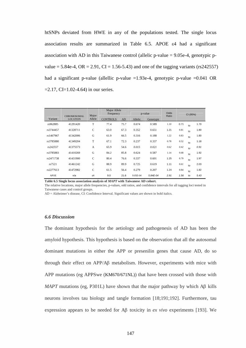
147
htSNPs deviated from HWE in any of the populations tested. The single locus
association results are summarized in Table 6.5. APOE ε4 had a significant
association with AD in this Taiwanese control (allelic p-value = 9.05e-4, genotypic p-
value = 5.84e-4, OR = 2.91, CI = 1.56-5.43) and one of the tagging variants (rs242557)
had a significant p-value (allellic p-value =1.93e-4, genotypic p-value =0.041 OR
=2.17, CI=1.02-4.64) in our series.
Major AlleleFrequency p-value
VariantCHROMOSOMAL
LOCATION
MajorAllele CONTROLS AD Allelic Genotypic
OddsRatio
CI (95%)
rs962885 41291420 T 77.4 75.7 0.674 0.589 1.10 0.71to
1.70
rs3744457 41328711 C 63.0 67.3 0.352 0.651 1.21 0.81to
1.80
rs1467967 41342006 G 61.9 66.5 0.316 0.188 1.22 0.83to
1.80
rs3785880 41349204 T 67.1 72.3 0.237 0.337 0.78 0.52to
1.18
rs242557 41375573 A 65.9 54.6 0.015 0.021 0.62 0.42to
0.91
rs3785883 41410269 G 84.2 85.8 0.624 0.587 1.14 0.68to
1.92
rs2471738 41431900 C 80.4 76.6 0.337 0.601 1.25 0.79to
1.97
rs7521 41461242 G 88.9 89.9 0.725 0.619 1.11 0.61to
2.03
rs2277613 41472982 C 61.5 56.4 0.279 0.207 1.24 0.84to
1.82
APOE n/a ε4 9.0 21.6 9.05E-04 5.84E-04 2.91 1.56 to 5.43
Table 6.5 Single locus association analysis of MAPT with Taiwanese AD cohort.The relative locations, major allele frequencies, p-values, odd ratios, and confidence intervals for all tagging loci tested inTaiwanese cases and control groups.
AD = Alzheimer’s disease, CI: Confidence Interval. Significant values are shown in bold italics.
6.6 Discussion
The dominant hypothesis for the aetiology and pathogenesis of AD has been the
amyloid hypothesis. This hypothesis is based on the observation that all the autosomal
dominant mutations in either the APP or presenilin genes that cause AD, do so
through their effect on APP/A metabolism. However, experiments with mice with
APP mutations (eg APPSwe (KM670/671NL)) that have been crossed with those with
MAPT mutations (eg, P301L) have shown that the major pathway by which A kills
neurons involves tau biology and tangle formation [18;191;192]. Furthermore, tau
expression appears to be needed for A toxicity in ex vivo experiments [193]. We

148
believe our data are most consistent with the view that, in the presence of an amyloid
load, those individuals with MAPT variants that are either highly expressing or prone
to express a more pathogenic species of tau through alternate splicing [194], are more
prone to disease. Our single locus results would indicate that, as we found with PSP,
the most likely region for a pathogenic variant(s) to occurs from just upstream of exon
1 to just within intron 9, as only rs242557 (5’ of exon 1) and rs2471738 (within intron
9) give significant single locus associations. The SNP rs242557 is within an 181 bp
region that is conserved in human, chimp, mouse, dog and rat (ch17: 41375547 –
41375728, UCSC genome browser build 35, May 2004), and is ~19 kb upstream of
the first coding exon of MAPT. The SNP rs2471738 does not lie within a conserved
region of intron 9 and does not appear to interrupt the donor or acceptor splice sites,
as it is ~2 kb away from the nearest intron–exon junction. This suggests that perhaps
this variant is not functional, but is associated with risk because it is in linkage
disequilibrium with another variant. In our analysis, it appears as though there might
be a stronger association between the H1c variant of MAPT and risk in individuals
who do not possess APOE-ε4 alleles. However, it should be noted that our APOE sub-
analyses are underpowered because control individuals possessing APOE-ε4 alleles
are fairly rare. Analysis in larger populations will need to be performed to confirm
these initially interesting results. Irrespective of APOE status, we obtained significant
associations of the H1c haplotype with both of our pathological AD series; the same
haplotype shows robust association with PSP. This suggests that modulation of MAPT
gene expression is a worthwhile approach to consider for the treatment of AD [62].
The MAPT gene has four blocks of high LD in the Taiwanese population (Figure 6.3).
This haplotype structure is not unusual for the human genome but differs from the
149
haplotype structure in European populations which is distorted by the occurrence of
the non-recombining and inverted H2 haplotype [131].
Our study showed that rs242557 is associated with AD. This SNP is 5’ of exon 1
(intron 0) and is the same SNP that we have previously reported to show a strong
association with AD in European populations [167]. We have found that both the
European and Taiwanese Alzheimer populations show the association with this locus
but in different directions. Meanwhile, the A-allele is the major allele of this
polymorphism in the Taiwanese population (~65%) while it is the minor allele in the
European population (~35%) [167]. We propose that risk variants of MAPT associated
with AD should be in the region within linkage disequilibrium region containing
rs242557. This suggests that the variability that leads to predisposition is within a
short distance of this SNP in intron 0 of MAPT. Furthermore, previously, in Chapter 4,
this same SNP also showed a robust association with PSP and CBD. Together, these
results indicate that this risk domain is involved in a common pathway in developing
several of these neurodegenerative disorders. The SNP rs242557 lies within a ~181bp
region that is conserved between human, chimp, mouse, dog and rat (ch17:41375547-
41375728, USC Human Genome build 35). Sequencing of this 181bp region in all of
our Taiwanese AD cases failed to reveal additional variability. This suggests either
that rs242557 itself or other variants that lie outside this conserved region but within
this LD block are likely to contribute the risk of development of AD and other related
neurodegenerative disorders. Given these data and the position of this SNP, the most
plausible explanation from this work and previous studies is that genetic variability in
MAPT expression contributes to the risk of developing AD. This interpretation would
be favoured either by the confirmation of this association with AD in another Asian
150
population, or, indirectly, through the analysis of PSP or CBD in Taiwanese
population; since in Caucasians, these share the same haplotypic association, but have
a higher relative risk. Further investigation of this region with functional approaches
should help to elucidate the pathogenesis of these neurodegenerative disorders.

151
Chapter 7 First-Stage Whole-Genome Association Study of
Parkinson’s disease
7.1 Overview
The previous decade has witnessed considerable progress in the identification of
genes underlying rare monogenic forms of Parkinson’s disease (PD). Despite
evidence supporting a role for genetics in sporadic PD, no common genetic variants
have been unequivocally linked to this disorder. In this study, a whole genome single
nucleotide polymorphism genotyping was performed in a cohort of publicly available
PD cases and neurologically normal controls using >408,000 unique SNPs from the
Illumina Infinium I and Infinium II assays. These experiments were performed with
the primary aim to detect if there is common genetic variability that exerts a large
effect in risk for disease in our cohort.
Approximately 220 million genotypes were produced in 539 subjects. For the 408,803
SNPs studied, the genotype call rate was >95% for each of 396,591 SNPs. A total of
219,577,497 unique genotype calls were made. Analyzing the current data, seven loci
were shown with a p-value less than 1x10⁻⁶, with OR ranging from 0.24 to 0.37 and
2.59 to 3.08
7.2 Background
Parkinson’s disease (PD) is a chronic neurodegenerative disease with a cumulative
prevalence greater than 1 per thousand [195]. The estimated sibling risk ratio (λs) for

152
PD is approximately 1.7 (70% increase risk for PD if a sibling has PD) for all ages,
and increases by more than seven times for those younger than 66 years [196]. These
data are consistent with a significant genetic contribution to disease risk.
While attempts to define the underlying lesions in monogenic forms of PD have been
particularly successful [31-36], traditional testing of candidate-gene associations has
been less successful. Few common variants have shown repeatable association with
risk for Parkinson’s disease, the notable exception being common variation in SNCA,
a gene originally implicated by results from family-based studies.
The completion of stages I and II of the International HapMap Project [197;198] in
concert with the arrival of efficient, affordable high density SNP typing methods,
promises to provide an approach with which to define the role of common genetic
variation in risk for disease. This approach, much like traditional linkage methods,
provides researchers with the ability to test variation in the genome in a relatively
unbiased global manner, and thus does not rely on a priori hypotheses regarding
mechanistic underpinnings of disease.
The International HapMap Project has provided a resource with which to calculate a
minimum set of SNPs, often called tagging SNPs (tSNPs), which act a proxy markers
for neighbouring genetic variation (also discussed in Chapter 2). Thus, a well-chosen
set of several hundred thousand tSNPs will provide information about several million
common genetic variants throughout the genome.
To begin to address the role of common genetic variation in idiopathic PD, a genome-
wide SNP typing was performed using more than 408,000 unique SNPs across the

153
genome. By using Illumina Infinium I and HumanHap300 assays, a genome-wide
association study in 276 patients with Parkinson’s disease and 276 neurologically
normal controls was done.
7.3 Case-Control Samples
The samples used for this study were derived from the National Institute of
Neurological Disorders and Stroke (NINDS) funded Neurogenetics repository, which
includes collections of patients with PD, cerebrovascular disease, epilepsy and
amyotrophic lateral sclerosis, in addition to neurologically normal controls
(http://ccr.coriell.org/ninds/).
7.3.1 Subject Collection
Samples were derived from the NINDS Neurogenetics repository hosted by Coriell
Institute for Medical research (NJ, USA) (http://ccr.coriell.org/ninds/). All subjects
gave written informed consent to participate in the study. Six pre-compiled panels
each consisting of 92 cases or controls were selected for the analysis. The panels that
containing samples from patients with PD were NDPT001, NDPT005 and NDPT007;
these included DNA from 273 unique participants and three replicate samples. The
panels that contained samples from neurologically normal controls were NDPT002,
NDPT006 and NDPT008; these comprised DNA from 275 unique subjects and one
replicate sample. For the control population utilized in these experiments, blood
samples were drawn from neurologically normal, unrelated, white individuals at many

154
different sites within the USA. Each participant underwent a detailed medical history
interview. None had a history of the following neurological diseases: Alzheimer's
disease, amyotrophic lateral sclerosis, ataxia, autism, bipolar disorder, brain
aneurysm, dementia, dystonia, or PD. Folstein mini-mental state examination scores
ranged from 26-30. All participants were interviewed for family history in detail.
None have any first-degree relative with a known primary neurological disorder
including amyotrophic lateral sclerosis, ataxia, autism, brain aneurysm, dystonia,
Parkinson’s disease and schizophrenia. The mean age at collection was 68 years
(range 55-88 years).
For the PD cohort, blood was drawn from unique and unrelated white individuals with
idiopathic PD. The age of patients at onset of the disease ranged from 55 years to 84
years. Disease onset was defined as the time when symptoms of the disease were first
noted, including at least one of the following: resting tremor, rigidity, bradykinesia,
gait disorder or postural instability. All patients were queried about family history of
parkinsonism, dementia, tremor, gait disorders, and other neurological dysfunction.
Both those with and without a reported family history of PD were included on this
panel. None were included who had three or more relatives with parkinsonism, nor
with apparent Mendelian inheritance of PD.
7.3.2 Sample Preparation
DNA for the genotyping experiments was extracted using a salting out procedure
from the Epstein-Barr virus immortalised lymphocyte cell lines. The average passage
number for each line was five (range five to seven). Epstein-Barr virus
155
immortalisation was undertaken as previously described [199]. At the same time,
DNA was extracted from 0.5mL of blood from all participants for subsequent quality
control steps in the cell-banking process.
7.4 Genotyping
All samples were assayed with the Illumina Infinium I and Infinium HumanHap300
SNP chips (Illumina Inc, San Diego, CA). These products assay 109,365 gene-centric
SNPs (Infinium I) and 317,511 haplotype tagging SNPs derived from phase I of the
international HapMap project (HumanMap300). There are 18,073 SNPs in common
between the two arrays; thus the assays combined provide data on 408,803 unique
SNPs. Any samples with a call rate below 95% were repeated on a fresh DNA aliquot,
if the call rate persisted below this level the sample was excluded from the analysis.
Low quality genotyping led us to repeat eleven individual samples; of which seven
were ultimately excluded from the analysis.
7.5 Data Analysis
For each SNP, a series of estimates and tests was computed using a program
developed at Wake Forest University called Snpgwa. Each SNP was tested for
departures from HWE. Five tests of genotypic association were computed: 2 degree of
freedom overall test for 2x3 tables, dominant model, additive model (Cochran-
Armitage trend test), recessive model and lack of fit to an additive model (LOF). The
odd ratios (OR), 95% CIs and p values were calculated for each of the association
156
models. The computational program Dandelion which ran within Snpgwa was
employed to perform two-marker and three-marker moving-window haplotype
association analysis for those SNPs that were consistent with HWE in controls. For all
p values with an uncorrected significance of less than 0.05 we did permutation tests
within Snpgwa using a variable number of permutations based on the p value of the
test. For each permutation, Snpgwa permutes the affection status (case or control) of
the entire sample represented in the input file while preserving the total number of
cases and total number of controls in each permutation. The permutation is done using
a Wichman-Hill random number generator.
In an attempt to detect the presence of significant population sub-structure or
ethnically mismatched individuals. 267 random, unlinked SNPs were selected
throughout the genome and ran the program STRUCTURE on these data from all
genotyped individuals [200].
7.6 Results
Two hundred and seventy six samples from patients with PD and 276 from unrelated
population controls were genotyped. In the PD cohort there were 273 unique
individuals, and in the control cohort there were 275 unique individuals. Genotyping
of the four replicate samples with the Infinium I assay gave genotype concordance
rates of greater than 99.99%. Analysis of the 18,073 SNPs that overlap between the
Infinium I and HumanHap300 products revealed genotype concordance rates of
99.94% between the assays across 537 samples. Four samples were dropped from the
control cohort due to low-quality genotyping; further analysis revealed that two of

157
these samples (ND01630 and ND01666) were contaminated; the other two samples
(ND03447 and ND03704) failed to meet the genotype quality threshold (95% call
rate) after repeated assay. Thus, the total number of fully genotyped samples in the
control cohort was 271. Six samples were dropped from the PD cohort, this included
three young-onset samples that were erroneously included in panel NDPT007
(ND05074, ND05416 and ND05841). Samples ND01500, ND04424 and ND04744
were excluded from analysis because of genotype call rates below 95% after being
assayed twice.
For the 408 803 SNPs studied, the genotype call rate was greater than 99% for each of
395,275 SNPs (96.6%) and greater than 95% for 406,312 SNPs (99.4%). The Hardy-
Weinberg equilibrium p-value was higher than 0·001 for 395,493 SNPs and higher
than 0.05 for 375,527 SNPs. The average minor allele frequency in autosomes was
26.47%. A total of 219,577,497 unique genotype calls were made and the average call
rate across all samples was 99.6%.
Statistical analysis of association was done for all genotypes, irrespective of Hardy-
Weinberg disequilibrium or minor allele frequency. The most significantly associated
SNPs are shown in Table 7.1

158
Chr.
LocationdbSNP ID Location bp
(genome build 36.1)No. Geno Gene Putative function HWE p-value p-value 2DF Empirical p-value 2DF p-value Dom/Add/Rec OR (95% CI) Empirical p-value
11q14 rs10501570 84095494 536 DLG2 a member of the membrane-associated guanylate kinase family, may interact at postsynaptic sites 0.396 7.3E-6 2.0E-06 5.3E-4R
0.2 (0.0-0.5) 4.9E-4R
17p11.2 rs281357 19683106 537 ULK2 similar to a serine/threonine kinase in C. elegans which is involved in axonal elongation 0.852 9.8E-6 4.0E-06 0.0002R
0.4 (0.2-0.6) 1.5E-5R
4q13.2 rs2242330+
68129844 537 BRDG1 docking protein acting downstream of Tec tyrosine kinase in B cell antigen receptor signaling 0.708 1.7E-5 1.2E-05 2.9E-6A
0.5 (0.4-0.7) <1E-6A
10q11.21 rs1480597* 44481115 525 Intergenic 1.000 1.9E-5 7.0E-06 3.2E-6D
0.4 (0.3-0.6) 2.0E-6D
4q13.2 rs6826751+
68116450 536 BRDG1 as above 0.024 2.0E-5 1.8E-05 3.5E-6A
0.6 (0.4-0.7) 5.0E-6A
16q23.1 rs4888984 78066835 537 Intergenic 1.000 2.7E-5 1.1E-05 4.6E-6A
0.5 (0.3-0.7) 3.0E-6A
4q35.2 rs4862792 188438344 511 Intergenic 0.358 3.5E-5 8.0E-06 6.8E-6D
2.9 (1.8-4.6) 7.0E-6D
4q13.2 rs3775866+
68126775 537 BRDG1 as above 0.911 4.6E-5 3.3E-05 7.8E-6A
0.5 (0.4-0.7) 8.0E-6A
20q13.13 rs2235617‡
47988384 530 ZNF313 metal ion binding, protein binding, zinc ion binding, involved in cell differentiation and spermatogenesis 0.034 4.7E-5 4.7E-05 8.8E-6D
0.4 (0.3-0.6) 1.2E-5D
1p31 rs988421 72322424 536 NEGR1 Neuronal growth regulator 0.667 4.9E-5 4.3E-05 7.0E-4R
2.0 (1.3-3.0) 8.2E-4R
10q11.21 rs7097094* 44530696 537 Intergenic 0.294 5.0E-5 2.7E-05 8.9E-6D
0.5 (0.3-0.7) 8.0E-6D
10q11.21 rs999473* 44502322 537 Intergenic 0.294 5.0E-5 3.8E-05 8.9E-6D
2.2 (1.5-3.1) 8.0E-6D
11q11 rs1912373 56240441 537 Intergenic 0.375 5.6E-5 6.1E-05 9.7E-6D
2.2 (1.6-3.2) 1.2E-5D
1q25 rs1887279#
182176783 537 GLT25D2 Glycosyltransferase 25 domain containing 2 0.424 5.7E-5 3.5E-05 1.2E-5A
0.5 (0.4-0.7) 6.0E-6A
1q25 rs2986574#
182173237 536 GLT25D2 Glycosyltransferase 25 domain containing 2 0.350 6.3E-5 2.4E-05 1.3E-5A
2.0 (1.4-2.7) 6.0E-6A
22q13 rs11090762 46133989 536 Intergenic 0.730 6.3E-5 4.2E-05 1.2E-5D
0.4 (0.3-0.6) 8.0E-6D
20q13.13 rs6125829‡
48002336 509 ZNF313 metal ion binding, protein binding, zinc ion binding, involved in cell differentiation and spermatogenesis 0.004 6.6E-5 7.2E-05 1.4E-5D
2.2 (1.6-3.2) 1.8E-5D
7p12 rs7796855 49627992 537 Intergenic 0.931 6.6E-5 7.2E-05 1.3E-5D
0.4 (0.3-0.6) 1.2E-5D
4q13.2 rs355477+
68079120 533 BRDG1 as above 0.207 7.9E-5 7.4E-05 1.5E-5A
0.6 (0.5-0.8) 1.7E-5A
1q25 rs3010040#
182174845 537 GLT25D2 Glycosyltransferase 25 domain containing 2 0.421 8.0E-5 6.2E-05 1.6E-5A
0.5 (0.4-0.7) 1.2E-5A
1q25 rs2296713#
182176340 537 GLT25D2 Glycosyltransferase 25 domain containing 2 0.421 8.0E-5 6.2E-05 1.6E-5A
2.0 (1.4-2.7) 1.2E-5A
4q13.2 rs355461+
68063319 537 BRDG1 as above 0.150 8.3E-5 6.0E-05 1.6E-5A
1.7 (1.3-2.2) 1.9E-5A
4q13.2 rs355506+
68068677 537 BRDG1 as above 0.150 8.3E-5 6.0E-05 1.6E-5A
1.7 (1.3-2.2) 1.9E-5A
4q13.2 rs355464+
68061719 531 BRDG1 as above 0.086 8.9E-5 9.3E-05 1.7E-5A
1.7 (1.3-2.2) 2.1E-5A
4q13.2 rs1497430+
68040409 535 BRDG1 as above 0.150 9.7E-5 8.0E-05 1.8E-5A
1.7 (1.3-2.2) 1.9E-5A
4q13.2 rs11946612+
68018566 535 BRDG1 as above 0.150 9.7E-5 8.5E-05 1.8E-5A
0.6 (0.5-0.8) 2.1E-5A
Table 7.1 Summary of the most significant associated SNPs in Genome-wide genotyping. p-values with uncorrected significant > 0.0001 for SNPs that gave successful genotypes in > 95% of samples. HWE=HardyWeinberg equilibrium. D=dominant. R=recessive. A=additive. No. geno=number of successful genotypes generated. +,#,‡,* : Closely associated SNPs.

159
Analysis with STRUCTURE [200] showed that there is no discernable difference in
the population sub-structure between cases and controls (Figure 7.1). Furthermore,
comparison of the cases and controls pooled together versus genotypes from a cohort
of 173 non-white participants showed clear separation of the PD and control group
from the non-white group, with the exception of a single patient from the former
cohort, who, based on these analyses, had significant non-white genetic background.
This individual was removed from the association analysis.
Figure 7.1 Bar and triangle plots from STRUCTURE using 267 random autosomal SNPsA) Bar plot for K=2, sorted by putative population 1 consists of 271 white controls and population 2consists of 267 patients with sporadic PD. B) Bar plot for K=2, sorted by putative population using thesame set of 267 SNPs where population 1 consists of 538 whites (sporadic PD case/control series) andpopulation 2 consists of 173 non-white participants. C) Triangle plot with same putative population asbar plot A) but with K=4, where blue dots are population 1 (controls) and red dots are population 2(PD). D) Triangle plot with same putative population as bar plot B) but with K=4, where blue dots arepopulation 1 (white sporadic PD patients and controls) and red dots are population 2 (non-whiteparticipants). The non-white population are self-idenitified African American subjects from the NIAsponsored study Health Aging in Neighbourhoods of Diversity across the Life Span(http://handis.nih.gov/)

160
7.7 Discussion
The aim of the present experiments was two-fold; first, to generate publicly available
genotype data for Parkinson’s disease patients and controls so that these data could be
mined and augmented by other researchers; second, to perform a preliminary analysis
in an attempt to localize common genetic variation exerting a large influence on risk
for PD in a white north American cohort. These are the first genome-wide SNP
genotype data, outside of the International HapMap Project, to be made publicly
available.
Our data provides 80% power to detect an allelic association with an odds ratio of
more than 2.09 and less than 0.40 at an uncorrected significance level of p=0.000001.
This calculation is based on the average observed minor allele frequency of 26% and
assumes that either the causal variant is typed or that there is complete and efficient
tagging of common variation by the genotyped tSNPs. Although the sample size here
is of limited power there is precedence for the use of small cohorts to identify genes
of large effect by gene-wide association studies; the analysis of around 100,000 SNPs
in only 96 cases with age-related macular degeneration and 50 controls led to the
identification of variability within the gene encoding complement factor H as a risk
factor for disease [212]. These data on macular degeneration draw attention to the use
of genome-wide association studies in localisation of common genetic variability
associated with disease, although the size of effect in that study was much higher than
would generally be expected in most complex diseases (in macular degeneration the
OR for homozygous carriers was 7.4). Illustrating the size of effects expected in
complex disorders, the locus most robustly associated with risk of PD is the SNCA
gene. A significant association was not identified at this particular locus; however,
given that the OR associated with this locus is estimated at 1.4 it is not surprising that
we were unable to identify an association.
161
Analysis of our data showed 26 loci with a two-degree of freedom p value less than
0.0001 (Table 7.1), with ORs ranging from 0.2 (95% CI 0.04–0.5) to 0.6 (0.5–0.8)
and from 1.7 (1.3–2.2) to 2.2 (1.6–3.2). A stringent Bonferroni correction based on
408,803 independent tests means that a precorrection p value of less than 1.2×10⁻⁷
would be needed to provide a corrected significant p value of less than 0.05. Thus,
none of the values listed were significant after correction. Although speculation on the
plausibility and biological significance of these candidate loci is tempting, we regard
these data as hypothesis generating. Furthermore, given the inevitably high false-
positive rate of genome-wide association studies, the next step in these analyses
should involve genotyping in additional sample series. In the first instance, this work
should be done in a cohort comprising patients and controls of similar demographic
characteristics to reduce the confounds of allelic and genetic heterogeneity between
ethnic groups. This approach would involve continued whole-genome SNP
genotyping in the additional PD cases and controls available from the Coriell
Neurogenetics repository. However, a more cost-effective measure would be to do
follow-up genotyping of several thousand of the most significantly associated SNPs in
additional cases and controls. The release of genotype data and not just allele
frequency data means that genotype data from additional samples can be added easily
to the current set allowing investigators to undertake joint analysis rather than
replication-based analysis. The former approach is more powerful than the latter in
identifying common genetic risk factors [201]. The control samples in the current
study have been specifically obtained so that they can be used for other neurological
disorders, including but not restricted to stroke and amyotrophic lateral sclerosis, so
these data will also be of use to other researchers outside of the PD speciality.
A genome-wide association analysis of Parkinson’s disease has been done with a two-
tiered design with slightly fewer than 200 000 SNPs [215]. Although this study used
fewer than half of the SNPs used in our study, the multistage design added substantial

162
power and sensitivity to the results. The authors of these experiments suggested that
their data revealed 13 SNPs associated with risk for Parkinson’s disease. We, and
others, have not been able to confirm these findings in independent cohorts [216,217].
Side-by-side comparison of the current data and the most significantly associated
SNPs, published by Maraganore and colleagues, did not show a replication of any of
these published associations [215]. One plausible approach is to combine or compare
odds ratios of physically close SNPs, although data compared between studies and
across platforms should be viewed with appropriate caution.
Our data suggest that there are no common genetic variants that exert an effect of
greater than an OR of 4 in PD. From the standpoint of experimental design this
information is very useful. However, there are important drawbacks to this
interpretation. First, these results can strictly only be applied to the current population.
Second, analysis of young-onset PD cases, where a genetic effect is thought to be
stronger, could reveal genetic variants with an effect of this size [202]. Third, this
statement is reliant on either genotyping the causal variant or efficient and complete
tagging of the causal variant.
In summary, the generation and release of genotype data derived from publicly
available PD and neurologically normal control samples were presented. These data
suggest that there is no common genetic variant that exerts a large genetic risk for
late-onset PD in white north Americans. These data are now available for future
mining and augmentation to identify common genetic variability that results in minor
and moderate risk for disease.
163
Chapter 8 Discussion
8.1 Summary
Under the framework of this thesis, the geographical distribution worldwide of MAPT
H1 and H2 haplotype were studied. There is an almost complete association between
the H2 haplotype and Caucasian ancestry as this H2 locus is not found in other
populations. This extended haplotype block has not just shown a unique distribution,
but is also the longest region of complete linkage disequilibrium, which spans over
~1.8 Mb, in the genome. The pattern of this LD over the MAPT region was also found
to be shared by different ethnic groups, including French, Orkney Islanders, Italian,
Russian, Pakistani and United Kingdom populations, but not in the Japanese or
Taiwanese populations, which have only H1 haplotype. A series of association studies
between this MAPT and different neurodegenerative diseases, PSP, CBD, AD and PD
were conducted with different populations globally. Locus-by-locus association
analysis revealed the defined MAPT haplotype block was associated with PSP. In this
study, several common variants of H1 and the sole H2 haplotype were identified.
With the common haplotypes of the MAPT defined, a set of 6 haplotype-tagging SNPs
(htSNPs) for Caucasian populations (United Kingdom, Finnish, Greek, United States)
and 9 htSNPs for Taiwanese population were selected that together captured almost
all (> 95%) the genetic diversity of the gene. Association analysis revealed that two
common haplotypes were associated with PSP, AD and the same trend in CBD. In
2007, Myers et al has revealed that the H2 haplotype as defined by del-In9 is a
significant protective factor while there is an increased expression of 4 microtuble
binding repeat containing MAPT transcripts driven by the variant of H1 (H1c) in AD.
164
These findings also gave support to the hypothesis of our studies in this thesis [213].
In PSP association study, the strongest associated H1-specific SNPs in the US
population was rs242557 (A/G) and in the UK population was rs2471738 (C/T)
though both associations were highly significant in both populations. In AD, the
single locus analysis revealed that the same rs242557 and rs2471738 were associated
with AD in the US and UK populations, while only the rs242557 in Taiwanese
population. Significant associations were obtained in both US and UK of the
pathological AD series with the H1c haplotypes. In the PD series, the rs242557 and
rs7521 were found to be associated in Finnish cohort, while rs3785883 was
moderately associated with the disease in the Finnish population. No significant
differences in genotype or allele distribution were identified between cases and
controls in the Taiwanese cohort. To address the role of common genetic variation,
besides MAPT in idiopathic PD (Chapter 5), a whole-genome association analysis
was performed with 26 loci with a two degree of freedom p-value less than 0.0001,
with odds ratios ranging from 0.2 (95% CI 0.04-0.5) to 0.6 (0.5-0.8) and 1.7 (1.3-2.2)
to 2.2 (1.6-3.2).
8.2 General Discussion
8.2.1 A general association between common MAPT haplotypes and
neurodegenerative diseases
Testing for association between the MAPT locus and PSP, CBD, AD and PD was
facilitated by the ease and consistency by which one could test H1 and H2 haplotypes
in European populations. In this work, PSP, CBD and AD showed robust associations,
165
but PD did not show a consistent association. Furthermore, the absence of the H2
haplotype from Asian populations meant it was not possible to test simple H1/H2
associations in those populations. However, with the delineation of further variability
on the H1 background, the assessments were done in this work. The results in this
thesis have suggested that the H1 variant that showed the strongest association to PSP
(H1c) also showed associations with AD and CBD. Furthermore, the H1-specific SNP
rs242557, showed associations with the AD, PSP and CBD in all the studied
populations in this study. These observations raise the possibility, which is perhaps
not surprising, that genetic variability in MAPT expression and/or splicing contributes
to the risk of developing all tangle diseases.
Recently, Myers et al further extended their study from confirming the association
between H1c clade and AD in an autopsy confirmed series to found that the H1 clade
increases the expression of total MAPT transcript and especially of 4 microtubule
binding repeat containing transcripts [213]. Caffrey et al independently showed that
the protective MAPT H2 haplotype significantly expresses two-fold more MAPT
transcripts with both exon 2 and 3 than the disease-associated H1 haplotype in both
cell culture and post-mortem brain tissue. Caffrey et al suggested in the report that
inclusion of exon 3 in MAPT transcripts may contribute to protecting H2 carrier from
neurodegeneration [214]. Though these findings may imply that different mechanisms
lead to the ultimate neurodegeneration, they support the notion that the basis of
genetic associations could be by control of expression or splicing. From the
perspective of the pathogenesis of AD, these data suggest that the up-regulatinon of
production of the disease related, four-repeat isoform of the tau protein (4R-tau) is the
major drive of pathogenesis. Its role in tangle pathogenesis may be analogous to that
166
of Aβ42 in plaque pathogenesis. This speculation ostensibly has therapeutic
implications.
In PD, several case-control association studies have evaluated the association of
MAPT [49;161;163;203-207]. However, these studies are largely underpowered and
failed to give a definite answer. In 2004, Healy et al reported a significant association
was identified (OR:1.57, 95% CI: 1.33-1.85) and when combined with all previous
studies in a meta-analysis, there was an overall association for homozygosity of the
H1 haplotype [49]. However, this association was weaker. This could be because PD
is a synucleinopathy, rather than a sole tauopathy. One of the plausible explanations is
the changes in expression of MAPT caused by the genetic variations could contribute
to increase vulnerability but without the concomitant production of tangle pathology;
and the risks conferred by those genetic variations to PD is less than those to the
tauopathies.
This observation supports the hypothesis that the neurodegenerative diseases could be
classified by molecular variations in the genes. Molecular genetic analysis is having
an impact on this approach to disease by two ways. First, it is offering a window on
the aetiology and pathogenesis of disease, making clear how disease may be initiated.
Second, it is showing that the boundaries of diseases are not where they might have
been expected to be [208] .

167
8.3 Future Research
8.3.1 Population-based genetic association studies
The diverse clinical manifestations of the neurodegenerative diseases, with similar
pathological findings in different populations, suggests that the underlying genetic
variations could be a key to understand the pathogenesis of neurodegenerative
diseases. Current and developing genetic techniques make it possible to study
genomic variation between population groups and offer the opportunity to test the
hypothesis that diseases have divergent clinical features between populations. There is
growing evidence that race and ethnicity modulate disease via genetic background,
although it is difficult to consider this separately from the influence of environment
on the phenotype, since ethnicity may correlate with geographic and behavioural
differences.
The present study identified the association of the H1c haplotype and H1-specific
SNP (rs242557) with PSP, CBD and AD in different populations. In order to
recognise the genetic risk factors of the neurodegenerative diseases, it would be
important to extend this study in two directions: First, by genotyping the same set of
htSNPs in other neurodegenerative diseases; second, by performing the association
studies of other candidate genes, such as alpha-synuclein (SNCA) and APOE, with
other neurodegenerative diseases in different ethnic groups. These investigations of
neurodegenerative diseases in different populations are required to further clarify the
phenotypes differ within as well as across different racial and ethnic groups. Such
investigations would also have a significant impact on the appropriate diagnosis and
treatment of neurodegenerative diseases globally. Once the diagnosis and treatment
168
for neurodegenerative diseases are based on an understanding of pathogenesis, these
differences in the phenotype will be of even greater clinical relevance.[209]
8.3.2 Whole-genome association studies in neurodegenerative diseases
The many possible approaches to mapping the genes that underlie common disease
and quantitative traits fall broadly into two categories: candidate gene studies, which
use either association or re-sequencing approaches, and genome-wide studies, which
include linkage mapping and genome-wide association study [106].
In this study, the genome-wide association study in PD was carried out. This genome
wide association approach is an association study that surveys most of the genome for
causal genetic variants. Because no assumption is made about the genomic location of
the causal variants, this approach could exploit the strengths of association studies
without having to guess or be biased by the identity of the causal genes. Thus, the
genome-wide association approach represents an unbiased yet fairly comprehensive
option that can be attempted even in the absence of convincing evidence regarding the
function or location of the causal gene. The genome-wide genotyping is an emerging
powerful tool for detecting the genetic variants which significantly increase disease
risk but insufficient to actually cause a specific disorder. Association studies will be
carried out in this setting more cost-effectively to find out the common variants which
cause the neurodegenerative diseases with complex genetic traits. [106;210;211]

169
9 Reference List
[1] Hardy,J., Orr,H., The genetics of neurodegenerative diseases, J. Neurochem.,
97 (2006) 1690-1699.
[2] Brown,R.C., Lockwood,A.H., Sonawane,B.R., Neurodegenerative diseases: an
overview of environmental risk factors, Environ. Health Perspect., 113 (2005)
1250-1256.
[3] Soto,C., Unfolding the role of protein misfolding in neurodegenerative
diseases, Nat. Rev. Neurosci., 4 (2003) 49-60.
[4] Langston,J.W., Ballard,P., Tetrud,J.W., Irwin,I., Chronic Parkinsonism in
humans due to a product of meperidine-analog synthesis, Science, 219 (1983)
979-980.
[5] Parsons,R.B., Smith,M.L., Williams,A.C., Waring,R.H., Ramsden,D.B.,
Expression of nicotinamide N-methyltransferase (E.C. 2.1.1.1) in the
Parkinsonian brain, J. Neuropathol. Exp. Neurol., 61 (2002) 111-124.
[6] Garruto,R.M., Gajdusek,D.C., Chen,K.M., Amyotrophic lateral sclerosis and
parkinsonism-dementia among Filipino migrants to Guam, Ann. Neurol., 10
(1981) 341-350.
[7] Hardy,J., Gwinn-Hardy,K., Genetic classification of primary
neurodegenerative disease, Science, 282 (1998) 1075-1079.
[8] Cummings JL, Neurodegenerative Disorders as Proteinopathies: Phenotypic
Relationships. In Genotype-Proteotype-Phenotype Relationships in
Neurodegenerative Diseases Springer-Verlag Berlin Heidelberg New York,
New York, 2005, pp. 1-10.
[9] Bertram,L., Tanzi,R.E., The genetic epidemiology of neurodegenerative

170
disease, J. Clin. Invest, 115 (2005) 1449-1457.
[10] Lander,E.S., The new genomics: global views of biology, Science, 274 (1996)
536-539.
[11] Tanzi,R.E., A genetic dichotomy model for the inheritance of Alzheimer's
disease and common age-related disorders, J. Clin. Invest, 104 (1999) 1175-
1179.
[12] Goate,A., Chartier-Harlin,M.C., Mullan,M., Brown,J., Crawford,F., Fidani,L.,
Giuffra,L., Haynes,A., Irving,N., James,L., ., Segregation of a missense
mutation in the amyloid precursor protein gene with familial Alzheimer's
disease, Nature, 349 (1991) 704-706.
[13] Sherrington,R., Rogaev,E.I., Liang,Y., Rogaeva,E.A., Levesque,G., Ikeda,M.,
Chi,H., Lin,C., Li,G., Holman,K., ., Cloning of a gene bearing missense
mutations in early-onset familial Alzheimer's disease, Nature, 375 (1995) 754-
760.
[14] Rogaev,E.I., Sherrington,R., Rogaeva,E.A., Levesque,G., Ikeda,M., Liang,Y.,
Chi,H., Lin,C., Holman,K., Tsuda,T., ., Familial Alzheimer's disease in
kindreds with missense mutations in a gene on chromosome 1 related to the
Alzheimer's disease type 3 gene, Nature, 376 (1995) 775-778.
[15] Levy-Lahad,E., Wasco,W., Poorkaj,P., Romano,D.M., Oshima,J.,
Pettingell,W.H., Yu,C.E., Jondro,P.D., Schmidt,S.D., Wang,K., ., Candidate
gene for the chromosome 1 familial Alzheimer's disease locus, Science, 269
(1995) 973-977.
[16] Rogaeva,E., The solved and unsolved mysteries of the genetics of early-onset
Alzheimer's disease, Neuromolecular. Med., 2 (2002) 1-10.
[17] Alzheimer Disease & Frontotemporal Dementia Mutation Database 2007

171
(URL: http://www.molgen.ua.ac.be/ADMutations/)
[18] Hardy,J., Selkoe,D.J., The amyloid hypothesis of Alzheimer's disease:
progress and problems on the road to therapeutics, Science, 297 (2002) 353-
356.
[19] Wilquet,V., De Strooper,B., Amyloid-beta precursor protein processing in
neurodegeneration, Curr. Opin. Neurobiol., 14 (2004) 582-588.
[20] Mayeux,R., Sano,M., Chen,J., Tatemichi,T., Stern,Y., Risk of dementia in
first-degree relatives of patients with Alzheimer's disease and related disorders,
Arch. Neurol., 48 (1991) 269-273.
[21] Strittmatter,W.J., Saunders,A.M., Schmechel,D., Pericak-Vance,M., Enghild,J.,
Salvesen,G.S., Roses,A.D., Apolipoprotein E: high-avidity binding to beta-
amyloid and increased frequency of type 4 allele in late-onset familial
Alzheimer disease, Proc. Natl. Acad. Sci. U. S. A, 90 (1993) 1977-1981.
[22] Schmechel,D.E., Saunders,A.M., Strittmatter,W.J., Crain,B.J., Hulette,C.M.,
Joo,S.H., Pericak-Vance,M.A., Goldgaber,D., Roses,A.D., Increased amyloid
beta-peptide deposition in cerebral cortex as a consequence of apolipoprotein
E genotype in late-onset Alzheimer disease, Proc. Natl. Acad. Sci. U. S. A, 90
(1993) 9649-9653.
[23] Farrer,L.A., Cupples,L.A., Haines,J.L., Hyman,B., Kukull,W.A., Mayeux,R.,
Myers,R.H., Pericak-Vance,M.A., Risch,N., van Duijn,C.M., Effects of age,
sex, and ethnicity on the association between apolipoprotein E genotype and
Alzheimer disease. A meta-analysis. APOE and Alzheimer Disease Meta
Analysis Consortium, JAMA, 278 (1997) 1349-1356.
[24] Daw,E.W., Payami,H., Nemens,E.J., Nochlin,D., Bird,T.D.,
Schellenberg,G.D., Wijsman,E.M., The number of trait loci in late-onset

172
Alzheimer disease, Am. J. Hum. Genet., 66 (2000) 196-204.
[25] Cruts,M., van Duijn,C.M., Backhovens,H., Van den,B.M., Wehnert,A.,
Serneels,S., Sherrington,R., Hutton,M., Hardy,J., George-Hyslop,P.H.,
Hofman,A., Van Broeckhoven,C., Estimation of the genetic contribution of
presenilin-1 and -2 mutations in a population-based study of presenile
Alzheimer disease, Hum. Mol. Genet., 7 (1998) 43-51.
[26] Campion,D., Dumanchin,C., Hannequin,D., Dubois,B., Belliard,S., Puel,M.,
Thomas-Anterion,C., Michon,A., Martin,C., Charbonnier,F., Raux,G.,
Camuzat,A., Penet,C., Mesnage,V., Martinez,M., Clerget-Darpoux,F.,
Brice,A., Frebourg,T., Early-onset autosomal dominant Alzheimer disease:
prevalence, genetic heterogeneity, and mutation spectrum, Am. J. Hum. Genet.,
65 (1999) 664-670.
[27] Finckh,U., van Hadeln,K., Muller-Thomsen,T., Alberici,A., Binetti,G.,
Hock,C., Nitsch,R.M., Stoppe,G., Reiss,J., Gal,A., Association of late-onset
Alzheimer disease with a genotype of PLAU, the gene encoding urokinase-
type plasminogen activator on chromosome 10q22.2, Neurogenetics., 4 (2003)
213-217.
[28] Tanzi,R.E., Bertram,L., New frontiers in Alzheimer's disease genetics, Neuron,
32 (2001) 181-184.
[29] Tanner,C.M., Ottman,R., Goldman,S.M., Ellenberg,J., Chan,P., Mayeux,R.,
Langston,J.W., Parkinson disease in twins: an etiologic study, JAMA, 281
(1999) 341-346.
[30] Maher,N.E., Golbe,L.I., Lazzarini,A.M., Mark,M.H., Currie,L.J., Wooten,G.F.,
Saint-Hilaire,M., Wilk,J.B., Volcjak,J., Maher,J.E., Feldman,R.G.,
Guttman,M., Lew,M., Waters,C.H., Schuman,S., Suchowersky,O.,
Lafontaine,A.L., Labelle,N., Vieregge,P., Pramstaller,P.P., Klein,C., Hubble,J.,
173
Reider,C., Growdon,J., Watts,R., Montgomery,E., Baker,K., Singer,C.,
Stacy,M., Myers,R.H., Epidemiologic study of 203 sibling pairs with
Parkinson's disease: the GenePD study, Neurology, 58 (2002) 79-84.
[31] Polymeropoulos,M.H., Lavedan,C., Leroy,E., Ide,S.E., Dehejia,A., Dutra,A.,
Pike,B., Root,H., Rubenstein,J., Boyer,R., Stenroos,E.S.,
Chandrasekharappa,S., Athanassiadou,A., Papapetropoulos,T., Johnson,W.G.,
Lazzarini,A.M., Duvoisin,R.C., Di Iorio,G., Golbe,L.I., Nussbaum,R.L.,
Mutation in the alpha-synuclein gene identified in families with Parkinson's
disease, Science, 276 (1997) 2045-2047.
[32] Kitada,T., Asakawa,S., Hattori,N., Matsumine,H., Yamamura,Y.,
Minoshima,S., Yokochi,M., Mizuno,Y., Shimizu,N., Mutations in the parkin
gene cause autosomal recessive juvenile parkinsonism, Nature, 392 (1998)
605-608.
[33] Bonifati,V., Rizzu,P., van Baren,M.J., Schaap,O., Breedveld,G.J., Krieger,E.,
Dekker,M.C., Squitieri,F., Ibanez,P., Joosse,M., van Dongen,J.W.,
Vanacore,N., van Swieten,J.C., Brice,A., Meco,G., van Duijn,C.M.,
Oostra,B.A., Heutink,P., Mutations in the DJ-1 gene associated with
autosomal recessive early-onset parkinsonism, Science, 299 (2003) 256-259.
[34] Valente,E.M., Abou-Sleiman,P.M., Caputo,V., Muqit,M.M., Harvey,K.,
Gispert,S., Ali,Z., Del Turco,D., Bentivoglio,A.R., Healy,D.G., Albanese,A.,
Nussbaum,R., Gonzalez-Maldonado,R., Deller,T., Salvi,S., Cortelli,P.,
Gilks,W.P., Latchman,D.S., Harvey,R.J., Dallapiccola,B., Auburger,G.,
Wood,N.W., Hereditary early-onset Parkinson's disease caused by mutations
in PINK1, Science, 304 (2004) 1158-1160.
[35] Paisan-Ruiz,C., Jain,S., Evans,E.W., Gilks,W.P., Simon,J., van der,B.M.,
Lopez,d.M., Aparicio,S., Gil,A.M., Khan,N., Johnson,J., Martinez,J.R.,

174
Nicholl,D., Carrera,I.M., Pena,A.S., de Silva,R., Lees,A., Marti-Masso,J.F.,
Perez-Tur,J., Wood,N.W., Singleton,A.B., Cloning of the gene containing
mutations that cause PARK8-linked Parkinson's disease, Neuron, 44 (2004)
595-600.
[36] Ramirez,A., Heimbach,A., Gründemann,J., Stiller,B., Hampshire,D., Cid,L.P.,
Goebel,I., Mubaidin,A.F., Wriekat,A.L., Roeper,J., Al-Din,A., Hillmer,A.M.,
Karsak,M., Liss,B., Woods,C.G., Behrens,M.I., Kubisch,C., Hereditary
parkinsonism with dementia is caused by mutations in ATP13A2, encoding a
lysosomal type 5 P-type ATPase, Nat Genet, 38 (2006) 1184-1191.
[37] Singleton,A.B., Farrer,M., Johnson,J., Singleton,A., Hague,S., Kachergus,J.,
Hulihan,M., Peuralinna,T., Dutra,A., Nussbaum,R., Lincoln,S., Crawley,A.,
Hanson,M., Maraganore,D., Adler,C., Cookson,M.R., Muenter,M.,
Baptista,M., Miller,D., Blancato,J., Hardy,J., Gwinn-Hardy,K., alpha-
Synuclein locus triplication causes Parkinson's disease, Science, 302 (2003)
841.
[38] Chartier-Harlin,M.C., Kachergus,J., Roumier,C., Mouroux,V., Douay,X.,
Lincoln,S., Levecque,C., Larvor,L., Andrieux,J., Hulihan,M., Waucquier,N.,
Defebvre,L., Amouyel,P., Farrer,M., Destee,A., Alpha-synuclein locus
duplication as a cause of familial Parkinson's disease, Lancet, 364 (2004)
1167-1169.
[39] Albrecht,M., LRRK2 mutations and Parkinsonism, Lancet, 365 (2005) 1230.
[40] Matsumine,H., Saito,M., Shimoda-Matsubayashi,S., Tanaka,H., Ishikawa,A.,
Nakagawa-Hattori,Y., Yokochi,M., Kobayashi,T., Igarashi,S., Takano,H.,
Sanpei,K., Koike,R., Mori,H., Kondo,T., Mizutani,Y., Schaffer,A.A.,
Yamamura,Y., Nakamura,S., Kuzuhara,S., Tsuji,S., Mizuno,Y., Localization
of a gene for an autosomal recessive form of juvenile Parkinsonism to

175
chromosome 6q25.2-27, Am. J. Hum. Genet., 60 (1997) 588-596.
[41] Petrucelli,L., O'Farrell,C., Lockhart,P.J., Baptista,M., Kehoe,K., Vink,L.,
Choi,P., Wolozin,B., Farrer,M., Hardy,J., Cookson,M.R., Parkin protects
against the toxicity associated with mutant alpha-synuclein: proteasome
dysfunction selectively affects catecholaminergic neurons, Neuron, 36 (2002)
1007-1019.
[42] van Duijn,C.M., Dekker,M.C., Bonifati,V., Galjaard,R.J., Houwing-
Duistermaat,J.J., Snijders,P.J., Testers,L., Breedveld,G.J., Horstink,M.,
Sandkuijl,L.A., van Swieten,J.C., Oostra,B.A., Heutink,P., Park7, a novel
locus for autosomal recessive early-onset parkinsonism, on chromosome 1p36,
Am. J. Hum. Genet., 69 (2001) 629-634.
[43] Abou-Sleiman,P.M., Healy,D.G., Quinn,N., Lees,A.J., Wood,N.W., The role
of pathogenic DJ-1 mutations in Parkinson's disease, Ann. Neurol., 54 (2003)
283-286.
[44] Valente,E.M., Bentivoglio,A.R., Dixon,P.H., Ferraris,A., Ialongo,T.,
Frontali,M., Albanese,A., Wood,N.W., Localization of a novel locus for
autosomal recessive early-onset parkinsonism, PARK6, on human
chromosome 1p35-p36, Am. J. Hum. Genet., 68 (2001) 895-900.
[45] Leroy,E., Boyer,R., Auburger,G., Leube,B., Ulm,G., Mezey,E., Harta,G.,
Brownstein,M.J., Jonnalagada,S., Chernova,T., Dehejia,A., Lavedan,C.,
Gasser,T., Steinbach,P.J., Wilkinson,K.D., Polymeropoulos,M.H., The
ubiquitin pathway in Parkinson's disease, Nature, 395 (1998) 451-452.
[46] Le,W.D., Xu,P., Jankovic,J., Jiang,H., Appel,S.H., Smith,R.G., Vassilatis,D.K.,
Mutations in NR4A2 associated with familial Parkinson disease, Nat. Genet.,
33 (2003) 85-89.
176
[47] Maraganore,D.M., Lesnick,T.G., Elbaz,A., Chartier-Harlin,M.C., Gasser,T.,
Kruger,R., Hattori,N., Mellick,G.D., Quattrone,A., Satoh,J., Toda,T., Wang,J.,
Ioannidis,J.P., de Andrade,M., Rocca,W.A., UCHL1 is a Parkinson's disease
susceptibility gene, Ann. Neurol., 55 (2004) 512-521.
[48] Scott,W.K., Nance,M.A., Watts,R.L., Hubble,J.P., Koller,W.C., Lyons,K.,
Pahwa,R., Stern,M.B., Colcher,A., Hiner,B.C., Jankovic,J., Ondo,W.G.,
Allen,F.H., Jr., Goetz,C.G., Small,G.W., Masterman,D., Mastaglia,F.,
Laing,N.G., Stajich,J.M., Slotterbeck,B., Booze,M.W., Ribble,R.C.,
Rampersaud,E., West,S.G., Gibson,R.A., Middleton,L.T., Roses,A.D.,
Haines,J.L., Scott,B.L., Vance,J.M., Pericak-Vance,M.A., Complete genomic
screen in Parkinson disease: evidence for multiple genes, JAMA, 286 (2001)
2239-2244.
[49] Healy,D.G., Abou-Sleiman,P.M., Lees,A.J., Casas,J.P., Quinn,N., Bhatia,K.,
Hingorani,A.D., Wood,N.W., Tau gene and Parkinson's disease: a case-control
study and meta-analysis, J. Neurol. Neurosurg. Psychiatry, 75 (2004) 962-965.
[50] Martin,E.R., Scott,W.K., Nance,M.A., Watts,R.L., Hubble,J.P., Koller,W.C.,
Lyons,K., Pahwa,R., Stern,M.B., Colcher,A., Hiner,B.C., Jankovic,J.,
Ondo,W.G., Allen,F.H., Jr., Goetz,C.G., Small,G.W., Masterman,D.,
Mastaglia,F., Laing,N.G., Stajich,J.M., Ribble,R.C., Booze,M.W., Rogala,A.,
Hauser,M.A., Zhang,F., Gibson,R.A., Middleton,L.T., Roses,A.D., Haines,J.L.,
Scott,B.L., Pericak-Vance,M.A., Vance,J.M., Association of single-nucleotide
polymorphisms of the tau gene with late-onset Parkinson disease, JAMA, 286
(2001) 2245-2250.
[51] Conrad,C., Andreadis,A., Trojanowski,J.Q., Dickson,D.W., Kang,D., Chen,X.,
Wiederholt,W., Hansen,L., Masliah,E., Thal,L.J., Katzman,R., Xia,Y.,
Saitoh,T., Genetic evidence for the involvement of tau in progressive
supranuclear palsy, Ann. Neurol., 41 (1997) 277-281.
177
[52] Huang,X., Chen,P.C., Poole,C., APOE-[epsilon]2 allele associated with higher
prevalence of sporadic Parkinson disease, Neurology, 62 (2004) 2198-2202.
[53] Farrer,M., Maraganore,D.M., Lockhart,P., Singleton,A., Lesnick,T.G., de
Andrade,M., West,A., de Silva,R., Hardy,J., Hernandez,D., alpha-Synuclein
gene haplotypes are associated with Parkinson's disease, Hum. Mol. Genet., 10
(2001) 1847-1851.
[54] Bower,J.H., Maraganore,D.M., McDonnell,S.K., Rocca,W.A., Incidence of
progressive supranuclear palsy and multiple system atrophy in Olmsted
County, Minnesota, 1976 to 1990, Neurology, 49 (1997) 1284-1288.
[55] Litvan,I., Agid,Y., Calne,D., Campbell,G., Dubois,B., Duvoisin,R.C.,
Goetz,C.G., Golbe,L.I., Grafman,J., Growdon,J.H., Hallett,M., Jankovic,J.,
Quinn,N.P., Tolosa,E., Zee,D.S., Clinical research criteria for the diagnosis of
progressive supranuclear palsy (Steele-Richardson-Olszewski syndrome):
report of the NINDS-SPSP international workshop, Neurology, 47 (1996) 1-9.
[56] Sergeant,N., Delacourte,A., Buee,L., Tau protein as a differential biomarker of
tauopathies, Biochim. Biophys. Acta, 1739 (2005) 179-197.
[57] de Yebenes,J.G., Sarasa,J.L., Daniel,S.E., Lees,A.J., Familial progressive
supranuclear palsy. Description of a pedigree and review of the literature,
Brain, 118 ( Pt 5) (1995) 1095-1103.
[58] Rojo,A., Pernaute,R.S., Fontan,A., Ruiz,P.G., Honnorat,J., Lynch,T., Chin,S.,
Gonzalo,I., Rabano,A., Martinez,A., Daniel,S., Pramstaller,P., Morris,H.,
Wood,N., Lees,A., Tabernero,C., Nyggard,T., Jackson,A.C., Hanson,A., de
Yebenes,J.G., Clinical genetics of familial progressive supranuclear palsy,
Brain, 122 ( Pt 7) (1999) 1233-1245.
[59] Evans,W., Fung,H.C., Steele,J., Eerola,J., Tienari,P., Pittman,A., Silva,R.,

178
Myers,A., Vrieze,F.W., Singleton,A., Hardy,J., The tau H2 haplotype is
almost exclusively Caucasian in origin, Neurosci. Lett., 369 (2004) 183-185.
[60] Stefansson,H., Helgason,A., Thorleifsson,G., Steinthorsdottir,V., Masson,G.,
Barnard,J., Baker,A., Jonasdottir,A., Ingason,A., Gudnadottir,V.G.,
Desnica,N., Hicks,A., Gylfason,A., Gudbjartsson,D.F., Jonsdottir,G.M.,
Sainz,J., Agnarsson,K., Birgisdottir,B., Ghosh,S., Olafsdottir,A., Cazier,J.B.,
Kristjansson,K., Frigge,M.L., Thorgeirsson,T.E., Gulcher,J.R., Kong,A.,
Stefansson,K., A common inversion under selection in Europeans, Nat. Genet.,
37 (2005) 129-137.
[61] Hutton,M., Missense and splice site mutations in tau associated with FTDP-17:
multiple pathogenic mechanisms, Neurology, 56 (2001) S21-S25.
[62] Singleton,A., Myers,A., Hardy,J., The law of mass action applied to
neurodegenerative disease: a hypothesis concerning the etiology and
pathogenesis of complex diseases, Hum. Mol. Genet., 13 Spec No 1 (2004)
R123-R126.
[63] Gibb,W.R., Luthert,P.J., Marsden,C.D., Clinical and pathological features of
corticobasal degeneration, Adv. Neurol., 53 (1990) 51-54.
[64] Komori,T., Tau-positive glial inclusions in progressive supranuclear palsy,
corticobasal degeneration and Pick's disease, Brain Pathol., 9 (1999) 663-679.
[65] Baba,Y., Putzke,J.D., Tsuboi,Y., Josephs,K.A., Thomas,N., Wszolek,Z.K.,
Dickson,D.W., Effect of MAPT and APOE on prognosis of progressive
supranuclear palsy, Neurosci. Lett., 405 (2006) 116-119.
[66] Di Maria,E., Tabaton,M., Vigo,T., Abbruzzese,G., Bellone,E., Donati,C.,
Frasson,E., Marchese,R., Montagna,P., Munoz,D.G., Pramstaller,P.P.,
Zanusso,G., Ajmar,F., Mandich,P., Corticobasal degeneration shares a

179
common genetic background with progressive supranuclear palsy, Ann.
Neurol., 47 (2000) 374-377.
[67] Houlden,H., Baker,M., Morris,H.R., MacDonald,N., Pickering-Brown,S.,
Adamson,J., Lees,A.J., Rossor,M.N., Quinn,N.P., Kertesz,A., Khan,M.N.,
Hardy,J., Lantos,P.L., George-Hyslop,P., Munoz,D.G., Mann,D., Lang,A.E.,
Bergeron,C., Bigio,E.H., Litvan,I., Bhatia,K.P., Dickson,D., Wood,N.W.,
Hutton,M., Corticobasal degeneration and progressive supranuclear palsy
share a common tau haplotype, Neurology, 56 (2001) 1702-1706.
[68] Witman,G.B., Cleveland,D.W., Weingarten,M.D., Kirschner,M.W., Tubulin
requires tau for growth onto microtubule initiating sites, Proc. Natl. Acad. Sci.
U. S. A, 73 (1976) 4070-4074.
[69] Binder,L.I., Frankfurter,A., Rebhun,L.I., The distribution of tau in the
mammalian central nervous system, J. Cell Biol., 101 (1985) 1371-1378.
[70] Gu,Y., Oyama,F., Ihara,Y., Tau is widely expressed in rat tissues, J.
Neurochem., 67 (1996) 1235-1244.
[71] LoPresti,P., Szuchet,S., Papasozomenos,S.C., Zinkowski,R.P., Binder,L.I.,
Functional implications for the microtubule-associated protein tau:
localization in oligodendrocytes, Proc. Natl. Acad. Sci. U. S. A, 92 (1995)
10369-10373.
[72] Avila,J., Tau phosphorylation and aggregation in Alzheimer's disease
pathology, FEBS Lett., 580 (2006) 2922-2927.
[73] Kanemaru,K., Takio,K., Miura,R., Titani,K., Ihara,Y., Fetal-type
phosphorylation of the tau in paired helical filaments, J. Neurochem., 58 (1992)
1667-1675.
[74] Andreadis,A., Brown,W.M., Kosik,K.S., Structure and novel exons of the

180
human tau gene, Biochemistry, 31 (1992) 10626-10633.
[75] Goedert,M., Spillantini,M.G., Potier,M.C., Ulrich,J., Crowther,R.A., Cloning
and sequencing of the cDNA encoding an isoform of microtubule-associated
protein tau containing four tandem repeats: differential expression of tau
protein mRNAs in human brain, EMBO J., 8 (1989) 393-399.
[76] Goedert,M., Wischik,C.M., Crowther,R.A., Walker,J.E., Klug,A., Cloning and
sequencing of the cDNA encoding a core protein of the paired helical filament
of Alzheimer disease: identification as the microtubule-associated protein tau,
Proc. Natl. Acad. Sci. U. S. A, 85 (1988) 4051-4055.
[77] Goedert,M., Tau gene mutations and their effects, Mov Disord., 20 Suppl 12
(2005) S45-S52.
[78] Goedert,M., Spillantini,M.G., Jakes,R., Rutherford,D., Crowther,R.A.,
Multiple isoforms of human microtubule-associated protein tau: sequences and
localization in neurofibrillary tangles of Alzheimer's disease, Neuron, 3 (1989)
519-526.
[79] de Silva,R., Lashley,T., Strand,C., Shiarli,A.M., Shi,J., Tian,J., Bailey,K.L.,
Davies,P., Bigio,E.H., Arima,K., Iseki,E., Murayama,S., Kretzschmar,H.,
Neumann,M., Lippa,C., Halliday,G., MacKenzie,J., Ravid,R., Dickson,D.,
Wszolek,Z., Iwatsubo,T., Pickering-Brown,S.M., Holton,J., Lees,A.,
Revesz,T., Mann,D.M., An immunohistochemical study of cases of sporadic
and inherited frontotemporal lobar degeneration using 3R- and 4R-specific tau
monoclonal antibodies, Acta Neuropathol. (Berl), 111 (2006) 329-340.
[80] Delacourte,A., Sergeant,N., Wattez,A., Gauvreau,D., Robitaille,Y., Vulnerable
neuronal subsets in Alzheimer's and Pick's disease are distinguished by their
tau isoform distribution and phosphorylation, Ann. Neurol., 43 (1998) 193-
204.

181
[81] Arai,T., Ikeda,K., Akiyama,H., Shikamoto,Y., Tsuchiya,K., Yagishita,S.,
Beach,T., Rogers,J., Schwab,C., McGeer,P.L., Distinct isoforms of tau
aggregated in neurons and glial cells in brains of patients with Pick's disease,
corticobasal degeneration and progressive supranuclear palsy, Acta
Neuropathol. (Berl), 101 (2001) 167-173.
[82] Togo,T., Sahara,N., Yen,S.H., Cookson,N., Ishizawa,T., Hutton,M., de
Silva,R., Lees,A., Dickson,D.W., Argyrophilic grain disease is a sporadic 4-
repeat tauopathy, J. Neuropathol. Exp. Neurol., 61 (2002) 547-556.
[83] Sergeant,N., Wattez,A., Delacourte,A., Neurofibrillary degeneration in
progressive supranuclear palsy and corticobasal degeneration: tau pathologies
with exclusively "exon 10" isoforms, J. Neurochem., 72 (1999) 1243-1249.
[84] de Silva,R., Lashley,T., Gibb,G., Hanger,D., Hope,A., Reid,A.,
Bandopadhyay,R., Utton,M., Strand,C., Jowett,T., Khan,N., Anderton,B.,
Wood,N., Holton,J., Revesz,T., Lees,A., Pathological inclusion bodies in
tauopathies contain distinct complements of tau with three or four
microtubule-binding repeat domains as demonstrated by new specific
monoclonal antibodies, Neuropathol. Appl. Neurobiol., 29 (2003) 288-302.
[85] Sergeant,N., David,J.P., Goedert,M., Jakes,R., Vermersch,P., Buee,L.,
Lefranc,D., Wattez,A., Delacourte,A., Two-dimensional characterization of
paired helical filament-tau from Alzheimer's disease: demonstration of an
additional 74-kDa component and age-related biochemical modifications, J.
Neurochem., 69 (1997) 834-844.
[86] Golbe,L.I., Neurodegeneration in the age of molecular biology, BMJ, 324
(2002) 1467-1468.
[87] Goedert,M., Sisodia,S.S., Price,D.L., Neurofibrillary tangles and beta-amyloid
deposits in Alzheimer's disease, Curr. Opin. Neurobiol., 1 (1991) 441-447.
182
[88] Ikeda,S., Tokuda,T., Yanagisawa,N., Kametani,F., Ohshima,T., Allsop,D.,
Variability of beta-amyloid protein deposited lesions in Down's syndrome
brains, Tohoku J. Exp. Med., 174 (1994) 189-198.
[89] Yoshimura,N., Kubota,S., Fukushima,Y., Kudo,H., Ishigaki,H., Yoshida,Y.,
Down's syndrome in middle age. Topographical distribution and
immunoreactivity of brain lesions in an autopsied patient, Acta Pathol. Jpn.,
40 (1990) 735-743.
[90] Pearce,J.M., Pick's disease, J. Neurol. Neurosurg. Psychiatry, 74 (2003) 169.
[91] Burn,D.J., Lees,A.J., Progressive supranuclear palsy: where are we now?,
Lancet Neurol., 1 (2002) 359-369.
[92] Rademakers,R., Cruts,M., Van Broeckhoven,C., The role of tau (MAPT) in
frontotemporal dementia and related tauopathies, Hum. Mutat., 24 (2004) 277-
295.
[93] Nirmalananthan,N., Greensmith,L., Amyotrophic lateral sclerosis: recent
advances and future therapies, Curr. Opin. Neurol., 18 (2005) 712-719.
[94] Vanier,M.T., Millat,G., Niemann-Pick disease type C, Clin. Genet., 64 (2003)
269-281.
[95] Greenberg,S.G., Davies,P., A preparation of Alzheimer paired helical
filaments that displays distinct tau proteins by polyacrylamide gel
electrophoresis, Proc. Natl. Acad. Sci. U. S. A, 87 (1990) 5827-5831.
[96] Weeks,D.E., Lathrop,G.M., Polygenic disease: methods for mapping complex
disease traits, Trends Genet., 11 (1995) 513-519.
[97] Elston,R.C., The genetic dissection of multifactorial traits, Clin. Exp. Allergy,
25 Suppl 2 (1995) 103-106.
183
[98] Devlin,B., Risch,N., A comparison of linkage disequilibrium measures for
fine-scale mapping, Genomics, 29 (1995) 311-322.
[99] LEWONTIN,R.C., THE INTERACTION OF SELECTION AND LINKAGE.
II. OPTIMUM MODELS, Genetics, 50 (1964) 757-782.
[100] Cordell,H.J., Clayton,D.G., Genetic association studies, Lancet, 366 (2005)
1121-1131.
[101] Hirose,S., Mitsudome,A., Okada,M., Kaneko,S., Genetics of idiopathic
epilepsies, Epilepsia, 46 Suppl 1 (2005) 38-43.
[102] Hattersley,A.T., McCarthy,M.I., What makes a good genetic association
study?, Lancet, 366 (2005) 1315-1323.
[103] Ioannidis,J.P., Trikalinos,T.A., Khoury,M.J., Implications of small effect sizes
of individual genetic variants on the design and interpretation of genetic
association studies of complex diseases, Am. J. Epidemiol., 164 (2006) 609-
614.
[104] Cardon,L.R., Palmer,L.J., Population stratification and spurious allelic
association, Lancet, 361 (2003) 598-604.
[105] Pritchard,J.K., Donnelly,P., Case-control studies of association in structured or
admixed populations, Theor. Popul. Biol., 60 (2001) 227-237.
[106] Hirschhorn,J.N., Daly,M.J., Genome-wide association studies for common
diseases and complex traits, Nat. Rev. Genet., 6 (2005) 95-108.
[107] Kruglyak,L., Nickerson,D.A., Variation is the spice of life, Nat. Genet., 27
(2001) 234-236.
[108] Syvanen,A.C., Accessing genetic variation: genotyping single nucleotide
polymorphisms, Nat. Rev. Genet., 2 (2001) 930-942.

184
[109] A haplotype map of the human genome, Nature, 437 (2005) 1299-1320.
[110] Kruglyak,L., Prospects for whole-genome linkage disequilibrium mapping of
common disease genes, Nat. Genet., 22 (1999) 139-144.
[111] Jorde,L.B., Linkage disequilibrium and the search for complex disease genes,
Genome Res., 10 (2000) 1435-1444.
[112] Daly,M.J., Rioux,J.D., Schaffner,S.F., Hudson,T.J., Lander,E.S., High-
resolution haplotype structure in the human genome, Nat. Genet., 29 (2001)
229-232.
[113] Patil,N., Berno,A.J., Hinds,D.A., Barrett,W.A., Doshi,J.M., Hacker,C.R.,
Kautzer,C.R., Lee,D.H., Marjoribanks,C., McDonough,D.P., Nguyen,B.T.,
Norris,M.C., Sheehan,J.B., Shen,N., Stern,D., Stokowski,R.P., Thomas,D.J.,
Trulson,M.O., Vyas,K.R., Frazer,K.A., Fodor,S.P., Cox,D.R., Blocks of
limited haplotype diversity revealed by high-resolution scanning of human
chromosome 21, Science, 294 (2001) 1719-1723.
[114] Gabriel,S.B., Schaffner,S.F., Nguyen,H., Moore,J.M., Roy,J., Blumenstiel,B.,
Higgins,J., DeFelice,M., Lochner,A., Faggart,M., Liu-Cordero,S.N., Rotimi,C.,
Adeyemo,A., Cooper,R., Ward,R., Lander,E.S., Daly,M.J., Altshuler,D., The
structure of haplotype blocks in the human genome, Science, 296 (2002)
2225-2229.
[115] Johnson,G.C., Esposito,L., Barratt,B.J., Smith,A.N., Heward,J., Di Genova,G.,
Ueda,H., Cordell,H.J., Eaves,I.A., Dudbridge,F., Twells,R.C., Payne,F.,
Hughes,W., Nutland,S., Stevens,H., Carr,P., Tuomilehto-Wolf,E.,
Tuomilehto,J., Gough,S.C., Clayton,D.G., Todd,J.A., Haplotype tagging for
the identification of common disease genes, Nat. Genet., 29 (2001) 233-237.
[116] Dawson,E., Abecasis,G.R., Bumpstead,S., Chen,Y., Hunt,S., Beare,D.M.,
185
Pabial,J., Dibling,T., Tinsley,E., Kirby,S., Carter,D., Papaspyridonos,M.,
Livingstone,S., Ganske,R., Lohmussaar,E., Zernant,J., Tonisson,N., Remm,M.,
Magi,R., Puurand,T., Vilo,J., Kurg,A., Rice,K., Deloukas,P., Mott,R.,
Metspalu,A., Bentley,D.R., Cardon,L.R., Dunham,I., A first-generation
linkage disequilibrium map of human chromosome 22, Nature, 418 (2002)
544-548.
[117] Crawford,D.C., Carlson,C.S., Rieder,M.J., Carrington,D.P., Yi,Q., Smith,J.D.,
Eberle,M.A., Kruglyak,L., Nickerson,D.A., Haplotype diversity across 100
candidate genes for inflammation, lipid metabolism, and blood pressure
regulation in two populations, Am. J. Hum. Genet., 74 (2004) 610-622.
[118] Carlson,C.S., Eberle,M.A., Kruglyak,L., Nickerson,D.A., Mapping complex
disease loci in whole-genome association studies, Nature, 429 (2004) 446-452.
[119] Goldstein,D.B., Ahmadi,K.R., Weale,M.E., Wood,N.W., Genome scans and
candidate gene approaches in the study of common diseases and variable drug
responses, Trends Genet., 19 (2003) 615-622.
[120] Zhang,K., Deng,M., Chen,T., Waterman,M.S., Sun,F., A dynamic
programming algorithm for haplotype block partitioning, Proc. Natl. Acad. Sci.
U. S. A, 99 (2002) 7335-7339.
[121] Stram,D.O., Haiman,C.A., Hirschhorn,J.N., Altshuler,D., Kolonel,L.N.,
Henderson,B.E., Pike,M.C., Choosing haplotype-tagging SNPS based on
unphased genotype data using a preliminary sample of unrelated subjects with
an example from the Multiethnic Cohort Study, Hum. Hered., 55 (2003) 27-36.
[122] Ke,X., Cardon,L.R., Efficient selective screening of haplotype tag SNPs,
Bioinformatics., 19 (2003) 287-288.
[123] Weale,M.E., Depondt,C., Macdonald,S.J., Smith,A., Lai,P.S., Shorvon,S.D.,

186
Wood,N.W., Goldstein,D.B., Selection and evaluation of tagging SNPs in the
neuronal-sodium-channel gene SCN1A: implications for linkage-
disequilibrium gene mapping, Am. J. Hum. Genet., 73 (2003) 551-565.
[124] Carlson,C.S., Eberle,M.A., Rieder,M.J., Yi,Q., Kruglyak,L., Nickerson,D.A.,
Selecting a maximally informative set of single-nucleotide polymorphisms for
association analyses using linkage disequilibrium, Am. J. Hum. Genet., 74
(2004) 106-120.
[125] Halldorsson,B.V., Bafna,V., Lippert,R., Schwartz,R., De La Vega,F.M.,
Clark,A.G., Istrail,S., Optimal haplotype block-free selection of tagging SNPs
for genome-wide association studies, Genome Res., 14 (2004) 1633-1640.
[126] Poorkaj,P., Bird,T.D., Wijsman,E., Nemens,E., Garruto,R.M., Anderson,L.,
Andreadis,A., Wiederholt,W.C., Raskind,M., Schellenberg,G.D., Tau is a
candidate gene for chromosome 17 frontotemporal dementia, Ann. Neurol., 43
(1998) 815-825.
[127] Hutton,M., Lendon,C.L., Rizzu,P., Baker,M., Froelich,S., Houlden,H.,
Pickering-Brown,S., Chakraverty,S., Isaacs,A., Grover,A., Hackett,J.,
Adamson,J., Lincoln,S., Dickson,D., Davies,P., Petersen,R.C., Stevens,M., de
Graaff,E., Wauters,E., van Baren,J., Hillebrand,M., Joosse,M., Kwon,J.M.,
Nowotny,P., Che,L.K., Norton,J., Morris,J.C., Reed,L.A., Trojanowski,J.,
Basun,H., Lannfelt,L., Neystat,M., Fahn,S., Dark,F., Tannenberg,T.,
Dodd,P.R., Hayward,N., Kwok,J.B., Schofield,P.R., Andreadis,A., Snowden,J.,
Craufurd,D., Neary,D., Owen,F., Oostra,B.A., Hardy,J., Goate,A., van
Swieten,J., Mann,D., Lynch,T., Heutink,P., Association of missense and 5'-
splice-site mutations in tau with the inherited dementia FTDP-17, Nature, 393
(1998) 702-705.
[128] Abou-Sleiman,P.M., Hanna,M.G., Wood,N.W., Genetic association studies of
187
complex neurological diseases, J. Neurol. Neurosurg. Psychiatry, 77 (2006)
1302-1304.
[129] Spillantini,M.G., Goedert,M., Tau protein pathology in neurodegenerative
diseases, Trends Neurosci., 21 (1998) 428-433.
[130] Baker,M., Litvan,I., Houlden,H., Adamson,J., Dickson,D., Perez-Tur,J.,
Hardy,J., Lynch,T., Bigio,E., Hutton,M., Association of an extended haplotype
in the tau gene with progressive supranuclear palsy, Hum. Mol. Genet., 8
(1999) 711-715.
[131] Pittman,A.M., Myers,A.J., Duckworth,J., Bryden,L., Hanson,M., Abou-
Sleiman,P., Wood,N.W., Hardy,J., Lees,A., de Silva,R., The structure of the
tau haplotype in controls and in progressive supranuclear palsy, Hum. Mol.
Genet., 13 (2004) 1267-1274.
[132] Fung,H.C., Evans,J., Evans,W., Duckworth,J., Pittman,A., de Silva,R.,
Myers,A., Hardy,J., The architecture of the tau haplotype block in different
ethnicities, Neurosci. Lett., 377 (2005) 81-84.
[133] Skipper,L., Wilkes,K., Toft,M., Baker,M., Lincoln,S., Hulihan,M., Ross,O.A.,
Hutton,M., Aasly,J., Farrer,M., Linkage disequilibrium and association of
MAPT H1 in Parkinson disease, Am. J. Hum. Genet., 75 (2004) 669-677.
[134] Poorkaj,P., Tsuang,D., Wijsman,E., Steinbart,E., Garruto,R.M., Craig,U.K.,
Chapman,N.H., Anderson,L., Bird,T.D., Plato,C.C., Perl,D.P., Weiderholt,W.,
Galasko,D., Schellenberg,G.D., TAU as a susceptibility gene for amyotropic
lateral sclerosis-parkinsonism dementia complex of Guam, Arch. Neurol., 58
(2001) 1871-1878.
[135] Mulder,D.W., Kurland,L.T., Iriarte,L.L., Neurologic diseases on the island of
Guam, U. S. Armed. Forces. Med. J., 5 (1954) 1724-1739.
188
[136] Plato,C.C., Garruto,R.M., Galasko,D., Craig,U.K., Plato,M., Gamst,A.,
Torres,J.M., Wiederholt,W., Amyotrophic lateral sclerosis and parkinsonism-
dementia complex of Guam: changing incidence rates during the past 60 years,
Am. J. Epidemiol., 157 (2003) 149-157.
[137] Morris,H.R., Steele,J.C., Crook,R., Wavrant-De Vrieze,F., Onstead-
Cardinale,L., Gwinn-Hardy,K., Wood,N.W., Farrer,M., Lees,A.J.,
McGeer,P.L., Siddique,T., Hardy,J., Perez-Tur,J., Genome-wide analysis of
the parkinsonism-dementia complex of Guam, Arch. Neurol., 61 (2004) 1889-
1897.
[138] Cann,H.M., de Toma,C., Cazes,L., Legrand,M.F., Morel,V., Piouffre,L.,
Bodmer,J., Bodmer,W.F., Bonne-Tamir,B., Cambon-Thomsen,A., Chen,Z.,
Chu,J., Carcassi,C., Contu,L., Du,R., Excoffier,L., Ferrara,G.B.,
Friedlaender,J.S., Groot,H., Gurwitz,D., Jenkins,T., Herrera,R.J., Huang,X.,
Kidd,J., Kidd,K.K., Langaney,A., Lin,A.A., Mehdi,S.Q., Parham,P., Piazza,A.,
Pistillo,M.P., Qian,Y., Shu,Q., Xu,J., Zhu,S., Weber,J.L., Greely,H.T.,
Feldman,M.W., Thomas,G., Dausset,J., Cavalli-Sforza,L.L., A human genome
diversity cell line panel, Science, 296 (2002) 261-262.
[139] Eerola,J., Hernandez,D., Launes,J., Hellstrom,O., Hague,S., Gulick,C.,
Johnson,J., Peuralinna,T., Hardy,J., Tienari,P.J., Singleton,A.B., Assessment
of a DJ-1 (PARK7) polymorphism in Finnish PD, Neurology, 61 (2003) 1000-
1002.
[140] Perez-Tur,J., Buee,L., Morris,H.R., Waring,S.C., Onstead,L., Wavrant-De
Vrieze,F., Crook,R., Buee-Scherrer,V., Hof,P.R., Petersen,R.C., McGeer,P.L.,
Delacourte,A., Hutton,M., Siddique,T., Ahlskog,J.E., Hardy,J., Steele,J.C.,
Neurodegenerative diseases of Guam: analysis of TAU, Neurology, 53 (1999)
411-413.

189
[141] Zerjal,T., Dashnyam,B., Pandya,A., Kayser,M., Roewer,L., Santos,F.R.,
Schiefenhovel,W., Fretwell,N., Jobling,M.A., Harihara,S., Shimizu,K.,
Semjidmaa,D., Sajantila,A., Salo,P., Crawford,M.H., Ginter,E.K.,
Evgrafov,O.V., Tyler-Smith,C., Genetic relationships of Asians and Northern
Europeans, revealed by Y-chromosomal DNA analysis, Am. J. Hum. Genet.,
60 (1997) 1174-1183.
[142] Kwok,J.B., Teber,E.T., Loy,C., Hallupp,M., Nicholson,G., Mellick,G.D.,
Buchanan,D.D., Silburn,P.A., Schofield,P.R., Tau haplotypes regulate
transcription and are associated with Parkinson's disease, Ann. Neurol., 55
(2004) 329-334.
[143] Zietkiewicz,E., Modern human origins and prehistoric demography of Europe
in light of the present-day genetic diversity, J. Appl. Genet., 42 (2001) 509-
530.
[144] Dickman,M.S., von Economo encephalitis, Arch. Neurol., 58 (2001) 1696-
1698.
[145] Ishii,T., Takeyasu,K., The C-terminal 165 amino acids of the plasma
membrane Ca(2+)-ATPase confer Ca2+/calmodulin sensitivity on the
Na+,K(+)-ATPase alpha-subunit, EMBO J., 14 (1995) 58-67.
[146] Garg,R.K., Subacute sclerosing panencephalitis, Postgrad. Med. J., 78 (2002)
63-70.
[147] Ikeda,K., Akiyama,H., Kondo,H., Arai,T., Arai,N., Yagishita,S., Numerous
glial fibrillary tangles in oligodendroglia in cases of subacute sclerosing
panencephalitis with neurofibrillary tangles, Neurosci. Lett., 194 (1995) 133-
135.
[148] Holzer,M., Craxton,M., Jakes,R., Arendt,T., Goedert,M., Tau gene (MAPT)

190
sequence variation among primates, Gene, 341 (2004) 313-322.
[149] Conrad,C., Vianna,C., Schultz,C., Thal,D.R., Ghebremedhin,E., Lenz,J.,
Braak,H., Davies,P., Molecular evolution and genetics of the Saitohin gene
and tau haplotype in Alzheimer's disease and argyrophilic grain disease, J.
Neurochem., 89 (2004) 179-188.
[150] Maher,E.R., Lees,A.J., The clinical features and natural history of the Steele-
Richardson-Olszewski syndrome (progressive supranuclear palsy), Neurology,
36 (1986) 1005-1008.
[151] Daniel,S.E., de Bruin,V.M., Lees,A.J., The clinical and pathological spectrum
of Steele-Richardson-Olszewski syndrome (progressive supranuclear palsy): a
reappraisal, Brain, 118 ( Pt 3) (1995) 759-770.
[152] Wszolek,Z.K., Tsuboi,Y., Uitti,R.J., Reed,L., Hutton,M.L., Dickson,D.W.,
Progressive supranuclear palsy as a disease phenotype caused by the S305S
tau gene mutation, Brain, 124 (2001) 1666-1670.
[153] Poorkaj,P., Muma,N.A., Zhukareva,V., Cochran,E.J., Shannon,K.M.,
Hurtig,H., Koller,W.C., Bird,T.D., Trojanowski,J.Q., Lee,V.M.,
Schellenberg,G.D., An R5L tau mutation in a subject with a progressive
supranuclear palsy phenotype, Ann. Neurol., 52 (2002) 511-516.
[154] Morris,H.R., Osaki,Y., Holton,J., Lees,A.J., Wood,N.W., Revesz,T., Quinn,N.,
Tau exon 10 +16 mutation FTDP-17 presenting clinically as sporadic young
onset PSP, Neurology, 61 (2003) 102-104.
[155] Morris,H.R., Katzenschlager,R., Janssen,J.C., Brown,J.M., Ozansoy,M.,
Quinn,N., Revesz,T., Rossor,M.N., Daniel,S.E., Wood,N.W., Lees,A.J.,
Sequence analysis of tau in familial and sporadic progressive supranuclear
palsy, J. Neurol. Neurosurg. Psychiatry, 72 (2002) 388-390.

191
[156] Ezquerra,M., Pastor,P., Valldeoriola,F., Molinuevo,J.L., Blesa,R., Tolosa,E.,
Oliva,R., Identification of a novel polymorphism in the promoter region of the
tau gene highly associated to progressive supranuclear palsy in humans,
Neurosci. Lett., 275 (1999) 183-186.
[157] de Silva,R., Weiler,M., Morris,H.R., Martin,E.R., Wood,N.W., Lees,A.J.,
Strong association of a novel Tau promoter haplotype in progressive
supranuclear palsy, Neurosci. Lett., 311 (2001) 145-148.
[158] de Silva,R., Hope,A., Pittman,A., Weale,M.E., Morris,H.R., Wood,N.W.,
Lees,A.J., Strong association of the Saitohin gene Q7 variant with progressive
supranuclear palsy, Neurology, 61 (2003) 407-409.
[159] Conrad,C., Vianna,C., Freeman,M., Davies,P., A polymorphic gene nested
within an intron of the tau gene: implications for Alzheimer's disease, Proc.
Natl. Acad. Sci. U. S. A, 99 (2002) 7751-7756.
[160] Ponting,C.P., Hutton,M., Nyborg,A., Baker,M., Jansen,K., Golde,T.E.,
Identification of a novel family of presenilin homologues, Hum. Mol. Genet.,
11 (2002) 1037-1044.
[161] Morris,H.R., Janssen,J.C., Bandmann,O., Daniel,S.E., Rossor,M.N., Lees,A.J.,
Wood,N.W., The tau gene A0 polymorphism in progressive supranuclear
palsy and related neurodegenerative diseases, J. Neurol. Neurosurg. Psychiatry,
66 (1999) 665-667.
[162] Sham,P.C., Curtis,D., Monte Carlo tests for associations between disease and
alleles at highly polymorphic loci, Ann. Hum. Genet., 59 (1995) 97-105.
[163] Golbe,L.I., Lazzarini,A.M., Spychala,J.R., Johnson,W.G., Stenroos,E.S.,
Mark,M.H., Sage,J.I., The tau A0 allele in Parkinson's disease, Mov Disord.,
16 (2001) 442-447.

192
[164] Oliveira,S.A., Scott,W.K., Zhang,F., Stajich,J.M., Fujiwara,K., Hauser,M.,
Scott,B.L., Pericak-Vance,M.A., Vance,J.M., Martin,E.R., Linkage
disequilibrium and haplotype tagging polymorphisms in the Tau H1 haplotype,
Neurogenetics., 5 (2004) 147-155.
[165] Pastor,P., Ezquerra,M., Perez,J.C., Chakraverty,S., Norton,J., Racette,B.A.,
McKeel,D., Perlmutter,J.S., Tolosa,E., Goate,A.M., Novel haplotypes in
17q21 are associated with progressive supranuclear palsy, Ann. Neurol., 56
(2004) 249-258.
[166] Buee,L., Delacourte,A., Comparative biochemistry of tau in progressive
supranuclear palsy, corticobasal degeneration, FTDP-17 and Pick's disease,
Brain Pathol., 9 (1999) 681-693.
[167] Myers,A.J., Kaleem,M., Marlowe,L., Pittman,A.M., Lees,A.J., Fung,H.C.,
Duckworth,J., Leung,D., Gibson,A., Morris,C.M., de Silva,R., Hardy,J., The
H1c haplotype at the MAPT locus is associated with Alzheimer's disease,
Hum. Mol. Genet., 14 (2005) 2399-2404.
[168] Martin,E.R., Scott,W.K., Nance,M.A., Watts,R.L., Hubble,J.P., Koller,W.C.,
Lyons,K., Pahwa,R., Stern,M.B., Colcher,A., Hiner,B.C., Jankovic,J.,
Ondo,W.G., Allen,F.H., Jr., Goetz,C.G., Small,G.W., Masterman,D.,
Mastaglia,F., Laing,N.G., Stajich,J.M., Ribble,R.C., Booze,M.W., Rogala,A.,
Hauser,M.A., Zhang,F., Gibson,R.A., Middleton,L.T., Roses,A.D., Haines,J.L.,
Scott,B.L., Pericak-Vance,M.A., Vance,J.M., Association of single-nucleotide
polymorphisms of the tau gene with late-onset Parkinson disease, JAMA, 286
(2001) 2245-2250.
[169] Spillantini,M.G., Schmidt,M.L., Lee,V.M., Trojanowski,J.Q., Jakes,R.,
Goedert,M., Alpha-synuclein in Lewy bodies, Nature, 388 (1997) 839-840.
[170] Jensen,P.H., Hager,H., Nielsen,M.S., Hojrup,P., Gliemann,J., Jakes,R., alpha-
193
synuclein binds to Tau and stimulates the protein kinase A-catalyzed tau
phosphorylation of serine residues 262 and 356, J. Biol. Chem., 274 (1999)
25481-25489.
[171] Lee,V.M., Giasson,B.I., Trojanowski,J.Q., More than just two peas in a pod:
common amyloidogenic properties of tau and alpha-synuclein in
neurodegenerative diseases, Trends Neurosci., 27 (2004) 129-134.
[172] Polymeropoulos,M.H., Higgins,J.J., Golbe,L.I., Johnson,W.G., Ide,S.E., Di
Iorio,G., Sanges,G., Stenroos,E.S., Pho,L.T., Schaffer,A.A., Lazzarini,A.M.,
Nussbaum,R.L., Duvoisin,R.C., Mapping of a gene for Parkinson's disease to
chromosome 4q21-q23, Science, 274 (1996) 1197-1199.
[173] Zhang,J., Song,Y., Chen,H., Fan,D., The tau gene haplotype h1 confers a
susceptibility to Parkinson's disease, Eur. Neurol., 53 (2005) 15-21.
[174] Hughes,A.J., Daniel,S.E., Kilford,L., Lees,A.J., Accuracy of clinical diagnosis
of idiopathic Parkinson's disease: a clinico-pathological study of 100 cases, J.
Neurol. Neurosurg. Psychiatry, 55 (1992) 181-184.
[175] Fung,H.C., Chen,C.M., Hardy,J., Singleton,A.B., Lee-Chen,G.J., Wu,Y.R.,
Analysis of the PINK1 gene in a cohort of patients with sporadic early-onset
parkinsonism in Taiwan, Neurosci. Lett., 394 (2006) 33-36.
[176] Clarimon,J., Xiromerisiou,G., Eerola,J., Gourbali,V., Hellstrom,O.,
Dardiotis,E., Peuralinna,T., Papadimitriou,A., Hadjigeorgiou,G.M.,
Tienari,P.J., Singleton,A.B., Lack of evidence for a genetic association
between FGF20 and Parkinson's disease in Finnish and Greek patients, BMC.
Neurol., 5 (2005) 11.
[177] Pittman,A.M., Myers,A.J., Abou-Sleiman,P., Fung,H.C., Kaleem,M.,
Marlowe,L., Duckworth,J., Leung,D., Williams,D., Kilford,L., Thomas,N.,

194
Morris,C.M., Dickson,D., Wood,N.W., Hardy,J., Lees,A.J., de Silva,R.,
Linkage disequilibrium fine mapping and haplotype association analysis of the
tau gene in progressive supranuclear palsy and corticobasal degeneration, J.
Med. Genet., 42 (2005) 837-846.
[178] Shi,Y.Y., He,L., SHEsis, a powerful software platform for analyses of linkage
disequilibrium, haplotype construction, and genetic association at
polymorphism loci, Cell Res., 15 (2005) 97-98.
[179] Crawford,F., Freeman,M., Town,T., Fallin,D., Gold,M., Duara,R., Mullan,M.,
No genetic association between polymorphisms in the Tau gene and
Alzheimer's disease in clinic or population based samples, Neurosci. Lett., 266
(1999) 193-196.
[180] Lilius,L., Froelich,F.S., Basun,H., Forsell,C., Axelman,K., Mattila,K.,
Andreadis,A., Viitanen,M., Winblad,B., Fratiglioni,L., Lannfelt,L., Tau gene
polymorphisms and apolipoprotein E epsilon4 may interact to increase risk for
Alzheimer's disease, Neurosci. Lett., 277 (1999) 29-32.
[181] Bullido,M.J., Aldudo,J., Frank,A., Coria,F., Avila,J., Valdivieso,F., A
polymorphism in the tau gene associated with risk for Alzheimer's disease,
Neurosci. Lett., 278 (2000) 49-52.
[182] Green,E.K., Thaker,U., McDonagh,A.M., Iwatsubo,T., Lambert,J.C., Chartier-
Harlin,M.C., Harris,J.M., Pickering-Brown,S.M., Lendon,C.L., Mann,D.M., A
polymorphism within intron 11 of the tau gene is not increased in frequency in
patients with sporadic Alzheimer's disease, nor does it influence the extent of
tau pathology in the brain, Neurosci. Lett., 324 (2002) 113-116.
[183] Roks,G., Dermaut,B., Heutink,P., Julliams,A., Backhovens,H., Van de,B.M.,
Serneels,S., Hofman,A., Van Broeckhoven,C., van Duijn,C.M., Cruts,M.,
Mutation screening of the tau gene in patients with early-onset Alzheimer's
195
disease, Neurosci. Lett., 277 (1999) 137-139.
[184] Russ,C., Powell,J.F., Zhao,J., Baker,M., Hutton,M., Crawford,F., Mullan,M.,
Roks,G., Cruts,M., Lovestone,S., The microtubule associated protein Tau gene
and Alzheimer's disease--an association study and meta-analysis, Neurosci.
Lett., 314 (2001) 92-96.
[185] Myers,A.J., Goate,A.M., The genetics of late-onset Alzheimer's disease, Curr.
Opin. Neurol., 14 (2001) 433-440.
[186] McKhann,G., Drachman,D., Folstein,M., Katzman,R., Price,D., Stadlan,E.M.,
Clinical diagnosis of Alzheimer's disease: report of the NINCDS-ADRDA
Work Group under the auspices of Department of Health and Human Services
Task Force on Alzheimer's Disease, Neurology, 34 (1984) 939-944.
[187] Folstein,M.F., Folstein,S.E., McHugh,P.R., "Mini-mental state". A practical
method for grading the cognitive state of patients for the clinician, J. Psychiatr.
Res., 12 (1975) 189-198.
[188] Rosen,W.G., Terry,R.D., Fuld,P.A., Katzman,R., Peck,A., Pathological
verification of ischemic score in differentiation of dementias, Ann. Neurol., 7
(1980) 486-488.
[189] Abecasis,G.R., Cookson,W.O., GOLD--graphical overview of linkage
disequilibrium, Bioinformatics., 16 (2000) 182-183.
[190] Hixson,J.E., Vernier,D.T., Restriction isotyping of human apolipoprotein E by
gene amplification and cleavage with HhaI, J. Lipid Res., 31 (1990) 545-548.
[191] Lewis,J., Dickson,D.W., Lin,W.L., Chisholm,L., Corral,A., Jones,G.,
Yen,S.H., Sahara,N., Skipper,L., Yager,D., Eckman,C., Hardy,J., Hutton,M.,
McGowan,E., Enhanced neurofibrillary degeneration in transgenic mice
expressing mutant tau and APP, Science, 293 (2001) 1487-1491.

196
[192] Hardy,J., Duff,K., Hardy,K.G., Perez-Tur,J., Hutton,M., Genetic dissection of
Alzheimer's disease and related dementias: amyloid and its relationship to tau,
Nat. Neurosci., 1 (1998) 355-358.
[193] Oddo,S., Caccamo,A., Shepherd,J.D., Murphy,M.P., Golde,T.E., Kayed,R.,
Metherate,R., Mattson,M.P., Akbari,Y., LaFerla,F.M., Triple-transgenic
model of Alzheimer's disease with plaques and tangles: intracellular Abeta and
synaptic dysfunction, Neuron, 39 (2003) 409-421.
[194] Rapoport,M., Dawson,H.N., Binder,L.I., Vitek,M.P., Ferreira,A., Tau is
essential to beta -amyloid-induced neurotoxicity, Proc. Natl. Acad. Sci. U. S.
A, 99 (2002) 6364-6369.
[195] Kuopio,A.M., Marttila,R.J., Helenius,H., Rinne,U.K., Changing epidemiology
of Parkinson's disease in southwestern Finland, Neurology, 52 (1999) 302-308.
[196] de Rijk,M.C., Breteler,M.M., Graveland,G.A., Ott,A., Grobbee,D.E., van der
Meche,F.G., Hofman,A., Prevalence of Parkinson's disease in the elderly: the
Rotterdam Study, Neurology, 45 (1995) 2143-2146.
[197] Maraganore,D.M., de Andrade,M., Elbaz,A., Farrer,M.J., Ioannidis,J.P.,
Kruger,R., Rocca,W.A., Schneider,N.K., Lesnick,T.G., Lincoln,S.J.,
Hulihan,M.M., Aasly,J.O., Ashizawa,T., Chartier-Harlin,M.C., Checkoway,H.,
Ferrarese,C., Hadjigeorgiou,G., Hattori,N., Kawakami,H., Lambert,J.C.,
Lynch,T., Mellick,G.D., Papapetropoulos,S., Parsian,A., Quattrone,A.,
Riess,O., Tan,E.K., Van Broeckhoven,C., Collaborative analysis of alpha-
synuclein gene promoter variability and Parkinson disease, JAMA, 296 (2006)
661-670.
[198] The International HapMap Project, Nature, 426 (2003) 789-796.
[199] Miller,G., Shope,T., Lisco,H., Stitt,D., Lipman,M., Epstein-Barr virus:

197
transformation, cytopathic changes, and viral antigens in squirrel monkey and
marmoset leukocytes, Proc. Natl. Acad. Sci. U. S. A, 69 (1972) 383-387.
[200] Falush,D., Stephens,M., Pritchard,J.K., Inference of population structure using
multilocus genotype data: linked loci and correlated allele frequencies,
Genetics, 164 (2003) 1567-1587.
[201] Skol,A.D., Scott,L.J., Abecasis,G.R., Boehnke,M., Joint analysis is more
efficient than replication-based analysis for two-stage genome-wide
association studies, Nat. Genet., 38 (2006) 209-213.
[202] Payami,H., Zareparsi,S., James,D., Nutt,J., Familial aggregation of Parkinson
disease: a comparative study of early-onset and late-onset disease, Arch.
Neurol., 59 (2002) 848-850.
[203] de Silva,R., Hardy,J., Crook,J., Khan,N., Graham,E.A., Morris,C.M.,
Wood,N.W., Lees,A.J., The tau locus is not significantly associated with
pathologically confirmed sporadic Parkinson's disease, Neurosci. Lett., 330
(2002) 201-203.
[204] Farrer,M., Skipper,L., Berg,M., Bisceglio,G., Hanson,M., Hardy,J., Adam,A.,
Gwinn-Hardy,K., Aasly,J., The tau H1 haplotype is associated with
Parkinson's disease in the Norwegian population, Neurosci. Lett., 322 (2002)
83-86.
[205] Hoenicka,J., Perez,M., Perez-Tur,J., Barabash,A., Godoy,M., Vidal,L.,
Astarloa,R., Avila,J., Nygaard,T., de Yebenes,J.G., The tau gene A0 allele and
progressive supranuclear palsy, Neurology, 53 (1999) 1219-1225.
[206] Maraganore,D.M., Hernandez,D.G., Singleton,A.B., Farrer,M.J.,
McDonnell,S.K., Hutton,M.L., Hardy,J.A., Rocca,W.A., Case-Control study
of the extended tau gene haplotype in Parkinson's disease, Ann. Neurol., 50

198
(2001) 658-661.
[207] Pastor,P., Ezquerra,M., Munoz,E., Marti,M.J., Blesa,R., Tolosa,E., Oliva,R.,
Significant association between the tau gene A0/A0 genotype and Parkinson's
disease, Ann. Neurol., 47 (2000) 242-245.
[208] Hardy,J., Gwinn-Hardy,K., Neurodegenerative disease: a different view of
diagnosis, Mol. Med. Today, 5 (1999) 514-517.
[209] Katrina Gwinn-Hardy, Racial and Ethnic influences on the Expression of the
Genotype in Neurodegenerative diseases. In Genotype-Proteotype-Phenotype
Relationships in Neurodegenerative Diseases Springer-Verlag Berlin
Heidelberg New York, New York, 2005, pp. 25-36.
[210] Fung,H.C., Scholz,S., Matarin,M., Simon-Sanchez,J., Hernandez,D.,
Britton,A., Gibbs,J.R., Langefeld,C., Stiegert,M.L., Schymick,J., Okun,M.S.,
Mandel,R.J., Fernandez,H.H., Foote,K.D., Rodriguez,R.L., Peckham,E., de
Vrieze,F.W., Gwinn-Hardy,K., Hardy,J.A., Singleton,A., Genome-wide
genotyping in Parkinson's disease and neurologically normal controls: first
stage analysis and public release of data, Lancet Neurol., 5 (2006) 911-916.
[211] Gunderson,K.L., Kuhn,K.M., Steemers,F.J., Ng,P., Murray,S.S., Shen,R.,
Whole-genome genotyping of haplotype tag single nucleotide polymorphisms,
Pharmacogenomics., 7 (2006) 641-648.
[212] Klein,R.J., Zeiss,C., Chew,E.Y., Tsai,J.Y., Sackler,R.S., Haynes,C.,
Henning,A.K., SanGiovanni,J.P., Mane,S.M., Mayne,S.T., Bracken,M.B.,
Ferris,F.L., Ott,J., Barnstable,C., Hoh,J., Complement factor H polymorphism
in age-related macular degeneration, Science., 308 (2005) 385-389.
[213] Myers,A.J., Pittman,A.M., Zhao,A.S., Rohrer,K., Kaleem,M., Marlowe,L.,
Lees,A., Leung,D., McKeith,I.G., Perry,R.H., Morris,C.M., Trojanowski,J.Q.,
199
Clark, C., Karlawish,J., Arnold,S., Forman,M.S., Van Deerlin,V., de Silva,R.,
Hardy, J., The MAPT H1c risk haplotype is associated with increased
expression of tau and especially of 4 repeat containing transcripts.
Neurobiol Dis., 25 (2007) 561-570.
[214] Caffrey,T.M., Joachim,C., Wade-Martins,R.,Haplotype-specific expression of
the N-terminal exons 2 and 3 at the human MAPT locus, Neurobiol Aging.,
2007 [doi:10.1016/j.neurobiolaging.2007.05.002]
[215] Maraganore DM, de Andrade M, Lesnick TG, et al. High-resolution whole-
genome association study of Parkinson disease. Am J Hum Genet 2005; 77:
685–93.
[216] Farrer MJ, Haugarvoll K, Ross OA, et al. Genomewide association, Parkinson
disease, and PARK10. Am J Hum Genet 2006; 78: 1084–88.
[217] Goris A, Williams-Gray CH, Foltynie T, Compston DA, Barker RA, Sawcer
SJ. No evidence for association with Parkinson disease for 13 single-
nucleotide polymorphisms identified by whole-genome association screening.
Am J Hum Genet 2006; 78: 1088–90.
[218] Baker M, Graff-Radford D, Wavrant DeVrieze F, Graff-Radford N, Petersen
RC, Kokmen E, Boeve B, Myllykangas L, Polvikoski T, Sulkava R,
Verkoniemmi A, Tienari P, Haltia M, Hardy J, Hutton M, Perez-Tur J: No
association between TAU haplotype and Alzheimer's disease in population or
clinic based series or in familial disease. Neurosci Lett 2000, 285(2):147-9.

200
10. Appendices
10.1 Chapter 3 Genotyping assays:
dbSNP ID Forward ReverseSize of
Amplicon(bp)
rs758391 ACCGTGGAGACATCTGTAGT TGGTAGGCCTGTGGTAAA 228
rs1662577 TCCAAGTGGTTTAGCCATA ACGTATTTAGGCCTTCCTCT 108
rs70602 GCGGAAATCTAACCATCTGTGC GAACGGCTTCTTGACCTAAGTGG 419
rs2668643 AGAACCAAAGATGGAATCCT AAGCGAAAACCCTAAGACA 395
rs894685 AAGTCTCCCCAAACAACAG TTGCCTGTCTGTCCATCT 119
del_In9 GGAAGACGTTCTCACTGATCTG AGGAGTCTGGCTTCAGTCTCTC 484
rs1801353 TCCAGACTAAGTTCCGAATG CAGCACCTTCTCATCATTG 101
rs1047833 GTTTGCTCCACCCTTAGAT GCTGAGCCTGTGTTTCAG 266
rs393152 ATCAGGTGACTCCCAAGAA TTAGCATCAAGGGTAGATCC 121
rs1052553 GGTGAACCTCCAAAATCAG GGACTTGACATTCTTCAGGT 219
rs7687 ATAGTATCAGCCCTCCACAC AACTCAACAGGGTGCAGAT 201
rs2240758 ATTACAAAAGCGCTTACAGG TCTGAAGTGGTGGTCTCACT 195
rs199533 CATACGGAGAACGAAGTACC AACCAATGTTGATGTGTTCA 197
Table 10.1 Sequences of PCR primer pairs used for genotying in Chapter 3
dbSNP ID Pyrosequencing primer Enzyme for RFLP assay
rs758391 --- Hph I (A)
rs1662577 --- BsrG I (C)
rs70602 TCTCCTGTGGTCATTTT
rs2668643 --- ApoI (A)
rs894685 --- Acc I (T)
del_In9 ---
rs1801353 TCTGGCTGGGTTTCA
rs1047833 GCAGCCTTCAGCTTG
rs393152 GCTGTGGCTCTTTCC
rs1052553 GGAGTACGGACCAC
rs7687 CCTTGGAAATGGTTCTTT
rs2240758 GCCAAACTTGGAATC
rs199533 CAAGTCAAAGGGAAGAATable 10.2 Genotyping assays for the SNP in Chapter 3Genotyping assays for the SNP, either by Pyrosequencing or by RFLP. In the cases of RFLP assay, therestriction enzymes were listed above, the enzyme cuts at the (N) allele.

201
10.2 Genotyping assays for Association Studies
dbSNP ID Forward ReverseSize of
Amplicon(bp)
Enzyme forRFLP assay
rs1467967 GAAGGGAGGAGCTCACACAG CCACCCTTCAGTTTTGGATG 365 Dra I (A)
rs242557 ACAGAGAAAGCCCCTGTTGG ATGCTGGGAAGCAAAAGAAA 384 Apa L I (A)
rs3785883 CATTGCCATCACCTTGTCAG AGTTTCCTGGAAGCCATGTG 293 Bsa H I (G)
rs2471738 GAACACAGGAGGGAGGGAAG GAACCGAATGAGGACTGGAA 292 Bste II (C)
rs7521 ACCTCTGTGCCACCTCTCAC AGGTGAGGCTCTAGGCCAGT 232 Pst I (A)
del_In9 GGAAGACGTTCTCACTGATCTG AGGAGTCTGGCTTCAGTCTCTC 484
Table 10.3 Haplotyping tagging SNPs for the MAPT association study (PSP, CBD (ch.4), PD (ch.5)and AD (ch.6))Genotyping assays for the SNP by RFLP. The restriction enzymes were listed above; the enzyme cutsat the (N) allele.
10.3 Genotyping assays for the SNP in LD study of the Taiwanese Population
dbSNP ID Forward ReverseSize of Amplicon
(bp)
rs962885 GGTCCTGGAGATGAACATAA TCTGAGAAATTCCTGTGTCC 158
rs2301689 GGAAGCCAGGTAGATTCTCT GGGAACTGGGAATTAGAAAG 238
rs3744457 CGCGCTGTCACTTTAGTT GAGCGCTGAGAAAGAAATC 247
rs2280004 TAACGAGGCAATGGTTTTAG GTTCCTTTGCCCTACTTCA 124
rs1467967 CAACCTCACAGGGTACTTTC CTCCAAACCCTGATAAAACA 365
rs3785880 CTGGTTTGAAAGGGAGGT CTGAAAGAGAAACCTAGGAATG 235
rs1001945 CCACAGCAATGAGTGACATA ACTTTGGCAACTCCATCTC 497
rs242557 ACAGAGAAAGCCCCTGTTGG ATGCTGGGAAGCAAAAGAAA 384
rs242562 ACTGAAGGGATAAGGAGGAC GGGAGCTGACGTTCATTA 316
rs2303867 GAGTTCGAAGTGATGGAAGA ATTCTGGATGCAAACTGTTC 191
rs3785882 TCAGGAAGATTGCTGGAGT TCGCTGATTCAACAGATAGA 161
rs3785883 GAATCTGCACTCAGAGTTGTG CTAGCACTAGCATGACACAGA 293
rs3785885 TTACCTTTGTGTGTCCATGA TTTGAACACCCCATCTAGTC 157
rs2258689 TTCCTCTGCTAAAACCTTGA TTTCAAAGGTGGTTTCCTTA 208
rs2471738 ATCCGGGTTAAATTAAGGTC AATGAGGACTGGAAAGTCTG 292
rs916896 AAGCTTCCAGAGACTGTGAG GCAGTAAGCTTCTCTGCTGT 191
rs7521 ACCTCTGTGCCACCTCTCAC AGGTGAGGCTCTAGGCCAGT 232
rs2074432 CACTTTGCTATGGACTCACC GAGTTGAGGCAAACAAACAC 151
rs2277613 ACTGATACTCTGGGGGACTT TCAAACCTCCCAAAAAGTTA 154
rs876944 AGGTTCAAATTACGGTCATC TCTTCAGGCTTGTTCTTGAC 173
rs2301732 CCTGCTTCTGCTCTAGAATG GAGATTCAGTCGTTGCTTCT 238
Table 10.4 PCR Primer pairs of linkage equilibrium structure study in the Taiwanese populationSNPs used to determine the linkage disequilibrium structure in the Taiwanese population

202
dbSNP ID Pyrosequencing primer Enzyme for RFLP assay
rs962885 GCGGGAGAGGGTCA
rs2301689 GGCCTCCACTTCCTCT
rs3744457 ACAGCCGCAGCCA
rs2280004 CTGCTATTATTATCAGCATC
rs1467967 --- Dra I (A)
rs3785880 GGTCTCCCCTGGAGTA
rs1001945 GGAAGGCAGTGGAAA
rs242557 --- Apa L I (A)
rs242562 GAGACCAGCCCGACT
rs2303867 TCTGGGCCTGCTG
rs3785882 CAACCAGTCCTGGAAC
rs3785883 --- Bsa H I (G)
rs3785885 CCCCATCTAGTCCCA
rs2258689 GGGAAGTGACAGAAGAGA
rs2471738 --- Bste II (C)
rs916896 CAGCCTCGGGGCA
rs7521 --- Pst I (A)
rs2074432 TTTTCTTGGGATGGTAA
rs2277613 AGAGCACCCATGCC
rs876944 CAAATTACGGTCATCCC
rs2301732 AGTTCAACCTCTATTTGCT
Table 10.5 Genotyping assays for the SNP in LD study of the Taiwanese PopulationGenotyping assays for the SNP, either by Pyrosequencing or by RFLP. In the cases of RFLP assay, therestriction enzymes were listed above, the enzyme cuts at the (N) allele.

203
10.4 Selected reprints of publications arising from the work in this thesis
Fung HC, Scholz S, Matarin M, Simon-Sanchez J, Hernandez D, Britton A, Gibbs JR,
Langefeld C, Stiegert ML, Schymick J, Okun MS, Mandel RJ, Fernandez HH, Foote
KD, Rodriguez RL, Peckham E, De Vrieze FW, Gwinn-Hardy K, Hardy JA,
Singleton A. Genome-wide genotyping in Parkinson's disease and neurologically
normal controls: first stage analysis and public release of data. Lancet Neurol. 2006
Nov;5(11):911-6.
Fung HC, Xiromerisiou G, Gibbs JR, Wu YR, Eerola J, Gourbali V, Hellstrom O,
Chen CM, Duckworth J, Papadimitriou A, Tienari PJ, Hadjigeorgiou GM, Hardy J,
Singleton AB. Association of tau haplotype-tagging polymorphisms with Parkinson's
disease in diverse ethnic Parkinson's disease cohorts. Neurodegener Dis.
2006;3(6):327-33.
Fung HC, Evans J, Evans W, Duckworth J, Pittman A, de Silva R, Myers A, Hardy J.
The architecture of the tau haplotype block in different ethnicities. Neurosci Lett.
2005 Mar 29;377(2):81-4.
Evans W, Fung HC, Steele J, Eerola J, Tienari P, Pittman A, de Silva R, Myers A,
Vrieze FW, Singleton A, Hardy J. The tau H2 haplotype is almost exclusively
Caucasian in origin. Neurosci Lett. 2004 Oct 21;369(3):183-5.
Pittman AM, Myers AJ, Abou-Sleiman P, Fung HC, Kaleem M, Marlowe L,
Duckworth J, Leung D, Williams D, Kilford L, Thomas N, Morris CM, Dickson D,
Wood NW, Hardy J, Lees AJ, de Silva R. Linkage disequilibrium fine mapping and
haplotype association analysis of the tau gene in progressive supranuclear palsy and
corticobasal degeneration. J Med Genet. 2005 Nov;42(11):837-46.
Related Documents