Statistical Aspects of Imaging Cancer with PET Finbarr O’Sullivan Department of Statistics University College Cork Ireland 50’th Celebration Madison, WI June, 2010. Collaborators: Janet Eary, Ken Krohn, David Mankoff, Mark Muzi, Alex Spence (UW) Jian Huang, Niall Fitzgerald, Eric Wolsztynski (UCC) Supported by: MI-2007 (SFI), P01-CA-42045 & RO1-CA-65537 (NIH)

Welcome message from author
This document is posted to help you gain knowledge. Please leave a comment to let me know what you think about it! Share it to your friends and learn new things together.
Transcript

Statistical Aspects of Imaging Cancer with PET
Finbarr O’Sullivan
Department of StatisticsUniversity College Cork
Ireland
50’th CelebrationMadison, WIJune, 2010.
Collaborators: Janet Eary, Ken Krohn, David Mankoff, Mark Muzi, Alex Spence (UW)Jian Huang, Niall Fitzgerald, Eric Wolsztynski (UCC)
Supported by: MI-2007 (SFI), P01-CA-42045 & RO1-CA-65537 (NIH)

Positron Emission Tomography (PET) BASICS
Data ~ Poisson(S+ARλ)λ is the target isotope emission distribution (where the tracer ends up)
R (Radon Transform); A (Attenuation); S (Scatter)
Dose Limited Resolution -> Statistical Aspects are Important (Vardi et al,…Nychka, Wahba…Leahy..)
Imaging Model

CLINICAL PET IMAGINGScanner (PET/CT)
ThyroidBrain
Heart
Bladder
Metabolic State of Cancer?
Normal Glucose (FDG) Pattern Source: Radiological Society of North America

PET Scans used in Cancer Medicine
• Diagnosis/Staging
• Treatment Response
• Recurrence Assessment
Increasing Emphasis on Clinical Validation: PET measurements Patient Outcomes [Survival, Disease Progression,Morbibity ]
18 year PET-FDG study at UW ~ 900 Sarcoma patients (scans and outcome data)

Human Sarcoma
• Class of malignant tumors affecting soft conjonctive tissue, cartilage and bone
• Can arise anywhere in the body, frequently hidden deep in the limbs
• Represents ~1% of adult cancers, more prevalent with children (~15-20%), ~10% of all cancers overall
• 5-year mean survival rate: ~90% (stage 1), ~75% (stage 2), ~54% (Stage 3) [statistics for the USA]
• Soft tissue sarcomas usually appear as a lump or mass, rarely cause pain, swelling, or other symptoms. Often misdiagnosed. Sometimes thought to be sports injuries.
• “Late detection” is not unusual → potentially advanced stage of development

PET-FDGSarcomaStudies
Soft TissueHigh Grade
Bone TissueHigh Grade
Soft TissueHigh Grade
Soft TissueHigh Grade
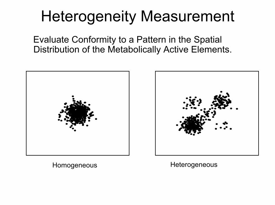
Heterogeneity Measurement Evaluate Conformity to a Pattern in the Spatial Distribution of the Metabolically Active Elements.
Homogeneous Heterogeneous

CV Spatially Insensitive
Spatially Coherent Spatially Incoherent
0.0 0.2 0.4 0.6 0.8 1.0
010
020
030
040
0
Histogram
CV is 0.71 for Both!

Ellipsoidal Model for Homogeneous Tumor
x1
0.1
0.2
0.3
0.4
0.5
0.60.7
0.8
0.9
1
1
2
Heterogeneity
| , (( ) ' ( ))
(monotone); ( , )
1-
( ) x g g x x
g
H R
θ µ µ
θ µ
λ −− Σ −
= Σ
=
≈
H=0.06g
O’Sullivan, Roy, Eary et al (2003,2005,2009)

HeterogeneityMeasurements
H=0.02 H=0.06
H=0.26 H=0.34

Heterogeneity
and
Patient Outcome
PredictorVariable (X)
Scale %Change in Risk(unitchange in X)
95%C.I.
P-value
AGE(years)
16.8 34 (-12,101) 0.150
SUV(max)(ml/gm)
6.14 -38 (-60,-29) 0.037
Heterogeneity 7.4% 87 (35,160) 0.0002

Roose, Chapman and Maini, SIAM Review, 2008.Cristini, Gatenby, Sutherland, Casciari, Rasey, Krohn 1986...2010
Necrosis
NecroticCenter

Tumor Synthesis (Growth Pattern)
Transverse Sagittal
Coronal
1 2 3 1 2 3
1 2 3
1 2 3
, , ) ( , , )( , , )
Uptake Model
, , )( )
Co-ordinate TransformationsPrincipal Axes : ( Flexible Cyclinder : ( , , )
( ) ( , , ) (h hh
x x x z z zz z z h r
rx x x h r gσ
θ
λ λ θ α τθ
→→
= ≈ +
Sarcoma
Bladder
PhaseInformation


Chemotherapy Response
MODEL:
PRE POST
GLM-Test:ˆ
ˆRESPONSE
β
βσ
=
Pre
) PRE quasi-Poisson( )POST quasi-Poisson(eβ
µµ
::
Co-
Reg
istra
tion
MarginMargin
Post
CorrelationAdjusted!

Dynamic PET Studies: Scans after Tracer Input

Quantitative Data Analysis:Separate Delivery and Retention
0( )( () ) ( )T B P
tPC t V Ct s sC dR t s= −∆ + −∆− ⋅∫
Residue
AIF
•Parametric (compartmental)
•Non-Parametric (non-compartmental)O’Sullivan et al. JASA (2009)
•Directly Sampled
•Image Extracted (Statistically Guided)O’Sullivan et al. IEEE-TMI (2010)
Data

Quantitative Analysis of Dynamic PET Data
BLOOD
FDG
TISSUE
FDG FDG-6-P
0.0
0.2
0.4
0.6
0.8
1.0
0 30 60 90 120Time (minutes)
Cp FLT Cp metArterial Input (AIF)
0.00
0.01
0.02
0.03
0.04
0.05
0.06
0 30 60 90 120
Time (minutes)
Tumor BrainTissue Data
Residue(Impulse Response)
0( )( () ) ( )T B P
tPC t V Ct s sC dRt s= −∆ + −∆− ⋅∫
Flux Flow
VD Extraction
Functionals

Nonparametric Residue Analysis
0
0( ) (( )
1
extraction
)
( ) (Survival Function)
is the residence density for tracer label is flow, is blood volume an
(
d
)
( )t
P Pt
T B
B
C t V t s ds
V
C C
t d
hK
R
hR
t s
γ
γ
τ τ
= −∆ + −∆
= −
− ⋅
∫
∫
Meier and Zierler (1954), Bassingthwaighte (1971), Ostergaard et al. (1996)
Estimation based a cross-validated regularization procedure involving Positivity/Monotonicity and Smoothness Constraints.

1 2
1 1 2 2
1 2
1 1 2 2 3
( ) ( ) ( ) .... ( )
( ) ....
( ) ( ) ( ) .... ( )
p
B p p
C p
M p
h B B B
h e e e
h h h h
λ τλ τ λ τ
τ φ τ φ τ φ τ
τ α α α
τ π τ π τ π τ
−− −
≈ + + +
≈ + + +
≈ + + +
B - splines
C om partm ental
M ixtures
Numerical Approximations for Residence
Mendelsohn and Rice (1984); Cunningham and Jones(1993) , O’Sullivan et al (2009)

Most Widely Used Compartmental Model for PET
BLOOD
FDG
K1
k2
TISSUE
FDG FDG-6-PO4 k4
k3
1 2
1 1 2 2
1 2
1 1 2 2 3
( ) ( ) ( ) .... ( )
( ) ....
( ) ( ) ( ) .... ( )
p
B p p
C p
M p
h B B B
h e e e
h h h h
λ τλ τ λ τ
τ φ τ φ τ φ τ
τ α α α
τ π τ π τ π τ
−− −
≈ + + +
≈ + + +
≈ + + +
B - splines
C om partm ental
M ixturesMay be reasonable in-vitro, but for in-vivo PET ROI data???
Implies a Residence Density of the form:

PET FDG Data from Normal Brain ROIs
Nonparametric and Compartmental Analysis[ A formal statistical test rejects the compartmental model, p-value=0.046 ]
Cerebellum ROI
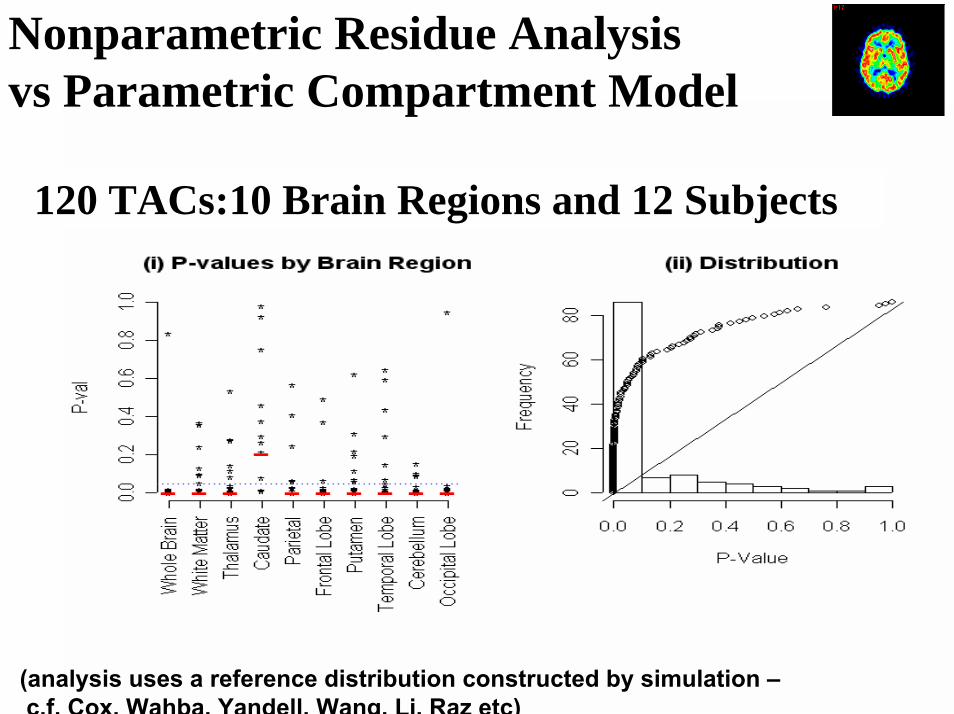
Nonparametric Residue Analysisvs Parametric Compartment Model
120 TACs:10 Brain Regions and 12 Subjects
(analysis uses a reference distribution constructed by simulation –c.f. Cox, Wahba, Yandell, Wang, Li, Raz etc)
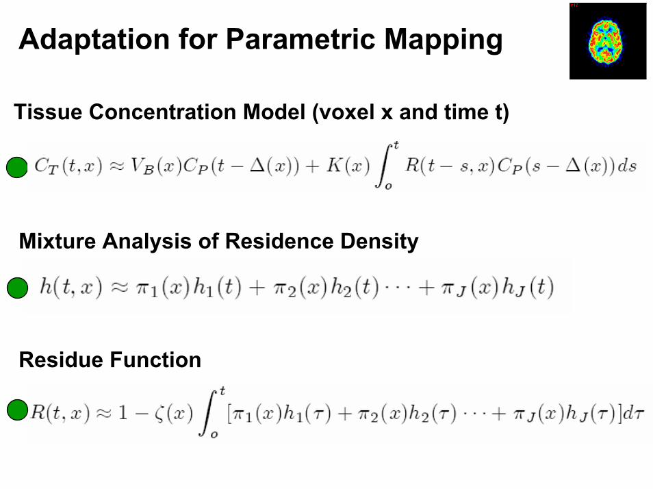
Adaptation for Parametric Mapping
Tissue Concentration Model (voxel x and time t)
Mixture Analysis of Residence Density
Residue Function

Residue Analysis of Segment Time Course Patterns
A
Residence
Residence
Time-Course
Time-Course
B

RMSData
ResidualPCA
RMSResidual
Temporal
Spatial
Diagnostic Assessment:
Voxel-Level Residuals

ml/gm
l/g
ml/g
/min
K VD VP
ml/1
0g/m
in
min
Flux MTT Ext0.0
0.0 0.0 0.0
0.00.0
0.5
0.52.0
0.60.3
0.5

Thym
idin
eVe
rapa
mil
mL
/g/m
in
0.0
Wat
er0.025
0.0
0.20
Uptake
Uptake
Uptake
mL
/g
0.0
2.21
mL
/g/m
in
0.0
0.37
2.60
K
K
K
mL
/g/m
inm
L/g
/min
mL
/g
0.0
0.13
VP
VD
Flux

Variance of Residues
(Greenwood’s Formula)
Approximation:
Flow AIFMean
-> Variation in Functionals by the Delta-Method

RegionalVoxel-LevelResidues andFlow Distribution
StandardizedVoxel-LevelResidues(Measured)

Some Analysis
Poisson with mean
Asymptotic Variance of MLEs

Summary
• PET in Cancer Imaging Diagnosis/StagingResponse AssessmentTreatment Planning
• Spatial and Temporal Aspects of PET Data Important
• Detailed Measurement and Modeling of the Disease Process is key to adaptive treatment
Statistics (Wisconsin style) has much to offer.(Please keep it going for another 50… at least!)
Related Documents