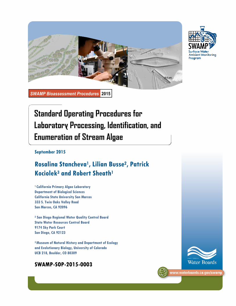
SWAMP Bioassessment Procedures 2015 Standard Operating Procedures for Laboratory Processing, Identification, and Enumeration of Stream Algae September 2015 Rosalina Stancheva 1 , Lilian Busse 2 , Patrick Kociolek 3 and Robert Sheath 1 1 California Primary Algae Laboratory Department of Biological Sciences California State University San Marcos 333 S. Twin Oaks Valley Road San Marcos, CA 92096 2 San Diego Regional Water Quality Control Board State Water Resources Control Board 9174 Sky Park Court San Diego, CA 92123 3 Museum of Natural History and Department of Ecology and Evolutionary Biology, University of Colorado UCB 218, Boulder, CO 80309 SWAMP-SOP-2015-0003

Welcome message from author
This document is posted to help you gain knowledge. Please leave a comment to let me know what you think about it! Share it to your friends and learn new things together.
Transcript
SWAMP Bioassessment Procedures 2015
Standard Operating Procedures for
Laboratory Processing, Identification, and
Enumeration of Stream Algae
September 2015
Rosalina Stancheva1, Lilian Busse2, Patrick
Kociolek3 and Robert Sheath1
1 California Primary Algae Laboratory
Department of Biological Sciences
California State University San Marcos
333 S. Twin Oaks Valley Road
San Marcos, CA 92096
2 San Diego Regional Water Quality Control Board
State Water Resources Control Board
9174 Sky Park Court
San Diego, CA 92123
3 Museum of Natural History and Department of Ecology
and Evolutionary Biology, University of Colorado
UCB 218, Boulder, CO 80309
SWAMP-SOP-2015-0003

September 2015 Page 2
SWAMP Laboratory Processing, Identification, and Enumeration of Stream Algae SOP
TABLE OF CONTENTS
TABLE OF CONTENTS ...................................................................................................... 2
ACKNOWLEDGMENTS ...................................................................................................... 4
LIST OF ABBREVIATIONS AND ACRONYMS .................................................................. 5
REQUIREMENTS AND RECOMMENDATIONS FOR SWAMP FUNDED PROJECTS……..6
INTRODUCTION ............................................................................................................... 18
SECTION 1: LABORATORY PRACTICES ....................................................................... 19
1.1 Taxonomist Qualifications ..................................................................................... 19
1.2 Laboratory Technician Qualifications .................................................................... 21
1.3 Taxonomic Literature ............................................................................................ 22
1.4 Taxonomic Nomenclature ..................................................................................... 23
1.5 Photographic Documentation of Algae .................................................................. 24
1.5.1 Photographic Documentation of Newly Reported Taxa .............................. 24
1.5.1.1 Photographic Documentation of Newly Reported SBA Taxa ............. 25
1.5.1.2 Photographic Documentation of Newly Reported Diatom Taxa ........ 25
1.5.2 Photographic Documentation of Previously Reported Taxa ........................ 25
1.5.3 General Requirements for Photographic Documentation ............................ 26
1.6 Standard Taxonomic Effort ................................................................................... 26
1.6.1 Standard Taxonomic Effort for SBA ........................................................... 27
1.6.2 Standard Taxonomic Effort for Diatoms ..................................................... 27
1.7 External Taxonomic Harmonization Process ......................................................... 28
1.8 Training ................................................................................................................. 30
1.9 General Taxonomic Laboratory Practices ............................................................. 30
SECTION 2: LABORATORY SAMPLE RECEIPT ............................................................. 31
2.1 Sample Receipt ..................................................................................................... 32
2.2 Sample Integrity Check ......................................................................................... 32
2.2.1 SBA Qualitative Sample Integrity Check ................................................... 32
2.2.2 SBA Quantitative Sample Integrity Check ................................................ 32
2.2.3 Diatom Quantitative Sample Integrity Check ............................................ 33
2.2.4 Receipt of Broken Sample Vials ............................................................... 33
SECTION 3: SAMPLE PREPARATION ............................................................................ 33
3.1 SBA Qualitative and Quantitative Sample Preparation .......................................... 35
3.1.1 SBA Qualitative Sample Preparation ......................................................... 35
3.1.2 SBA Quantitative Sample Preparation – Macroalgal Fraction .................... 36
3.1.3 SBA Quantitative Sample Preparation – Microalgal Fraction ..................... 38

September 2015 Page 3
SWAMP Laboratory Processing, Identification, and Enumeration of Stream Algae SOP
3.1.4 SBA Semi-permanent Slide Preparation of Quantitative
Microalgal Fraction .................................................................................... 39
3.2 Diatom Quantitative Sample Preparation .............................................................. 41
3.2.1 Cleaning of Diatom Samples: Nitric Acid Method ...................................... 41
3.2.2 Cleaning of Diatom Samples: Hydrogen Peroxide Method ........................ 43
3.2.3 Permanent Slide Preparation of Diatom Samples ..................................... 45
SECTION 4: IDENTIFICATION AND ENUMERATION ANALYSIS OF ALGAE .............. 47
4.1 Identification and Enumeration Analysis of SBA ................................................. 47
4.1.1 SBA Qualitative Sample Analysis.............................................................. 49
4.1.2 SBA Quantitative Sample Analysis – Macroalgal Fraction ........................ 50
4.1.3 SBA Quantitative Sample Analysis – Microalgal Fraction .......................... 53
4.1.4 Biovolume calculations for SBA ................................................................ 55
4.1.4.1 Biovolume Calculations: SBA Quantitative Sample –
Macroalgal Fraction........................................................................ 56
4.1.4.2 Biovolume Calculations: SBA Quantitative Sample –
Microalgal Fraction ......................................................................... 56
4.2 Identification and Enumeration Analysis of Diatoms ........................................... 58
4.3 Sample Labeling and Archiving .......................................................................... 60
4.3.1 Archiving of SBA – Qualitative Samples.................................................... 61
4.3.2 Archiving of SBA – Quantitative Samples ................................................. 61
4.3.3 Archiving of Diatom Samples .................................................................... 62
SECTION 5: QUALITY ASSURANCE AND QUALITY CONTROL .................................. 62
5.1 Laboratory Quality Control .................................................................................. 63
5.2 Laboratory Quality Assurance ............................................................................. 63
5.3 Sample Handling Requirements ......................................................................... 63
5.3.1 Sample Handling Requirements – Point of Receipt .................................. 63
5.3.2 Sample Handling Requirements – Archiving ............................................. 65
5.4 Photomicrographic Documentation Requirements .............................................. 65
5.5 Requirements for External Harmonization of Taxonomic Results........................ 66
SECTION 6: REPORTING OF ALGAL TAXONOMY RESULTS ..................................... 66
6.1 Reporting of SBA Results ................................................................................... 66
6.2 Reporting of Diatom Results ............................................................................... 67
6.3 Data Management and Reporting ....................................................................... 67
TERMS AND DEFINITIONS............................................................................................. 70
APPENDICES .................................................................................................................. 73
September 2015 Page 4
SWAMP Laboratory Processing, Identification, and Enumeration of Stream Algae SOP
ACKNOWLEDGEMENTS
The current version of the protocol was established with contributions from the following people:
Christina Fuller (California State University San Marcos) - technical assistance and preparing Appendix A
Eric von der Geest (Moss Landing Marine Laboratories) – preparing Section 5, discussion and input on quality assurance procedures
Candice Heinz (State Water Resource Control Board) - discussion and input on quality assurance procedures
Kalina Manoylov (Georgia College and State University) - SOP review and comments
Melissa Morris (State Water Resource Control Board) - preparing Section 5, discussion and input on quality assurance procedures
Marco Sigala (Moss Landing Marine Laboratories) – preparing Section 6 and Appendix G
Sarah Spaulding (University of Colorado) – discussion on taxonomy quality assurance and quality control process
Evtim Topalov (Palomar College) - graphic design of Figure 2
John Wehr (Fordham University) - SOP review and comments
Citation for this document:
Stancheva, R., Busse, L., P. Kociolek, and R. Sheath, 2015. Standard Operating
Procedures for Laboratory Processing and Identification of Stream Algae in California.
California State Water Resources Control Board Surface Water Ambient Monitoring Pro-
gram (SWAMP) Bioassessment SOP 0003.
September 2015 Page 5
SWAMP Laboratory Processing, Identification, and Enumeration of Stream Algae SOP
LIST OF ACRONYMS AND ABBREVIATIONS
Term Definition
CalPAL SWAMP California Primary Algae Laboratory of the State Water Board
CEDEN California Environmental Data Exchange Network
COC Chain of Custody
DI water DI water - Deionized Water
DIC Differential Interference Contrast
DMT Data Management Team
ID Identification
MQOs Measurement Quality Objectives
MSDS Material Safety Data Sheets
NCE Natural Counting Entity
PTA Percent Taxonomic Agreement
QA Quality Assurance
QC Quality Control
Sample ID Unique sample name
SBA Soft-Bodied Algae
SOP Standard Operating Procedures
STE Standard Taxonomic Effort
SWAMP Surface Water Ambient Monitoring Program
September 2015 Page 6
SWAMP Laboratory Processing, Identification, and Enumeration of Stream Algae SOP
REQUIREMENTS AND RECOMMENDATIONS FOR SWAMP
FUNDED PROJECTS
Relevant SOP
Sections Section Element Description of Requirements
Description of
Recommendations
1.1 Laboratory
Practices
Taxonomist
Qualifications
The laboratory must have at least
one taxonomist (preferably two) who
meets the minimum qualifications
specified in Section 1.1.
Maintain current documentation of
taxonomist qualifications, and be
prepared to submit these for review
upon request.
None
1.2 Laboratory
Practices
Laboratory
Technician
Qualifications
Laboratory technicians must meet the
minimum qualifications specified in
Section 1.2.
Laboratories must maintain current
training documentation of all
laboratory technicians, and be
prepared to submit these for review
upon request.
None
1.3 Laboratory
Practices
Taxonomic
Literature
Remain current with taxonomic
literature related to local algal flora.
List of algal
taxonomic resources
is included in
Appendix I:
References
1.4 Laboratory
Practices
Taxonomic
Nomenclature
Use the taxon names compiled in the
SWAMP Algae Master Taxa list.
Newly reported species must be well
documented and submitted for
harmonization.
Taxon names of newly reported
species must be approved prior to
reporting.
None

September 2015 Page 7
SWAMP Laboratory Processing, Identification, and Enumeration of Stream Algae SOP
Relevant SOP
Sections Section Element Description of Requirements
Description of
Recommendations
1.5.1 Laboratory
Practices
Photographic
Documentation
of Newly
Reported Taxa
Collect high-quality
photomicrographs for each newly
reported species submitted to the
Algae Master Taxa list.
Refer to Section 5.4
and Appendix H
1.5.2 Laboratory
Practices
Photographic
Documentation
of Previously
Reported Taxa
None
Collect representative
photomicrographs of
each SBA and diatom
taxon identified in the
sample
1.5.3 Laboratory
Practices
General
Requirements
for
Photographic
Documentation
Photomicrograph documentation
must have the following:
Scale bar in the lower right corner
of the image measuring 10, 20 or
50 µm proportional to the size of
the algae and magnification used;
Be saved in TIFF format using the
maximum resolution afforded by
the equipment in use (minimum of
300 dpi);
Each photo should have a filename
consisting of the following elements
in the order indicated: SWAMP
Sample ID, Sampling date (MM/
DD/YYYY), Species ID,
magnification for objective (i.e.
40x).
Store all
photomicrographs on a
high-capacity internal
hard drive of the
laboratory computer
and periodically backed
up onto an external
hard drive.
1.6.1 Laboratory
Practices
Standard
Taxonomic
Effort for SBA
SBA specimens to be identified to
species level, or the lowest
taxonomic level possible
Identify each SBA to
species level or lower.
If species identification
is not possible due to
insufficient taxonomic
vegetative or
reproductive data,
identify the specimen to
the lowest taxonomic
level possible, such as
genus or above.
September 2015 Page 8
SWAMP Laboratory Processing, Identification, and Enumeration of Stream Algae SOP
Relevant SOP
Sections Section Element Description of Requirements
Description of
Recommendations
1.6.2 Laboratory
Practices
Standard
Taxonomic
Effort for
Diatoms
Diatom specimens to be identified to
species level, or the lowest
taxonomic level possible
Identify each diatom
to species level or
lower. If species
identification is not
possible due to
insufficient taxonomic
data, identify the
specimen to the
lowest taxonomic level
possible, such as
genus or above.
1.7 Laboratory
Practices
External
Taxonomic
Harmonization
Process
Submit all newly reported species
identifications for taxonomic
harmonization prior to reporting.
SWAMP
recommends
harmonization of the
entire dataset
(including results from
previously reported
species), but does not
currently require this
due to resource
limitations.
1.8 Laboratory
Practices Training
Laboratories must have internal
procedures for executing and
documenting the training of
laboratory technicians and
taxonomists in the use of these
procedures.
Documentation of
training should
include demonstration
of performance in the
procedures.
1.9 Laboratory
Practices
General
Taxonomic
Laboratory
Practices
None None
2.1
Laboratory
Sample
Receipt
Sample
Receipt
Confirm that the sample labels
match the chain of custody (COC)
forms and all samples are accounted
for.
Retain copies of the COCs.
None

September 2015 Page 9
SWAMP Laboratory Processing, Identification, and Enumeration of Stream Algae SOP
Relevant SOP
Sections Section Element Description of Requirements
Description of
Recommendations
2.2.1
Laboratory
Sample
Receipt
SBA
Qualitative
Sample
Integrity Check
Confirm the sample is received in a
100 mL Whirl-Pak® bag.
Confirm the sample is cool (4 °C)
upon receipt.
Confirm sample has not been frozen.
Note evidence of freezing on the
COC. Confirm sample did not leak
prior to receipt. Note evidence of
leaking on the COC.
Confirm the sample has been
received within 2 weeks of collection.
None
2.2.2
Laboratory
Sample
Receipt
SBA
Quantitative
Sample
Integrity Check
Confirm that the SBA quantitative
sample has been preserved. If the
sample received is unpreserved, it
must be preserved ASAP within 4
days of collection.
If the sample is preserved following
receipt, record the volume of the
unpreserved sample, amount of
glutaraldehyde added, and date and
time of preservation on the COC.
Inspect the volume in
the sample vial. Vials
received with less
than 50 mL of
preserved sample
may indicate the
sample was not
preserved or had
leaked during
transport.
2.2.3
Laboratory
Sample
Receipt
Diatom
Quantitative
Sample
Integrity Check
Confirm the samples received are
preserved in the field with 1%
formalin.
Inspect the volume in
the sample vial. Vials
with less than 50 mL
of preserved sample
may indicate the
sample was not
preserved or had
leaked during
transport.
2.2.4
Laboratory
Sample
Receipt
Receipt of
Broken Sample
Vials
Transfer leaking sample to a new 50
mL plastic centrifuge tube labeled
with the sample information.
Measure and record the remaining
sample volume. Document the
sample condition.
Add additional preservative and note
volume added on COC.
None

September 2015 Page 10
SWAMP Laboratory Processing, Identification, and Enumeration of Stream Algae SOP
Relevant SOP
Sections Section Element Description of Requirements
Description of
Recommendations
3.1.1 Sample
Preparation
SBA Qualitative
Sample
Preparation
None
Archiving of the SBA
Qualitative sample
should be conducted
as soon as possible
following completion
of the taxonomic
analysis.
3.1.2 Sample
Preparation
SBA
Quantitative
Sample
Preparation –
Macroalgal
Fraction
Confirm the absence of
macroalgae using the dissecting
microscope before proceeding
with microalgae preparation.
Determine the biovolume of
macroalgae by water
displacement.
None
3.1.3 Sample
Preparation
SBA
Quantitative
Sample
Preparation –
Microalgal
Fraction
Homogenize the microalgal
fraction of the SBA Quantitative
sample by gently but thoroughly
inverting the 50 mL centrifuge tube
several times.
None
3.1.4 Sample
Preparation
SBA Semi-
permanent Slide
Preparation of
Quantitative
Microalgal
Fraction
Confirm the prepared slide
contains a random distribution of
microalgae that is sufficiently
dense for species identification
and enumeration.
Gently tap on the
cover slip to reduce
the algae clumping
and air bubbles, if
present.
3.2.1 Sample
Preparation
Cleaning of
Diatom
Samples: Nitric
Acid Method
Diatom Quantitative samples are
preserved in formalin, so they
must be handled carefully.
None
3.2.2 Sample
Preparation
Cleaning of
Diatom
Samples:
Hydrogen
Peroxide
Method
Specific handling and disposal
procedures must be in place for
handling hydrogen peroxide and
potassium dichromate.
None
September 2015 Page 11
SWAMP Laboratory Processing, Identification, and Enumeration of Stream Algae SOP
Relevant SOP
Sections Section Element Description of Requirements
Description of
Recommendations
3.2.3 Sample
Preparation
Slide
Preparation
of Diatom
Samples
Confirm the prepared slide
contains a random distribution
of diatoms that is sufficiently
dense for conducting
identification and enumeration
procedures.
Add 10% HCl to the
cleaned diatom
suspension to achieve a
more even distribution of
diatom valves on the
coverslip.
4.1.1
Identification
and
Enumeration
Analysis of
Algae
SBA
Qualitative
Sample
Analysis
Record all macroalgal taxa
identified in the SBA
qualitative sample in the ID
Datasheet for SBA Sample
under the heading Qualitative
sample – list of taxa
Take sufficient high-quality
photomicrographs of all newly
recorded species to support
harmonization of results.
Collect photomicrographs
of previously reported
species to demonstrate the
key aspects of vegetative
morphology and
reproduction used in
identification.
When reproducing
filaments of zygnematalean
algae are observed, but
completely matured
zygospores/aplanospores
are not available, further
incubation under nutrient
stress facilitates completion
of sexual or asexual
reproduction. The resulting
mature zygospores (or
akinetes, aplanospores)
can provide the taxonomist
with the additional
information needed to
identify the species.
4.1.2
Identification
and
Enumeration
Analysis of
Algae
SBA
Quantitative
Sample
Analysis -
Macroalgal
Fraction
Record the fraction represented
by non-algal matter on the ID
Datasheet for SBA Sample
Heading: non-algal matter xx
%.
Take photomicrographs of
previously reported species
to demonstrate the key
aspects of vegetative
morphology and
reproduction used in
identification.
September 2015 Page 12
SWAMP Laboratory Processing, Identification, and Enumeration of Stream Algae SOP
Relevant SOP
Sections Section Element Description of Requirements
Description of
Recommendations
4.1.2 (cont)
Identification
and
Enumeration
Analysis of
Algae
SBA
Quantitative
Sample
Analysis -
Macroalgal
Fraction
Record the identification for
each macroalgal taxon
identified and the corresponding
proportion of each in the ID
Datasheet for SBA Sample-
Heading: Macroalgae taxon ID;
Proportion of each taxon (%).
Enumerate 100 NCEs of
epiphytic SBA alga attached to
the surface of the macroalgae.
Record each epiphytic algae
taxa identified and the
corresponding number of NCEs
enumerated on the ID
Datasheet for SBA Sample-
Heading: Epiphyte taxon ID;
#NCE.
Take sufficient high-quality
photomicrographs of all newly
recorded species to support
harmonization of results.
Take photomicrographs of
previously reported
species to demonstrate the
key aspects of vegetative
morphology and
reproduction used in
identification.
4.1.3
Identification
and
Enumeration
Analysis of
Algae
SBA
Quantitative
Sample
Analysis -
Microalgal
Fraction
Record any additional dilution or
concentration performed on the
sample in the ID Datasheet for
SBA Sample-Heading:
Quantitative Sample-Microalgal
fraction-sample volume after
additional dilution/concentration:
xx mL; dilution factor
Identify and enumerate 300
SBA NCEs across a known
number of horizontal optical
transects.
Ensure that the volume of
the drop is not so large
that it creates the
formation of bubbles or
causes the cover slip to
float.
Avoid having too much or
too little material on the
slide.
Gentle tapping on the
cover slip or spread
clumps apart with a pair of
dissecting needles will
reduce clumping of algae.

September 2015 Page 13
SWAMP Laboratory Processing, Identification, and Enumeration of Stream Algae SOP
Relevant SOP
Sections Section Element Description of Requirements
Description of
Recommendations
4.1.3 (cont)
Identification
and
Enumeration
Analysis of
Algae
SBA
Quantitative
Sample
Analysis -
Microalgal
Fraction
Record each microalgal SBA
species identified and the
corresponding number of NCEs
enumerated on the ID Datasheet
for SBA Sample-Heading:
Microalgal taxon ID; #NCE).
Record the number of transects
traversed in ID Datasheet for
SBA Sample-Heading:
Microalgal fraction-number of
horizontal transects counted: xx.
Determine the appropriate
geometric model for each
microalgal species identified and
perform microscopic
measurements of the cell
dimensions for each. Record
measurements on the ID
Datasheet for SBA Sample-
Heading: Cell diameter (µm);
Cell/NCE length (µm); Cell
Depth (µm); Total number of
cells; Total filament length(µm).
Take sufficient high-quality
photomicrographs of all newly
recorded species to support
harmonization of results.
Take
photomicrographs of
previously reported
species to demonstrate
the key aspects of
vegetative morphology
and reproduction used in
identification.
4.1.4.1
Identification
and
Enumeration
Analysis of
Algae
Biovolume
Calculations:
SBA
Quantitative
Sample-
Macroalgal
Fraction
Calculate the biovolume of each
macroalgal taxon using the
formulas in Section 4.1.4.1.
None
September 2015 Page 14
SWAMP Laboratory Processing, Identification, and Enumeration of Stream Algae SOP
Relevant SOP
Sections Section Element Description of Requirements
Description of
Recommendations
4.1.4.2
Identification
and
Enumeration
Analysis of
Algae
Biovolume
Calculations:
SBA
Quantitative
Sample-
Microalgal
Fraction
Calculate the biovolume of each
microalgal taxon using the
formulas in Section 4.1.4.2.
None
4.2
Identification
and
Enumeration
Analysis of
Algae
Enumeration
and
Identification
Analysis
of Diatoms
Identify and enumerate 600
diatom valves across a known
length of horizontal optical
transects.
Partial valves are defined as
having more than 50% of the
valve including the central area.
Enumerate only complete and
partial valves. The valve (both
complete and partial) must
extend at least halfway into the
transect, and must include the
center of the valve in the
transect.
Record each diatom taxon
identified and the corresponding
number of valves enumerated
on the ID Datasheet for Diatom
Sample-Heading: Diatom taxon
ID; Number of valves).
Record the number of transects
traversed, starting and ending
field of view for each transect in
ID Datasheet for Diatom
Sample-Heading: Number of
transects counted: xx.
Take sufficient high-quality
photomicrographs of all newly
recorded species to support
harmonization of results.
If the sample is very
sparse, continue
counting for 4 hours or
until 300 valves are
enumerated (whichever
comes first), excluding
time spent learning new
species.
Take photomicrographs of
previously reported
species to demonstrate
the key aspects of
vegetative morphology
and reproduction used in
identification.

September 2015 Page 15
SWAMP Laboratory Processing, Identification, and Enumeration of Stream Algae SOP
Relevant SOP
Sections Section Element Description of Requirements
Description of
Recommendations
4.3
Identification
and
Enumeration
Analysis of
Algae
Sample
Labeling and
Archiving
All SBA and diatom samples must
be retained as voucher specimens
until harmonization and reporting
of data is complete.
Archives of samples and
slides should be
retained by the
laboratory for two years.
4.3.1
Identification
and
Enumeration
Analysis of
Algae
Archiving of
SBA –
Qualitative
Samples
Select a representative subsample
that contains all identified
macroalgal taxa and fix it with 2%
glutaraldehyde final concentration.
None
4.3.2
Identification
and
Enumeration
Analysis of
Algae
Archiving of
SBA –
Quantitative
Samples
Slides-microalgal fraction: Seal the
cover slip with nail polish, label the
microscopic slide by sample ID,
collection date (MM/DD/YYYY),
and note “microalgae”.
Return analyzed macroalgae and
archive the macroalgal fraction
adding glutaraldehyde to 2% final
concentration. Label the tube by
sample ID, collection date (MM/
DD/YYYY), and note
“macroalgae”.
Refix the subsample with 2%
glutaraldehyde final concentration
and keep separately from original
sample for reference purposes.
Label it by sample ID, collection
date (MM/DD/YYYY), and note
“microalgae”.
None
4.3.3
Identification
and
Enumeration
Analysis of
Algae
Archiving of
Diatoms
Label each slide by sample ID,
collection date (MM/DD/YYYY),
and note “diatoms”.
Fix remaining cleaned diatom
material with ethanol to 50% final
concentration. Label each vial by
sample ID, collection date (MM/
DD/YYYY), and note “diatoms”.
None
September 2015 Page 16
SWAMP Laboratory Processing, Identification, and Enumeration of Stream Algae SOP
Relevant SOP
Sections Section Element
Description of
Requirements
Description of
Recommendations
5.1
Quality
Assurance
and Quality
Control
Laboratory Quality
Control None None
5.2
Quality
Assurance
and Quality
Control
Laboratory Quality
Assurance None None
5.3.1
Quality
Assurance
and Quality
Control
Sample
Handling
Requirements-
Point of
Receipt
Confirm samples meet the
sample handling
requirements in Table 3:
Required Sample
Conditions for Laboratory
Receipt.
Follow corrective actions in
Table 4: Required
Corrective Actions for
Laboratory Receipt.
None
5.3.2
Quality
Assurance
and Quality
Control
Sample
Handling
Requirements-
Archiving
All samples must be
archived by the laboratory
until results have been
harmonized and reported.
None
5.4
Quality
Assurance
and Quality
Control
Photomicrographic
Documentation
Requirements
Collect high-quality
photomicrographic
documentation of newly
recorded species sufficient
to support harmonization of
results.
Recommendations for
producing high-quality
photomicrographs are
included in
Appendix H.
Collect
photomicrographic
documentation of
previously reported
species sufficient to
demonstrate the key
aspects of vegetative
morphology and
reproduction used in
identification.
September 2015 Page 17
SWAMP Laboratory Processing, Identification, and Enumeration of Stream Algae SOP
Relevant SOP
Sections Section Element Description of Requirements
Description of
Recommendations
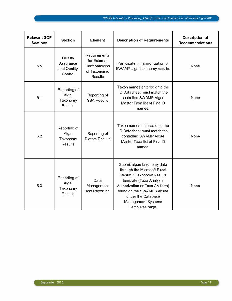
5.5
Quality
Assurance
and Quality
Control
Requirements
for External
Harmonization
of Taxonomic
Results
Participate in harmonization of
SWAMP algal taxonomy results. None
6.1
Reporting of
Algal
Taxonomy
Results
Reporting of
SBA Results
Taxon names entered onto the
ID Datasheet must match the
controlled SWAMP Algae
Master Taxa list of FinalID
names.
None
6.2
Reporting of
Algal
Taxonomy
Results
Reporting of
Diatom Results
Taxon names entered onto the
ID Datasheet must match the
controlled SWAMP Algae
Master Taxa list of FinalID
names.
None
6.3
Reporting of
Algal
Taxonomy
Results
Data
Management
and Reporting
Submit algae taxonomy data
through the Microsoft Excel
SWAMP Taxonomy Results
template (Taxa Analysis
Authorization or Taxa AA form)
found on the SWAMP website
under the Database
Management Systems
Templates page.
None

September 2015 Page 18
SWAMP Laboratory Processing, Identification, and Enumeration of Stream Algae SOP
INTRODUCTION
This standard operating procedure (SOP) is applicable to the analysis of benthic
soft-bodied algae (SBA) and diatoms collected using SWAMP standard operating
procedures for collection of field data for ambient bioassessments of California wadeable
streams: benthic macroinvertebrates, algae and physical habitat (Ode et al., 2015).
It describes the staff qualifications, laboratory and taxonomy methods to be used
whenever algae stream bioassessment is conducted under the SWAMP program. Since
both algal groups, SBA and diatoms, require different laboratory treatment, their separate
laboratory processing, identification and enumeration, species documentation, archiving of
samples and slides, quality assurance procedures, and data reporting to SWAMP are
described. SBA analysis is conducted from two types of samples collected from each
stream reach — fresh qualitative and preserved quantitative, resulting in a comprehensive
taxa list with corresponding biovolume of algal taxa recorded in the quantitative sample.
Diatom analysis from a separate quantitative sample provides a taxa list with the relative
abundance of each diatom taxon identified.

September 2015 Page 19
SWAMP Laboratory Processing, Identification, and Enumeration of Stream Algae SOP
Section 1: Laboratory Practices
This section describes policies for establishing and maintaining necessary infrastructure for
processing and identifying soft-bodied algae (SBA) and diatoms. These practices are
critical to the production of high-quality algae data.
Laboratories performing identification and enumeration of algal samples using these
procedures are required to have the following resources and tools:
Highly qualified freshwater SBA and diatom taxonomists;
Highly trained laboratory technicians;
Research-grade compound microscopes and stereoscopes with capability for attached
digital camera;
Access to up-to-date taxonomic literature;
A photomicrographic reference collection of SBA and diatom specimens;
Good general laboratory practices;
Infrastructure for sample tracking and data management;
A standard taxonomic effort.
1.1 Taxonomist Qualifications
The laboratory must have at least one person (preferably two) with considerable experience
in identification and enumeration of all taxonomic groups of stream SBA (called SBA
taxonomist) and/or diatoms (called diatom taxonomist, or diatomist). This experience can
only be obtained by hands-on algal studies from a variety of freshwater habitats (preferably
from streams) with algal identifications corroborated by experts. This experience should
also include knowledge of and ability to use the detailed taxonomic references listed at the
reference section (Appendix I). In order to remain current with changing algal systematics
and nomenclature, the experienced taxonomist(s) must maintain contact with other
taxonomists through professional societies and other interactions.
Laboratories must maintain current documentation of taxonomist’s qualification, and be
prepared to submit these for review upon request.
September 2015 Page 20
SWAMP Laboratory Processing, Identification, and Enumeration of Stream Algae SOP
Taxonomists are responsible for performing identification and enumeration of algae
samples, and for training new laboratory personnel in the procedures detailed in this SOP.
The experienced taxonomist(s) educate new staff on the specifics of the local algal flora
composition, information important to ensure the quality of results.
Algal taxonomists performing analysis for SWAMP projects must meet the following
minimum qualifications:
Education Taxonomists must have at least a Master of Science (MS) degree in botany, ecology,
biology or related degree, in addition to one or more of the following:
Coursework related to plant taxonomy, aquatic ecology or limnology;
Graduate thesis or undergraduate research projects in algal taxonomy, or ecology of
benthic freshwater algae;
University-level phycology class or SBA taxonomy class (for SBA taxonomist) and
diatom taxonomy class (for diatom taxonomist).
Experience and Training
The following experience is required for all taxonomists:
At least two years of experience identifying freshwater algae, preferably from stream
benthos;
Regularly attend taxonomy training workshops offered by professional meetings;
New personnel must be trained by more experienced taxonomy staff until the new
taxonomist demonstrates the ability to correctly identify local algal species and
produces datasets that meet laboratory QA standards.
Knowledge and Skills
The following knowledge and skills are required for all taxonomists:
Proficiency in the use of appropriate algal taxonomic literature and dichotomous
identification keys;
Current knowledge of the most recent changes in algal taxonomy;

September 2015 Page 21
SWAMP Laboratory Processing, Identification, and Enumeration of Stream Algae SOP
Navigate the California Online Algae Identification Resource Tools effectively;
Ability to identify and document algae accurately;
Experience in the use of light microscopy and digital microphotography for taking
high-quality pictures of algal specimens;
Good record-keeping skills.
1.2 Laboratory Technician Qualifications
Laboratory technicians are responsible for:
Sample receipt;
Tracking samples from receipt to archiving;
Cleaning and preparation of SBA and diatom samples for taxonomic identification and
enumeration.
Preparation procedures include subsampling of SBA for macroalgae, measuring the
macroalgal fraction volume, concentrating the microalgal subsample, and the cleaning of
diatoms and preparing diatom slides for identification by taxonomists.
Laboratory technicians processing algae samples for SWAMP projects must meet the
following minimum qualifications:
Experience
At least two years laboratory experience, with preference given to those who have
experience processing bioassessment algal samples;
Experience in the safe handling of laboratory chemicals.
Skills
Good record-keeping skills;
Good hand-eye coordination in sample processing;
Good skills in quantitative sample processing;
Ability to process fractions of liquid algal samples with high precision and accuracy;
Ability to avoid cross-contamination of algal samples;
Experience in the use of technical equipment and light microscopy for standard algal
September 2015 Page 22
SWAMP Laboratory Processing, Identification, and Enumeration of Stream Algae SOP
specimen preparation techniques, including slide preparation.
Laboratories must maintain current training documentation of all laboratory technicians, and
be prepared to submit these for review upon request.
1.3 Taxonomic Literature
To properly perform algae identifications, the taxonomist must be up-to-date on the most
current taxonomic literature and online resources. Maintaining current knowledge of the
taxonomy of local algal flora is critical to ensuring data quality. A number of standard
references and online tools have been employed for the identification of freshwater algae
across the United States. A list of these resources is included in Appendix I: References.
SBA
Although not yet complete, the most comprehensive references for SBA are the 14-volume
set, The Freshwater Flora of Central Europe (1978-2014), and The Freshwater Algal Flora
of the British Isles 2nd Ed. (John et al., 2011). These references must be used with caution,
as not all species are identical to those present in California.
The main floristic works on freshwater algae from the United States are summarized in
Freshwater Algae of North America: Ecology and Classification 2nd Ed. (Wehr et al., 2015).
This book notes key references for species identification of SBA from all taxonomic groups
documented in the United States, such as Smith (1950), Transeau (1951), Prescott (1951)
Prescott et al. (1977-1982), Dillard (1989-2007).
While utilizing these resources, it is important to remember that current knowledge of the
freshwater algal diversity of California is incomplete and no one flora is currently available
to address all species. Some species have recently been recorded from streams in
California (Wehr et al. 2013), or are newly described to science, such as several Spirogyra
and Zygnema species (Stancheva et al. 2012b, 2013), and a new genus of green algae –
Caespitula (Hall et al., in preparation). Therefore, algal identifications should be done
carefully with good knowledge of current literature and local algal flora.
Diatoms
The most commonly employed reference for identification of diatoms is the five-volume set,
The Freshwater Flora of Central Europe (Krammer and Lange-Bertalot, 1986-1991, 2000).
September 2015 Page 23
SWAMP Laboratory Processing, Identification, and Enumeration of Stream Algae SOP
More recent work from Lange-Bertalot (Diatoms of Europe, 2000-2013) adopts a finer
taxonomic perspective. Bahls (2012) estimated that for the north and central portions of the
western United States, only half of the taxa are documented in Krammer and Lange-
Bertalot (1986-1991). Patrick and Reimer (1966, 1975) in The Diatoms of the United States
brought a huge advantage over previous floristic works on diatoms. It is important to note
that these references consider only a limited number of species, and exclude the centric
and keel-forming taxa.
No one flora is currently available to address all the diatom species occurring in California,
therefore, the taxonomic laboratory must combine resources (floristic and primary literature)
to accurately identify the diatoms, keeping in mind that many new freshwater diatom
species have recently been described to science from the western US (Kociolek et al.,
2014) and elsewhere in the US (Morales, 2005, Morales et al., 2012, Morales and
Manoylov, 2009, Potapova, 2012, Spaulding et al., 2010, etc.).
1.4 Taxonomic Nomenclature
When reporting algae results to SWAMP, all laboratories are required to use the same
compilation of taxon names. To ensure data comparability, SWAMP maintains an Algae
Master Taxa list. The list is accessible online at http://swamp.waterboards.ca.gov/
swamp_checker/LookUpLists.php, and organized into two sections by the type of sample:
SWAMP Master Taxa List-SBA (OrganismLookUp - CAD-TWG Algae List)
SWAMP Master Taxa List-Diatoms (OrganismLookUp - CAD-TWG Diatom List)
The SWAMP Master Taxa List attempts to represent the most up-to-date, commonly
accepted taxonomic scheme for each name. For each final ID, it includes taxonomic
classification (phylum, class, order, family, genus, species, variety, form) and the taxonomic
authors of the name.
When taxon names of SBA or diatoms are not available in the current SWAMP Algae
Master Taxa List, the specimens must be well documented (see Section 1.5 and Appendix
H) and submitted for taxonomic harmonization (see Section 1.7). Following approval, all
final ID names for newly reported species must be reported in the Organism_DetailLookUp
September 2015 Page 24
SWAMP Laboratory Processing, Identification, and Enumeration of Stream Algae SOP
tab of the Taxonomy Results template (see Section 6 and Appendix G) using the following
standard:
Taxonomic classification should follow Algaebase (Guiry and Guiry, 2015);
Species names with taxonomic authors should be obtained from the Algaebase
website. Only use names that are currently accepted taxonomically (indicated by 'C');
Taxonomic authors should be abbreviated according to the International Plant Names
Index. Since the SWAMP reporting format does not allow the use of periods in the
name (in abbreviations, such as var., f., cf.), they cannot be included in the result. For
example, Cocconeis placentula var. lineata (Ehrenberg) Grunow should be submitted
as Cocconeis placentula var lineata (Ehrenb.) Grunow; and Rhizoclonium cf.
hieroglyphicum (C. Agardh) Kützing should be submitted as Rhizoclonium cf
hieroglyphicum (C. Agardh) Kütz.
For taxa identified at genus or coarser taxonomic levels, a unique number in numerical
order should be added to the name in agreement with existing names and numbers in
the SWAMP Algae Master Taxa list. For example, Calothrix spp. should be submitted
as Calothrix sp 9.
1.5 Photographic Documentation of Algae
Photomicrographs of algae provide an excellent source of documented information about
the samples, and laboratories should include collection of photomicrographs in their
standard procedures. Taking multiple photomicrographs of every species would generate a
large amount of potentially useful supporting documentation.
In order to minimize the resources required, while maximizing the impact on data quality,
SWAMP has identified two situations encountered during taxonomic analysis in regards to
photographic documentation of algae (e. g. newly recorded algae taxa and previously
reported taxa to SWAMP) The requirements and recommendations related to photographic
documentation have been determined for each.
1.5.1 Photographic Documentation of Newly Reported Taxa
Newly reported taxa are those which have been previously described elsewhere or not, but
have not been previously reported to the SWAMP Algae Master Taxa list. Some of these

September 2015 Page 25
SWAMP Laboratory Processing, Identification, and Enumeration of Stream Algae SOP
newly reported species to the Algae Master Taxa list can be potentially newly discovered to
science. SWAMP requires collection of high-quality photomicrographs for each newly
reported taxon submitted to the Algae Master Taxa list. Newly reported taxa require the
largest number of photomicrographs, as they provide critical information for the
harmonization process.
While SWAMP requires taking photomicrographs of newly reported algae, a specific
minimum number of photomicrographs is not established. The appropriate number of
photomicrographs needed varies and should be determined by the taxonomist. The number
of photomicrographs taken should be sufficient to support identification and harmonization
of the new taxa.
1.5.1.1 Photographic Documentation of Newly Reported SBA Taxa
Photomicrographs must be taken of each newly reported SBA taxon identified. The number
of photomicrographs taken should be sufficient to illustrate all diagnostic features and
morphological variation needed for identification (see Appendix H). Some macroalgal taxa
may require more than one photomicrograph at low and high magnification if several
features are necessary for identification (e.g., key vegetative and reproductive
characteristics). Microalgae may also require multiple photomicrographs of the
characteristics of the colony, single cells from the colony, and specific diagnostic
organelles. Images should be well focused on the key features. For the definition of
macroalgae and microalgae see section 4.1.
1.5.1.2 Photographic Documentation of Newly Reported Diatom Taxa
For every newly reported diatom taxon that is identified, the slide on which it was seen and
its position on the slide should be documented. Short descriptions with detailed
observations about its frustular morphology as well as photomicrographs of the taxonomic
entity should be provided. Depending upon the number of specimens and the variability
expressed in the taxon, approximately five images per taxon showing the morphological
variability should be collected (see Appendix H).
1.5.2 Photographic Documentation of Previously Reported Taxa
Previously reported taxa are those already included in the Algae Master Taxa list. SWAMP
September 2015 Page 26
SWAMP Laboratory Processing, Identification, and Enumeration of Stream Algae SOP
strongly recommends that laboratories take representative photomicrographs of each SBA
and diatom taxon identified in the sample, however, these do not warrant the same level of
documentation required for newly reported species. Regardless, photomicrographs of
reported species provide valuable information that supports the data quality at multiple
levels (laboratory, project, and program).
SWAMP recommends laboratories take multiple photomicrographs of highly variable
species from samples originating from distant locations. These photographs support
consistency between identification of the algae taxa across the region.
1.5.3 General Requirements for Photographic Documentation
All photomicrographs must be taken using TIFF format (without compression). Although
TIFF files are significantly larger than files using alternate formats, they provide the
high-resolution required, in addition to being the standard format required by many scientific
publications.
Photomicrograph documentation of SBA or diatom specimens must have the following:
Scale bar in the lower right corner of the image measuring 10, 20 or 50 µm proportional
to the size of the algae and magnification used;
Be saved in TIFF format using the maximum resolution afforded by the equipment in
use (minimum of 300 dpi);
Each photomicrograph should have a filename consisting of the following elements in
the order indicated: SWAMP Sample ID, Sampling date (MM/DD/YYYY), Species ID,
magnification of the objective (i.e., 40x).
For example, the filename would be: 503ABC015_Calothrix epiphytica_07292014_40x.tiff
The laboratory should store all photomicrographs on a high-capacity internal hard drive of
the laboratory computer which is periodically backed up onto an external hard drive.
1.6 Standard Taxonomic Effort
A standard taxonomic effort (STE) refers to the taxonomic level at which specimens must

September 2015 Page 27
SWAMP Laboratory Processing, Identification, and Enumeration of Stream Algae SOP
be identified. Effort is required to achieve species level of algae identification, or the lowest
taxonomic level possible. In the sections below, some limitations in achieving species level
identification of SBA and diatoms are outlined. Laboratories are responsible for following
the STE to ensure proper level of algae identification.
1.6.1 Standard Taxonomic Effort for SBA
SBA species level identifications require observations of large portions of the filaments or
colonies, and the presence of specific vegetative and reproductive structures. Absence of
these structures can limit identification for some taxa to genus or coarser levels. The
analysis of fresh algal qualitative samples and separate identification of the macroalgal
fraction of quantitative samples applied in this SOP supplies additional morphological
information facilitating species identification of problematic genera (Stancheva et al.,
2012a). For instance, the species level identification of the following genera: Anabaena,
Dolichospermum, Cylindrospermum, Batrachospermum, Sirodotia, Oedogonium, Spirogyra,
Zygnema, Mougeotia, Vaucheria, etc. is largely based on their reproductive structures or
specialized cells, such as akinetes. Non-reproducing specimens are more commonly
observed. Therefore well-defined “morphospecies” are assigned for the non-reproductive
specimens based on their vegetative morphology and are available in the California Online
Algae Identification Resource Tools - Soft-Bodied Stream Algae of California (Stancheva et
al. 2014). SWAMP requires that laboratories follow the taxonomic concept of accepted
names presented in the California Online Algae Identification Resource Tools in order to
facilitate consistent usage of SBA names.
Current taxonomic literature does not include all SBA taxa present in the US flora. The
number of unknown and newly described species in the freshwater SBA flora of California
is significant (Stancheva et al., 2012b, 2013, Hall et al., in preparation). During the analysis
of samples, SBA taxonomists will record specimens that may appear either new to science
or previously unreported in the SWAMP Algae Master Taxa list. Therefore, it is best to
describe the unknown morphological entities well and to distinguish them from the
established nomenclature.
1.6.2 Standard Taxonomic Effort for Diatoms
Diatoms are typically identified to species level or lower because: (1) reproductive features
are typically not required; (2) detailed and diagnostic features of the frustules can be seen

September 2015 Page 28
SWAMP Laboratory Processing, Identification, and Enumeration of Stream Algae SOP
with good optics; and (3) frustules can be mounted on permanent slides with no loss of
critical features facilitating detailed study and repeated observations over time among
multiple specimens and researchers.
Every attempt should be made to make the identification of specimens to the finest level,
however, there are situations where identification of specimens cannot be made to the level
of species or finer due to its permanent position on the slide (for instance in girdle view). In
these instances, the taxonomist should identify the specimen to the finest taxonomic level
afforded such as genus, or occasionally family.
Current publications do not consider all taxa present in the US flora, therefore it is best not
to “shoehorn” unknown morphological entities into established nomenclature. The number
of unknown taxa in the freshwater diatom flora of California is significant (Kociolek et al.,
2014). During the evaluation of samples, diatom taxonomists will undoubtedly encounter
specimens that may appear either new to science or previously unreported in the SWAMP
Algae Master Taxa list. It is always easier to combine names or designations with other
species during harmonization than it is to try to tease out counts for two taxa that were
originally reported under one name. A developing, critical mass of on-line guides such as
Diatoms of the United States (Spaulding et al., 2010) is emerging and although not mature
in a variety of ways (number of taxa presented, utility and ease of navigation of the sites),
they are still very helpful and will become even more helpful in the future. The California
Online Algae Identification Resource Tools - Diatoms of the Southern California Bight
(Kociolek, 2012), provides a useful online reference for stream diatoms from southern
California.
1.7 External Taxonomic Harmonization Process
All newly reported taxa, some of which may be potentially newly discovered to science
species, must undergo taxonomic harmonization before they are reported to SWAMP. The
harmonization is a requirement for SWAMP datasets, and is recommended for non-
SWAMP datasets. Harmonization is needed in order to load data into the SWAMP
database (see appendix G for details). Taxonomic harmonization ensures that:
The taxonomic nomenclature used to report SWAMP data is consistent with the Algae
Master Taxa list;

September 2015 Page 29
SWAMP Laboratory Processing, Identification, and Enumeration of Stream Algae SOP
Identification of newly reported taxa is verified prior to reporting; and
The Algae Master Taxa list is consistently updated to include newly reported taxa
names.
An algal taxonomist from the California Primary Algae Laboratory (CalPAL) with extensive
experience in SBA and diatom taxonomy of the local algal flora included in the SWAMP
data set is authorized to lead the taxonomic harmonization process. The CalPAL
taxonomist is also responsible for reviewing and approving new algal names produced by
all laboratories performing algae analysis for SWAMP.
Harmonization is mandatory for newly reported taxa included in the dataset; however, it is
not required for all previously reported species. Harmonization of the entire dataset would
improve the overall quality of the reported results and has been identified as a future goal.
SWAMP recommends harmonization of the entire dataset (including results from previously
reported species), but does not currently require this step due to resource limitations.
The taxonomic harmonization process is identical for both SBA and diatoms. Harmonization
requires communication between both taxonomists, achieved in part by the exchange of
photographic documentation and text descriptions of SBA and diatoms. This process is
time consuming for both taxonomists, therefore, efforts should be made from the primary
taxonomist to reduce the number of new SBA and diatom names submitted for approval as
follows:
Each new species ID name must be checked for synonyms available in the Algae
Master Taxa list.
Each new genus level ID must be checked for comparability with all “morphospecies” in
numerical order belonging to the same genus using the resources available online:
Soft-Bodied Stream Algae of California, and Diatoms of the Southern California Bight.
When observation and documentation of the morphological features of a new genus
level ID are not possible due to the limitations outlines in Section 1.6, loose genus name
categories should be used, such as Achnathes, Navicula, Gomphonema, Anabaena,
etc. Genus identifications can be confirmed by consultation with the CalPAL taxonomist
if needed.

September 2015 Page 30
SWAMP Laboratory Processing, Identification, and Enumeration of Stream Algae SOP
All newly reported SBA and diatom names identified and verified as indicated above should
be submitted to the CalPAL taxonomist along with high-quality photomicrographs of the
determined taxon and a short morphological description, including the cell dimensions (see
Section 1.5 and Appendix D for details). For distinct taxa identified to the genus or coarser
taxonomic levels, the description should be focused on important morphological taxonomic
features that make the taxon unique, including size measurements, allowing assignment of
an unique number in numerical order. Some of these taxa may eventually be identified to
species level when more information is accrued. It is critical that all documentation,
characteristics, and descriptions are clear and provide enough detail to allow another
taxonomist to understand the new diagnosis. The Taxonomic Harmonization Datasheet
(Appendix D) is prepared by the primary taxonomist and submitted to the CalPAL
taxonomist, who provides comments and recommendations, and approves the final taxa
IDs after communication with the primary taxonomist. The review of some taxa ID may
require checking the original sample from the CalPAL taxonomist. When the harmonization
process is completed, all approved new algal names must be entered by the primary
taxonomist in the Organism_DetailLookUp tab of the Taxonomy Results template (see
Section 1.4) and then all data can be reported to SWAMP (see Section 6 and Appendix H).
1.8 Training
Laboratories must have internal procedures for executing and documenting the training of
laboratory technicians and taxonomists in the use of these procedures. Training is
conducted by the experienced taxonomist. Documentation of training should include
demonstration of performance in the procedures.
1.9 General Taxonomic Laboratory Practices
Good general laboratory practices include, but are not limited to, maintenance of the
following:
Written laboratory procedures clearly documenting all laboratory processes;
Sample tracking and data management systems including, but not limited to, data
sheets;
Clean working conditions, including clean instrumentation and tools, such as forceps,
scissors, and other tools that come into contact with sample matrices;
September 2015 Page 31
SWAMP Laboratory Processing, Identification, and Enumeration of Stream Algae SOP
Clean microscopes, including objective lenses, eyepieces and light sources as
necessary, or as recommended by the manufacturer;
Access to all relevant scientific literature;
For all chemicals, current Material Safety Data Sheets (MSDS) for all chemicals in the
laboratory. MSDS sheets should be available to all laboratory staff;
Adherence to safety rules for glassware, hot plates, and chemicals such as oxidizers,
toluene, naphrax, formalin, and glutaraldehyde (see Appendix B).
Section 2: Laboratory Sample Receipt
Three separate stream algae samples are collected in the field and delivered to the lab as
described by Ode et al. (2015).
SBA qualitative sample: Unpreserved sample consisting of a composite of all types of
SBA macroalgae visible within the stream reach. This sample is collected in a 100 mL Whirl
-Pak® bag and kept cool (4⁰C) and dark until it is received by the laboratory.
SBA quantitative sample: Sample preserved with 2% glutaraldehyde in 50 mL plastic
centrifuge tube. If the samples arrive unpreserved, follow steps listed in Section 2.2.2.
Diatom quantitative sample: Sample preserved with formalin in 50 mL plastic centrifuge
tube.
Upon delivery, the laboratory technician receives, inspects, and documents the incoming
samples.
A unique laboratory sample identification code (lab sample ID) for internal tracking
purposes may be assigned to each sample.
The condition of each sample upon receipt is assessed against the SWAMP required
sample handling criterion (see Section 5.3).

September 2015 Page 32
SWAMP Laboratory Processing, Identification, and Enumeration of Stream Algae SOP
2.1 Sample Receipt
Upon receipt, the laboratory must confirm that the sample labels match the chain of custody
(COC) forms and all samples are accounted for. Sample site IDs should be written legibly
on labels. Copies of the COCs must be retained as a record.
2.2 Sample Integrity Check
Following receipt, the laboratory must inspect each sample and confirm sample integrity
has been maintained to the level indicated. Sample handling requirements and associated
corrective actions are specified in Table 1 and Table 2 of Section 5.3.1.
2.2.1 SBA Qualitative Sample Integrity Check
Confirm the sample is received in a 100 mL Whirl-Pak® bag.
Confirm the sample is cool (4°C) upon receipt. Note if warm on the COC.
Inspect the sample for evidence of freezing. Note evidence of freezing on the COC.
Inspect the sample for evidence of leaking during shipping. Leaking can result in cross
contamination of samples. Note evidence of leaking on the COC.
Confirm the sample has been received within 2 weeks of collection.
If the qualitative SBA sample is received more than 2 weeks from collection, or if the
integrity of the sample upon receipt is in question, the taxonomist must inspect the sample
to determine the extent of sample degradation and document these findings on the COC.
2.2.2 SBA Quantitative Sample Integrity Check
SBA quantitative sample may arrive unpreserved.
Confirm that the SBA quantitative sample has been preserved. If the sample is received
unpreserved, it must be preserved as soon as possible within 4 days of collection with
2% glutaraldehyde final concentration. The volume of the unpreserved sample, amount
of glutaraldehyde added, and date and time of preservation must be documented on the
COC.
Samples preserved in the field are preserved with 2% glutaraldehyde in 50 mL plastic
centrifuge tube.
September 2015 Page 33
SWAMP Laboratory Processing, Identification, and Enumeration of Stream Algae SOP
The total volume of the field-preserved sample should be 50 mL (45 mL sample and 5
mL preservative). Vials received with less than 50 mL of preserved sample may indicate
the sample was not preserved or had leaked during transport.
2.2.3 Diatom Quantitative Sample Integrity Check
Confirm the samples are received preserved in the field with formalin in 50 mL plastic
centrifuge tube.
Inspect the volume in the sample vial. The total volume of the field-preserved sample
should be 50 mL (40 mL sample and 10 mL preservative). Vials received with less than
50 mL of preserved sample may indicate the sample was not preserved or had leaked
during transport.
2.2.4 Receipt of Broken Sample Vials
If a vial is cracked or leaking it must be transferred to a new vial according to the
following procedure:
Transfer the affected sample to a new 50 mL plastic centrifuge tube with a label
containing the sample information.
Measure and record the remaining sample volume.
Document the sample condition.
Add additional preservative and note volume added on COC.
Note any action taken on the COC and notes section of the laboratory database sample
log in.
Section 3: Sample Preparation
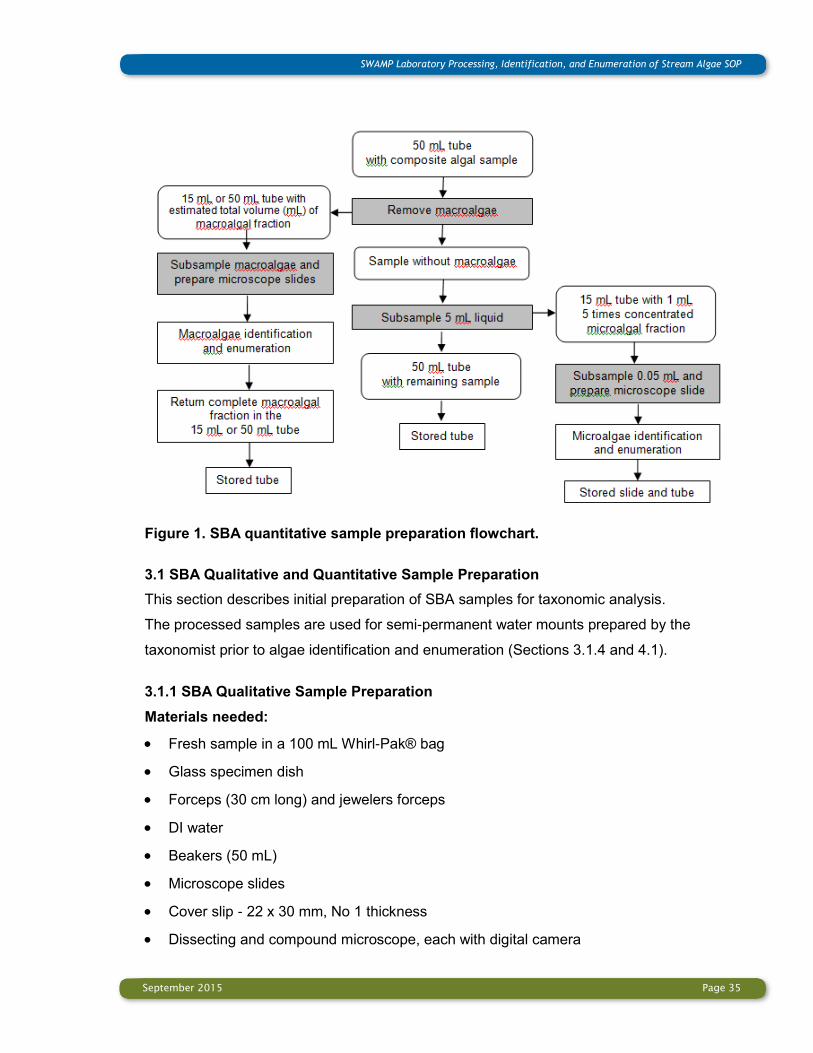
After algal samples are received, samples are prepared for taxonomic analysis. The sample
preparation process is different for the three different algae samples: (1) SBA qualitative
sample; (2) SBA quantitative sample (macroalgae and microalgae fractions), and
(3) Diatom quantitative sample.

September 2015 Page 34
SWAMP Laboratory Processing, Identification, and Enumeration of Stream Algae SOP
The purpose of analysis of qualitative SBA samples is to identify as many macroalgal taxa
present in the sample as possible. Macroalgal species identification requires observation of
enough unfixed material representing different life stages to determine vegetative features,
reproductive mode, and characteristics of completely developed reproductive structures of
each species. All macroalgal taxa are identified to lowest possible taxonomic level (usually
to species).
Quantitative SBA samples contain algae of different sizes requiring detailed observations of
many cellular, vegetative and reproductive structures in order for the species to be
identified. For proper identification and enumeration of SBA taxa, macroalgal and
microalgal fractions of each sample are processed separately (Figure 1).
The purpose of analysis of quantitative SBA samples is to identify as many SBA taxa
present in the sample as possible, to provide an accurate algal taxa list and uniform
biovolume estimate of each algal taxon in a sampled stream reach. This procedure is
designed to produce a comprehensive list of all algal taxa identified to lowest possible
taxonomic level (usually species) together with a precise estimate of their individual
volumetric contribution per unit area sampled.
The purpose of the quantitative analysis of diatoms is to identify 600 valves of diatoms to
the lowest possible taxonomic level (usually species) and to determine the relative
abundance of the diatom taxa. For proper identification of diatoms, the diatom frustules
need to be cleaned by removing all organic contents of the diatom cells.
During the sample preparation and consequent taxonomic analysis, care should be given to
avoid sample cross contamination by using disposable materials, or carefully washed and
DI rinsed materials. Instrumentation should be used only for an individual sample and then
immediately stored for decontamination. Dropper bottles with DI or Lugol’s Iodine Solution,
used multiple times, should not touch the algal material. Sample splashing should be
avoided when multiple samples are processed.
September 2015 Page 35
SWAMP Laboratory Processing, Identification, and Enumeration of Stream Algae SOP
Figure 1. SBA quantitative sample preparation flowchart.
3.1 SBA Qualitative and Quantitative Sample Preparation
This section describes initial preparation of SBA samples for taxonomic analysis.
The processed samples are used for semi-permanent water mounts prepared by the
taxonomist prior to algae identification and enumeration (Sections 3.1.4 and 4.1).
3.1.1 SBA Qualitative Sample Preparation
Materials needed:
Fresh sample in a 100 mL Whirl-Pak® bag
Glass specimen dish
Forceps (30 cm long) and jewelers forceps
DI water
Beakers (50 mL)
Microscope slides
Cover slip - 22 x 30 mm, No 1 thickness
Dissecting and compound microscope, each with digital camera
September 2015 Page 36
SWAMP Laboratory Processing, Identification, and Enumeration of Stream Algae SOP
Step 1: Very gently transfer the fresh macroalgae from the field plastic bag into a glass dish
containing DI water.
Step 2: When the taxonomic work on the sample is completed (see Section 4.1.1) archive a
portion of the fresh sample (see Section 4.3.1). Archiving of the SBA qualitative sample
should be conducted as soon as possible following completion of the taxonomic analysis.
Return the remaining material to the original plastic bag, loosely capped, adding DI water if
needed. The fresh sample should be archived for two more weeks in the refrigerator at 4ºC
in case further examination is needed.
3.1.2 SBA Quantitative Sample Preparation – Macroalgal Fraction
Materials needed:
Preserved composite sample in 50 mL plastic centrifuge tube
Forceps (30 cm long) and jewelers forceps
DI water
15 mL graduated centrifuge tube with graduations in 0.1 mL increments up to 1 mL, and
0.5 mL increments above
50 mL graduated centrifuge tube with graduations in 2.5 mL increments
Grid bottom culture dish
Microscope slides
Cover slips - 22 x 30 mm, No 1 thickness
Dissecting and compound microscope, each with digital camera
Step 1: Obtain the 50 mL centrifuge tube with preserved composite sample and visually
inspect its content to estimate whether a 15 mL or 50 mL tube is needed for macroalgae
fraction collection.
Step 2: Label a 15 mL or 50 mL graduated centrifuge tube with the following information:
SWAMP sample ID
Date of collection (MM/DD/YYYY)
Note “macroalgae” on the label to distinguish from the microalgae fraction
Macroalgae volume: xx mL
September 2015 Page 37
SWAMP Laboratory Processing, Identification, and Enumeration of Stream Algae SOP
If the macroalgal fraction is very large, use a 50 mL graduated centrifuge tube with 2.5 mL
increments.
Step 3: Place 10 mL of DI water into the labeled centrifuge tube.
Step 4: Using the forceps (30 cm long), very gently pinch the material at the bottom of the
tube. Search for visible macroalgal clumps, and any solid particles in the sample, such as
mosses, vascular plant tissues, roots, etc. Gently pull up the forceps and slowly move the
macroalgae and all solid particles grasped between the forceps in the solution to remove
extra clinging sediment and isolate any macroalgal filaments in the sample. Repeat this
step at least three times before proceeding to the next step.
Step 5: If macroalgal clumps are present in the sample continue onto Step 6. If no
macroalgal clumps are present, proceed with preparation of the microalgae fraction
(Section 3.1.3). If no macroalgae and any solid particles are visible to the naked eye,
inspect the sample tube under a dissecting microscope before proceeding with microalgae
preparation.
Step 6: Using forceps, remove the macroalgae from sample very gently, squeeze it to
remove as much liquid as possible and then place it into the tube with 10 mL DI water.
Continue until no macroalgae remain.
Step 7: Determine the volume of macroalgal fraction by the increase (displacement) from
the original 10 mL of water. When using 15 mL centrifuge tubes with graduated markings
measuring 0.5 mL, estimate the water displacement to 0.1 mL (See Note 1 below). Record
the volume of the macroalgal fraction (mL) in the ID Datasheet for SBA Sample- Heading:
Qualitative sample – Heading: Macroalgal fraction-total volume: xx mL (Appendix C1) and
on the label of the tube with the macroalgal fraction.
Note 1: The surface of water in a tube is not completely flat. Instead, the surface curves in
a shallow U-shape meniscus. When measuring, read the line just at the bottom of the
meniscus.
September 2015 Page 38
SWAMP Laboratory Processing, Identification, and Enumeration of Stream Algae SOP
3.1.3 SBA Quantitative Sample Preparation – Microalgal Fraction
Materials needed:
Preserved composite sample in 50 mL plastic centrifuge tube remaining after
macroalgae removal
10 mL pipette
50 mL centrifuge tube with graduations in 2.5 mL increments
15 mL centrifuge tube with graduations in 0.1 mL increments up to 1 mL, and 0.5 mL
increments above
Dissecting needles
Table-top centrifuge
146 mm borosilicate pipette
Microscope slides
Cover slip - 22 x 30 mm, No 1 thickness
Compound microscope with digital camera
Step 1: Obtain the 50 mL centrifuge tube containing the SBA quantitative sample following
removal of the macroalgae fraction. Homogenize the microalgal fraction of the SBA
quantitative sample by gently but thoroughly inverting the centrifuge tube several times.
The sample must be well homogenized prior to sub-sampling (Step 2).
Step 2: Pipette 5 mL of homogenized microalgae fraction into a 50 mL centrifuge tube
labeled with the sample information.
Step 3: Fill the centrifuge tube with DI water to the 50 mL mark. Let the sample settle for a
minimum of 12 hours.
Step 4: Once the sample has thoroughly settled, gently remove the supernatant layer down
to a volume of 5 mL by using a pipette. Avoid disturbing the algal material on the bottom of
the tube.
Step 5: Label a 15 mL graduated centrifuge tube with the following information:
SWAMP sample ID

September 2015 Page 39
SWAMP Laboratory Processing, Identification, and Enumeration of Stream Algae SOP
Date of collection (MM/DD/YYYY)
Note “microalgae” on the label to distinguish from the macroalgae fraction
Step 6: Transfer the 5 mL of sample material from the 50 mL centrifuge tube to the labeled
15 mL graduated centrifuge tube.
Step 7: Rinse down the sides of the 50 mL centrifuge tube several times with DI water to
capture any remaining algae clinging to the sides. Transfer the rinse liquid to the labeled 15
mL graduated centrifuge tube.
Step 8: Fill the labeled 15 mL graduated centrifuge tube with DI water to the 15 mL mark.
Step 9: Centrifuge the sample for 5 min at 4000 RPM on a table-top centrifuge.
Step 10: Remove the supernatant layer until 1 mL sample is left by using a pipette. Avoid
disturbing the algal material on the bottom of the tube.
This procedure concentrates the microalgal fraction 5 times while removing most of the
glutaraldehyde before microscopic examination. From this 1 mL sample, a semi-permanent
slide is prepared for analysis (Section 3.1.4 below).
3.1.4 SBA Semi-permanent Slide Preparation of Quantitative Microalgal Fraction
Materials needed:
15 mL centrifuge tube with microalgal fraction of 1 mL
DI water
Dissecting needles
146 mm borosilicate pipette
Microscope slides
Cover slip - 22 x 30 mm, No 1 thickness
Nail polish
Compound microscope with digital camera
Step 1: Obtain the 15 mL centrifuge tube containing 1 mL concentrated microalgal fraction.
Step 2: Visually inspect the sample and evaluate the amount of material (sediment and

September 2015 Page 40
SWAMP Laboratory Processing, Identification, and Enumeration of Stream Algae SOP
algae) settled on the bottom of the tube. If the sample contains more than 0.5 mL of
material (sediment and algae) settled on the bottom, dilute with DI water to a final volume of
2, 3, 4 or 5 mL.
If a small amount of material (sediment and algae) is present, centrifuge for 5 min at 4000
RPM on a table-top centrifuge and concentrate the sample to 0.5 mL.
Record any additional dilution or concentration performed on the sample and the final
sample volume used for slide preparation in the ID Datasheet for SBA Sample-Heading:
Quantitative sample-Microalgal fraction-Sample volume after additional dilution/
concentration: xx mL (Appendix C1). This information is required for SBA biovolume
calculations (see Section 4.1.4.2, Appendix F).
Step 3: Vortex or pipet-mix the sample and subsample with pipette from the center of the
well-mixed material. Place 1 drop (0.05 mL) of sample on a standard microscope slide and
cover with a 22 x 30 mm cover slip.
Proper preparation of the slides is vital to performing identifications. The following should
be noted while preparing slides:
Ensure that the volume of the drop is not so large that it creates the formation of
bubbles or causes the cover slip to float.
Avoid having too much or too little material on the slide. Too much material results in
layers of cells, specimen overlap and a non-flat cover slip which interferes with accurate
identification. Too little material increases the amount of time required to complete
analysis and may not be adequate for proper identification and enumeration.
Small thick clumps of spreading filaments intermixed with colonial algae can sometimes
occur in the microalgal fraction. Clumping of material not only interferes with accurate
identification and enumeration, but can circumvent the assumption of random
distribution of specimens on the sides. These clumps usually contain several different
species, so they should to be dispersed before proceeding with analysis. Gentle tapping
on the cover slip or spreading the clump apart with a pair of dissecting needles will
reduce clumping.
September 2015 Page 41
SWAMP Laboratory Processing, Identification, and Enumeration of Stream Algae SOP
Step 4: Inspect the semi-permanent microalgal slide at lower magnification (200x) using a
compound microscope to confirm that microalgae are evenly distributed. Gently adjust the
cover slip if algal clumps are present. Cover slip may be sealed with nail polish to prevent
evaporation. This semi-permanent microalgal mount is good for analysis for at least two
hours.
3.2 Diatom Quantitative Sample Preparation
Two methods are available for diatom cleaning: the nitric acid method (American Public
Health Association, 1981) and the hydrogen peroxide and potassium dichromate method
(Van der Werff, 1955).
3.2.1 Cleaning of Diatom Samples: Nitric Acid Method
Concentrated nitric acid is extremely hazardous, and therefore specific handling and
disposal procedures must be in place. Staff should consult the appropriate MSDS provided
by the supplier. Nitric acid should always be handled in a positive-draw fume hood by
trained staff wearing safety goggles, rubber gloves, and lab coats. Diatom quantitative
samples are preserved in formalin, so they must be handled carefully.
Materials needed:
Preserved diatom sample in 50 mL plastic centrifuge tube
Beakers (250 mL)
Concentrated nitric acid (HNO3)
HCE (10%)
Hot plate
Centrifuge
15 mL centrifuge tubes
DI water
pH paper
Waste container
Scissors
Step 1: Label each beaker with tape and the corresponding sample number.
Step 2: Obtain the preserved diatom sample in 50 mL plastic centrifuge tube. Check

September 2015 Page 42
SWAMP Laboratory Processing, Identification, and Enumeration of Stream Algae SOP
sample to determine if macroalgal clumps are present. If large algal clumps are observed in
the sample, cut them into smaller pieces with scissors.
Step 3: Shake the sample vial vigorously and pour 20 mL (30 mL for sparse samples) of
homogenized sample into the beaker labeled with the sample information.
Step 4: Add a small amount of nitric acid to the beaker to test if a violent or exothermic
reaction occurs. If violent reaction does occur, or if carbonates are abundant in the sample,
the sample can be pre-treated by adding 10% HCl.
Step 5: When it has been determined that no violent reaction will occur, slowly add the
remaining volume of nitric acid to the beaker. In all, a volume of nitric acid approximately
equal to the volume of sample processed is added to the beaker.
Step 6: Place the beaker on a hot plate under a positive-draw fume hood. Boil the sample
and nitric acid mixture until the organic content turns white. This white material is the
siliceous cell walls of the diatoms. This step typically takes 30 minutes to 1 hour, during
which the volume of the material will be reduced to about ½ (see Note 2 below).
Step 7: Once the boiling step is complete, allow the sample to cool. Transfer the sample to
a 15 mL centrifuge tube labeled with the SWAMP sample ID and centrifuge at 3500 rpm for
8 minutes.
Step 8: Pour the supernatant off into an appropriately designated waste container. Add DI
water to the centrifuge tube containing the diatom sample and centrifuge at 3500 rpm for 8
minutes.
Step 9: Repeat the cycle of decantation, addition of new DI water, and centrifugation 5
times, or until the pH of the water is neutral (or the same as the deionized water being
used – may not be pH 7). The result should be a pellet of nearly white material at the
bottom of the tube.
Step 10: Label a 15 mL graduated centrifuge tube with the following information:
SWAMP sample ID
Date of collection (MM/DD/YYYY)
Note “diatoms” on the label to distinguish from the other samples

September 2015 Page 43
SWAMP Laboratory Processing, Identification, and Enumeration of Stream Algae SOP
Step 11: Transfer the cleaned diatom material to the labeled 15 mL centrifuge tube. Add DI
water up to the 10 mL mark. This is the material from which the slides will be made.
Note 2: The surfaces of hot plates can get hot enough to cause boiling over or explosive
conditions, especially with samples containing high amounts of organics or carbonates.
Alternative methods such as using a heating block with 26 mm diameter glass test tubes
(the lower portion of each tube stay hot while the upper portion stays cool, creating a reflux
action, minimizing the risk of over-boiling or drying the sample) or microwave apparatus
(Acker et al., 2002) may be used to clean the diatoms.
3.2.2 Cleaning of Diatom Samples: Hydrogen Peroxide Method
The principal reagents used in this method, hydrogen peroxide and potassium dichromate,
are extremely hazardous. 30% hydrogen peroxide is a strong oxidizer, and may require
special handling and storage. Specific handling and disposal procedures must be in place
for the handling these materials. Staff should consult the appropriate MSDS provided by
the supplier. Both chemicals must always be handled in a positive-draw fume hood, and
staff should wear safety goggles, nitrile gloves and lab coats.
Materials needed:
Preserved diatom sample in 50 mL plastic centrifuge tube
Beakers (250 mL)
Hydrogen peroxide (H2O2 30%)
Potassium dichromate (K2Cr2O7)
Microspatula
Squirt bottle of DI water
DI water
Centrifuge
15 mL centrifuge tubes
Waste container
Scissors
Step 1: Label each beaker with tape and the corresponding sample number.
Step 2: Obtain the preserved diatom sample in 50 mL plastic centrifuge tube. Check the
September 2015 Page 44
SWAMP Laboratory Processing, Identification, and Enumeration of Stream Algae SOP
sample to determine if macroalgal clumps are present. If large algal clumps are observed in
the sample, cut them into smaller pieces with scissors.
Step 3: Shake the sample vial vigorously and pour 20 mL (30 mL for sparse samples) of
homogenized sample into the beaker labeled with the sample information.
Step 4: Add approximately 20-30 mL of 30% hydrogen peroxide to the sample.
Step 5: Place the beaker on a hot plate under a positive-draw fume hood. Bring to boiling.
Step 6: Remove from heat and immediately add a small amount (several crystals) of
potassium dichromate to the mixture using a microspatula. The addition of the potassium
dichromate will catalyze a strong exothermic reaction, therefore, the potassium dichromate
should be added slowly. Have a squirt bottle of DI water at the ready in case the reaction
begins to boil over the top of the beaker. The reaction takes approximately 5 to 10 minutes
to complete. Completion of the reaction is indicated by solution changing in color from dark
purple to orange.
Step 7: Add DI water up to 200 mL.
Step 8: Allow the diatom material to settle for at least 8 hours.
Step 9: Slowly and gently, to avoid disturbing the diatom material on the bottom of the
beaker, decant the liquid into an appropriately designated waste container.
Step 10: Refill beaker with the diatom material on the bottom with DI water to 200 mL.
Step 11: Repeat steps 8-10 several times (approximately 3 to 5) until the cleaned diatom
material is mostly colorless.
Step 12: Let the cleaned diatom material settle overnight and decant the supernatant as
low as possible.
Step 13: Label a 15 mL graduated centrifuge tube with the following information:
SWAMP sample ID
Date of collection (MM/DD/YYYY)
Note “diatoms” on the label to distinguish from the other samples
September 2015 Page 45
SWAMP Laboratory Processing, Identification, and Enumeration of Stream Algae SOP
Step 14: Transfer the cleaned diatom material to the labeled 15 mL centrifuge tube. Add DI
water up to the 10 mL mark. This is the material from which the slides will be made.
Note 3: The method of allowing the material to settle over 8 hours can also be applied to
the nitric acid procedure; however, since the resulting nitric acid solution is often colorless,
testing the pH of the solution is a more reliable way to determine when the material has
been sufficiently rinsed. Centrifugation of the sample to remove the remaining chemicals
from hydrogen peroxide procedure can also be used.
3.2.3 Permanent Slide Preparation of Diatom Samples
Permanent slide preparation is the same, regardless of which cleaning method is chosen.
Materials needed:
15 mL centrifuge tube containing the cleaned diatom material
Cover slips – 18 x 18 mm or 22 x 22 mm, No 1 thickness
Microscope slide
146 mm borosilicate pipette
Hot plate
Naphrax
Forceps
Razor blade
70% Ethanol
10% HCL
DI water
Step 1: Drip an amount of DI water onto the cover slip with a glass pipette. The amount
should be sufficient to form a thin layer of water over the entire cover slip when the diatom
suspension is added. If the clean diatom suspension is very sparse, skip this step.
Step 2: Obtain the 15 mL centrifuge tube with cleaned diatom material. Agitate the vial
containing the cleaned diatom suspension and quickly withdraw material from near the
central portion of the sample using the glass pipette.
Step 3: Eject one or two drops of diatom suspension smoothly and carefully into the layer

September 2015 Page 46
SWAMP Laboratory Processing, Identification, and Enumeration of Stream Algae SOP
of DI water on the cover slip (see Note 4). If the clean diatom suspension is very sparse
eject three to five drops directly onto a cover slip without DI water. If the cover slip
overflows, discard it, clean the area, and prepare a new cover slip with diatoms.
Step 4: Air dry the material or gently dry the material on a warm hot plate. The temperature
of the hot plate must not exceed 40ºC. Higher temperatures cause the water to circulate or
bubble, resulting in a non-random distribution or loss of diatom valves. Avoid any procedure
that rapidly evaporates the suspension. Rapid evaporation could produce strong patterns of
diatoms settling on the cover slip.
Step 5: When the cover slips have visibly dried, place them on a hot plate at an elevated
temperature to drive off any remaining moisture.
Step 6: Confirm the prepared cover slip contains a random distribution of diatoms
sufficiently dense for conducting identification and enumeration procedures. On average,
15 to 30 diatom valves should be visible in a single field of view. Confirm density and
random distribution in 5 fields of view. If clumps of diatom valves are on the slide to the
point where individual specimens cannot be viewed prepare another cover slip.
Step 7: Add a small amount of mounting medium (Naphrax) to a cleaned microscope slide
and put the cover slip (diatoms down) on the mounting medium with forceps.
Step 8: Put the microscope slide with the cover slip on a hot plate preheated to 120 to
150ºC. Leave on hot plate until bubbles stop forming under the cover slip, indicating that all
the solvent from the mounting material has been driven out of the medium.
Step 9: Use forceps to safely remove the slide from the hot plate. Gently tap down on the
cover slip to remove any air bubbles and to even the distribution of diatoms.
Step 10: Once the slide cools, scrape any excess mounting medium that remains outside
the cover slip with a single-edged razor.
Step 11: Attach adhesive labels to the completed slides and include the following
information:
SWAMP sample ID
Date of collection (MM/DD/YYYY)
September 2015 Page 47
SWAMP Laboratory Processing, Identification, and Enumeration of Stream Algae SOP
Note “diatoms” on the label to distinguish from the other slides
Step 12: Preserve the unused cleaned diatom material with ethanol to 50% final
concentration and archive the vial (see Section 4.3.3).
Note 4: To achieve a more even distribution of diatom valves on the coverslip, 10% HCL
can be added to the cleaned diatom suspension (1 drop per 10 mL of material in the vial).
Section 4: Identification and Enumeration Analysis of Algae
4.1 Identification and Enumeration Analysis of SBA
Correct identification of benthic SBA taxa requires separation of the algae in the sample
based on size classes into a macroalgal fraction and a microalgal fraction defined as
follows:
Macroalgae are large macroscopic filamentous, colonial, tuft-forming, crustose,
tissue-like or coenocytic eukaryotic algae and cyanobacteria that have forms
recognizable with the naked eye (e.g., Nostoc, Rivularia, Batrachospermum, Lemanea,
Cladophora, Draparnaldia, Oedogonium, Rhizoclonium, Spirogyra, Zygnema,
Mougeotia, Vaucheria) [see Sheath and Cole (1992) for definitions of forms].
Microalgae are small, microscopic forms not recognizable with the naked eyes but
consisting of unicellular, colonial or filamentous non-diatom algae.
Proper identification of SBA requires a different approach for each fraction. Macroalgal
species identification needs observation of enough material to adequately characterize
vegetative and reproductive structures under a combination of dissecting and compound
microscopes. Examination of microalgae should reveal cellular details, such as
chloroplasts, pyrenoids, cell wall, among other features. Detailed and careful observations
are necessary for accurate identification. Some diagnostic features are not evident without
specific techniques, such as staining (e. g. starch with Lugol’s iodine solution). Use of
taxonomic resources (see Section 1.3 and Appendix I) and taking photomicrographs while
making identifications will facilitate taxonomic consistency and qualitative assurance.
Enumeration of the microscopic algae observed in the microalgal fraction, and as epiphytes
in macroalgal fraction is based on a counting unit called a natural counting entity (NCE).

September 2015 Page 48
SWAMP Laboratory Processing, Identification, and Enumeration of Stream Algae SOP
Natural counting entity (NCE) is each natural occurring form of algae (i.e., each
unicell, colony, filament, tissue-like form, coenocyte, tuft, or crust) regardless of the
number of cells in the thallus or colony.
The main purpose of using “natural counting entity” is to prevent numerous small cells in a
sample with macroscopic forms from dominating a count relative to their actual contribution
to the community biomass. It also facilitates the counting of algal forms which have linked
cells that may be hard to distinguish.
Laboratory set up for separate analysis of macroalgal and microalgal fractions of the SBA
quantitative sample is illustrated in Figure 2 and explained in the Sections 3.1 and 4.1.
Figure 2. Laboratory set up for separate analysis of macroalgal and microalgal
fractions of SBA quantitative sample.
Legend: A. 50 mL centrifuge tube containing SBA composite quantitative sample; B.
Microalgal fraction placed on microscope slide with 22x30 mm cover slip. Typically 0.25 mL
of the composite sample liquid is analyzed. C. Macroalgal fraction with measured total
volume (i. e. 0.5 mL) placed in a grid Petri dish to estimate the proportions of species. In
this case: Cladophora glomerata (31%), Nostoc verrucosum (24%), Batrachospermum
gelatinosum (15%), and Spirogyra varians (30%).

September 2015 Page 49
SWAMP Laboratory Processing, Identification, and Enumeration of Stream Algae SOP
4.1.1 SBA Qualitative Sample Analysis
Step 1: Using a research-grade dissecting microscope carefully examine the macroalgal
fresh sample placed into glass dish and determine the number of macroalgal genera in the
sample appearing different in morphology or color.
Thoroughly examine all the material and identify key macroalgal features needed to
separate the algae by genus. These may include:
Colonial shape, size and color in cyanobacteria (such as Nostoc, Dichothrix, Rivularia);
Different life stages, heterocyst position and akinete development in cyanobacteria
(such as Anabaena, Cylindrospermum, Gloeotrichia);
Male and female specimens with developed reproductive structures in red and green
algae (such as Batrachospermum, Sirodotia, Oedogonium);
Different life stages and completely matured reproductive structures in zygnematalean
algae and tribophytes (such as Spirogyra, Zygnema, Mougeotia, Vaucheria).
Step 2: Place each macroalgal genus identified aside. When small rocks are collected,
carefully examine the surface of the rocks for attached algae using dissecting microscope.
If algae are present, scrape them out and place them on a microscope slide.
Step 3: Prepare microscope slides for each macroalgal genus which may be presented
with more than one species in the sample. The number of slides prepared depends on the
need to obtain sufficient information to successfully perform species identification.
When reproducing filaments of zygnematalean algae are observed, but completely matured
zygospores/aplanospores are not available, further incubation under nutrient stress
facilitates completion of sexual or asexual reproduction. The resulting mature zygospores
(or akinetes, aplanospores) can provide the taxonomist with the additional information
needed to identify the species.
To incubate the algae under nutrient stress:
Select the conjugating filaments of Spirogyra, Zygnema or Mougeotia and place them in
a 50 mL glass beaker filled with DI water.
Keep the samples out of direct sunlight (in a north-facing window) at room temperature
September 2015 Page 50
SWAMP Laboratory Processing, Identification, and Enumeration of Stream Algae SOP
until reproductive structures completely develop. Check for reproductive filaments under
the microscope every three days and document the different stages of conjugation and
development of zygospores (or akinetes, aplanospores).
Step 4: Examine prepared slides under the compound microscope and identify SBA
macroalgae to species level. If large colonial diatoms are observed in the sample, record
them following the recommendations in Section 4.1.2 Step 5.
Step 5: Take sufficient photomicrographs of all newly recorded species to support
harmonization of results. Take photomicrographs of previously reported species to
demonstrate the key aspects of vegetative morphology and reproduction used in
identification (see Section 1.5 and Appendix H).
Step 6: Record all macroalgal taxa identified in the SBA qualitative sample in the ID
Datasheet for SBA Sample under the heading Qualitative sample – list of taxa
(Appendix C1).
Step 7: Submit the remaining SBA qualitative sample and the algal material from the slides
for archiving (see Section 4.3.1).
4.1.2 SBA Quantitative Sample Analysis – Macroalgal Fraction
Macroalgal fractions extracted from different quantitative SBA samples vary considerably in
total volume and content. Typically this fraction consists of soft-bodied macroalgae
sometimes mixed with large colonial diatoms. However, due to the field sampling protocol
(Ode et al., 2015), the macroalgal fraction may contain non-algal matter, such as mosses,
vascular plant tissues, roots or debris, macroinvertebrates, etc., which should be subtracted
from the macroalgal sample total volume. Many SBA and diatom macroalgae, as well as
vascular plants, support development of epiphytic SBA algae, which are identified and
enumerated also.
The approach for species identification of the macroalgae from the quantitative and
qualitative samples (Section 4.1.1) is similar except for the process of algae incubation.
Microscope slides for macroalgae and SBA epiphytic species identification need to be
prepared during the identification process following the observations under the dissecting
microscope.
September 2015 Page 51
SWAMP Laboratory Processing, Identification, and Enumeration of Stream Algae SOP
Step 1: Start with the 15 mL or 50 mL centrifuge tube containing the macroalgal fraction
extracted from the SBA quantitative sample.
Step 2: Transfer the contents of the centrifuge tube to a gridded Petri dish.
Step 3: Using forceps gently spread the material in the sample evenly throughout the Petri
dish.
Step 4: Carefully examine the material under the dissecting microscope. If the sample
contains non-algal matter such as mosses, vascular plant tissues, roots or debris, separate
this material from the macroalgae and determine its volume. If it is possible to remove the
non-algal matter, place it in centrifuge tube and measure the volume using water
displacement. If the non-algal matter is not possible to remove, visually estimate its
proportion. Calculate the fraction of the total sample volume represented by non-algal
matter. Record the fraction represented by non-algal matter in the ID Datasheet for SBA
Sample-Heading: non-algal matter xx % (Appendix C1).
Step 5: After the non-algal matter has been separated, gently distribute the soft-bodied
macroalgae evenly using forceps. When working with the macroalgal fraction, care should
be given to avoid breaking up large filaments or colonies and to preserve key features
needed for species identification. Since large reproductive structures are easily damaged,
the clumps should be spread very carefully.
If the sample contains macroalgal colonial diatoms (such as Melosira, Pleurosira,
Terpsinoe, Cymbella, Gomphonema, Didymosphenia, Bacillaria, etc.) separate this material
from the soft-bodied macroalgae and determine the representative proportion of the
diatoms as a percent of total macroalgal fraction volume. If possible, identify diatoms to
genus level, if not – use the general category “diatoms”. This information can help recording
invasive or bloom-forming macroalgal diatom taxa, which may be omitted in diatom analysis
due to numerical dominance of smaller diatom species.
Step 6: Using a dissecting microscope, thoroughly examine the sample and identify key
macroalgal features needed to segregate the SBA by genus. Determine the number of
distinct soft-bodied macroalgal genera present in the sample.
September 2015 Page 52
SWAMP Laboratory Processing, Identification, and Enumeration of Stream Algae SOP
Step 7: Prepare microscope slides for each soft-bodied macroalgal genus. The number of
slides depends on the need to obtain sufficient information to successfully perform
identification and support the STE.
Step 8: Examine prepared slides under the compound microscope. Identify soft-bodied
macroalgae to species level.
Step 9: Estimate the representative proportion of each soft-bodied macroalgal species as a
percent of total macroalgal fraction volume in order to determine their biovolume (see
Section 4.1.4.1).
Step 10: Record the name and the representative proportion of each soft-bodied
macroalgal taxon identified in the ID Datasheet for SBA Sample-Heading: Macroalgae
taxon ID; Proportion of each taxon (%) (Appendix C1).
Step 11: When the total volume of macroalgal fraction is so low that it is not possible to be
measured by water displacement, prepare a microscope slide containing all macroalgal
material. Under compound microscope identify all macroalgal taxa and determine directly
their biovolume by individual measuring of each specimen using geometric shapes (such as
cylinder for filamentous algae, or sphere for young colonies of Nostoc, for details see
Section 4.1.3 Step 4). Calculate the total biovolume of each macroalgal species by
summing the biovolumes of all specimens measured. In this case the biovolume of each
macroalgal species is directly measured in μm3.
Step 12: Identify to species level and enumerate 100 NCEs of epiphytic SBA attached to
the surface of the soft-bodied macroalgae and other aquatic substrates. If fewer than 100
NCEs of epiphytic SBA are observed, enumerate as many as there are in the entire
macroalgal fraction.
Step 13: Record each SBA epiphytic taxon identified and the corresponding number of
NCEs enumerated in the ID Datasheet for SBA Sample-Heading: Epiphyte taxon ID; #NCE
(Appendix C1).
Step 14: Take sufficient photomicrographs of all newly recorded species to support
harmonization of results. Take photomicrographs of previously reported species to
September 2015 Page 53
SWAMP Laboratory Processing, Identification, and Enumeration of Stream Algae SOP
demonstrate the key aspects of vegetative morphology and reproduction used in
identification (see Section 1.5 Photographic Documentation of Algae and Appendix H).
Step 15: Return the entire content of the macroalgal fraction, including the material from
the slides, and the non-algal matter into the tube with macroalgal fraction of the SBA
quantitative sample and submit it for archiving (see Section 4.3.1).
4.1.3 SBA Quantitative Sample Analysis – Microalgal Fraction
The microalgal SBA fraction is examined on a semi-permanent water mount for best
observation of algal cellular morphology. Water mounts allow adjusting of the cover slip to
change the position of the cells and spreading out of multilayered cell clumps for
observation of critical taxonomic features. Furthermore, specific techniques, such as
staining (e. g. starch with Lugol’s iodine solution) are possible to apply on water mount.
Step 1: Using a research quality compound microscope, scan the semi-permanent slide
with microalgae at magnification 200x to assess the taxonomic composition of the sample.
Step 2: Switch to a magnification of 400x (e.g., 40x objective with 10x eyepieces). At a
magnification of 400x, the cover slip is composed of many horizontal optical transects.
Step 3: Identify and enumerate 300 SBA NCEs across a known number of horizontal
optical transects. Count only intact cells with complete cell contents. If 300 SBA NCEs have
been counted before reaching the end of the last transect, continue enumeration and
complete the transect. This ensures that a known fraction of the sample is analyzed.
Record the number of horizontal transects traversed, each SBA microalgal taxon identified
and the corresponding number of NCEs enumerated in ID Datasheet for SBA Sample-
Heading: Microalgal fraction-number of transects counted: xx, Microalgae taxon ID; #NCE
(Appendix C1).
The taxonomist should adjust the total of NCEs enumerated under the following
circumstances:
If the sample contains numerous small single cells or new taxa appeared after counting
300 SBA NCEs, continue enumeration to 400 or 500 NCEs.
September 2015 Page 54
SWAMP Laboratory Processing, Identification, and Enumeration of Stream Algae SOP
If the morphological information obtained during the enumeration of 300 NCEs is not
sufficient for species identification of certain taxon, continue observations of more
specimens outside the counted area until species level identification is achieved.
Stop counting after enumeration of the first complete slide if fewer than 20 SBA NCEs
are recorded. Enumeration is stopped regardless of whether the sample received
additional dilution or concentration prior to slide preparation.
Prepare additional slides if more than 20 SBA NCEs are recorded in the first slide.
Continue enumeration for 4 hours or until 150 SBA NCEs have been enumerated
(whichever comes first). Exclude the time spent identifying and documenting new
species from the total enumeration time.
Step 4: Using the techniques described by Hillebrand et al. (1999), determine the
appropriate geometric model for each microalgal species identified. The list of geometric
shapes corresponding to the SBA genera reported for SWAMP are detailed in Appendix E.
Perform microscopic measurements of the cell dimensions for each algal entity according
to the closest geometric shape Record measurements in the ID Datasheet for SBA Sample-
Heading: Cell diameter (µm); Cell/Filament length (µm); Cell depth (µm); Total number of
cells; Total filament length (µm) (Appendix C1).
The following should be noted while estimating biovolumes:
Measurements for each single SBA NCE are made concurrent with identification and
enumeration. Most microalgal NCE can be viewed as cylinders, spheres, spheroids, or
cones in order to estimate biovolume. Proper biovolume estimate requires
measurements of two or three dimensions, such as cell width, length and depth. Depth
measurements are made on specimens from a side view. This can be achieved by
lightly tapping the coverslip to turn the specimen. If the specimen cannot be measured
from the side, estimate the depth and include a note in the data record. SBA size
measurements should be made using an ocular micrometer or by image analysis
software when photomicrographs are available.
A multicellular coccoid colony is considered to be a single NCE. However, the number
of cells in the colony varies among the species and in many taxa increases with the age
September 2015 Page 55
SWAMP Laboratory Processing, Identification, and Enumeration of Stream Algae SOP
(for instance, cyanobacterial species belonging to Aphanocapsa and Aphanothece).
When the colony consists of a small number of cells, measure the dimensions of all
individual cells. If the colony contains a large number of cells average the cell
dimensions (diameter and length) obtained by measurement of 20 cells and record the
total cell number.
Often the colonies break into fragments with different sizes, each one of which is
considered a NCE. The biovolume of colonial NCE is calculated by multiplying
averaged cell biovolume by number of cells per colony.
For a filament, which is considered a single NCE, measure the width and the whole
length of the filament. When some filamentous taxa are abundant in the sample (such
as, species belonging to Leptolyngbya and Heteroleibleinia) average the width and the
length of the filaments based on the measurements of 20 NCE.
However, size averaging should be applied only for an individual sample, and not
extrapolated to other samples, because filaments and colonies break apart into
fragments with different lengths under variable conditions.
Step 5: Take sufficient photomicrographs of all newly recorded species to support
harmonization of results. Take photomicrographs of previously reported species to
demonstrate the key aspects of vegetative morphology and reproduction used in
identification (see Section 1.5 and Appendix H).
Step 6: Submit remaining microalgal fraction of the SBA quantitative sample for archiving
(see Section 4.3.2).
4.1.4 Biovolume Calculations for SBA
The biovolume for each identified SBA taxon is measured during the identification and
enumeration. Precision of the SBA biovolume estimates is achieved by separate
processing of macroalgal and microalgal fractions and by cell size measurements made for
each single microalgal NCE (see Stancheva et al., 2012a for details).
September 2015 Page 56
SWAMP Laboratory Processing, Identification, and Enumeration of Stream Algae SOP
4.1.4.1 Biovolume Calculations: SBA Quantitative Sample – Macroalgal Fraction
Step 1: Determine the representative proportion of each macroalgal species as a
percentage of the total volume of macroalgal fraction, from which the non-algal matter has
been subtracted, to calculate the biovolume of each species.
Determine the biovolume of each species in mL and convert it to μm3 when multiplied by
1012. Note that in some cases the biovolume of macroalgal species is directly estimated in
μm3 (see Section 4.1.2 Step 11).
This is a total biovolume of i-species (Va) in macroalgal fraction of sample in µm3.
Step 2: The value (Va) is multiplied by 4 in order to estimate the biovolume of each
macroalgal species in the original field composite sample (Va’), from which only 1/4 of the
macroalgae were transferred into the 50 mL tube received in the laboratory for SBA
analysis (Ode et al., 2015, p. F-1).
Va’ = 4 Va (µm3)
Use the following formula to calculate the biovolume of each macroalgal species in μm3 per
cm2 stream bottom area sampled. The result unit is μm3cm-2.
Vi=Va’ A-1 (μm3 cm-2)
where:
Vi = biovolume of i-species (μm3) per 1 cm2 stream bottom area sampled
Va’ = biovolume of i-species (μm3) in the original composite sample
A = stream bottom area of substratum sampled. It is the total area of substratum surface
from which benthic algae were collected (Ode et al., 2015). This information is provided for
each sample in cm2.
4.1.4.2 Biovolume Calculations: SBA Quantitative Sample – Microalgal Fraction
Step 1: Calculate the biovolume of each microalgal NCE using the measured dimensions
and formulae for geometric shapes closest to the cell’s shape, proposed by Hillebrand et al.
(1999) and specified for the SBA genera recorded by SWAMP (see Appendix E). For each
September 2015 Page 57
SWAMP Laboratory Processing, Identification, and Enumeration of Stream Algae SOP
microalgal taxon, sum the biovolumes of all NCE recorded in the known number of
analyzed optical transects.
This is a total biovolume of i-species (Va) in the analyzed optical transects in µm3.
Step 2: This value (Va) needs to be corrected for the sample dilution, caused by addition of
5 mL of glutaraldehyde to the composite sample. The correction factor (Vcr) is calculated
as follows:
Vcr= (Vt-Vm) (Vt-Vm-5)-1
where:
Vcr = a correction factor for sample dilution with fixative (assuming 5 mL of fixative was
added to the sample)
Vt = total initial sample volume in the sample vial (generally~50 mL)
Vm = volume of macroalgal fraction in the sample (will be 0 if no macroalgae detected)
Then to correct biovolume of i-species (Va) for fixative dilution use the following formula:
Va’=Va Vcr (µm3)
Step 3: Use the following formula to calculate the biovolume of each microalgal species
(μm3) per 1 cm2 stream bottom area sampled. The result unit is μm3cm-2.
Vi=Va’ Vs Vc-1 A-1 (μm3 cm-2)
where:
Vi = biovolume of i-species (μm3) per 1 cm2 stream bottom area sampled
Va’ = biovolume of i-species (μm3) per sample counted (known number of optical transects
in which the enumeration has been done) corrected for the dilution with fixative (Vcr)
Vs = composite sample volume (mL). It is the volume of all the liquid material amassed
during sampling, including water used for rinsing substrate and sampling devices. Final
composite volume typically does not exceed 400-500 mL (Ode et al., 2015). This
information is provided for each sample in mL.

September 2015 Page 58
SWAMP Laboratory Processing, Identification, and Enumeration of Stream Algae SOP
Vc = volume of sample counted (mL) [this is the number of transects counted multiplied by
the sample volume per transect and by dilution factor (see Note 5 below)];
A = stream bottom area of substratum sampled. It is the total area of substratum surface
from which benthic algae were collected (Ode et al., 2015). This information is provided for
each sample in cm2.
Note 5: The sample volume contained in one horizontal transect is determined as follows: a
transect is a rectangular area of the slide in which the width is equal to the field of view and
the length is equal to the length of the cover slip. With our microscope condition, at a 40x
objective, the 0.55 mm width of the transect results in a cover slip (22 x 30 mm) consisting
of 40 optical horizontal transects. For microscopes where the 40x field of view differs from
0.55 mm, calculate the transect width required.
Sample volume held by one horizontal optical transect is calculated as follows: on the
counting slide, 0.05 mL of subsample is placed. This subsample has been concentrated 5
times the original sample, thus 0.25 mL from the original sample is analyzed. Therefore the
original sample volume held by one horizontal optical transect is 0.00625 mL (=0.25 mL/40
horizontal transects).
When additional dilutions or concentrations are applied to the initial microalgal subsample
of 1 mL (see Section 4.1.3 Step 2), the sample volume per transect must be corrected by
multiplying with the dilution factor (DF). Most often, the subsample of 1 mL is counted
without dilutions/concentrations (DF 1), but sometimes is concentrated to 0.5 mL (DF 2), or
diluted to 2 mL (DF 1/2), to 3 mL (DF 1/3), to 4 mL (DF 1/4), to 5 mL (DF 1/5). Examples of
microalgal biovolume calculation with our microscope are shown in Appendix F.
4.2 Identification and Enumeration Analysis of Diatoms
Identification of diatoms requires an understanding of how the cells are put together (each
cell frustule being comprised of two valves and one to several girdle bands) and what
frustule components are being identified and enumerated. The different views one may
have of a frustule and/or its components is also important since attempts to key out
specimens will require one to know the view in which one is seeing an individual cell. There
are many fine structural elements of diatom cell walls and knowledge of this terminology is

September 2015 Page 59
SWAMP Laboratory Processing, Identification, and Enumeration of Stream Algae SOP
imperative for the identification of species. Guides to this information can be found in the
published literature, and commonly used books and floras include Patrick and Reimer
(1966), Krammer and Lange-Bertalot (1986-1991), Round et al. (1990), and Kociolek et al.
(2015 a, b) (see Section 1.3 and Appendix I). The fine structure of diatoms is the basis for
the taxonomy of the group, and modern approaches to taxonomy are relying more and
more on these fine structures to make effective distinctions that are not only reflected at
species level, but also at the genus level.
Step 1: Position the slide on the microscope stage with its label to the right. Scan slide at
medium magnification (200x or 400x) to confirm that diatoms are evenly distributed on the
cover slip and to assess the taxonomic composition of the sample to be analyzed.
Step 2: Establish a horizontal transect for counting by positioning the 100x objective a short
distance from the edge of slide, where valves are no longer optically distorted. A transect is
a rectangular area of the slide in which the width is equal to the field of view and the length
is equal to the length of the cover slip.
Step 3: Identify and enumerate all complete and partial valves visible in the first field of
view. A partial valve is defined as having more than 50% of the valve including the central
area. The valve (both complete and partial) must extend at least halfway into the transect,
and must include the center of the valve in the transect. Once the diatoms in the first field of
view have been enumerated move on to the next field of view in the direction of the
horizontal transect. If a second transect needs to be counted, move to the first field of view
of the second transect.
Step 4: Record the first and last field of view for each counted transect and the upper right
corner of the cover slip by taking the coordinates from the microscope stage. Enter the
coordinates in the ID datasheet for Diatom Sample (Appendix C2).
Step 5: Record each diatom taxon identified and the corresponding number of valves
enumerated in the ID Datasheet for Diatom Sample-Heading: Diatom taxon ID; Number of
valves (Appendix C2).
Step 6: Identify and enumerate 600 diatom valves across a known length of horizontal
optical transects. Avoid counting valves in any disrupted areas of the mount, particularly
September 2015 Page 60
SWAMP Laboratory Processing, Identification, and Enumeration of Stream Algae SOP
edges that have optical aberrations. When the diatom enumeration is completed, record the
last field of view counted by taking the coordinates from the microscope stage. The last
field of view counted can be located at any transect point. If the sample is very sparse,
continue counting for 4 hours or until 300 valves are enumerated (whichever comes first),
excluding time spent learning new species.
Record the number of transects traversed and the coordinates of the last field of view
counted in the ID Datasheet for Diatom Sample-Heading: Number of transects counted: xx
(Appendix C2).
Step 7: Take sufficient photomicrographs of all newly recorded species to support
harmonization of results. Take photomicrographs of previously reported species to
demonstrate the key aspects of vegetative morphology and reproduction used in
identification (see Section 1.5 and Appendix H).
4.3 Sample Labeling and Archiving
All SBA and diatom samples and slides must be retained as voucher specimens until
harmonization and reporting of data is complete. All samples will be archived and stored as
reference collections. Archives of samples and slides should be retained by the laboratory
for two years. The following types of samples will be archived:
SBA
Vials with qualitative SBA sample fixed with 2% glutaraldehyde
15 mL or 50 mL graduated centrifuge tube with minimally disturbed SBA macroalgal
fraction fixed with 2% glutaraldehyde
15 mL graduated centrifuge tube with remaining SBA microalgal fraction fixed with 2%
glutaraldehyde
50 mL graduated centrifuge tube with remaining content of original SBA sample fixed
with 2% glutaraldehyde
Sealed semi-permanent slides with analyzed quantitative SBA microalgal fraction
Diatoms
Vials with unused cleaned diatom sample fixed with ethanol to 50% final concentration

September 2015 Page 61
SWAMP Laboratory Processing, Identification, and Enumeration of Stream Algae SOP
50 mL graduated centrifuge tube with remaining content of original diatom sample fixed
with 1% formalin
Permanent diatom slides
All sample IDs in the archive follow the original SWAMP sample IDs, sampling date
(MM/DD/YYYY), type of sample (see below). The fixed SBA samples should be kept dark
and cool. Archived slides and samples must be retained by the laboratory until
harmonization and reporting of data is completed.
4.3.1 Archiving of SBA – Qualitative Samples
Materials needed:
Plastic scintillation vials (20-40 mL)
2% glutaraldehyde
Parafilm
Step 1: Select a representative subsample that contains all identified macroalgal taxa and
the algal material from the slides. Place the material in plastic scintillation vial, fix it with 2%
glutaraldehyde final concentration and parafilm the cap.
Step 2: Labeled the vial by SWAMP sample ID, sampling date (MM/DD/YYYY), and note
“qualitative sample” or “Q”.
4.3.2 Archiving of SBA – Quantitative Samples
Materials needed:
Nail polish
Slide box
Parafilm
Macroalgal fraction
Step 1: Place the entire content of the macroalgal fraction which has been analyzed back in
the 15 mL or 50 mL tube, including the macroalgae investigated on microscope slide under
compound microscope. This should be done very careful preserving the entirety of the
sample.
Step 2: Refix the sample with glutaraldehyde to 2% final concentration and parafilm the

September 2015 Page 62
SWAMP Laboratory Processing, Identification, and Enumeration of Stream Algae SOP
cap. Label the tube by SWAMP sample ID, collection date (MM/DD/YYYY), and note
“macroalgae”.
Microalgal fraction
Step 1: When finished with taxonomic work and enumeration, seal the cover slip with nail
polish, label the microscopic slide by SWAMP sample ID, collection date (MM/DD/YYYY),
and note “microalgae”, and keep it in a slide box.
Step 2: Refix the subsample with 2% glutaraldehyde final concentration, parafilm the cap,
and keep it separately from original sample for reference purposes. Label it by SWAMP
sample ID, collection date (MM/DD/YYYY), and note “microalgae”.
4.3.3 Archiving of Diatoms
Materials needed:
Plastic scintillation vials (20 mL)
Slide box
70% Ethanol
Parafilm
Step 1: Label each slide by SWAMP sample ID, collection date (MM/DD/YYYY), and note “diatoms”, and keep it in a slide box.
Step 2: The remaining cleaned diatom material is kept in a vial with small amount of water
and fixed with ethanol to 50% final concentration. Label each vial by SWAMP sample ID,
collection date (MM/DD/YYYY), and note “diatoms”. This material can be used for
additional light microscope or scanning electron microscope observations.
Section 5: Quality Assurance and Quality Control This SOP has been created to provide a method for generating SWAMP algal taxonomy
data. In addition, this document represents the initial attempt at establishing quality
assurance (QA) and quality control (QC) activities for algal taxonomic analysis.

September 2015 Page 63
SWAMP Laboratory Processing, Identification, and Enumeration of Stream Algae SOP
Section 5.1 Laboratory Quality Control
The procedures covering preparation and taxonomic analysis of diatoms are adapted from
established methods. The QC procedures historically employed for diatoms subject a
percentage of the permanent slides from a sample batch to identification and enumeration
by a second taxonomist. QC assessment is achieved by calculation of the Percent
Taxonomic Agreement for each pair of results, which is validated against an associated
Measurement Quality Objective. These protocols are not included in current SOP.
However, QC procedures that include validation by a secondary taxonomist, both internally
and externally, are being discussed for inclusion in future versions. The primary QC focus is
currently on taxonomic harmonization of the dataset. Harmonization of data has been
identified as a valuable tool for improving data quality and consistency.
Section 5.2 Laboratory Quality Assurance
The procedures outlined below work to strengthen the QA activities related to algal
taxonomic analysis. The experience and knowledge of the taxonomists and laboratory
technicians generating data using this SOP have an enormous impact on the results.
Although appropriate QC measures are not yet developed, QA procedures designed to
support generation of algal taxonomy data of known and documented quality are included.
These include:
Requiring laboratory documentation of taxonomist qualifications and training of new
staff.
Establishing sample handling requirements to ensure information vital to performing
data assessment is properly and consistently documented and included in the
meta-data.
Requiring collection of photomicrographs, with guidelines for determining the
appropriate amount of documentation needed.
Requiring external taxonomic harmonization of the results generated using these
procedures.
Section 5.3 Sample Handling Requirements
Section 5.3.1 Sample Handling Requirements – Point of Receipt
Proper execution of identification and enumeration procedures requires algae samples in
good condition. Poor sample handling can lead to degradation of algae samples and
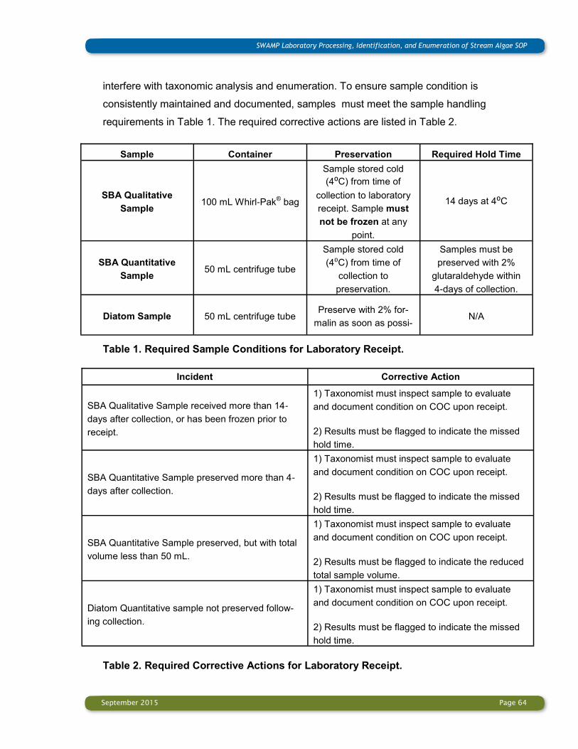
September 2015 Page 64
SWAMP Laboratory Processing, Identification, and Enumeration of Stream Algae SOP
interfere with taxonomic analysis and enumeration. To ensure sample condition is
consistently maintained and documented, samples must meet the sample handling
requirements in Table 1. The required corrective actions are listed in Table 2.
Table 1. Required Sample Conditions for Laboratory Receipt.
Table 2. Required Corrective Actions for Laboratory Receipt.
Sample Container Preservation Required Hold Time
SBA Qualitative
Sample 100 mL Whirl-Pak
® bag
Sample stored cold
(4⁰C) from time of
collection to laboratory
receipt. Sample must
not be frozen at any
point.
14 days at 4⁰C
SBA Quantitative
Sample 50 mL centrifuge tube
Sample stored cold
(4⁰C) from time of
collection to
preservation.
Samples must be
preserved with 2%
glutaraldehyde within
4-days of collection.
Diatom Sample 50 mL centrifuge tube Preserve with 2% for-
malin as soon as possi-N/A
Incident Corrective Action
SBA Qualitative Sample received more than 14-
days after collection, or has been frozen prior to
receipt.
1) Taxonomist must inspect sample to evaluate
and document condition on COC upon receipt.
2) Results must be flagged to indicate the missed
hold time.
SBA Quantitative Sample preserved more than 4-
days after collection.
1) Taxonomist must inspect sample to evaluate
and document condition on COC upon receipt.
2) Results must be flagged to indicate the missed
hold time.
SBA Quantitative Sample preserved, but with total
volume less than 50 mL.
1) Taxonomist must inspect sample to evaluate
and document condition on COC upon receipt.
2) Results must be flagged to indicate the reduced
total sample volume.
Diatom Quantitative sample not preserved follow-
ing collection.
1) Taxonomist must inspect sample to evaluate
and document condition on COC upon receipt.
2) Results must be flagged to indicate the missed
hold time.
September 2015 Page 65
SWAMP Laboratory Processing, Identification, and Enumeration of Stream Algae SOP
Section 5.3.2 Sample Handling Requirements – Archiving
This procedure establishes requirements for archiving the SBA and diatom samples. All
samples must be archived by the laboratory until results have been harmonized and
reported. Beyond this, the following guidelines are recommended:
The SBA qualitative sample can be archived for a maximum of 28-days after the
collection if kept cold (4°C).
All other samples are preserved prior to archiving, and can be held for at least 2-years.
Section 5.4 Photomicrographic Documentation Requirements
Requirements related to photomicrographic documentation are specified throughout the
procedure as follows:
Take sufficient photomicrographs of all newly reported species to support
harmonization of results. Take photomicrographs of previously identified taxa to
demonstrate the key aspects of vegetative morphology and reproduction used in
identification (see Section 1.5 and Appendix H).
At this point, SWAMP has not included a specific number of photomicrographs in this
requirement. The number of images needed to support identification and harmonization
may vary greatly; establishment of a minimum number would result in collection of too
many photos in some instances and too few in others. It is therefore left to the judgment of
the experienced taxonomist to determine how many photomicrographs need to be
collected. The standard used to evaluate the photomicrographic documentation is whether
or not it supports the stated goals:
Photomicrographic documentation of newly reported species is sufficient to support
harmonization of results.
Photomicrographic documentation of previously reported species is sufficient to
demonstrate the key aspects of vegetative morphology and reproduction used in
identification.
Recommendations for producing high-quality photomicrographs are included in Appendix
H. Photomicrographs displayed on online resources, Soft-Bodied Stream Algae of
California and Diatoms of the Southern California Bight, set the quality standard for

September 2015 Page 66
SWAMP Laboratory Processing, Identification, and Enumeration of Stream Algae SOP
photomicrographs. Questions regarding photomicrographic documentation should be
discussed with the algal taxonomist from CalPAL conducting harmonization of the results.
Section 5.5 Requirements for External Harmonization of Taxonomic Results
A fully detailed procedure covering harmonization of taxonomic results is currently being
developed (see Section 1.7). With respect to these procedures, laboratories should be
aware that participation in external harmonization of results is required for SWAMP
projects.
Harmonization requires communication between laboratory and CalPal taxonomists. This is
achieved in part by laboratory submission of the photographic documentation and text
descriptions of SBA and diatoms. Sometimes exchange of samples between the
taxonomists is needed. While this process is time consuming for both taxonomists, it is
critical to assuring the quality and comparability of the SWAMP algal taxonomy data and
participation in this process is therefore required by SWAMP.
Taxonomic harmonization currently is focused on newly recorded algal names which have
to be approved by an external taxonomist before the data is reported to the SWAMP
database. Thus, harmonization should take place with enough time before the deadline for
data reporting. It is recommended for new taxa recorded from the fresh SBA qualitative
samples to be harmonized during the identification process, because unpreserved samples
allow the best observation of taxonomic features and can be used for molecular analysis.
Section 6: Reporting of Algal Taxonomy Results
6.1 Reporting of SBA Results
All results collected for SBA sample (qualitative sample, quantitative sample - macroalgal
fraction, quantitative sample - microalgal fraction) are entered into the ID Datasheet for
SBA Sample (Appendix C1). Each taxonomic entry must have a corresponding numerical
entry (e.g., counts and cell size measurements) with the exception of the qualitative sample
which is not enumerated. After all data have been entered for a given sample, the numeric
entries for all NCEs should be summed to ensure the total target count has been reached.
SBA biovolumes should be calculated and entered in Biovolume Calculation Datasheet

September 2015 Page 67
SWAMP Laboratory Processing, Identification, and Enumeration of Stream Algae SOP
(Appendix F).Taxon names entered onto the SWAMP Taxonomy Results template should
match the SWAMP Algae Mater Taxa list. Any new FinalID name generated from an
analysis and confirmed during the taxonomy harmonization process should be added to the
Organism_DetailLookUp sheet in the SWAMP Taxonomy Results template and submitted
to CalPAL for approval (see Sections 1.4 and 6.3, and Appendix G).
6.2 Reporting of Diatom Results
All data from the diatom identification and enumeration are entered into the ID Datasheet
for Diatoms (Appendix C2). Each taxonomic entry must have a corresponding numerical
entry (i.e., counts). After all data has been entered for a given sample, the numeric entries
for counts should be summed to ensure the total target count has been reached.
Taxon names entered onto SWAMP Taxonomy Results template must match the SWAMP
Algae Master Taxa list. Any new FinalID name generated from an analysis and confirmed
during the taxonomy harmonization process should be added to the
Organism_DetailLookUp sheet in the SWAMP Taxonomy Results template and submitted
to CalPAL for approval (see Section 1.4 and 6.3., and Appendix G).
6.3 Data Management and Reporting
The primary way for submitting algae taxonomy data is through the Microsoft Excel
SWAMP Taxonomy Results template (Taxa Analysis Authorization or Taxa AA form) found
on the SWAMP website under the Database Management Resources Templates page
http://www.waterboards.ca.gov/water_issues/programs/swamp/
data_management_resources/templates_docs.shtml#taxonomy. Template documentation
explaining specific data fields, business rules, and how to complete this file can be found at
this link as well.
The procedure for using the template to report results is included in Appendix G. The
procedure focuses on three tabs: AlgaeInfo, BenthicResults, and Organism_DetailLookUp.
It is important to understand which party (project management, field crew, or laboratory) is
responsible for populating specific data within each tab. View the SWAMP and CEDEN
online LookUp lists for the most current valid values for given fields.
The AlgaeInfo tab contains composite sample volume information for each sample
September 2015 Page 68
SWAMP Laboratory Processing, Identification, and Enumeration of Stream Algae SOP
necessary for SBA biovolume calculations. The project manager (PM) and/or field crew is
responsible for populating and providing this information to the taxonomy lab prior to
completion of the data file.
The BenthicResults tab is used by the laboratory to report algae taxonomy results. The tab
has three sections used for aligning and reporting data.
The first section (columns A through Y) details Sample through Collection information for
each sample. This information is provided by the PM and/or field crew. Column T contains
the data for stream bottom area of substratum sampled for each sample necessary for SBA
biovolume calculations. The Chain of Custody (COC) contains some of the information
within this section but it usually does not provide all of the necessary information.
The second section (columns Z through AL) contains the lab effort/sorting information for
each sample. This information is populated by the taxonomy lab.
The third section (columns AM through BB) includes the taxonomy and numerical results
for each FinalID name and LifeStageCode combination for each sample. This information is
populated by the taxonomy lab. LifeStageCode can be found in separate tab:
LifeStageLookUp.
It is important to be able to link the second and third sections with the first section to report
the data to SWAMP or CEDEN. It is preferable for the taxonomy lab to receive the first
section before data is reported so data can be aligned. If this does not occur, the
LabSampleID can be used to link the taxonomy data with the sample information from
the COC.
The Organism_DetailLookUp tab is used for reporting FinalID names not currently listed on
the Algae Master Taxa list. Each new FinalID name, including attribute information, should
be submitted to CalPAL for review and approval before the Taxonomy Results template can
be finalized (see Section 1.7).
After taxonomy harmonization is completed, results that need to be revised to reflect the
outcome of harmonization are updated. The final data is added to the Taxonomy Result
template BenthicResults tab in columns Z through BB aligning the data with the

September 2015 Page 69
SWAMP Laboratory Processing, Identification, and Enumeration of Stream Algae SOP
corresponding Sample Collection information in columns A through Y. The data file is
submitted by the taxonomy lab through an online checker to check for errors associated
with formatting and business rules. SWAMP and CEDEN maintain their own checkers,
thus, be sure to visit the appropriate website depending on where your data should be
submitted as directed by the PM. The taxonomy lab should maintain an archive of all
submitted data files.

September 2015 Page 70
SWAMP Laboratory Processing, Identification, and Enumeration of Stream Algae SOP
TERMS AND DEFINITIONS
Algae. Photosynthetic aquatic organisms, including soft-bodied algae and diatoms with
uncovered reproductive structures;
Akinete. Thick-walled resting spore formed by transformation of a vegetative cell (John
et al., 2011);
Aplanospore. Non-motile spore and not morphologically identical to mother cell (John
et al., 2011);
Benthic algae. Bottom-living algae attached to or resting on the stream bottom (John et al.,
2011), in contrast to planktonic algae which are free-floating in the water column;
Biovolume. Volume of biological material being measured;
Chloroplast. Double-membrane bound organelle in eukaryotic algae containing chlorophyll
and other pigments (John et al., 2011);
Coccoid. Single-celled and non-flagellated algae;
Coenocyte. Thallus containing many nuclei, with few or no cell walls (John et al., 2011);
Colony. Group of individual cells enclosed into a common sheath/envelope or joined
together and having characteristic form and structure (John et al., 2011);
Conjugation. In zygnematalean algae a process of fertilization of two gametes either within
a tube which develops between two filaments in which the gametes were formed, or in the
cells of the filament;
Composite sample volume. Volume of all the liquid material amassed during sampling,
including water used for rinsing substrate and sampling devices. Final composite volume
should not exceed 400-500 mL (Ode et al., 2015, Glossary, and p. 27). This information is
provided for each sample;
Cyanobacteria (cyanoprokaryotes, blue-green algae) are photosynthetic prokaryotes,

September 2015 Page 71
SWAMP Laboratory Processing, Identification, and Enumeration of Stream Algae SOP
that is, cells that have no membrane-bound organelles, including chloroplasts. For the
purposes of this SOP, cyanobacteria are subsumed under SBA.
Diatom. A unicellular alga that possesses a rigid, silicified (silica-based) cell wall in the
form of a “pill box” (Ode et al., 2015);
Epiphytic algae. Growing on another plant, including another alga (John et al., 2011);
Filament. Cells united or arranged in one or more rows to form a chain or thread (John et
al., 2011);
Frustule. The complete silicified cell-wall of diatoms, consisting of the epi- and hypotheca
(Krammer and Lange-Bertalot, 2000);
Girdle. Collective term for all structural elements between two diatom valves (Krammer and
Lange-Bertalot, 2000);
Heterocyst (or heterocyte). A tick-walled, multilayered, and weakly pigmented cell in
certain cyanobacteria, contains the nitrogenase enzyme, which enables fixation of gaseous
nitrogen to ammonium (John et al., 2011);
ID datasheet. Excel spreadsheet file filled out on the computer that contains all algal taxa
with corresponding counts and size measurements;
Macroalgae. Large macroscopic filamentous, colonial, tuft-forming, crustose, tissue-like or
coenocytic eukaryotic algae and cyanobacteria that have forms recognizable with the
naked eye (e.g., Nostoc, Rivularia, Batrachospermum, Lemanea, Cladophora,
Draparnaldia, Oedogonium, Rhizoclonium, Spirogyra, Zygnema, Mougeotia, Vaucheria)
[see Sheath and Cole (1992) for definitions of forms];
Microalgae. Small microscopic form not recognizable with the naked eyes but consisting of
unicellular, colonial or filamentous non-diatom algae;
Natural counting entity (NCE). Each natural occurring form of algae (i.e., each unicell,
colony, filament, tissue-like form, coenocyte, tuft, or crust) is defined as a natural counting
entity regardless of the number of cells in the thallus or colony;

September 2015 Page 72
SWAMP Laboratory Processing, Identification, and Enumeration of Stream Algae SOP
Pyrenoid. Organelle associated with the chloroplast of many algae, which contains a high
content of enzyme responsible for fixing carbon dioxide in photosynthesis and around
which starch grains may accumulate (John et al., 2011);
Relative abundance of diatoms. The number of valves of particular diatom taxon as a
percentage of the total number of diatom valves counted per sample (which is at least 600
diatom valves in this SOP);
Soft-bodied algae (SBA). Non-diatom algal taxa; for the purposes of this SOP,
cyanobacteria are subsumed under this assemblage (Ode et al., 2015);
Stream bottom area of substratum sampled. Total area of substratum surface from
which benthic algae were collected (Ode et al., 2015, pp. 10-18). This information is
provided for each sample in cm2;
Thallus (Thalli). Body of simple plants not differentiated into a true root, stem, leaf or
leaves (John et al., 2011);
Tuft. Group of filaments which are close together but without a common sheath, usually
aggregations at right angles to a surface (John et al., 2011);
Zygospore. Spore formed sexually following gamete fusion, often thick-walled,
characteristically colored and sometimes ornamented (John et al., 2011);
Valve. Lid-top of the epi-and hypotheca in diatom. It consist of the valve face and that part
of the valve, that connects with the girdle (Krammer and Lange-Bertalot, 2000).

September 2015 Page 73
SWAMP Laboratory Processing, Identification, and Enumeration of Stream Algae SOP
LIST OF APPENDICES
Appendix A: Laboratory Equipment List
Appendix B: Health and Safety Issues
Appendix B1: Health and Safety Issues for SBA Processing
Appendix B2: Health and Safety Issues for Diatom Processing
Appendix C: ID Datasheets
Appendix C1: ID Datasheet for SBA Sample with Example Form
Appendix C2: ID Datasheet for Diatom Sample
Appendix D: Taxonomic Harmonization Datasheet
Appendix E: List of Geometric Shapes Applied in Biovolume Calculation for SBA
Appendix F: Biovolume Calculation Datasheet for SBA Microalgal Fraction with
Example Form
Appendix G: Protocol for the SWAMP Database Reporting
Appendix H: Recommendations for producing high-quality algal photomicrographs
Appendix I: References

September 2015 Page 74
SWAMP Laboratory Processing, Identification, and Enumeration of Stream Algae SOP
Appendix A: Laboratory Equipment List

September 2015 Page 75
SWAMP Laboratory Processing, Identification, and Enumeration of Stream Algae SOP
September 2015 Page 76
SWAMP Laboratory Processing, Identification, and Enumeration of Stream Algae SOP
Appendix B: Health and Safety Issues
Appendix B1: Health and Safety Issues for SBA Processing
As with all laboratory procedures, care should be given when working with glass and any
chemicals. Specific safety issues in the laboratory related to SBA processing is to work safe
with samples which are fixed with 2% glutaraldehyde. These samples include all
quantitative algal samples collected and fixed in the field, as well as the vials with
preserved and stored qualitative algal samples as reference collections. All procedures with
samples fixed with 2% glutaraldehyde are to be performed in a positive-draw fume hood.
Staff members should wear laboratory coats, nitrile gloves and protective eyewear. When
fixed qualitative samples are reinvestigated, the algal material should be washed with DI
water prior to microscopic examination by soaking in beakers with DI water for 2 hours.
Processing of quantitative algal samples includes a dilution of 2% glutaraldehyde in the
sample by at least 20x (see Section 3.1.2 and 3.1.3).
SOP for using glutaraldehyde for the preservation of SBA
1. Scope and Application
Glutaraldehyde is a colorless liquid with a pungent odor used as a fixative. This SOP
covers the use of glutaraldehyde by SWAMP laboratories as a fixative for SBA.
2. Health Hazards
The health hazards associated with the use of glutaraldehyde include:
Inhalation
• Regulatory limit of 0.05 ppm as a ceiling level
• Chemical burns to the respiratory tract
• Asthma and shortness of breath
• Headache, dizziness, and nausea
Skin
• Sensitization or allergic reactions, hives
• Irritations and burns
• Staining of the hands (brownish or tan)

September 2015 Page 77
SWAMP Laboratory Processing, Identification, and Enumeration of Stream Algae SOP
Eyes
• Irritation and burns. Eye contact causes moderate to severe irritation, experienced as
discomfort or pain, excessive blinking and tear production
• May cause permanent visual impairment
• Conjunctivitis and corneal damage
Ingestion
• Gastrointestinal tract burns
• Central nervous system depression, excitement
• Nausea, vomiting
• Unconsciousness, coma, respiratory failure, death
3. Safety Shower and Eyewash
All employees using glutaraldehyde must be aware of the location and use of the laboratory
safety shower and eyewash, and must be able to reach it within 10 seconds from the time
of contamination. At no time will processes using glutaraldehyde be allowed that do not
provide access to a safety shower and eyewash.
Employees who have skin or eye contact with glutaraldehyde will immediately stop all
processes and proceed to the safety shower and eyewash station. The employee will rinse
the affected area for a minimum of 15 minutes. If eye contact has occurred, the upper and
lower eyelids must be lifted to allow adequate flushing of the eyes.
4. Special Handling Procedures and Storage Requirements
Procedures will be followed that reduce exposure to glutaraldehyde vapor to the lowest
reasonable level.
This includes:
• Ensure glutaraldehyde is only used under a positive-draw fume hood.
• Use only enough glutaraldehyde to perform the required procedure.
• Every effort must be made to minimize splashing, spilling, and personnel exposure
• Once specimens are preserved, they will be capped or secured in a way that does not
allow glutaraldehyde to evaporate into the lab.
• At no time will open containers be removed from the positive-draw fume hood.

September 2015 Page 78
SWAMP Laboratory Processing, Identification, and Enumeration of Stream Algae SOP
• All containers of glutaraldehyde or solutions containing glutaraldehyde will be
appropriately marked with the chemical name, and hazard warning label.
• Glutaraldehyde will be stored in tightly closed containers in a cool, secure, and properly
marked location.
• Glutaraldehyde storage vials should meet the following safety requirements: If they are
plastic - high-density Polyethylene (HDPE) will be one of the best plastic not corroded by
the glutardehyde. Low-density Polyethylene (LDPE) loses some of its capabilities at
temperatures higher then 50ºC. Polypropylene (PP) works excellently just as well. For any
scintillation vial, no metal/foil liners should be inside the cap. Borosilicate glass vials or any
polypropylene copolymers will work well also. No flexible PVP (Polyvinylpyrrolidone) or
PMP (polymethylpentene) should be used.
5. Waste Disposal
Excess glutaraldehyde and all waste material containing glutaraldehyde must be placed in
an unbreakable secondary container labeled with the following “HAZARDOUS WASTE
GLUTARALDEHYDE.” Wastes will be disposed of through the laboratory hazardous waste
contract.
Appendix B2: Health and Safety Issues for Diatom Processing
As with all laboratory procedures, care should be given when working with glass and any
chemicals. For all chemicals, the laboratory should maintain current files /documentation in
the form of Material Safety Data Sheets (MSDS) and those files should be available to all
laboratory staff. Should there be any concern, laboratory staff should take appropriate
action (relative to their setting). Specific safety issues in the laboratory related to diatom
processing include:
Glassware. There are many places in these procedures where glassware is used,
including use of beakers for sample process, pipettes, microscope slides, and especially
the thin cover glasses used. Breakage of glassware should be cleaned up immediately,
with broken glass being disposed of in specific, marked containers.
Oxidizers. This includes nitric acid and 30% hydrogen peroxide. Laboratory personnel
should have eye protection and wear gloves when using these materials. Should there be a
September 2015 Page 79
SWAMP Laboratory Processing, Identification, and Enumeration of Stream Algae SOP
spill, spill kits should be used to contain and clean up the spill.
Fixatives. Formalin, the primary fixative used in the preservation of fresh diatom samples,
requires care in handling and disposal. It should be handled only in a positive-draw fume
hood, and disposal should be made in accordance with a company’s hazardous waste
disposals procedures and protocols.
Naphrax/Toluene. The solvent toluene is used to keep the mounting medium Naphrax as a
liquid. It is evaporated when heated. In both its liquid and vapor forms, toluene can be
hazardous to health, so it should be used exclusively in a positive-draw fume hood.
Potassium dichromate. This chemical is highly hazardous to health and environment. It is
a strong oxidizer, corrosive, irritant, sensitizer, carcinogen, highly toxic acts upon eyes,
skin, lungs, mucous membranes, reproductive toxin, fatal if inhaled. It should be used ex-
clusively in a positive-draw fume hood and disposed of as hazardous material. Not recom-
mended for use.
Hot plates. Just as their name indicates, use of hot plates means that care must be taken
not to burn oneself.
September 2015 Page 80
SWAMP Laboratory Processing, Identification, and Enumeration of Stream Algae SOP
Appendix C. ID Datasheets
Appendix C1: ID Datasheet for SBA Sample with Example Form

September 2015 Page 81
SWAMP Laboratory Processing, Identification, and Enumeration of Stream Algae SOP
Example Form
Note: * Indicates averaged dimensions based on measurements of 20 NCEs per species.

September 2015 Page 82
SWAMP Laboratory Processing, Identification, and Enumeration of Stream Algae SOP
Appendix C2: ID Datasheet for Diatom Sample
September 2015 Page 83
SWAMP Laboratory Processing, Identification, and Enumeration of Stream Algae SOP
Appendix D. Taxonomic Harmonization Datasheet
The taxonomic harmonization datasheet is a modification of the Organism_DetaiLookUp
tab in the SWAMP Taxonomy Results template (see Appendix G). The additional columns
presented below are distributed in the Organism_DetaiLookUp tab after the Final ID column
for each new SBA and diatom name.
Note: * Each photomicrograph should have a filename consisting of the following elements
in the order indicated: SWAMP Sample ID, Sampling date (MM/DD/YYYY), Species ID,
magnification of the objective (i.e., 40x) (see Section 1.5 and Appendix H).

September 2015 Page 84
SWAMP Laboratory Processing, Identification, and Enumeration of Stream Algae SOP
Appendix E: List of Geometric Shapes Applied in Biovolume Calculation for SBA This list includes all geometric shapes (after Hillebrand et al., 1999) applied in biovolume
calculation of stream SBA for the SWAMP program. Many stream SBA genera recorded in
the SWAMP Master list and illustrated in the online identification tool Soft-Bodied Stream
Algae of California (Stancheva et al., 2014) are missing in Hillebrand et al. (1999). They are
listed below in the corresponding geometric shape category.
Cylinder. It is one of the most common shapes applied in the biovolume calculation of all
filamentous algae and some coccoids with cylindrical cells. In the case of tapering filaments
the diameter is averaged by measuring the cells in the widest, narrowest and middle part of
the filament.
Cyanobacteria: Anabaena, Capsosira, Chamaesiphon confervicola, C. incrustans, C.
investiens, Dolichospermum, Trichormus;
Chlorophyta: all green branched and unbranched filamentous genera, Teilingia;
Xanthophyta: Xanthonema
Sphere. Common shape applied in biovolume calculation of many flagellated and
nonmotile unicells or colonial algae. The shape is applied to a single cell, and in the case of
colony the biovolume of a single cell is multiplied by the total number of cells in the colony.
Chlorophyta: Apiocystis, Asterococcus, Botryosphaerella, Hariotina, Radiococcus,
Schizochlamys, Tetrasporidium;
Chrysophyceae: Chrysopyxis;
Xanthophyta: Mischococcus
Prolate spheroid. Common shape applied in biovolume calculation of many flagellated and
nonmotile unicells or colonial algae. The shape is applied to a single cell, and in the case of
a colony, the biovolume of a single cell is multiplied by the total number of cells in the
colony.
Cyanobacteria: Coelomoron, Pleurocapsa, Chamaesiphon species, excluding the species
with cells approximated as cylinders
Chlorophyta: Apatococcus, Chlamydomonadopsis, Comasiella, Nephrocytium,

September 2015 Page 85
SWAMP Laboratory Processing, Identification, and Enumeration of Stream Algae SOP
Sphaerobotrys, Planotaenium;
Chrysophyceae: Stylococcus;
Xanthophyta: Chadefaudiothrix, Chlorosaccus
Cryptophyta: Chroomonas
Ellipsoid. Chlorophyta: Oocardium
Prism on elliptic base. This shape refers to entire colony of Pediastrum, Lacunastrum,
and Stauridium (Chlorophyta), where the transapical section height is approximated to 2
µm.
Two cones. Chlorophyta: Quadrigula; Xanthophyta: Chytridiochloris
Two truncated cones. Chlorophyta: Stauridium
Ellipsoid + cylinder. Euglenophyta: Monomorphina, Strombomonas
More rarely applied shapes are cube, box, cylinder + two cones, cylinder+cone,
ellipsoid+two cones+cylinder, half ellipsoid+cone on elliptic base.

September 2015 Page 86
SWAMP Laboratory Processing, Identification, and Enumeration of Stream Algae SOP
Appendix F: Biovolume Calculation Datasheet for SBA Microalgal Fraction with
Example Form
Formulas (see Section 4.1.4.2)
Va’=Va Vcr (µm3), Vcr= (Vt-Vm) (Vt-Vm-5)-1
Vi=Va’ Vs Vc-1 A-1 (μm3 cm-2)
Sample and Microscope condition
● 1 mL microalgal fraction, 5 times concentrated original sample
● 0.05 mL subsample analyzed = 0.25 mL original sample
● Transect width with 40x objective = 0.55 mm
● 40 horizontal optical transects on the slide
● Sample volume per transect = 0.00625 (= 0.25/40) mL in 1 mL microalgal fraction
● Sample volume per transect should be corrected by multiplication with dilution factor (DF)
when additionally concentrated to 0.5 mL (DF 2), or diluted to 2 mL (DF 1/2), to 3 mL (DF
1/3), to 4 mL (DF 1/4), or to 5 mL (DF 1/5)
Vc = Volume of sample counted (mL)
Transects counted (#)
Sample volume per
transect (mL) Dilution factor (DF) Vc
Va' = Biovolume of i-species per sample counted (µm3)
Vt Vm Vt-Vm Vt-Vm-5 Vcr Va Va'
Vi = Volume of i-species per unit bottom area (µm3 cm-2)
A Vs Va' Vc Va' Vs Vc A Vi

September 2015 Page 87
SWAMP Laboratory Processing, Identification, and Enumeration of Stream Algae SOP
Example form for calculation of biovolume of i-species in two different sample conditions
Sample 1: Total biovolume of i-species in 3 horizontal optical transect analyzed is 523 µm3
(Va); no additional dilutions/concentrations of microalgal fraction (DF = 1); no macroalgae
present (Vm = 0 mL); Vt = 50 mL; A = 138.6 cm2; Vs = 345 mL
Sample 2: Total biovolume of i-species in 3 horizontal optical transect analyzed is 523 µm3
(Va); dilution of microalgal fraction to 2 mL (DF = 1/2); 0.3 mL macroalgae present (Vm =
0.3 mL); Vt = 50 mL; A = 138.6 cm2; Vs = 465 mL

September 2015 Page 88
SWAMP Laboratory Processing, Identification, and Enumeration of Stream Algae SOP
Appendix G: Protocol for the SWAMP Database Reporting
The following protocol for data entry and submittal to the SWAMP database is used by
laboratories after SBA and diatom identification and enumeration data are entered in the ID
datasheets (Appendix C), and SBA biovolumes are calculated (Appendix F). A similar
process can be followed for submitting CEDEN data but the template may differ slightly.
The primary way for submitting algae taxonomy data is through the Microsoft Excel
SWAMP Taxonomy Results template (Taxa Analysis Authorization or Taxa AA form) found
on the SWAMP website under the Database Management Resources Templates page
[http://www.waterboards.ca.gov/water_issues/programs/swamp/
data_management_resources/templates_docs.shtml#taxonomy]. Template documentation
explaining specific data fields, business rules, and how to complete this file can be found at
this link as well. It is important to understand who is responsible for populating specific data
within each template tab and when each task should occur.
Many tabs exist in the Taxonomy Results template, but only one (BenthicResults) is used to
report algal taxonomy data. Most tabs are used for submitting new LookUp values for
given data fields, but users should view the SWAMP and CEDEN online LookUp lists for
the most current valid and comparable values. The Header row of each tab contains the
corresponding field names for those data tables. Any field name in bold text indicates it is a
required field and should be populated. If the value is unknown, default values may be used
but it is preferable to use correct values. Three tabs will be discussed in greater detail in
this document: AlgaeInfo, BenthicResults, and Organism_DetailLookUp.
The AlgaeInfo tab contains sample volume information for each sample necessary for
biovolume calculations. The Project Manager (PM) and/or field crew is responsible for
populating and providing this information in the template file to the taxonomy lab prior to
completion of the data file. If the physical habitat (PHAB) and sample collection data are
entered into a SWAMP database (shell or replica), the SWAMP Bioassessment field forms
can be used to run an existing query (Algae Sample Information) to export this data to the
template file.

September 2015 Page 89
SWAMP Laboratory Processing, Identification, and Enumeration of Stream Algae SOP
The BenthicResults tab is used by the lab to report SBA and diatom taxonomy results. The
tab has three sections used for aligning and reporting data. The first section (columns A
through Y) details Sample through Collection information for each sample and is to be
provided by the PM and/or field crew. A COC contains some of the information within this
section but it usually does not provide all of the necessary information. If the sample
collection information exists in a SWAMP database (shell or replica), the PM or field crew
can export this data using the SWAMP Bioassessment field forms by running the Taxa
Template query. The second section (columns Z through AL) contains the lab effort/sorting
information for each sample and is to be populated by the taxonomy lab. The third section
(columns AM through BB) includes taxonomy results for each FinalID name and
LifeStageCode combination for each sample, and it is to be populated by the taxonomy lab.
It is important to be able to link the second and third sections with the first section to report
data to SWAMP or CEDEN. It is preferable for the taxonomy lab to receive the first section
before data is reported so data can be aligned. If this does not occur, a LabSampleID can
be used to link the taxonomy data with the sample information from the COC.
The Organism_DetailLookUp tab is used for reporting FinalID names not in the Algae
Master Taxa list. Each new FinalID name, including attribute information such as
taxonomic hierarchy and taxonomic authority, should be submitted to CalPAL for review
and approval before the Taxonomy data template can be finalized. Additional supporting
documentation such as photographs should also be submitted during the taxonomic
harmonization process (see Sections 1.4 and 1.7).
After FinalID harmonization is completed, the final data is added to the Taxonomy Results
template BenthicResults tab in columns Z through BB aligning the data with the
corresponding Sample Collection information in columns A through Y. The data file is then
submitted by the taxonomy lab through an online checker to check for errors associated
with formatting and business rules. SWAMP and CEDEN maintain their own online
checkers so please visit the appropriate web site depending on where your data should be
submitted [SWAMP online checker: http://swamp.waterboards.ca.gov/swamp_checker/].
The SWAMP online checker will ask for the Data Category (Taxonomy), your email address
and agency, and for you to browse to your file for loading. After the file is checked, any
associated errors will be shown on a Summary page where users can choose to hide errors

September 2015 Page 90
SWAMP Laboratory Processing, Identification, and Enumeration of Stream Algae SOP
from showing if they want. Any critical errors must be fixed in the data file before it can be
submitted to SWAMP. Any Regular or Warning errors should be reviewed and, if possible,
fixed by either the taxonomy lab, PM, and/or field crew. When the green ‘Submit Data to
SWAMP’ button is available, users click on the button to submit the file along with adding
any comments and/or emailing other people with their submittal. Please note the entity
responsible for submitting the final data set to SWAMP or CEDEN will depend on the
project. That is, the PM may decide the taxonomy lab is to submit the data directly through
the online checker, or the PM may want the taxonomy lab to submit the file to the PM first
and the PM will be responsible for running the file through the online checker for submittal.
In either case, the taxonomy lab should maintain an archive of all final data files whether it
is submitted directly to the PM, SWAMP, or CEDEN.
The database protocol in this SOP is current as of the publication date but is subject to
change as updates are made to SWAMP and CEDEN. Always confirm the current protocol
with the SWAMP or CEDEN Data Management team and/or your PM.
September 2015 Page 91
SWAMP Laboratory Processing, Identification, and Enumeration of Stream Algae SOP
Appendix H: Recommendations for producing high-quality algal
photomicrographs
Soft-Bodied Macroalgae
1. Use a clean slide and cover slip to prepare water mount.
2. Select specimens, either fresh or preserved, which are representative and in good
shape. If the specimen which has to be pictured is already mounted in an overcrowded
slide, try to isolate it and move it to another slide. Overcrowded slides will create too
many depths of view and overlap for clear 2D imaging.
3. Prepare a water mount with the algal specimen spread out well enough so that it lies flat
on the slide. Avoid breaking or overlapping filaments or other structures. Avoid using too
much or too little water to prepare the slide; too much will flood the slide and create
depths of view or bubbles that can overlap the image, while too little will often cause
plasmolysis of the sample.
4. Large mucilaginous cyanobacterial colonies should be gently flattened with the cover
slip to make sure the cells are visible. Large red algae, such as Batrachospermum,
should be gently flattened with cover slip to disclose the reproductive cells, and
chopping with a razor blade may be necessary. Protect large spores of Zygnemataceae,
Oedogonium, Vaucheria from breaking while mounting the material.
5. When the macroalga is mounted properly in a flat, single-cell layer, adjust the
background light and white balance, and take a picture at lower magnifications (10x,
20x objective) to show the gross morphology of the filaments or colonies.
6. Take additional pictures at higher magnification (40x objective) focused on critical
taxonomic features, such as akinetes and mucilage in cyanobacteria, chloroplast, type
of branching, specialized cells, reproductive cells and spore cell walls in filamentous
eukaryotic algae, etc.
7. Stain the cells with Lugol`s solution if needed to distinguish the starch and take
additional pictures.
Soft-Bodied Microalgae
1. Use clean slide and cover slip to prepare water mount.
2. Place a drop of microalgal sample on the slide and avoid the formation of air bubbles
while covering with the cover slip.
3. Make sure the sample is not too dense and the cells are not overlapping.
September 2015 Page 92
SWAMP Laboratory Processing, Identification, and Enumeration of Stream Algae SOP
4. For photographing specimens, whether filaments, colonies or single cells, select those
which lie flat on the slide. Press or tap the cover slip gently to adjust the position of the
specimen if needed.
5. When the microalga is mounted properly in a flat, single-cell layer, adjust the
background light and white balance, and take pictures at higher magnifications (40x
objective) focusing on critical taxonomic features, such as chloroplasts, pyrenoids, cell
walls, mucilage, etc.
6. Stain the cells with Lugol`s solution if needed to distinguish the starch and take
additional pictures.
Diatoms
1. Make sure that the permanent slide and cover slip are clean before starting.
2. Select a valve which lies flat on the slide in valve view.
3. Avoid photographing valves which are tilted or covered by debris, in clumps or broken.
4. Avoid photographing valves located in disrupted areas of the mount, particularly edges
that have optical aberrations.
5. When the diatom valve of interest is selected, adjust the background light and white
balance, as well as the DIC with the 100x oil objective.
6. Focus on the surface of the valve so that the gross morphology, i.e. valve shape and
structural elements are clearly visible.
7. Take additional pictures of the same valve focusing on critical taxonomic features, such
as stigma in the central area in Gomphonema, pseudocepta and longitudinal lines in
Gomphoneis, annulae at the poles in Geissleria, central nodule and fibulae in Nitzschia,
central area and striae in Navicula, etc.

September 2015 Page 93
SWAMP Laboratory Processing, Identification, and Enumeration of Stream Algae SOP
Appendix I: References
General References
Acker, F., Russell, B., and E. Hagan. 2002. Diatom Cleaning by Nitric Acid Digestion with a
Microwave Apparatus. Protocol P- 13-42. In: Charles, D.F., C. Knowles, and R.S.
Davis, eds. 2002. Protocols for the analysis of algal samples collected as part of
the U.S. Geological Survey National Water-Quality Assessment Program. Report
No. 02-06, Patrick Center for Environmental Research, The Academy of Natural
Sciences, Philadelphia, PA. 124 pp. Also at http://diatom.ansp.org/nawqa/pdfs/
ProtocolPublication.pdf
American Public Health Association. 1981. Standard Methods for the Examination of Water
and Wastewater, 15th ed. Am. Pub. Health Assoc., Washington, D.C. 1,134 pp.
Fetscher, A. E., R. Stancheva, J. P. Kociolek, R. G. Sheath, E. D., Stein, R. D., Mazor, P.
R. Ode, and L. B. Busse. 2014. Development and comparison of stream indices of
biotic integrity using diatoms vs. non-diatom algae vs. a combination. Journal of
Applied Phycology 26: 433-450.
Guiry, M. D., and G. M. Guiry. 2015. AlgaeBase. World-wide electronic publication, National
University of Ireland, Galway. http://www.algaebase.org; searched on 28 Septem-
ber 2015.
Hillebrand, H., C. D. Durselen, D. Kirschel, U. Pollinger, and T. Zohary. 1999. Biovolume
calculation for pelagic and benthic microalgae. Journal of Phycology 35:403-424.
Ode, P. R., A. E. Fetscher, and L. B. Busse. 2015. Standard operating procedures for the
collection of field data for ambient bioassessments of California wadeable streams:
benthic macroinvertebrates, algae, and physical habitat. California State Water
Resources Control Board Surface Water Ambient Monitoring Program (SWAMP)
Bioassessment.
Sheath, R. G., and K. M. Cole. 1992. Biogeography of stream macroalgae in North
America. Journal of Phycology 28:448-460.

September 2015 Page 94
SWAMP Laboratory Processing, Identification, and Enumeration of Stream Algae SOP
Stancheva, R., Fetscher, A. E., and R. G. Sheath. 2012a. A novel quantification method for
stream-inhabiting, non-diatom benthic algae, and its application in bioassessment.
Hydrobiologia 684: 225-239.
USEPA. 2013 (draft). National Rivers and Streams Assessment: Quality Assurance Project
Plan. EPA‐841‐B‐12‐007. U.S. Environmental Protection Agency, Office of Water,
Washington, DC.
Van der Werff, A. 1955. A new method of concentrating and cleaning diatoms and other
organisms. Official Journal of the International Association of Theoretical and Ap-
plied Limnology, 12:276-277.
Woodard, M.E., J. Slusark, and P.R. Ode. 2012. Standard Operating Procedures for
Laboratory Processing and Identification of Benthic Macroinvertebrates in
California. California State Water Resources Control Board Surface Water
Ambient Monitoring Program (SWAMP) Bioassessment SOP 003.
Taxonomic Identification Literature and Online Resources
* Indicates the references to start identification, including most recent taxonomic
publications on the local algal flora, and then to expand the search to references for
particular algal groups
References for Identification of SBA
Bourrelly, P. 1968. Les Algues d'Eau Douce. Tome II. Les Algues Jaunes et Brunes,
Chromophycees, Chrysophycees, Phéophycées, Xanthophycées et Diatomées.
N. Boubée et Cie, Paris.
Bourrelly, P. 1985. Les Algues d'Eau Douce. Tome III. Les Algues Bleues et Rouges, Les
Eugléniens, Peridiniens et Cryptomonadines. N. Boubée et Cie, Paris.
Bourrelly, P. 1990. Les Algues d'Eau Douce. Tome I. Les Algues Vertes. N. Boubée et Cie,
Paris.
Desikachary, T.V. 1959. Cyanophyta. Indian Council of Agricultur Research, New Delhi,
India.

September 2015 Page 95
SWAMP Laboratory Processing, Identification, and Enumeration of Stream Algae SOP
Dillard, G.E. 1989. Bibliotheca Phycologica. Band 81. Freshwater Algae of the
Southeastern United States. Part 1. Chlorophyceae: Volvocales, Tetrasporales
and Chlorococcales. J. Cramer, Stuttgart.
Dillard, G.E. 1989a. Bibliotheca Phycologica. Band 83. Freshwater Algae of the
Southeastern United States. Part 2. Chlorophyceae: Ulotrichales, Microsporales,
Cylindrocapsales, Sphaeropleales, Chaetophorales, Cladophorales,
Schizogoniales, Siphonales and Oedogoniales. J. Cramer, Stuttgart.
Dillard, G.E. 1990. Bibliotheca Phycologica. Band 85. Freshwater Algae of the
Southeastern United States. Part 3. Chlorophyceae: Zynematales:
Zygnemataceae, Mesotaeniaceae and Desmidiaceae (Section 1). J. Cramer,
Stuttgart.
Dillard, G.E. 1991. Bibliotheca Phycologica. Band 89. Freshwater Algae of the
Southeastern United States. Part 4. Chlorophyceae: Zygnematales: Desmidiaceae
(Section 2). J. Cramer, Stuttgart.
Dillard, G.E. 1991a. Bibliotheca Phycologica. Band 90. Freshwater Algae of the
Southeastern United States. Part 5. Chlorophyceae: Zygnematales: Desmidiaceae
(Section 3). J. Cramer, Stuttgart.
Dillard, G.E. 1993. Bibliotheca Phycologica. Band 93. Freshwater Algae of the
Southeastern United States. Part 6. Chlorophyceae: Zygnematales: Desmidiaceae
(Section 4). J. Cramer, Stuttgart.
Dillard, G.E. 2000. Bibliotheca Phycologica. Band 106. Freshwater Algae of the
Southeastern United States. Part 7. Pigmented Euglenophyceae. J. Cramer,
Stuttgart.
Dillard, G.E. 2007. Bibliotheca Phycologica. Band 112. Freshwater Algae of the
Southeastern United States. Part 8. Chrysophyceae, Xanthophyceae,
Raphidophyceae, Crysophyceae and Dinophyceae. J. Cramer, Stuttgart.
Ettl, H., and G. Gärtner 1988. Chlorophyta II: Tetrsporales, Chlorococcales,
Gloeodendrales. In Ettl, H., J. Gerloff, H. Heynig, and D. Mollenhauer (eds),
Süsswasserflora von Mitteleuropa 10. Gustav Fischer, Sttutgart.

September 2015 Page 96
SWAMP Laboratory Processing, Identification, and Enumeration of Stream Algae SOP
Ettl, H. 1978. Xanthophyceae. In Ettl, H., J. Gerloff & H. Heynig (eds), Süsswasserflora von
Mitteleuropa 1. Gustav Fischer, Sttutgart.
Hall, J. D., R. Stancheva, R. M. McCourt, and R. G. Sheath.* Vegetative morphology and
phylogenetic position of Caespitula incrustans gen. et sp. nov. (Ulvales,
Chlorophyta) from streams in California. Phycologia (in preparation).
Hindák, F. 2008. Colour Atlas of Cyanophytes. VEDA, Bratislava.
John, D. M., B. A. Whitton, and A. J. Brook (eds). 2011.* The Freshwater Algal Flora of the
British Isles. 2nd
Ed. Cambridge University Press, Cambridge.
Kadłubowska, J. Z. 1984. Conjugatophyceae I. Chlorophyta VIII. Zygnemales. In Ettl, H., J.
Gerloff, H. Heynig & D. Mollenhauer (eds), Süsswasserflora von Mitteleuropa 16.
Gustav Fischer, Sttutgart.
Komárek, J., and K. Anagnostidis 1999.* Cyanoprokaryota: Chroococcales. In Ettl, H., G.
Gärtner, H. Heynig & D. Mollenhauer (eds), Süsswasserflora von Mitteleuropa
19/1. Gustav Fischer, Sttutgart.
Komárek, J., and K. Anagnostidis 2005.* Cyanoprokaryota: Oscillatoriales. In Büdel, B., G.
Gärtner, L. Krienitz & M. Schagerl (eds), Süsswasserflora von Mitteleuropa 19/2.
Elsevier, München.
Komárek, J. 2013.* Cyanoprokaryota: Heterocytous Genera. In Büdel, B., G. Gärtner, L.
Krienitz & M. Schagerl (eds), Süsswasserflora von Mitteleuropa 19/3. Springer,
Berlin.
Kristiansen, J., and H. R. Preisig. 2001. Encyclopedia of Chrysophyte Genera. Bibliotheca
Phycologica, Band 110, J. Cramer, Stuttgart.
Kristiansen, J., and H. R. Preisig. 2007. Chrysophyte and Haptophyte algae. Part 2:
Synurophyceae. In Büdel, B., G. Gärtner, L. Krienitz, H. R. Preisig & M. Schagerl,
(eds), Süsswasserflora von Mitteleuropa 2/2. Springer-Verlag Berlin Heidelberg.
Prescott, G. W. 1951.* Algae of the Western Great Lakes Area. WM.C. Brown Publishers,
Dubuque, Iowa, 977 pp.

September 2015 Page 97
SWAMP Laboratory Processing, Identification, and Enumeration of Stream Algae SOP
Prescott, G. W., C. E. M Bicudo, and W. C. Vinyard. 1982. A Synopsis of North American
Desmids. Part II. Desmidiaceae: Placodermae, Section 4. University of Nebraska
Press, Lincoln.
Prescott, G. W., H. T. Croasdale, and W. C. Vinyard. 1975. A Synopsis of North American
Desmids. Part II. Desmidiaceae: Placodermae, Section 1. University of Nebraska
Press, Lincoln.
Prescott, G. W., H. T. Croasdale, and W. C. Vinyard. 1977. A Synopsis of North American
Desmids. Part II. Desmidiaceae: Placodermae, Section 2. University of Nebraska
Press, Lincoln.
Prescott, G. W., H. T. Croasdale, W. C. Vinyard, and C. E. M. Bicudo. 1981. A Synopsis of
North American Desmids. Part II. Desmidiaceae: Placodermae, Section 3.
University of Nebraska Press, Lincoln.
Reith, A.1980. Xanthophyceae. In Ettl, H., J. Gerloff & H. Heynig (eds), Süsswasserflora
von Mitteleuropa 2. Gustav Fischer, Sttutgart.
Sheath, R. G. 2003. Red algae. In Wehr, J. D. & R. G. Sheath (eds), Freshwater Algae of
North America: Ecology and Classification. Academic Press, San Diego,
California: 197-221.
Smith, G. M. 1950. The Fresh-Water Algae of the United States. 2nd ed. McGraw-Hill Book
Co., New York.
Stancheva, R., J. D. Hall, and R. G. Sheath. 2012b.* Systematics of the genus Zygnema
(Zygnematophyceae, Charophyta) from Californian watersheds. Journal of
Phycology 48: 409-422.
Stancheva, R., J. D., Hall, R. M. McCourt, and Sheath. 2013.* Identity and phylogenetic
placement of Spirogyra species (Zygnematophyceae, Charophyta) from California
streams and elsewhere. Journal of Phycology 49: 588–607.
Stancheva, R., C. Fuller,and R. G. Sheath. 2014.* Soft-Bodied Stream Algae of California.
http://dbmuseblade.colorado.edu/DiatomTwo/sbsac_site/IDResourceTool.htmL

September 2015 Page 98
SWAMP Laboratory Processing, Identification, and Enumeration of Stream Algae SOP
Starmach, K. 1985. Chrysophyceae und Haptophyceae. In Ettl, H., J. Gerloff, H. Heynig &
D. Mollenhauer (eds), Süsswasserflora von Mitteleuropa 1. Gustav Fischer,
Sttutgart.
Transeau, E. N. 1951.* The Zygnemataceae. The Ohio State University Press, Columbus,
Ohio.
Wehr, J. D., R. Stancheva, K. Truhn, K., and R. G. Sheath. 2013.* Discovery of the rare
freshwater brown alga Pleurocladia lacustris (Ectocarpales, Phaeophyceae) in
California streams. Western North American Naturalist 73: 148-157.
Wehr, J. D., R. G. Sheath, and J. P. Kociolek (eds). 2015.* Freshwater Algae of North
America: Ecology and Classification, 2nd Ed. Elsevier Academic Press, San Diego,
California.
Whitford, L. A., and G. L. Schumacher. 1973. A Manual of Fresh-water Algae. Sparks
Press, Raliegh.
Wołowski, K., and F. Hindák, 2008. Atlas of Euglenophytes. VEDA, Bratislava.
Wood, R. D. 1967. Charophytes of North America. URI Bookstore, Kingston, RI.
References for Identification of Diatoms
Bahls, L. L. 2012. Seven new species in Navicula sensu stricto from the Northern Great
Plains and Northern Rocky Mountains. In A. Witkowski, P. Kociolek and P.
Compere, editors, Festschrift in honor of Prof. Dr. Horst Lange-Bertalot’s 75th
Birthday. Nova Hedwigia Beiheft 141, J. Cramer, Stuttgart, pp. 19-38.
Germain, H. 1981. Flore des Diatomées eaux douces et saumatres. Editions Boubée,
Paris. 444pp.
Kociolek, J. P., B. Lanslandes, D., Bennet, E., Thomas, M., Brady, and C. Graeff. 2014.*
Diatoms of the United States, Volume 1: Taxonomy, Ultrastructure and
Descriptions of New Species and Other Rarely Reported Taxa from Lake
Sediments in the Western U.S.A., Bibliotheca Diatomologica, Volume 61, J.
Cramer, Stuttgart.
September 2015 Page 99
SWAMP Laboratory Processing, Identification, and Enumeration of Stream Algae SOP
Kociolek, J. P., Spaulding, S. A., and R. L. Lowe. 2015a*. Bacillariophyceae: The Raphid
Diatoms. In: Wehr, J. D., R. G. Sheath, and J. P. Kociolek (eds). Freshwater Al-
gae of North America: Ecology and Classification, 2nd Ed. Elsevier Academic
Press, San Diego, California. Pp. 709-772.
Kociolek, J. P., Theriot, E. C., Williams, D. M., Julius, M., Stoermer, E. F., and J. C.
Kingston. 2015b*. Centric and Araphid Diatoms. In: Wehr, J. D., R. G. Sheath,
and J. P. Kociolek (eds). Freshwater Algae of North America: Ecology and Classi-
fication, 2nd Ed. Elsevier Academic Press, San Diego, California. Pp. 653-708.
Kociolek, J. P. 2012.* Diatoms of the Southern California Bight http://
dbmuseblade.colorado.edu/DiatomTwo/dscb_site/index.php
Krammer, K., and H. Lange-Bertalot. 1986-1991.* Süsswasserflora von Mitteleuropa.
2/1-4. Bacillariophyceae. Gustav Fisher Verlag, Stuttgart.
Krammer, K., and H. Lange-Bertalot. 2000. Süsswasserflora von Mitteleuropa. 2/5.
Bacillariophyceae, Part 5: English and French translation of the keys. Gustav
Fisher Verlag, Stuttgart.
Lange-Bertalot, H. 2000-2013.* The Diatoms of Europe. Diatoms of Inland Waters and
Comparable Habitats. Vol. 1-7, A. R. A. Gantner Verlag, Ruggel/Liechtenstain.
Morales, E. A. 2005. Observations of the morphology of some known and new fragilarioid
diatoms (Bacillariophyceae) from rivers in the USA. Phycological Research, 53:
113–133.
Morales, E. A., and K. M. Manoylov. 2009. Mayamaea cahabaensis sp. nov.
(Bacillariophyceae), a New Freshwater Diatom from Streams in the Southern
United States. Proceedings of the Academy of Natural Sciences of Philadelphia
158:49-59.
Morales, E. A., K. M. Manoylov, and L. L. Bahls. 2012. Fragilariforma horstii sp. nov.
(Bacillariophyceae) a new araphid species from the northern United States of
America, Nova Hedwigia, Beiheft 141:141–154.
September 2015 Page 100
SWAMP Laboratory Processing, Identification, and Enumeration of Stream Algae SOP
Patrick, R. M., and Reimer, C. W. 1966, 1975.* The Diatoms of the United States.
Monograph 13. Academy of Natural Sciences of Philadelphia.
Potapova, M. G. 2012. New species and combinations in monoraphid diatoms (family
Achnanthidiaceae) from North America, Diatom Research, 27:29-42.
Round, F. E., R. M. Crawford, and D. G. Mann. 1990. The Diatoms. Biology and
Morphology of the Genera. Cambridge University Press, Cambridge.
Spaulding, S. A., D. J. Lubinski, and M. Potapova. 2010.* Diatoms of the United States.
http://westerndiatoms.colorado.edu
Wehr, J. D., R. G. Sheath, and J. P. Kociolek (eds). 2015.* Freshwater Algae of North
America: Ecology and Classification, 2nd Ed. Elsevier Academic Press, San Diego,
California.
Related Documents