B uruli ulcer (BU) is a devastating necrotic human skin disease caused by Mycobacterium ulcerans (1). It is the third most common mycobacterial disease af- ter tuberculosis and leprosy; ≈2,000 cases are reported each year worldwide, mostly in rural areas of West and Central Africa. The high number of patients with massive skin ulcers is a major problem because treat- ment of advanced disease is complex, and the conse- quent long-term disabilities can lead to social stigma- tization and economic consequences for families and rural communities (2). BU is characterized by a focal endemicity, and M. ulcerans has potential primary environmental res- ervoirs in wetlands, rivers, and stagnant bodies of water (3,4). The exact mode of transmission to hu- mans remains unclear, but studies have shown that inoculation into the subcutaneous tissues is required (5,6). Thus, suspicions have arisen that aquatic in- sects, mollusks, and fishes are reservoirs and that in- sect bites are the mode of transmission (7–9). Trans- mission through human-to-human contact has been ruled out as a potential mode of transmission because living near an infected family member does not pose a higher risk for infection (10). However, fundamen- tal questions remain concerning the participation of humans in dissemination of the bacterium (11,12). Developing adapted preventive strategies re- quires identification of the environment that enables M. ulcerans development and the dynamics of the mycobacterium in the environment and in patients. However, because M. ulcerans cannot yet be cultured directly from environmental samples, comparison of M. ulcerans isolates retrieved in the environment with those in humans is impossible. Whole-genome sequencing (WGS), coupled with single-nucleotide polymorphism (SNP)–based ge- notyping, has led to major advances in M. ulcerans genomics. This approach was applied recently to provide a description of the M. ulcerans population structure in Ghana (13). It has also been used to pro- vide insights into the circulating genotypes in BU- endemic regions of Cameroon (14) and to study the evolution of M. ulcerans in Africa and southeastern Stable and Local Reservoirs of Mycobacterium ulcerans Inferred from the Nonrandom Distribution of Bacterial Genotypes, Benin Clément Coudereau, Alban Besnard, Marie Robbe-Saule, Céline Bris, Marie Kempf, Roch Christian Johnson, Télésphore Yao Brou, Ronald Gnimavo, Sara Eyangoh, Fida Khater, Estelle Marion Emerging Infectious Diseases • www.cdc.gov/eid • Vol. 26, No. 3, March 2020 491 RESEARCH Author affiliations: Université d’Angers, Angers, France (C. Coudereau, A. Besnard, M. Robbe-Saule, M. Kempf, F. Khater, E. Marion); INSERM, Angers (C. Coudereau, A. Besnard, M. Robbe-Saule, M. Kempf, F. Khater, E. Marion); Centre Hospitalo-Universitaire d’Angers, Angers (C. Bris, M. Kempf); Université d’Abomey Calavi, Abomey Calavi, Benin (R.C. Johnson); Fondation Raoul Follereau, Paris, France (R.C. Johnson); Maison de la Télédétection, Montpellier, France (T.Y. Brou); Centre de Diagnostic et Traitement de la Lèpre et de l’Ulcère de Buruli, Pobè, Bénin (R. Gnimavo); International Pasteur Institute Network, Yaoundé, Cameroon (S. Eyangoh) DOI: https://doi.org/10.3201/eid2603.190573 Mycobacterium ulcerans is the causative agent of Buruli ulcer, a neglected tropical disease found in rural areas of West and Central Africa. Despite the ongoing efforts to tackle Buruli ulcer epidemics, the environmental reservoir of its pathogen remains elusive, underscoring the need for new approaches to improving disease prevention and management. In our study, we implemented a local-scale spatial clustering model and deciphered the genetic diver- sity of the bacteria in a small area of Benin where Buruli ulcer is endemic. Using 179 strain samples from West Af- rica, we conducted a phylogeographic analysis combin- ing whole-genome sequencing with spatial scan statistics. The 8 distinct genotypes we identified were by no means randomly spread over the studied area. Instead, they were divided into 3 different geographic clusters, associated with landscape characteristics. Our results highlight the ability of M. ulcerans to evolve independently and differen- tially depending on location in a specific ecologic reservoir.

Welcome message from author
This document is posted to help you gain knowledge. Please leave a comment to let me know what you think about it! Share it to your friends and learn new things together.
Transcript

Buruli ulcer (BU) is a devastating necrotic human skin disease caused by Mycobacterium ulcerans (1).
It is the third most common mycobacterial disease af-ter tuberculosis and leprosy; ≈2,000 cases are reported each year worldwide, mostly in rural areas of West and Central Africa. The high number of patients with
massive skin ulcers is a major problem because treat-ment of advanced disease is complex, and the conse-quent long-term disabilities can lead to social stigma-tization and economic consequences for families and rural communities (2).
BU is characterized by a focal endemicity, and M. ulcerans has potential primary environmental res-ervoirs in wetlands, rivers, and stagnant bodies of water (3,4). The exact mode of transmission to hu-mans remains unclear, but studies have shown that inoculation into the subcutaneous tissues is required (5,6). Thus, suspicions have arisen that aquatic in-sects, mollusks, and fishes are reservoirs and that in-sect bites are the mode of transmission (7–9). Trans-mission through human-to-human contact has been ruled out as a potential mode of transmission because living near an infected family member does not pose a higher risk for infection (10). However, fundamen-tal questions remain concerning the participation of humans in dissemination of the bacterium (11,12).
Developing adapted preventive strategies re-quires identification of the environment that enables M. ulcerans development and the dynamics of the mycobacterium in the environment and in patients. However, because M. ulcerans cannot yet be cultured directly from environmental samples, comparison of M. ulcerans isolates retrieved in the environment with those in humans is impossible.
Whole-genome sequencing (WGS), coupled with single-nucleotide polymorphism (SNP)–based ge-notyping, has led to major advances in M. ulcerans genomics. This approach was applied recently to provide a description of the M. ulcerans population structure in Ghana (13). It has also been used to pro-vide insights into the circulating genotypes in BU-endemic regions of Cameroon (14) and to study the evolution of M. ulcerans in Africa and southeastern
Stable and Local Reservoirs of Mycobacterium ulcerans Inferred from the Nonrandom Distribution
of Bacterial Genotypes, BeninClément Coudereau, Alban Besnard, Marie Robbe-Saule, Céline Bris, Marie Kempf, Roch Christian Johnson,
Télésphore Yao Brou, Ronald Gnimavo, Sara Eyangoh, Fida Khater, Estelle Marion
Emerging Infectious Diseases • www.cdc.gov/eid • Vol. 26, No. 3, March 2020 491
RESEARCH
Author affiliations: Université d’Angers, Angers, France (C. Coudereau, A. Besnard, M. Robbe-Saule, M. Kempf, F. Khater, E. Marion); INSERM, Angers (C. Coudereau, A. Besnard, M. Robbe-Saule, M. Kempf, F. Khater, E. Marion); Centre Hospitalo-Universitaire d’Angers, Angers (C. Bris, M. Kempf); Université d’Abomey Calavi, Abomey Calavi, Benin (R.C. Johnson); Fondation Raoul Follereau, Paris, France (R.C. Johnson); Maison de la Télédétection, Montpellier, France (T.Y. Brou); Centre de Diagnostic et Traitement de la Lèpre et de l’Ulcère de Buruli, Pobè, Bénin (R. Gnimavo); International Pasteur Institute Network, Yaoundé, Cameroon (S. Eyangoh)
DOI: https://doi.org/10.3201/eid2603.190573
Mycobacterium ulcerans is the causative agent of Buruli ulcer, a neglected tropical disease found in rural areas of West and Central Africa. Despite the ongoing efforts to tackle Buruli ulcer epidemics, the environmental reservoir of its pathogen remains elusive, underscoring the need for new approaches to improving disease prevention and management. In our study, we implemented a local-scale spatial clustering model and deciphered the genetic diver-sity of the bacteria in a small area of Benin where Buruli ulcer is endemic. Using 179 strain samples from West Af-rica, we conducted a phylogeographic analysis combin-ing whole-genome sequencing with spatial scan statistics. The 8 distinct genotypes we identified were by no means randomly spread over the studied area. Instead, they were divided into 3 different geographic clusters, associated with landscape characteristics. Our results highlight the ability of M. ulcerans to evolve independently and differen-tially depending on location in a specific ecologic reservoir.

RESEARCH
Australia (11,15). Recently, Vandelannoote et al. de-scribed the bacterial distribution on a local scale in Congo (12).
In using a representative collection of 208 M. ul-cerans isolates, our objective was to identify and track on a local scale the genotypes circulating in the BU-endemic regions of Ouémé and Plateau in southeast Benin and in Ogun State in southwest Nigeria. We evaluated the presence of specific clusters according to the geographic localization of patients and per-formed local-scale clustering by using a phylogenetic analysis approach based on SNP typing, coupled with spatial scan statistics.
Materials and Methods
Bacterial Isolates and PatientsWe conducted WGS on 208 M. ulcerans strains isolat-ed from patients diagnosed with and treated for BU during 2007–2016 at the Centre de Diagnostic et Trait-ement de la Lèpre et de l’Ulcère de Buruli (CDTLUB) in Pobè, Benin. We first sequenced and analyzed 179 strains; then, to perform validation of the model, we sequenced and analyzed a second set of 29 strains.
DNA SequencingWe cultivated isolates on a Lowenstein-Jensen medium for 5 months. We performed DNA extraction as previ-ously described (16). We sequenced genomes by using either MiSeq or HiSeq sequencer (Illumina, https://www.illumina.com) with Nextrera XT DNA preparation kit or Ion Torrent S5XL technology with IonXpress Plus Fragment library kit (Life Technologies, https://www.thermofisher.com/us/en/home/brands/life-technol-ogies.html). We submitted generated reads to the Na-tional Center for Biotechnology Information Sequence Read Archive (BioProjectID no. PRJNA499075). (See additional methods in Appendix 1, https://wwwnc. cdc.gov/EID/article/26/3/19-0573-App1.pdf.)
Variant Detection and Maximum-Likelihood PhylogeneticsAfter checking quality with FastQC version 0.11.7 (17), we performed a quality trimming by using Trim-momatic version 0.36 (18) and read mapping and SNP detection by using Snippy version 3.2 (19). We used the Burrows-Wheeler Aligner version 0.7.12 (20) with default parameters to map clipped read-pairs to the Agy99 reference genome (Genbank accession no. CP000325) and to the pMUM001 plasmid (accession no. BX649209) (21). Agy99 is the only annotated strain from Africa available for M. ulcerans species. By using the alignment of core genome SNPs of the first 179
genomes, we generated a maximum-likelihood phy-logenetic tree with PhyML 3.020120412 using the general time-reversible model (22). For the second set of sequencing of 29 strains, we generated another tree by using the alignment of all the sequenced ge-nomes. We performed bootstrapping by using 1,000 replicates to assess the reliability of the phylogenies. All phylogenies were rooted by using strains from Mu_A2 lineage. We used TreeCollapseCL 4.0 (23) to collapse nodes in the tree with bootstrap values be-low a set threshold of 70% to soft polytomies, thereby preserving the length of the tree.
Phylogeographic AnalysisWe performed a Kulldorf spatial scan statistic, imple-mented in SaTScan 9.6 (24), to verify the presence and location of spatial clusters by identifying spatial clus-ters on the basis of geographic coordinates (Appendix 1). We used QGIS 2.10 (25) to generate figures on the geographic distribution of M. ulcerans.
Satellite Data and ProcessingWe acquired satellite imagery by using Sentinel-2 (European Space Agency, https://www.esa.int). Im-ages used were recorded on January 6, 2018, with a spatial resolution of 10 m. Land use and cover were recovered with a supervised classification using min-imum distance algorithm. We created training sam-ples on the basis of expert knowledge of West Africa topography and Google Earth.
Statistical AnalysisWe tested for statistical significance by using either Fisher exact test or 1-way analysis of variance. We validated the model by calculating accuracy and Mat-thews correlation coefficient on the confusion matri-ces (Appendix 1). We performed data analysis and visualization in R 3.4.4 (26) with the ade4, plot3D, seqinr, and ggplot2 packages and data-intensive computations by using a GenOuest computer cluster (https://www.genouest.org).
Results
Presentation of Selected Strains and AreasWe analyzed 179 strains isolated from patients diag-nosed with and treated for BU at CDTLUB during 2007–2016. Patients originated from regions of Ouémé (111 [52%]) and Plateau (47 [26%]) in southeast Benin and from Ogun State in southwest Nigeria (21 [12%]). The proportion of genomes selected was in accordance with the geographic origin of patients visiting CDT-LUB (Figure 1). Moreover, the 158 bacterial strains
492 Emerging Infectious Diseases • www.cdc.gov/eid • Vol. 26, No. 3, March 2020

Reservoirs of Mycobacterium ulcerans, Benin
selected from Benin (Ouémé and Plateau) were repre-sentative of the epidemiologic data of BU in patients in Benin (Appendix 2 Tables 1, 2, https://wwwnc.cdc.gov/EID/article/26/3/19-0573-App2.xlsx).
Genome Sequence Comparisons of 179 M. ulcerans Strains
SNP Identification on M. ulcerans StrainsAfter WGS, a total of 6,163 core genome SNPs were uncovered after mapping the 179 strains against the referent genome Agy99 (Appendix 2 Table 3a); 35 SNPs (0.5%) were nonsense mutations, 2,544 (41.2%) were missense mutations, 1,539 (25%) were synony-mous mutations, and 2,045 (33.2%) were outside of genes. Among these SNPs, 85% (5,223) belonged to 5 isolates identified as coming from Mu_A2 lineage and thus were used as a tree rooting outgroup. The 174 other isolates belonged to the West Africa lin-eage Mu_A1, and their genomes displayed highly re-stricted intrastrain genetic variation, having 940 SNP differences across a 5.2 Mbp core genome. Among these 940 SNPs (Appendix 2 Table 3b), 9 (1%) were nonsense, 398 (42%) were missense, 228 were synon-ymous (24%), and 305 (33%) were outside of genes. Also, although the plasmid accounted for 3.1% of the total amount of the bacterial genome, only 9 SNPs
(0.9%) were found, none occurring on genes that en-coded enzymes required for mycolactone synthesis. Thus, most (99%) SNPs were located on the bacterial chromosome.
Identification of 8 Genetically Distinct M. ulcerans GenotypesWe used an Eigenstrat-like principal component anal-ysis approach to identify groups of genomes based on their SNP. We identified 8 groups with similar geno-typic features and defined them as genotypes (Figure 2, panel A). We displayed the 940 SNPs at each ge-nomic position (Figure 2, panel B).
Phylogenetic Inference of the 8 Genetically Distinct M. ulcerans GenotypesThe 8 genotypes were also identifiable in the phyloge-ny of the 174 strains identified as belonging to Mu_A1 lineage (West Africa lineage) (Figure 3; Appendix 1 Figure 1). Almost half (46%) of the strains belonged to genotype 8; the rest belonged to genotypes 1–7 at proportions ranging from 4% to 12% (Figure 2). Each genotype seemed to be a monophyletic group, with the exception of genotypes 4 and 5, which were para-phyletic. Each group had a bootstrap value ranging from 88% to 100%. Therefore, we proposed a muta-tion profile for each genotype, thereby providing a specific molecular signature as a basis for bacterial
Emerging Infectious Diseases • www.cdc.gov/eid • Vol. 26, No. 3, March 2020 493
Figure 1. Spatial distribution of Buruli ulcer patients in Benin and Nigeria. The 179 sequenced genomes of Mycobacterium ulcerans were isolated from patients in southeastern Benin; 62% came from the Ouémé region, 26% came from the Plateau region, and the remaining genomes originated from patients in Nigeria. Red dots indicate precise locations of patients’ declared place of residence. In cases where several patients were from the same village, points were slightly displaced in a circle fashion to obtain the most accurate rendering of geographic density of Buruli ulcer cases. Insets show location of Benin in West Africa and of the Ouémé and Plateau regions in Benin. CDTLUB, Centre de Diagnostic et Traitement de la Lèpre et de l’Ulcère de Buruli.

RESEARCH
494 Emerging Infectious Diseases • www.cdc.gov/eid • Vol. 26, No. 3, March 2020
Figure 2. Graphical representations of the 8 Mycobacterium ulcerans genomes and their 940 single-nucleotide polymorphisms from Buruli ulcer patients in Benin and Nigeria. A) Principal component analysis (PCA) projection on the first 3 principal components with 8 groups of genomes clustering together, which we defined as genotypes. PCA was performed based on the Eigenstrat algorithm but applied to a haploid organism. Image on the left displays a PCA performed on all 174 genomes; image on the right displays a PCA performed after removing genomes from the first 3 genotypes (shown for better visualization of genome clustering). Axes x, y, and z represent the principal components 1, 2, and 3, respectively; inertia was 7% for component 1, 5% for component 2, and 4% for component 3. B) Graphical representation of the 940 single-nucleotide polymorphisms specific to the 8 genotypes, showing interdifferences and intradifferences of all genomes. Each line represents 1 genomic position, and each column represents 1 M. ulcerans genome. A color code has been chosen for each nucleotide (blue, adenine; green, guanine; red, cytosine; yellow, thymine). Each representation has been ordered and referenced against the genome 1232–13 belonging to genotype 1 (first column).

Reservoirs of Mycobacterium ulcerans, Benin
strain genotyping (Appendix 1 Figure 2). We com-piled each SNP specific relationship to a genotype (Appendix 2 Table 3).
Effect of Genotype Specificity on Clinical FeaturesTo verify whether the 8 genotypes could be related to any of the basic characteristics of patients, we per-formed the Fisher exact test to analyze severity and sex and analysis of variance to analyze age. We found no significant association regarding severity, sex, or age (data not shown). We also considered finding an as-sociation between genotypes and presence of bone le-sions. Our results showed no association between gen-otype and higher or lower incidence of osteomyelitis
(data not shown). However, this lack of finding could be attributable to the limited amount of reported bone damage in our sampling (only 4 cases). We found no association between genotype and the year of strain isolation (Figure 4).
Identification of Spatial Clusters in BeninTo examine the relationship between phylogenetic classification and M. ulcerans geographic origin, we used a multinomial spatial scan statistic (Appendix 1). We found a first significant cluster (p = 0.002), with a radius of 15.7 km2, that included 68 cases and was lo-cated in northern Ouémé (Figure 5). This cluster con-tained strains belonging mainly to genotypes 4 and 8;
Emerging Infectious Diseases • www.cdc.gov/eid • Vol. 26, No. 3, March 2020 495
Figure 3. Eight genotypes emerging from phylogenetic analysis of Mycobacterium ulcerans isolates from Buruli ulcer patients in Benin and Nigeria. This rooted circular phylogenetic tree was built by using PhyML (22) on the basis of the core alignment of all single-nucleotide polymorphisms obtained with Snippy 3.2 (19). The bootstrap values are only represented on primitive branches. Branches with bootstrap values <70% were collapsed as polytomies. The outgroup (Papua New Guinea genomes) and the reference genome (Agy99) are not represented (see Appendix 1 Figure 1, https://wwwnc.cdc.gov/EID/article/26/3/19-0573-App1.pdf). On the basis of the segregation indicated by this tree, the genomes were divided in 8 genotypes, which are either monophyletic or paraphyletic. Each taxon was assigned a specific color. Subgenotypes of genotype 8 also are indicated. Scale bar indicates the Nei genetic distance.

RESEARCH
relative risk (RR) for infection was 1.5 for genotype 4 and 1.9 for genotype 8 (Table 1). In contrast, within this area, the RR for infection with a strain of geno-type 2, 3, 5, and 7 was low (RR 0.6, 0.2, 1.0, and 0.4, respectively), and the RR for infection with a strain belonging to genotype 1 or 6 was null. The second sig-nificant cluster (p = 0.0024) was located in southern Ouémé, with a radius of 18.8 km2, and included 17 strains. The most notable feature of this cluster was the high risk for infection with a strain of genotype 7 (Table 1). Indeed, a patient with BU living in this area was 20 times more likely to have been infected with this genotype than a BU person living outside this area. Surprisingly, the multinomial spatial scan statistic did not identify any significant cluster in Pla-teau, meaning that strains in Plateau are similar to a random distribution of all M. ulcerans genotypes (Fig-ure 5). These data suggest a difference in bacterial life cycle between Ouémé and Plateau in terms of bacte-rial persistence.
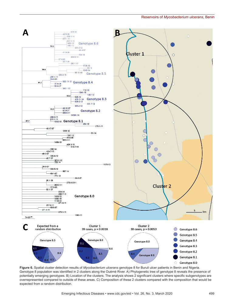
Identification and Distribution of Genetic Subgroups Belonging to Genotype 8Among the 8 genotypes identified from the West Africa lineage, genotype 8 held almost half of the M. ulcerans strains and was present in Ouémé and Plateau. We focused on this genotype and found 6 subgenotypes phylogenetically distinguished, with well-supported nodes and bootstrap values ranging from 84% to 99% (Figures 3, 6; Appendix 2 Table 3). Subgenotypes 8.1 to 8.6 contained half of the total strains belonging to genotype 8. The other half was
assigned to the denomination 8.0 as not belonging to a particular subgenotype. Because genotype 8 was found mainly near the Ouémé River, we assessed the distribution of subgenotypes in this area by using a spatial scan statistic similar to that we described pre-viously. We found 2 statistically significant clusters along the Ouémé River, 1 in the north (cluster 1) and 1 in the south (cluster 2) (Figure 6). Subgenotypes 8.1, 8.2, 8.3, and 8.5 were found only in cluster 1 (i.e., in the north (Table 2). Subgenotypes 8.1 and 8.3 had higher RRs (4.31 and 2.69, respectively) within this area compared with outside the area. The risk for carrying subgenotype 8.6 strains within this cluster was significantly low (RR 0.1) (Table 2). However, we found only 2 subgenotypes (8.4 and 8.6) in cluster 2 (RR 9.4 for subgenotype 8.6 and 1.7 for subgenotype 8.4) (Table 2). Clusters 1 and 2 (Figure 6) were slightly displaced compared with the northern and southern clusters (Figure 5) but overlapped considerably.
Land Cover and Genotype DistributionGenotypes were not distributed randomly between southern and northern Ouémé, suggesting that the clusters might be associated with specific land cover. Globally, the 2 regions differed significantly in terms of land use and cover, and a high soil heterogene-ity existed between the north and south (Figure 7). Whereas the south has flooded soils suitable for mar-ket gardening and marshes for tree cultivation, the north mainly consists of forests and palm groves, and agricultural land is sparse. This difference might in-dicate that M. ulcerans strains of genotype 7 will most likely be found in bare soils and rice fields, whereas M. ulcerans strains of genotype 8 will most likely be found in areas with riparian vegetation, herbaceous vegetation, and woodlands.
Specificity of the Nigeria StrainsThe CDTLUB in Pobè is located near the Nigeria–Be-nin border, and several BU patients from Nigeria were treated in the center. Of the 179 strains sequenced in our study, 21 were isolated from patients from Ogun State in southwestern Nigeria. This area is dependent on a different drainage basin than that used by Oué-mé, thus providing an opportunity to study M. ulcer-ans diversity and distribution in another independent BU-endemic area. Spatial analysis showed that the most significant cluster (p<0.0001) covered the area of Ogun State. This cluster was drastically differ-ent from those found in the Ouémé region of Benin. In this region of Nigeria, the RRs for infection with strains from genotype 1, 2, 3, or 6 were significantly higher than for any other genotype (Figure 8). The
496 Emerging Infectious Diseases • www.cdc.gov/eid • Vol. 26, No. 3, March 2020
Figure 4. Distribution of Mycobacterium ulcerans genotypes according to diagnosis date for Buruli ulcer patients in Benin and Nigeria. The distribution of genotypes was tested on 2 × 8 contingency tables (Fisher exact test) to compare each year to one another.

Reservoirs of Mycobacterium ulcerans, Benin
RR for infection with these genotypes was 9.9 times higher for genotype 1, 3.2 times higher for genotype 2, 5 times higher for genotype 3, and 13 times higher for genotype 6 in this area compared with outside the area (Table 1). Furthermore, the RR for infection with genotype 8 was negligible (0.2), even though it was the most widespread genotype along the Ouémé River (Table 1). We observed a similar nonrandom distribution in Ouémé and in Ogun State.
Validation of Multinomial Model Demonstrating Geographic ClustersTo validate the distribution of genotypes in both BU-endemic areas, we developed a spatial model of genotype dispersion on the basis of phylogenetic classification. To verify the power of our model to correctly associate a genotype to a genome given its
geographic origin, we performed WGS on 29 addi-tional bacterial strains. These strains were isolated from patients at CDTLUB who had been diagnosed with BU during 2015–2017 and were living in north-ern Ouémé (11 patients), southern Ouémé (9 pa-tients), Plateau (4 patients), and Nigeria (5 patients). We identified 2 of the 29 strains as part of the MU_A2 lineage and added them to the outgroup to build a new phylogenetic tree. The 27 other strains were eas-ily included in the phylogenetic tree (Figure 2) with-out altering the classification of the 8 different geno-types or the cluster classifications. Plateau contained no statistically significant clusters.
For each of the 3 significant geographic clus-ters, we compared the observed repartition of new genomes to the expected distribution given by each cluster. The 2 clusters in Ouémé exhibited strong
Emerging Infectious Diseases • www.cdc.gov/eid • Vol. 26, No. 3, March 2020 497
Figure 5. Spatial cluster detection results of Mycobacterium ulcerans genotypes for Buruli ulcer patients in Benin and Nigeria. A) Two significant areas detected along the Ouémé River. Three regions of interest are shown on the map. Two (northern Ouémé and southern Ouémé) show significant spatial clustering of genotypes; a nonsignificant area (Plateau) is given for reference. B) Composition of these 3 clusters, compared with the composition that would be expected from a random distribution.

RESEARCH
accuracy (91%) and a Matthews coefficient >0.6, cor-responding to a strong relationship (Table 3). The Nigeria cluster model showed lower accuracy (85%) and a Matthews coefficient of 0.371, corresponding to a moderately positive relationship. These results sup-port the existence of a spatial cluster of M. ulcerans genotypes in some BU-endemic areas.
DiscussionBU occurs in poor rural communities with little economic or political influence. A key epidemio-logic feature of this disease is the distribution of cases in very well-delimited foci. However, in these areas, the precise zones of high-risk contamination in environments are not identified. As with other neglected tropical diseases, fighting BU will require integrated approaches to reduce transmission of the causative mycobacterium and ensure earlier patient management.
Socioeconomic factors, environmental changes, ecologic factors, and the conquest of new territo-ries promote infections caused by pathogens with a wildlife origin (28–30). In the field of BU, all epide-miologic studies show that environmental changes, particularly wetland creation, deforestation, and so-cioeconomic factors that promote contact with non-protected water, enhance the spread of the disease (3,28,31–37). Although all epidemiologic and envi-ronmental studies underline the main role of ecologic factors in M. ulcerans transmission, the precise route of M. ulcerans transmission to humans remains un-clear. Molecular epidemiology studies conducted on a local scale can be adapted to elucidate the structure, diversity, evolution, dissemination, and life of the bacterial population.
The genome of M. ulcerans consists of a main chromosome and a giant plasmid containing the gene encoding for enzymes synthesizing the mycolactone. Because this genome has low variation, conventional genetic methods can only differentiate isolates on a continental scale (38). WGS offers a much greater resolution and could be used for studying M. ulcerans
diversity on a local scale by analyzing SNPs (11). SNP analysis of our 174 M. ulcerans isolates belonging to the West Africa lineage Mu_A1 enabled us to identify 8 genotypes on the basis of 940 SNP positions. This analysis revealed a high conservation, especially on plasmid sequences, highlighting the crucial role of mycolactone toxin to colonize specific environmental niches, including human (39,40). The main role of my-colactone in host colonization was affirmed because no link could be established between this genomic di-versity and clinical manifestations. Furthermore, the distribution of gene mutations based on a functional annotation is similar to the distribution of all the clas-sified genes of M. ulcerans (Appendix 1 Figure 3), sup-porting the hypothesis that acquisition of a mutation has no relation to its ability to colonize a host or with its virulence.
The particularity of our study was the spatial local-scale analysis of the isolates. We used a phy-logenetic analysis approach based on SNP-typing, coupled with spatial scan statistics. This method is more suitable for working in a well-defined BU-en-demic area in a short period (a few years), whereas a Bayesian phylogenetic approach is suitable for study-ing temporal distribution of M. ulcerans over a much longer period (decades) (11,15).
Our spatial analysis revealed the existence of a geographic clustering of M. ulcerans genotypes in southeastern Benin and southwestern Nigeria. On this scale, our results showed a strong association between hydrologic drainage areas and M. ulcerans genotypes, because a clear difference was observed in the distribution of genotypes between BU patients living around Nigeria’s Yewa basin and Benin’s Ouémé basin. Our clustering revealed that bacteria evolved independently and differentially, depending on their specific ecologic reservoir. Moreover (and more surprisingly), we were able to detect clustering of M. ulcerans genotypes along a same drainage basin (in this case the Ouémé basin). Inside the main geno-type (genotype 8), we were also able to detect sub-genotypes with a similar clustering along the river,
498 Emerging Infectious Diseases • www.cdc.gov/eid • Vol. 26, No. 3, March 2020
Table 1. Cluster detection analysis for predominance of Mycobacterium ulcerans genotypes using the maximum reported spatial window of 50% of the sample population and a univariate scan statistic, Benin and Nigeria*
Spatial cluster
Radius, km2 LLR Ob
Genotype 1
2
3
4
5
6
7
8
RR (%) RR (%) RR (%) RR (%) RR (%) RR (%) RR (%) RR (%) Northern Ouémé
15.7 19.2 68 0 0.6 (4.1) 0.2 (1.3) 1.5 (15) 1 (9.6) 0 0.4 (4.1) 1.9 (66) Southern Ouémé
17.9 18.8 17 1.5 (5) 1 (6) 0 0 0 0.9 (6) 20 (53) 0.6 (29) Nigeria† 54.3 27.1 20 9.9 (20) 3.2 (15) 5 (20) 0 0 13 (35) 0 0.2 (10) *Percentages indicate falling in each group within a cluster. LLR: log likelihood ratio; Ob, number of observations in a cluster; RR, relative risk, computed as the ratio of the proportions of the number of Buruli ulcer cases in each category out of the total number of cases inside the cluster versus outside. †Ogun State.
Reservoirs of Mycobacterium ulcerans, Benin
Emerging Infectious Diseases • www.cdc.gov/eid • Vol. 26, No. 3, March 2020 499
Figure 6. Spatial cluster detection results of Mycobacterium ulcerans genotype 8 for Buruli ulcer patients in Benin and Nigeria. Genotype 8 population was identified in 2 clusters along the Ouémé River. A) Phylogenetic tree of genotype 8 reveals the presence of potentially emerging genotypes. B) Location of the clusters. The analysis shows 2 significant clusters where specific subgenotypes are overrepresented compared to outside of these areas. C) Composition of these 2 clusters compared with the composition that would be expected from a random distribution.

RESEARCH
indicating dissemination of M. ulcerans on a local scale and then a persistence of M. ulcerans in indepen-dently endemic niches. These findings are consistent with previous scenarios in which M. ulcerans, once in-troduced into a new environment, expands instead of becoming a quiescent pathogen (11).
In considering the nature of the land cover, we observed striking heterogeneity along the river, pin-pointing the compartmentalization of different envi-ronmental niches (Figure 7). On the other hand, the predominance of 1 genotype in 1 area associated with a particular land cover suggests that patients frequent the same type of contamination source, and the hy-pothesis that acquisition of infection is local has al-ready been proposed (11). Altogether, our study gives a precise cartography of M. ulcerans genotype distribution, revealing a well delimited high-risk area where preventive strategies, active diagnosis, and epidemiologic surveillance must be focused.
Unlike the Ouémé region and southwestern Ni-geria, the lack of any spatial cluster in the Plateau re-gion suggests differences in terms of dissemination and environmental persistence. Plateau separates the Ouémé and Yewa draining basins, and the bacterial genotypes in the Plateau area are a mix of the geno-types in these 2 basins. This signature could be ex-plained by different hypotheses. First, there might be a contamination site different from the place of resi-dence given that persons living on the Plateau might be contaminated in Ouémé or Nigeria during their travels. This hypothesis does not explain all contami-nations because patients’ histories revealed that some of them had never left their village. Second, there might be a nonpersistent presence of M. ulcerans in the Plateau environment, in which M. ulcerans might be disseminated from Ouémé and Nigeria to Plateau by mammals (including humans) or flying insects and might be present in aquatic niches for only the few months when wetlands exist just after the rainy seasons (41). This situation rules out humans as the main carrier and reservoir of M. ulcerans and supports the position that humans with active infection are un-likely to play a major role in the bacterial ecology.
500 Emerging Infectious Diseases • www.cdc.gov/eid • Vol. 26, No. 3, March 2020
Table 2. Cluster detection analysis for predominance of subgenotypes of Mycobacterium ulcerans genotype 8, Benin*
Spatial cluster Radius, km2 LLR Ob
Subgenotype 8.0 8.1 8.2 8.3 8.4 8.5 8.6
RR (%) RR (%) RR (%) RR (%) RR (%) RR (%) RR (%) Cluster 1 14.7 15.7 39 0.7
(38.5) 4.3
(10.3) Inf
(12.8) 2.69
(12.8) 1.08
(7.7) Inf
(15.4) 0.1 (2.6)
Cluster 2 20.0 17.3 30 1.2 (53.3)
0 (0) 0 (0) 0 (0) 1.7 (10) 0 (0) 9.4 (36.7)
*Percentages indicate falling in each group within a cluster. Inf, infinite; LLR: log likelihood ratio; Ob, number of observations in a cluster; RR, relative risk, computed as the ratio of the proportions of the number of Buruli ulcer cases in each category out of the total number of cases inside the cluster versus outside.
Figure 7. Land use and land cover assessment from Sentinel-2 imaging of Benin. The Ouémé region has specific land and plant formations, such as grassy savanna, grasslands, and swamps. Soils easily become saturated with water because of a shallow water table and the proximity of a river, which causes floods and a natural delta formation in the south of the region. Circles indicate the detected northern and southern Ouémé Mycobacterium ulcerans clusters.

Reservoirs of Mycobacterium ulcerans, Benin
Moreover, BU is known to be a locally acquired infec-tion rather than an imported one (42). Thus, the most plausible hypothesis in the particular case of the Pla-teau region is based on the inability of the bacterium to develop for a period long enough in transitional aquatic reservoirs, thereby preventing the production of new genotypes. Treating humans against M. ulcer-ans infection might not be sufficient to break disease transmission chains as previously suggested (12).
Because only M. ulcerans strains isolated from pa-tients were analyzed, it would have been interesting, as a next step, to compare diversity of M. ulcerans from
Emerging Infectious Diseases • www.cdc.gov/eid • Vol. 26, No. 3, March 2020 501
Figure 8. Difference in Mycobacterium ulcerans genotype distribution between Ogun State (Nigeria) and Benin. A) Locations of patients and the genotypes of the strains. B) Composition of the cluster in the Ogun area compared with the composition that would be expected from a random distribution. The genotype distribution of the Ogun was fundamentally different from those in Benin.
Table 3. Statistical measurements of the performance of the multinomial models on a real dataset of newly sequenced genomes, Benin and Nigeria*
Locale Accuracy† Matthews correlation
coefficient‡ Northern Ouémé 91.44% 0.608* Southern Ouémé 91.73% 0.637* Nigeria (Ogun State) 86.25% 0.371** †Sum of the true positive and true negative divided by the total population. ‡Contingency matrix method of calculating the Pearson product–moment correlation coefficient (27). Its value ranges from −1 to +1 with +1 being a perfect positive prediction model, 0 no better than random prediction, and −1 being a model where predicted class is in total contradiction with observed class. Values indicated the strength of the relationship (*, strong positive relationship; **, moderate positive relationship).

RESEARCH
humans and environmental samples to better under-stand the interactions between this pathogen and the host. Combined with the tools we developed to reveal the genetic diversity of M. ulcerans in humans, DNA enrichment techniques, such those reported in stud-ies on Borrelia spp., could be improved to meet this challenge (43). In conclusion, our approach allowed the identification of delimited high-risk contamina-tion areas, paving a new avenue to develop preven-tion and intervention strategies.
AcknowledgmentsWe thank Laurent Marsollier for proofreading and advice, Tim Stinear and Jessica Porter for support with DNA sequencing, all staff at CDTLUB-Pobè in Benin who performed diagnosis and treatment, and David Goudenege and Vincent Procaccio from the Biochemistry and Genetics Department at Angers Hospital, France.
This work was carried out with support from the GenOuest bioinformatics platform.
Funding for this work was provided by Agence Nationale de la Recherche (grants nos. ANR 11 CEPL 00704 EXTRA6MU and 17-BSV3-0013-01), Fondation Raoul Follereau, INSERM, and Agence Nationale de la Recherche Région Pays de la Loire.
Use of patients’ data was approved by the CDTLUB Institutional Review Board, the national BU control authorities of Benin (approval no. IRB00006860), and the Ethics Committee of the Angers University Hospital in France (Comité d’Ethique du Centre Hospitalo-Universitaire d’Angers).
About the AuthorDr. Coudereau is a former postdoctoral fellow at the Centre de Recherche en Cancérologie et Immunologie Nantes–Angers (CRCINA) pursuing a career in bioinformatics. His research interests include next- generation sequencing, phylogeny, and spatial analysis related to the genetic population of M. ulcerans in endemic regions of Africa.
References 1. Sizaire V, Nackers F, Comte E, Portaels F. Mycobacterium
ulcerans infection: control, diagnosis, and treatment. Lancet Infect Dis. 2006;6:288–96. https://doi.org/10.1016/ S1473-3099(06)70464-9
2. Asiedu K, Etuaful S. Socioeconomic implications of Buruli ulcer in Ghana: a three-year review. Am J Trop Med Hyg. 1998;59:1015–22. https://doi.org/10.4269/ajtmh.1998.59.1015
3. Aiga H, Amano T, Cairncross S, Adomako J, Nanas OK, Coleman S. Assessing water-related risk factors for Buruli
ulcer: a case-control study in Ghana. Am J Trop Med Hyg. 2004;71:387–92. https://doi.org/10.4269/ ajtmh.2004.71.387
4. Merritt RW, Walker ED, Small PL, Wallace JR, Johnson PD, Benbow ME, et al. Ecology and transmission of Buruli ulcer disease: a systematic review. PLoS Negl Trop Dis. 2010;4:e911. https://doi.org/10.1371/journal.pntd.0000911
5. Wallace JR, Mangas KM, Porter JL, Marcsisin R, Pidot SJ, Howden B, et al. Mycobacterium ulcerans low infectious dose and mechanical transmission support insect bites and puncturing injuries in the spread of Buruli ulcer. PLoS Negl Trop Dis. 2017;11:e0005553. https://doi.org/10.1371/ journal.pntd.0005553
6. Williamson HR, Mosi L, Donnell R, Aqqad M, Merritt RW, Small PL. Mycobacterium ulcerans fails to infect through skin abrasions in a guinea pig infection model: implications for transmission. PLoS Negl Trop Dis. 2014;8:e2770. https://doi.org/10.1371/journal.pntd.0002770
7. Johnson PD, Azuolas J, Lavender CJ, Wishart E, Stinear TP, Hayman JA, et al. Mycobacterium ulcerans in mosquitoes captured during outbreak of Buruli ulcer, southeastern Australia. Emerg Infect Dis. 2007;13:1653–60. https://doi.org/10.3201/eid1311.061369
8. Marsollier L, Robert R, Aubry J, Saint André JP, Kouakou H, Legras P, et al. Aquatic insects as a vector for Mycobacterium ulcerans. Appl Environ Microbiol. 2002;68:4623–8. https://doi.org/10.1128/AEM.68.9.4623-4628.2002
9. Portaels F, Elsen P, Guimaraes-Peres A, Fonteyne PA, Meyers WM. Insects in the transmission of Mycobacterium ulcerans infection. Lancet. 1999;353:986. https://doi.org/ 10.1016/S0140-6736(98)05177-0
10. van der Werf TS, Stinear T, Stienstra Y, van der Graaf WT, Small PL. Mycolactones and Mycobacterium ulcerans disease. Lancet. 2003;362:1062–4. https://doi.org/10.1016/ S0140-6736(03)14417-0
11. Buultjens AH, Vandelannoote K, Meehan CJ, Eddyani M, de Jong BC, Fyfe JAM, et al. Comparative genomics shows that Mycobacterium ulcerans migration and expansion preceded the rise of Buruli ulcer in southeastern Australia. Appl Environ Microbiol. 2018;84:e02612–7. https://doi.org/ 10.1128/AEM.02612-17
12. Vandelannoote K, Phanzu DM, Kibadi K, Eddyani M, Meehan CJ, Jordaens K, et al. Mycobacterium ulcerans population genomics to inform on the spread of Buruli ulcer across central Africa. MSphere. 2019;4:e00472–18. https://doi.org/10.1128/mSphere.00472-18
13. Wu J, Tschakert P, Klutse E, Ferring D, Ricciardi V, Hausermann H, et al. Buruli ulcer disease and its association with land cover in southwestern Ghana. PLoS Negl Trop Dis. 2015;9:e0003840. https://doi.org/10.1371/journal.pntd.0003840
14. Bolz M, Bratschi MW, Kerber S, Minyem JC, Um Boock A, Vogel M, et al. Locally confined clonal complexes of Mycobacterium ulcerans in two Buruli ulcer endemic regions of Cameroon. PLoS Negl Trop Dis. 2015;9:e0003802. https://doi.org/10.1371/journal.pntd.0003802
15. Vandelannoote K, Meehan CJ, Eddyani M, Affolabi D, Phanzu DM, Eyangoh S, et al. Multiple introductions and recent spread of the emerging human pathogen Mycobacterium ulcerans across Africa. Genome Biol Evol. 2017;9:414–26. https://doi.org/10.1093/gbe/evx003
16. Kambarev S, Corvec S, Chauty A, Marion E, Marsollier L, Pecorari F. Draft genome sequence of Mycobacterium ulcerans S4018 isolated from a patient with an active Buruli ulcer in Benin, Africa. Genome Announc. 2017;5:e00248–17. https://doi.org/10.1128/genomeA.00248-17
502 Emerging Infectious Diseases • www.cdc.gov/eid • Vol. 26, No. 3, March 2020

Reservoirs of Mycobacterium ulcerans, Benin
17. Andrew S. FastQC: a quality control tool for high throughput sequence data. 2010 [cited 2019 Apr 1].
18. Bolger AM, Lohse M, Usadel B. Trimmomatic: a flexible trimmer for Illumina sequence data. Bioinformatics. 2014;30:2114–20. https://doi.org/10.1093/bioinformatics/btu170
19. Seemann T. Snippy: fast bacterial variant calling from NGS reads. 2015 [cited 2019 Apr 1].
20. Li H, Durbin R. Fast and accurate short read alignment with Burrows-Wheeler transform. Bioinformatics. 2009;25:1754–60. https://doi.org/10.1093/bioinformatics/btp324
21. Stinear TP, Seemann T, Pidot S, Frigui W, Reysset G, Garnier T, et al. Reductive evolution and niche adaptation inferred from the genome of Mycobacterium ulcerans, the causative agent of Buruli ulcer. Genome Res. 2007;17:192–200. https://doi.org/10.1101/gr.5942807
22. Guindon S, Dufayard JF, Lefort V, Anisimova M, Hordijk W, Gascuel O. New algorithms and methods to estimate maximum-likelihood phylogenies: assessing the performance of PhyML 3.0. Syst Biol. 2010;59:307–21. https://doi.org/10.1093/sysbio/syq010
23. Hodcroft E. TreeCollapserCL. 2012 [cited 2019 Apr 1].24. Kulldorff M. SatScan: software for the spatial and space-time
scan statistics. 2019 [cited 2019 Apr 1].25. Team QGIS Development. QGIS Geographic Information
System. Project OSGF. 2019 [cited 2019 Apr 1].26. R Core Team. R: a language and environment for statistical
computing. Computing RFfS. 2019 [cited 2019 Apr 1].27. Powers DMW. Evaluation: from precision, recall and
F-Measure to ROC, informedness, markedness, and correlation. J Mach Learn Technol. 2011;2:37–63.
28. Bennett SD, Lowther SA, Chingoli F, Chilima B, Kabuluzi S, Ayers TL, et al. Assessment of water, sanitation and hygiene interventions in response to an outbreak of typhoid fever in Neno District, Malawi. PLoS One. 2018;13:e0193348. https://doi.org/10.1371/journal.pone.0193348
29. Davies HG, Bowman C, Luby SP. Cholera - management and prevention. J Infect. 2017;74(Suppl 1):S66–73. https://doi.org/10.1016/S0163-4453(17)30194-9
30. Mitjà O, Marks M, Bertran L, Kollie K, Argaw D, Fahal AH, et al. Integrated control and management of neglected tropical skin diseases. PLoS Negl Trop Dis. 2017;11:e0005136. https://doi.org/10.1371/journal.pntd.0005136
31. Brou T, Broutin H, Elguero E, Asse H, Guegan JF. Landscape diversity related to Buruli ulcer disease in Côte d’Ivoire. PLoS Negl Trop Dis. 2008;2:e271. https://doi.org/10.1371/journal.pntd.0000271
32. Landier J, Gaudart J, Carolan K, Lo Seen D, Guégan JF, Eyangoh S, et al. Spatio-temporal patterns and landscape-associated risk of Buruli ulcer in Akonolinga, Cameroon. PLoS Negl Trop Dis. 2014;8:e3123. https://doi.org/10.1371/journal.pntd.0003123
33. Marion E, Landier J, Boisier P, Marsollier L, Fontanet A, Le Gall P, et al. Geographic expansion of Buruli ulcer disease, Cameroon. Emerg Infect Dis. 2011;17:551–3. https://doi.org/10.3201/eid1703.091859
34. Nackers F, Johnson RC, Glynn JR, Zinsou C, Tonglet R, Portaels F. Environmental and health-related risk factors for Mycobacterium ulcerans disease (Buruli ulcer) in Benin. Am J Trop Med Hyg. 2007;77:834–6. https://doi.org/10.4269/ajtmh.2007.77.834
35. Pouillot R, Matias G, Wondje CM, Portaels F, Valin N, Ngos F, et al. Risk factors for buruli ulcer: a case control study in Cameroon. PLoS Negl Trop Dis. 2007;1:e101. https://doi.org/10.1371/journal.pntd.0000101
36. Raghunathan PL, Whitney EA, Asamoa K, Stienstra Y, Taylor TH Jr, Amofah GK, et al. Risk factors for Buruli ulcer disease (Mycobacterium ulcerans Infection): results from a case-control study in Ghana. Clin Infect Dis. 2005;40:1445–53. https://doi.org/10.1086/429623
37. Wagner T, Benbow ME, Burns M, Johnson RC, Merritt RW, Qi J, et al. A Landscape-based model for predicting Mycobacterium ulcerans infection (Buruli Ulcer disease) presence in Benin, West Africa. EcoHealth. 2008;5:69–79. https://doi.org/10.1007/s10393-007-0148-7
38. Röltgen K, Stinear TP, Pluschke G. The genome, evolution and diversity of Mycobacterium ulcerans. Infect Genet Evol. 2012;12:522–9. https://doi.org/10.1016/j.meegid.2012.01.018
39. Demangel C, Stinear TP, Cole ST. Buruli ulcer: reductive evolu-tion enhances pathogenicity of Mycobacterium ulcerans. Nat Rev Microbiol. 2009;7:50–60. https://doi.org/10.1038/nrmicro2077
40. Rondini S, Käser M, Stinear T, Tessier M, Mangold C, Dernick G, et al. Ongoing genome reduction in Mycobacterium ulcerans. Emerg Infect Dis. 2007;13:1008–15. https://doi.org/10.3201/eid1307.060205
41. Yerramilli A, Tay EL, Stewardson AJ, Fyfe J, O’Brien DP, Johnson PDR. The association of rainfall and Buruli ulcer in southeastern Australia. PLoS Negl Trop Dis. 2018;12:e0006757. https://doi.org/10.1371/ journal.pntd.0006757
42. Tanser FC, Le Sueur D. The application of geographical information systems to important public health problems in Africa. Int J Health Geogr. 2002;1:4. https://doi.org/ 10.1186/1476-072X-1-4
43. Carpi G, Walter KS, Bent SJ, Hoen AG, Diuk-Wasser M, Caccone A. Whole genome capture of vector-borne pathogens from mixed DNA samples: a case study of Borrelia burgdorferi. BMC Genomics. 2015;16:434. https://doi.org/10.1186/s12864-015-1634-x
Address for correspondence: Estelle Marion, ATOMycA Team, CRCINA Inserm U1232, 4 rue Larrey, 49933 Angers, France; email: [email protected]
Emerging Infectious Diseases • www.cdc.gov/eid • Vol. 26, No. 3, March 2020 503

Page 1 of 4
Article DOI: https://doi.org/10.3201/eid2603.190573
Stable and Local Reservoirs of Mycobacterium ulcerans Inferred from the
Nonrandom Distribution of Bacterial Genotypes, Benin
Appendix 1
Supplementary Methods
Bacterial Isolates and Patients
This study was performed by working closely with the CDTLUB of Pobè. This
specialized hospital has been actively monitoring the disease since 2006 and has collected
substantial epidemiologic data (age, sex, village/city of residence, etc.). Indeed, between 2006
and 2015, the CDTLUB of Pobe diagnosed and treated 1761 PCR-confirmed Buruli ulcer cases
coming from Oueme (63%), Plateau (28%) and Nigeria (9%).
Genome Sequencing
DNA sequencing of 179 samples was undertaken using either an Illumina MiSeq or
HiSeq with Nextera XT DNA preparation kit (Illumina, San Diego, California) and 29 strains
was performed using Ion Torrent S5XL with IonXpress Plus Fragment Library kit (Life
Technologies, California). The quality of raw reads was assessed using FastQC v0.11.7 (1).
DNA sequences have been submitted to the public Sequence Read Archive (SRA) -NCBI open
access database. All genomes possessed a mean sequencing coverage superior to 30 reads and
less than 20% of the bacterial chromosome was covered by less than 15 reads for efficient
variant discovery. Prior to further analysis, reads were cleaned using Trimmomatic v0.36 (2).
Reads were filtered to remove potentials adapters, ambiguous and unidentified base calls or any
reads inferior to 35bp as well as portion of reads whose average quality drops below 15 on a 4-
base wide sliding window. During mapping of reads on the reference genome, the repetitive and
ubiquitous IS2404 and IS2606 genomic elements were hard-masked and consequently eliminated
because of the unreliability of mapping on repetitive and mobile regions.

Page 2 of 4
Multinomial Spatial Scan Statistic
The method uses a circular window of varying radius centers at each location that moves
across the map so that, at any given position, the window includes different sets of neighboring
residencies. At each position, the radius of the circular window varies repeatedly from zero up to
a predefined maximum radius, corresponding to a window including 50% of the total study
population. This method allows the circular window to continuously vary in both location and
size, thereby creating a large number of distinct potential clusters. The detection of clusters was
performed by comparing the number of cases within the window with the number expected if
cases were randomly distributed in space, using a multinomial model (3). The unit of space was
defined by the coordinates of the patient's villages. Clusters were scanned where specific
genotypes of M. ulcerans were more or less prominent, i.e., areas where the observed number of
cases differs from the expected number of cases. The significance of the identified clusters was
tested with a likelihood ratio test and statistical inference for the spatial clusters was evaluated
based on a Monte Carlo hypothesis test with 999 replications.
Statistical Analysis and Performance Metrics
Comparisons of the expected genotype distribution inside each statistically significant
clusters found in SatScan with the genotype distribution of the second set of sequencing were
performed by calculating accuracy and Matthews correlation coefficient on confusion matrices.
Accuracy measures the fraction of all instances that are correctly categorized while Matthews
correlation coefficient summarizes the overall correlation like the Pearson correlation coefficient
(4) and thus possesses the same interpretation (5).
References
1. Andrews S. FastQC: a quality control tool for high throughput sequence data. 2010 [cited 2019 Apr 1].
https://www.bioinformatics.babraham.ac.uk/projects/fastqc
2. Bolger AM, Lohse M, Usadel B. Trimmomatic: a flexible trimmer for Illumina sequence data.
Bioinformatics. 2014;30:2114–20. PubMed https://doi.org/10.1093/bioinformatics/btu170
3. Jung I, Kulldorff M, Richard OJ. A spatial scan statistic for multinomial data. Stat Med. 2010;29:1910–
8. PubMed https://doi.org/10.1002/sim.3951

Page 3 of 4
4. Matthews BW. Comparison of the predicted and observed secondary structure of T4 phage lysozyme.
Biochim Biophys Acta. 1975;405:442–51. PubMed https://doi.org/10.1016/0005-2795(75)90109-
9
5. Powers DMW. Evaluation: from precision, recall and F-Measure to ROC, informedness, markedness,
and correlation. J Mach Learn Technol. 2011;2:37–63.
Appendix 1 Figure 1. Phylogenetic tree showing the relationship between the genomes from our study
and genomes obtained from two other studies performed in central and West Africa. Genomes from the
Mu_A2 lineage were obtained from the NCBI Sequence Read Archive, accessible under the BioProject
accession PRJNA313185, declares country of origin is precised for each genome. The genomes from the
Agogo lineages were obtained from the BioProject PRJEB8235 and are all coming from Ghana.
Page 4 of 4
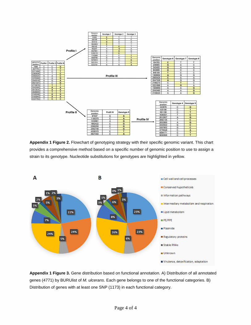
Appendix 1 Figure 2. Flowchart of genotyping strategy with their specific genomic variant. This chart
provides a comprehensive method based on a specific number of genomic position to use to assign a
strain to its genotype. Nucleotide substitutions for genotypes are highlighted in yellow.
Appendix 1 Figure 3. Gene distribution based on functional annotation. A) Distribution of all annotated
genes (4771) by BURUlist of M. ulcerans. Each gene belongs to one of the functional categories. B)
Distribution of genes with at least one SNP (1173) in each functional category.
Related Documents