Investigating the Role of Free-living Amoebae as a Reservoir for Mycobacterium ulcerans Nana Ama Amissah 1 *, Sophie Gryseels 2 , Nicholas J. Tobias 3 , Bahram Ravadgar 4 , Mitsuko Suzuki 5 , Koen Vandelannoote 2,6 , Lies Durnez 6 , Herwig Leirs 2 , Timothy P. Stinear 3,4 , Franc ¸oise Portaels 6 , Anthony Ablordey 1 , Miriam Eddyani 6 1 Bacteriology Department, Noguchi Memorial Institute for Medical Research, Accra, Ghana, 2 Evolutionary Ecology Group, Department of Biology, University of Antwerp, Antwerp, Belgium, 3 Department of Microbiology, University of Melbourne, Melbourne, Victoria, Australia, 4 Department of Microbiology, Monash University, Victoria, Australia, 5 Parasitology Department, Noguchi Memorial Institute for Medical Research, Accra, Ghana, 6 Department of Biomedical Sciences, Institute of Tropical Medicine, Antwerp, Belgium Abstract Background: The reservoir and mode of transmission of Mycobacterium ulcerans, the causative agent of Buruli ulcer, still remain a mystery. It has been suggested that M. ulcerans persists with difficulty as a free-living organism due to its natural fragility and inability to withstand exposure to direct sunlight, and thus probably persists within a protective host environment. Methodology/Principal Findings: We investigated the role of free-living amoebae as a reservoir of M. ulcerans by screening the bacterium in free-living amoebae (FLA) cultures isolated from environmental specimens using real-time PCR. We also followed the survival of M. ulcerans expressing green fluorescence protein (GFP) in Acanthameoba castellanii by flow cytometry and observed the infected cells using confocal and transmission electron microscopy for four weeks in vitro. IS2404 was detected by quantitative PCR in 4.64% of FLA cultures isolated from water, biofilms, detritus and aerosols. While we could not isolate M. ulcerans, 23 other species of mycobacteria were cultivated from inside FLA and/or other phagocytic microorganisms. Laboratory experiments with GFP-expressing M. ulcerans in A. castellani trophozoites for 28 days indicated the bacteria did not replicate inside amoebae, but they could remain viable at low levels in cysts. Transmission electron microscopy of infected A. castellani confirmed the presence of bacteria within both trophozoite vacuoles and cysts. There was no correlation of BU notification rate with detection of the IS2404 in FLA (r = 0.07, n = 539, p = 0.127). Conclusion/Significance: This study shows that FLA in the environment are positive for the M. ulcerans insertion sequence IS2404. However, the detection frequency and signal strength of IS2404 positive amoabae was low and no link with the occurrence of BU was observed. We conclude that FLA may host M. ulcerans at low levels in the environment without being directly involved in the transmission to humans. Citation: Amissah NA, Gryseels S, Tobias NJ, Ravadgar B, Suzuki M, et al. (2014) Investigating the Role of Free-living Amoebae as a Reservoir for Mycobacterium ulcerans. PLoS Negl Trop Dis 8(9): e3148. doi:10.1371/journal.pntd.0003148 Editor: Pamela L. C. Small, University of Tennessee, United States of America Received June 11, 2014; Accepted July 25, 2014; Published September 4, 2014 Copyright: ß 2014 Amissah et al. This is an open-access article distributed under the terms of the Creative Commons Attribution License, which permits unrestricted use, distribution, and reproduction in any medium, provided the original author and source are credited. Data Availability: The authors confirm that all data underlying the findings are fully available without restriction. All relevant data are within the paper and its Supporting Information files. Funding: This study was supported by the Flemish Interuniversity Council – University Development Cooperation (VLIR-UOS) and by the Stop Buruli Initiative funded by the UBS Optimus Foundation (Zurich, Switzerland). SG was an FWO PhD fellow (1.1.671.10.N.00) during part of this study. The funders had no role in study design, data collection and analysis, decision to publish, or preparation of the manuscript. Competing Interests: The authors have declared that no competing interests exist. * Email: [email protected] Introduction Mycobacterium ulcerans is a slow growing environmental pathogen responsible for a necrotizing cutaneous infection called Buruli ulcer (BU). The disease has been reported in over 30 countries worldwide mainly in tropical and subtropical climates and emerged as an increasing cause of morbidity in endemic rural communities in some West and Central African countries with Benin, Co ˆte d’Ivoire and Ghana bearing the highest burden of disease [1]. Most BU endemic areas are found close to slow flowing or stagnant water bodies and it is therefore assumed that the aquatic ecosystem may be a source of M. ulcerans from which the bacterium is transmitted to humans. This is supported by several studies that have detected M. ulcerans DNA sequences in a variety of environmental specimens including fish, snails, detritus, biofilms, soil, water filtrands, insects and protozoa [2–5]. Recently in Australia, M. ulcerans DNA has been detected in mosquitoes, faecal matter and skin lesions of small terrestrial mammals (ringtail and brushtail possums) that are thought to harbor and vector the PLOS Neglected Tropical Diseases | www.plosntds.org 1 September 2014 | Volume 8 | Issue 9 | e3148

Welcome message from author
This document is posted to help you gain knowledge. Please leave a comment to let me know what you think about it! Share it to your friends and learn new things together.
Transcript
Investigating the Role of Free-living Amoebae as aReservoir for Mycobacterium ulceransNana Ama Amissah1*, Sophie Gryseels2, Nicholas J. Tobias3, Bahram Ravadgar4, Mitsuko Suzuki5,
Koen Vandelannoote2,6, Lies Durnez6, Herwig Leirs2, Timothy P. Stinear3,4, Francoise Portaels6,
Anthony Ablordey1, Miriam Eddyani6
1 Bacteriology Department, Noguchi Memorial Institute for Medical Research, Accra, Ghana, 2 Evolutionary Ecology Group, Department of Biology, University of Antwerp,
Antwerp, Belgium, 3 Department of Microbiology, University of Melbourne, Melbourne, Victoria, Australia, 4 Department of Microbiology, Monash University, Victoria,
Australia, 5 Parasitology Department, Noguchi Memorial Institute for Medical Research, Accra, Ghana, 6 Department of Biomedical Sciences, Institute of Tropical Medicine,
Antwerp, Belgium
Abstract
Background: The reservoir and mode of transmission of Mycobacterium ulcerans, the causative agent of Buruli ulcer, stillremain a mystery. It has been suggested that M. ulcerans persists with difficulty as a free-living organism due to its naturalfragility and inability to withstand exposure to direct sunlight, and thus probably persists within a protective hostenvironment.
Methodology/Principal Findings: We investigated the role of free-living amoebae as a reservoir of M. ulcerans by screeningthe bacterium in free-living amoebae (FLA) cultures isolated from environmental specimens using real-time PCR. We alsofollowed the survival of M. ulcerans expressing green fluorescence protein (GFP) in Acanthameoba castellanii by flowcytometry and observed the infected cells using confocal and transmission electron microscopy for four weeks invitro. IS2404 was detected by quantitative PCR in 4.64% of FLA cultures isolated from water, biofilms, detritus and aerosols.While we could not isolate M. ulcerans, 23 other species of mycobacteria were cultivated from inside FLA and/or otherphagocytic microorganisms. Laboratory experiments with GFP-expressing M. ulcerans in A. castellani trophozoites for 28days indicated the bacteria did not replicate inside amoebae, but they could remain viable at low levels in cysts.Transmission electron microscopy of infected A. castellani confirmed the presence of bacteria within both trophozoitevacuoles and cysts. There was no correlation of BU notification rate with detection of the IS2404 in FLA (r = 0.07, n = 539,p = 0.127).
Conclusion/Significance: This study shows that FLA in the environment are positive for the M. ulcerans insertion sequenceIS2404. However, the detection frequency and signal strength of IS2404 positive amoabae was low and no link with theoccurrence of BU was observed. We conclude that FLA may host M. ulcerans at low levels in the environment without beingdirectly involved in the transmission to humans.
Citation: Amissah NA, Gryseels S, Tobias NJ, Ravadgar B, Suzuki M, et al. (2014) Investigating the Role of Free-living Amoebae as a Reservoir for Mycobacteriumulcerans. PLoS Negl Trop Dis 8(9): e3148. doi:10.1371/journal.pntd.0003148
Editor: Pamela L. C. Small, University of Tennessee, United States of America
Received June 11, 2014; Accepted July 25, 2014; Published September 4, 2014
Copyright: � 2014 Amissah et al. This is an open-access article distributed under the terms of the Creative Commons Attribution License, which permitsunrestricted use, distribution, and reproduction in any medium, provided the original author and source are credited.
Data Availability: The authors confirm that all data underlying the findings are fully available without restriction. All relevant data are within the paper and itsSupporting Information files.
Funding: This study was supported by the Flemish Interuniversity Council – University Development Cooperation (VLIR-UOS) and by the Stop Buruli Initiativefunded by the UBS Optimus Foundation (Zurich, Switzerland). SG was an FWO PhD fellow (1.1.671.10.N.00) during part of this study. The funders had no role instudy design, data collection and analysis, decision to publish, or preparation of the manuscript.
Competing Interests: The authors have declared that no competing interests exist.
* Email: [email protected]
Introduction
Mycobacterium ulcerans is a slow growing environmental
pathogen responsible for a necrotizing cutaneous infection called
Buruli ulcer (BU). The disease has been reported in over 30
countries worldwide mainly in tropical and subtropical climates
and emerged as an increasing cause of morbidity in endemic rural
communities in some West and Central African countries with
Benin, Cote d’Ivoire and Ghana bearing the highest burden of
disease [1].
Most BU endemic areas are found close to slow flowing or
stagnant water bodies and it is therefore assumed that the aquatic
ecosystem may be a source of M. ulcerans from which the
bacterium is transmitted to humans. This is supported by several
studies that have detected M. ulcerans DNA sequences in a variety
of environmental specimens including fish, snails, detritus,
biofilms, soil, water filtrands, insects and protozoa [2–5]. Recently
in Australia, M. ulcerans DNA has been detected in mosquitoes,
faecal matter and skin lesions of small terrestrial mammals (ringtail
and brushtail possums) that are thought to harbor and vector the
PLOS Neglected Tropical Diseases | www.plosntds.org 1 September 2014 | Volume 8 | Issue 9 | e3148
bacterium [6,7]. However, the main reservoir and modes of
transmission of BU outside Australia still remain unknown.
Since the discovery that Legionella pneumophila is able to infect
and replicate in free-living amoebae (FLA) [8], there has been an
increasing number of studies on the role of FLA in the survival of
pathogenic organisms [9]. Also, several species of mycobacteria
(M. shottsii, M. pseudoshottsii, M. tuberculosis, M. leprae, M.ulcerans, M. marinum, M. bovis, M. avium subsp paratuberculosisand M. avium) have been shown to survive within protozoa [4,10–
16]. M. ulcerans bears characteristic genomic signatures that are
typical of host restricted pathogens suggesting that M. ulcerans is
unlikely to be free-living in the environment but is instead
undergoing or has undergone adaptation to a specific ecological
niche [17]. Internalization of infectious agents inside other
parasites is a recurring theme in biology and represents an
evolutionary strategy for survival that may sometimes enhance
pathogenesis or transmissibility [18]: Bacteria ‘‘hidden’’ in their
protozoan hosts may more easily infect vertebrate end hosts,
multiplying within protozoans to escape immune reactions
[15,18].
Water bodies in areas of high BU endemicity have been
reported to contain significantly more FLA than in low endemic
areas [19]. Recently, we demonstrated that M. ulcerans can be
phagocytosed in vitro by Acanthamoeba polyphaga and persist for
at least 2 weeks [4]. This study also showed a higher detection
frequency of the IS2404 target in FLA cultures as compared to
crude samples from the environment. The aim of the present study
was to further explore FLA as a reservoir for M. ulcerans by
screening M. ulcerans in FLA from aquatic environment sampled
for 10 months and relating this to the BU notification rate in the
same endemic area. Furthermore, we experimentally investigated
the ability of M. ulcerans to survive and replicate within A.castellanii by infecting these amoebae with M. ulcerans expressing
green fluorescence protein (GFP).
Materials and Methods
Study sites and specimen collectionThe study was carried out in five endemic communities (with
recorded human BU cases): Ananekrom, Nshyieso, Serebouso,
Dukusen and Bebuso, and two non-endemic communities (no
recorded human BU cases): Mageda and Pataban in the Asante
Akim North Municipal of Ghana (Table 1, Fig. 1). These
communities are on average 18 km apart and were selected based
on number of BU cases reported at the Agogo Presbyterian
Hospital (APH), the Municipal health facility serving all commu-
nities (Fig. 1). Week-long monthly field visits were made for 10
months between October 2008 and July 2009 to randomly collect
environmental specimens: water, biofilm from plants and tree
trunks, detritus and aerosols. The specimens were taken between
6:00 am and 8:00 am, the peak period of human contact activities
in the water bodies. Biofilms (n = 428) were taken by scraping
surfaces of tree trunks, floating logs and tree stumps with sterile
scalpels and cotton swabs into 50 mL sterile Falcon tubes. Detritus
(n = 45) were scooped by hand into 50 mL sterile Falcon tubes.
Water specimens (n = 53) were taken from mid column with
buckets of which 3–10 liters were concentrated via 0.45 mm
membrane filters (Sartorius Stedim Biotech GmbH, Germany)
depending on the turbidity of the water. During the last two
months of sampling, non-nutrient agar (NNA) plates (n = 13)
seeded with Escherichia coli were exposed for 30 minutes for
isolation of FLA from aerosols generated next to the water bodies.
Specimen processingSeven milliliters phosphate buffered saline (PBS) were added to
the membrane filters (cut into smaller pieces), swabs and scalpels
contained in 50 mL Falcon tubes and shaken vigorously to
dislodge the substrate and biofilms from the surface. For detritus,
specimens were processed as described by Gryseels et al. [4].
Isolation of FLAA piece of the membrane filter and 2 to 3 drops of the
suspensions (biofilms and detritus) were inoculated at the centre of
1.5% NNA plates seeded with E. coli for cultivation of FLA [20] at
28.5uC. The inoculated NNA plates were examined daily for the
presence of trophozoites and cysts using the 106 objective of a
bright field microscope. When FLA were observed, they were
subcultured on new NNA plates seeded with E. coli [21]. After 3
or 4 subcultures, the FLA were harvested by scraping the surface
with an inoculating loop and suspending them in 1.5 mL sterile
distilled water.
DNA preparationThe modified Boom method was used for the extraction of
DNA from FLA cultures as described previously [22,23].
Detection of M. ulcerans DNATwo multiplex real-time PCR assays were performed on the
DNA extracts to test the presence of three distinct sequences:
IS2404, IS2606 and the ketoreductase B (KRB) domain in the M.ulcerans genome as described by Fyfe et al. [24]. All DNA extracts
were first screened for the IS2404 target multiplexed with an
internal positive control to check for PCR inhibitors such as humic
and fulvic acids (commonly found in environmental specimens)
[24]. The second PCR assay for detecting the presence of IS2606and KRB was done on FLA cultures that turned out positive for
the IS2404 target. Amplification and detection was carried out
using the 7500 real-time PCR system (Applied Biosystems).
Identification of FLAThe identification of FLA of the genera Acanthamoeba,
Naegleria and Vahlkampfiidae was confirmed using the primer
sets JDP1/JDP2, ITSfw/ITSrv and JITSfw/JITSrv respectively as
described by Gryseels et al. [4] and Eddyani et al. [19].
Author Summary
Mycobacterium ulcerans, the causative agent of Buruli ulcer(BU) is an environmental pathogen known to reside inaquatic habitat. However, the reservoir and modes oftransmission to humans still remain unknown. M. ulceranscan probably not live freely due to its natural fragility andinability to withstand exposure to direct sunlight. Thisstudy investigated the hypothesis that free-living amoebae(FLA) can serve as a reservoir of M. ulcerans by testing forits presence in amoebae isolated from water bodies in BUendemic and non-endemic communities and whether thepathogen can remain viable when experimentally infectedin amoebae in the laboratory. We detected only one(IS2404) of the three (IS2606 and KRB) targets for thepresence of M. ulcerans in amoebae cultures and found nocorrelation between its presence in the environment andBU notification rate. M. ulcerans remained viable at lowlevels in amoebae for 28 days in vitro. We thereforeconclude that FLA may host M. ulcerans at low levels in theenvironment without being directly involved in thetransmission to humans.
FLA Reservoir of Mycobacterium ulcerans
PLOS Neglected Tropical Diseases | www.plosntds.org 2 September 2014 | Volume 8 | Issue 9 | e3148

Isolation of M. ulcerans and other intracellularmycobacteria from specimens
Specimens were processed to kill extracellular mycobacteria as
described by Gryseels et al. [4], decontaminated using the oxalic
acid method [25], inoculated on LJ medium, and incubated at
30uC with weekly examination for a year. Supernatants containing
extracellular mycobacteria were cultured for week at the same
temperature to monitor the effectiveness of the isolation method.
Identification of intracellular mycobacteriaDirect smear examination (DSE) by ZN staining for acid fast
bacilli (AFB) was performed on single colonies grown on LJ media.
Cultures positive for AFB were subjected to a nested PCR specific
for the mycobacterial 16S rRNA gene [26,27] after heat-
inactivation of mycobacterial suspensions in TE for 10 minutes.
Amplicons were sequenced by the VIB Genetic Service Facility
(Antwerp, Belgium). The sequenced data were compared with
Figure 1. Distribution of the IS2404 target and intracellular mycobacteria from the sampling sites.doi:10.1371/journal.pntd.0003148.g001
FLA Reservoir of Mycobacterium ulcerans
PLOS Neglected Tropical Diseases | www.plosntds.org 3 September 2014 | Volume 8 | Issue 9 | e3148
known sequences in the GenBank database and interpreted using
the BlastN algorithm (available on http://www.ncbi.nlm.nih.gov/
BLAST/). The 16S rRNA sequences were also matched against
entries in the RIDOM (Ribosomal Differentiation of Medical
Microorganisms) database (http://www.ridom-rdna.de/). Direct
detection of mycobacterial DNA in FLA cultures was done using
the same 16S rRNA PCR assay.
Infection of A. castellanii with M. ulceransM. ulcerans strains JKD8083 (which expresses GFP) and
04126204 (which does not express GFP) were grown in 7H9
broth or 7H10 agar supplemented with OADC (Difco). Real-time
PCR was performed on M. ulcerans strains to estimate cell
numbers as previously described [28]. Primers targeting the 16S
rRNA gene were used for detection of mycobacteria. Known
amounts of M. ulcerans Agy99 genomic DNA were used to
construct a standard curve and cell numbers were estimated based
on the predicted mass of an M. ulcerans chromosome [24].
A. castellanii was cultured in peptone-yeast extract-glucose
(PYG) medium at 22uC in the dark as described by Moffat and
Tompkins [29]. Trophozoites were harvested at 4006 g for
10 minutes (Eppendorf, 5810R) and adjusted to a final concen-
tration of 106 cells ml21 in Acanthamoebae (AC) buffer as
described [29].
Bacterial strains were concentrated by centrifugation at 60006g for 15 minutes at room temperature and then resuspended in
AC buffer. A preparation of 16106 cells of A. castellanii was
mixed with 16106 cells of either M. ulcerans (JKD8083) or
(04126204) in 20 mL PYG broth and incubated at 22uC for
30 minutes. Co-cultures were washed three times in AC buffer and
treated with amikacin (150 mg/ml) as described [30] to kill
extracellular bacteria. Trophozoites were then washed and
resuspended in 50 mL AC buffer. Three milliliters samples were
taken at 1, 2, 7, 14, 21 and 28 days for analysis.
Flow cytometryAt each time point, samples were washed with FACS buffer six
times before a final resuspension in 500 ml of FACS buffer. FACS
was carried out using uninfected amoebae to identify trophozoite
populations. The subsequent infected samples were gated only on
these relevant populations. All samples, including the controls
(uninfected amoebae and bacteria only) were analyzed with a
FACS (Becton Dickinson) equipped with a 488 nm argon laser. At
each time point 50,000 events were counted. Background
fluorescence was determined by using infections of A. castellaniiwith non-fluorescent M. ulcerans (04126204). Percentages of
fluorescing amoebae were then calculated using Flowjo (v8.7).
Confocal and electron microscopyAliquots of infected amoebae in AC buffer were again pelleted
and washed in 16 PBS before DAPI staining according to the
manufacturer’s instructions (Invitrogen). Samples were imaged
using a LAS700 confocal microscope (Zeiss) with a 1006 oil
immersion lens.
At different time points 1 mL aliquots were used to examine for
M. ulcerans within A. castelanii as described previously [31] using
the electron microscope.
Statistical analysisStatistical analyses were performed in SPSS 18.0 (SPSS Inc.,
Chicago, IL) software. Standard multiple regression was used to
investigate whether the isolation frequency of FLA in communities
(water bodies) was related to the waterbody-specific prevalence of
Ta
ble
1.
Info
rmat
ion
on
sam
plin
gsi
tes.
Cli
nic
all
yd
iag
no
sed
hu
ma
nca
ses
po
siti
ve
by
IS2
40
4P
CR
Co
mm
un
itie
sW
ate
rb
od
yA
ctiv
itie
sP
op
ula
tio
nsi
ze
20
08
(%)
20
09
(%)
20
10
(%)
20
11
(%)
An
ane
kro
mEg
yaah
Riv
er
up
pe
ran
dlo
we
r,B
eto
mR
ive
rFi
shin
g,
recr
eat
ion
,p
ath
way
for
hu
man
san
dan
imal
s,*d
om
est
icac
tivi
tie
s
19
51
28
(1.4
4)
31
(1.5
9)
39
(2.0
0)
33
(1.6
9)
Nsh
yie
soEs
uo
-Efi
Riv
er
(sta
gn
ant
wat
er)
*Do
me
stic
acti
viti
es
14
29
8(0
.56
)1
0(0
.70
)5
(0.3
5)
17
(1.1
9)
Sere
bo
uso
On
wam
tifi
Riv
er
*Do
me
stic
acti
viti
es
12
75
7(0
.55
)1
6(1
.25
)1
1(0
.86
)6
(0.4
7)
Du
kuse
nO
nw
amR
ive
r*D
om
est
icac
tivi
tie
s6
75
4(0
.59
)2
(0.3
0)
7(1
.04
)2
(0.3
0)
Be
bu
soP
up
un
aso
eR
ive
r(s
tag
nan
tw
ate
r)*D
om
est
icac
tivi
tie
s,h
um
anan
dan
imal
cro
ssin
g,
fish
ing
96
66
(0.6
2)
4(0
.41
)5
(0.5
2)
4(0
.41
)
Mag
ed
aA
be
na
Sup
un
iR
ive
rM
arke
ttr
ade
rsd
rin
kfr
om
it7
73
0(0
)0
(0)
0(0
)0
(0)
Pat
aban
Pat
aban
Riv
er
Dri
nki
ng
14
21
0(0
)0
(0)
0(0
)0
(0)
*Do
me
stic
acti
viti
es:
bat
hin
g,
was
hin
g,
coo
kin
gan
dso
me
tim
es
dri
nki
ng
.d
oi:1
0.1
37
1/j
ou
rnal
.pn
td.0
00
31
48
.t0
01
FLA Reservoir of Mycobacterium ulcerans
PLOS Neglected Tropical Diseases | www.plosntds.org 4 September 2014 | Volume 8 | Issue 9 | e3148
the IS2404 target and mycobacterial DNA (in those FLA).
Hierarchical multiple regression was also used to assess the ability
of some parameters (detection of IS2404 positive FLA, detection
of mycobacterial DNA in FLA, and isolation of intracellular
mycobacteria) to predict BU notifications (number of reported
cases/number of inhabitants/month), after controlling for the
influence of time. Logistic regression was performed to assess the
influence of time (months) on the detection of IS2404 in FLA,
isolation of intracellular mycobacteria and frequency of isolated
FLA. The relationship between the isolation of FLA and the type
of specimen and the communities sampled was investigated using
the Pearson product-moment and Spearman Rank Order
correlation (rho) coefficient. Kruskal-Wallis and Mann-Whitney
U Tests were used to compare the isolation frequency of FLA and
detection frequency of IS2404 between the different types of
specimen and communities. P values ,0.05 were considered
significant.
Results
Isolation and identification of FLAFive hundred and thirty nine environmental specimens were
collected from October 2008 to July 2009. FLA were cultured
from 405 (75.10%) specimens. Confirmation using three different
PCR primer sets permitted the classification into three genera of
FLA from 370 (68.65%) specimens with some cultures harboring
more than one genus of FLA (Acanthamoeba [n = 157], Vahlk-amfiidae [n = 306] and Naegleria [n = 118]) (Table S2). FLA were
isolated from all specimen types (Table 2), and showed a
statistically significant difference across the specimen types, (x2
(4, n = 539) = 14.532, p = 0.006) with aerosols recording the
highest mean rank (354.50) compared to the other specimen
types. A Mann-Whitney U Test also showed that FLA were more
frequently isolated from aerosols than plant biofilm (p = 0.006)
(Bonferroni adjustment alpha level = 0.008). FLA were isolated
from all communities, the isolation frequency showed a significant
difference across communities ((x2 (6, n = 539) = 14.955, p = 0.021)
with Nshyieso recording the highest mean rank (308.22). A Mann-
Whitney U Test showed no significant difference in FLA isolation
between Mageda and Nshyieso (p = 0.878) but showed a difference
between Nshyieso and the communities Dukusen, Ananekrom and
Serebouso (p = 0.000, p = 0.002, p = 0.006).
Detection of M. ulcerans DNA in FLA culturesTwenty five out of 370 FLA cultures obtained from 539
specimens (4.64%) tested positive for the IS2404 target (Table S1).
CT values ranged from 29.46 to 38.05 corresponding to #1–10
genomes ml21 DNA extract of M. ulcerans. All IS2404 positive
FLA cultures tested negative for the IS2606 and KRB targets. The
IS2404 target was detected significantly more often in Acantha-moeba and Vahlkampfiidae (x2 (1, n = 539) = 5.532 p = 0.019,
phi = 0.111 and x2 (1, n = 539) = 4.814 p = 0.028, phi = 0.103)
than in Naegleria (x2 (1, n = 539) = 0.259, p = 0.611, phi = 0.033).
None of the mycobacterial cultures isolated intracellularly from six
of these specimens tested positive for IS2404. IS2404 positive
FLA were detected in all endemic and non-endemic communities
(Table 3, Fig. 1). There was no significant difference in detection
of the IS2404 target from FLA across the specimen types (x2 (4,
n = 539) = 6.715, p = 0.152).
Some of the IS2404 positive cultures harbored more than one
genus of FLA (Acanthamoeba [n = 13 (2.4%)], Vahlkampfiidae[n = 20 (3.7%)] and Naegleria [n = 7 (1.3%)]). The amoebae had
similar ITS sequences to those of Acanthamoeba sp., A. lenticulata,
A. castellanii, Naegleria sp. strain WTP29, N. lovaniensis, N.philippinensis and V. avara, V. inornata, Acanthamoeba sp. T11
genotype and Acanthamoeba spp. T4 genotype as reported by
Gryseels et al. [4]. Three of the positive cultures could not be
identified with the primers used (Table S1).
Identification of intracellular mycobacteriaWe could not cultivate M. ulcerans from the FLA, however,
other mycobacteria were cultured from intracellular origins in 162
(30.06%) specimens; 109 (67.28%) among these originated from
specimens from which we also isolated FLA. All isolates were
confirmed by DSE for AFB and partial sequencing of the 16S
rRNA gene. There was no growth of bacteria after the
supernatants containing the killed extracellular mycobacteria were
cultured for a week. One hundred and thirty one isolates showed
.99% sequence similarity match with the available database in
GenBank (NCBI and RIDOM 16S rDNA) comprising a total of
Table 2. Isolation of FLA per type of specimen and sampling site.
Sampling sites Aerosols (%) Biofilm (%) Detritus (%) Water (%) Total (%)
Ananekrom site 1 1/1 (100) 41/70 (58.57) 4/4 (100) 9/11 (81.82) 55/86 (63.95)
Ananekrom site 2-upper 0/0 (0) 1/2 (50.0) 1/1 (100) 0/0 (0) 2/3 (66.67)
Ananekrom site 2-lower 0/0 (0) 0/2 (0) 1/1 (100) 0/0 (0) 1/3 (33.33)
Ananekrom site 3 0/0 (0) 3/4 (75) 2/2 (100) 0/1 (0) 5/7 (71.43)
Bebuso site 1 1/1 (100) 33/54 (61.11) 2/3 (66.67) 5/7 (71.43) 41/65 (63.08)
Bebuso site 2 2/2 (100) 19/24 (79.17) 0/0 (0) 2/2 (100) 23/28 (82.14)
Dukusen 4/4 (100) 52/88 (59.09) 6/8 (75) 5/10 (50.0) 67/110 (60.91)
Mageda 0/0 (0) 8/10 (80.0) 5/5 (100) 0/1 (0) 13/16 (81.25)
Nshyieso site 1 0/0 (0) 2/3 (66.67) 2/2 (100) 0/1 (0) 4/6 (66.67)
Nshyieso site 2 1/1 (100) 64/77 (83.12) 4/6 (66.67) 9/9 (100) 78/93 (83.87)
Pataban 0/0 (0) 7/10 (70.0) 4/5 (80.0) 0/1 (0) 11/16 (68.75)
Serebouso 4/4 (100) 51/84 (60.71) 6/8 (75.0) 9/10 (90.0) 70/106 (66.04)
Total 13/13 (100) 281/428 (65.65) 37/45 (82.22) 39/53 (75.58) 370/539 (68.65)
doi:10.1371/journal.pntd.0003148.t002
FLA Reservoir of Mycobacterium ulcerans
PLOS Neglected Tropical Diseases | www.plosntds.org 5 September 2014 | Volume 8 | Issue 9 | e3148
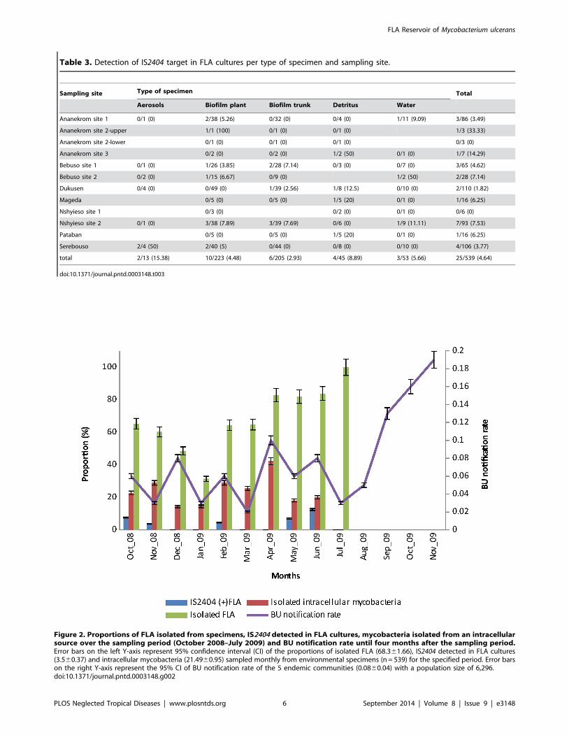
Table 3. Detection of IS2404 target in FLA cultures per type of specimen and sampling site.
Sampling site Type of specimen Total
Aerosols Biofilm plant Biofilm trunk Detritus Water
Ananekrom site 1 0/1 (0) 2/38 (5.26) 0/32 (0) 0/4 (0) 1/11 (9.09) 3/86 (3.49)
Ananekrom site 2-upper 1/1 (100) 0/1 (0) 0/1 (0) 1/3 (33.33)
Ananekrom site 2-lower 0/1 (0) 0/1 (0) 0/1 (0) 0/3 (0)
Ananekrom site 3 0/2 (0) 0/2 (0) 1/2 (50) 0/1 (0) 1/7 (14.29)
Bebuso site 1 0/1 (0) 1/26 (3.85) 2/28 (7.14) 0/3 (0) 0/7 (0) 3/65 (4.62)
Bebuso site 2 0/2 (0) 1/15 (6.67) 0/9 (0) 1/2 (50) 2/28 (7.14)
Dukusen 0/4 (0) 0/49 (0) 1/39 (2.56) 1/8 (12.5) 0/10 (0) 2/110 (1.82)
Mageda 0/5 (0) 0/5 (0) 1/5 (20) 0/1 (0) 1/16 (6.25)
Nshyieso site 1 0/3 (0) 0/2 (0) 0/1 (0) 0/6 (0)
Nshyieso site 2 0/1 (0) 3/38 (7.89) 3/39 (7.69) 0/6 (0) 1/9 (11.11) 7/93 (7.53)
Pataban 0/5 (0) 0/5 (0) 1/5 (20) 0/1 (0) 1/16 (6.25)
Serebouso 2/4 (50) 2/40 (5) 0/44 (0) 0/8 (0) 0/10 (0) 4/106 (3.77)
total 2/13 (15.38) 10/223 (4.48) 6/205 (2.93) 4/45 (8.89) 3/53 (5.66) 25/539 (4.64)
doi:10.1371/journal.pntd.0003148.t003
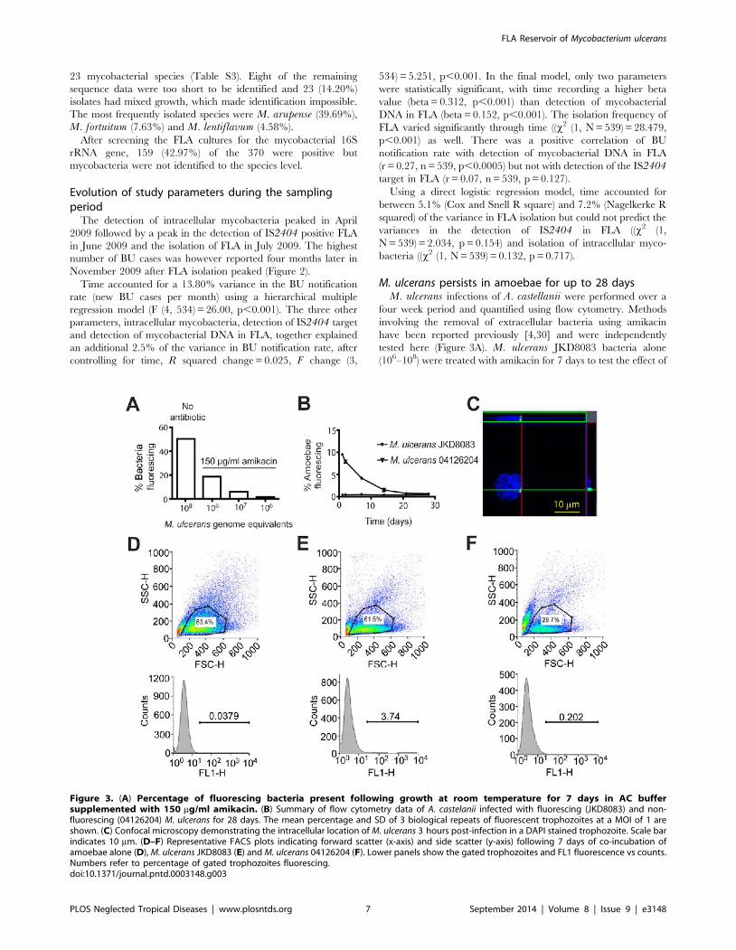
Figure 2. Proportions of FLA isolated from specimens, IS2404 detected in FLA cultures, mycobacteria isolated from an intracellularsource over the sampling period (October 2008–July 2009) and BU notification rate until four months after the sampling period.Error bars on the left Y-axis represent 95% confidence interval (CI) of the proportions of isolated FLA (68.361.66), IS2404 detected in FLA cultures(3.560.37) and intracellular mycobacteria (21.4960.95) sampled monthly from environmental specimens (n = 539) for the specified period. Error barson the right Y-axis represent the 95% CI of BU notification rate of the 5 endemic communities (0.0860.04) with a population size of 6,296.doi:10.1371/journal.pntd.0003148.g002
FLA Reservoir of Mycobacterium ulcerans
PLOS Neglected Tropical Diseases | www.plosntds.org 6 September 2014 | Volume 8 | Issue 9 | e3148
23 mycobacterial species (Table S3). Eight of the remaining
sequence data were too short to be identified and 23 (14.20%)
isolates had mixed growth, which made identification impossible.
The most frequently isolated species were M. arupense (39.69%),
M. fortuitum (7.63%) and M. lentiflavum (4.58%).
After screening the FLA cultures for the mycobacterial 16S
rRNA gene, 159 (42.97%) of the 370 were positive but
mycobacteria were not identified to the species level.
Evolution of study parameters during the samplingperiod
The detection of intracellular mycobacteria peaked in April
2009 followed by a peak in the detection of IS2404 positive FLA
in June 2009 and the isolation of FLA in July 2009. The highest
number of BU cases was however reported four months later in
November 2009 after FLA isolation peaked (Figure 2).
Time accounted for a 13.80% variance in the BU notification
rate (new BU cases per month) using a hierarchical multiple
regression model (F (4, 534) = 26.00, p,0.001). The three other
parameters, intracellular mycobacteria, detection of IS2404 target
and detection of mycobacterial DNA in FLA, together explained
an additional 2.5% of the variance in BU notification rate, after
controlling for time, R squared change = 0.025, F change (3,
534) = 5.251, p,0.001. In the final model, only two parameters
were statistically significant, with time recording a higher beta
value (beta = 0.312, p,0.001) than detection of mycobacterial
DNA in FLA (beta = 0.152, p,0.001). The isolation frequency of
FLA varied significantly through time ((x2 (1, N = 539) = 28.479,
p,0.001) as well. There was a positive correlation of BU
notification rate with detection of mycobacterial DNA in FLA
(r = 0.27, n = 539, p,0.0005) but not with detection of the IS2404target in FLA (r = 0.07, n = 539, p = 0.127).
Using a direct logistic regression model, time accounted for
between 5.1% (Cox and Snell R square) and 7.2% (Nagelkerke R
squared) of the variance in FLA isolation but could not predict the
variances in the detection of IS2404 in FLA ((x2 (1,
N = 539) = 2.034, p = 0.154) and isolation of intracellular myco-
bacteria ((x2 (1, N = 539) = 0.132, p = 0.717).
M. ulcerans persists in amoebae for up to 28 daysM. ulcerans infections of A. castellanii were performed over a
four week period and quantified using flow cytometry. Methods
involving the removal of extracellular bacteria using amikacin
have been reported previously [4,30] and were independently
tested here (Figure 3A). M. ulcerans JKD8083 bacteria alone
(106–108) were treated with amikacin for 7 days to test the effect of
Figure 3. (A) Percentage of fluorescing bacteria present following growth at room temperature for 7 days in AC buffersupplemented with 150 mg/ml amikacin. (B) Summary of flow cytometry data of A. castelanii infected with fluorescing (JKD8083) and non-fluorescing (04126204) M. ulcerans for 28 days. The mean percentage and SD of 3 biological repeats of fluorescent trophozoites at a MOI of 1 areshown. (C) Confocal microscopy demonstrating the intracellular location of M. ulcerans 3 hours post-infection in a DAPI stained trophozoite. Scale barindicates 10 mm. (D–F) Representative FACS plots indicating forward scatter (x-axis) and side scatter (y-axis) following 7 days of co-incubation ofamoebae alone (D), M. ulcerans JKD8083 (E) and M. ulcerans 04126204 (F). Lower panels show the gated trophozoites and FL1 fluorescence vs counts.Numbers refer to percentage of gated trophozoites fluorescing.doi:10.1371/journal.pntd.0003148.g003
FLA Reservoir of Mycobacterium ulcerans
PLOS Neglected Tropical Diseases | www.plosntds.org 7 September 2014 | Volume 8 | Issue 9 | e3148
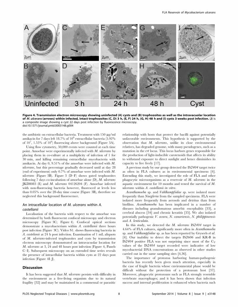
the antibiotic on extracellular bacteria. Treatment with 150 mg/ml
amikacin for 7 days left 18.7% of 108 extracellular bacteria (5.92%
of 107, 1.53% of 106) fluorescing above background (Figure 3A).
Using flow cytometry, 50,000 events were counted at each time
point. Amoebae were experimentally infected with M. ulcerans by
placing them in co-culture at a multiplicity of infection of 1 for
30 min, and killing remaining extracellular mycobacteria with
amikacin. At day 0, 9.51% of the amoebae were infected with M.ulcerans, but this percentage gradually decreased until at day 28
(end of experiment) only 0.7% of amoebae were infected with M.ulcerans (Figure 3B). Figure 3 (D–F) shows gated trophozoites
following 7 days co-incubation of amoebae alone (D), M. ulceransJKD8083 (E) and M. ulcerans 04126204 (F). Amoebae infected
with non-fluorescing bacteria however, fluoresced at levels less
than 0.05% over the 28-day time course (Figure 3B), therefore we
neglected this background fluorescence.
An intracellular location of M. ulcerans within A.castellanii
Localisation of the bacteria with respect to the amoebae was
determined by both fluorescent confocal microscopy and electron
microscopy (Figure 3C; Figure 4). Confocal optical sections
demonstrate a mycobacterium within A. castellanii three hours
post infection (Figure 3C). Video S1. shows fluorescing bacteria in
A. castelanii at 24 h post infection. Examination of 1 mL aliquots
of M. ulcerans-infected trophozoites and cysts by transmission
electron microscopy demonstrated an intravacuolar location for
M. ulcerans at 3, 24 and 48 hours post infection (Figure 4, Panels
C–I). Subsequent microscopy on an extended time series reveals
the presence of intracellular bacteria within cysts at 22 days post
infection (Figure 4I–J).
Discussion
It has been suggested that M. ulcerans persists with difficulty in
the environment as a free-living organism due to its natural
fragility [32] and may be maintained in a commensal or parasitic
relationship with hosts that protect the bacilli against potentially
unfavorable environments. This hypothesis is supported by the
observation that M. ulcerans, unlike its close environmental
relatives, has degraded genome, with many pseudogenes, such as a
mutation in the crt locus. This locus harbors genes responsible for
the production of light-inducible carotenoids that affects its ability
to withstand exposure to direct sunlight and hence diminishes its
capacity to live freely [17].
A previous study by our group detected the IS2404 target twice
as often in FLA cultures as in environmental specimens [4].
Extending this study, we investigated the role of FLA and other
phagocytic microorganisms as a reservoir of M. ulcerans in the
aquatic environment for 10 months and tested the survival of M.ulcerans within A. castellanii in vitro.
Acanthamoeba sp. and Vahlkampfiidae sp. were isolated more
frequently than Naegleria from the sampled specimens. FLA were
isolated more frequently from aerosols and detritus than from
biofilms. Acanthamoeba has been implicated in a number of
diseases including granulomatous amoebic encephalitis [33], a
cerebral abscess [34] and chronic keratitis [35]. We also isolated
potentially pathogenic V. avara, N. canariensis, N. philippinensisand A. lenticulata.
In this study, we detected the M. ulcerans IS2404 target in
4.64% of FLA cultures, significantly more often in Acanthamoebasp. and Vahlkampfiidae sp. as has been reported by Gryseels et al.[4]. Our inability to detect the targets IS2606 and KR-B in
IS2404 positive FLA was not surprising since most of the CT
values of the IS2404 target recorded were indicative of low
mycobacterial DNA concentrations as observed in other studies
carried out in the same sampling sites [4,36].
The importance of protozoa harboring human-pathogenic
bacteria has recently been given much attention, especially in
the case of fragile bacteria whose environmental phase would be
difficult without the protection of a protozoan host [37].
Moreover, phagocytic protozoans such as FLA strongly resemble
vertebrate macrophages; and it has been shown that infection
success and internal proliferation is enhanced when bacteria such
Figure 4. Transmission electron microscopy showing uninfected (A) cysts and (B) trophozoites as well as the intravacuolar locationof M. ulcerans (arrows) within infected, intact trophozoites (C, D) 3 h, (E, F) 24 h, (G, H) 48 h and (I) cysts 3 weeks post infection. (J) isa composite image showing a cyst 22 days post infection by fluorescence microscopy.doi:10.1371/journal.pntd.0003148.g004
FLA Reservoir of Mycobacterium ulcerans
PLOS Neglected Tropical Diseases | www.plosntds.org 8 September 2014 | Volume 8 | Issue 9 | e3148
as Legionella and M. avium had previously resided inside
protozoans [15,38]. A number of intracellular Mycobacterium sp.
were isolated from unknown hosts in the specimens, which
previously have been shown to live/survive intracellularly in
amoebae: M. simiae, M. fortuitum, M. septicum, M. peregrinum,
M. terrae, M. gordonae, M. intracellulare and M. lentiflavum[16,39]. The most frequently isolated species were: M. arupense,
M. fortuitum and M. lentiflavum are potentially pathogenic
species. These species were isolated from the environment [40–42]
and M. arupense was isolated from wild African rodents [43].
Intracellular mycobacteria were more frequently isolated from
specimens from which we also isolated FLA that may indicate their
role as a reservoir for these mycobacteria. Thomas et al. [37] also
reported a significant association between the presence of
amoebae and the presence of mycobacteria. FLA have the
additional advantage that they can form cysts, which allow them
to persist through harsh periods and be dispersed via the air. It has
been suggested that some infections can be acquired by inhaling
aerosols containing FLA cells filled with bacteria [44], for example
in the case of Legionella [45]. The detection of the IS2404 target
in two of the thirteen aerosolized FLA suggests that they may act
as vehicles for these mycobacteria. The aerosol transmission
hypothesis of M. ulcerans was first postulated by Hayman [46],
but received little attention, due to the unlikelihood of M. ulceransbeing airborne as a free-living organism. The possibility that
IS2404 positive mycobacteria including M. ulcerans are carried
by aerosolized protozoan cysts changes this perspective. More
research is, however, needed to explore this transmission route
further, and in a subsequent study we investigated the presence of
M. ulcerans on the skin of healthy inhabitants in the same endemic
communities (manuscript in preparation).
BU notification rates varied significantly through time with the
highest number of cases recorded in November 2009. BU
prevalence has increased during the last quarter of the year in
this locality as well as in some endemic regions of Ghana (data not
shown); in this case there was no community awareness during this
period. BU notification rates correlated positively with detection
frequency of mycobacterial DNA in FLA cultures but not with
detection of IS2404 in FLA cultures, suggesting that detection of
IS2404 in FLA cannot predict concurrent BU incidence.
However, the time series of this data set was not long enough to
test for a potential lagging phase between peaks of IS2404detection in FLA and BU incidence.
Similar to our previous study [4], over the time course of
experimental infection of A. castellanii with M. ulcerans, M.ulcerans is present within amoebae for up to 28 days albeit at low
levels. In addition, both electron microscopy and standard
fluorescence microscopy revealed the presence of intracellular
bacteria within cysts at 22 days post infection (Figure 4I–J). This is
not unexpected due to the previously demonstrated presence and
survival of a variety of environmental mycobacteria in cysts [39].
The persistence of strong GFP fluorescence of M. ulceranswithin A. castellanii throughout the experiment indicated that the
mycolactone polyketide synthases genes are abundantly expressed
intracellularly, as GFP gene expression is under the control of the
mlsA1 promoter [26]. These data also suggest that mycolactone
may be produced by the bacteria within the vacuole. While this
study was not designed to test the effect of M. ulcerans on A.castellanii, our observations of fluorescing M. ulcerans persisting
through 28 days within intact A. castellanii suggest that A.castellanii is not adversely affected by mycolactone or the presence
of the bacteria as was also shown for A. polyphaga [4].
The ability of M. marinum to persist within amoebae is widely
documented [39,47,48]. Following the initial time points a
decrease in M. ulcerans-infected amoebae as reported previously
for M. ulcerans, M. shottsii and M. pseudoshottsii [4,10] was seen
which suggests that M. ulcerans does not replicate within amoebae
and is not as well adapted as M. marinum to resist initial amoebic
digestion, but is perhaps able to persist once within the vacuolar
compartment by preventing lysosomal maturation of the vacuole
by as yet undetermined mechanisms.
This study showed the occurrence of the IS2404 marker in
FLA, especially in the genera Acanthamoeba and Vahlkampfiidae.
After co-culturing amoebae and M. ulcerans the pathogen
persisted at low levels suggesting that it probably only transiently
occupies FLA and that it is unlikely that protozoa are a long-term
reservoir for this pathogen. While the data we present here
confirm that FLA can host mycobacteria that harbor the IS2404marker (including M. ulcerans), the lack of predictive power of
detection of IS2404 positive FLA in predicting BU notifications
suggests FLA are not directly involved in transmission of M.ulcerans to humans. We suggest future work should focus on
reservoirs that act as M. ulcerans amplifiers that link protozoans
with humans.
Supporting Information
Table S1 Real-time PCR CT values of IS2404 target in FLA
cultures.
(DOCX)
Table S2 Identification of FLA per sample and sampling site.
(DOCX)
Table S3 Identification of intracellular mycobacteria per sample
and sampling site.
(DOCX)
Video S1 Fluorescing bacteria in A. castelanii at 24 h post
infection.
(ZIP)
Author Contributions
Conceived and designed the experiments: NAA AA FP ME LD TPS NJT.
Performed the experiments: NAA SG NJT BR ME AA. Analyzed the data:
NAA NJT BR TPS. Contributed reagents/materials/analysis tools: AA
HL FP MS KV. Contributed to the writing of the manuscript: NAA SG
NJT BR TPS.
References
1. Portaels F, Silva MT, Meyers WM (2009) Buruli ulcer. Clin Dermatol 27: 291–
305.
2. Marion E, Deshayes C, Chauty A, Cassisa V, Tchibozo S, et al. (2011) Detection
of Mycobacterium ulcerans DNA in water bugs collected outside the aquatic
environment in Benin. Med Trop (Mars) 71: 169–172.
3. Williamson HR, Benbow ME, Campbell LP, Johnson CR, Sopoh G, et al.
(2012) Detection of mycobacterium ulcerans in the environment predicts
prevalence of Buruli ulcer in Benin. PLoS Negl Trop Dis 6: e1506.
4. Gryseels S, Amissah D, Durnez L, Vandelannoote K, Leirs H, et al. (2012)
Amoebae as Potential Environmental Hosts for Mycobacterium ulcerans and
Other Mycobacteria, but Doubtful Actors in Buruli Ulcer Epidemiology. PLoS
Negl Trop Dis 6: e1764.
5. Merritt RW, Walker ED, Small PLC, Wallace JR, Johnson PDR, et al. (2010)
Ecology and transmission of buruli ulcer disease: a systematic review. PLoS Negl
Trop Dis 4: e911. Available: http://www.pubmedcentral.nih.gov/articlerender.
fcgi?artid = 3001905&tool = pmcentrez&rendertype = abstract. Accessed 4 May
2014.
6. Fyfe JAM, Lavender CJ, Handasyde KA, Legione AR, OBrien CR, et al. (2010)
A major role for mammals in the ecology of Mycobacterium ulcerans. PLoS
Negl Trop Dis 4: e791.
FLA Reservoir of Mycobacterium ulcerans
PLOS Neglected Tropical Diseases | www.plosntds.org 9 September 2014 | Volume 8 | Issue 9 | e3148
7. Johnson PDR, Azuolas J, Lavender CJ, Wishart E, Stinear TP, et al. (2007)
Mycobacterium ulcerans in mosquitoes captured during outbreak of Buruliulcer, southeastern Australia. Emerg Infect Dis 13: 1653–1660. doi:10.3201/
eid1311.061369.
8. Rowbotham TJ (1980) Preliminary report on the pathogenicity of Legionellapneumophila for freshwater and soil amoebae. J Clin Pathol 33: 1179–1183.
9. Landers P, Kerr KG, Rowbotham TJ, Tipper JL, Keig PM, et al. (2000)Survival and growth of Burkholderia cepacia within the free-living amoeba
Acanthamoeba polyphaga. Eur J Clin Microbiol Infect Dis 19: 121–123.
doi:10.1007/s100960050442.10. Gupta T, Fine-Coulson K, Karls R, Gauthier D, Quinn F (2013) Internalization
of Mycobacterium shottsii and Mycobacterium pseudoshottsii by Acanthamoebapolyphaga. Can J Microbiol 59: 570–576. Available: http://www.ncbi.nlm.nih.
gov/pubmed/23899000.11. Mba Medie F, Ben Salah I, Henrissat B, Raoult D, Drancourt M (2011)
Mycobacterium tuberculosis complex mycobacteria as amoeba-resistant organ-
isms. PLoS One 6: e20499.12. Thomas V, McDonnell G (2007) Relationship between mycobacteria and
amoebae: ecological and epidemiological concerns. Lett Appl Microbiol 45:349–357. Available: http://www.ncbi.nlm.nih.gov/pubmed/17897376. Ac-
cessed 4 May 2014.
13. Brown MRW, Barker J (1999) Unexplored reservoirs of pathogenic bacteria:Protozoa and biofilms. Trends Microbiol 7: 46–50.
14. Steinert M, Birkness K, White E, Fields B, Quinn F (1998) Mycobacteriumavium bacilli grow saprozoically in coculture with Acanthamoeba polyphaga
and survive within cyst walls. Appl Environ Microbiol 64: 2256–2261.15. Cirillo JD, Falkow S, Tompkins LS, Bermudez LE (1997) Interaction of
Mycobacterium avium with environmental amoebae enhances virulence. Infect
Immun 65: 3759–3767.16. Krishna-Prasad BN, Gupta SK (1978) Preliminary report on engulfment and
retention of mycobacteria by trophozoites of axenically grown Acanthamoebacastellanii Douglas. Curr Sci 47: 245–247.
17. Stinear TP, Seemann T, Pidot S, Frigui W, Reysset G, et al. (2007) Reductive
evolution and niche adaptation inferred from the genome of Mycobacteriumulcerans, the causative agent of Buruli ulcer. Genome Res 17: 192–
200. Available: http://www.pubmedcentral.nih.gov/articlerender.fcgi?artid =1781351&tool = pmcentrez&rendertype = abstract. Accessed 4 May 2014.
18. Greub G, Raoult D (2004) Microorganisms resistant to free-living amoebae. ClinMicrobiol Rev 17: 413–433. doi:10.1128/CMR.17.2.413.
19. Eddyani M, De Jonckheere JF, Durnez L, Suykerbuyk P, Leirs H, et al. (2008)
Occurrence of free-living amoebae in communities of low and high endemicityfor Buruli ulcer in southern Benin. Appl Environ Microbiol 74: 6547–6553.
20. Page F (1976) An illustrated key to freshwater and soil amoebae. Ambleside,England: Titus Wilson & Son, Ltd.
21. Ash LR, Orihel T (1987) Parasites: a guide to laboratory procedures and
identification. Ed. Chicago: American Society of Clinical Pathologists.22. Durnez L, Stragier P, Roebben K, Ablordey A, Leirs H, et al. (2009) A
comparison of DNA extraction procedures for the detection of Mycobacteriumulcerans, the causative agent of Buruli ulcer, in clinical and environmental
specimens. J Microbiol Methods 76: 152–158. doi:10.1016/j.mimet.2008.10.002.
23. Boom R, Sol CJ, Salimans MM, Jansen CL, Wertheim-van Dillen PM, et al.
(1990) Rapid and simple method for purification of nucleic acids. J ClinMicrobiol 28: 495–503.
24. Fyfe JAM, Lavender CJ, Johnson PDR, Globan M, Sievers A, et al. (2007)Development and application of two multiplex real-time PCR assays for the
detection of Mycobacterium ulcerans in clinical and environmental samples.
Appl Environ Microbiol 73: 4733–4740.25. Portaels F, De Muynck A, Sylla MP (1988) Selective isolation of mycobacteria
from soil: a statistical analysis approach. J Gen Microbiol 134: 849–855.doi:10.1099/00221287-134-3-849.
26. Kirschner P, Springer B, Vogel U, Meier A, Wrede A, et al. (1993) Genotypic
identification of mycobacteria by nucleic acid sequence determination: report ofa 2-year experience in a clinical laboratory. J Clin Microbiol 31: 2882–2889.
27. Boddinghaus B, Rogall T, Flohr T, Blocker H, Bottger EC (1990) Detection andidentification of mycobacteria by amplification of rRNA. J Clin Microbiol 28:
1751–1759.28. Tobias NJ, Seemann T, Pidot SJ, Porter JL, Marsollier L, et al. (2009)
Mycolactone gene expression is controlled by strong SigA-like promoters with
utility in studies of Mycobacterium ulcerans and buruli ulcer. PLoS Negl TropDis 3: e553.
29. Moffat JF, Tompkins LS (1992) A quantitative model of intracellular growth ofLegionella pneumophila in Acanthamoeba castellanii. Infect Immun 60: 296–
301.
30. Bermudez LE, Young LS (1994) Factors affecting invasion of HT-29 and HEp-2
epithelial cells by organisms of the Mycobacterium avium complex. InfectImmun 62: 2021–2026.
31. Abd H, Johansson T, Golovliov I, Sandstrom G, Forsman M (2003) Survivaland growth of Francisella tularensis in Acanthamoeba castellanii. Appl Environ
Microbiol 69: 600–606.
32. Portaels F, Chemlal K, Elsen P, Johnson PD, Hayman JA, et al. (2001)Mycobacterium ulcerans in wild animals. Rev Sci Tech 20: 252–264. Available:
http://www.ncbi.nlm.nih.gov/entrez/query.fcgi?cmd = Retrieve&db =PubMed&dopt = Citation&list_uids = 11288515.
33. Visvesvara GS, Mirra SS, Brandt FH, Moss DM, Mathews HM, et al. (1983)Isolation of two strains of Acanthamoeba castellanii from human tissue and their
pathogenicity and isoenzyme profiles. J Clin Microbiol 18: 1405–1412.
34. Harwood CR, Rich GE, McAleer R, Cherian G (1988) Isolation of
Acanthamoeba from a cerebral abscess. Med J Aust 148: 47–49.
35. Auran JD, Starr MB, Jakobiec FA (1987) Acanthamoeba Keratitis. Cornea 6: 2–
26. Available: http://journals.lww.com/corneajrnl/Fulltext/1987/06010/Acanthamoeba_Keratitis.2.aspx.
36. Vandelannoote K, Durnez L, Amissah D, Gryseels S, Dodoo A, et al. (2010)
Application of real-time PCR in Ghana, a Buruli ulcer-endemic country,confirms the presence of Mycobacterium ulcerans in the environment. FEMS
Microbiol Lett 304: 191–194. doi:10.1111/j.1574-6968.2010.01902.x.
37. Thomas V, Herrera-Rimann K, Blanc DS, Greub G (2006) Biodiversity of
amoebae and amoeba-resisting bacteria in a hospital water network. ApplEnviron Microbiol 72: 2428–2438. doi:10.1128/AEM.72.4.2428.
38. Cirillo JD, Falkow S, Tompkins LS (1994) Growth of Legionella pneumophila inAcanthamoeba castellanii enhances invasion. Infect Immun 62: 3254–3261.
39. Adekambi T, Ben Salah S, Khlif M, Raoult D, Drancourt M (2006) Survival ofenvironmental mycobacteria in Acanthamoeba polyphaga. Appl Environ
Microbiol 72: 5974–5981. Available: http://www.pubmedcentral.nih.gov/articlerender.fcgi?artid = 1563627&tool = pmcentrez&rendertype = abstract. Ac-
cessed 4 May 2014.
40. Liu R, Yu Z, Zhang H, Yang M, Shi B, et al. (2012) Diversity of bacteria andmycobacteria in biofilms of two urban drinking water distribution systems.
Can J Microbiol 58: 261–270. doi:10.1139/w11-129.
41. Thomson R, Tolson C, Carter R, Coulter C, Huygens F, et al. (2013) Isolation
of nontuberculous mycobacteria (NTM) from household water and showeraerosols in patients with pulmonary disease caused by NTM. J Clin Microbiol
51: 3006–3011. Available: http://www.pubmedcentral.nih.gov/articlerender.fcgi?artid = 3754680&tool = pmcentrez&rendertype = abstract.
42. Castillo-Rodal AI, Mazari-Hiriart M, Lloret-Sanchez LT, Sachman-Ruiz B,Vinuesa P, et al. (2012) Potentially pathogenic nontuberculous mycobacteria
found in aquatic systems. Analysis from a reclaimed water and water distribution
system in Mexico City. Eur J Clin Microbiol Infect Dis 31: 683–694. Available:http://www.ncbi.nlm.nih.gov/pubmed/21805195.
43. Durnez L, Eddyani M, Mgode GF, Katakweba A, Katholi CR, et al. (2008) Firstdetection of mycobacteria in African rodents and insectivores, using stratified
pool screening. Appl Environ Microbiol 74: 768–773.
44. Angenent LT, Kelley ST, St Amand A, Pace NR, Hernandez MT (2005)
Molecular identification of potential pathogens in water and air of a hospitaltherapy pool. Proc Natl Acad Sci U S A 102: 4860–4865.
45. Abu Kwaik Y, Gao LY, Stone BJ, Venkataraman C, Harb OS (1998) Invasionof protozoa by Legionella pneumophila and its role in bacterial ecology and
pathogenesis. Appl Environ Microbiol 64: 3127–3133.
46. Hayman J (1991) Postulated epidemiology of Mycobacterium ulcerans infection.
Int J Epidemiol 20: 1093–1098.
47. Kennedy GM, Morisaki JH, DiGiuseppe Champion PA (2012) ConservedMechanisms of Mycobacterium marinum Pathogenesis within the Environmen-
tal Amoeba Acanthamoeba castellanii. Appl Environ Microbiol 78: 2049–2052.doi:10.1128/AEM.06965-11.
48. Solomon JM, Leung GS, Isberg RR (2003) Intracellular replication ofMycobacterium marinum within Dictyostelium discoideum: efficient replication
in the absence of host coronin. Infect Immun 71: 3578–3586.
FLA Reservoir of Mycobacterium ulcerans
PLOS Neglected Tropical Diseases | www.plosntds.org 10 September 2014 | Volume 8 | Issue 9 | e3148
http://www.pubmedcentral.nih.gov/articlerender.fcgi?artid=1781351&tool=pmcentrez&rendertype=abstract
http://www.pubmedcentral.nih.gov/articlerender.fcgi?artid=1781351&tool=pmcentrez&rendertype=abstract
http://www.pubmedcentral.nih.gov/articlerender.fcgi?artid=1563627&tool=pmcentrez&rendertype=abstract
http://www.pubmedcentral.nih.gov/articlerender.fcgi?artid=1563627&tool=pmcentrez&rendertype=abstract
http://www.pubmedcentral.nih.gov/articlerender.fcgi?artid=3754680&tool=pmcentrez&rendertype=abstract
Related Documents