Spliceosomal snRNAs in the unicellular eukaryote Trichomonas vaginalis are structurally conserved but lack a 59-cap structure AUGUSTO SIMOES-BARBOSA, 1,2 DIONIGIA MELONI, 1 JAMES A. WOHLSCHLEGEL, 3 MARIA M. KONARSKA, 4 and PATRICIA J. JOHNSON 1 1 Department of Microbiology, Immunology, and Molecular Genetics, University of California, Los Angeles, California 90095-1489, USA 2 Universidade Catolica de Brasilia, Programa de Pos-graduacao em Ciencias Genomicas e Biotecnologia, SGAN 916, Brasilia DF 70790-160, Brazil 3 Department of Biological Chemistry, University of California, Los Angeles, California 90095-1737, USA 4 Laboratory of Molecular Biology & Biochemistry, The Rockefeller University, New York, New York 10021, USA ABSTRACT Few genes in the divergent eukaryote Trichomonas vaginalis have introns, despite the unusually large gene repertoire of this human-infective parasite. These introns are characterized by extended conserved regulatory motifs at the 59 and 39 boundaries, a feature shared with another divergent eukaryote, Giardia lamblia, but not with metazoan introns. This unusual characteristic of T. vaginalis introns led us to examine spliceosomal small nuclear RNAs (snRNAs) predicted to mediate splicing reactions via interaction with intron motifs. Here we identify T. vaginalis U1, U2, U4, U5, and U6 snRNAs, present predictions of their secondary structures, and provide evidence for interaction between the U2/U6 snRNA complex and a T. vaginalis intron. Structural models predict that T. vaginalis snRNAs contain conserved sequences and motifs similar to those found in other examined eukaryotes. These data indicate that mechanisms of intron recognition as well as coordination of the two catalytic steps of splicing have been conserved throughout eukaryotic evolution. Unexpectedly, we found that T. vaginalis spliceosomal snRNAs lack the 59 trimethylguanosine cap typical of snRNAs and appear to possess unmodified 59 ends. Despite the lack of a cap structure, U1, U2, U4, and U5 genes are transcribed by RNA polymerase II, whereas the U6 gene is transcribed by RNA polymerase III. Keywords: Trichomonas vaginalis; splicing; snRNA; capping INTRODUCTION RNA splicing, an essential step of mRNA maturation in eukaryotic genes containing introns, is catalyzed by a large ribonucleoprotein particle, the spliceosome (Burge et al. 1999). This dynamic particle is composed of U1, U2, U4, U5, and U6 small nuclear RNAs (snRNAs) and hundreds of proteins (Nilsen 2003). Spliceosomes have been best char- acterized in yeast and mammals, where well-established in vitro splicing systems are available (Nilsen 1998; Burge et al. 1999). Although spliceosomal protein components may vary between organisms and even within different tissues of a single organism, all spliceosomes share a conserved core of proteins and the five snRNAs (Burge et al. 1999). Spliceosomal introns have been found in all examined eukaryotes (Vanacova et al. 2005), but are absent in eubac- terial and archeal genomes. The prevalence and number of introns vary greatly in unicellular eukaryotes (Gardner et al. 2002; Abrahamsen et al. 2004; El-Sayed et al. 2005; Loftus et al. 2005; Carlton et al. 2007). For example, conventional cis introns are widespread in apicomplexan parasites (Gardner et al. 2002) and Entamoeba (Loftus et al. 2005; Davis et al. 2007), but are found only infrequently in trypanosomes (El-Sayed et al. 2005) and in the diplomonad Giardia lamblia (Nixon et al. 2002; Morrison et al. 2007). Euglenoids, relatives of trypanosomes, contain many cis introns, but also have a subset of unconventional introns that are excised by a spliceosome-independent mechanism (Ebel et al. 1999). Although the unicellular eukaryote Trichomonas vaginalis has an unusually large repertoire of Reprint requests to: Patricia J. Johnson, Department of Microbiology, Immunology, and Molecular Genetics, University of California Los Angeles, 609 Charles E. Young Drive East, Los Angeles, California 90095-1489, USA; e-mail: [email protected]; fax: (310) 206-5231. Article published online ahead of print. Article and publication date are at http://www.rnajournal.org/cgi/doi/10.1261/rna.1045408. RNA (2008), 14:1617–1631. Published by Cold Spring Harbor Laboratory Press. Copyright Ó 2008 RNA Society. 1617

Welcome message from author
This document is posted to help you gain knowledge. Please leave a comment to let me know what you think about it! Share it to your friends and learn new things together.
Transcript
Spliceosomal snRNAs in the unicellular eukaryote
Trichomonas vaginalis are structurally conservedbut lack a 59-cap structure
AUGUSTO SIMOES-BARBOSA,1,2 DIONIGIA MELONI,1 JAMES A. WOHLSCHLEGEL,3
MARIA M. KONARSKA,4 and PATRICIA J. JOHNSON1
1Department of Microbiology, Immunology, and Molecular Genetics, University of California, Los Angeles, California 90095-1489, USA2Universidade Catolica de Brasilia, Programa de Pos-graduacao em Ciencias Genomicas e Biotecnologia, SGAN 916,Brasilia DF 70790-160, Brazil3Department of Biological Chemistry, University of California, Los Angeles, California 90095-1737, USA4Laboratory of Molecular Biology & Biochemistry, The Rockefeller University, New York, New York 10021, USA
ABSTRACT
Few genes in the divergent eukaryote Trichomonas vaginalis have introns, despite the unusually large gene repertoire of thishuman-infective parasite. These introns are characterized by extended conserved regulatory motifs at the 59 and 39 boundaries,a feature shared with another divergent eukaryote, Giardia lamblia, but not with metazoan introns. This unusual characteristicof T. vaginalis introns led us to examine spliceosomal small nuclear RNAs (snRNAs) predicted to mediate splicing reactions viainteraction with intron motifs. Here we identify T. vaginalis U1, U2, U4, U5, and U6 snRNAs, present predictions of theirsecondary structures, and provide evidence for interaction between the U2/U6 snRNA complex and a T. vaginalis intron.Structural models predict that T. vaginalis snRNAs contain conserved sequences and motifs similar to those found in otherexamined eukaryotes. These data indicate that mechanisms of intron recognition as well as coordination of the two catalyticsteps of splicing have been conserved throughout eukaryotic evolution. Unexpectedly, we found that T. vaginalis spliceosomalsnRNAs lack the 59 trimethylguanosine cap typical of snRNAs and appear to possess unmodified 59 ends. Despite the lack of acap structure, U1, U2, U4, and U5 genes are transcribed by RNA polymerase II, whereas the U6 gene is transcribed by RNApolymerase III.
Keywords: Trichomonas vaginalis; splicing; snRNA; capping
INTRODUCTION
RNA splicing, an essential step of mRNA maturation ineukaryotic genes containing introns, is catalyzed by a largeribonucleoprotein particle, the spliceosome (Burge et al.1999). This dynamic particle is composed of U1, U2, U4,U5, and U6 small nuclear RNAs (snRNAs) and hundreds ofproteins (Nilsen 2003). Spliceosomes have been best char-acterized in yeast and mammals, where well-established invitro splicing systems are available (Nilsen 1998; Burgeet al. 1999). Although spliceosomal protein components mayvary between organisms and even within different tissues of a
single organism, all spliceosomes share a conserved core ofproteins and the five snRNAs (Burge et al. 1999).
Spliceosomal introns have been found in all examinedeukaryotes (Vanacova et al. 2005), but are absent in eubac-terial and archeal genomes. The prevalence and number ofintrons vary greatly in unicellular eukaryotes (Gardner et al.2002; Abrahamsen et al. 2004; El-Sayed et al. 2005; Loftuset al. 2005; Carlton et al. 2007). For example, conventionalcis introns are widespread in apicomplexan parasites(Gardner et al. 2002) and Entamoeba (Loftus et al. 2005;Davis et al. 2007), but are found only infrequently intrypanosomes (El-Sayed et al. 2005) and in the diplomonadGiardia lamblia (Nixon et al. 2002; Morrison et al. 2007).Euglenoids, relatives of trypanosomes, contain many cisintrons, but also have a subset of unconventional intronsthat are excised by a spliceosome-independent mechanism(Ebel et al. 1999). Although the unicellular eukaryoteTrichomonas vaginalis has an unusually large repertoire of
rna10454 Simoes-Barbosa et al. ARTICLE RA
Reprint requests to: Patricia J. Johnson, Department of Microbiology,Immunology, and Molecular Genetics, University of California LosAngeles, 609 Charles E. Young Drive East, Los Angeles, California90095-1489, USA; e-mail: [email protected]; fax: (310) 206-5231.
Article published online ahead of print. Article and publication date areat http://www.rnajournal.org/cgi/doi/10.1261/rna.1045408.
RNA (2008), 14:1617–1631. Published by Cold Spring Harbor Laboratory Press. Copyright � 2008 RNA Society. 1617
JOBNAME: RNA 14#8 2008 PAGE: 1 OUTPUT: Friday July 4 20:33:46 2008
csh/RNA/164291/rna10454

genes, only z65 of these genes appear to have an intron(Vanacova et al. 2005; Carlton et al. 2007).
Conserved canonical 59 splice site (SS), 39SS, andbranch-site signals are present in T. vaginalis introns; how-ever, the unusually short branch site–39SS distance (7 nt)creates a highly conserved 12-nt motif (59-ACTAACACACAG/-39) encompassing the branch-site adenosine (under-lined) and the 39SS (/), which is required for splicing(Vanacova et al. 2005). To further investigate RNA struc-tural requirements for splicing in this divergent eukaryote,here, we have identified and characterized T. vaginalis snRNAs.
Spliceosomal snRNAs interact with conserved intronmotifs, positioning the pre-mRNA in the complex anddirecting splicing specificity (Ares and Weiser 1995). Studiesin yeast and metazoa have demonstrated that base pairingbetween snRNAs and intronic 59SS, 39SS, and branch sitealso provide a scaffold for spliceosomal assembly andcatalysis (Nilsen 1998). First, the 59SS and branch-site basepair with U1 and U2 snRNAs, respectively, followed bythe entry of a U4/U6.U5 tri-snRNP complex (Burge et al.1999). Subsequent conformational changes result in thereplacement of U1 snRNA by U6 snRNA at the 59SS, andunwinding of the U4–U6 snRNA complex. Base pairingbetween U6 and U2 snRNAs facilitates juxtaposition of the59SS and branch site at the catalytic center, allowing forthe first transesterification step. In addition to positioningcritical splicing motifs, U6 snRNA is thought to coordinatedivalent cations needed for catalysis (Yean et al. 2000).
Spliceosomal snRNAs have been proposed to be theevolutionary descendents of self-splicing introns (Sharp1985). The U2–U6 complex reveals structural similarity toa self-splicing group II intron-like domain, consistent withderivation of these snRNAs from this ancestral ribozyme.Furthermore, the human U2–U6 minimal catalytic domainhas been shown to form a covalent product with a branch-site RNA in a reaction that resembles the first step ofsplicing catalysis (Valadkhan and Manley 2001). The U2–U6 core domain is structurally conserved and allows forthe autonomous formation of a four-way helical junction(Valadkhan and Manley 2000; Sashital et al. 2004;Valadkhan 2005) that appears to be required for splicingin both yeast and mammals.
snRNAs are transcribed by RNA polymerase (RNAP) II,with the exception of U6 snRNA, which is a RNAP IIItranscript (Hernandez 2001). Trypanosomes break this ruleby transcribing all of their snRNAs using RNAP III and anupstream inverted tRNA gene as promoter (Fantoni et al.1994). As is typical of RNAP II transcripts, U1, U2, U4, andU5 snRNAs in yeast and mammals acquire a m7G capcotranscriptionally. This cap is subsequently hypermeth-ylated, creating a m2,2,7G (trimethylguanosine; TMG) capstructure (Busch et al. 1982). Hypermethylation of snRNAcap structures occurs in the cytoplasm in humans and inthe nucleolus in yeast. The biological significance of thedistinctive 59-cap structure found in snRNAs is unclear, but
it has been suggested to play roles in snRNA compartmen-talization and/or transport (Huber et al. 2002; Mouaikelet al. 2002; Narayanan et al. 2003).
Our previous work revealed that introns in T. vaginalisand G. lamblia are similar to one another yet divergentfrom most other eukaryotic introns, in that they contain aconserved 12-nt motif encompassing the branch site and39SS (Vanacova et al. 2005). This motif was shown to becritical for splicing; however, the snRNAs predicted to in-teract with the motif were not identified in either T. vaginalisor G. lamblia. To this end, we have now identified andcharacterized all five T. vaginalis spliceosomal snRNAs.
RESULTS
Identification of putative spliceosomal snRNAsin Trichomonas vaginalis
Introns were recently identified in z65 T. vaginalis genes(Vanacova et al. 2005; Carlton et al. 2007); however, little isknown about the splicing machinery in this highly diver-gent eukaryote (Baldauf 2003). As a step toward charac-terizing splicing mechanisms in this unicellular parasite,we searched for the presence of evolutionarily conservedspliceosomal snRNAs. Short conserved regions in thespliceosomal snRNAs were used to screen the 7X coverageT. vaginalis genome database (Carlton et al. 2007). Thesequence of U2 snRNA (CAAGTGTAGTATCTG; positionnt 28 to nt 42 in human U2 snRNA) known to interact withthe intron branch site, and a phylogenetically conservedsequence comprising the apical loop of U5 snRNA helix I(TTCGCCTTTTACTA, position nt 34 to nt 47 in humanU5 snRNA) were utilized in Blastn searches. These revealeda number of candidate snRNA genes that were subse-quently analyzed using structural prediction software RNA-fold (Hofacker 2003). Sequences immediately upstreamand downstream from the target sequence were tested forthe presence of predicted secondary structures and con-served nucleotides in these regions. This approach resultedin the identification of a single gene encoding each of theputative U2 and U5 snRNAs in the T. vaginalis genome.
Using INFERNAL software and multiple alignments ofU6 snRNA databases to screen the T. vaginalis genome, aputative U6 snRNA gene was also found. U6 snRNA formsintermolecular complexes with both U2 and U4 snRNAs,essential for spliceosome assembly and function (Brow andGuthrie 1988; Wu and Manley 1991; Madhani and Guthrie1992; Sun and Manley 1995). Using a short region of theidentified U6 gene (GTCCTTGGACAAAGA, nt 71 to nt 85)predicted to base pair with U4 snRNA, we then identifieda candidate U4 snRNA gene utilizing Blastn and the RNA-folding approaches described above for identification ofU2 and U5 snRNAs.
A Blastn search for U1 snRNA T. vaginalis gene usinga conserved sequence comprising the U1A binding site
Simoes-Barbosa et al.
1618 RNA, Vol. 14, No. 8
JOBNAME: RNA 14#8 2008 PAGE: 2 OUTPUT: Friday July 4 20:33:47 2008
csh/RNA/164291/rna10454
(CCATTGCACTCCGG, position nt 63 to nt 76 in humanU1 snRNA) was unsuccessful. Thus, we turned to a func-tional approach to identity this snRNA. We first identifieda gene with similarity to human U1-70K (e-value of 2e�13
by Blastp) and a central RRM domain (Nelissen et al.1994) in the T. vaginalis genome. This protein, taggedwith streptavidin binding protein (SBP) to allow for affin-ity purification on streptavidin sepharose, was expressedin T. vaginalis transfectants and subsequently isolatedfrom nuclear extracts. Bound RNA (100–200 nt) wasextracted from this material, cloned, and sequenced, allow-ing for identification of U1 snRNA.
Subsequent to identifying the T. vaginalis snRNA, aBLASTn search of the genome database using each indi-vidual snRNA revealed only one gene copy for each ofthe five putative snRNA. However, given the possibility ofsequence gaps, nucleotide divergence and loss of sequencedata during the assembly of this large, repetitive genome(Carlton et al. 2007), it is possible that additional unde-tected genes are present. Northern analysis confirmed theexpression of the five snRNAs identified (Fig. 4, below; datanot shown) and their 59 and 39 ends were mapped by RNAligase-mediated 59 end rapid amplification of cDNA (RLM-59 RACE), 39 RACE, and DNA sequencing (Fig. 4, below;data not shown) except for the 59 end of U5 snRNA, whichwas mapped by primer extension (data not shown).
Sequence and secondary structure comparisons of thefull-length snRNAs revealed each to be similar to othereukaryotic snRNA homologs (Figs. 1, 2) (Burge et al. 1999).The putative T. vaginalis U2 snRNA was found to possessall five conserved loops (I, IIa, IIb, III, and IV) withinterspersion and length similar to those found in U2 ofother organisms (Fig. 1A). T. vaginalis U2 snRNA hasextensive primary sequence conservation at the 59 end(Supplemental Fig. 1B), allowing formation of stem–loopsI, IIa, and IIb (Fig. 1A). Interestingly, the loop andnucleotides at the 39 side of helix IIa are complementaryto a single-stranded region downstream from helix IIb (Fig.1A,B), and thus can form stem IIc (Ares and Weiser 1995).Conformational changes between the phylogenetically con-served IIa and IIc forms are implicated in spliceosomaltransitions (Hilliker et al. 2007; Perriman and Ares 2007).The putative U4 snRNA can be base paired with theputative U6 snRNA, forming two conserved helices sepa-rated by an intramolecular stem–loop. Although the Y-shaped U4–U6 interaction domain is conserved (Fig. 1B),the primary sequence of U4 snRNA is not (SupplementalFig. 1C). Nevertheless, stem–loops II and III, with theuridine-rich Sm-binding site in between, can be formed inthe U4 snRNA (Fig. 1B). The short U4–U6 stem I observedin T. vaginalis may be compensated by a longer stable U4–U6 stem II. The latter does not contain any disruption ofWatson-Crick base pair thought to be important inbalancing stability during spliceosomal structural transi-tions in vertebrates (Ares and Weiser 1995).
T. vaginalis U1 snRNA has a predicted secondary structure(Fig. 1C) more similar to human U1 snRNA than its counter-part in Saccharomyces cerevisiae U1 (Kretzner et al. 1987). Theparasite U1 snRNA also exhibits a phylogenetically con-served region that could potentially interact with the 59SS.This sequence ACUUAUU is almost identical to that foundin metazoan U1 snRNAs (ACUUACC) (Supplemental Fig.1A). Although the last two positions (underlined) differbetween T. vaginalis and metazoan U1, these should stillinteract with 59SS positions (Fig. 1C). This finding indi-cates that recognition of the 59SS, an important step inintron recognition, is conserved throughout eukaryoticevolution. As we have previously reported (Vanacova et al.2005), the 59SS of T. vaginalis intron is more conservedthan typically found in metazoan introns (Fig. 2A). The T.vaginalis U1 snRNA helices are also more highly conservedin comparison with those of metazoa U1 snRNAs. Helix IIIand IV are most dissimilar in primary sequence, as observedin other eukaryotic U1 snRNAs (Supplemental Fig. 1A).Helix IV is preceded by a uridine-rich region that maycorrespond to the Sm-binding domain in T. vaginalis. Theprimary sequence of U1 helix II apical loop is highlyconserved in T. vaginalis, suggesting that role for an U1Aprotein homolog in the T. vaginalis U1 snRNP. The bindingregion for U1-70K that was used to isolate the U1 snRNA,the helix I loop, lacks two nucleotides that are invariantin other U1 snRNAs (Yuo and Weiner 1989). There is anapparent deletion of an adenosine (position 2 in the loopof other U1 snRNAs, GAUC > GUC; Supplemental Fig. 1A)that is required for binding of U1-70K to human U1snRNA (Surowy et al. 1989). Additionally, there is an A > Gsubstitution in the second to last position of this loop.
The T. vaginalis U5 snRNA sequence is also conserved,particularly near the top of helix I (Fig. 1C). Notably, theapical 11-nt loop is identical to that found in the humanU5 snRNA. Such conservation throughout eukaryoticevolution likely reflects its essential role in splicing (O’Keefeet al. 1996). This predicted helix I in the T. vaginalis U5 alsocontains internal loops with the invariant CCG sequencein a loop on the 39 side of the helix. Helix II, which ispreceded by a putative Sm-binding site, has a lowersequence conservation than helix I.
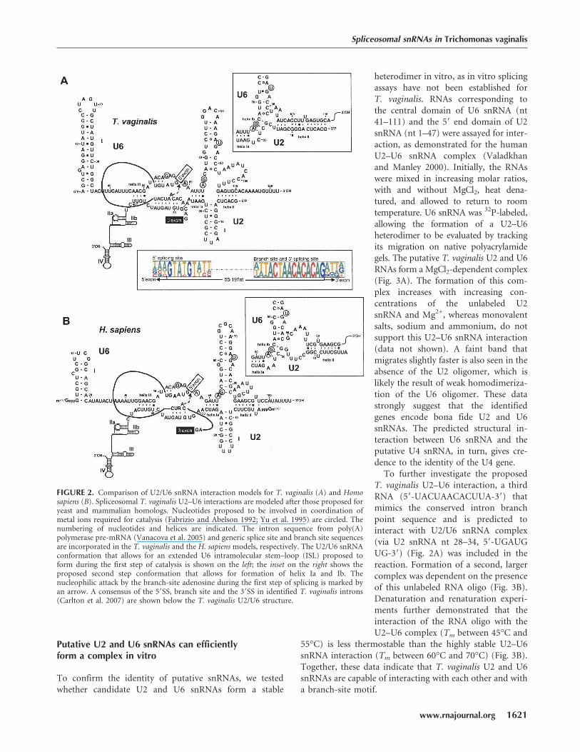
Consistent with splicing mechanisms in other organisms,the T. vaginalis U6 snRNA has the potential to interact withboth U2 and U4 snRNAs (Figs. 1B, 2). Notably, the putativeU6 snRNA has the predicted invariant sequences (A53CAGAGA59
and A65GC67) found in all examined U6 snRNAs (Figs. 1B,2A). Sites in U6 snRNA thought to coordinate a metal ionrequired for catalysis (Yu et al. 1995) are also conserved,and U6/U2 snRNA interaction model reveals three con-served U2–U6 helices and the intramolecular U6 stem–loopISL I (Fig. 2A). The predicted T. vaginalis U2/U6 snRNAcatalytic model is strikingly similar to yeast and humanmodels (Fig. 2A,B), involving two possible conformations(Sun and Manley 1995; Hilliker and Staley 2004).
Spliceosomal snRNAs in Trichomonas vaginalis
www.rnajournal.org 1619
JOBNAME: RNA 14#8 2008 PAGE: 3 OUTPUT: Friday July 4 20:33:47 2008
csh/RNA/164291/rna10454
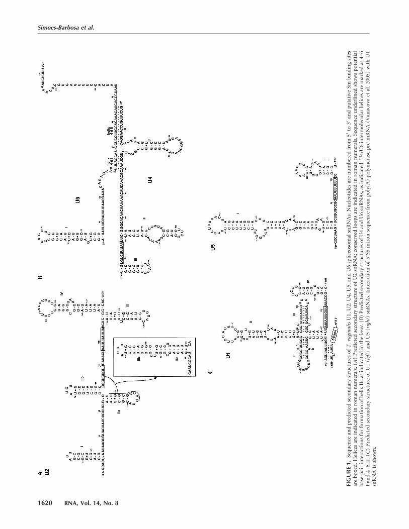
FIG
UR
E1.
Seq
uen
cean
dp
red
icte
dse
con
dar
yst
ruct
ure
so
fT
.vag
inal
isU
1,U
2,U
4,U
5,an
dU
6sp
lice
oso
mal
snR
NA
s.N
ucl
eoti
des
are
nu
mb
ered
fro
m59
to39
and
pu
tati
veSm
bin
din
gsi
tes
are
bo
xed
.H
elic
esar
ein
dic
ated
inro
man
nu
mer
als.
(A)
Pre
dic
ted
seco
nd
ary
stru
ctu
reo
fU
2sn
RN
A;
con
serv
edlo
op
sar
ein
dic
ated
inro
man
nu
mer
als.
Seq
uen
ceu
nd
erli
ned
sho
ws
po
ten
tial
bas
e-p
air
inte
ract
ion
sfo
rfo
rmat
ion
of
hel
ixII
cas
ind
icat
edin
the
inse
t.(B
)P
red
icte
dse
con
dar
yst
ruct
ure
so
fU
4an
dU
6sn
RN
As,
asin
dic
ated
.U4/
U6
inte
rmo
lecu
lar
hel
ices
are
mar
ked
as4–
6I
and
4–6
II.
(C)
Pre
dic
ted
seco
nd
ary
stru
ctu
reo
fU
1(l
eft)
and
U5
(rig
ht)
snR
NA
s.In
tera
ctio
no
f59
SSin
tro
nse
qu
ence
fro
mp
oly
(A)
po
lym
eras
ep
re-m
RN
A(V
anac
ova
etal
.20
05)
wit
hU
1sn
RN
Ais
sho
wn
.
Simoes-Barbosa et al.
1620 RNA, Vol. 14, No. 8
JOBNAME: RNA 14#8 2008 PAGE: 4 OUTPUT: Friday July 4 20:33:48 2008
csh/RNA/164291/rna10454
Putative U2 and U6 snRNAs can efficientlyform a complex in vitro
To confirm the identity of putative snRNAs, we testedwhether candidate U2 and U6 snRNAs form a stable
heterodimer in vitro, as in vitro splicingassays have not been established forT. vaginalis. RNAs corresponding tothe central domain of U6 snRNA (nt41–111) and the 59 end domain of U2snRNA (nt 1–47) were assayed for inter-action, as demonstrated for the humanU2–U6 snRNA complex (Valadkhanand Manley 2000). Initially, the RNAswere mixed in increasing molar ratios,with and without MgCl2, heat dena-tured, and allowed to return to roomtemperature. U6 snRNA was 32P-labeled,allowing the formation of a U2–U6heterodimer to be evaluated by trackingits migration on native polyacrylamidegels. The putative T. vaginalis U2 and U6RNAs form a MgCl2-dependent complex(Fig. 3A). The formation of this com-plex increases with increasing con-centrations of the unlabeled U2snRNA and Mg2+, whereas monovalentsalts, sodium and ammonium, do notsupport this U2–U6 snRNA interaction(data not shown). A faint band thatmigrates slightly faster is also seen in theabsence of the U2 oligomer, which islikely the result of weak homodimeriza-tion of the U6 oligomer. These datastrongly suggest that the identifiedgenes encode bona fide U2 and U6snRNAs. The predicted structural in-teraction between U6 snRNA and theputative U4 snRNA, in turn, gives cre-dence to the identity of the U4 gene.
To further investigate the proposedT. vaginalis U2–U6 interaction, a thirdRNA (59-UACUAACACUUA-39) thatmimics the conserved intron branchpoint sequence and is predicted tointeract with U2/U6 snRNA complex(via U2 snRNA nt 28–34, 59-UGAUGUG-39) (Fig. 2A) was included in thereaction. Formation of a second, largercomplex was dependent on the presenceof this unlabeled RNA oligo (Fig. 3B).Denaturation and renaturation experi-ments further demonstrated that theinteraction of the RNA oligo with theU2–U6 complex (Tm between 45°C and
55°C) is less thermostable than the highly stable U2–U6snRNA interaction (Tm between 60°C and 70°C) (Fig. 3B).Together, these data indicate that T. vaginalis U2 and U6snRNAs are capable of interacting with each other and witha branch-site motif.
FIGURE 2. Comparison of U2/U6 snRNA interaction models for T. vaginalis (A) and Homosapiens (B). Spliceosomal T. vaginalis U2–U6 interactions are modeled after those proposed foryeast and mammalian homologs. Nucleotides proposed to be involved in coordination ofmetal ions required for catalysis (Fabrizio and Abelson 1992; Yu et al. 1995) are circled. Thenumbering of nucleotides and helices are indicated. The intron sequence from poly(A)polymerase pre-mRNA (Vanacova et al. 2005) and generic splice site and branch site sequencesare incorporated in the T. vaginalis and the H. sapiens models, respectively. The U2/U6 snRNAconformation that allows for an extended U6 intramolecular stem–loop (ISL) proposed toform during the first step of catalysis is shown on the left; the inset on the right shows theproposed second step conformation that allows for formation of helix Ia and Ib. Thenucleophilic attack by the branch-site adenosine during the first step of splicing is marked byan arrow. A consensus of the 59SS, branch site and the 39SS in identified T. vaginalis introns(Carlton et al. 2007) are shown below the T. vaginalis U2/U6 structure.
Spliceosomal snRNAs in Trichomonas vaginalis
www.rnajournal.org 1621
JOBNAME: RNA 14#8 2008 PAGE: 5 OUTPUT: Friday July 4 20:34:44 2008
csh/RNA/164291/rna10454
Fig. 2 live 4/C
Trichomonas vaginalis snRNAs have uncapped59-termini
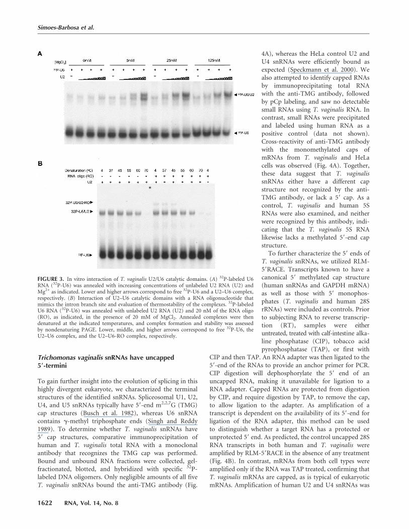
To gain further insight into the evolution of splicing in thishighly divergent eukaryote, we characterized the terminalstructures of the identified snRNAs. Spliceosomal U1, U2,U4, and U5 snRNAs typically have 59-end m2,2,7G (TMG)cap structures (Busch et al. 1982), whereas U6 snRNAcontains g-methyl triphosphate ends (Singh and Reddy1989). To determine whether T. vaginalis snRNAs have59 cap structures, comparative immunoprecipitation ofhuman and T. vaginalis total RNA with a monoclonalantibody that recognizes the TMG cap was performed.Bound and unbound RNA fractions were collected, gel-fractionated, blotted, and hybridized with specific 32P-labeled DNA oligomers. Only negligible amounts of all fiveT. vaginalis snRNAs bound the anti-TMG antibody (Fig.
4A), whereas the HeLa control U2 andU4 snRNAs were efficiently bound asexpected (Speckmann et al. 2000). Wealso attempted to identify capped RNAsby immunoprecipitating total RNAwith the anti-TMG antibody, followedby pCp labeling, and saw no detectablesmall RNAs using T. vaginalis RNA. Incontrast, small RNAs were precipitatedand labeled using human RNA as apositive control (data not shown).Cross-reactivity of anti-TMG antibodywith the monomethylated caps ofmRNAs from T. vaginalis and HeLacells was observed (Fig. 4A). Together,these data suggest that T. vaginalissnRNAs either have a different capstructure not recognized by the anti-TMG antibody, or lack a 59 cap. As acontrol, T. vaginalis and human 5SRNAs were also examined, and neitherwere recognized by this antibody, indi-cating that the T. vaginalis 5S RNAlikewise lacks a methylated 59-end capstructure.
To further characterize the 59 ends ofT. vaginalis snRNAs, we utilized RLM-59RACE. Transcripts known to have acanonical 59 methylated cap structure(human snRNAs and GAPDH mRNA)as well as those with 59 monophos-phates (T. vaginalis and human 28SrRNAs) were included as controls. Priorto subjecting RNA to reverse transcrip-tion (RT), samples were eitheruntreated, treated with calf-intestine alka-line phosphatase (CIP), tobacco acidpyrophosphatase (TAP), or first with
CIP and then TAP. An RNA adapter was then ligated to the59-end of the RNAs to provide an anchor primer for PCR.CIP digestion will dephosphorylate the 59 end of anuncapped RNA, making it unavailable for ligation to aRNA adapter. Capped RNAs are protected from digestionby CIP, and require digestion by TAP, to remove the cap,to allow ligation to the adapter. As amplification of atranscript is dependent on the availability of its 59-end forligation of the RNA adapter, this method can be usedto distinguish whether a target RNA has a protected orunprotected 59 end. As predicted, the control uncapped 28SRNA transcripts in both human and T. vaginalis wereamplified by RLM-59RACE in the absence of any treatment(Fig. 4B). In contrast, mRNAs from both cell types wereamplified only if the RNA was TAP treated, confirming thatT. vaginalis mRNAs are capped, as is typical of eukaryoticmRNAs. Amplification of human U2 and U4 snRNAs was
FIGURE 3. In vitro interaction of T. vaginalis U2/U6 catalytic domains. (A) 32P-labeled U6RNA (32P-U6) was annealed with increasing concentrations of unlabeled U2 RNA (U2) andMg2+ as indicated. Lower and higher arrows correspond to free 32P-U6 and a U2–U6 complex,respectively. (B) Interaction of U2–U6 catalytic domains with a RNA oligonucleotide thatmimics the intron branch site and evaluation of thermostability of the complexes. 32P-labeledU6 RNA (32P-U6) was annealed with unlabeled U2 RNA (U2) and 20 nM of the RNA oligo(RO), as indicated, in the presence of 20 mM of MgCl2. Annealed complexes were thendenatured at the indicated temperatures, and complex formation and stability was assessedby nondenaturing PAGE. Lower, middle, and higher arrows correspond to free 32P-U6, theU2–U6 complex, and the U2–U6-RO complex, respectively.
Simoes-Barbosa et al.
1622 RNA, Vol. 14, No. 8
JOBNAME: RNA 14#8 2008 PAGE: 6 OUTPUT: Friday July 4 20:35:19 2008
csh/RNA/164291/rna10454
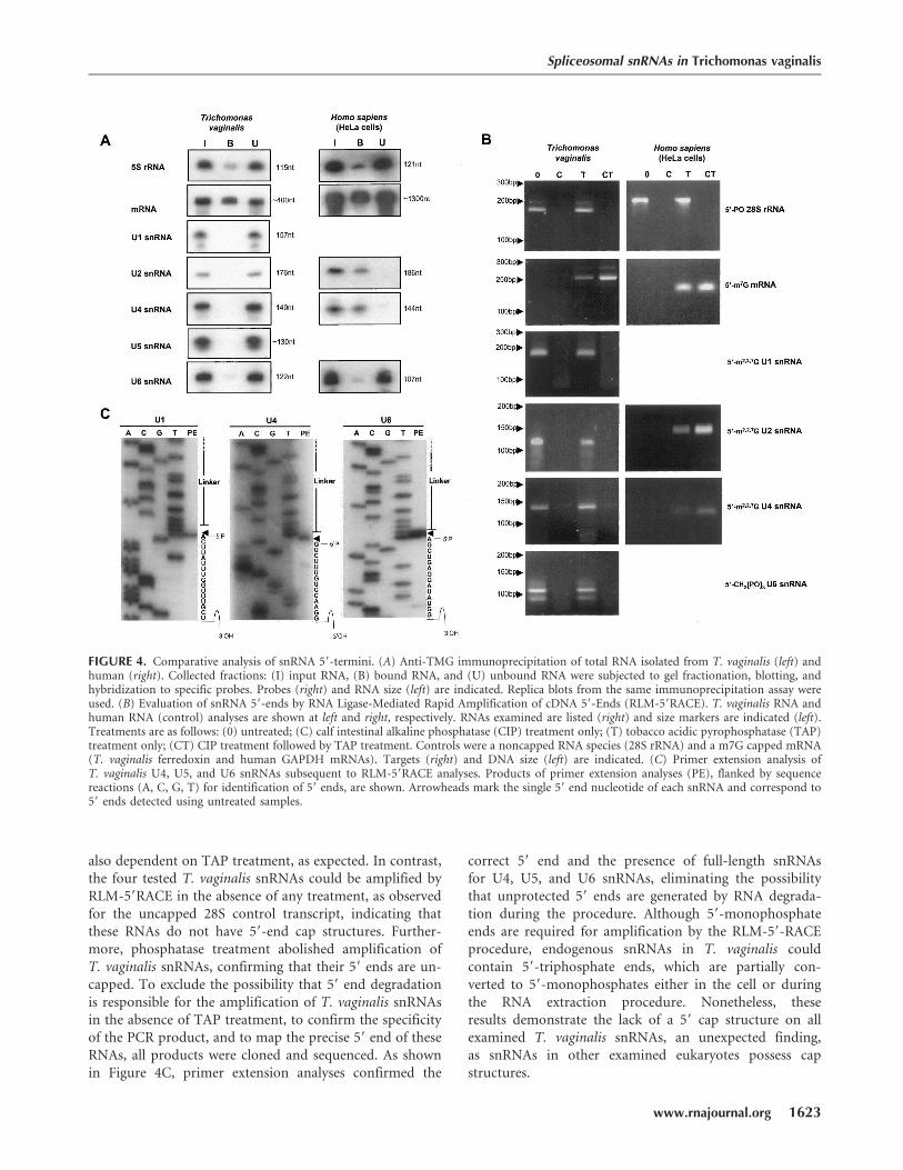
also dependent on TAP treatment, as expected. In contrast,the four tested T. vaginalis snRNAs could be amplified byRLM-59RACE in the absence of any treatment, as observedfor the uncapped 28S control transcript, indicating thatthese RNAs do not have 59-end cap structures. Further-more, phosphatase treatment abolished amplification ofT. vaginalis snRNAs, confirming that their 59 ends are un-capped. To exclude the possibility that 59 end degradationis responsible for the amplification of T. vaginalis snRNAsin the absence of TAP treatment, to confirm the specificityof the PCR product, and to map the precise 59 end of theseRNAs, all products were cloned and sequenced. As shownin Figure 4C, primer extension analyses confirmed the
correct 59 end and the presence of full-length snRNAsfor U4, U5, and U6 snRNAs, eliminating the possibilitythat unprotected 59 ends are generated by RNA degrada-tion during the procedure. Although 59-monophosphateends are required for amplification by the RLM-59-RACEprocedure, endogenous snRNAs in T. vaginalis couldcontain 59-triphosphate ends, which are partially con-verted to 59-monophosphates either in the cell or duringthe RNA extraction procedure. Nonetheless, theseresults demonstrate the lack of a 59 cap structure on allexamined T. vaginalis snRNAs, an unexpected finding,as snRNAs in other examined eukaryotes possess capstructures.
FIGURE 4. Comparative analysis of snRNA 59-termini. (A) Anti-TMG immunoprecipitation of total RNA isolated from T. vaginalis (left) andhuman (right). Collected fractions: (I) input RNA, (B) bound RNA, and (U) unbound RNA were subjected to gel fractionation, blotting, andhybridization to specific probes. Probes (right) and RNA size (left) are indicated. Replica blots from the same immunoprecipitation assay wereused. (B) Evaluation of snRNA 59-ends by RNA Ligase-Mediated Rapid Amplification of cDNA 59-Ends (RLM-59RACE). T. vaginalis RNA andhuman RNA (control) analyses are shown at left and right, respectively. RNAs examined are listed (right) and size markers are indicated (left).Treatments are as follows: (0) untreated; (C) calf intestinal alkaline phosphatase (CIP) treatment only; (T) tobacco acidic pyrophosphatase (TAP)treatment only; (CT) CIP treatment followed by TAP treatment. Controls were a noncapped RNA species (28S rRNA) and a m7G capped mRNA(T. vaginalis ferredoxin and human GAPDH mRNAs). Targets (right) and DNA size (left) are indicated. (C) Primer extension analysis ofT. vaginalis U4, U5, and U6 snRNAs subsequent to RLM-59RACE analyses. Products of primer extension analyses (PE), flanked by sequencereactions (A, C, G, T) for identification of 59 ends, are shown. Arrowheads mark the single 59 end nucleotide of each snRNA and correspond to59 ends detected using untreated samples.
Spliceosomal snRNAs in Trichomonas vaginalis
www.rnajournal.org 1623
JOBNAME: RNA 14#8 2008 PAGE: 7 OUTPUT: Friday July 4 20:35:26 2008
csh/RNA/164291/rna10454
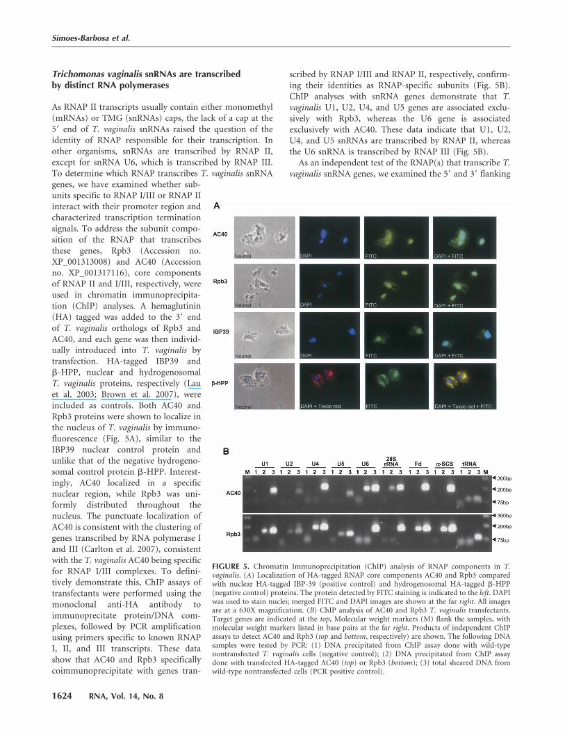
Trichomonas vaginalis snRNAs are transcribedby distinct RNA polymerases
As RNAP II transcripts usually contain either monomethyl(mRNAs) or TMG (snRNAs) caps, the lack of a cap at the59 end of T. vaginalis snRNAs raised the question of theidentity of RNAP responsible for their transcription. Inother organisms, snRNAs are transcribed by RNAP II,except for snRNA U6, which is transcribed by RNAP III.To determine which RNAP transcribes T. vaginalis snRNAgenes, we have examined whether sub-units specific to RNAP I/III or RNAP IIinteract with their promoter region andcharacterized transcription terminationsignals. To address the subunit compo-sition of the RNAP that transcribesthese genes, Rpb3 (Accession no.XP_001313008) and AC40 (Accessionno. XP_001317116), core componentsof RNAP II and I/III, respectively, wereused in chromatin immunoprecipita-tion (ChIP) analyses. A hemaglutinin(HA) tagged was added to the 39 endof T. vaginalis orthologs of Rpb3 andAC40, and each gene was then individ-ually introduced into T. vaginalis bytransfection. HA-tagged IBP39 andb-HPP, nuclear and hydrogenosomalT. vaginalis proteins, respectively (Lauet al. 2003; Brown et al. 2007), wereincluded as controls. Both AC40 andRpb3 proteins were shown to localize inthe nucleus of T. vaginalis by immuno-fluorescence (Fig. 5A), similar to theIBP39 nuclear control protein andunlike that of the negative hydrogeno-somal control protein b-HPP. Interest-ingly, AC40 localized in a specificnuclear region, while Rpb3 was uni-formly distributed throughout thenucleus. The punctuate localization ofAC40 is consistent with the clustering ofgenes transcribed by RNA polymerase Iand III (Carlton et al. 2007), consistentwith the T. vaginalis AC40 being specificfor RNAP I/III complexes. To defini-tively demonstrate this, ChIP assays oftransfectants were performed using themonoclonal anti-HA antibody toimmunoprecitate protein/DNA com-plexes, followed by PCR amplificationusing primers specific to known RNAPI, II, and III transcripts. These datashow that AC40 and Rpb3 specificallycoimmunoprecipitate with genes tran-
scribed by RNAP I/III and RNAP II, respectively, confirm-ing their identities as RNAP-specific subunits (Fig. 5B).ChIP analyses with snRNA genes demonstrate that T.vaginalis U1, U2, U4, and U5 genes are associated exclu-sively with Rpb3, whereas the U6 gene is associatedexclusively with AC40. These data indicate that U1, U2,U4, and U5 snRNAs are transcribed by RNAP II, whereasthe U6 snRNA is transcribed by RNAP III (Fig. 5B).
As an independent test of the RNAP(s) that transcribe T.vaginalis snRNA genes, we examined the 59 and 39 flanking
FIGURE 5. Chromatin Immunoprecipitation (ChIP) analysis of RNAP components in T.vaginalis. (A) Localization of HA-tagged RNAP core components AC40 and Rpb3 comparedwith nuclear HA-tagged IBP-39 (positive control) and hydrogenosomal HA-tagged b-HPP(negative control) proteins. The protein detected by FITC staining is indicated to the left. DAPIwas used to stain nuclei; merged FITC and DAPI images are shown at the far right. All imagesare at a 630X magnification. (B) ChIP analysis of AC40 and Rpb3 T. vaginalis transfectants.Target genes are indicated at the top, Molecular weight markers (M) flank the samples, withmolecular weight markers listed in base pairs at the far right. Products of independent ChIPassays to detect AC40 and Rpb3 (top and bottom, respectively) are shown. The following DNAsamples were tested by PCR: (1) DNA precipitated from ChIP assay done with wild-typenontransfected T. vaginalis cells (negative control); (2) DNA precipitated from ChIP assaydone with transfected HA-tagged AC40 (top) or Rpb3 (bottom); (3) total sheared DNA fromwild-type nontransfected cells (PCR positive control).
Simoes-Barbosa et al.
1624 RNA, Vol. 14, No. 8
JOBNAME: RNA 14#8 2008 PAGE: 8 OUTPUT: Friday July 4 20:35:35 2008
csh/RNA/164291/rna10454
Fig. 5 live 4/C
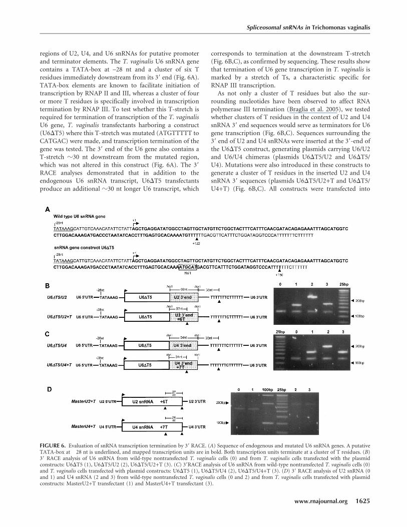
regions of U2, U4, and U6 snRNAs for putative promoterand terminator elements. The T. vaginalis U6 snRNA genecontains a TATA-box at –28 nt and a cluster of six Tresidues immediately downstream from its 39 end (Fig. 6A).TATA-box elements are known to facilitate initiation oftranscription by RNAP II and III, whereas a cluster of fouror more T residues is specifically involved in transcriptiontermination by RNAP III. To test whether this T-stretch isrequired for termination of transcription of the T. vaginalisU6 gene, T. vaginalis transfectants harboring a construct(U6DT5) where this T-stretch was mutated (ATGTTTTT toCATGAC) were made, and transcription termination of thegene was tested. The 39 end of the U6 gene also contains aT-stretch z30 nt downstream from the mutated region,which was not altered in this construct (Fig. 6A). The 39
RACE analyses demonstrated that in addition to theendogenous U6 snRNA transcript, U6DT5 transfectantsproduce an additional z30 nt longer U6 transcript, which
corresponds to termination at the downstream T-stretch(Fig. 6B,C), as confirmed by sequencing. These results showthat termination of U6 gene transcription in T. vaginalis ismarked by a stretch of Ts, a characteristic specific forRNAP III transcription.
As not only a cluster of T residues but also the sur-rounding nucleotides have been observed to affect RNApolymerase III termination (Braglia et al. 2005), we testedwhether clusters of T residues in the context of U2 and U4snRNA 39 end sequences would serve as terminators for U6gene transcription (Fig. 6B,C). Sequences surrounding the39 end of U2 and U4 snRNAs were inserted at the 39-end ofthe U6DT5 construct, generating plasmids carrying U6/U2and U6/U4 chimeras (plasmids U6DT5/U2 and U6DT5/U4). Mutations were also introduced in these constructs togenerate a cluster of T residues in the inserted U2 and U4snRNA 39 sequences (plasmids U6DT5/U2+T and U6DT5/U4+T) (Fig. 6B,C). All constructs were transfected into
FIGURE 6. Evaluation of snRNA transcription termination by 39 RACE. (A) Sequence of endogenous and mutated U6 snRNA genes. A putativeTATA-box at �28 nt is underlined, and mapped transcription units are in bold. Both transcription units terminate at a cluster of T residues. (B)39 RACE analysis of U6 snRNA from wild-type nontransfected T. vaginalis cells (0) and from T. vaginalis cells transfected with the plasmidconstructs: U6DT5 (1), U6DT5/U2 (2), U6DT5/U2+T (3). (C) 39RACE analysis of U6 snRNA from wild-type nontransfected T. vaginalis cells (0)and T. vaginalis cells transfected with plasmid constructs: U6DT5 (1), U6DT5/U4 (2), U6DT5/U4+T (3). (D) 39 RACE analysis of U2 snRNA (0and 1) and U4 snRNA (2 and 3) from wild-type nontransfected T. vaginalis cells (0 and 2) and from T. vaginalis cells transfected with plasmidconstructs: MasterU2+T transfectant (1) and MasterU4+T transfectant (3).
Spliceosomal snRNAs in Trichomonas vaginalis
www.rnajournal.org 1625
JOBNAME: RNA 14#8 2008 PAGE: 9 OUTPUT: Friday July 4 20:36:04 2008
csh/RNA/164291/rna10454
T. vaginalis and 39 RACE and DNA sequencing were usedto evaluate transcription termination. In this context, theinserted T cluster was also recognized as a terminator. Todirectly examine the termination of transcription of endo-geneous U2 or U4 snRNA transcription, the same T clusterwas inserted into constructs containing U2 or U4 geneswith their endogeneous upstream and downstream se-quences (Fig. 6D). Mapping of the 39 end of the resultingtranscripts, by sequencing cloned RT–PCR products, illus-trates that U2 and U4 gene transcription is not terminatedby this T stretch, and instead, termination occurred at thesame 39-end nucleotide as the endogenous genes. Thesefindings confirm the ChIP data indicating that the sameRNAP transcribes U2 and U4 genes and a different onetranscribes the U6 gene. Taken together, these data indicatethat T. vaginalis U6 gene transcribed by RNAP IIIand T. vaginalis U2 and U4 genes are transcribed byRNAP II.
DISCUSSION
Relatively few genes (z65) in the T. vaginalis genomeappear to contain an intron (Carlton et al. 2007). Theintrons that have been identified are uniformly short andcharacterized by a conserved 12-nt sequence (59-ACTAACACACAG/-39) that contains both the branch site (under-lined) and 39 splice site (/), with the former separated byonly 7 nt from the latter (Vanacova et al. 2005; Carlton et al.2007). Unusually short introns and the fusion of the branchsite and 39 splice site are also found in the four knownintrons in G. lamblia, another highly divergent eukaryote(Morrison et al. 2007). Prior to this study, spliceosomalsnRNAs required for splicing catalysis in other eukary-otes had not been identified from either Trichomonas orGiardia. Here we have identified all five T. vaginalisspliceosomal snRNAs. These snRNAs can be folded intoevolutionarily conserved secondary structures and containsequences and structural motifs known to be required forsplicing activity in other organisms (Guthrie and Patterson1988). Sm-binding motifs, necessary for binding the corecomponents of spliceosomal snRNPs, were found at con-served positions in all T. vaginalis snRNAs (Fig. 1). Theconsensus of these motifs, AAU3NR3, is similar to theestablished consensus RAU5GR, in contrast to the degen-erate sequence found in kinetoplastids (Mottram et al.1989; Hinas et al. 2006). Once snRNAs are identified inGiardia, should they be found to also be conserved, itwould be tempting to propose that the core structure ofmodern spliceosomes was established in basal eukaryotes.Should this be the case, the kinetoplastids would appear tohave diverged from this basal core structure.
The high degree of conservation observed for T. vaginalisspliceosomal snRNAs suggests that not only the mecha-nism of splicing, but also most steps involved in intronrecognition and rearrangement of spliceosomal RNA struc-
ture have been conserved throughout eukaryote evolution.These include the formation of commitment and pre-spliceosome complexes. Both primary sequence and sec-ondary structure of T. vaginalis U1 snRNA exhibit a highdegree of conservation with other eukaryotes. Conservationof its 59 end indicates an invariant mechanism for recog-nition of the 59SS. In addition, the presence of a highlyconserved apical loop at U1 snRNA helix II suggestsconservation of interacting U1-specific splicing factors.Furthermore, the T. vaginalis U1 snRNA was isolated basedon its interaction with a T. vaginalis U1-70k homolog,known to interact with the helix I loop of other eukaryoticU1 snRNAs (Surowy et al. 1989; Yuo and Weiner 1989).Despite the conservation of this interaction in T. vaginalis,its U1-70 k homolog (GenBank XP_001303000) is signifi-cantly smaller (z31 kDa) than its counterparts in othereukaryotes (Yuo and Weiner 1989). Moreover, the conservedregion in helix I (GAUC) (Surowy et al. 1989), to which it ispredicted to bind, has a nucleotide deletion (GUC).
U2 snRNA also binds to pre-mRNA within the pre-spliceosomal complex and undergoes changes in secondarystructure during spliceosome rearrangements and splicingcatalysis (Hilliker et al. 2007; Perriman and Ares Jr. 2007).Our structural prediction models strongly support theability of the T. vaginalis U2 snRNA to adopt secondarystructures and to interact with U6 snRNA and the con-served intron branch-site motif as previously described(Ares Jr. and Weiser 1995). The T. vaginalis U2 snRNAcontains the invariant branch-site interaction sequence59-GUAGUA-39 that can stably base pair to the unusuallyconserved branch site found in all T. vaginalis introns(Vanacova et al. 2005). T. vaginalis introns lack polypyr-imidine tracts between the branch site and 39SS, and thusare unlikely to bind U2 auxiliary factors (U2AFs) known tostabilize the branch site–U2 snRNA interaction in metazoa(Ares and Weiser 1995), suggesting that such factors maynot be required/present in the T. vaginalis spliceosome.
We have demonstrated that T. vaginalis U2 and U6snRNAs form stable complexes in vitro (Fig. 3). Sequenceanalysis allows for building models of base-pairing inter-actions between U6/U2 and U6/U4 snRNAs. By analogy tosnRNAs in other organisms, these models accommodateconformational changes thought to take place during thespliceosome assembly leading to the formation of acatalytically active spliceosome (Nilsen 1998). Two strictlyconserved U6 sequence motifs, ACAGAGA and AGC, areimplicated in formation of the catalytic center and itsrearrangements (Nilsen 1998). The strong similarity ofT. vaginalis U2/U6 snRNA interaction model to that ofother examined eukaryotes underscores the phylogeneticconservation of critical interactions (Fig. 2) that allow theformation of two mutually exclusive conformations pro-posed to facilitate the two-step splicing catalysis (Sashitalet al. 2004). Furthermore, similar U2/U6 snRNA structurehas been argued to be involved in an in vitro protein-free
Simoes-Barbosa et al.
1626 RNA, Vol. 14, No. 8
JOBNAME: RNA 14#8 2008 PAGE: 10 OUTPUT: Friday July 4 20:36:13 2008
csh/RNA/164291/rna10454
reaction that resembles certain aspects of splicing (Valadkhanand Manley 2001, 2003; Sashital et al. 2004). As discussedabove, our model also gives support to the phylogeneticconservation of U2/U6 helix I, divided by a 2-nt bulge intohelix Ia and Ib (Madhani and Guthrie 1992; McPheetersand Abelson 1992). In yeast, genetic evidence supports theformation of a tertiary interaction mediated by a bulged U2adenosine (A25) found between helix Ia and Ib and theguanosine G52 found in the U6 evolutionary conservedsequence A47CAGAGA53 (yeast numbering) (Madhani andGuthrie 1994). Although we do not have evidence of suchtertiary interaction, conservation of helix Ia and Ib inT. vaginalis U2/U6 interactions further supports a role forhelix I in splicing (Fig. 2).
T. vaginalis U6 intramolecular stem–loop (ISL) appearsto be less stable than its counterparts in yeast or metazoa.However, genetic analyses in yeast indicate that the stabilityof U6-ISL is not as important as its sequence (McPheeters1996) and that it is possible that other RNA structures orproteins that interact with U6-ISL compensate for itsapparent destabilization in T. vaginalis. The lack of stableU6-AGC base pairing to the U6-ISL in T. vaginalis mightindicate folding instability of this structure. Nevertheless,our model supports the importance of the upper portion ofthis stem (McPheeters 1996; Shukla and Padgett 2001) aswell as the formation of the U6 ISL pentaloop GNR(N)A,similar to that proposed in other organisms (Huppleret al. 2002). Structure-function analyses of the T. vaginalisspliceosomal proteins and snRNAs will be necessary to testwhether these structural differences impact splicing mech-anisms in this divergent eukaryote. It is possible that thisand other differences in stabilities of various RNA–RNAinteractions within snRNAs in different organisms reflectmodulation of relative stabilities of spliceosomal confor-mations along the splicing pathway. The U6 ISL sequencealso affects U4–U6 pairing, which in T. vaginalis representsa stable, perfect Watson-Crick interaction, whereas humanU4–U6 stem II contains several U-G pairs. At present, ourunderstanding of such structural rearrangements is insuf-ficient to predict detailed consequences of the observed se-quence differences between T. vaginalis and metazoan snRNAs.
In addition to the sequence and structural similarities ofthe newly identified T. vaginalis spliceosomal snRNAs com-pared with those in yeast and metazoa, a high degree ofconservation in another important component of the spliceo-some, Prp8p, has been previously described (Fast andDoolittle 1999). This large (280 kDa) component of U5snRNP is involved in multiple interactions near the cat-alytic center (Grainger and Beggs 2005) and implicated inmodulation of spliceosomal transitions between the catalyticsteps (Liu et al. 2007). The T. vaginalis PRP8 is also highlyconserved; over its entire length it shares 56% amino acididentity with the human and Caenorhabditis elegans pro-teins. These observations further support our argumentbased on structural similarities of snRNAs that the splicing
apparatus and splicing mechanisms have been highlyconserved among all eukaryotes.
An unusual property of T. vaginalis snRNAs, however, isthe absence of a characteristic 59-terminal TMG cap, foundin other organisms (Mouaikel et al. 2002). Instead, wefound that T. vaginalis snRNAs have unmodified 59 termini.The biological significance of a TMG 59-cap structure onsnRNAs is not fully understood; however, roles in traffickingand snRNP assembly have been proposed (Huber et al.2002; Mouaikel et al. 2002; Narayanan et al. 2003; Cougotet al. 2004). Whether the lack of a 59 cap influences traffick-ing and/or assembly of T. vaginalis snRNAs awaits fur-ther analyses. It is notable that although S. cerevisiae andSchizosaccharomyces pombe snRNAs have TMG caps, itspresence is not essential, as deletion of the enzyme requiredfor formation of a TMG cap, trimethylguanosine synthase(Tgs), results in snRNAs lacking TMG caps, but does notaffect viability (Mouaikel et al. 2002; Hausmann et al. 2007).On the other hand, similar studies in Drosophila have shownthat the absence of TMG cap synthesis and TMG-containingRNAs result in embryonic lethality (Komonyi et al. 2005).
As capping of transcripts is coordinated with transcrip-tion and is specific for different RNAPs, we determinedwhich RNAP(s) transcribe T. vaginalis snRNAs. These dataindicate that U1, U2, U4, and U5 genes are transcribed byRNAP II, despite the absence of a 59 cap structure typicallyformed on nascent transcripts early during RNAP IItranscription (Cougot et al. 2004). Using the same exper-imental approaches, T. vaginalis mRNAs, also transcribedby RNAP II (Liston et al. 2001; Schumacher et al. 2003),were found to contain a 59 cap structure. Thus, the absenceof a cap on the snRNA transcripts does not reflect a gen-eral lack of capping of T. vaginalis RNAP II transcripts.Whether T. vaginalis RNAP II snRNA transcripts aretranscribed by a variant RNAP II complex that does notassociate with fully active capping machinery or theyoriginally contain a cap that is subsequently removed by59 end processing, is unknown.
T. vaginalis U6 snRNA was found to be transcribed byRNAP III, consistent with structural properties of the gene.This U6 gene lacks an A/B box and has a TATA-boxat position �28 nt similar to the mammalian RNAP IIIU6 snRNA genes that rely on a extragenic 59 promoter(Hernandez 2001). The 39 end of the T. vaginalis U6snRNA is also flanked by a cluster of T-residues, whichwas demonstrated to mark termination of transcription, acharacteristic of RNAP III transcripts.
Together, our analyses illustrate that T. vaginalis snRNAsare unique among their eukaryotic counterparts, based onthe absence of a 59-end cap structure, but are otherwiseextremely conserved based on predicted secondary struc-ture models, indicating a strong conservation of splicingmechanisms throughout eukaryotic evolution. Availabilityof these snRNAs sets the stage for the isolation and charac-terization of spliceosomes from this divergent eukaryote,
Spliceosomal snRNAs in Trichomonas vaginalis
www.rnajournal.org 1627
JOBNAME: RNA 14#8 2008 PAGE: 11 OUTPUT: Friday July 4 20:36:13 2008
csh/RNA/164291/rna10454
allowing further analyses of the conservation of core spli-ceosomal proteins in diverse eukaryotes.
MATERIALS AND METHODS
Identification of T. vaginalis snRNAs
The 73 coverage of the T. vaginalis genome sequence database(http://www.tigr.org/tdb/e2k1/tvg/) was downloaded to create alocal database that was searched using BioEdit software (matrixPAM40 setting) to identify trichomonad snRNAs. Candidate U2,U4, and U5 snRNAs were identified initially using a BLASTnsearch, followed by structural analyses (described below); U6 andU1 snRNA identification required a more involved bioinformaticsapproach and functional assays, respectively. Three candidate U2genes resulted from a BLASTn search, using CAAGTGTAGTATCTG (conserved nucleotides 28–42 in human U2 snRNA).RNAfold software (Hofacker 2003) was then used to generatepredicted structures from these three sequences and theirupstream and downstream regions. A subsequent search forconserved nucleotides and predicted U2 helix I and IIa lead tothe identification of a single U2 snRNA gene. A BLASTn search,using TTCGCCTTTTACTA (conserved nucleotides 34–n47 inhuman U5 snRNA) likewise resulted in three candidate U5 genesthat were then subjected to the structural analyses. A single U5snRNA gene, capable of forming the stem–loop I with its 39
conserved CCG, was identified. The U6 snRNA was identifiedusing INFERNAL software (ftp://ftp.genetics.wustl.edu/pub/eddy/software/infernal), snRNA databases, and covariance models fromThe Sanger Institute Rfam database (http://www.sanger.ac.uk/Software/Rfam/index.shtml). A multiple U6 snRNA alignmentwas built and used to screen the 7X T. vaginalis genome databaseusing the default parameters of the cmsearch program. Theresulting output revealed three hits; however, only the top hithad a significant score value. Further examination of this hitshowed it to have the two U6 snRNA invariant sequencesACAGAGA and AGC, whereas the other two did not. To identifyU4 snRNA, a BLASTn search using U6 snRNA nucleotides 71–85(CTCCTTGGACAAGA) predicted to interact with U4 was done,resulting in 12 candidate genes. Each sequence was searched for apossible intramolecular stem–loop in nucleotide positions 30–35,followed by 10–15 nt that could interact with the candidate U6snRNA to form a Y-shaped U4–U6 interaction domain. Thisapproach resulted in the identification of a single U4 snRNA gene.
U1 snRNA was not successfully identified using bioinformatics.To isolate U1 snRNA, the T. vaginalis homolog of U1-70K(GenBank accession number XM_001302999.1) was tagged withstreptoavidin binding protein (SBP) at its C terminus and clonedinto the Master-Neo plasmid (Dyall et al. 2003). Nuclear extractsof T. vaginalis cells transfected with the resulting construct wereprepared as described (Mayeda and Krainer 1999), except para-sites were lysed by passage through a 25G5/8 needle syringe.Approximately 0.6 mg of nuclear protein extract was mixed with300 mL of prewashed streptavidin Sepharose (Amersham) in afinal volume of 1.2 mL of 20 mM Hepes (pH7.9); 70 mM KCl,20 mM MgCl2, 2 mM EDTA, 0.05% NP-40, 0.5 mM DTT, plusprotease inhibitors (Roche complete mini, EDTA-free, cocktail).After 1 h incubation at 4°C, the Sepharose was washed 8X with1 mL of the same buffer, and RNA was extracted by Trizol LS
extraction (Invitrogen). [32P]pCp labeling of the recovered RNAwas performed and visualized in a TBE-Urea 5% polyacrylamidegel by autoradiography. RNAs ranging in size from 100 to 200 ntwere recovered from the gel, polyadenylated with recombinantEscherichia coli Poly-A Polymerase I (Ambion), phenol extracted,ethanol precipitated, and reverse transcribed using a polydT 2-base anchor primer adapter, as described (FirstChoice RLM-RACEkit, Ambion). cDNA was then isolated by gel electrophoresisand poly-C tailed at the 59 terminus by terminal deoxynucleotidyltransferase. Outer and inner PCR products were obtained by usingGI-rich adapter primers (59 RACE system, Invitrogen) andspecific primers for the polydT adapter (FirstChoice RLM-RACEkit, Ambion). All PCR products were cloned into the Topo pCR-4vector (Invitrogen) and sequenced using M13 primers and the BigDye version 3 labeling kit (Applied Biosystems).
In vitro interaction of U2–U6 minimalcatalytic domains
The region corresponding to the central domain of U6 snRNA (nt41–111) was transcribed in a large scale T7-transcription system(Ambion) and oligonucleotides corresponding to the 59 domainof U2 snRNA (nt 1–47) and RNA branch oligonucleotide (RO;UACUAACACUUA) were purchased from Invitrogen. Followingacrylamide gel purification of the RNAs, an in vitro interactionassay was performed as described previously (Valadkhan andManley 2000). U2 or U6 RNA domains labeled at the 39-end with[32P]pCp and RNA ligase (Ambion) were incubated in thepresence of 5 nM of the unlabeled U2 snRNA and 0, 1, 5, and10 nM of the 32P-labeled U6 snRNA, and vice versa, in thepresence of 0, 5, 25, and 125 mM of MgCl2. Reactions, includingthe RO, contained 5 nM of 32P-labeled U6 RNA, 10 nM of theunlabeled U2 RNA (U2), 20 nM of the RO), and 20 mM of MgCl2.Complexes were then treated for 15 min at 4, 37, 45, 55, 60, or70°C before loading on a polyacrylamide gel and analyzed by gelelectrophoresis as described (Valadkhan and Manley 2000).
T. vaginalis culture and transfections
T. vaginalis strain T1 was maintained in TYM medium supple-mented with 10% horse serum and iron (Clark and Diamond2002). Transfections were conducted as described (Delgadilloet al. 1997), and cultures were selected with Gentamicin (GIBCO).Within 10 d of cell culturing, DNA, RNA, or protein preparationswere prepared from transfectants.
Plasmid constructs, chimeras, and mutagenesis
All gene constructs used the Master-Neo plasmid, a T. vaginalisexpression vector for stable transfection that contains the neo-mycin resistance gene for selection and NdeI and Asp718 sites forcloning genes of interest immediately upstream of a double-HAepitope tag (Dyall et al. 2003). Constructs of genes containingnative 59 UTR and/or 39 UTRs were made by digesting Master-Neo, followed by ligation with PCR-generated Rpb3, U2, U4, orU6 genes. Site-directed mutagenesis was performed by PCR, andthe mutation was confirmed by sequencing. U6–U2 and U6–U4chimeras were made by inserting a NsiI restriction site to deletethe U6 snRNA terminator. The last 37 and 34 nt of U2 and U4snRNA sequences, respectively, were inserted into the 39 end of
Simoes-Barbosa et al.
1628 RNA, Vol. 14, No. 8
JOBNAME: RNA 14#8 2008 PAGE: 12 OUTPUT: Friday July 4 20:36:14 2008
csh/RNA/164291/rna10454
U6 snRNA to produce the chimeras. In U2 snRNA, positions 147and 149 were mutated to generate a stretch of six T residues, asunderlined (146TGTCTTCAC154 to 146TTTTTTCAC154). In U4snRNA, positions 124 and 125 were mutated to generate a stretchof seven T residues, as underlined (121CTTAATTTC129 to
121CTTTTTTTC129).
RNA isolation and analysis
Total RNA was isolated by mirVana� PARIS� isolation kit(Ambion). For snRNA analyses, RNA was separated on eitherTBE-Urea 5% acrylamide or formaldehyde agarose gels. RNAgels were transferred to nylon membranes using standard proce-dures (GE Healthcare) and hybridized with 32P-labeled probesusing recommended stringency (Ambion). For the snRNAs, hybrid-izations were confirmed using three independent DNA oligomers asprobes.
Immunoprecipitation of RNAby antitrimethylguanosine(TMG) monoclonal antibody
Twenty microliters of prewashed anti-2,2,7-trimethylguanosine(TMG) antibody agarose beads (Calbiochem), were incubatedwith 100 mg of total RNA in 0.5 mL 01.% Triton X-100, 50 mMNaCl, and 20 mM HEPES (pH 7.9) for 1 h at 4°C. The super-natant was recovered as the unbound RNA and precipitated withethanol. Beads were washed eight times with 0.5 mL incubationbuffer by repeated centrifugation. Elution was then performedtwice by incubating beads with 100 mL of 0.5% SDS, 10 mM Tris-HCl (pH 8.0), 10 mM EDTA, and 10 mg/mL of proteinase Kat 55°C for 15 min each. Recovered bound RNA was phenol/chloroform extracted and ethanol precipitated.
Mapping of 39 and 59-ends of snRNAs by primerextension and DNA sequencing
RNA ligase-mediated rapid amplification of cDNA 59-Ends(RLM-59 RACE) was performed with DNAse treated (Ambion)RNA from T. vaginalis and HeLa cells, following recommenda-tions (Invitrogen). From the same RNA sample, different treat-ments were performed prior to RNA oligomer ligation and reversetranscription, as follows: (1) no treatment, (2) calf-intestinealkalyne phosphatase (CIP) only, (3) tobacco acid pyrophospha-tase (TAP) only, (4) CIP followed by TAP treatment. The treatedRNA samples were then split in equal parts for RT–PCR reactionsusing RNA adapter specific primers and gene-specific primers fordetection of the following transcript targets: (1) T. vaginalisferredoxin mRNA and human GAPDH mRNA, (2) T. vaginalisand human 28S RNAs, (3) T. vaginalis and human snRNAs U2,U4, and U6. Results were reproduced in an independent duplicateexperiment. All RT–PCR products were cloned and sequenced.For T. vaginalis snRNAs, manual DNA sequencing (SequiThermExcel II, Epicentre) was performed in parallel to primer extensionreactions using the respective gene-specific reverse 32P-labeledprimer used in the RLM-59 RACE. T. vaginalis total RNA wassubjected to polyadenylation and 39 ends of the snRNAs weremapped using 39 RACE as recommended (Poly(A) tailing kit andFirstChoice RLM-RACE, Ambion).
Immunofluorescence microscopy
T. vaginalis cells were washed with warm phosphate-bufferedsaline (PBS) and allowed to attach to coverslips for 1–2 h in ahumidifying chamber at 37°C. All procedures thereafter weredone at room temperature. The cells were fixed with 3.5%formalin in PBS for 20 min, quenched with 100 mM glycine inPBS for 5 min, blocked with 3% BSA in PBS for 15 min, andpermeabilized with 3% BSA, 0.2% Triton X-100 in PBS for 15min. Rpb3 and AC40 HA-tagged transfectants were visualizedusing mouse anti-HA monoclonal antibody (Covance) andsecondary Alexa Fluor 488 (FITC) donkey antimouse antibodies(Invitrogen). HA-tagged IBP39 (Lau et al. 2003) and b-HPP(Brown et al. 2007) transfectants were localized as previouslydescribed. For each antibody, 1 h incubation was followed by fivePBS washes. Coverslips were then mounted onto slides usingProLong Gold with DAPI (Invitrogen). An Axioscop2 microscopeoutfitted with Axiovision v. 3.2 software was used to visualize andprocess images (Zeiss).
Chromatin immunoprecipitation (ChIP) analysis
T. vaginalis transfectants expressing AC40 (GenBank� accessionno. XP_001317116) or Rpb3 (GenBank accession no. XP_001313008)tagged with double-HA epitope at the C terminus were used forChIP assays as previously described (Ong et al. 2006), exceptwashes were conducted twice with each of the following buffers:(1) 1% Triton X-100, 1 mM EDTA, 50 mM HEPES (pH 7.5), 150mM NaCl, 0.1% deoxycholate; (2) 1% Triton X-100, 1 mMEDTA, 50 mM HEPES (pH 7.5), 500 mM NaCl, 0.1% deoxy-cholate; (3) 0.5% NP-40, 1 mM EDTA, 10 mM Tris-HCl (pH 8.0),250 mM LiCl, 0.5% deoxycholate. For negative controls, eitherwild-type cells were included or the primary anti-HA antibodywas omitted for the transfected cell samples. Sheared DNA fromwild-type cells before going to ChIP was used as a positive PCRcontrol. Different sets of primers were used to detect the followingtranscription targets; (1) 28S rRNA for a RNAP I gene; (2)ferredoxin or a-SCS for RNAP II genes; (3) tRNA for RNAP IIIgenes; (4) U2, U4, and U6 snRNA genes. Forward and reverseprimers were designed to detect a product about 150–200 nt,having the forward primer to anneal about 100 nt upstream ofthe transcription unit. The sole exception was the tRNA genethat primers were designed to detect the full transcript. PCRproducts were resolved in 1.5% TBE-agarose gels. The experi-ment was performed in duplicate for each AC40 and Rpb3transfectants.
SUPPLEMENTAL DATA
Supplemental material can be found at http://www.rnajournal.org.
ACKNOWLEDGMENTS
We thank Maria Delgadillo-Correa and Camila L. Correa fortechnical assistance, Alias J. Smith for help with the ChIP analyses,and Drs. Cheryl Okumura, Chris Ryan, and Nancy Sturm forcritical comments on the manuscript and our colleagues in the labfor helpful discussions. This work was supported by the NationalInstitute of Allergy and Infectious Diseases, National Institutes of
Spliceosomal snRNAs in Trichomonas vaginalis
www.rnajournal.org 1629
JOBNAME: RNA 14#8 2008 PAGE: 13 OUTPUT: Friday July 4 20:36:14 2008
csh/RNA/164291/rna10454
Health grant AI30537 to P.J.J., NIH grant GM49044 to M.M.K.,and a fellowship to A.S.B. from CNPq-Brazil (PDE 200065/2004-1).
Received February 26, 2008; accepted April 24, 2008.
REFERENCES
Abrahamsen, M.S., Templeton, T.J., Enomoto, S., Abrahante, J.E.,Zhu, G., Lancto, C.A., Deng, M., Liu, C., Widmer, G., Tzipori, S.,et al. 2004. Complete genome sequence of the apicomplexan,Cryptosporidium parvum. Science 304: 441–445.
Ares Jr., M. and Weiser, B. 1995. Rearrangement of snRNA structureduring assembly and function of the spliceosome. Prog. NucleicAcid Res. Mol. Biol. 50: 131–159.
Baldauf, S.L. 2003. The deep roots of eukaryotes. Science 300: 1703–1706.Braglia, P., Percudani, R., and Dieci, G. 2005. Sequence context effects
on oligo(dT) termination signal recognition by Saccharomycescerevisiae RNA polymerase III. J. Biol. Chem. 280: 19551–19562.
Brow, D.A. and Guthrie, C. 1988. Spliceosomal RNA U6 is remarkablyconserved from yeast to mammals. Nature 334: 213–218.
Brown, M.T., Goldstone, H.M., Bastida-Corcuera, F., Delgadillo-Correa, M.G., McArthur, A.G., and Johnson, P.J. 2007. Afunctionally divergent hydrogenosomal peptidase with protomi-tochondrial ancestry. Mol. Microbiol. 64: 1154–1163.
Burge, C.B., Thomas, T., and Sharp, P.A. 1999. Splicing of precursors tomRNAs by the Spliceosome. Cold Spring Harbor Laboratory Press,Cold Spring Harbor, New York.
Busch, H., Reddy, R., Rothblum, L., and Choi, Y.C. 1982. SnRNAs,SnRNPs, and RNA processing. Annu. Rev. Biochem. 51: 617–654.
Carlton, J.M., Hirt, R.P., Silva, J.C., Delcher, A.L., Schatz, M.,Zhao, Q., Wortman, J.R., Bidwell, S.L., Alsmark, U.C.,Besteiro, S., et al. 2007. Draft genome sequence of the sexuallytransmitted pathogen Trichomonas vaginalis. Science 315: 207–212.
Clark, C.G. and Diamond, L.S. 2002. Methods for cultivation ofluminal parasitic protists of clinical importance. Clin. Microbiol.Rev. 15: 329–341.
Cougot, N., van Dijk, E., Babajko, S., and Seraphin, B. 2004. ‘Cap-tabolism’. Trends Biochem. Sci. 29: 436–444.
Davis, C.A., Brown, M.P., and Singh, U. 2007. Functional character-ization of spliceosomal introns and identification of U2, U4, andU5 snRNAs in the deep-branching eukaryote Entamoeba histoly-tica. Eukaryot. Cell 6: 940–948.
Delgadillo, M.G., Liston, D.R., Niazi, K., and Johnson, P.J. 1997.Transient and selectable transformation of the parasitic protistTrichomonas vaginalis. Proc. Natl. Acad. Sci. 94: 4716–4720.
Dyall, S.D., Lester, D.C., Schneider, R.E., Delgadillo-Correa, M.G.,Plumper, E., Martinez, A., Koehler, C.M., and Johnson, P.J. 2003.Trichomonas vaginalis Hmp35, a putative pore-forming hydro-genosomal membrane protein, can form a complex in yeastmitochondria. J. Biol. Chem. 278: 30548–30561.
Ebel, C., Frantz, C., Paulus, F., and Imbault, P. 1999. Trans-splicingand cis-splicing in the colourless Euglenoid, Entosiphon sulcatum.Curr. Genet. 35: 542–550.
El-Sayed, N.M., Myler, P.J., Bartholomeu, D.C., Nilsson, D.,Aggarwal, G., Tran, A.N., Ghedin, E., Worthey, E.A., Delcher, A.L.,Blandin, G., et al. 2005. The genome sequence of Trypanosoma cruzi,etiologic agent of Chagas disease. Science 309: 409–415.
Fabrizio, P. and Abelson, J. 1992. Thiophosphates in yeast U6 snRNAspecifically affect pre-mRNA splicing in vitro. Nucleic Acids Res.20: 3659–3664.
Fantoni, A., Dare, A.O., and Tschudi, C. 1994. RNA polymerase III-mediated transcription of the trypanosome U2 small nuclear RNAgene is controlled by both intragenic and extragenic regulatoryelements. Mol. Cell. Biol. 14: 2021–2028.
Fast, N.M. and Doolittle, W.F. 1999. Trichomonas vaginalis possesses agene encoding the essential spliceosomal component, PRP8. Mol.Biochem. Parasitol. 99: 275–278.
Gardner, M.J., Hall, N., Fung, E., White, O., Berriman, M.,Hyman, R.W., Carlton, J.M., Pain, A., Nelson, K.E., Bowman, S.,et al. 2002. Genome sequence of the human malaria parasitePlasmodium falciparum. Nature 419: 498–511.
Grainger, R.J. and Beggs, J.D. 2005. Prp8 protein: At the heart of thespliceosome. RNA 11: 533–557.
Guthrie, C. and Patterson, B. 1988. Spliceosomal snRNAs. Annu. Rev.Genet. 22: 387–419.
Hausmann, S., Ramirez, A., Schneider, S., Schwer, B., and Shuman, S.2007. Biochemical and genetic analysis of RNA cap guanine-N2methyltransferases from Giardia lamblia and Schizosaccharomycespombe. Nucleic Acids Res. 35: 1411–1420.
Hernandez, N. 2001. Small nuclear RNA genes: A model system tostudy fundamental mechanisms of transcription. J. Biol. Chem.276: 26733–26736.
Hilliker, A.K. and Staley, J.P. 2004. Multiple functions for theinvariant AGC triad of U6 snRNA. RNA 10: 921–928.
Hilliker, A.K., Mefford, M.A., and Staley, J.P. 2007. U2 togglesiteratively between the stem IIa and stem IIc conformations topromote pre-mRNA splicing. Genes & Dev. 21: 821–834.
Hinas, A., Larsson, P., Avesson, L., Kirsebom, L.A., Virtanen, A., andSoderbom, F. 2006. Identification of the major spliceosomal RNAsin Dictyostelium discoideum reveals developmentally regulated U2variants and polyadenylated snRNAs. Eukaryot. Cell 5: 924–934.
Hofacker, I.L. 2003. Vienna RNA secondary structure server. NucleicAcids Res. 31: 3429–3431.
Huber, J., Dickmanns, A., and Luhrmann, R. 2002. The importin-bbinding domain of snurportin1 is responsible for the Ran- andenergy-independent nuclear import of spliceosomal U snRNPs invitro. J. Cell Biol. 156: 467–479.
Huppler, A., Nikstad, L.J., Allmann, A.M., Brow, D.A., andButcher, S.E. 2002. Metal binding and base ionization in the U6RNA intramolecular stem-loop structure. Nat. Struct. Biol. 9: 431–435.
Komonyi, O., Papai, G., Enunlu, I., Muratoglu, S., Pankotai, T.,Kopitova, D., Maroy, P., Udvardy, A., and Boros, I. 2005. DTL, theDrosophila homolog of PIMT/Tgs1 nuclear receptor coactivator-interacting protein/RNA methyltransferase, has an essential role indevelopment. J. Biol. Chem. 280: 12397–12404.
Kretzner, L., Rymond, B.C., and Rosbash, M. 1987. S. cerevisiae U1RNA is large and has limited primary sequence homology tometazoan U1 snRNA. Cell 50: 593–602.
Lau, A.O., Liston, D.R., Vanacova, S., and Johnson, P.J. 2003.Trichomonas vaginalis initiator binding protein, IBP39, containsa novel DNA binding motif. Mol. Biochem. Parasitol. 130: 167–171.
Liston, D.R., Lau, A.O., Ortiz, D., Smale, S.T., and Johnson, P.J. 2001.Initiator recognition in a primitive eukaryote: IBP39, an initiator-binding protein from Trichomonas vaginalis. Mol. Cell. Biol. 21:7872–7882.
Liu, L., Query, C.C., and Konarska, M.M. 2007. Opposing classes ofprp8 alleles modulate the transition between the catalytic steps ofpre-mRNA splicing. Nat. Struct. Mol. Biol. 14: 519–526.
Loftus, B., Anderson, I., Davies, R., Alsmark, U.C., Samuelson, J.,Amedeo, P., Roncaglia, P., Berriman, M., Hirt, R.P., Mann, B.J.,et al. 2005. The genome of the protist parasite Entamoebahistolytica. Nature 433: 865–868.
Madhani, H.D. and Guthrie, C. 1992. A novel base-pairing interactionbetween U2 and U6 snRNAs suggests a mechanism for thecatalytic activation of the spliceosome. Cell 71: 803–817.
Madhani, H.D. and Guthrie, C. 1994. Randomization-selectionanalysis of snRNAs in vivo: evidence for a tertiary interaction inthe spliceosome. Genes & Dev. 8: 1071–1086.
Mayeda, A. and Krainer, A.R. 1999. Preparation of HeLa cell nuclearand cytosolic S100 extracts for in vitro splicing. Methods Mol. Biol.118: 309–314.
McPheeters, D.S. 1996. Interactions of the yeast U6 RNA with thepre-mRNA branch site. RNA 2: 1110–1123.
McPheeters, D.S. and Abelson, J. 1992. Mutational analysis of theyeast U2 snRNA suggests a structural similarity to the catalyticcore of group I introns. Cell 71: 819–831.
Simoes-Barbosa et al.
1630 RNA, Vol. 14, No. 8
JOBNAME: RNA 14#8 2008 PAGE: 14 OUTPUT: Friday July 4 20:36:15 2008
csh/RNA/164291/rna10454
Morrison, H.G., McArthur, A.G., Gillin, F.D., Aley, S.B., Adam, R.D.,Olsen, G.J., Best, A.A., Cande, W.Z., Chen, F., Cipriano, M.J., et al.2007. Genomic minimalism in the early diverging intestinalparasite Giardia lamblia. Science 317: 1921–1926.
Mottram, J., Perry, K.L., Lizardi, P.M., Luhrmann, R., Agabian, N.,and Nelson, R.G. 1989. Isolation and sequence of four smallnuclear U RNA genes of Trypanosoma brucei subsp. brucei:Identification of the U2, U4, and U6 RNA analogs. Mol. Cell.Biol. 9: 1212–1223.
Mouaikel, J., Verheggen, C., Bertrand, E., Tazi, J., and Bordonne, R.2002. Hypermethylation of the cap structure of both yeast snRNAsand snoRNAs requires a conserved methyltransferase that islocalized to the nucleolus. Mol. Cell 9: 891–901.
Narayanan, A., Eifert, J., Marfatia, K.A., Macara, I.G., Corbett, A.H.,Terns, R.M., and Terns, M.P. 2003. Nuclear RanGTP is notrequired for targeting small nucleolar RNAs to the nucleolus.J. Cell Sci. 116: 177–186.
Nelissen, R.L., Will, C.L., van Venrooij, W.J., and Luhrmann, R. 1994.The association of the U1-specific 70K and C proteins with U1snRNPs is mediated in part by common U snRNP proteins. EMBOJ. 13: 4113–4125.
Nilsen, T.W. 1998. RNA–RNA interactions in nuclear pre-mRNA splicing.Cold Spring Harbor Laboratory Press, Cold Spring Harbor, NY.
Nilsen, T.W. 2003. The spliceosome: The most complex macromo-lecular machine in the cell? Bioessays 25: 1147–1149.
Nixon, J.E., Wang, A., Morrison, H.G., McArthur, A.G., Sogin, M.L.,Loftus, B.J., and Samuelson, J. 2002. A spliceosomal intron inGiardia lamblia. Proc. Natl. Acad. Sci. 99: 3701–3705.
O’Keefe, R.T., Norman, C., and Newman, A.J. 1996. The invariant U5snRNA loop 1 sequence is dispensable for the first catalytic step ofpre-mRNA splicing in yeast. Cell 86: 679–689.
Ong, S.J., Hsu, H.M., Liu, H.W., Chu, C.H., and Tai, J.H. 2006.Multifarious transcriptional regulation of adhesion protein geneap65-1 by a novel Myb1 protein in the protozoan parasiteTrichomonas vaginalis. Eukaryot. Cell 5: 391–399.
Perriman, R.J. and Ares Jr., M. 2007. Rearrangement of competing U2RNA helices within the spliceosome promotes multiple steps insplicing. Genes & Dev. 21: 811–820.
Sashital, D.G., Cornilescu, G., McManus, C.J., Brow, D.A., andButcher, S.E. 2004. U2-U6 RNA folding reveals a group IIintron-like domain and a four-helix junction. Nat. Struct. Mol.Biol. 11: 1237–1242.
Schumacher, M.A., Lau, A.O., and Johnson, P.J. 2003. Structural basis ofcore promoter recognition in a primitive eukaryote. Cell 115: 413–424.
Sharp, P.A. 1985. On the origin of RNA splicing and introns. Cell 42:397–400.
Shukla, G.C. and Padgett, R.A. 2001. The intramolecular stem-loopstructure of U6 snRNA can functionally replace the U6atac snRNAstem-loop. RNA 7: 94–105.
Singh, R. and Reddy, R. 1989. g-monomethyl phosphate: A capstructure in spliceosomal U6 small nuclear RNA. Proc. Natl. Acad.Sci. 86: 8280–8283.
Speckmann, W.A., Terns, R.M., and Terns, M.P. 2000. The box C/Dmotif directs snoRNA 59-cap hypermethylation. Nucleic Acids Res.28: 4467–4473.
Sun, J.S. and Manley, J.L. 1995. A novel U2-U6 snRNA structure isnecessary for mammalian mRNA splicing. Genes & Dev. 9: 843–854.
Surowy, C.S., van Santen, V.L., Scheib-Wixted, S.M., and Spritz, R.A.1989. Direct, sequence-specific binding of the human U1-70Kribonucleoprotein antigen protein to loop I of U1 small nuclearRNA. Mol. Cell. Biol. 9: 4179–4186.
Valadkhan, S. 2005. snRNAs as the catalysts of pre-mRNA splicing.Curr. Opin. Chem. Biol. 9: 603–608.
Valadkhan, S. and Manley, J.L. 2000. A tertiary interaction detected ina human U2-U6 snRNA complex assembled in vitro resembles agenetically proven interaction in yeast. RNA 6: 206–219.
Valadkhan, S. and Manley, J.L. 2001. Splicing-related catalysis byprotein-free snRNAs. Nature 413: 701–707.
Valadkhan, S. and Manley, J.L. 2003. Characterization of the catalyticactivity of U2 and U6 snRNAs. RNA 9: 892–904.
Vanacova, S., Yan, W., Carlton, J.M., and Johnson, P.J. 2005.Spliceosomal introns in the deep-branching eukaryote Trichomo-nas vaginalis. Proc. Natl. Acad. Sci. 102: 4430–4435.
Wu, J.A. and Manley, J.L. 1991. Base pairing between U2 and U6snRNAs is necessary for splicing of a mammalian pre-mRNA.Nature 352: 818–821.
Yean, S.L., Wuenschell, G., Termini, J., and Lin, R.J. 2000. Metal-ioncoordination by U6 small nuclear RNA contributes to catalysis inthe spliceosome. Nature 408: 881–884.
Yu, Y.T., Maroney, P.A., Darzynkiwicz, E., and Nilsen, T.W. 1995. U6snRNA function in nuclear pre-mRNA splicing: A phosphoro-thioate interference analysis of the U6 phosphate backbone. RNA1: 46–54.
Yuo, C.Y. and Weiner, A.M. 1989. Genetic analysis of the roleof human U1 snRNA in mRNA splicing: I. Effect of mutationsin the highly conserved stem-loop I of U1. Genes & Dev. 3: 697–707.
Spliceosomal snRNAs in Trichomonas vaginalis
www.rnajournal.org 1631
JOBNAME: RNA 14#8 2008 PAGE: 15 OUTPUT: Friday July 4 20:36:17 2008
csh/RNA/164291/rna10454
Related Documents