Specific Syndecan-1 Domains Regulate Mesenchymal Tumor Cell Adhesion, Motility and Migration Fang Zong 1 * . , Eleni Fthenou 2. , Filip Mundt 1 , Tu ¨ nde Szatma ´ri 1 , Ilona Kovalszky 3 , La ´ szlo ´ Szila ´k 4 , David Brodin 5 , George Tzanakakis 2 , Anders Hjerpe 1 , Katalin Dobra 1 1 Division of Pathology, Department of Laboratory Medicine, Karolinska Institutet, Stockholm, Sweden, 2 Division of Morphology, Department of Histology, School of Medicine, University of Crete, Heraklion, Greece, 3 1st Institute of Pathology and Experimental Cancer Research, Semmelweis University, Budapest, Hungary, 4 Institute of Biology, Savaria University Center, Western Hungarian University, Szombathely, Hungary, 5 Bioinformatics and Expression Analysis Core Facility, Department of Biosciences and Nutrition, Karolinska Institutet, Stockholm, Sweden Abstract Background: Syndecans are proteoglycans whose core proteins have a short cytoplasmic domain, a transmembrane domain and a large N-terminal extracellular domain possessing glycosaminoglycan chains. Syndecans are involved in many important cellular processes. Our recent publications have demonstrated that syndecan-1 translocates into the nucleus and hampers tumor cell proliferation. In the present study, we aimed to investigate the role of syndecan-1 in tumor cell adhesion and migration, with special focus on the importance of its distinct protein domains, to better understand the structure-function relationship of syndecan-1 in tumor progression. Methodology/Principal Findings: We utilized two mesenchymal tumor cell lines which were transfected to stably overexpress full-length syndecan-1 or truncated variants: the 78 which lacks the extracellular domain except the DRKE sequence proposed to be essential for oligomerization, the 77 which lacks the whole extracellular domain, and the RMKKK which serves as a nuclear localization signal. The deletion of the RMKKK motif from full-length syndecan-1 abolished the nuclear translocation of this proteoglycan. Various bioassays for cell adhesion, chemotaxis, random movement and wound healing were studied. Furthermore, we performed gene microarray to analyze the global gene expression pattern influenced by syndecan-1. Both full-length and truncated syndecan-1 constructs decrease tumor cell migration and motility, and affect cell adhesion. Distinct protein domains have differential effects, the extracellular domain is more important for promoting cell adhesion, while the transmembrane and cytoplasmic domains are sufficient for inhibition of cell migration. Cell behavior seems to depend also on the nuclear translocation of syndecan-1. Many genes are differentially regulated by syndecan-1 and a number of genes are actually involved in cell adhesion and migration. Conclusions/Significance: Our results demonstrate that syndecan-1 regulates mesenchymal tumor cell adhesion and migration, and different domains have differential effects. Our study provides new insights into better understanding of the role of syndecans in tumor progression. Citation: Zong F, Fthenou E, Mundt F, Szatma ´ri T, Kovalszky I, et al. (2011) Specific Syndecan-1 Domains Regulate Mesenchymal Tumor Cell Adhesion, Motility and Migration. PLoS ONE 6(6): e14816. doi:10.1371/journal.pone.0014816 Editor: Mike Klymkowsky, University of Colorado, Boulder, United States of America Received May 26, 2010; Accepted March 31, 2011; Published June 23, 2011 Copyright: ß 2011 Zong et al. This is an open-access article distributed under the terms of the Creative Commons Attribution License, which permits unrestricted use, distribution, and reproduction in any medium, provided the original author and source are credited. Funding: This research was funded by grants from the Swedish Heart and Lung Fund (20080549, http://www.hjart-lungfonden.se) and a Swedish AFA research grant (56301063, http://www.afa.se). The funders had no role in study design, data collection and analysis, decision to publish, or preparation of the manuscript. Competing Interests: The authors have declared that no competing interests exist. * E-mail: [email protected] . These authors contributed equally to this work. Introduction Tumor cell invasion of surrounding tissue is one key factor for tumor aggressiveness and is dependent on the complex interplay of tumor cell adhesion, motility and migration. Tumor cells must first adhere to extracellular matrix (ECM) and cell surface molecules as they invade. Cell motility and migration are dynamic processes that require continuous assembly and disassembly of cell-cell and cell-matrix adhesions [1], since these cell behaviors are conse- quences of the interactions between tumor cells and their surrounding microenvironment. Among the many interacting cellular molecules, syndecans are emerging as important regulators for these processes and thus crucial for tumor invasion. Syndecans are a family of transmembrane proteoglycans (PGs) consisting of a type I membrane core protein with glycosamino- glycan (GAG) chains covalently attached to the extracellular portion of protein core [2]. In mammals, there are four syndecan members transcribed from four genes. According to the similarities in core protein structure and GAG chain substitution they are divided into two sub-families: syndecan-1 and -3, and syndecan-2 and -4. All syndecans have a large extracellular domain (EC domain), a single transmembrane domain and a short cytoplasmic domain, each of which contributes to syndecan function [3,4,5]. The N-terminal EC domains are different in each syndecan with the exception of the conserved GAG attachment sites. The EC domains of syndecan-1, -2 and -4 have all been associated with PLoS ONE | www.plosone.org 1 June 2011 | Volume 6 | Issue 6 | e14816

Welcome message from author
This document is posted to help you gain knowledge. Please leave a comment to let me know what you think about it! Share it to your friends and learn new things together.
Transcript
Specific Syndecan-1 Domains Regulate MesenchymalTumor Cell Adhesion, Motility and MigrationFang Zong1*., Eleni Fthenou2., Filip Mundt1, Tunde Szatmari1, Ilona Kovalszky3, Laszlo Szilak4, David
Brodin5, George Tzanakakis2, Anders Hjerpe1, Katalin Dobra1
1 Division of Pathology, Department of Laboratory Medicine, Karolinska Institutet, Stockholm, Sweden, 2 Division of Morphology, Department of Histology, School of
Medicine, University of Crete, Heraklion, Greece, 3 1st Institute of Pathology and Experimental Cancer Research, Semmelweis University, Budapest, Hungary, 4 Institute of
Biology, Savaria University Center, Western Hungarian University, Szombathely, Hungary, 5 Bioinformatics and Expression Analysis Core Facility, Department of
Biosciences and Nutrition, Karolinska Institutet, Stockholm, Sweden
Abstract
Background: Syndecans are proteoglycans whose core proteins have a short cytoplasmic domain, a transmembranedomain and a large N-terminal extracellular domain possessing glycosaminoglycan chains. Syndecans are involved in manyimportant cellular processes. Our recent publications have demonstrated that syndecan-1 translocates into the nucleus andhampers tumor cell proliferation. In the present study, we aimed to investigate the role of syndecan-1 in tumor celladhesion and migration, with special focus on the importance of its distinct protein domains, to better understand thestructure-function relationship of syndecan-1 in tumor progression.
Methodology/Principal Findings: We utilized two mesenchymal tumor cell lines which were transfected to stablyoverexpress full-length syndecan-1 or truncated variants: the 78 which lacks the extracellular domain except the DRKEsequence proposed to be essential for oligomerization, the 77 which lacks the whole extracellular domain, and the RMKKKwhich serves as a nuclear localization signal. The deletion of the RMKKK motif from full-length syndecan-1 abolished thenuclear translocation of this proteoglycan. Various bioassays for cell adhesion, chemotaxis, random movement and woundhealing were studied. Furthermore, we performed gene microarray to analyze the global gene expression patterninfluenced by syndecan-1. Both full-length and truncated syndecan-1 constructs decrease tumor cell migration and motility,and affect cell adhesion. Distinct protein domains have differential effects, the extracellular domain is more important forpromoting cell adhesion, while the transmembrane and cytoplasmic domains are sufficient for inhibition of cell migration.Cell behavior seems to depend also on the nuclear translocation of syndecan-1. Many genes are differentially regulated bysyndecan-1 and a number of genes are actually involved in cell adhesion and migration.
Conclusions/Significance: Our results demonstrate that syndecan-1 regulates mesenchymal tumor cell adhesion andmigration, and different domains have differential effects. Our study provides new insights into better understanding of therole of syndecans in tumor progression.
Citation: Zong F, Fthenou E, Mundt F, Szatmari T, Kovalszky I, et al. (2011) Specific Syndecan-1 Domains Regulate Mesenchymal Tumor Cell Adhesion, Motilityand Migration. PLoS ONE 6(6): e14816. doi:10.1371/journal.pone.0014816
Editor: Mike Klymkowsky, University of Colorado, Boulder, United States of America
Received May 26, 2010; Accepted March 31, 2011; Published June 23, 2011
Copyright: � 2011 Zong et al. This is an open-access article distributed under the terms of the Creative Commons Attribution License, which permitsunrestricted use, distribution, and reproduction in any medium, provided the original author and source are credited.
Funding: This research was funded by grants from the Swedish Heart and Lung Fund (20080549, http://www.hjart-lungfonden.se) and a Swedish AFA researchgrant (56301063, http://www.afa.se). The funders had no role in study design, data collection and analysis, decision to publish, or preparation of the manuscript.
Competing Interests: The authors have declared that no competing interests exist.
* E-mail: [email protected]
. These authors contributed equally to this work.
Introduction
Tumor cell invasion of surrounding tissue is one key factor for
tumor aggressiveness and is dependent on the complex interplay of
tumor cell adhesion, motility and migration. Tumor cells must first
adhere to extracellular matrix (ECM) and cell surface molecules as
they invade. Cell motility and migration are dynamic processes
that require continuous assembly and disassembly of cell-cell and
cell-matrix adhesions [1], since these cell behaviors are conse-
quences of the interactions between tumor cells and their
surrounding microenvironment. Among the many interacting
cellular molecules, syndecans are emerging as important regulators
for these processes and thus crucial for tumor invasion.
Syndecans are a family of transmembrane proteoglycans (PGs)
consisting of a type I membrane core protein with glycosamino-
glycan (GAG) chains covalently attached to the extracellular
portion of protein core [2]. In mammals, there are four syndecan
members transcribed from four genes. According to the similarities
in core protein structure and GAG chain substitution they are
divided into two sub-families: syndecan-1 and -3, and syndecan-2
and -4. All syndecans have a large extracellular domain (EC
domain), a single transmembrane domain and a short cytoplasmic
domain, each of which contributes to syndecan function [3,4,5].
The N-terminal EC domains are different in each syndecan
with the exception of the conserved GAG attachment sites. The
EC domains of syndecan-1, -2 and -4 have all been associated with
PLoS ONE | www.plosone.org 1 June 2011 | Volume 6 | Issue 6 | e14816

cell adhesion [6,7], for a review see reference [8]. Shedding of the
EC domain occurs at protease sensitive sites close to the cell
membrane. One identified cleavage site is G245-L246, about 7
amino acids from the cell membrane in human syndecan-1 [9].
Another juxtamembrane cleavage site is identified to amino acids
A243 and S244 in murine syndecan-1, which sequence equivalent
is present in human syndecan-1. It is speculated that cleavage at
the A243-244 maybe partly utilized in human syndecan-1
[10,11,12].
The transmembrane domain is highly conserved among the
four syndecan family members. The GXXXG motif positioned
very close to the cell membrane promotes homo- and hetero-
dimerizations of syndecans, thus characterizing the transmem-
brane domain as essential for the activation of the cytoplasmic
domain and downstream signaling [13,14,15]. For syndecan-3
oligomerization both the transmembrane domain and the adjacent
EKRE motif of the EC domain are needed [13]. The function of
the corresponding DRKE sequence in syndecan-1 molecule is still
not clear.
The cytoplasmic domain contains two highly conserved regions
(C1 and C2), which are identical in all four syndecans (with the
exception of a R for K substitution in C1 of syndecan-2). The
cytoplasmic domains bind cytoskeletal and PDZ-domain proteins
through the C1 and C2 regions, respectively, and thus regulate
dynamics of the actin cytoskeleton and membrane trafficking.
These interactions control syndecan recycling through endosomal
compartments, promote internalization of accompanying protein
cargo, and regulate cell adhesion and various signaling systems
(For a review see references [14,15,16]). The central variable
region (V), flanked by C1 and C2, is unique for each family
member. The V region of syndecan-1 plays an essential role in
lamellipodial spreading, actin bundling and cell migration [17].
Syndecan-1 is present not only on the cell surface but also at
intracellular locations [18,19,20]. In particular, it accumulates in
the nucleus in a time-and tubulin-dependent manner [18]. We
were the first to show that the RMKKK motif present at the
juxtamembrane region of the cytoplasmic domain, may serve as a
nuclear localization signal (NLS) guiding syndecan-1 to the
nucleus and, in parallel, decreases the proliferation of two
mesenchymal tumor cell lines [21,22].
Publications on myeloma [23,24] and mammary carcinoma
models [25,26] point to a syndecan-1 structure-function relation-
ship in tumor cell adhesion and migration. A stretch of 5
hydrophobic amino acids, AVAAV (amino acids 222–226) within
the EC domain close to the plasma membrane, was identified to be
critical forsyndecan-1-mediated inhibition of cell invasion [27].
Syndecan-1 can cooperate with integrins to regulate adhesion-
complex formation, cytoskeletal organization and cell spreading
and directional migration [28]. Recently, a site for direct binding
and activating integrins, without the apparent involvement of
GAG chains [7], has been mapped to a region of the EC domain
(amino acids 88–121) in syndecan-1 (For a review see reference
[8]). However, most studies of syndecans on cell adhesion and
migration have been focused on syndecan-4 and syndecan-2 (For a
review see references [4,29,30]). The role of syndecans in tumor
development may vary with tumor stage and type. Information on
syndecan-1 involvement in mesenchymal tumor cell adhesion and
migration is still lacking, especially with regard to the specific
contribution of its distinct functional domains.
In the light of our recent findings, showing that full-length
syndecan-1 and its specific domains inhibit mesenchymal tumor
cell proliferation [22], we investigate, in the present study, its role
in cell adhesion and migration. We modulated syndecan-1
expression levels in two mesenchymal tumor cell lines: a human
fibrosarcoma B6FS cell line and a human malignant mesothelioma
(MM) STAV-AB cell line. We examined subsequent effects on
different parameters associated with cell adhesion, motility and
migration, focusing not only on the contribution of the specific
syndecan-1 domains, but also on the expression levels of these
constructs.
Results
Generation of strong and weak syndecan-1 expressersB6FS stable transfectants of the full-length syndecan-1/EGFP
and the RMKKK/EGFP were separated into two subpopulations
based on their EGFP fluorescence intensity by using FACS sorting.
The subpopulations with high or low EGFP intensity were
separated and isolated (Figure 1A), and sub-cultured into strong
or weak expressers, respectively. The EGFP intensity of the
RMKKK strong expresser (RMKKK S) was about 8 fold higher
than the RMKKK weak expresser (RMKKK W); the full-length
strong expresser (FL S) had EGFP intensity 6 fold higher than the
full-length weak expresser (FL W) (Figure 1B). FACS sorting was
also performed on the STAV-AB stable transfectants of the full-
length syndecan-1/EGFP and the RMKKK/EGFP. The subpop-
ulations with high or low EGFP intensity were obtained (data not
shown), but they failed to grow out in culture.
Effects of syndecan-1 on cell adhesionOverexpression of the full-length syndecan-1 enhanced fibro-
sarcoma cell adhesion, and the high expresser gave a more
pronounced effect (Figure 1 and Figure 2A), suggesting that
syndecan-1 stimulates cell adhesion in a dose-dependent manner.
The 78 transfection marginally enhanced cell adhesion, while the
77 construct had no significant effect. The RMKKK construct, on
the other hand, reduced cell adhesion. As for the effect on the
STAV-AB cell line, cell adhesion was hampered to some extent by
the full-length construct (Figure 2B). A common feature in both
cell lines was that cell adhesion was dependent mainly on the EC
domain and seemed to correlate to the size of the construct.
Syndecan-1 inhibits cell chemotaxisFurther, we studied whether overexpression of syndecan-1 may
affect the basal and chemoattractant-induced mesenchymal tumor
cell migration. While the basal level of cell migration was not
affected by transfection of the full-length syndecan-1 construct in
fibrosarcoma cells, FBS-induced cell migration was hampered.
The effect was more pronounced in cells with the highest
syndecan-1 expression, indicating that syndecan-1 reduces cell
migration in a dose-dependent manner. Similarly, none of the
truncated constructs affected the basal level of cell migration
(Figure 3A), but they all inhibited chemotactic migration along the
FBS gradient (Figure 3B). Syndecan-1 constructs also decreased
serum induced cell migration in STAV-AB transfectants
(Figure 3D), and full-length syndecan-1 even decreased the basal
level of cell migration (Figure 3C).
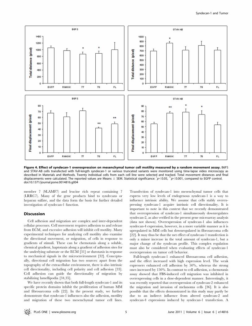
Syndecan-1 inhibits cell motility and migrationIn order to measure the changes in cell motility following
syndecan-1 overexpression, the movement of individual cells was
visualized using time-lapse video microscopy. All fibrosarcoma
transfectants had a tendency for migrating shorter total distances,
suggesting decreased level of cell motility. More interestingly, the
full-length syndecan-1 and 78 transfectants showed significantly
reduced final displacements, indicating that their cell movement
was restricted to a smaller area. However, the STAV-AB MM
cells showed a tendency of smaller final displacement only in the
Syndecan-1 and Tumor
PLoS ONE | www.plosone.org 2 June 2011 | Volume 6 | Issue 6 | e14816

full-length and actually increased effects in the other transfectants
(Figure 4 and Videos S1, S2, S3, S4, S5, S6, S7, S8, S9, and S10).
Furthermore, when we examined the effect of syndecan-1 in a
wound healing assay, both the full-length syndecan-1 and its
truncated variants inhibited MM cell migration shown as slower
wound closure, the RMKKK construct giving the most pronounced
effect. As for the effect on fibrosarcoma cells, all transfectants
displayed a trend of decreased ability to migrate (Figure 5).
Figure 2. Effect of syndecan-1 overexpression on mesenchymal cell adhesion. Stable transfectants were serum starved for 24 h beforeseeding into a black 96 well plate in RPMI with 10% of the respective sera. After 5 min the floating cells were washed away with culture medium. Thenumbers of the attached cells were measured using a fluorometer. All analyses were performed in triplicate. The reported values are Means 6 SD.Statistical significance: *p,0.01, **p,0.001, compared to EGFP control.doi:10.1371/journal.pone.0014816.g002
Figure 1. Strong and weak expressers generated by FACS sorting. B6FS stable transfectants of the full-length syndecan-1/EGFP and theRMKKK/EGFP were separated by using FACS sorting based on their EGFP intensity. The subpopulations with high (P2) or low (P3) EGFP intensitywere isolated and subcultured as strong or weak expressers respectively. A. FACS plots showing the separation of the subpopulations. B. Meanvalue of EGFP intensity of the subpopulations. As for the suffix for RMKKK and FL, ‘‘W’’ stands for weak expresser while as ‘‘S’’ stands for strongexpresser.doi:10.1371/journal.pone.0014816.g001
Syndecan-1 and Tumor
PLoS ONE | www.plosone.org 3 June 2011 | Volume 6 | Issue 6 | e14816

RMKKK deletion hampers the nuclear translocation ofsyndecan-1
The deletion of the RMKKK sequence was verified by
sequencing and by gel electrophoresis, yielding a shorter fragment
in cells transfected with the RMKKK deleted construct compared
to the full-length syndecan-1 (Figure 6A). Syndecan-1 mRNA
levels, both in full-length and RMKKK deleted transfectants were
approximately three fold increased compared to cells transfected
with the corresponding vector (Figure 6B). Overexpression of
syndecan-1 at protein level corresponded to 1.5 to two fold
increase compared to cells transfected with the vector control
(Figure 6C).
Overexpression of the RMKKK deleted construct resulted in
considerable decrease of the nuclear syndecan-1 as compared to
cells transfected with the wild type full-length syndecan-1. The
intensity of nuclear reactivity was decreased two fold in cells
transfected with the RMKKK deleted construct compared to cells
transfected with full-length one (data not shown).
Simultaneously, the proportion of cells showing nuclear staining
was 76% after transfection with full-length syndecan-1 compared
to 15% in cells transfected with the empty vector and 8% in cells
transfected with the RMKKK deleted mutant (Figure 6D).
Syndecan-1 influences cell adhesion/migration/chemotaxis-related genes
The performed bioassays show that the overexpression of
syndecan-1 may influence mesenchymal tumor cell adhesion, and
decrease the tumor cell motility and migration. To investigate the
effects of syndecan-1 overexpression on general transcriptional
modulation in STAV-AB cells, we performed microarray analysis,
which showed a total of 2878 genes regulated by overepression of
syndecan-1. Of these genes, 138 were identified to be adhesion,
migration and chemotaxis related genes based on the Gene
Ontology (GO) selection. We further clustered these terms into
different combinations to show their overlapping functions
(Figure 7A and Table S1). Among the total 138 genes, 71 genes
were up-regulated (Figure 7B) and 67 genes were down-regulated
(Figure 7C). The majority of these genes (53%) were associated
with adhesion, compared to migration (21%) and chemotaxis
(5%). The intersections of Venn diagram showed that many genes
were involved in two or three of these functional categories. Thus,
17 genes were involved in both adhesion and migration, 6 genes
were involved in both migration and chemotaxis, and 5 genes were
associated with all three groups (Figure 7A).
When genes modulated by syndecan-1 overexpression were
grouped according to the class of protein, some of these genes were
associated with more than one of the above functional groups.
Thus, for example, overexpression of syndecan-1 affected
extracellular matrix molecules, growth factors and growth factor
receptors, cytokines and chemokines, adhesion molecules, cyto-
plasmic proteins and cell motility-associated molecules (Table S1).
Top two up-regulated genes were formyl peptide receptor 1 and
metastasis suppressor 1 (MTSS1) and top two down-regulated
genes were signaling lymphocyte activation molecule family
Figure 3. Effect of syndecan-1 overexpression on mesenchymal cell chemotaxis. Stable transfectants were serum starved for 24 h beforeseeding into the upper well of a two chamber system with serum-free medium. Medium containing 10% of the respective sera was placed in thebottom chamber. After 6 h incubation, those cells that had migrated to the bottom of the membrane insert were dislodged and their numbermeasured using a fluorometer. All analyses were performed in triplicate. The reported values are Means 6 SD. Statistical significance: *p,0.01,**p,0.001, compared to EGFP control.doi:10.1371/journal.pone.0014816.g003
Syndecan-1 and Tumor
PLoS ONE | www.plosone.org 4 June 2011 | Volume 6 | Issue 6 | e14816
member 7 (SLAMF7) and leucine rich repeat containing 7
(LRRC7). Many of the gene products bind to syndecans or
heparan sulfate, and the data form the basis for further detailed
investigation of syndecan-1 function.
Discussion
Cell adhesion and migration are complex and inter-dependent
cellular processes. Cell movement requires adhesion to and release
from ECM, and excessive adhesion will inhibit cell motility. Many
experimental techniques for analyzing cell motility also examine
the directional movement, or migration, of cells in response to
gradients of stimuli. These can be chemotaxis along a soluble,
chemical gradient, haptotaxis along a gradient of adhesion sites for
the underlying substrate or the ECM [31] or durotaxis in response
to mechanical signals in the microenvironment [32]. Conceptu-
ally, directional cell migration has two sources: apart from the
topography of the extracellular environment, there is also intrinsic
cell directionality, including cell polarity and cell adhesion [33].
Cell adhesion can guide the directionality of migration by
stabilizing lamellipodia [34,35].
We have recently shown that both full-length syndecan-1 and its
specific protein domains inhibit the proliferation of human MM
and fibrosarcoma cells [22]. In the present study, we further
demonstrate that syndecan-1 influences also the adhesion, motility
and migration of these two mesenchymal tumor cell lines.
Transfection of syndecan-1 into mesenchymal tumor cells that
express very low levels of endogenous syndecan-1 is a way to
influence intrinsic ability. We assume that cells stably overex-
pressing syndecan-1 acquire intrinsic cell directionality. It is
important to note in this context that we recently demonstrated
that overexpression of syndecan-1 simultaneously downregulates
syndecan-2, as also verified in the present gene microarray analysis
(data not shown). Overexpression of syndecan-1 also influences
syndecan-4 expression, however, in a more variable manner as it is
upregulated in MM cells but downregulated in fibrosarcoma cells
[22]. It may thus be that the net effect of syndecan-1 transfection is
only a minor increase in the total amount of syndecan-1, but a
major change of the syndecan profile. This complex regulation
must also be considered when evaluating effects of syndecan-1
overexpression on tumor cell behavior.
Full-length syndecan-1 enhanced fibrosarcoma cell adhesion,
and the effect increased with high expression level. The weak
expressers enhanced cell adhesion by 50%, whereas the strong
ones increased by 150%. In contrast to cell adhesion, a chemotaxis
assay showed that FBS-induced cell migration was inhibited in
overexpressing cells in a dose-dependent manner. Interestingly, it
was recently reported that overexpression of syndecan-2 enhanced
the migration and invasion of melanoma cells [36]. It is also
possible that the effects demonstrated in this study may partly be
due to an indirect influence from altered syndecan-2 and
syndecan-4 expressions induced by syndecan-1 transfection. A
Figure 4. Effect of syndecan-1 overexpression on mesenchymal tumor cell motility measured by a random movement assay. B6FSand STAV-AB cells transfected with full-length syndecan-1 or various truncated variants were monitored using time-lapse video microscopy asdescribed in Materials and Methods. Twenty individual cells from each cell line were selected and tracked. Total movement distances and finaldisplacements were calculated. The reported values are Means 6 SEM. Statistical significance: *p,0.05, **p,0.001, compared to EGFP control.doi:10.1371/journal.pone.0014816.g004
Syndecan-1 and Tumor
PLoS ONE | www.plosone.org 5 June 2011 | Volume 6 | Issue 6 | e14816

dose-dependent regulation of cell motility by syndecan-1 is also
supported by the fact that in the absence of syndecan-2, syndecan-4
may govern cell movement according to a dose-related mechanism;
both high and low levels of syndecan-4 diminish cell motility by
alternatively providing a too tight or a too loose contact with the
substratum [37,38,39]. All these observations imply that overlap-
ping functions exist between members of the syndecan family
[14,40].
A similar syndecan-1-associated inhibition of fibrosarcoma cell
motility was also seen with time-lapse microscopy, where the total
distance moved was unaltered in the overexpressing cells, whereas
the final displacement was decreased. This reduced migration
distance with a preserved level of motility indicates that syndecan-
1 overexpression restricted cell movement to a more limited area.
Such restricted migration and decreased chemotaxis of syndecan-1
transfectants may thus correlate to the enhanced cell adhesion
seen in overexpressing cells. When we examined the effect of
syndecan-1 on the motility/migration of mesenchymal tumor cells
using the wound-healing assay, the overexpressing cells showed
slower wound closure. This effect may not only be a consequence
of decreased cell migration but possibly also of the reduced cell
proliferation as we have recently shown [22]. Influence of
durotaxis from the wound can’t be ignored when evaluating the
net effect on cell migration as cells have a tendency to migrate into
a region denuded of cells.
Overexpression of full-length syndecan thus affects both cell
adhesion and migration, as two tightly-related cellular processes.
The migratory effect may partly be secondary to the changes in
adhesion, and the gene expression analysis shows that majority of
regulated genes are adhesion related. However, there are many
genes associated to migration and chemotaxis, and many of them
have multiple functions.
MTSS1, one of the most up-regulated genes, is an actin and
membrane binding protein. It has been demonstrated as a tumor
suppressor and to be downregulated in bladder cancer, hepato-
cellular carcinoma and gastric cancer [41,42,43]. It has also been
shown to suppress growth as well as invasive, migratory, and
adhesive properties of a breast cancer cell line [44]. While the most
down-regulated gene, SLAMF7 (CD319), is expressed on cytotoxic
lymphoctes, activated B-cells, and mature dendritic cells [45,46]. It
has been shown that it mediates cytotoxicity of NK-cells [47], and
induces proliferation and autocrine cytokine expression on human
B lymphocytes [48]. The SLAM family of receptors plays roles in
lymphocyte development, cytotoxicity, immunity, cell survival and
cell adhesion [49]. The second down-regulated gene LRRC7,
also known as Densin-180, is a transmembrane protein containing
one PDZ domain. LRRC7 has been shown to interact with
N-Cadherin [50] and may thus provide a mechanism for tran-
sendothelial migration in cancer cells. Hyaluronan- and pro-
teoglycan link protein 1 (HAPLN1), which was also downregulat-
ed, has been shown to play a pro-tumorigenic role in malignant
pleural mesothelioma [51]. These reports are in line with our
present findings demonstrating that overexpression of syndecan-1
negatively regulated MM cell migration. Integrin alpha5 (ITGA5)
and integrin beta1 (ITGB1) were upregulated whereas integrin
beta5 was downregulated, thus supporting the notion of close
cooperation between syndecans and integrins. Together, they
regulate the global gene expression and a crosstalk of signaling
pathways. The varying effects of syndecan-1 overexpression on cell
behavior comparing the two cell lines must be put into this
context.
In an attempt to dissect the contribution of the different
functional protein domains of this PG, the two mesenchymal
tumor cell lines were transfected with three truncated variants of
syndecan-1. The effects on enhanced cell adhesion seem mainly to
depend upon the EC domain with its GAG chains, as in its
absence cell adhesion is not enhanced. Indeed the two shorter
constructs with no EC domain, i.e. 77/EGFP and RMKKK/
EGFP, actually inhibited cell adhesion. Interestingly, the 78/
EGFP construct, which contains the short DRKE motif remnant
of the EC domain (mimicking the structure that remains in the
plasma membrane after syndecan-1 shedding) also enhanced
adhesion to some extent. The different effects seen when
comparing the 77/EGFP and 78/EGFP constructs, which only
differ in the presence of the four extra amino acid residues in the
latter, are intriguing. It may indicate a biological significance for
the juxtamembrane DRKE motif, presumably on the oligomer-
ization of the syndecan-1. The finding that the 78/EGFP
construct, but not the 77/EGFP constructs also decreased
the final displacement in the cell random movement assay,
in a similar way to full-length syndecan-1, also supports the
idea that reduced migration may correlate with increased
adhesion.
Figure 5. Effect of syndecan-1 overexpression on mesenchymaltumor cell motility/migration measured by a wound healingassay. B6FS and STAV-AB cells transfected with full-length syndecan-1or various truncated variants were cultured in 60 mm petri-dishes untilmonolayer confluence was achieved. The cell layer was then woundedand the wound closure was monitored at various time points using adigital image processor connected to a microscope. Width measure-ments were taken across the wound at six different positions along it.Progression of the cell front was quantified and calculated as migrationspeed (mm/h). The assay was performed three times independently,with triplicate measurements in each. The reported values are Means 6SEM of 18 observations from 1 representative experiment. Statisticalsignificance: *p,0.05, **p,0.001, compared to EGFP control.doi:10.1371/journal.pone.0014816.g005
Syndecan-1 and Tumor
PLoS ONE | www.plosone.org 6 June 2011 | Volume 6 | Issue 6 | e14816

Although none of the truncated constructs had any effect on the
basal level of fibrosarcoma cell migration in the Transwell assay,
they all inhibited the chemotactic migration along a FBS gradient.
Thus, this kind of migration seems to depend more on the
cytoplasmic portion of the PG rather than on its EC domain and
attached GAGs. All the truncated constructs also inhibited wound
closure of the two mesenchymal tumor cells. This was seen even
with the short RMKKK/EGFP construct, which had the most
pronounced effect on MM cells. It therefore seems that the
syndecan-1 effect on cell migration is not only dependent on its cell
surface interaction, but also related to its presence in the nucleus.
The function of nuclear syndecan-1 is still obscure. Deletion of the
RMKKK motif abolished the nuclear translocation of syndecan-1.
This RMKKK motif binds to cytoskeleton components and it may
in this way influence cell adhesion-related function. Moreover, it
acts as a NLS for syndecan-1, and transfection of the RMKKK/
EGFP construct results in a nuclear accumulation of the transcript,
suggesting the existence of nuclear ligands to the motif [21]. The
finding that heparanase decreases the amount of nuclear
syndecan-1 indicates that syndecan-1 in the nucleus can also have
other nuclear binding sites [19]. It can be speculated that the free
RMKKK fragment blocks the translocation of native syndecan-1
into the nucleus, and this could be one way to hamper the function
of nuclear syndecan-1, resulting in transcriptional regulation. The
effects seen with this motif unique to the syndecans, indicate that
the function of syndecans on the migratory capacity is also
influenced by events in the nucleus.
A study of the differential roles for membrane-bound and
soluble syndecan-1 in breast cancer progression has recently been
published. Proteolytic conversion of syndecan-1 from a mem-
brane-bound into a soluble molecule marks a switch from a
proliferative to an invasive phenotype [20]. Increased invasiveness
was observed in another fibrosarcoma cell line HT-1080 when
transfected with the same full-length and 78 truncated syndecan-1
constructs as we used, and local growth was faster in the full-length
syndecan-1 than the 78 transfectants. It is presumed that the whole
protein and maybe the shedding is needed for the local
development of the tumor, but the intracellular and transmem-
brane domain is sufficient to promote the formation of metastasis
[52]. We found that the full-length syndecan-1 and the remnants
of syndecan-1 after shedding (the 78/EGFP construct), negatively
regulated fibrosarcoma B6FS cell migration, and this can be partly
Figure 6. Syndecan-1 expression and subcellular distribution in B6FS cells transfected with full-length and RMKKK deletedconstructs. A. Electrophoresis on agarose gel showed the expression of RMKKKdeleted construct in the transfected cells, giving a shorteramplimer(lane 3) than the full-length syndecan-1 product (lane 2) or the endogeneous syndecan-1 (lane 4). B. Upregulation of syndecan-1 mRNA inthe transfected cells. Fold change was calculated based on the intensity of the bands normalized to the vector control, with GAPDH as referencegene. Results represent mean of 4 independent experiments. Expression of syndecan-1 protein (C) and the level of nuclear translocation (D) wasexamined by immunocytochemistry. Fold change in protein level was calculated by measuring the fluorescence intensity. Nuclear translocation ofsyndecan-1 was quantified by calculating the percent of cells showing nuclear immunoreactivity to the total number of cells. Results presented areMeans 6SEM from at least 2 independent experiments. Statistical significance: *p,0.05, compared to vector control as indicated (D).doi:10.1371/journal.pone.0014816.g006
Syndecan-1 and Tumor
PLoS ONE | www.plosone.org 7 June 2011 | Volume 6 | Issue 6 | e14816

due to the different phenotypes of these two fibrosarcoma cell
lines. We also found that the 78/EGFP construct displayed
opposite effects on cell motility in terms of final displacement
between fibrosarcoma and MM cell lines, showing the cell type-
specific effects. Further investigations on syndecan-1 interacting
ligands and signaling pathways are on-going in our laboratory.
Taken together, our results show that syndecan-1 decreases
migration and motility, and enhances adhesion of mesenchymal
tumor cells in an expression level-dependent manner. Distinct
protein domains have different effects: the extracellular domain is
more important for promoting cell adhesion, while the transmem-
brane and cytoplasmic domains are sufficient for inhibition of cell
migration; cell adhesion and migration seem to depend also on the
nuclear localization of syndecans. Our study demonstrates that
syndecan-1 plays an important role in mesenchymal tumor
behavior. Moreover, our study provides new inputs into the better
understanding of structure-function relationship of this PG in
tumor progression.
Materials and Methods
Cell lines and their cultureSTAV-AB human MM cells were grown in RPMI 1640
medium containing 25 mM HEPES (42401, Gibco, Grand Island,
NY, USA) and 2 mM L-Glutamine, supplemented with 10%
human AB serum (growth medium) [53]. B6FS human fibrosar-
coma cells were grown in RPMI 1640+ glutaMAXTM-I (72400,
Gibco) supplemented with 10% foetal bovine serum (FBS) and
Gentamicin 50 mg/ml (Gibco) (growth medium) [54]. All cells
were cultured in 75 cm2 Tissue Culture Flasks (Sarstedt, Newton,
NC, USA), in humidified 5% (v/v) CO2 at 37uC and culture
medium was changed twice a week. Both cell lines are of
mesenchymal origin and were selected based on their low
endogenous expression levels of syndecan-1 [22,55].
Plasmids and generation of stably-transfected cell linesThe plasmids and subsequent stable transfection of cells were
described in detail in our previous publication [22]. The
78/EGFP, 77/EGFP and RMKKK/EGFP constructs are three
truncated variants of the human full-length syndecan-1/EGFP
construct. The 78/EGFP lacks the extracellular domain with the
exception of the juxtamembrane DRKE sequence, the 77/EGFP
lacks the entire extracellular domain, and the RMKKK/EGFP
contains only the nuclear localization signal (NLS). The pEGFP-N1
vector was used as a negative control.
MM and fibrosarcoma cells were transfected with the constructs
above, using Effectene Transfection Reagent (Qiagen GmbH,
Hilden, Germany). Optimization of the transfection was carried
out according to the manufacturer’s guidelines. To obtain stable
transfectants, the EGFP positive cells were selected by Geneticin
incubation (G418, Roche Diagnostics GmbH, Mannheim, Ger-
many). Mock-transfected cells were used as a reference for
selection. In this study, only the stably-transfected cell lines were
used and cultured under geneticin pressure (200 mg/mL) B6FS
stable tansfectantsof the full-length syndecan-1/EGFP and the
RMKKK/EGFP were further separated based on their EGFP
intensity by using fluorescence-activated cell sorting (FACS, BD
FACSAriaTM, BD Biosciences, San Jose, California, USA). The
FACS was performed by the Flow Cytometry Unit at Karolinska
University Hospital Huddinge, Sweden). The subpopulations with
high or low EGFP intensity were isolated and subcultured as
strong or weak expressers respectively.
Generation of the RMKKK deletion constructIn order to study the importance of the RMKKK sequence as
a NLS, we designed a new construct that lacks the RMKKK se-
quence from a plasmid carrying the gene encoding the full-length
syndecan-1 (pN1-flsyn1). The pN1-flsyn1 plasmid was constructed
by deleting the EGFP gene from the commercially available pN1-
EGFP plasmid (BD Biosciences, Clontech, Palo Alto, CA, USA) by
digestion with EcoRI and NotI restriction enzymes and replacing
it with the gene encoding the full-length syndecan-1. In order to
construct the RMKKK deletion plasmid, we designed a primer
pair flanking the RMKKK region (FwRMKKKdel: gacgaagg-
cagctactccttggag; RevRMKKKdel: gtacagcatgaaacccaccaggca) in
the syndecan-1 gene. We amplified the pN1-flsyn1 plasmid by
PCR reaction using a PhusionH Hot Start DNA Polyme-
rase (Phusion Site-directed Mutagenesis Kit, Finnzymes, Espoo,
Figure 7. Affymetrix microarray for gene expression pattern influenced by syndecan-1 overexpression in MM STAV-AB cells. Venndiagram showing both distribution and proportion of genes connected to individual GO groups and the combinations of different GO groups(threshold is above 5% significance level and two fold changes). A. All regulated genes. B. upregulated genes. C. downregulated genes.doi:10.1371/journal.pone.0014816.g007
Syndecan-1 and Tumor
PLoS ONE | www.plosone.org 8 June 2011 | Volume 6 | Issue 6 | e14816

Finland). Both primers were phosphorylated at the 59 end to allow
direct ligation after the PCR. The PCR reaction was carried out in
a final volume of 50 ml. The reaction mixture contained 10 ml of
56 Phusion HF Buffer, 1 ml of 10 mMdNTPs, 50 pmol of both
primers, 200 ng template DNA and 1 U of PhusionH Hot Start
DNA Polymerase. The amplification was done using a two-step
cycling protocol: denaturation at 98uC for 30 sec, annealing and
elongation at 72uC for 80 sec, followed by a final incubation at
72uC for 5 min. The resulted linearized DNA represents the
pN1flsyn1_RMKKKdel plasmid (RMKKK-del), lacking the
RMKKK sequence. The full-length syndecan-1 sequence was
deleted from the same pN1-flsyn1 construct. Briefly, a primer pair
flanking the full-length syndecan-1 gene was designed (Fwsyn1del:
agcggccgcgactctagatcataat; Revsyn1del: aagcttgagctcgagatct-
gagtcc). The PCR product amplified with the aid of Site-directed
Mutagenesis Kit (Finnzymes) was the pN1-empty plasmid (vector),
and used as negative control.
25 ng of the PCR products were recircularized using Quick T4
DNA ligase (provided with the Phusion Site-directed Mutagenesis
Kit, Finnzymes) in a reaction at 25uC for 5 minutes. Chemically
competent E. coli cells were transformed with these plasmids; the cells
were plated on LB-agar plates containing 50 mg/mL kanamycin as a
selection agent. Plasmid DNA was isolated using QiAmpMiniprep
spin kit (Qiagen GmbH, Hilden, Germany), as suggested by the
manufacturer. These newly designed constructs were sequenced to
verify if the deletion was correct and complete. The sequencing was
performed by CybergeneAB, Stockholm, Sweden.
Reverse transcription polymerase chain reaction (RT-PCR)Total cellular RNA was isolated from subconfluent B6FS cells,
using the High Pure RNA Isolation Kit (Roche Diagnostics GmbH
Mannheim, Germany). The yield and purity of RNA preparations
were estimated spectrophotometrically by measuring the absor-
bance at 260 nm, and the A260/A230 and A260/A280 ratio,
respectively. From each sample, 2 mg of RNA was reverse
transcribed and the cDNAs were amplified using the First Strand
cDNA Synthesis Kit (Pharmacia Biotech, Uppsala, Sweden). A
region of 94 base-pair containing the RMKKK sequence was
amplified using the following primers in the vicinity of the RMKKK
region: fwRMKKKdel2: ggctcatctttgctgtgtgc; revRMKKKdel2:
gcttgtttcggctcctccaa. The product of RMKKK deleted syndecan-1
(79 bp) can be distinguished from the product of native full-length
syndecan-1, which is longer (94 bp). For the PCR reaction, 3 ml of
the RT reaction was used. The reaction mixture contained 5 ml 56Phusion HF Buffer, 0,5 ml of 10 mMdNTPs, 25 pmol of both
primers, and 0,5 U of PhusionH Hot Start DNA Polymerase
(Finnzymes) in a final volume of 25 ml. To allow the semiquanti-
tative analysis of the sequences, we also added to the reaction
25 pmol of a primer pair for glyceraldehyde-3- phosphatase
dehydrogenase(GAPDH) (GAPDH-se: acatcatccctgcctctactgg,
GAPDHas: agtgggtgtcgctgttgaagtc) [55], which resulted in a
214 bp product. Amplification of the cDNA product was done for
25 cycles using the following parameters: denaturation at 98uC for
30 sec, annealing at 67uC for 30 sec and elongation at 72uC for
1 min, followed by a final incubation at 72uC for 5 min. RT-PCR
products were analyzed on 3% agarose gels. Semiquantitative
measurement of mRNA expression was carried out with the
FluorChem Imaging System and AlphaEase FC Software V5.0.0
(Alpha Innotech Inc.) using GAPDH as a reference gene.
Detection and subcellular localization of newlysynthesized syndecan-1
Expression and distribution of syndecan-1 proteins was further
examined using immunocytochemical analysis and fluorescent
microscopy. Stably transfected cells were seeded on to POLYSINE
coated microscopy slides (Menzel-Glaser, Braunschweig, Ger-
many) and allowed to adhere for 48 h before fixation in 3%
paraformaldehyde. After permeabilization with 0.1% Triton X-
100 (Sigma, Steinheim, Germany) non-specific binding was
blocked with 3% goat serum (Dako A/S, Glostrup, Denmark)
for 30 min. Incubation with the primary antibody diluted 1:4,
(Mouse anti Human CD138 monoclonal antibody (MCA-681)
Serotec LTD, Kidlington, Oxford, England) or with mouse IgG1
(Dako A/S, Glostrup, Denmark)as negative control was performed
overnight at 4uC, followed by 30 min incubation with the
secondary antibody (Alexa 488 goat anti-mouse F(ab9)2 fragment
of IgG (H+L), (Molecular Probes, Leiden, The Netherlands,
A11017) in the dark at room temperature. Samples were then
counterstained with 1 mg/L bisbenzimide H33342 (Fluka,
Steinheim, Germany). Detailed visualization was performed using
Nikon microphot-FXA EPI-FL3 fluorescence microscope. Images
were processed using ImageJ 1.43 software allowing measurement
of fluorescence intensity. For expression of syndecan-1 protein,
background was subtracted, and fluorescence intensity of the cells
was measured in 7 randomly selected visual fields for each slide.
For quantification of nuclear syndecan-1, the intensity of nuclear
fluorescence was measured in 50 randomly selected cell nuclei for
each transfectant. We also calculated the proportion of cells
showing nuclear staining to total number of cells in the same visual
field, 100–150 cells were evaluated for each sample.
Adhesion assayB6FS syndecan-1 overexpressing cells were seeded at 5.000 cells
per well in a 96-well plate (Corning Incorporated, Corning, NY,
USA), in its growth medium containing geneticin. After 5 minutes,
the wells were washed twice with the culture medium to remove
the floating cells. Adherent cells were lysed using Cell Lysis Buffer
(Cat. 90130, Chemicon, Billerica, MA, USA) and the cell numbers
were quantified using CyQUANTGRH Dye (Cat. 90132,
Chemicon) as measured with a fluorometer (BioTek Instruments,
Winooski, VT, USA) with a 480/520 nm filter set. This assay was
performed in triplicate.
Chemotaxis assayDirectional migration of B6FS syndecan-1 overexpressing cells
was assessed using a 24-Transwell plate (Cat. 3422, Corning
Incorporated). After 24 h serum starvation, 50.000 cells were
seeded into an upper well of the chamber in serum-free medium.
FBS (10%), as a chemoattractant was placed in the bottom
chamber. After 6 h the cells that had migrated through the
microporous membrane were dislodged from the outer surface of
the insert using Trypsin 0.5% EDTA (Gibco), and then lysed using
Cell Lysis Buffer (Cat. 90130, Chemicon). The cell numbers were
quantified as described above for the cell adhesion assay. This
assay was performed in triplicate.
Random movement assaySTAV-AB and B6FS syndecan-1 overexpressing cells were
seeded in a 12-well glass bottom culture plate (MatTek
Corporation, Ashland, MA, USA), and incubated at 37uC for
6 hours before transfer to a Leica DMIRE2 Inverted Laboratory
Microscope (McBain Instruments, Simi Valley, CA,USA) for
imaging. Cells were maintained during imaging in normal culture
condition. Five observation fields were randomly selected and
time-lapse imaging was performed every 15 minutes over 16 hours
with a 106 dry objective. This yielded a video of 65 photos for
each single cell monitored. The movements of 20 individual cells
for each cell line were tracked using ImageJ software (NIH). Total
Syndecan-1 and Tumor
PLoS ONE | www.plosone.org 9 June 2011 | Volume 6 | Issue 6 | e14816
distances and final displacements of moving cells were calculated
based on path tracking data as pixels.
Wound healing assaySTAV-AB and B6FS syndecan-1 overexpressing cells were
seeded in 60 mm petri-dishes and cultured to confluence in their
growth media containing geneticin. The cell monolayer was then
wounded by scratching with a sterile 200 ml pipette tip. Detached
cells were removed by washing twice with culture medium.
Subsequent wound closure was monitored at 3, 6 and 12 hours
using a digital image processor connected to a microscope. Width
measurements were taken across the gap at six different positions
along the wound. Cell motility was quantified by image analysis
(ImageJ 1.4.3.67 Launcher Symmetry Software, NIH, Bethesda,
MD, USA) and calculated as migration speed (mm/h). This assay
was performed three times independently with triplicates in each.
RNA isolation and microarray analysisThe STAV-AB MM cells stably overexpressing full-length
syndecan-1, or the EGFP vector were cultured for about 36 hours.
Total RNA was isolated from sub-confluent cell cultures, using the
High Pure RNA Isolation Kit (Roche, Mannheim, Germany), with
optional on-column DNase 1 digestion, according to the supplied
protocol. The yield and purity of the RNA preparations were
estimated spectrophotometrically by measuring the UV absor-
bance at 260 nm and calculating the A260/A280 ratio, respec-
tively. Three independent experiments were performed.
The above RNA samples were applied to gene microarray
analysis using GeneChipH Human Gene 1.0 ST Arrays (Affymetrix,
Inc., Santa Clara, CA, USA), representing more than 22.000 well-
annotated human genes. Target synthesis and hybridizations were
performed in the Affymetrix core facility (NOVUM, Karolinska
Institutet, Huddinge, Sweden) according to standard proto-
cols. Affymetrix Expression Console (http://www.affymetrix.com/
products_services/software/specific/expression_console_ software.
affx) was used for the data preprocessing, with the PLIER method
for summarization, PM-GCBG for background correction and
Global Median for normalization. Expression levels in the different
groups were compared using Student’s t-tests, and, as a measure of
corresponding false discovery rates, q-values were calculated using
the q-value package in R (http://www.rproject.org/). All data is
MIAME compliant and the raw data has been deposited in the
MIAME compliant database Gene Expression Omnibus (the
accession number GSE21401, http://www.ncbi.nlm.nih.gov/geo/
info/linking.html). By applying a filter of at least a two-fold change
in mean expression level with P,0.05 for significance, we screened
for modulated genes in the full-length syndecan-1 overexpressing
cells with the EGFP mock-transfectants as reference. Gene
Ontology (GO) (http://amigo.geneontology.org/cgi-bin/amigo/
go.cgi) was used to select genes related to cell adhesion/
migration/chemotaxis from the total regulated genes based on
their GO annotations. Venn diagrams were constructed for analysis
of these GO terms using a web-based application [56].
Statistical analysisStatistical significance was evaluated using the Student’s t-test
and the one way completely randomized variance analysis
(ANOVA) using the Microcal Origin (version 5.0) software. The
null hypothesis of no difference was rejected at a=0.05.
Supporting Information
Table S1 Genes modulated by syndecan-1 overexpression in
MM STAV-AB cells. All genes are selected with at least a 95%
confidence interval and filtered with two fold changes. The table is
sectioned according to GO terms: adhesion, migration and
chemotaxis; as well as their respective combinations. Figure 7
was generated based on the data in this table.
Found at: doi:10.1371/journal.pone.0014816.s001 (0.28 MB
DOC)
Video S1 Videos S1-S10 for Figure 4. Random movement assay
on the syndecan-1 overexpressing cells. The cells were seeded in
a 12-well glass bottom culture plate and incubated at 37uC for
6 hours before transfer to a Leica DMIRE2 Inverted Laboratory
Microscope for imaging. Cells were maintained during imaging in
normal culture condition. Five observation fields were randomly
selected and time-lapse imaging was performed every 15 minutes
over 16 hours with a 106dry objective. Thus a video of 65 photos
for each single cell monitored was yielded and can be played
using Quicktime player or Windows Media player software. A
representative video of each transfectant was selected to show its
random movement: Video S1, B6FS EGFP; Video S2, B6FS
RMKKK/EGFP; Video S3, B6FS 77/EGFP; Video S4, B6FS
78/EGFP; Video S5, B6FS FL/EGFP. Video S6, STAV-AB
EGFP; Video S7, STAV-AB RMKKK/EGFP; Video S8, STAV-
AB 77/EGFP; Video S9, STAV-AB 78/EGFP; Video S10,
STAV-AB FL/EGFP. The difference of motility between different
transfectants can be seen by comparison of videos of respective
transfectants.
Found at: doi:10.1371/journal.pone.0014816.s002 (6.93 MB AVI)
Video S2
Found at: doi:10.1371/journal.pone.0014816.s003 (4.82 MB AVI)
Video S3
Found at: doi:10.1371/journal.pone.0014816.s004 (4.34 MB AVI)
Video S4
Found at: doi:10.1371/journal.pone.0014816.s005 (3.79 MB AVI)
Video S5
Found at: doi:10.1371/journal.pone.0014816.s006 (4.56 MB AVI)
Video S6
Found at: doi:10.1371/journal.pone.0014816.s007 (3.59 MB AVI)
Video S7
Found at: doi:10.1371/journal.pone.0014816.s008 (3.19 MB AVI)
Video S8
Found at: doi:10.1371/journal.pone.0014816.s009 (3.10 MB AVI)
Video S9
Found at: doi:10.1371/journal.pone.0014816.s010 (5.09 MB AVI)
Video S10
Found at: doi:10.1371/journal.pone.0014816.s011 (3.84 MB AVI)
Acknowledgments
The authors are grateful to Asa-Lena Dackland at the Flow Cytometry
Unit at Karolinska University Hospital Huddinge for her expert FACS skill
to isolate transfectants with different expression levels. Dr. Zhengwen An,
Dr. Zhilun Li and Dr. Hongquan Zhang for their generous and skilled
technical assistance with cell tracking experiments.
Author Contributions
Conceived and designed the experiments: FZ EF TS GT AH KD.
Performed the experiments: FZ EF FM TS DB. Analyzed the data: FZ EF
FM TS DB GT AH KD. Contributed reagents/materials/analysis tools:
IK LS DB. Wrote the paper: FZ EF. Critically revised the paper: IK LS
GT AH KD.
Syndecan-1 and Tumor
PLoS ONE | www.plosone.org 10 June 2011 | Volume 6 | Issue 6 | e14816

References
1. Webb DJ, Parsons JT, Horwitz AF (2002) Adhesion assembly, disassembly andturnover in migrating cells – over and over and over again. Nat Cell Biol 4:
E97–100.2. Couchman JR (2003) Syndecans: proteoglycan regulators of cell-surface
microdomains? Nat Rev Mol Cell Biol 4: 926–937.3. Bernfield M, Gotte M, Park PW, Reizes O, Fitzgerald ML, et al. (1999)
Functions of cell surface heparan sulfate proteoglycans. Annu Rev Biochem 68:
729–777.4. Okina E, Manon-Jensen T, Whiteford JR, Couchman JR (2009) Syndecan
proteoglycan contributions to cytoskeletal organization and contractility.Scand J Med Sci Sports 19: 479–489.
5. Chakravarti R, Adams JC (2006) Comparative genomics of the syndecans
defines an ancestral genomic context associated with matrilins in vertebrates.BMC Genomics 7: 83.
6. Whiteford JR, Behrends V, Kirby H, Kusche-Gullberg M, Muramatsu T, et al.(2007) Syndecans promote integrin-mediated adhesion of mesenchymal cells in
two distinct pathways. Exp Cell Res 313: 3902–3913.
7. Beauvais DM, Ell BJ, McWhorter AR, Rapraeger AC (2009) Syndecan-1regulates alphavbeta3 and alphavbeta5 integrin activation during angiogenesis
and is blocked by synstatin, a novel peptide inhibitor. J Exp Med 206: 691–705.8. Xian X, Gopal S, Couchman JR (2009) Syndecans as receptors and organizers
of the extracellular matrix. Cell Tissue Res.9. Endo K, Takino T, Miyamori H, Kinsen H, Yoshizaki T, et al. (2003) Cleavage
of syndecan-1 by membrane type matrix metalloproteinase-1 stimulates cell
migration. J Biol Chem 278: 40764–40770.10. Subramanian SV, Fitzgerald ML, Bernfield M (1997) Regulated shedding of
syndecan-1 and -4 ectodomains by thrombin and growth factor receptoractivation. J Biol Chem 272: 14713–14720.
11. Jalkanen M, Rapraeger A, Saunders S, Bernfield M (1987) Cell surface
proteoglycan of mouse mammary epithelial cells is shed by cleavage of itsmatrix-binding ectodomain from its membrane-associated domain. J Cell Biol
105: 3087–3096.12. Wang Z, Gotte M, Bernfield M, Reizes O (2005) Constitutive and accelerated
shedding of murine syndecan-1 is mediated by cleavage of its core protein at aspecific juxtamembrane site. Biochemistry 44: 12355–12361.
13. Asundi VK, Carey DJ (1995) Self-association of N-syndecan (syndecan-3) core
protein is mediated by a novel structural motif in the transmembrane domainand ectodomain flanking region. J Biol Chem 270: 26404–26410.
14. Alexopoulou AN, Multhaupt HA, Couchman JR (2007) Syndecans in woundhealing, inflammation and vascular biology. Int J Biochem Cell Biol 39:
505–528.
15. Woods A, Couchman JR (2001) Syndecan-4 and focal adhesion function. CurrOpin Cell Biol 13: 578–583.
16. Zimmermann P, Zhang Z, Degeest G, Mortier E, Leenaerts I, et al. (2005)Syndecan recycling [corrected] is controlled by syntenin-PIP2 interaction and
Arf6. Dev Cell 9: 377–388.17. Chakravarti R, Sapountzi V, Adams JC (2005) Functional role of syndecan-1
cytoplasmic V region in lamellipodial spreading, actin bundling, and cell
migration. Mol Biol Cell 16: 3678–3691.18. Brockstedt U, Dobra K, Nurminen M, Hjerpe A (2002) Immunoreactivity to cell
surface syndecans in cytoplasm and nucleus: tubulin-dependent rearrangements.Exp Cell Res 274: 235–245.
19. Chen L, Sanderson RD (2009) Heparanase regulates levels of syndecan-1 in the
nucleus. PLoS One 4: e4947.20. Nikolova V, Koo CY, Ibrahim SA, Wang Z, Spillmann D, et al. (2009)
Differential roles for membrane-bound and soluble syndecan-1 (CD138) inbreast cancer progression. Carcinogenesis 30: 397–407.
21. Zong F, Fthenou E, Wolmer N, Hollosi P, Kovalszky I, et al. (2009) Syndecan-1and FGF-2, but not FGF receptor-1, share a common transport route and co-
localize with heparanase in the nuclei of mesenchymal tumor cells. PLoS One 4:
e7346.22. Zong F, Fthenou E, Castro J, Peterfia B, Kovalszky I, et al. (2009) Effect of
syndecan-1 overexpression on mesenchymal tumour cell proliferation with focuson different functional domains. Cell Prolif.
23. Stanley MJ, Liebersbach BF, Liu W, Anhalt DJ, Sanderson RD (1995) Heparan
sulfate-mediated cell aggregation. Syndecans-1 and -4 mediate intercellularadhesion following their transfection into human B lymphoid cells. J Biol Chem
270: 5077–5083.24. Liu W, Litwack ED, Stanley MJ, Langford JK, Lander AD, et al. (1998)
Heparan sulfate proteoglycans as adhesive and anti-invasive molecules.
Syndecans and glypican have distinct functions. J Biol Chem 273: 22825–22832.25. Burbach BJ, Ji Y, Rapraeger AC (2004) Syndecan-1 ectodomain regulates
matrix-dependent signaling in human breast carcinoma cells. Exp Cell Res 300:234–247.
26. Beauvais DM, Burbach BJ, Rapraeger AC (2004) The syndecan-1 ectodomainregulates alphavbeta3 integrin activity in human mammary carcinoma cells.
J Cell Biol 167: 171–181.
27. Langford JK, Yang Y, Kieber-Emmons T, Sanderson RD (2005) Identificationof an invasion regulatory domain within the core protein of syndecan-1. J Biol
Chem 280: 3467–3473.
28. Beauvais DM, Rapraeger AC (2003) Syndecan-1-mediated cell spreading
requires signaling by alphavbeta3 integrins in human breast carcinoma cells.Exp Cell Res 286: 219–232.
29. Morgan MR, Humphries MJ, Bass MD (2007) Synergistic control of celladhesion by integrins and syndecans. Nat Rev Mol Cell Biol 8: 957–969.
30. Bass MD, Humphries MJ (2002) Cytoplasmic interactions of syndecan-4orchestrate adhesion receptor and growth factor receptor signalling. Biochem J
368: 1–15.
31. Carter SB (1965) Principles of cell motility: the direction of cell movement and
cancer invasion. Nature 208: 1183–1187.
32. Lo CM, Wang HB, Dembo M, Wang YL (2000) Cell movement is guided by therigidity of the substrate. Biophys J 79: 144–152.
33. Petrie RJ, Doyle AD, Yamada KM (2009) Random versus directionallypersistent cell migration. Nat Rev Mol Cell Biol 10: 538–549.
34. Bailly M, Yan L, Whitesides GM, Condeelis JS, Segall JE (1998) Regulation ofprotrusion shape and adhesion to the substratum during chemotactic responses
of mammalian carcinoma cells. Exp Cell Res 241: 285–299.
35. Harms BD, Bassi GM, Horwitz AR, Lauffenburger DA (2005) Directional
persistence of EGF-induced cell migration is associated with stabilization of
lamellipodial protrusions. Biophys J 88: 1479–1488.
36. Lee JH, Park H, Chung H, Choi S, Kim Y, et al. (2009) Syndecan-2 regulates
the migratory potential of melanoma cells. J Biol Chem 284: 27167–27175.
37. Wilcox-Adelman SA, Denhez F, Iwabuchi T, Saoncella S, Calautti E, et al.
(2002) Syndecan-4: dispensable or indispensable? Glycoconj J 19: 305–313.
38. Cattaruzza S, Perris R (2005) Proteoglycan control of cell movement during
wound healing and cancer spreading. Matrix Biol 24: 400–417.
39. Chalkiadaki G, Nikitovic D, Berdiaki A, Sifaki M, Krasagakis K, et al. (2009)
Fibroblast growth factor-2 modulates melanoma adhesion and migration
through a syndecan-4-dependent mechanism. Int J Biochem Cell Biol 41:1323–1331.
40. Kirn-Safran C, Farach-Carson MC, Carson DD (2009) Multifunctionality ofextracellular and cell surface heparan sulfate proteoglycans. Cell Mol Life Sci 66:
3421–3434.
41. Lee YG, Macoska JA, Korenchuk S, Pienta KJ (2002) MIM, a potential
metastasis suppressor gene in bladder cancer. Neoplasia 4: 291–294.
42. Ma S, Guan XY, Lee TK, Chan KW (2007) Clinicopathological significance ofmissing in metastasis B expression in hepatocellular carcinoma. Hum Pathol 38:
1201–1206.
43. Liu K, Wang G, Ding H, Chen Y, Yu G, et al. (2010) Downregulation of
metastasis suppressor 1(MTSS1) is associated with nodal metastasis and pooroutcome in Chinese patients with gastric cancer. BMC Cancer 10: 428.
44. Parr C, Jiang WG (2009) Metastasis suppressor 1 (MTSS1) demonstratesprognostic value and anti-metastatic properties in breast cancer. Eur J Cancer
45: 1673–1683.
45. Boles KS, Mathew PA (2001) Molecular cloning of CS1, a novel human naturalkiller cell receptor belonging to the CD2 subset of the immunoglobulin
superfamily. Immunogenetics 52: 302–307.
46. Bouchon A, Cella M, Grierson HL, Cohen JI, Colonna M (2001) Activation of
NK cell-mediated cytotoxicity by a SAP-independent receptor of the CD2family. J Immunol 167: 5517–5521.
47. Kumaresan PR, Lai WC, Chuang SS, Bennett M, Mathew PA (2002) CS1, a
novel member of the CD2 family, is homophilic and regulates NK cell function.Mol Immunol 39: 1–8.
48. Lee JK, Mathew SO, Vaidya SV, Kumaresan PR, Mathew PA (2007) CS1(CRACC, CD319) induces proliferation and autocrine cytokine expression on
human B lymphocytes. J Immunol 179: 4672–4678.
49. Cannons JL, Tangye SG, Schwartzberg PL (2011) SLAM family receptors and
SAP adaptors in immunity. Annu Rev Immunol 29: 665–705.
50. Izawa I, Nishizawa M, Ohtakara K, Inagaki M (2002) Densin-180 interacts with
delta-catenin/neural plakophilin-related armadillo repeat protein at synapses.
J Biol Chem 277: 5345–5350.
51. Ivanova AV, Goparaju CM, Ivanov SV, Nonaka D, Cruz C, et al. (2009)
Protumorigenic role of HAPLN1 and its IgV domain in malignant pleuralmesothelioma. Clin Cancer Res 15: 2602–2611.
52. Peterfia B, Hollosi P, Szilak L, Timar F, Paku S, et al. (2006) [Role of syndecan-1proteoglycan in the invasiveness of HT-1080 fibrosarcoma]. Magy Onkol 50:
115–120.
53. Klominek J, Robert K-H, Hjerpe A, Wickstrom B, Gahrton G (1989) Serum-
dependent growth patterns of two, newly established human mesothelioma cell
lines. Cancer Res 49: 6118–6122.
54. Thurzo V, Popovic M, Matoska J, Blasko M, Grofova M, et al. (1976) Human
neoplastic cells in tissue culture: two established cell lines derived from giant celltumor and fibrosarcoma. Neoplasma 23: 577–587.
55. Dobra K, Andang M, Syrokou A, Karamanos NK, Hjerpe A (2000)Differentiation of mesothelioma cells is influenced by the expression of
proteoglycans. Exp Cell Res 258: 12–22.
56. Hulsen T, de Vlieg J, Alkema W (2008) BioVenn - a web application for thecomparison and visualization of biological lists using area-proportional Venn
diagrams. BMC Genomics 9: 488.
Syndecan-1 and Tumor
PLoS ONE | www.plosone.org 11 June 2011 | Volume 6 | Issue 6 | e14816
Related Documents