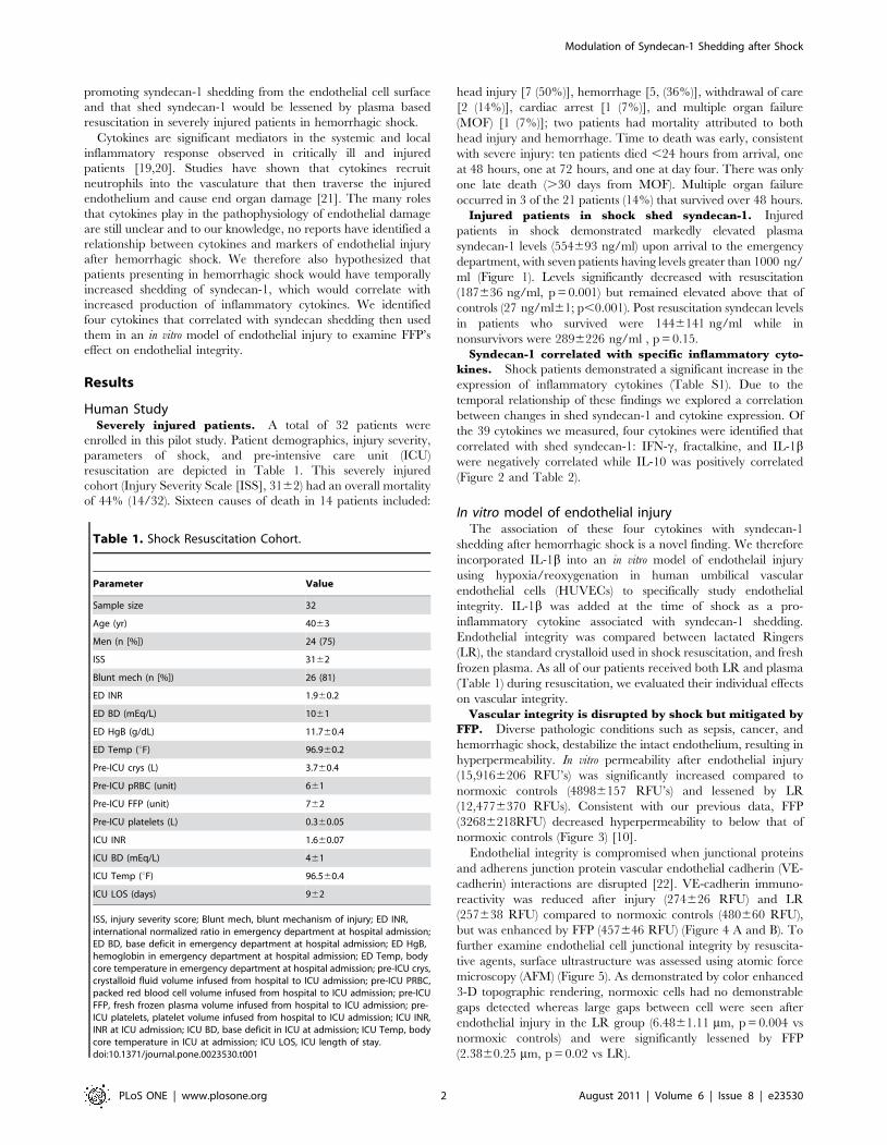
Modulation of Syndecan-1 Shedding after Hemorrhagic Shock and Resuscitation Ricky J. Haywood-Watson 1 , John B. Holcomb 1,2 , Ernest A. Gonzalez 3 , Zhanglong Peng 1 , Shibani Pati 1,2 , Pyong Woo Park 4 , WeiWei Wang 5 , Ana Maria Zaske 2 , Tyler Menge 1 , Rosemary A. Kozar 1 * 1 Department of Surgery, The University of Texas Health Science Center at Houston, Houston, Texas, United States of America, 2 Center for Translational Injury and Research (CeTIR), The University of Texas Health Science Center at Houston, Houston, Texas, United States of America, 3 Department of Surgery, University of Texas Southwestern Austin, Austin, Texas, United States of America, 4 Division of Respiratory Diseases, Children’s Hospital, Harvard Medical School, Boston, Massachusetts, United States of America, 5 Center for Clinical and Translational Sciences, The University of Texas Health Science Center at Houston, Houston, Texas, United States of America Abstract The early use of fresh frozen plasma as a resuscitative agent after hemorrhagic shock has been associated with improved survival, but the mechanism of protection is unknown. Hemorrhagic shock causes endothelial cell dysfunction and we hypothesized that fresh frozen plasma would restore endothelial integrity and reduce syndecan-1 shedding after hemorrhagic shock. A prospective, observational study in severely injured patients in hemorrhagic shock demonstrated significantly elevated levels of syndecan-1 (554693 ng/ml) after injury, which decreased with resuscitation (187636 ng/ ml) but was elevated compared to normal donors (2761 ng/ml). Three pro-inflammatory cytokines, interferon-c, fractalkine, and interleukin-1b, negatively correlated while one anti-inflammatory cytokine, IL-10, positively correlated with shed syndecan-1. These cytokines all play an important role in maintaining endothelial integrity. An in vitro model of endothelial injury then specifically examined endothelial permeability after treatment with fresh frozen plasma orlactated Ringers. Shock or endothelial injury disrupted junctional integrity and increased permeability, which was improved with fresh frozen plasma, but not lactated Ringers. Changes in endothelial cell permeability correlated with syndecan-1 shedding. These data suggest that plasma based resuscitation preserved endothelial syndecan-1 and maintained endothelial integrity, and may help to explain the protective effects of fresh frozen plasma after hemorrhagic shock. Citation: Haywood-Watson RJ, Holcomb JB, Gonzalez EA, Peng Z, Pati S, et al. (2011) Modulation of Syndecan-1 Shedding after Hemorrhagic Shock and Resuscitation. PLoS ONE 6(8): e23530. doi:10.1371/journal.pone.0023530 Editor: Paul McNeil, Medical College of Georgia, United States of America Received April 29, 2011; Accepted July 19, 2011; Published August 19, 2011 Copyright: ß 2011 Haywood-Watson et al. This is an open-access article distributed under the terms of the Creative Commons Attribution License, which permits unrestricted use, distribution, and reproduction in any medium, provided the original author and source are credited. Funding: The study was supported by the National Institutes of Health T-32 GM008792 and P-50 GM38529 and the Department of Defense W81XWH-08-C-0712. Statistical support was provided by the Biostatistics/ Epidemiology/ Research Design (BERD) component of the Center for Clinical and Translational Sciences (CCTS) for this project. CCTS is mainly funded by NIH CTSA grant (UL1 RR024148). The funders had no role in study design, data collection and analysis, decision to publish, or preparation of the manuscript. Competing Interests: The authors have declared that no competing interests exist. * E-mail: [email protected] Introduction Hemorrhagic shock is the most common cause of potentially preventable death after both civilian and combat traumatic injury [1]. Despite the significant effort expended on mechanistic resuscitation studies, several large randomized multicenter clinical trials have unfortunately failed to demonstrate any clinically significant outcome differences [2–4]. Recently, data from both military [5,6] and civilian studies [7–9] have associated survival benefit following massive transfusion (.10 units packed red cells in 24 hours) with the implementation of a high ratio fresh frozen plasma (FFP) to red cell resuscitation strategy. This change in resuscitation centers around the early and increased use of plasma and platelets and decreased crystalloid utilization. These changes have been associated with a significant increase in early survival, though the studies are retrospective and the mechanism of protection is unknown. To begin to investigate the molecular pathways responsible for protection by FFP-based resuscitation, we are focusing on the role of the endothelial cell in maintaining endothelial integrity [10]. Endothelial dysfunction and hyperper- meability have been implicated in the morbidity and mortality associated with sepsis, organ failure and hemorrhagic shock [11– 13]. The glycocalyx is a network of soluble plasma components that projects from the endothelial cell surface and plays a key role in maintaining endothelial integrity [14]. It consists of proteoglycans and glycoproteins attached to the cell surface. Cell adhesion molecules constitute several of the glycoproteins. With injury to the glycocalyx, adhesion molecules are exposed, allowing patho- logic neutrophil-endothelial cell interactions. Other glycoproteins within the glycocalyx are important to coagulation, fibrinolysis, and hemostasis [15]. The major cell surface proteoglycan is syndecan, whose extracellular domain is substituted with heparan sulfate chains and promotes interaction with plasma proteins [16]. There are four members (syndecan 1–4) that comprise the syndecan family. While syndecan-1 is found primarily on epithelial cells, recent data suggests that it also found on endothelial cells and plays an important role in endothelial cell function after hemorrhagic shock [17,18]. We therefore hypothesized that hemorrhagic shock would disrupt endothelial integrity by PLoS ONE | www.plosone.org 1 August 2011 | Volume 6 | Issue 8 | e23530

Welcome message from author
This document is posted to help you gain knowledge. Please leave a comment to let me know what you think about it! Share it to your friends and learn new things together.
Transcript
Modulation of Syndecan-1 Shedding after HemorrhagicShock and ResuscitationRicky J. Haywood-Watson1, John B. Holcomb1,2, Ernest A. Gonzalez3, Zhanglong Peng1, Shibani Pati1,2,
Pyong Woo Park4, WeiWei Wang5, Ana Maria Zaske2, Tyler Menge1, Rosemary A. Kozar1*
1 Department of Surgery, The University of Texas Health Science Center at Houston, Houston, Texas, United States of America, 2 Center for Translational Injury and
Research (CeTIR), The University of Texas Health Science Center at Houston, Houston, Texas, United States of America, 3 Department of Surgery, University of Texas
Southwestern Austin, Austin, Texas, United States of America, 4 Division of Respiratory Diseases, Children’s Hospital, Harvard Medical School, Boston, Massachusetts,
United States of America, 5 Center for Clinical and Translational Sciences, The University of Texas Health Science Center at Houston, Houston, Texas, United States of
America
Abstract
The early use of fresh frozen plasma as a resuscitative agent after hemorrhagic shock has been associated with improvedsurvival, but the mechanism of protection is unknown. Hemorrhagic shock causes endothelial cell dysfunction and wehypothesized that fresh frozen plasma would restore endothelial integrity and reduce syndecan-1 shedding afterhemorrhagic shock. A prospective, observational study in severely injured patients in hemorrhagic shock demonstratedsignificantly elevated levels of syndecan-1 (554693 ng/ml) after injury, which decreased with resuscitation (187636 ng/ml) but was elevated compared to normal donors (2761 ng/ml). Three pro-inflammatory cytokines, interferon-c,fractalkine, and interleukin-1b, negatively correlated while one anti-inflammatory cytokine, IL-10, positively correlatedwith shed syndecan-1. These cytokines all play an important role in maintaining endothelial integrity. An in vitro modelof endothelial injury then specifically examined endothelial permeability after treatment with fresh frozen plasmaorlactated Ringers. Shock or endothelial injury disrupted junctional integrity and increased permeability, which wasimproved with fresh frozen plasma, but not lactated Ringers. Changes in endothelial cell permeability correlated withsyndecan-1 shedding. These data suggest that plasma based resuscitation preserved endothelial syndecan-1 andmaintained endothelial integrity, and may help to explain the protective effects of fresh frozen plasma after hemorrhagicshock.
Citation: Haywood-Watson RJ, Holcomb JB, Gonzalez EA, Peng Z, Pati S, et al. (2011) Modulation of Syndecan-1 Shedding after Hemorrhagic Shock andResuscitation. PLoS ONE 6(8): e23530. doi:10.1371/journal.pone.0023530
Editor: Paul McNeil, Medical College of Georgia, United States of America
Received April 29, 2011; Accepted July 19, 2011; Published August 19, 2011
Copyright: � 2011 Haywood-Watson et al. This is an open-access article distributed under the terms of the Creative Commons Attribution License, whichpermits unrestricted use, distribution, and reproduction in any medium, provided the original author and source are credited.
Funding: The study was supported by the National Institutes of Health T-32 GM008792 and P-50 GM38529 and the Department of Defense W81XWH-08-C-0712.Statistical support was provided by the Biostatistics/ Epidemiology/ Research Design (BERD) component of the Center for Clinical and Translational Sciences(CCTS) for this project. CCTS is mainly funded by NIH CTSA grant (UL1 RR024148). The funders had no role in study design, data collection and analysis, decision topublish, or preparation of the manuscript.
Competing Interests: The authors have declared that no competing interests exist.
* E-mail: [email protected]
Introduction
Hemorrhagic shock is the most common cause of potentially
preventable death after both civilian and combat traumatic injury
[1]. Despite the significant effort expended on mechanistic
resuscitation studies, several large randomized multicenter clinical
trials have unfortunately failed to demonstrate any clinically
significant outcome differences [2–4]. Recently, data from both
military [5,6] and civilian studies [7–9] have associated survival
benefit following massive transfusion (.10 units packed red cells in
24 hours) with the implementation of a high ratio fresh frozen
plasma (FFP) to red cell resuscitation strategy. This change in
resuscitation centers around the early and increased use of plasma
and platelets and decreased crystalloid utilization. These changes
have been associated with a significant increase in early survival,
though the studies are retrospective and the mechanism of
protection is unknown. To begin to investigate the molecular
pathways responsible for protection by FFP-based resuscitation,
we are focusing on the role of the endothelial cell in maintaining
endothelial integrity [10]. Endothelial dysfunction and hyperper-
meability have been implicated in the morbidity and mortality
associated with sepsis, organ failure and hemorrhagic shock [11–
13].
The glycocalyx is a network of soluble plasma components that
projects from the endothelial cell surface and plays a key role in
maintaining endothelial integrity [14]. It consists of proteoglycans
and glycoproteins attached to the cell surface. Cell adhesion
molecules constitute several of the glycoproteins. With injury to
the glycocalyx, adhesion molecules are exposed, allowing patho-
logic neutrophil-endothelial cell interactions. Other glycoproteins
within the glycocalyx are important to coagulation, fibrinolysis,
and hemostasis [15]. The major cell surface proteoglycan is
syndecan, whose extracellular domain is substituted with heparan
sulfate chains and promotes interaction with plasma proteins [16].
There are four members (syndecan 1–4) that comprise the
syndecan family. While syndecan-1 is found primarily on epithelial
cells, recent data suggests that it also found on endothelial cells and
plays an important role in endothelial cell function after
hemorrhagic shock [17,18]. We therefore hypothesized that
hemorrhagic shock would disrupt endothelial integrity by
PLoS ONE | www.plosone.org 1 August 2011 | Volume 6 | Issue 8 | e23530
promoting syndecan-1 shedding from the endothelial cell surface
and that shed syndecan-1 would be lessened by plasma based
resuscitation in severely injured patients in hemorrhagic shock.
Cytokines are significant mediators in the systemic and local
inflammatory response observed in critically ill and injured
patients [19,20]. Studies have shown that cytokines recruit
neutrophils into the vasculature that then traverse the injured
endothelium and cause end organ damage [21]. The many roles
that cytokines play in the pathophysiology of endothelial damage
are still unclear and to our knowledge, no reports have identified a
relationship between cytokines and markers of endothelial injury
after hemorrhagic shock. We therefore also hypothesized that
patients presenting in hemorrhagic shock would have temporally
increased shedding of syndecan-1, which would correlate with
increased production of inflammatory cytokines. We identified
four cytokines that correlated with syndecan shedding then used
them in an in vitro model of endothelial injury to examine FFP’s
effect on endothelial integrity.
Results
Human StudySeverely injured patients. A total of 32 patients were
enrolled in this pilot study. Patient demographics, injury severity,
parameters of shock, and pre-intensive care unit (ICU)
resuscitation are depicted in Table 1. This severely injured
cohort (Injury Severity Scale [ISS], 3162) had an overall mortality
of 44% (14/32). Sixteen causes of death in 14 patients included:
head injury [7 (50%)], hemorrhage [5, (36%)], withdrawal of care
[2 (14%)], cardiac arrest [1 (7%)], and multiple organ failure
(MOF) [1 (7%)]; two patients had mortality attributed to both
head injury and hemorrhage. Time to death was early, consistent
with severe injury: ten patients died ,24 hours from arrival, one
at 48 hours, one at 72 hours, and one at day four. There was only
one late death (.30 days from MOF). Multiple organ failure
occurred in 3 of the 21 patients (14%) that survived over 48 hours.
Injured patients in shock shed syndecan-1. Injured
patients in shock demonstrated markedly elevated plasma
syndecan-1 levels (554693 ng/ml) upon arrival to the emergency
department, with seven patients having levels greater than 1000 ng/
ml (Figure 1). Levels significantly decreased with resuscitation
(187636 ng/ml, p = 0.001) but remained elevated above that of
controls (27 ng/ml61; p,0.001). Post resuscitation syndecan levels
in patients who survived were 1446141 ng/ml while in
nonsurvivors were 2896226 ng/ml , p = 0.15.
Syndecan-1 correlated with specific inflammatory cyto-
kines. Shock patients demonstrated a significant increase in the
expression of inflammatory cytokines (Table S1). Due to the
temporal relationship of these findings we explored a correlation
between changes in shed syndecan-1 and cytokine expression. Of
the 39 cytokines we measured, four cytokines were identified that
correlated with shed syndecan-1: IFN-c, fractalkine, and IL-1bwere negatively correlated while IL-10 was positively correlated
(Figure 2 and Table 2).
In vitro model of endothelial injuryThe association of these four cytokines with syndecan-1
shedding after hemorrhagic shock is a novel finding. We therefore
incorporated IL-1b into an in vitro model of endothelail injury
using hypoxia/reoxygenation in human umbilical vascular
endothelial cells (HUVECs) to specifically study endothelial
integrity. IL-1b was added at the time of shock as a pro-
inflammatory cytokine associated with syndecan-1 shedding.
Endothelial integrity was compared between lactated Ringers
(LR), the standard crystalloid used in shock resuscitation, and fresh
frozen plasma. As all of our patients received both LR and plasma
(Table 1) during resuscitation, we evaluated their individual effects
on vascular integrity.
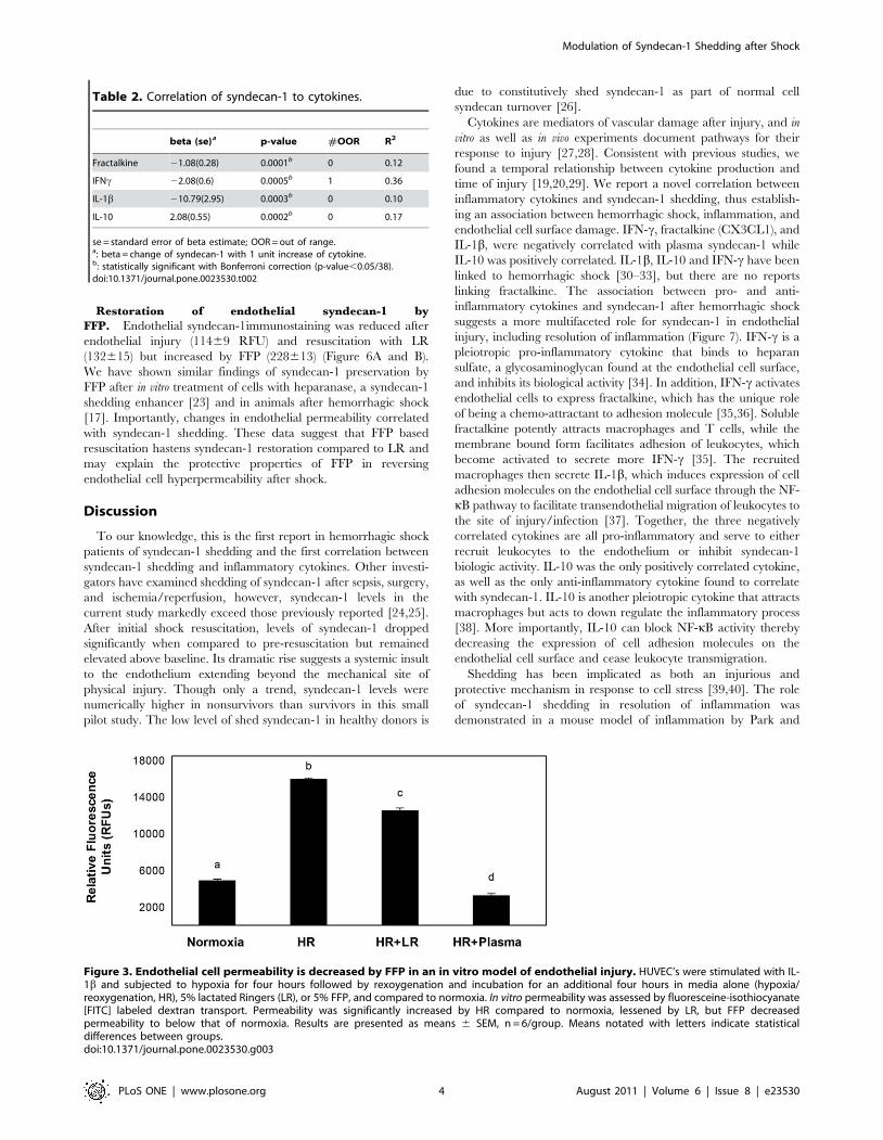
Vascular integrity is disrupted by shock but mitigated by
FFP. Diverse pathologic conditions such as sepsis, cancer, and
hemorrhagic shock, destabilize the intact endothelium, resulting in
hyperpermeability. In vitro permeability after endothelial injury
(15,9166206 RFU’s) was significantly increased compared to
normoxic controls (48986157 RFU’s) and lessened by LR
(12,4776370 RFUs). Consistent with our previous data, FFP
(32686218RFU) decreased hyperpermeability to below that of
normoxic controls (Figure 3) [10].
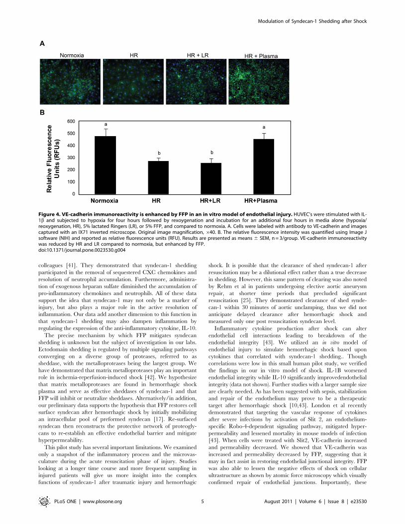
Endothelial integrity is compromised when junctional proteins
and adherens junction protein vascular endothelial cadherin (VE-
cadherin) interactions are disrupted [22]. VE-cadherin immuno-
reactivity was reduced after injury (274626 RFU) and LR
(257638 RFU) compared to normoxic controls (480660 RFU),
but was enhanced by FFP (457646 RFU) (Figure 4 A and B). To
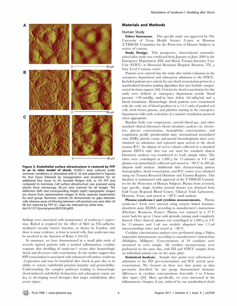
further examine endothelial cell junctional integrity by resuscita-
tive agents, surface ultrastructure was assessed using atomic force
microscopy (AFM) (Figure 5). As demonstrated by color enhanced
3-D topographic rendering, normoxic cells had no demonstrable
gaps detected whereas large gaps between cell were seen after
endothelial injury in the LR group (6.4861.11 mm, p = 0.004 vs
normoxic controls) and were significantly lessened by FFP
(2.3860.25 mm, p = 0.02 vs LR).
Table 1. Shock Resuscitation Cohort.
Parameter Value
Sample size 32
Age (yr) 4063
Men (n [%]) 24 (75)
ISS 3162
Blunt mech (n [%]) 26 (81)
ED INR 1.960.2
ED BD (mEq/L) 1061
ED HgB (g/dL) 11.760.4
ED Temp (uF) 96.960.2
Pre-ICU crys (L) 3.760.4
Pre-ICU pRBC (unit) 661
Pre-ICU FFP (unit) 762
Pre-ICU platelets (L) 0.360.05
ICU INR 1.660.07
ICU BD (mEq/L) 461
ICU Temp (uF) 96.560.4
ICU LOS (days) 962
ISS, injury severity score; Blunt mech, blunt mechanism of injury; ED INR,international normalized ratio in emergency department at hospital admission;ED BD, base deficit in emergency department at hospital admission; ED HgB,hemoglobin in emergency department at hospital admission; ED Temp, bodycore temperature in emergency department at hospital admission; pre-ICU crys,crystalloid fluid volume infused from hospital to ICU admission; pre-ICU PRBC,packed red blood cell volume infused from hospital to ICU admission; pre-ICUFFP, fresh frozen plasma volume infused from hospital to ICU admission; pre-ICU platelets, platelet volume infused from hospital to ICU admission; ICU INR,INR at ICU admission; ICU BD, base deficit in ICU at admission; ICU Temp, bodycore temperature in ICU at admission; ICU LOS, ICU length of stay.doi:10.1371/journal.pone.0023530.t001
Modulation of Syndecan-1 Shedding after Shock
PLoS ONE | www.plosone.org 2 August 2011 | Volume 6 | Issue 8 | e23530
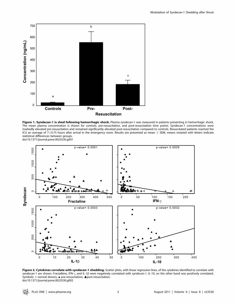
Figure 1. Syndecan-1 is shed following hemorrhagic shock. Plasma syndecan-1 was measured in patients presenting in hemorrhagic shock.The mean plasma concentration is shown for controls, pre-resuscitation, and post-resuscitation time points. Syndecan-1 concentrations weremarkedly elevated pre-resuscitation and remained significantly elevated post-resuscitation compared to controls. Resuscitated patients reached theICU an average of 760.75 hours after arrival in the emergency room. Results are presented as mean 6 SEM, means notated with letters indicatestatistical differences between groups.doi:10.1371/journal.pone.0023530.g001
Figure 2. Cytokines correlate with syndecan-1 shedding. Scatter plots, with linear regression lines, of the cytokines identified to correlate withsyndecan-1 are shown. Fractalkine, IFN-c, and IL-1b were negatively correlated with syndecan-1. IL-10, on the other hand was positively correlated.Symbols: + normal donors, N pre-resuscitation, mpost-resuscitation.doi:10.1371/journal.pone.0023530.g002
Modulation of Syndecan-1 Shedding after Shock
PLoS ONE | www.plosone.org 3 August 2011 | Volume 6 | Issue 8 | e23530
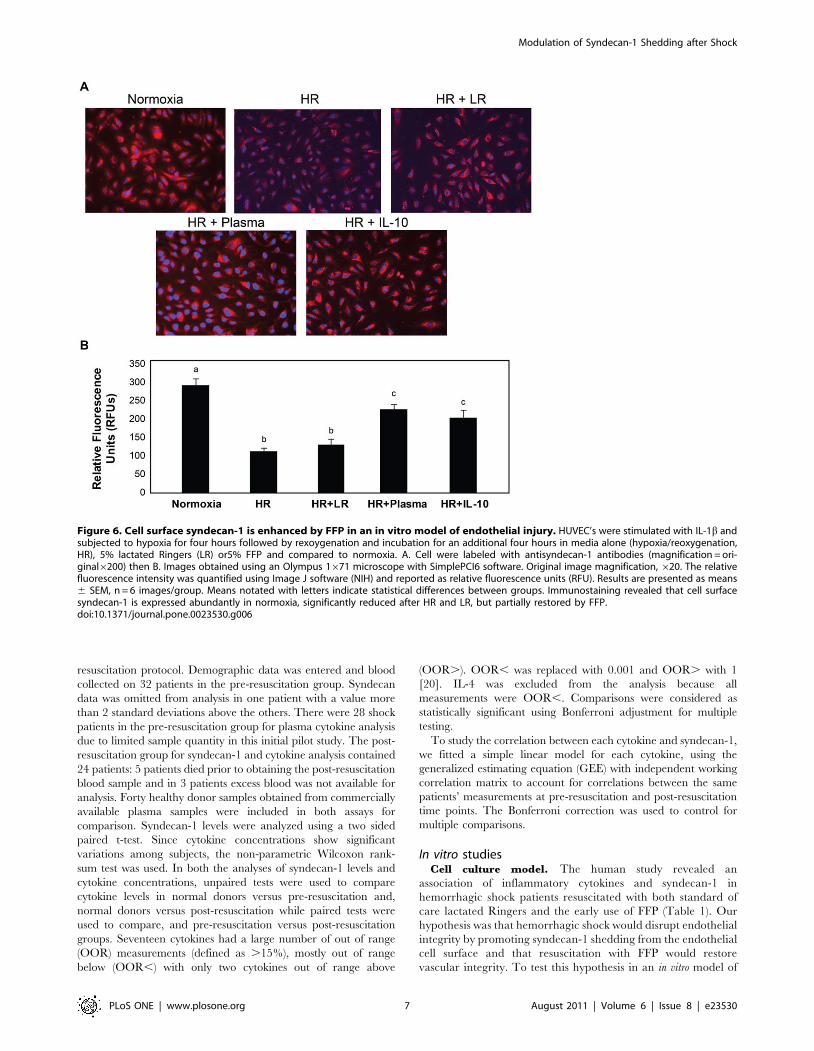
Restoration of endothelial syndecan-1 by
FFP. Endothelial syndecan-1immunostaining was reduced after
endothelial injury (11469 RFU) and resuscitation with LR
(132615) but increased by FFP (228613) (Figure 6A and B).
We have shown similar findings of syndecan-1 preservation by
FFP after in vitro treatment of cells with heparanase, a syndecan-1
shedding enhancer [23] and in animals after hemorrhagic shock
[17]. Importantly, changes in endothelial permeability correlated
with syndecan-1 shedding. These data suggest that FFP based
resuscitation hastens syndecan-1 restoration compared to LR and
may explain the protective properties of FFP in reversing
endothelial cell hyperpermeability after shock.
Discussion
To our knowledge, this is the first report in hemorrhagic shock
patients of syndecan-1 shedding and the first correlation between
syndecan-1 shedding and inflammatory cytokines. Other investi-
gators have examined shedding of syndecan-1 after sepsis, surgery,
and ischemia/reperfusion, however, syndecan-1 levels in the
current study markedly exceed those previously reported [24,25].
After initial shock resuscitation, levels of syndecan-1 dropped
significantly when compared to pre-resuscitation but remained
elevated above baseline. Its dramatic rise suggests a systemic insult
to the endothelium extending beyond the mechanical site of
physical injury. Though only a trend, syndecan-1 levels were
numerically higher in nonsurvivors than survivors in this small
pilot study. The low level of shed syndecan-1 in healthy donors is
due to constitutively shed syndecan-1 as part of normal cell
syndecan turnover [26].
Cytokines are mediators of vascular damage after injury, and in
vitro as well as in vivo experiments document pathways for their
response to injury [27,28]. Consistent with previous studies, we
found a temporal relationship between cytokine production and
time of injury [19,20,29]. We report a novel correlation between
inflammatory cytokines and syndecan-1 shedding, thus establish-
ing an association between hemorrhagic shock, inflammation, and
endothelial cell surface damage. IFN-c, fractalkine (CX3CL1), and
IL-1b, were negatively correlated with plasma syndecan-1 while
IL-10 was positively correlated. IL-1b, IL-10 and IFN-c have been
linked to hemorrhagic shock [30–33], but there are no reports
linking fractalkine. The association between pro- and anti-
inflammatory cytokines and syndecan-1 after hemorrhagic shock
suggests a more multifaceted role for syndecan-1 in endothelial
injury, including resolution of inflammation (Figure 7). IFN-c is a
pleiotropic pro-inflammatory cytokine that binds to heparan
sulfate, a glycosaminoglycan found at the endothelial cell surface,
and inhibits its biological activity [34]. In addition, IFN-c activates
endothelial cells to express fractalkine, which has the unique role
of being a chemo-attractant to adhesion molecule [35,36]. Soluble
fractalkine potently attracts macrophages and T cells, while the
membrane bound form facilitates adhesion of leukocytes, which
become activated to secrete more IFN-c [35]. The recruited
macrophages then secrete IL-1b, which induces expression of cell
adhesion molecules on the endothelial cell surface through the NF-
kB pathway to facilitate transendothelial migration of leukocytes to
the site of injury/infection [37]. Together, the three negatively
correlated cytokines are all pro-inflammatory and serve to either
recruit leukocytes to the endothelium or inhibit syndecan-1
biologic activity. IL-10 was the only positively correlated cytokine,
as well as the only anti-inflammatory cytokine found to correlate
with syndecan-1. IL-10 is another pleiotropic cytokine that attracts
macrophages but acts to down regulate the inflammatory process
[38]. More importantly, IL-10 can block NF-kB activity thereby
decreasing the expression of cell adhesion molecules on the
endothelial cell surface and cease leukocyte transmigration.
Shedding has been implicated as both an injurious and
protective mechanism in response to cell stress [39,40]. The role
of syndecan-1 shedding in resolution of inflammation was
demonstrated in a mouse model of inflammation by Park and
Table 2. Correlation of syndecan-1 to cytokines.
beta (se)a p-value #OOR R2
Fractalkine 21.08(0.28) 0.0001b 0 0.12
IFNc 22.08(0.6) 0.0005b 1 0.36
IL-1b 210.79(2.95) 0.0003b 0 0.10
IL-10 2.08(0.55) 0.0002b 0 0.17
se = standard error of beta estimate; OOR = out of range.a: beta = change of syndecan-1 with 1 unit increase of cytokine.b: statistically significant with Bonferroni correction (p-value,0.05/38).doi:10.1371/journal.pone.0023530.t002
Figure 3. Endothelial cell permeability is decreased by FFP in an in vitro model of endothelial injury. HUVEC’s were stimulated with IL-1b and subjected to hypoxia for four hours followed by rexoygenation and incubation for an additional four hours in media alone (hypoxia/reoxygenation, HR), 5% lactated Ringers (LR), or 5% FFP, and compared to normoxia. In vitro permeability was assessed by fluoresceine-isothiocyanate[FITC] labeled dextran transport. Permeability was significantly increased by HR compared to normoxia, lessened by LR, but FFP decreasedpermeability to below that of normoxia. Results are presented as means 6 SEM, n = 6/group. Means notated with letters indicate statisticaldifferences between groups.doi:10.1371/journal.pone.0023530.g003
Modulation of Syndecan-1 Shedding after Shock
PLoS ONE | www.plosone.org 4 August 2011 | Volume 6 | Issue 8 | e23530
colleagues [41]. They demonstrated that syndecan-1 shedding
participated in the removal of sequestered CXC chemokines and
resolution of neutrophil accumulation. Furthermore, administra-
tion of exogenous heparan sulfate diminished the accumulation of
pro-inflammatory chemokines and neutrophils. All of these data
support the idea that syndecan-1 may not only be a marker of
injury, but also plays a major role in the active resolution of
inflammation. Our data add another dimension to this function in
that syndecan-1 shedding may also dampen inflammation by
regulating the expression of the anti-inflammatory cytokine, IL-10.
The precise mechanism by which FFP mitigates syndecan
shedding is unknown but the subject of investigation in our labs.
Ectodomain shedding is regulated by multiple signaling pathways
converging on a diverse group of proteases, referred to as
sheddase, with the metalloproteases being the largest group. We
have demonstrated that matrix metalloproteases play an important
role in ischemia-reperfusion-induced shock [42]. We hypothesize
that matrix metalloproteases are found in hemorrhagic shock
plasma and serve as effective sheddases of syndecan-1 and that
FFP will inhibit or neutralize sheddases. Alternatively/in addition,
our preliminary data supports the hypothesis that FFP restores cell
surface syndecan after hemorrhagic shock by initially mobilizing
an intracellular pool of preformed syndecan [17]. Re-surfaced
syndecan then reconstructs the protective network of proteogly-
cans to re-establish an effective endothelial barrier and mitigate
hyperpermeability.
This pilot study has several important limitations. We examined
only a snapshot of the inflammatory process and the microvas-
culature during the acute resuscitation phase of injury. Studies
looking at a longer time course and more frequent sampling in
injured patients will give us more insight into the complex
functions of syndecan-1 after traumatic injury and hemorrhagic
shock. It is possible that the clearance of shed syndecan-1 after
resuscitation may be a dilutional effect rather than a true decrease
in shedding. However, this same pattern of clearing was also noted
by Rehm et al in patients undergoing elective aortic aneurysm
repair, at shorter time periods that precluded significant
resuscitation [25]. They demonstrated clearance of shed synde-
can-1 within 30 minutes of aortic unclamping, thus we did not
anticipate delayed clearance after hemorrhagic shock and
measured only one post resuscitation syndecan level.
Inflammatory cytokine production after shock can alter
endothelial cell interactions leading to breakdown of the
endothelial integrity [43]. We utilized an in vitro model of
endothelial injury to simulate hemorrhagic shock based upon
cytokines that correlated with syndecan-1 shedding.. Though
correlations were low in this small human pilot study, we verified
the findings in our in vitro model of shock. IL-1B worsened
endothelial integrity while IL-10 significantly improvedendothelial
integrity (data not shown). Further studies with a larger sample size
are clearly needed. As has been suggested with sepsis, stabilization
and repair of the endothelium may prove to be a therapeutic
target after hemorrhagic shock [10,43]. London et al recently
demonstrated that targeting the vascular response of cytokines
after severe infections by activation of Slit 2, an endothelium-
specific Robo-4-dependent signaling pathway, mitigated hyper-
permeability and lessened mortality in mouse models of infection
[43]. When cells were treated with Slit2, VE-cadherin increased
and permeability decreased. We showed that VE-cadherin was
increased and permeability decreased by FFP, suggesting that it
may in fact assist in restoring endothelial junctional integrity. FFP
was also able to lessen the negative effects of shock on cellular
ultrastructure as shown by atomic force microscopy which visually
confirmed repair of endothelial junctions. Importantly, these
Figure 4. VE-cadherin immunoreactivity is enhanced by FFP in an in vitro model of endothelial injury. HUVEC’s were stimulated with IL-1b and subjected to hypoxia for four hours followed by rexoygenation and incubation for an additional four hours in media alone (hypoxia/reoxygenation, HR), 5% lactated Ringers (LR), or 5% FFP, and compared to normoxia. A. Cells were labeled with antibody to VE-cadherin and imagescaptured with an IX71 inverted microscope. Original image magnification, 640. B. The relative fluorescence intensity was quantified using Image Jsoftware (NIH) and reported as relative fluorescence units (RFU). Results are presented as means 6 SEM, n = 3/group. VE-cadherin immunoreactivitywas reduced by HR and LR compared to normoxia, but enhanced by FFP.doi:10.1371/journal.pone.0023530.g004
Modulation of Syndecan-1 Shedding after Shock
PLoS ONE | www.plosone.org 5 August 2011 | Volume 6 | Issue 8 | e23530
findings were associated with maintenance of syndecan-1 expres-
sion. Robo4 is required for the effect of Slit2 on VE-cadherin-
mediated vascular barrier function, as shown by London, and
there is some evidence, at least in neural cells, that syndecans may
be involved in the function of Robo 4 [44,45].
In summary, we have demonstrated in a small pilot study of
severely injured patients with a marked inflammatory cytokine
response that shedding of syndecan-1 occurs after hemorrhagic
shock and decreases with resuscitation. In vitro studies suggest that
FFP resuscitation is associated with enhanced cell surface syndecan
-1expression and may be beneficial after shock in part due to its
ability to restore endothelial junction integrity and permeability.
Understanding the complex pathways leading to hemorrhagic
shock-induced endothelial dysfunction and subsequent repair are
key to developing novel therapies that target endothelium after
severe injury.
Materials and Methods
Human StudyEthics Statement. This specific study was approved by The
University of Texas Health Science Center at Houston
(UTHSCH) Committee for the Protection of Human Subjects as
waiver of consent.
Study Design. This prospective, observational nonrando-
mized pilot study was conducted from January to June 2009 in the
Emergency Department (ED) and Shock Trauma Intensive Care
Unit (STICU) at Memorial Hermann Hospital, Houston, TX, a
busy Level I trauma center.
Patients were entered into the study after initial evaluation in the
emergency department and subsequent admission to the STICU.
Included patients met criteria for our shock resuscitation protocol, a
standardized decision making algorithm that uses bedside comput-
erized decision support [46]. Criteria for shock resuscitation for this
study were defined as: emergency department systolic blood
pressure ,90 mmHg, and/or base deficit $6 mEq/ml and a
blood transfusion. Hemorrhagic shock patients were resuscitated
with the early use of blood products in a 1:1:1 ratio of packed red
cells, fresh frozen plasma, and platelets starting in the emergency
department with early activation of a massive transfusion protocol
when appropriate.
Baseline body core temperature, arterial blood gas, and other
standard clinical laboratory blood chemistry analyses (ie, electro-
lyte, glucose concentration, hemoglobin concentration), and
coagulation profile (prothrombin time, international normalized
ratio [INR], platelet count, and partial thromboplastin time) were
obtained on admission and repeated upon arrival to the shock
trauma ICU. An aliquot of excess volume collected in a standard
plasma EDTA tube that was not used for standard clinical
laboratory analyses was transferred to 3-mL sample tubes. The
tubes were centrifuged at 1,000 g for 15 minutes at 4uC and
plasma was immediately collected and stored at 280uC in 400 mL
aliquots until analysis. Additional data characterizing patient
demographics, shock resuscitation, and ICU course were obtained
using our Trauma Research Database and Trauma Registry. This
database is maintained with approval of the UTHSCH Commit-
tee for the Protection of Human Subjects. Plasma from screened,
type specific, single, healthy normal donors was obtained from
Gulf Coast Regional Blood Center, Clinical Trials Laboratory,
Houston, Texas, and stored at 280uC until analysis.
Plasma syndecan-1 and cytokine measurements. Plasma
syndecan-1 levels were assessed using enzyme linked immuno-
absorbent assay (ELISA) according to manufacturer’s instructions
(Diaclone; Besancon, France). Plasma was warmed in a 37uCwater bath for up to 1 hour with periodic mixing until completely
thawed. Once thawed, plasma was centrifuged at 1,000 g at 4uCfor 15 minutes and 1 ml was sterilely aliquoted into 1.5 ml
microcentrifuge tubes and stored at 280uC.
Cytokine concentration analyses were performed using a 39plex
suspension immunoassay according to manufacturer’s instructions
(Multiplex; Millipore). Concentrations of 39 cytokines were
measured in each sample. All cytokine measurements were
performed on the same day, with ED and STICU samples from
an individual patient run on the same plate.
Statistical Analysis. Sample time points were referenced as
admission to the ED (pre-resuscitation) and ICU arrival (post-
resuscitation). We focused on these two time points as data
previously described by our group demonstrated dramatic
differences in cytokine concentrations detectable 4 to 8 hours
after injury [20]. More specifically, we were interested in the
inflammatory changes, if any, induced by our standardized shock
Figure 5. Endothelial surface ultrastructure is restored by FFPin an in vitro model of shock. HUVEC’s were cultured undernormoxic conditions or stimulated with IL-1b and subjected to hypoxiafor four hours followed by rexoygenation and incubation for anadditional four hours in 5% lactated Ringers (LR), or 5% FFP andcompared to normoxia. Cell surface ultrastructure was assessed usingatomic force microscopy, 80 mm area scanned for all images. Thedeflection (left) and corresponding height (right) topographic imagesare shown from representative images of three separate experimentsfor each group. Normoxic controls (A) demonstrate no gaps betweencells whereas areas of thinning between cell junctions are seen after LR(B) but reduced by FFP (C). Gaps are indicated by white lines.doi:10.1371/journal.pone.0023530.g005
Modulation of Syndecan-1 Shedding after Shock
PLoS ONE | www.plosone.org 6 August 2011 | Volume 6 | Issue 8 | e23530
resuscitation protocol. Demographic data was entered and blood
collected on 32 patients in the pre-resuscitation group. Syndecan
data was omitted from analysis in one patient with a value more
than 2 standard deviations above the others. There were 28 shock
patients in the pre-resuscitation group for plasma cytokine analysis
due to limited sample quantity in this initial pilot study. The post-
resuscitation group for syndecan-1 and cytokine analysis contained
24 patients: 5 patients died prior to obtaining the post-resuscitation
blood sample and in 3 patients excess blood was not available for
analysis. Forty healthy donor samples obtained from commercially
available plasma samples were included in both assays for
comparison. Syndecan-1 levels were analyzed using a two sided
paired t-test. Since cytokine concentrations show significant
variations among subjects, the non-parametric Wilcoxon rank-
sum test was used. In both the analyses of syndecan-1 levels and
cytokine concentrations, unpaired tests were used to compare
cytokine levels in normal donors versus pre-resuscitation and,
normal donors versus post-resuscitation while paired tests were
used to compare, and pre-resuscitation versus post-resuscitation
groups. Seventeen cytokines had a large number of out of range
(OOR) measurements (defined as .15%), mostly out of range
below (OOR,) with only two cytokines out of range above
(OOR.). OOR, was replaced with 0.001 and OOR. with 1
[20]. IL-4 was excluded from the analysis because all
measurements were OOR,. Comparisons were considered as
statistically significant using Bonferroni adjustment for multiple
testing.
To study the correlation between each cytokine and syndecan-1,
we fitted a simple linear model for each cytokine, using the
generalized estimating equation (GEE) with independent working
correlation matrix to account for correlations between the same
patients’ measurements at pre-resuscitation and post-resuscitation
time points. The Bonferroni correction was used to control for
multiple comparisons.
In vitro studiesCell culture model. The human study revealed an
association of inflammatory cytokines and syndecan-1 in
hemorrhagic shock patients resuscitated with both standard of
care lactated Ringers and the early use of FFP (Table 1). Our
hypothesis was that hemorrhagic shock would disrupt endothelial
integrity by promoting syndecan-1 shedding from the endothelial
cell surface and that resuscitation with FFP would restore
vascular integrity. To test this hypothesis in an in vitro model of
Figure 6. Cell surface syndecan-1 is enhanced by FFP in an in vitro model of endothelial injury. HUVEC’s were stimulated with IL-1b andsubjected to hypoxia for four hours followed by rexoygenation and incubation for an additional four hours in media alone (hypoxia/reoxygenation,HR), 5% lactated Ringers (LR) or5% FFP and compared to normoxia. A. Cell were labeled with antisyndecan-1 antibodies (magnification = ori-ginal6200) then B. Images obtained using an Olympus 1671 microscope with SimplePCI6 software. Original image magnification, 620. The relativefluorescence intensity was quantified using Image J software (NIH) and reported as relative fluorescence units (RFU). Results are presented as means6 SEM, n = 6 images/group. Means notated with letters indicate statistical differences between groups. Immunostaining revealed that cell surfacesyndecan-1 is expressed abundantly in normoxia, significantly reduced after HR and LR, but partially restored by FFP.doi:10.1371/journal.pone.0023530.g006
Modulation of Syndecan-1 Shedding after Shock
PLoS ONE | www.plosone.org 7 August 2011 | Volume 6 | Issue 8 | e23530
shock, human umbilical vascular endothelial cells (HUVECs)
were exposed to hypoxia/reoxygenation plus IL-1b, as it was a
pro-inflammatory cytokine that correlated with syndecan
shedding.
HUVEC’s were grown on eight well Lab-Tek permanox
chamber slides (Nalge Nunc Int., Rochester, NY) coated with
200 ml of collagen (50 mg/ml) per well and incubated overnight.
Three 6104 endothelial cells were seeded per well and cells were
incubated for 48 hours or until confluency was reached. At the
time of experimentation, cells were placed in 1% hypoxia plus
20 ng/ml IL-1b for four hours. Upon reoxygenation, media was
replaced with either 5% lactated Ringers or 5% FFP for an
additional four hours [18].
VE-Cadherin and Syndecan-1 Immunofluorescence. Cells
were seeded into 8 well-CultureSlide (BD FalconTM) at 36104
endothelial cells per well and treated as described as above. At
end of reoxygenation, media was removed and cells fixed in
100% cold methanol on ice for 20 minutes followed by
paraformaldehyde fixation. Cells were blocked with 2.5% goat
serum and 2%BSA in TBS for 1 hour at room temperature, then
incubated with 1:200 primary antibodies for Syndecan-1 (Santa
Cruz 12765) or VE-cadherin (Cell Signaling 2500) in 2% BSA in
TBST overnight at 4uC. Secondary antibodies ( Alexa Fluor 488,
goat anti-rabbit IgG, 1:1000, or Alexa Fluor 568 goat anti-mouse
IgG, 1:500) were applied in 2% BSA in TBST for 1 hour at room
temperature. Cell nucleolus was stained with DAPI for 5 min.
Slides were cover-slipped using Fluoromount-G and images were
captured with 1671 inverted microscope (Olympus; Center
Valley, Penn). The relative fluorescence intensity was quantified
using Image J software (NIH) and reported as relative
fluorescence units (RFU).
Atomic Force Microscopy. HUVECs were cultured on 8
well chamber slides and treatments were administered as
described above. After treatment, the cells were fixed for
20 min in formalin at room temperature. Atomic Force
Microscopy was conducted using a BioScope IITM Controller
(Veeco Metrology Inc., Santa Barbara, CA) integrated with a
Nikon TE2000 inverted optical microscope. AFM studies were
accomplished on ‘never dried’ fixed cells to investigate the
topographical properties of the cell membrane. Liquid scanning
was performed in PBS 16 in contact mode using MLCT
cantilevers (fo = 4–10 kHz, k = 0.01 N/m) operated to 0.25 Hz
for cell description. The image analysis was performed with the
Research NanoScope software version 7.30.
In vitro permeability. 24 well transwell cell culture inserts
(BD Falcon, 353495) were coated with 1 mg/ml of collagen per
insert and incubated overnight at 4uC or at room temperature for
2 hours. One mL of Endothelial Growth Media (Lonza, CC-3162)
was added to each well of a 24 well plate. The transwell inserts
were placed into the 24 well plate and 46104 endothelial cells were
added to each transwell. The plate was incubated under normal
conditions until confluency was reached, usually 48 hours, then
fluoresceine-isothiocyanate [FITC] labeled dextran 1:50 was
added for 2 hours. After 15 minutes, 100 ml was taken from the
bottom of each well and read immediately at ,495/520 nm
(Biotek Synergy 2). Permeability was assayed using in vitro
Vascular Permeability Assay Kit (Millipore, ECM640). Twenty
four well transwell inserts were coated with 1 mg/ml of collagen/
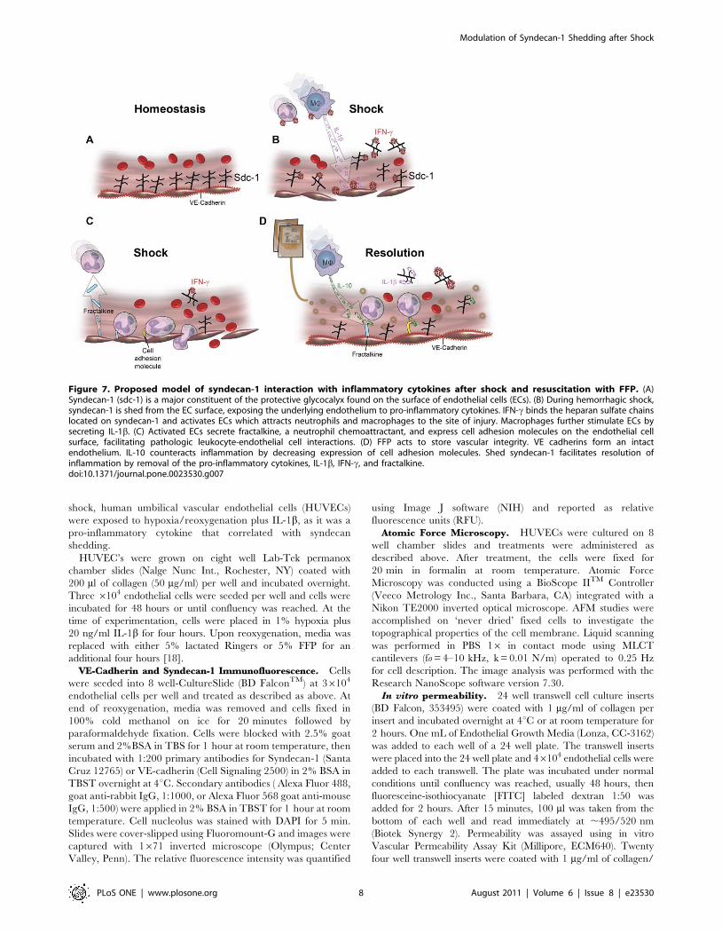
Figure 7. Proposed model of syndecan-1 interaction with inflammatory cytokines after shock and resuscitation with FFP. (A)Syndecan-1 (sdc-1) is a major constituent of the protective glycocalyx found on the surface of endothelial cells (ECs). (B) During hemorrhagic shock,syndecan-1 is shed from the EC surface, exposing the underlying endothelium to pro-inflammatory cytokines. IFN-c binds the heparan sulfate chainslocated on syndecan-1 and activates ECs which attracts neutrophils and macrophages to the site of injury. Macrophages further stimulate ECs bysecreting IL-1b. (C) Activated ECs secrete fractalkine, a neutrophil chemoattractant, and express cell adhesion molecules on the endothelial cellsurface, facilitating pathologic leukocyte-endothelial cell interactions. (D) FFP acts to store vascular integrity. VE cadherins form an intactendothelium. IL-10 counteracts inflammation by decreasing expression of cell adhesion molecules. Shed syndecan-1 facilitates resolution ofinflammation by removal of the pro-inflammatory cytokines, IL-1b, IFN-c, and fractalkine.doi:10.1371/journal.pone.0023530.g007
Modulation of Syndecan-1 Shedding after Shock
PLoS ONE | www.plosone.org 8 August 2011 | Volume 6 | Issue 8 | e23530

insert and incubated at room temperature for 1 hour. 500 ul of
Endothelial Growth Media (Lonza, CC-3162) was added to each
plate then HUVEC’s seeded at 26105 cells per insert and cultured
until formation of integrated cell monolayer. Cells were treated as
described above. At end of reoxygenation FITC-Dextran (150 ml)
was added to the upper chamber and fluorescent measurements
determined using excitation/emission wavelengths of 485 nm/
530 nm.
Statistical analysis. Data is reported as mean 6 SEM.
Results were analyzed by one-way ANOVA with Tukey post hoc
tests. In figures, means notated with letters indicate statistical
differences between groups.
Supporting Information
Table S1 Median cytokine concentrations.
(DOC)
Acknowledgments
The authors wish to thank Scott Holmes for his graphical assistance.
Author Contributions
Conceived and designed the experiments: RH-W RAK EAG JBH PP.
Performed the experiments: RH-W ZP AMZ TM SP. Analyzed the data:
WW RAK RH-W. Wrote the paper: RAK RH-W SP JBH. Coordinated
patient sample collection: EAG.
References
1. Sauaia A, Moore FA, Moore EE, Moore EE, Moser KS, et al. (1995)
Epidemiology of trauma deaths: a reassessment. J Trauma 38: 185–193.
2. Hauser CJ, Boffard K, Dutton R, Bernard GR, Croce MA, Holcomb JB, et al.
(2010) Results of the CONTROL trial: efficacy and safety of recombinant
activated Factor VII in the management of refractory traumatic hemorrhage.J Trauma 69: 489–500.
3. Bulger EM, May S, Brasel KJ, Schreiber M, Kerby JD, et al. (2010) Out-of-hospital hypertonic resuscitation following severe traumatic brain injury: a
randomized controlled trial. JAMA 304: 1455–64.
4. Moore EE, Moore FA, Fabian TC, Bernard AC, Fulda GJ, et al. (2009) Human
polymerized hemoglobin for the treatment of hemorrhagic shock when blood isunavailable: the USA multicenter trial. J Am Coll Surg 208: 1–13.
5. Borgman M, Spinella PC, Perkins JG, Grathwohl KW, Repine T, et al. (2007)
Blood product replacement affects survival in patients receiving massivetransfusions at a combat support hospital. J Trauma 63: 805–813.
6. Spinella PC, Perkins JG, Grathwohl KW, Beekley AC, Niles SE, et al. (2008)Effect of plasma and red blood cell transfusions on survival in patients with
combat related traumatic injuries. J Trauma 64: S69–S78.
7. Holcomb JB, Wade CE, Michalek JE, Chisholm GB, Zarzabal LA, et al. (2008)
Increased plasma and platelet to red blood cell ratios improves outcome in 466massively transfused civilian trauma patients. Ann Surg 248: 447–458.
8. Cotton BA, Gunter OL, Isbell J, Robertson AM, Morris JA, Jr., et al. (2008)Damage control hematology: the impact of a trauma exsanguination protocol on
survival and blood product utilization. J Trauma 64: 1177–1182.
9. Holcomb JB, Jenkins D, Rhee P, Johannigman J, Mahoney P, et al. (2007)Damage control resuscitation: directly addressing the early coagulopathy of
trauama. J Trauma 62: 307–310.
10. Pati S, Matijevic N, Doursout MF, Ko T, Cao Y, et al. (2010) Protective effects
of fresh frozen plasma on vascular endothelial permeability, coagulation, andresuscitation after hemorrhagic shock are time dependent and diminish between
days 0 and 5 after thaw. J Trauma 69: S55–63.
11. Lee WL, Slutsky AS (2010) Sepsis and endothelial permeability. N Engl J Med
363: 689–691.
12. Tharakan B, Hunter FA, Smythe WR, Childs EW (2010) Curcumin inhibits
reactive oxygen species formation and vascular hyperpermeability following
haemorrhagic shock. Clinical and Experimental Pharmacology and Physiology37: 939–944.
13. Childs EW, Tharakan B, Hunter FA, Smythe WR (2010) 17b-estradiolmediatedprotection against vascular leak after hemorrhagic shock: role of estrogen
receptors and apoptotic signaling. Shock 34: 229–235.
14. Reitsma S, Slaaf DW, Vink H, van Zandvoort MA, oude Egbrink MG (2007)
The endothelial glycocalyx: composition, functions, and visualization. PflugersArch 454: 345–359.
15. Nieuwdorp M, van Haeften TW, Gouverneur M, Mooij HL, van Lieshout MHP
(2006) Loss of endothelial glycocalyx during acute hyperglycemia coincides withendothelial dysfunction and coagulation activation in vivo. Diabetes 55:
480–486.
16. Fux L, Ilan N, Sanderson RD, Vlodavsky I (2009) Heparanase: busy at the cell
surface. Trends Biochem Sci 34: 511–519.
17. Kozar RA, Zhang R, Holcomb JB, Sondeen J, Gonzalez E, et al. (2011) Plasma
restoration of endothelial glycocalyx after hemorrhagic shock. Anes Anal 112(6):1289–1295.
18. Gonzalez EA, Kozar R, Hartwell B, Conyers J, Holcomb J (2009) Effect ofactivated human plasma on syndecan-1 expression. Shock 31(1): 56.
19. Maier B, Lefering R, Lehnert M, Laurer HL, Steudel WI, et al. (2007) Early
versus late onset of multiple organ failure is associated with differing patterns ofplasma cytokine biomarker expression and outcome after severe trauma. Shock
28: 668–674.
20. Jastrow KM, 3rd, Gonzalez EA, McGuire MF, Suliburk JW, Kozar RA, et al.
(2009) Early cytokine production risk stratifies trauma patients for multiple organfailure. J Am Coll Surg 209: 320–331.
21. Kadl A, Leitinger N (2005) The role of endothelial cells in the resolution of acuteinflammation. Antioxid Redox Signal 7: 1744–1754.
22. London NR, Whitehead KJ, Li DY (2009) Endogenous endothelial cell signaling
systems maintain vascular stability. Angiogenesis 12: 149–58.
23. Peng Z, Haywood-Watson R, Pati S, Dong J, Vijayan V, et al. (2010)
Restitution of syndecan-1 is modulated by age of fresh frozen plasma (FFP).
Shock 33: 61.
24. Steppan J, Hofer S, Funke B, Brenner T, Henrich M, et al. (2011) Sepsis and
major abdominal surgery lead to flaking of the endothelial glycocalix. J Surg Res
165: 136–141.
25. Rehm M, Bruegger D, Christ F, Conzen P, Thiel M, Jacob M, et al. (2007)
Shedding of the endothelial glycocalyx in patients undergoing major vascular
surgery with global and regional ischemia. Circulation 116: 1896–1906.
26. Lambaerts K, Wilcox-Adelman SA, Zimmermann P (2009) The signaling
mechanisms of syndecan heparan sulfate proteoglycans. Curr Opin Cell Biol 21:
662–669.
27. Takahashi T, Hato F, Yamane T, Fukumasu H, Suzuki K, Ogita S, et al. (2001)
Activation of human neutrophil by cytokine-activated endothelial cells. Circ Res
88: 422–429.
28. Maroszynska I, Fiedor P (2000) Leukocytes and endothelium interaction as rate
limiting step in the inflammatory response and a key factor in the ischemia-
reperfusion injury. Ann Transplant 5: 5–11.
29. Kellum JA, Kong L, Fink MP, Weissfeld LA, Yealy DM, et al. (2009)
Understanding the inflammatory cytokine response in pneumonia and sepsis:
results of the Genetic and Inflammatory Markers of Sepsis (GenIMS) Study.
Arch Intern Med 167: 1655–1663.
30. Matheson PJ, Mays CJ, Hurt RT, Zakaria ER, Richardson JD, et al. (2009)
Modulation of mesenteric lymph flow and composition by direct peritoneal
resuscitation from hemorrhagic shock. Arch Surg 144: 625–634.
31. Lozano FS, Rodriguez JM, Garcia-Criado FJ, Barros MB, Conde PS, et al.
(2005) Postoperative evolution of inflammatory response in a model of
suprarenal aortic cross-clamping with and without hemorrhagic shock. Systemic
and local reactions. World J Surg 29: 1248–1258.
32. Mees ST, Dohm C, Broetzmann K, Schroeder J, Faendrich F, et al. (2007) Age-
and gender-related differences of the immune function in a murine model of
hemorrhagic shock: IL-10 restores immunodepression in aged females without
reduction of mortality. Langenbecks Arch Surg 392: 629–638.
33. Makley AT, Goodman MD, Friend LA, Deters JS, Johannigman JA, et al.
(2010) Resuscitation with fresh whole blood ameliorates the inflammatory
response after hemorrhagic shock. J Trauma 68: 305–311.
34. Sadir R, Forest E, Lortat-Jacob H (1998) The heparan sulfate binding sequence
of interferon-gamma increased the on rate of the interferon-gamma-interferon-
gamma receptor complex formation. J Biol Chem 273: 10919–10925.
35. Umehara H, Bloom ET, Okazaki T, Nagano Y, Yoshie O, et al. (2004)
Fractalkine in vascular biology: from basic research to clinical disease.
Arterioscler Thromb Vasc Biol 24: 34–40.
36. Imai T, Hieshima K, Haskell C, Baba M, Nagira M, et al. (1997) Identification
and molecular characterization of fractalkine receptor CX3CR1, which
mediates both leukocyte migration and adhesion. Cell 91: 521–530.
37. Hiscott J, Marois J, Garoufalis J, D’Addario M, Roulston A, et al. (1993)
Characterization of a functional NF-kappa B site in the human interleukin 1 beta
promoter: evidence for a positive autoregulatory loop. Mol Cell Biol 13:
6231–6240.
38. Tedgui A, Mallat Z (2001) Anti-inflammatory mechanisms in the vascular wall.
Circ Res 88: 5877–887.
39. Matsuo M (2008) Increased expression of heparan sulfate proteoglycan on the
cultured renal epithelial cells during oxalate exposure. Kurume Med J 55:
19–28.
40. Kliment CR, Englert JM, Gochuico BR, Yu G, Kaminski N, et al. (2009)
Oxidative stress alters syndecan-1 distribution in lungs with pulmonary fibrosis.
J Biol Chem 284: 3537–3545.
41. Hayashida K, Parks WC, Park PW (2009) Syndecan-1 shedding facilitates the
resolution of neutrophilic inflammation by removing sequestered CXC
chemokines. Blood 114: 3033–3043.
Modulation of Syndecan-1 Shedding after Shock
PLoS ONE | www.plosone.org 9 August 2011 | Volume 6 | Issue 8 | e23530
42. Robinson EK, Kelly DP, Mercer DW, Kozar RA (2008) Differential effects of
luminal arginine and glutamine on metalloproteinase production in thepostischemic gut. JPEN 32: 433–438.
43. London NR, Zhu W, Bozza FA, Smith MC, Greif DM, et al. (2010) Targeting
Robo4-dependent Slit signaling to survive the cytokine storm in sepsis andinfluenza. Sci Transl Med 17: 23ra19.
44. Hu H (2001) Cell-surface heparan sulfate is involved in the repulsive guidanceactivities of Slit2 protein. Nat Neurosci 4: 695–701.
45. Steigemann P, Molitor A, Fellert S, Jackle H, Vorbruggen G (2004) Heparan
sulfate proteoglycan syndecan promotes axonal and myotube guidance by slit/
robo signaling. Curr Biol 14: 225–30.
46. Gonzalez EA, Moore FA, Holcomb JB, Miller CC, Kozar RA, et al. (2007)
Fresh frozen plasma should be given earlier to patients requiring massive
transfusion. J Trauma 62: 112–119.
Modulation of Syndecan-1 Shedding after Shock
PLoS ONE | www.plosone.org 10 August 2011 | Volume 6 | Issue 8 | e23530
Related Documents