Introduction Microtubules perform a variety of essential functions within the cell. The mitotic spindle supports chromosome segregation and determines the position of cleavage in animals (Ahringer, 2003). This array is focused at centrosomes in animals although other non-centrosomal arrays exist that are mostly involved in polarised morphologies of specialised animal cells (Keating and Borisy, 1999). In animals, cortical microtubule (MT) arrays have been described only in very specific cases, such as starfish or Xenopus oocytes and zebrafish zygotes, where they are involved in the establishment of the dorsoventral axis (Jesuthasan and Stahle, 1997; Schroeder and Gard, 1992; Schroeder and Otto, 1984). By contrast, cortical MTs are a common feature of vegetative plant cells, in which they play a key role in shaping the cell by directing deposition of cellulose microfibrils (Mfs) in the cell wall and thereby cell growth (Lloyd and Chan, 2004). In plants the interphase MT array in G1 consists of cortical MTs linked to the plasmalemma and is replaced in G2 by the pre-prophase band cortical array that marks the position of the division plane. This array disappears in late prophase and is followed in succession by the mitotic apparatus and the phragmoplast. The perinuclear MT array is in continuity with the cortical array although it is still not clear whether cortical MTs arise from the translocation of perinuclear MTs or are nucleated de novo at cortical sites or both since there is evidence for both mechanisms (Burk and Ye, 2002; Cyr and Palevitz, 1995; Shaw et al., 2003). Rather less is known about MT distributions in zygotes and embryos of higher plants, owing to their general inaccessibility for direct study. Zygotes of fucoid algae (Fucus, Silvetia) have long served as a cellular model to study fertilisation, polarisation and cell division. Oospheres are initially spherical with no detectable polarity (Jaffe, 1958). After fertilisation (AF) Fucus zygotes deposit a cell wall within minutes and establish an axis of polarity in response to external cues (such as unidirectional light) that becomes fixed in space (14-18 hours AF) and leads to the germination of a rhizoid at the side facing away from the incident light (16-20 hours AF) (Henry et al., 1996; Love et al., 1997; Novotny and Forman, 1974). Concomitantly zygotes progress through the first cell cycle and divide perpendicularly to the growth axis (Corellou et al., 2001). The first asymmetrical division gives rise to the rhizoid and thallus cells with distinct developmental fates (Bouget et al., 1998). The rhizoid remains the only site of growth of the embryos during several rounds of division. The primary division planes in the rhizoid are oriented transverse to the polar axis whereas the first thallus cell 2723 Fucus zygotes polarise and germinate a rhizoid before their first asymmetrical division. The role of microtubules (MTs) in orienting the first division plane has been extensively studied by immunofluorescence approaches. In the present study, the re-organisation of MT arrays during the development of Fucus zygotes and embryos was followed in vivo after microinjection of fluorescent tubulin. A dynamic cortical MT array that shows dramatic re- organization during zygote polarization was detected for the first time. Randomly distributed cortical MTs were redistributed to the presumptive rhizoid site by the time of polarisation and well before rhizoid germination. The cortical MT re-organisation occurs independently of centrosome separation and nucleation. By the time of mitosis the cortical array depolymerised to cortical foci in regions from which it also reformed following mitosis, suggesting that it is nucleated from cortical sites. We confirm previous indications from immunodetection studies that centrosomal alignment and nuclear rotation occur via MT connexions to stabilised cortical sites and that definitive alignment is post-metaphasic. Finally, we show that cortical MTs align parallel to the growth axis during rhizoid tip growth and our results suggest that they may be involved in regulating rhizoid growth by shaping the rhizoid and containing turgor pressure Supplementary material available online at http://jcs.biologists.org/cgi/content/full/118/12/2723/DC1 Key words: Microtubules, Polarity asymmetric division, Fucus Summary Spatial re-organisation of cortical microtubules in vivo during polarisation and asymmetric division of Fucus zygotes Florence Corellou 2 , Susana M. B. Coelho 2, *, François-Yves Bouget 2 and Colin Brownlee 1,‡ 1 Marine Biological Association, The Laboratory, Citadel Hill, Plymouth, PL1 2PB, UK 2 Station Biologique, UMR 1931 CNRS and Laboratoires Goëmar, 29680 Roscoff, France *Present adress: Laboratoire Arago, UMR 7628 CNRS and Université Pierre et Marie-Curie, 66651 Banyuls sur mer, France ‡ Author for correspondence (e-mail: [email protected]) Accepted 28 February 2005 Journal of Cell Science 118, 2723-2734 Published by The Company of Biologists 2005 doi:10.1242/jcs.02353 Research Article Journal of Cell Science

Welcome message from author
This document is posted to help you gain knowledge. Please leave a comment to let me know what you think about it! Share it to your friends and learn new things together.
Transcript
IntroductionMicrotubules perform a variety of essential functions withinthe cell. The mitotic spindle supports chromosome segregationand determines the position of cleavage in animals (Ahringer,2003). This array is focused at centrosomes in animalsalthough other non-centrosomal arrays exist that are mostlyinvolved in polarised morphologies of specialised animal cells(Keating and Borisy, 1999). In animals, cortical microtubule(MT) arrays have been described only in very specific cases,such as starfish or Xenopus oocytes and zebrafish zygotes,where they are involved in the establishment of thedorsoventral axis (Jesuthasan and Stahle, 1997; Schroeder andGard, 1992; Schroeder and Otto, 1984).
By contrast, cortical MTs are a common feature ofvegetative plant cells, in which they play a key role in shapingthe cell by directing deposition of cellulose microfibrils (Mfs)in the cell wall and thereby cell growth (Lloyd and Chan,2004). In plants the interphase MT array in G1 consists ofcortical MTs linked to the plasmalemma and is replaced in G2by the pre-prophase band cortical array that marks the positionof the division plane. This array disappears in late prophaseand is followed in succession by the mitotic apparatus and thephragmoplast. The perinuclear MT array is in continuity withthe cortical array although it is still not clear whether cortical
MTs arise from the translocation of perinuclear MTs or arenucleated de novo at cortical sites or both since there isevidence for both mechanisms (Burk and Ye, 2002; Cyr andPalevitz, 1995; Shaw et al., 2003).
Rather less is known about MT distributions in zygotes andembryos of higher plants, owing to their generalinaccessibility for direct study. Zygotes of fucoid algae(Fucus, Silvetia) have long served as a cellular model to studyfertilisation, polarisation and cell division. Oospheres areinitially spherical with no detectable polarity (Jaffe, 1958).After fertilisation (AF) Fucus zygotes deposit a cell wallwithin minutes and establish an axis of polarity in response toexternal cues (such as unidirectional light) that becomes fixedin space (14-18 hours AF) and leads to the germination of arhizoid at the side facing away from the incident light (16-20hours AF) (Henry et al., 1996; Love et al., 1997; Novotny andForman, 1974). Concomitantly zygotes progress through thefirst cell cycle and divide perpendicularly to the growth axis(Corellou et al., 2001). The first asymmetrical division givesrise to the rhizoid and thallus cells with distinct developmentalfates (Bouget et al., 1998). The rhizoid remains the only siteof growth of the embryos during several rounds of division.The primary division planes in the rhizoid are orientedtransverse to the polar axis whereas the first thallus cell
2723
Fucus zygotes polarise and germinate a rhizoid before theirfirst asymmetrical division. The role of microtubules (MTs)in orienting the first division plane has been extensivelystudied by immunofluorescence approaches. In the presentstudy, the re-organisation of MT arrays during thedevelopment of Fucus zygotes and embryos was followedin vivo after microinjection of fluorescent tubulin. Adynamic cortical MT array that shows dramatic re-organization during zygote polarization was detected forthe first time. Randomly distributed cortical MTs wereredistributed to the presumptive rhizoid site by the timeof polarisation and well before rhizoid germination. Thecortical MT re-organisation occurs independently ofcentrosome separation and nucleation. By the time ofmitosis the cortical array depolymerised to cortical foci in
regions from which it also reformed following mitosis,suggesting that it is nucleated from cortical sites. Weconfirm previous indications from immunodetectionstudies that centrosomal alignment and nuclear rotationoccur via MT connexions to stabilised cortical sites and thatdefinitive alignment is post-metaphasic. Finally, we showthat cortical MTs align parallel to the growth axis duringrhizoid tip growth and our results suggest that they may beinvolved in regulating rhizoid growth by shaping therhizoid and containing turgor pressure
Supplementary material available online athttp://jcs.biologists.org/cgi/content/full/118/12/2723/DC1
Key words: Microtubules, Polarity asymmetric division, Fucus
Summary
Spatial re-organisation of cortical microtubules in vivoduring polarisation and asymmetric division of FucuszygotesFlorence Corellou2, Susana M. B. Coelho2,*, François-Yves Bouget2 and Colin Brownlee1,‡
1Marine Biological Association, The Laboratory, Citadel Hill, Plymouth, PL1 2PB, UK2Station Biologique, UMR 1931 CNRS and Laboratoires Goëmar, 29680 Roscoff, France*Present adress: Laboratoire Arago, UMR 7628 CNRS and Université Pierre et Marie-Curie, 66651 Banyuls sur mer, France‡Author for correspondence (e-mail: [email protected])
Accepted 28 February 2005Journal of Cell Science 118, 2723-2734 Published by The Company of Biologists 2005doi:10.1242/jcs.02353
Research Article
Jour
nal o
f Cel
l Sci
ence
2724
division occurs parallel to the growth axis (Bouget et al.,1998).
The role of the cytoskeleton in the polarisation of fucoidzygotes and in nuclear rotation has been extensively studied(Allen and Kropf, 1992; Bisgrove and Kropf, 1998; Bisgroveand Kropf, 2001a; Hable and Kropf, 1998; Kropf et al., 1990;Quatrano, 1973). Whereas F-actin is thought to be essential forpolarisation and germination MTs were found to be importantfor centrosomal alignment with the growth axis but dispensablefor polarisation (Quatrano, 1973). Immunocytological studiesof MT arrays of Silvetia zygotes during polarisation revealeda two-step alignment of the centrosomal axis with the growthaxis (Bisgrove and Kropf, 2001a). Initially, prior to metaphasethe centrosomal axis is roughly aligned with the growth axis.This alignment appears highly dependent on the connection ofcentrosomal MTs to the cortex and is consistent with a search-capture process in which actin plays a key role. Cortical MTswere faintly detected at the thallus in premetaphase. The exactalignment of the centrosomal axis is post-metaphasic and doesnot appear to require cortical MT connections. The study byBisgrove and Kropf (Bisgrove and Kropf, 2001a) is the onlyreport of cortical MTs in fucoid algae and showed short andrare MTs at the thallus cortex. However, such studies haveemployed fixation techniques that may interfere with thenormal MT distributions and moreover do not allow re-organization of MTs to be followed in the same cell. Thedevelopment of in vivo techniques such as microinjection offluorescent tubulin and more recently, expression of GFP-tubulin has considerably improved our understanding of MTbehaviour in other systems (Khodjakov et al., 1997; Yu et al.,2001).
In the present work we microinjected fluorescent rhodamin-labelled tubulin (RT) into Fucus zygotes to gain insights intothe reorganisation of MTs in vivo during early developmentand the first cell cycles, We were able to detect for the first timein polarizing zygotes a dense cortical MT network that lies incontinuity with the perinuclear array. Cortical MTs appear tooriginate from cortical nucleation sites. During polarisation ofthe zygote, they progressively re-organise from random to apolarised array localised at the rhizoid pole. MTs parallelto the growth axis appear to support germination. MTconnections between the nucleus and the cortex that facilitatenuclear rotation (Bisgrove and Kropf, 2001a) were alsoconfirmed in vivo. The re-organisation of the cortical arrayappears to occur before or at the same time as the perinuclearMTs re-organize from the nuclear envelope to the centrosomes.Cortical MTs transiently disappear during mitosis but corticalpatches remain until early metaphase and may be involved inlate alignment of the spindle. Our data also suggest that thiscortical array is required for osmoregulation.
Materials and MethodsZygote cultureReceptacles from sexually mature Fucus serratus adults were cut anddry stored at 4°C until use. Gametes were released by placing maleor female receptacles in filtered seawater (FSW) in natural daylightafter a short rinse with tap water. Fertilisation was performed bymixing sperm with oospheres. After 1 hour, zygotes were washed withFSW and filtered through a 90 µm nylon mesh. Zygotes were gentlyheld in glass wedges (Speksnijder et al., 1989) and cultured in FSW
under unidirectional blue light at 14°C. Nocodazole (Sigma, Poole,UK) was dissolved in DMSO at 10 mg ml–1 and further diluted inseawater before use to give a final concentration of 1 µg ml–1.
Microinjection of fluorescent brain tubulinRhodamine conjugated tubulin (RT) was purchased from MolecularProbes (Eugene, OR). RT stock solution was centrifuged at 10,000 gat 4°C, aliquoted and frozen at –80°C. RT was diluted just before usein 50 mM KCl, 300 mM Mannitol, 5 mM Hepes pH 6.9 to achieve afinal concentration of 2 mg ml–1. Micropipettes fabricated from 1.2mm filamented borosilicate glass (Harvard Apparatus, Edenbridge,UK), and dry bevelled (Roberts et al., 1993), were back-filled withthe RT solution, which was maintained on ice. Zygotes were pressuremicroinjected using a Medical Systems (Greenval, NY) injectionsystem. During microinjection, embryos were maintained at 10°C ona cooled microscope stage to minimize RT polymerisation duringinjection. Turgor pressure was reduced by adding 0.2-0.4 M sorbitol(depending on the age of the zygote) in seawater to allow injectionand to minimize damage following pipette withdrawal. Based on co-injection with Oregon Green dextran 10,000 kDa (Molecular Probes),we estimate that the injected volume was less than 3% of cell volumecorresponding to a maximum intracellular RT concentration of 0.06mg ml–1. After microinjection zygotes were returned to culture andallowed to recover and polarise under unidirectional blue light, for atleast 1 hour during which the turgor pressure was readjustedprogressively by regular media changes with a decreasing amount ofsorbitol. The direction of the light was applied so that the germinationsite was at 90° or 180° to the injection site.
Images were acquired using a Bio-Rad 1024 confocal laserscanning microscope (CLSM) (Hemel Hempstead, UK) equippedwith an argon/krypton laser. Fluorescent tubulin was excited at 568nm and fluorescence was observed at 605 nm using a 10 nm band passfilter. Optical sections were acquired at 1.5 µm intervals unlessotherwise stated. Sections were projected using the maximumbrightness projection with LASERSHARP software (Bio-Rad).
ResultsFluorescent tubulin microinjection allows in vivoobservation of MT reorganisation during zygoticdevelopmentFucus zygotes were microinjected with rhodamine-labelledtubulin (RT) to examine microtubule distribution andmovement during the polarization process, germination andmitosis that leads to asymmetrical division. Of zygotes thatwere microinjected early in development (between 2-5 hoursAF, n=50) 74% polarized and germinated and of thoseobserved after germination, 66% (n=50) entered mitosisindicating that microinjection had little deleterious effect onzygote development. Germination never occurred at theinjection site but according to the light direction i.e.perpendicularly or opposite to the injection site showing thatthe injection was not a relevant stimulus for polarisationcompared with light. In a small proportion of injected zygotes(16%, n=81) the nucleus was seen to be slightly displaced fromthe centre to a more peripheral position and MT bundles werevery occasionally observed extending between the nucleus andthe injection site (see e.g. Fig. 5D) or transiently at the site ofinjection (not shown). However, zygotes with those featuresgrew as well as other injected zygotes with no apparentdisruption of the timing of developmental events. To check forartefacts (such as microtubule bundling or the appearancecortical MT foci) potentially arising from injection of excessive
Journal of Cell Science 118 (12)
Jour
nal o
f Cel
l Sci
ence
2725Cortical microtubules and polarization
RT levels, varying amounts of RT varying from very low (i.e.barely detectable above cellular autofluorescence) to high (i.e.2-fold or more than cell autofluorescence) (see Figs S1-7 insupplementary material) were injected. No differences in RTdistribution were observed between zygotes or embryosinjected with low or high levels of RT. These observationstogether with the observed MT re-organisation duringpolarisation and mitosis strongly indicate that the observed MTstructures are not artefacts arising from tubulin microinjection.
Repeated confocal scanning of the embryos was shown tobe deleterious since most embryos stopped their developmentafter five rounds of serial scanning (i.e. 100-150 scans). Toavoid this problem and to ensure a sufficient number of timepoints for each developmental sequence, zygotes were scannedfrom either the early stage of polarisation until first mitosisentry or from germination until the end of the first mitosis.Two cell embryos were also injected to observe furtherdevelopment.
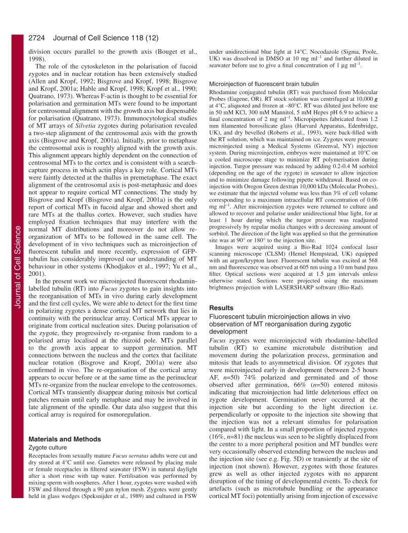
A homogenous and dense cortical MT network isdetected in unpolarised Fucus zygotes27 zygotes or oospheres (three different batches) were
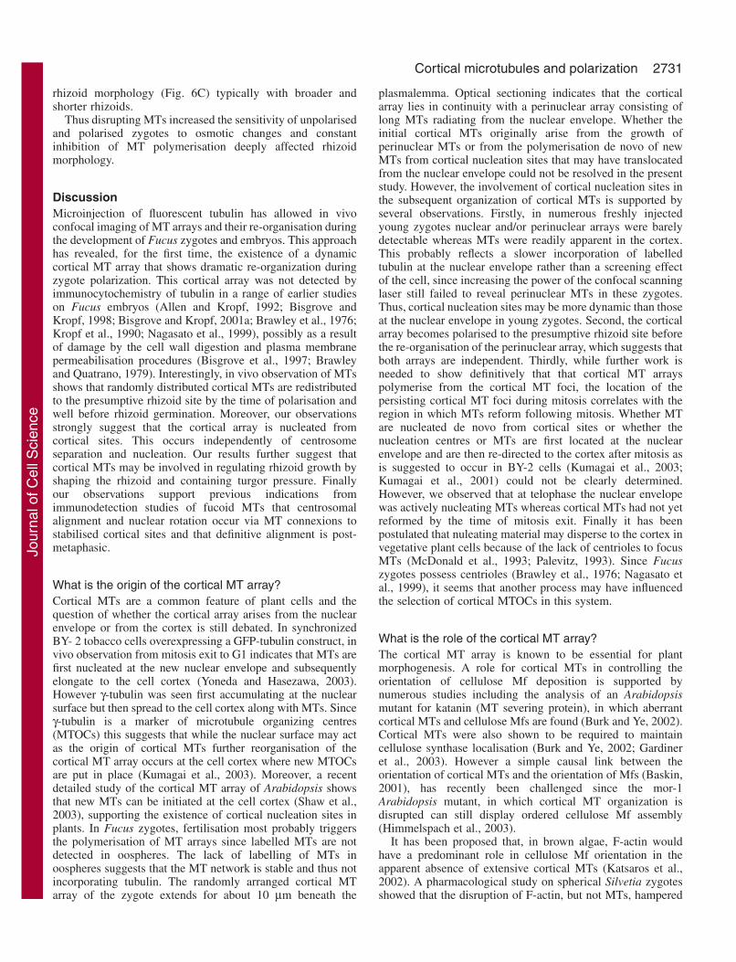
microinjected with RT between 0 and 4 hours (AF). MTdistribution was examined in zygotes using CLSM at 5-7 hoursAF (Fig. 1). Tubulin was not incorporated into MTs inoospheres and the diffusion of free fluorescent tubulin from theinjection site resulted in a diffuse fluorescence throughout thecytoplasmic network (Fig. 1A). By contrast, a cortical MTarray was detected within minutes after injection of zygotes.In unpolarised zygotes (5-7 hours AF) randomly arranged MTswere distributed homogeneously around the cell cortex (Fig.1B). MT arrays from the surface extended deeper into thecytoplasm. Cytoplasmic MTs (deeper than 10 µm) formed ameshwork around numerous organelles and vesicles thatappeared as dark ovoid areas (Fig. 1C). Perinuclear MTs wereuniformly nucleated from the nucleus through the cytoplasmtowards the cortex but appeared to incorporate tubulin moreslowly since some zygotes had a labelled cortex before anyperinuclear MTs were detected (data not shown). One or twobright spots of fluorescence were frequently (19% n=26)associated with the nuclear surface (Fig. 1D). These mostprobably correspond to the centrosomes (Bisgrove and Kropf,1998). MTs radiated from these bright spots as well as fromthe nuclear envelope suggesting that they may be nucleationcentres.
Fig. 1. Specificity of incorporation of fluorescent tubulin in different MT arrays of unpolarised zygotes. Zygotes are positioned with the lightcoming from the top of the page and were microinjected on the left side. (A) Oospheres 4 hours after injection (1 optical section). (B) Corticalarea of a 5 hours zygote (projection of the 2 upper optical sections): MTs are homogeneously distributed and randomly oriented. (C) One 15µm deep section shows the cytoplasmic MT array in a 6-hour-old zygote. (D) Projection of section from 30 to 38 µm deep nuclear MT array ina 6-hour-old zygote. (E-L). Effect of nocodazole on MT arrays. (E-H) 7-hour-old zygote before and (I-L) after 2 hours incubation withnocodazole. (E,I) Cortical MTs showing only a few MTs and fluorescent foci (free non incorporated tubulin) remaining after nocodazoletreatment. (F) Cytoplasmic region (projection from 7.5 to 27 µm deep inside the zygote) showing a dense cortical MT array at the periphery ofthe zygote. After incubation with nocodazole (J) very few and short MTs remain. (G,K) Nuclear region (projection from 28.5 to 33 µm deepinside the zygote). (G) The tubulin has not been well incorporated in the nuclear area, which is only faintly labelled. (H,L) Nuclear area fromanother zygote before (H) and after (L) nocodazole incubation. The bright patches on the left of zygotes in A, E, I, correspond tounincorporated tubulin on the zygote surface near the injection site. Bar, 15 µm.
Jour
nal o
f Cel
l Sci
ence
2726
We tested the sensitivity of different MT arrays in theunpolarized zygote to nocodazole which is known to beefficient in inhibiting MT polymerisation in Fucus zygotes(Brawley and Quatrano, 1979). All types of MT arrays ofunpolarised zygotes (6 hours AF) were shown to be sensitiveto this drug after 2 hours treatment and although a few MTswere barely detectable at the cortex of 2 zygotes, no MTs could
be detected in the majority of zygotes (60%, n=5) (Fig. 1E-L).MT depolymerisation was not due to a general toxic effect ofnocodazole since on return to seawater all zygotes germinatedand the majority underwent mitosis (60%, n=5). Together theseresults indicate that the microinjected fluorescent tubulin wasspecifically incorporated into the different MT arrays of Fucuszygotes.
Journal of Cell Science 118 (12)
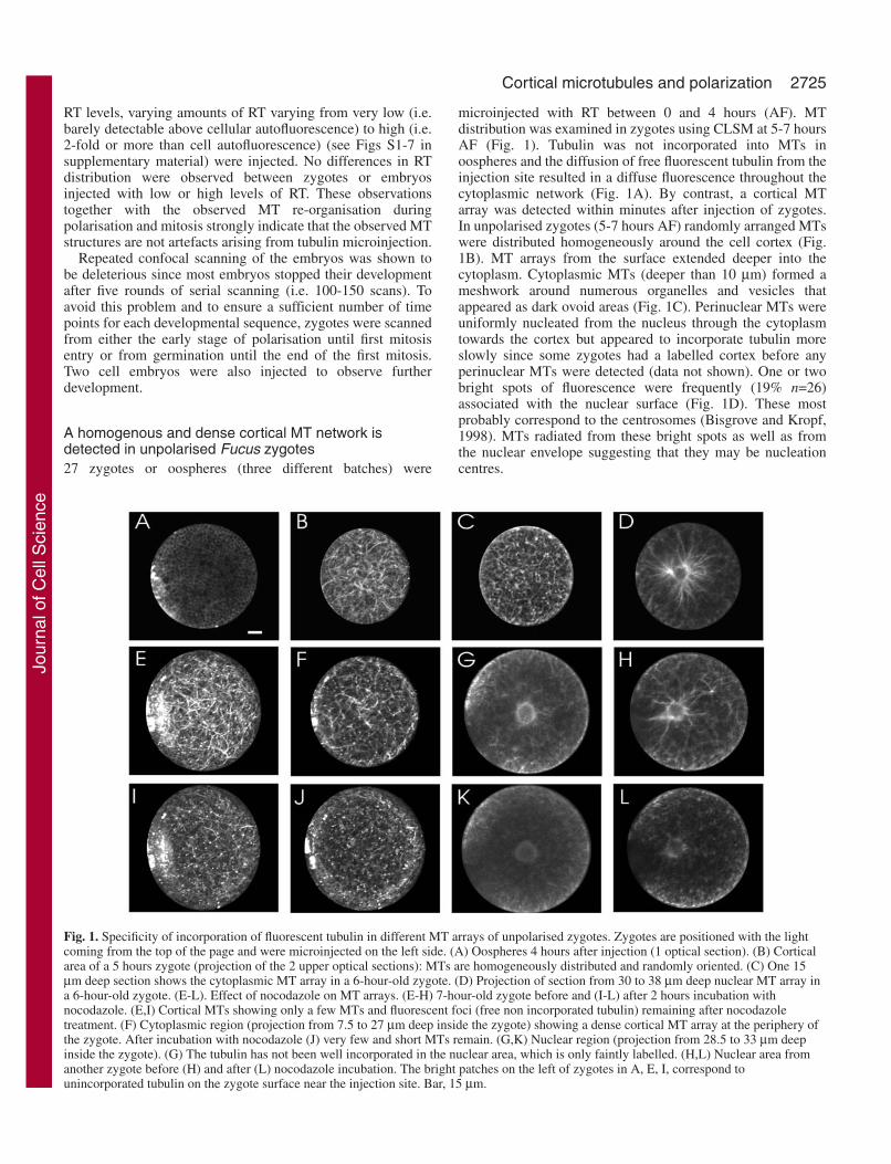
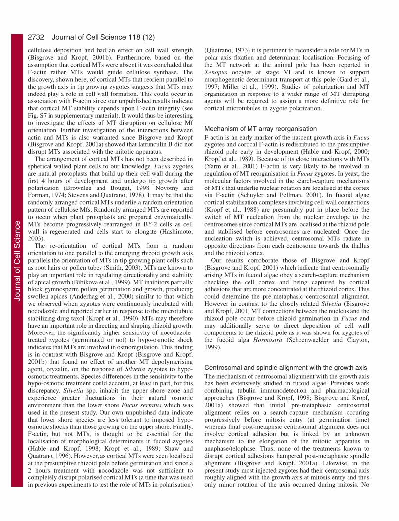
Fig. 2. Time course of corticalMT array reorganisation inpolarised zygotes.(A-H) Polarisation,germination and mitosis entryin the same zygote. Lightdirection is from the left.(A-E) All sections areprojected to reflect the shape ofthe zygote. (A) 17 hours (B) 19hours, (C) 21 hours and (D) 23hours AF showing progressivelocalisation of cortical MTstowards the rhizoid pole(projection of all sections).(E) 26 hours AF (projection ofall sections). The rhizoid hasgerminated and long corticalMTs are absent in the zygoteswith a prophase spindle butbright tubulin structures remainat the rhizoid collar.(F-H) Different areas of thesame 26-hour-old zygoteshowing that tubulin structuresat the collar start at the cortexand go deep inside thecytoplasm: (F) Cortical areathrough the upper 10 µm of thesame zygote. Bright tubulinstructures are localised as acollar around the rhizoid sub-apex. (G) Cytoplasmic area(11-22 µm) and (H) nucleararea (32-37 µm) with theprophase spindle. (I-N) MT re-organisation during rhizoidelongation (projection of allsections). (I) 17 hours AF withcortical MTs localised to therhizoid hemisphere. (J,K,L) 19,22 and 24 hours AF showingrhizoid emergence and theparallel arrangement of MTs tothe rhizoid axis. (M,N) 25 and27 hours AF: MTsdepolymerise from the rhizoidtip to bright cortical star-shapedstructures located at the collar.(O-Q) Projection of all sections during rhizoid germination and growth. (O) 16 hours AF showing convergence of MTs towards the site ofrhizoid emergence. (P) 20 hours AF: MT bundles form a crescent at the periphery of the growing rhizoid. (Q) 27 hours AF showingdisappearance of MTs from the rhizoid apex and MT bundles parallel to the growth axis extending from the rhizoid collar to a region below theapex. (R-U) Cortical network disappearance during mitosis and reformation after mitosis (projection of all sections). In this zygote rhizoidgermination occurred downwards out of the horizontal scanning plane but on the right side of the zygote. (R) 16-hour-old zygote. Cortical MTsare more localised to the rhizoid hemisphere and the nuclear envelope is labelled. (S) 20 hours AF. Cortical MTs are more focused to therhizoid and all cortical MTs are shortening. (T) Aster nucleation begins 26 hours AF showing mitosis entry. Both asters are detected in the cellcentre. Only cortical star-shaped structures remain at the cortex (arrows). (U) 38-hour-old zygote showing reformation of the cortical network,particularly at the rhizoid pole. Bar, 15 µm.
Jour
nal o
f Cel
l Sci
ence
2727Cortical microtubules and polarization
The cortical network is localised at thefuture germination site by the time ofpolarisationClear changes in the MT networks wereobserved as zygotes developed (Fig. 2).Interestingly the cortical array becamepreferentially localized at one hemispherein spherical zygotes as early as 10 hoursAF (80%, n=15) (see Figs 3 and 6). In the6 batches examined (n=42) cortical MTslocalised at the rhizoid pole inungerminated zygotes (Fig. 2A-D).Cortical MTs were first broadly localisedto one hemisphere of the cell and becamefocused at a more precise site (Fig. 2A-C).As germination occurred cortical MTsformed an even denser and concentratedarray (Fig. 2D), and eventually formed a crescent at the nascentrhizoid tip (see Fig. 2P). During rhizoid emergence the corticalMTs reoriented parallel to the growth axis and progressivelydisappeared from the thallus pole (Fig. 2I-L,O). As zygotesentered mitosis the cortical MT array was dramatically reducedto star-shaped patches from which the cortical MTs hadoriginally elongated. These remained brightly labelled at thecell cortex (Fig. 2E,N,S). These cortical structures were moreconcentrated at the sub-apical region of the rhizoid where theywere often aligned as a collar that extended over 10 µm intothe cytoplasm (Fig. 2E,N) and were also seen at the rhizoidapex. The time sequence acquisition shown in the lower rowof Fig. 2 shows the disappearance of the cortical network atmitosis entry and its subsequent reformation following mitosis(Fig. 2R-U). Although the reformation of MT was not followedfrom the beginning the re-formed MTs appeared to originate
from sites close to the star-shaped foci to which they retract atmitosis entry (arrows in T).
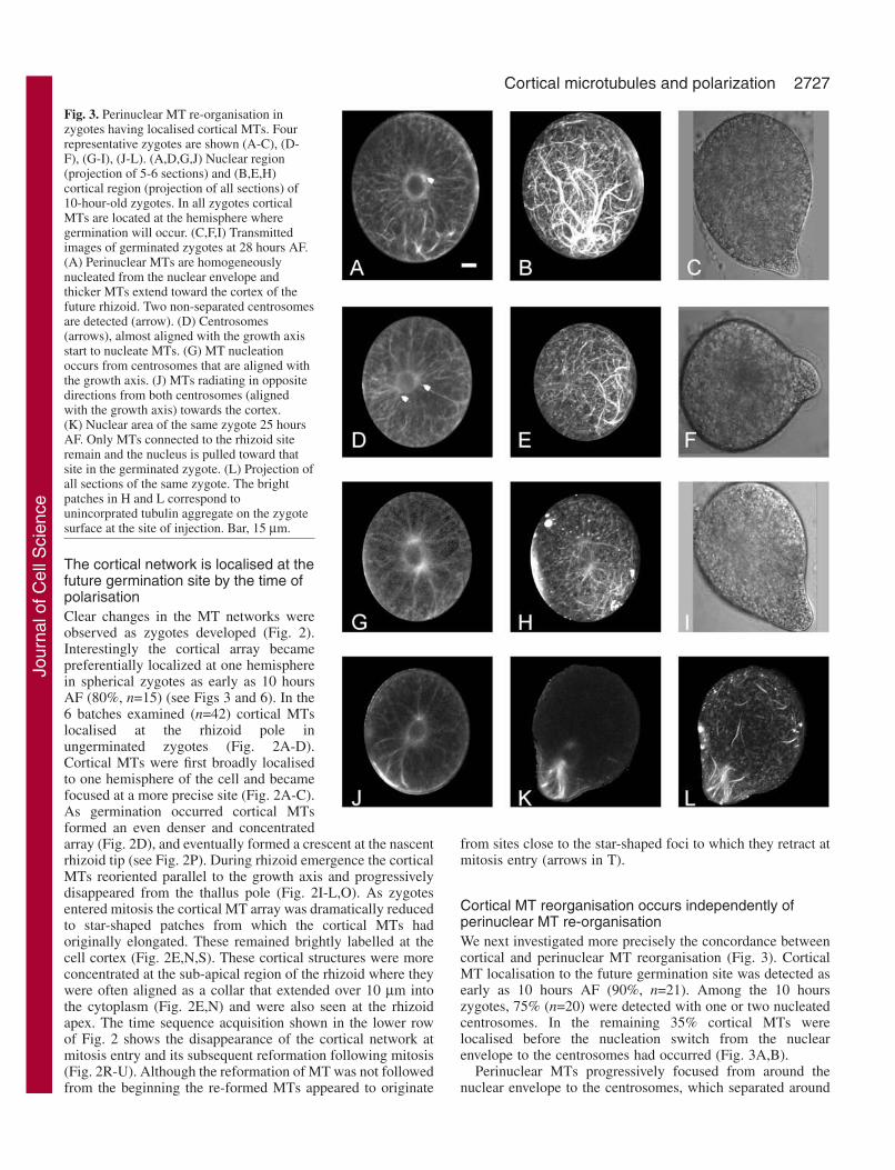
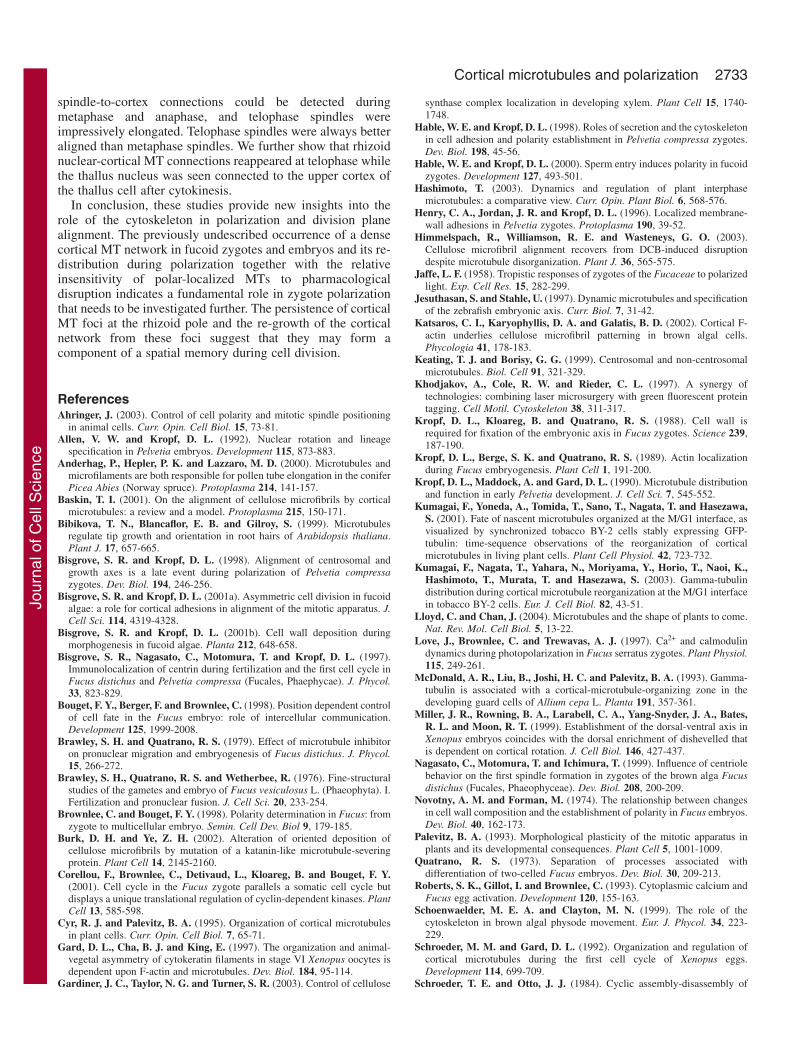
Cortical MT reorganisation occurs independently ofperinuclear MT re-organisationWe next investigated more precisely the concordance betweencortical and perinuclear MT reorganisation (Fig. 3). CorticalMT localisation to the future germination site was detected asearly as 10 hours AF (90%, n=21). Among the 10 hourszygotes, 75% (n=20) were detected with one or two nucleatedcentrosomes. In the remaining 35% cortical MTs werelocalised before the nucleation switch from the nuclearenvelope to the centrosomes had occurred (Fig. 3A,B).
Perinuclear MTs progressively focused from around thenuclear envelope to the centrosomes, which separated around
Fig. 3. Perinuclear MT re-organisation inzygotes having localised cortical MTs. Fourrepresentative zygotes are shown (A-C), (D-F), (G-I), (J-L). (A,D,G,J) Nuclear region(projection of 5-6 sections) and (B,E,H)cortical region (projection of all sections) of10-hour-old zygotes. In all zygotes corticalMTs are located at the hemisphere wheregermination will occur. (C,F,I) Transmittedimages of germinated zygotes at 28 hours AF.(A) Perinuclear MTs are homogeneouslynucleated from the nuclear envelope andthicker MTs extend toward the cortex of thefuture rhizoid. Two non-separated centrosomesare detected (arrow). (D) Centrosomes(arrows), almost aligned with the growth axisstart to nucleate MTs. (G) MT nucleationoccurs from centrosomes that are aligned withthe growth axis. (J) MTs radiating in oppositedirections from both centrosomes (alignedwith the growth axis) towards the cortex.(K) Nuclear area of the same zygote 25 hoursAF. Only MTs connected to the rhizoid siteremain and the nucleus is pulled toward thatsite in the germinated zygote. (L) Projection ofall sections of the same zygote. The brightpatches in H and L correspond tounincorprated tubulin aggregate on the zygotesurface at the site of injection. Bar, 15 µm.
Jour
nal o
f Cel
l Sci
ence
2728
the nucleus (68.6%, n=67). (Fig. 3A,D,G). Centrosomal MTsdiverged in opposite directions towards the cortex (Fig. 3G,J).Once the centrosomes were nucleated, one of them was alwaysconnected via MTs to the site where cortical MTs werelocalized even before the centrosomal alignment with the
growth axis was achieved (Fig. 3D). These observationssuggest that the re-organisation of cortical MTs takes placebefore or concomitant with perinuclear re-organisation but thatit does not depend upon the reorganisation of the perinucleararray.
Journal of Cell Science 118 (12)
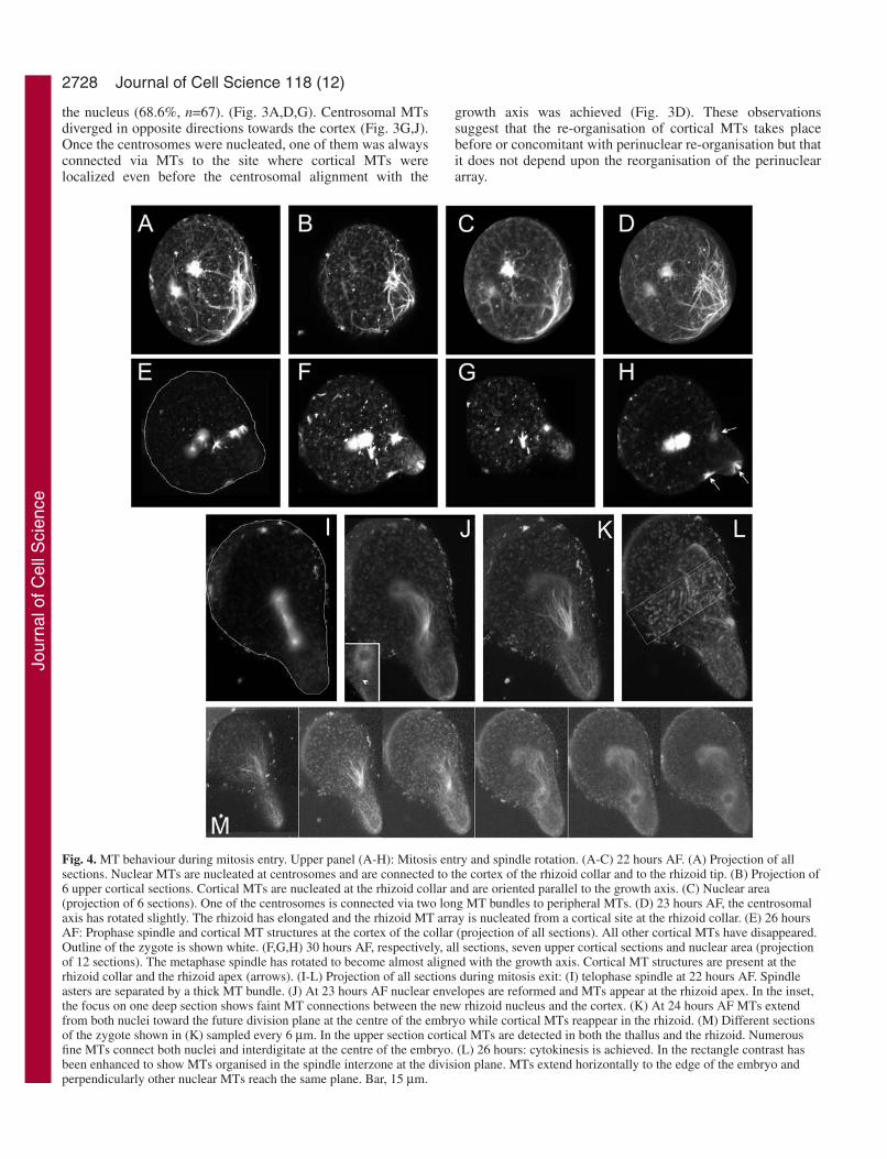
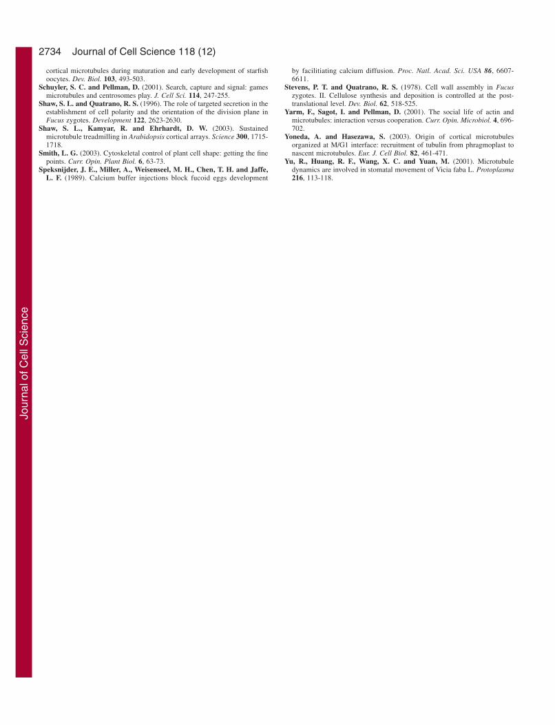
Fig. 4. MT behaviour during mitosis entry. Upper panel (A-H): Mitosis entry and spindle rotation. (A-C) 22 hours AF. (A) Projection of allsections. Nuclear MTs are nucleated at centrosomes and are connected to the cortex of the rhizoid collar and to the rhizoid tip. (B) Projection of6 upper cortical sections. Cortical MTs are nucleated at the rhizoid collar and are oriented parallel to the growth axis. (C) Nuclear area(projection of 6 sections). One of the centrosomes is connected via two long MT bundles to peripheral MTs. (D) 23 hours AF, the centrosomalaxis has rotated slightly. The rhizoid has elongated and the rhizoid MT array is nucleated from a cortical site at the rhizoid collar. (E) 26 hoursAF: Prophase spindle and cortical MT structures at the cortex of the collar (projection of all sections). All other cortical MTs have disappeared.Outline of the zygote is shown white. (F,G,H) 30 hours AF, respectively, all sections, seven upper cortical sections and nuclear area (projectionof 12 sections). The metaphase spindle has rotated to become almost aligned with the growth axis. Cortical MT structures are present at therhizoid collar and the rhizoid apex (arrows). (I-L) Projection of all sections during mitosis exit: (I) telophase spindle at 22 hours AF. Spindleasters are separated by a thick MT bundle. (J) At 23 hours AF nuclear envelopes are reformed and MTs appear at the rhizoid apex. In the inset,the focus on one deep section shows faint MT connections between the new rhizoid nucleus and the cortex. (K) At 24 hours AF MTs extendfrom both nuclei toward the future division plane at the centre of the embryo while cortical MTs reappear in the rhizoid. (M) Different sectionsof the zygote shown in (K) sampled every 6 µm. In the upper section cortical MTs are detected in both the thallus and the rhizoid. Numerousfine MTs connect both nuclei and interdigitate at the centre of the embryo. (L) 26 hours: cytokinesis is achieved. In the rectangle contrast hasbeen enhanced to show MTs organised in the spindle interzone at the division plane. MTs extend horizontally to the edge of the embryo andperpendicularly other nuclear MTs reach the same plane. Bar, 15 µm.
Jour
nal o
f Cel
l Sci
ence
2729Cortical microtubules and polarization
By the time of germination, centrosomes were seen to beseparated around the nucleus in most of the zygotes (74%,n=27). The nucleus rotated so that the centrosomal axis becameroughly aligned with the growth axis before mitosis. Howeverthis process failed in some of the zygotes (33% n=27) and thenucleus did not rotate so that some of the zygotes enteredmitosis with a spindle perpendicular to the growth axis andwere arrested at metaphase (see Fig. 2H).
MT arrays during mitosisZygotes entered mitosis between 20-25 hours AF (n=36). Tofollow mitosis in the same zygote we had to scan at close timeintervals. However, this often resulted in a metaphasic arrest ofnuclear division (61% n=22). Telophase figures were observedin neighbouring injected zygotes that were only scanned once(30% n=11). The time of mitosis progression (from prophaseto cytokinesis) was estimated to be 2-4 hours (n=3), consistentwith data reported for Fucus spiralis (Corellou et al., 2001).
At mitosis entry, the mitotic spindle formed and was roughlyaligned with the growth axis in most of the microinjectedzygotes (58%). However, in the remaining cases the spindlewas not aligned until just prior to mitosis (Fig. 4A-H). When
this late alignment was observed cortical MT foci remaineduntil the spindle was aligned (Fig. 4F-H). In about 30% of thezygotes astral MTs extending from the basal aster of the mitoticspindle towards the rhizoid region were detectable (not shown).The connection of the nucleus to the rhizoid region disappearedas zygotes progressed into metaphase (not shown). Anaphasespindles were rarely seen probably due to the rapidity of thisprocess. In contrast telophase figures were detected in 39% ofthe zygotes suggesting that this event is much slower (Fig.4I,L). Fig. 4 shows early telophase: the spindle asters wereseparated by a large bundle of MTs (Fig. 4K). Nuclearenvelope reformed and exhibited nucleating activity since itwas clearly labelled by the fluorescent tubulin (Fig. 4J,M). Attelophase a faint connection of the basal nuclei to the rhizoidtip was detected in 81% of the zygotes (Fig. 4J inset and M).Concomitantly cortical MTs reformed at the rhizoid (Fig.4K,M) and thallus pole. Newly formed nuclei remainedconnected via MT bundles emanating from the nuclearenvelope until the subsequent cytokinesis occurred (Fig. 4L).Multiple fine perinuclear MTs were still interdigitated whereasnew transverse MTs appeared at the centre of the embryos.Nuclear MTs remained connected to the cell plate that wasextending transversely towards the cell cortex.
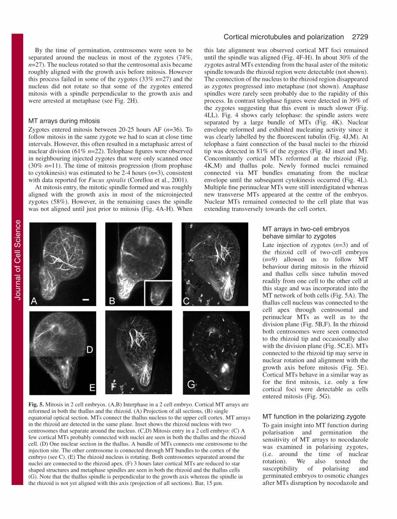
MT arrays in two-cell embryosbehave similar to zygotesLate injection of zygotes (n=3) and ofthe rhizoid cell of two-cell embryos(n=9) allowed us to follow MTbehaviour during mitosis in the rhizoidand thallus cells since tubulin movedreadily from one cell to the other cell atthis stage and was incorporated into theMT network of both cells (Fig. 5A). Thethallus cell nucleus was connected to thecell apex through centrosomal andperinuclear MTs as well as to thedivision plane (Fig. 5B,F). In the rhizoidboth centrosomes were seen connectedto the rhizoid tip and occasionally alsowith the division plane (Fig. 5C,E). MTsconnected to the rhizoid tip may serve innuclear rotation and alignment with thegrowth axis before mitosis (Fig. 5E).Cortical MTs behave in a similar way asfor the first mitosis, i.e. only a fewcortical foci were detectable as cellsentered mitosis (Fig. 5G).
MT function in the polarizing zygoteTo gain insight into MT function duringpolarisation and germination thesensitivity of MT arrays to nocodazolewas examined in polarising zygotes,(i.e. around the time of nuclearrotation). We also tested thesusceptibility of polarising andgerminated embryos to osmotic changesafter MTs disruption by nocodazole and
Fig. 5. Mitosis in 2 cell embryos. (A,B) Interphase in a 2 cell embryo. Cortical MT arrays arereformed in both the thallus and the rhizoid. (A) Projection of all sections, (B) singleequatorial optical section. MTs connect the thallus nucleus to the upper cell cortex. MT arraysin the rhizoid are detected in the same plane. Inset shows the rhizoid nucleus with twocentrosomes that separate around the nucleus. (C,D) Mitosis entry in a 2 cell embryo: (C) Afew cortical MTs probably connected with nuclei are seen in both the thallus and the rhizoidcell. (D) One nuclear section in the thallus. A bundle of MTs connects one centrosome to theinjection site. The other centrosome is connected through MT bundles to the cortex of theembryo (see C). (E) The rhizoid nucleus is rotating. Both centrosomes separated around thenuclei are connected to the rhizoid apex. (F) 3 hours later cortical MTs are reduced to starshaped structures and metaphase spindles are seen in both the rhizoid and the thallus cells(G). Note that the thallus spindle is perpendicular to the growth axis whereas the spindle inthe rhizoid is not yet aligned with this axis (projection of all sections). Bar, 15 µm.
Jour
nal o
f Cel
l Sci
ence
2730
observed the development of zygotes continuously incubatedin nocodazole.
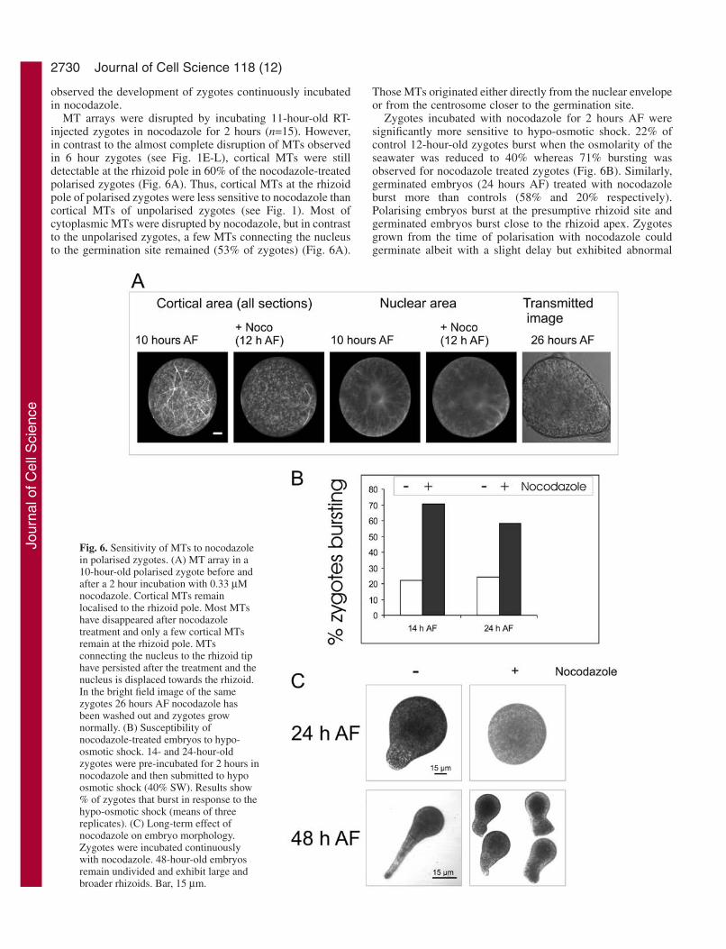
MT arrays were disrupted by incubating 11-hour-old RT-injected zygotes in nocodazole for 2 hours (n=15). However,in contrast to the almost complete disruption of MTs observedin 6 hour zygotes (see Fig. 1E-L), cortical MTs were stilldetectable at the rhizoid pole in 60% of the nocodazole-treatedpolarised zygotes (Fig. 6A). Thus, cortical MTs at the rhizoidpole of polarised zygotes were less sensitive to nocodazole thancortical MTs of unpolarised zygotes (see Fig. 1). Most ofcytoplasmic MTs were disrupted by nocodazole, but in contrastto the unpolarised zygotes, a few MTs connecting the nucleusto the germination site remained (53% of zygotes) (Fig. 6A).
Those MTs originated either directly from the nuclear envelopeor from the centrosome closer to the germination site.
Zygotes incubated with nocodazole for 2 hours AF weresignificantly more sensitive to hypo-osmotic shock. 22% ofcontrol 12-hour-old zygotes burst when the osmolarity of theseawater was reduced to 40% whereas 71% bursting wasobserved for nocodazole treated zygotes (Fig. 6B). Similarly,germinated embryos (24 hours AF) treated with nocodazoleburst more than controls (58% and 20% respectively).Polarising embryos burst at the presumptive rhizoid site andgerminated embryos burst close to the rhizoid apex. Zygotesgrown from the time of polarisation with nocodazole couldgerminate albeit with a slight delay but exhibited abnormal
Journal of Cell Science 118 (12)
Fig. 6. Sensitivity of MTs to nocodazolein polarised zygotes. (A) MT array in a10-hour-old polarised zygote before andafter a 2 hour incubation with 0.33 µMnocodazole. Cortical MTs remainlocalised to the rhizoid pole. Most MTshave disappeared after nocodazoletreatment and only a few cortical MTsremain at the rhizoid pole. MTsconnecting the nucleus to the rhizoid tiphave persisted after the treatment and thenucleus is displaced towards the rhizoid.In the bright field image of the samezygotes 26 hours AF nocodazole hasbeen washed out and zygotes grownormally. (B) Susceptibility ofnocodazole-treated embryos to hypo-osmotic shock. 14- and 24-hour-oldzygotes were pre-incubated for 2 hours innocodazole and then submitted to hypoosmotic shock (40% SW). Results show% of zygotes that burst in response to thehypo-osmotic shock (means of threereplicates). (C) Long-term effect ofnocodazole on embryo morphology.Zygotes were incubated continuouslywith nocodazole. 48-hour-old embryosremain undivided and exhibit large andbroader rhizoids. Bar, 15 µm.
Jour
nal o
f Cel
l Sci
ence
2731Cortical microtubules and polarization
rhizoid morphology (Fig. 6C) typically with broader andshorter rhizoids.
Thus disrupting MTs increased the sensitivity of unpolarisedand polarised zygotes to osmotic changes and constantinhibition of MT polymerisation deeply affected rhizoidmorphology.
DiscussionMicroinjection of fluorescent tubulin has allowed in vivoconfocal imaging of MT arrays and their re-organisation duringthe development of Fucus zygotes and embryos. This approachhas revealed, for the first time, the existence of a dynamiccortical MT array that shows dramatic re-organization duringzygote polarization. This cortical array was not detected byimmunocytochemistry of tubulin in a range of earlier studieson Fucus embryos (Allen and Kropf, 1992; Bisgrove andKropf, 1998; Bisgrove and Kropf, 2001a; Brawley et al., 1976;Kropf et al., 1990; Nagasato et al., 1999), possibly as a resultof damage by the cell wall digestion and plasma membranepermeabilisation procedures (Bisgrove et al., 1997; Brawleyand Quatrano, 1979). Interestingly, in vivo observation of MTsshows that randomly distributed cortical MTs are redistributedto the presumptive rhizoid site by the time of polarisation andwell before rhizoid germination. Moreover, our observationsstrongly suggest that the cortical array is nucleated fromcortical sites. This occurs independently of centrosomeseparation and nucleation. Our results further suggest thatcortical MTs may be involved in regulating rhizoid growth byshaping the rhizoid and containing turgor pressure. Finallyour observations support previous indications fromimmunodetection studies of fucoid MTs that centrosomalalignment and nuclear rotation occur via MT connexions tostabilised cortical sites and that definitive alignment is post-metaphasic.
What is the origin of the cortical MT array?Cortical MTs are a common feature of plant cells and thequestion of whether the cortical array arises from the nuclearenvelope or from the cortex is still debated. In synchronizedBY- 2 tobacco cells overexpressing a GFP-tubulin construct, invivo observation from mitosis exit to G1 indicates that MTs arefirst nucleated at the new nuclear envelope and subsequentlyelongate to the cell cortex (Yoneda and Hasezawa, 2003).However γ-tubulin was seen first accumulating at the nuclearsurface but then spread to the cell cortex along with MTs. Sinceγ-tubulin is a marker of microtubule organizing centres(MTOCs) this suggests that while the nuclear surface may actas the origin of cortical MTs further reorganisation of thecortical MT array occurs at the cell cortex where new MTOCsare put in place (Kumagai et al., 2003). Moreover, a recentdetailed study of the cortical MT array of Arabidopsis showsthat new MTs can be initiated at the cell cortex (Shaw et al.,2003), supporting the existence of cortical nucleation sites inplants. In Fucus zygotes, fertilisation most probably triggersthe polymerisation of MT arrays since labelled MTs are notdetected in oospheres. The lack of labelling of MTs inoospheres suggests that the MT network is stable and thus notincorporating tubulin. The randomly arranged cortical MTarray of the zygote extends for about 10 µm beneath the
plasmalemma. Optical sectioning indicates that the corticalarray lies in continuity with a perinuclear array consisting oflong MTs radiating from the nuclear envelope. Whether theinitial cortical MTs originally arise from the growth ofperinuclear MTs or from the polymerisation de novo of newMTs from cortical nucleation sites that may have translocatedfrom the nuclear envelope could not be resolved in the presentstudy. However, the involvement of cortical nucleation sites inthe subsequent organization of cortical MTs is supported byseveral observations. Firstly, in numerous freshly injectedyoung zygotes nuclear and/or perinuclear arrays were barelydetectable whereas MTs were readily apparent in the cortex.This probably reflects a slower incorporation of labelledtubulin at the nuclear envelope rather than a screening effectof the cell, since increasing the power of the confocal scanninglaser still failed to reveal perinuclear MTs in these zygotes.Thus, cortical nucleation sites may be more dynamic than thoseat the nuclear envelope in young zygotes. Second, the corticalarray becomes polarised to the presumptive rhizoid site beforethe re-organisation of the perinuclear array, which suggests thatboth arrays are independent. Thirdly, while further work isneeded to show definitively that that cortical MT arrayspolymerise from the cortical MT foci, the location of thepersisting cortical MT foci during mitosis correlates with theregion in which MTs reform following mitosis. Whether MTare nucleated de novo from cortical sites or whether thenucleation centres or MTs are first located at the nuclearenvelope and are then re-directed to the cortex after mitosis asis suggested to occur in BY-2 cells (Kumagai et al., 2003;Kumagai et al., 2001) could not be clearly determined.However, we observed that at telophase the nuclear envelopewas actively nucleating MTs whereas cortical MTs had not yetreformed by the time of mitosis exit. Finally it has beenpostulated that nuleating material may disperse to the cortex invegetative plant cells because of the lack of centrioles to focusMTs (McDonald et al., 1993; Palevitz, 1993). Since Fucuszygotes possess centrioles (Brawley et al., 1976; Nagasato etal., 1999), it seems that another process may have influencedthe selection of cortical MTOCs in this system.
What is the role of the cortical MT array?The cortical MT array is known to be essential for plantmorphogenesis. A role for cortical MTs in controlling theorientation of cellulose Mf deposition is supported bynumerous studies including the analysis of an Arabidopsismutant for katanin (MT severing protein), in which aberrantcortical MTs and cellulose Mfs are found (Burk and Ye, 2002).Cortical MTs were also shown to be required to maintaincellulose synthase localisation (Burk and Ye, 2002; Gardineret al., 2003). However a simple causal link between theorientation of cortical MTs and the orientation of Mfs (Baskin,2001), has recently been challenged since the mor-1Arabidopsis mutant, in which cortical MT organization isdisrupted can still display ordered cellulose Mf assembly(Himmelspach et al., 2003).
It has been proposed that, in brown algae, F-actin wouldhave a predominant role in cellulose Mf orientation in theapparent absence of extensive cortical MTs (Katsaros et al.,2002). A pharmacological study on spherical Silvetia zygotesshowed that the disruption of F-actin, but not MTs, hampered
Jour
nal o
f Cel
l Sci
ence
2732
cellulose deposition and had an effect on cell wall strength(Bisgrove and Kropf, 2001b). Furthermore, based on theassumption that cortical MTs were absent it was concluded thatF-actin rather MTs would guide cellulose synthase. Thediscovery, shown here, of cortical MTs that reorient parallel tothe growth axis in tip growing zygotes suggests that MTs mayindeed play a role in cell wall formation. This could occur inassociation with F-actin since our unpublished results indicatethat cortical MT stability depends upon F-actin integrity (seeFig. S7 in supplementary material). It would thus be interestingto investigate the effects of MT disruption on cellulose Mforientation. Further investigation of the interactions betweenactin and MTs is also warranted since Bisgrove and Kropf(Bisgrove and Kropf, 2001a) showed that latrunculin B did notdisrupt MTs associated with the mitotic apparatus.
The arrangement of cortical MTs has not been described inspherical walled plant cells to our knowledge. Fucus zygotesare natural protoplasts that build up their cell wall during thefirst 4 hours of development and undergo tip growth afterpolarisation (Brownlee and Bouget, 1998; Novotny andForman, 1974; Stevens and Quatrano, 1978). It may be that therandomly arranged cortical MTs underlie a random orientationpattern of cellulose Mfs. Randomly arranged MTs are reportedto occur when plant protoplasts are prepared enzymatically.MTs become progressively rearranged in BY-2 cells as cellwall is regenerated and cells start to elongate (Hashimoto,2003).
The re-orientation of cortical MTs from a randomorientation to one parallel to the emerging rhizoid growth axisparallels the orientation of MTs in tip growing plant cells suchas root hairs or pollen tubes (Smith, 2003). MTs are known toplay an important role in regulating directionality and stabilityof apical growth (Bibikova et al., 1999). MT inhibitors partiallyblock gymnosperm pollen germination and growth, producingswollen apices (Anderhag et al., 2000) similar to that whichwe observed when zygotes were continuously incubated withnocodazole and reported earlier in response to the microtubulestabilizing drug taxol (Kropf et al., 1990). MTs may thereforehave an important role in directing and shaping rhizoid growth.Moreover, the significantly higher sensitivity of nocodazole-treated zygotes (germinated or not) to hypo-osmotic shockindicates that MTs are involved in osmoregulation. This findingis in contrast with Bisgrove and Kropf (Bisgrove and Kropf,2001b) that found no effect of another MT depolymerisingagent, oryzalin, on the response of Silvetia zygotes to hypo-osmotic treatments. Species differences in the sensitivity to thehypo-osmotic treatment could account, at least in part, for thisdiscrepancy. Silvetia spp. inhabit the upper shore zone andexperience greater fluctuations in their natural osmoticenvironment than the lower shore Fucus serratus which wasused in the present study. Our own unpubished data indicatethat lower shore species are less tolerant to imposed hypo-osmotic shocks than those growing on the upper shore. Finally,F-actin, but not MTs, is thought to be essential for thelocalisation of morphological determinants in fucoid zygotes(Hable and Kropf, 1998; Kropf et al., 1989; Shaw andQuatrano, 1996). However, as cortical MTs were seen localisedat the presumptive rhizoid pole before germination and since a2 hours treatment with nocodazole was not sufficient tocompletely disrupt polarised cortical MTs (a time that was usedin previous experiments to test the role of MTs in polarisation)
(Quatrano, 1973) it is pertinent to reconsider a role for MTs inpolar axis fixation and determinant localisation. Focusing ofthe MT network at the animal pole has been reported inXenopus oocytes at stage VI and is known to supportmorphogenetic determinant transport at this pole (Gard et al.,1997; Miller et al., 1999). Studies of polarization and MTorganization in response to a wider range of MT disruptingagents will be required to assign a more definitive role forcortical microtubules in zygote polarization.
Mechanism of MT array reorganisationF-actin is an early marker of the nascent growth axis in Fucuszygotes and cortical F-actin is redistributed to the presumptiverhizoid pole early in development (Hable and Kropf, 2000;Kropf et al., 1989). Because of its close interactions with MTs(Yarm et al., 2001) F-actin is very likely to be involved inregulation of MT reorganisation in Fucus zygotes. In yeast, themolecular factors involved in the search-capture mechanismsof MTs that underlie nuclear rotation are localised at the cortexvia F-actin (Schuyler and Pellman, 2001). In fucoid algaecortical stabilisation complexes involving cell wall connections(Kropf et al., 1988) are presumably put in place before theswitch of MT nucleation from the nuclear envelope to thecentrosomes since cortical MTs are localised at the rhizoid poleand stabilised before centrosomes are nucleated. Once thenucleation switch is achieved, centrosomal MTs radiate inopposite directions from each centrosome towards the thallusand the rhizoid cortex.
Our results corroborate those of Bisgrove and Kropf(Bisgrove and Kropf, 2001) which indicate that centrosomallyarising MTs in fucoid algae obey a search-capture mechanismchecking the cell cortex and being captured by corticaladhesions that are more concentrated at the rhizoid cortex. Thiscould determine the pre-metaphasic centrosomal alignment.However in contrast to the closely related Silvetia (Bisgroveand Kropf, 2001) MT connections between the nucleus and therhizoid pole occur before rhizoid germination in Fucus andmay additionally serve to direct deposition of cell wallcomponents to the rhizoid pole as it was shown for zygotes ofthe fucoid alga Hormosira (Schoenwaelder and Clayton,1999).
Centrosomal and spindle alignment with the growth axisThe mechanism of centrosomal alignment with the growth axishas been extensively studied in fucoid algae. Previous workcombining tubulin immunodetection and pharmacologicalapproaches (Bisgrove and Kropf, 1998; Bisgrove and Kropf,2001a) showed that initial pre-metaphasic centrosomalalignment relies on a search-capture mechanism occuringprogressively before mitosis entry (at germination time)whereas final post-metaphsic centrosomal alignment does notinvolve cortical adhesion but is linked by an unknownmechanism to the elongation of the mitotic apparatus inanaphase/telophase. Thus, none of the treatments known todisrupt cortical adhesions hampered post-metaphasic spindlealignment (Bisgrove and Kropf, 2001a). Likewise, in thepresent study most injected zygotes had their centrosomal axisroughly aligned with the growth axis at mitosis entry and thusonly minor rotation of the axis occurred during mitosis. No
Journal of Cell Science 118 (12)
Jour
nal o
f Cel
l Sci
ence
2733Cortical microtubules and polarization
spindle-to-cortex connections could be detected duringmetaphase and anaphase, and telophase spindles wereimpressively elongated. Telophase spindles were always betteraligned than metaphase spindles. We further show that rhizoidnuclear-cortical MT connections reappeared at telophase whilethe thallus nucleus was seen connected to the upper cortex ofthe thallus cell after cytokinesis.
In conclusion, these studies provide new insights into therole of the cytoskeleton in polarization and division planealignment. The previously undescribed occurrence of a densecortical MT network in fucoid zygotes and embryos and its re-distribution during polarization together with the relativeinsensitivity of polar-localized MTs to pharmacologicaldisruption indicates a fundamental role in zygote polarizationthat needs to be investigated further. The persistence of corticalMT foci at the rhizoid pole and the re-growth of the corticalnetwork from these foci suggest that they may form acomponent of a spatial memory during cell division.
ReferencesAhringer, J. (2003). Control of cell polarity and mitotic spindle positioning
in animal cells. Curr. Opin. Cell Biol. 15, 73-81.Allen, V. W. and Kropf, D. L. (1992). Nuclear rotation and lineage
specification in Pelvetia embryos. Development 115, 873-883.Anderhag, P., Hepler, P. K. and Lazzaro, M. D. (2000). Microtubules and
microfilaments are both responsible for pollen tube elongation in the coniferPicea Abies (Norway spruce). Protoplasma 214, 141-157.
Baskin, T. I. (2001). On the alignment of cellulose microfibrils by corticalmicrotubules: a review and a model. Protoplasma 215, 150-171.
Bibikova, T. N., Blancaflor, E. B. and Gilroy, S. (1999). Microtubulesregulate tip growth and orientation in root hairs of Arabidopsis thaliana.Plant J. 17, 657-665.
Bisgrove, S. R. and Kropf, D. L. (1998). Alignment of centrosomal andgrowth axes is a late event during polarization of Pelvetia compressazygotes. Dev. Biol. 194, 246-256.
Bisgrove, S. R. and Kropf, D. L. (2001a). Asymmetric cell division in fucoidalgae: a role for cortical adhesions in alignment of the mitotic apparatus. J.Cell Sci. 114, 4319-4328.
Bisgrove, S. R. and Kropf, D. L. (2001b). Cell wall deposition duringmorphogenesis in fucoid algae. Planta 212, 648-658.
Bisgrove, S. R., Nagasato, C., Motomura, T. and Kropf, D. L. (1997).Immunolocalization of centrin during fertilization and the first cell cycle inFucus distichus and Pelvetia compressa (Fucales, Phaephycae). J. Phycol.33, 823-829.
Bouget, F. Y., Berger, F. and Brownlee, C. (1998). Position dependent controlof cell fate in the Fucus embryo: role of intercellular communication.Development 125, 1999-2008.
Brawley, S. H. and Quatrano, R. S. (1979). Effect of microtubule inhibitoron pronuclear migration and embryogenesis of Fucus distichus. J. Phycol.15, 266-272.
Brawley, S. H., Quatrano, R. S. and Wetherbee, R. (1976). Fine-structuralstudies of the gametes and embryo of Fucus vesiculosus L. (Phaeophyta). I.Fertilization and pronuclear fusion. J. Cell Sci. 20, 233-254.
Brownlee, C. and Bouget, F. Y. (1998). Polarity determination in Fucus: fromzygote to multicellular embryo. Semin. Cell Dev. Biol 9, 179-185.
Burk, D. H. and Ye, Z. H. (2002). Alteration of oriented deposition ofcellulose microfibrils by mutation of a katanin-like microtubule-severingprotein. Plant Cell 14, 2145-2160.
Corellou, F., Brownlee, C., Detivaud, L., Kloareg, B. and Bouget, F. Y.(2001). Cell cycle in the Fucus zygote parallels a somatic cell cycle butdisplays a unique translational regulation of cyclin-dependent kinases. PlantCell 13, 585-598.
Cyr, R. J. and Palevitz, B. A. (1995). Organization of cortical microtubulesin plant cells. Curr. Opin. Cell Biol. 7, 65-71.
Gard, D. L., Cha, B. J. and King, E. (1997). The organization and animal-vegetal asymmetry of cytokeratin filaments in stage VI Xenopus oocytes isdependent upon F-actin and microtubules. Dev. Biol. 184, 95-114.
Gardiner, J. C., Taylor, N. G. and Turner, S. R. (2003). Control of cellulose
synthase complex localization in developing xylem. Plant Cell 15, 1740-1748.
Hable, W. E. and Kropf, D. L. (1998). Roles of secretion and the cytoskeletonin cell adhesion and polarity establishment in Pelvetia compressa zygotes.Dev. Biol. 198, 45-56.
Hable, W. E. and Kropf, D. L. (2000). Sperm entry induces polarity in fucoidzygotes. Development 127, 493-501.
Hashimoto, T. (2003). Dynamics and regulation of plant interphasemicrotubules: a comparative view. Curr. Opin. Plant Biol. 6, 568-576.
Henry, C. A., Jordan, J. R. and Kropf, D. L. (1996). Localized membrane-wall adhesions in Pelvetia zygotes. Protoplasma 190, 39-52.
Himmelspach, R., Williamson, R. E. and Wasteneys, G. O. (2003).Cellulose microfibril alignment recovers from DCB-induced disruptiondespite microtubule disorganization. Plant J. 36, 565-575.
Jaffe, L. F. (1958). Tropistic responses of zygotes of the Fucaceae to polarizedlight. Exp. Cell Res. 15, 282-299.
Jesuthasan, S. and Stahle, U. (1997). Dynamic microtubules and specificationof the zebrafish embryonic axis. Curr. Biol. 7, 31-42.
Katsaros, C. I., Karyophyllis, D. A. and Galatis, B. D. (2002). Cortical F-actin underlies cellulose microfibril patterning in brown algal cells.Phycologia 41, 178-183.
Keating, T. J. and Borisy, G. G. (1999). Centrosomal and non-centrosomalmicrotubules. Biol. Cell 91, 321-329.
Khodjakov, A., Cole, R. W. and Rieder, C. L. (1997). A synergy oftechnologies: combining laser microsurgery with green fluorescent proteintagging. Cell Motil. Cytoskeleton 38, 311-317.
Kropf, D. L., Kloareg, B. and Quatrano, R. S. (1988). Cell wall isrequired for fixation of the embryonic axis in Fucus zygotes. Science 239,187-190.
Kropf, D. L., Berge, S. K. and Quatrano, R. S. (1989). Actin localizationduring Fucus embryogenesis. Plant Cell 1, 191-200.
Kropf, D. L., Maddock, A. and Gard, D. L. (1990). Microtubule distributionand function in early Pelvetia development. J. Cell Sci. 7, 545-552.
Kumagai, F., Yoneda, A., Tomida, T., Sano, T., Nagata, T. and Hasezawa,S. (2001). Fate of nascent microtubules organized at the M/G1 interface, asvisualized by synchronized tobacco BY-2 cells stably expressing GFP-tubulin: time-sequence observations of the reorganization of corticalmicrotubules in living plant cells. Plant Cell Physiol. 42, 723-732.
Kumagai, F., Nagata, T., Yahara, N., Moriyama, Y., Horio, T., Naoi, K.,Hashimoto, T., Murata, T. and Hasezawa, S. (2003). Gamma-tubulindistribution during cortical microtubule reorganization at the M/G1 interfacein tobacco BY-2 cells. Eur. J. Cell Biol. 82, 43-51.
Lloyd, C. and Chan, J. (2004). Microtubules and the shape of plants to come.Nat. Rev. Mol. Cell Biol. 5, 13-22.
Love, J., Brownlee, C. and Trewavas, A. J. (1997). Ca2+ and calmodulindynamics during photopolarization in Fucus serratus zygotes. Plant Physiol.115, 249-261.
McDonald, A. R., Liu, B., Joshi, H. C. and Palevitz, B. A. (1993). Gamma-tubulin is associated with a cortical-microtubule-organizing zone in thedeveloping guard cells of Allium cepa L. Planta 191, 357-361.
Miller, J. R., Rowning, B. A., Larabell, C. A., Yang-Snyder, J. A., Bates,R. L. and Moon, R. T. (1999). Establishment of the dorsal-ventral axis inXenopus embryos coincides with the dorsal enrichment of dishevelled thatis dependent on cortical rotation. J. Cell Biol. 146, 427-437.
Nagasato, C., Motomura, T. and Ichimura, T. (1999). Influence of centriolebehavior on the first spindle formation in zygotes of the brown alga Fucusdistichus (Fucales, Phaeophyceae). Dev. Biol. 208, 200-209.
Novotny, A. M. and Forman, M. (1974). The relationship between changesin cell wall composition and the establishment of polarity in Fucus embryos.Dev. Biol. 40, 162-173.
Palevitz, B. A. (1993). Morphological plasticity of the mitotic apparatus inplants and its developmental consequences. Plant Cell 5, 1001-1009.
Quatrano, R. S. (1973). Separation of processes associated withdifferentiation of two-celled Fucus embryos. Dev. Biol. 30, 209-213.
Roberts, S. K., Gillot, I. and Brownlee, C. (1993). Cytoplasmic calcium andFucus egg activation. Development 120, 155-163.
Schoenwaelder, M. E. A. and Clayton, M. N. (1999). The role of thecytoskeleton in brown algal physode movement. Eur. J. Phycol. 34, 223-229.
Schroeder, M. M. and Gard, D. L. (1992). Organization and regulation ofcortical microtubules during the first cell cycle of Xenopus eggs.Development 114, 699-709.
Schroeder, T. E. and Otto, J. J. (1984). Cyclic assembly-disassembly of
Jour
nal o
f Cel
l Sci
ence
2734
cortical microtubules during maturation and early development of starfishoocytes. Dev. Biol. 103, 493-503.
Schuyler, S. C. and Pellman, D. (2001). Search, capture and signal: gamesmicrotubules and centrosomes play. J. Cell Sci. 114, 247-255.
Shaw, S. L. and Quatrano, R. S. (1996). The role of targeted secretion in theestablishment of cell polarity and the orientation of the division plane inFucus zygotes. Development 122, 2623-2630.
Shaw, S. L., Kamyar, R. and Ehrhardt, D. W. (2003). Sustainedmicrotubule treadmilling in Arabidopsis cortical arrays. Science 300, 1715-1718.
Smith, L. G. (2003). Cytoskeletal control of plant cell shape: getting the finepoints. Curr. Opin. Plant Biol. 6, 63-73.
Speksnijder, J. E., Miller, A., Weisenseel, M. H., Chen, T. H. and Jaffe,L. F. (1989). Calcium buffer injections block fucoid eggs development
by facilitiating calcium diffusion. Proc. Natl. Acad. Sci. USA 86, 6607-6611.
Stevens, P. T. and Quatrano, R. S. (1978). Cell wall assembly in Fucuszygotes. II. Cellulose synthesis and deposition is controlled at the post-translational level. Dev. Biol. 62, 518-525.
Yarm, F., Sagot, I. and Pellman, D. (2001). The social life of actin andmicrotubules: interaction versus cooperation. Curr. Opin. Microbiol. 4, 696-702.
Yoneda, A. and Hasezawa, S. (2003). Origin of cortical microtubulesorganized at M/G1 interface: recruitment of tubulin from phragmoplast tonascent microtubules. Eur. J. Cell Biol. 82, 461-471.
Yu, R., Huang, R. F., Wang, X. C. and Yuan, M. (2001). Microtubuledynamics are involved in stomatal movement of Vicia faba L. Protoplasma216, 113-118.
Journal of Cell Science 118 (12)
Jour
nal o
f Cel
l Sci
ence
Related Documents


![Cell cycle-coupled [Ca oscillations in mouse zygotes and ... · Cell cycle-coupled [Ca2+] i oscillations in mouse zygotes and function of the inositol 1,4,5-trisphosphate receptor-1](https://static.cupdf.com/doc/110x72/5fb285c478c1117d6b731391/cell-cycle-coupled-ca-oscillations-in-mouse-zygotes-and-cell-cycle-coupled.jpg)