Molecular detection of ascomycetes associated with Fucus serratus Alga ZUCCARO 1 , Barbara SCHULZ 1 and Julian I. MITCHELL 2 * 1 Institut fu ¨r Mikrobiologie, Technische Universita ¨t Braunschweig, Spielmannstrasse 7, D-38106, Braunschweig, Germany. 2 School of Biological Sciences, University of Portsmouth, King Henry Building, King Henry I Street, Portsmouth PO1 2DY, UK. E-mail : [email protected] Received 1 July 2003; accepted 18 September 2003. The association of ascomycetes with Fucus serratus was investigated by comparing the broad-based molecular and cultural diversities of healthy and dead fronds. Four PCR primer pairs were used to amplify the 18S (primers NS1-FR1 ; NS1-EF3) or 28S rRNA (primers NL209-NL912 ; NL359-NL912) genes directly from the DNA of algal thalli. Two novel primer pairs, NL209-NL912 and NL359-NL912 giving product sizes of 700 and 559 bp respectively, were designed to amplify the 28S rDNA from ascomycetes specifically. All primer combinations amplified DNA from 33 reference taxa isolated from Fucus serratus, and the products generated by primers NS1-FR1GC and NL359-NL912GC were separated in 18–38 % and 38–60 % denaturant gradients respectively after DGGE. The 18S rDNA DGGE system resolved eight bands from algal DNA, but many of the sequences separated were not fungal, whereas the 28S rDNA system resolved seven bands that were all identified as ascomycetes. Phylogenetic analysis and BLAST search results of environmental sequences revealed the presence of four main ascomycete groups : (1) the Halosphaeriales, (2) the Hypocreales, (3) an unidentified Lulworthiales complex, and (4) the Pleosporales. Few fungal isolates were detected molecularly suggesting that fungal colonisation of fronds was limited, mainly to species in dead casts. INTRODUCTION The interactions between algae and microorganisms are important factors in the evolution, conservation and commercial exploitation of the marine environment (Hawksworth 1988, 1991, Mearns-Spragg et al. 1997, Jensen & Fenical 2002). Many microbial associations, such as epi- and endophytic relationships, generate chemical protection mechanisms that limit the colonis- ation of algae by biofouling organisms (Armstrong et al. 2001, Schultz et al. 2002). There is little infor- mation, however, on the role of fungi in any protection mechanisms or even, more generally, on fungi with keystone roles in marine systems (Hyde et al. 1998). Details on the distribution and occurrence of algicolous fungi are incomplete, primarily as a result of less inten- sive study compared to other fungal groups but also because of the inability to culture many algae in vitro (Kohlmeyer & Kohlmeyer 1979, Hyde, Sarma & Jones 2000, Kohlmeyer & Volkmann-Kohlmeyer 2003a). This paper describes the application of molecular biology techniques to assess fungal–algal interactions directly, with and without fungal cultivation, so that key, or novel, fungi might be identified. Reports describing the molecular diversity of fungi rely primarily on identifying small subunit rRNA (SSU), or ITS sequences, after DGGE/TGGE separ- ation and cloning (Smit et al. 1999, Vainio & Hantula 2000, van Elsas et al. 2000, Landeweert et al. 2003). The extent of viable but non-culturable fungi in the environment remains largely uncharted, but it is presumed to be extensive (Hawksworth 2001). Vandenkoornhuyse et al. (2002) estimated that five unknown groups of fungi existed associated with the rhizome of Arrhenatherum elatius, suggesting that fungal diversity might be far greater than expected with previously uncultured organisms dominating. In the marine environment, Buchan et al. (2002) reported a novel lineage associated with Spartina alterniflora based upon rRNA/ITS and laccase sequences as well as TFRLP patterns. The molecular identification of fungi, however, can be problematic using SSU sequences, where phylogenetic information is limiting (Kowalchuk 1998), or with ITS sequences for a broad-based identi- fication. Furthermore, the distribution of gene se- quences in databases is inconsistent making it essential * Corresponding author. Mycol. Res. 107 (12): 1451–1466 (December 2003). f The British Mycological Society 1451 DOI: 10.1017/S0953756203008657 Printed in the United Kingdom.

Welcome message from author
This document is posted to help you gain knowledge. Please leave a comment to let me know what you think about it! Share it to your friends and learn new things together.
Transcript

Molecular detection of ascomycetes associated with
Fucus serratus
Alga ZUCCARO1, Barbara SCHULZ1 and Julian I. MITCHELL2*
1 Institut fur Mikrobiologie, Technische Universitat Braunschweig, Spielmannstrasse 7, D-38106, Braunschweig, Germany.2School of Biological Sciences, University of Portsmouth, King Henry Building, King Henry I Street, PortsmouthPO1 2DY, UK.
E-mail : [email protected]
Received 1 July 2003; accepted 18 September 2003.
The association of ascomycetes with Fucus serratus was investigated by comparing the broad-based molecular andcultural diversities of healthy and dead fronds. Four PCR primer pairs were used to amplify the 18S (primers NS1-FR1;NS1-EF3) or 28S rRNA (primers NL209-NL912; NL359-NL912) genes directly from the DNA of algal thalli. Two novel
primer pairs, NL209-NL912 and NL359-NL912 giving product sizes of 700 and 559 bp respectively, were designed toamplify the 28S rDNA from ascomycetes specifically. All primer combinations amplified DNA from 33 reference taxaisolated from Fucus serratus, and the products generated by primers NS1-FR1GC and NL359-NL912GC were separated
in 18–38% and 38–60% denaturant gradients respectively after DGGE. The 18S rDNA DGGE system resolved eightbands from algal DNA, but many of the sequences separated were not fungal, whereas the 28S rDNA system resolvedseven bands that were all identified as ascomycetes. Phylogenetic analysis and BLAST search results of environmentalsequences revealed the presence of four main ascomycete groups: (1) the Halosphaeriales, (2) the Hypocreales, (3) an
unidentified Lulworthiales complex, and (4) the Pleosporales. Few fungal isolates were detected molecularly suggestingthat fungal colonisation of fronds was limited, mainly to species in dead casts.
INTRODUCTION
The interactions between algae and microorganisms areimportant factors in the evolution, conservation andcommercial exploitation of the marine environment(Hawksworth 1988, 1991, Mearns-Spragg et al. 1997,Jensen & Fenical 2002). Many microbial associations,such as epi- and endophytic relationships, generatechemical protection mechanisms that limit the colonis-ation of algae by biofouling organisms (Armstronget al. 2001, Schultz et al. 2002). There is little infor-mation, however, on the role of fungi in any protectionmechanisms or even, more generally, on fungi withkeystone roles in marine systems (Hyde et al. 1998).Details on the distribution and occurrence of algicolousfungi are incomplete, primarily as a result of less inten-sive study compared to other fungal groups but alsobecause of the inability to culture many algae in vitro(Kohlmeyer & Kohlmeyer 1979, Hyde, Sarma & Jones2000, Kohlmeyer & Volkmann-Kohlmeyer 2003a).This paper describes the application of molecularbiology techniques to assess fungal–algal interactions
directly, with and without fungal cultivation, so thatkey, or novel, fungi might be identified.
Reports describing the molecular diversity of fungirely primarily on identifying small subunit rRNA(SSU), or ITS sequences, after DGGE/TGGE separ-ation and cloning (Smit et al. 1999, Vainio & Hantula2000, van Elsas et al. 2000, Landeweert et al. 2003).The extent of viable but non-culturable fungi inthe environment remains largely uncharted, but itis presumed to be extensive (Hawksworth 2001).Vandenkoornhuyse et al. (2002) estimated that fiveunknown groups of fungi existed associated with therhizome of Arrhenatherum elatius, suggesting thatfungal diversity might be far greater than expected withpreviously uncultured organisms dominating. In themarine environment, Buchan et al. (2002) reported anovel lineage associated with Spartina alterniflorabased upon rRNA/ITS and laccase sequences as well asTFRLP patterns. The molecular identification of fungi,however, can be problematic using SSU sequences,where phylogenetic information is limiting (Kowalchuk1998), or with ITS sequences for a broad-based identi-fication. Furthermore, the distribution of gene se-quences in databases is inconsistent making it essential* Corresponding author.
Mycol. Res. 107 (12): 1451–1466 (December 2003). f The British Mycological Society 1451
DOI: 10.1017/S0953756203008657 Printed in the United Kingdom.

to generate a database of isolates from the environmentfor sequence comparison (Liesack et al. 1997, van Elsaset al. 2000).
The D2 region of the large ribosomal RNA subunit(LSU) offers an alternative to SSU sequences for abroad-based molecular identification (Fell 1993, Fellet al. 2000). It is the second most variable region of thegene and is encompassed by two variable regions(D1 and D3) allowing the design of fungal specific pri-mers (Hopple & Vilgalys 1999). The majority of fungiassociated with brown algae belong to the Ascomycota,including conidial fungi (Jones 1976, Kohlmeyer &Kohlmeyer 1979, 2003a, Stanley 1991). Phylogeneticanalyses of marine fungi have generated a diversity ofLSU sequences, particularly within theHalosphaeriales,Lulworthiales and some loculoascomycetes, providing abasis for molecular environmental studies (Kohlmeyer,Spatafora & Volkmann-Kohlmeyer 2000, Campbellet al. 2002).
The aim of this study was to use two molecularsystems, in conjunction with DGGE, to identify asco-mycetous fungi commonly found associated with livingand dead Fucus serratus. The first was based on pub-lished primers that amplify the 18S rRNA gene. Forthe second system, three novel PCR primers weredeveloped for the D2 region of the 28S rRNA gene.This communication describes these primers, detailsthe DGGE conditions for the separation of their PCRproducts and relates the ascomycete molecular diver-sity, and phylogenetic affiliations, detected by bothsystems compared with the cultured diversity.
MATERIALS AND METHODS
Sampling site and collection of algae
Specimens of attached Fucus serratus and casts were col-lected from the northeastern side of Heligoland Island,Germany, in April 2002. The material was transportedimmediately to the laboratory in cooled sterile con-tainers for fungal isolation and molecular analysis.
Isolation of fungi
Six healthy thalli, and one dead cast, approximately40 cm in length, bearing well-developed receptacleswere cleaned with sterile SDS (0.1% w/v) removingvisible debris and epiphytes except calcareous wormtubes. Half of the healthy thalli were surface-sterilisedby washing with sodium hypochloride (3% v/v) con-taining Tween 80 for 10 s. All specimens were rinsed insterile artificial seawater (ASW) three times. The successof the surface sterilisation treatment was assessed afterimprinting the tissue on agar media (Schultz et al.1998). Healthy and dead algae were cut aseptically into0.5 cm3 pieces, and randomly subdivided into two frac-tions (ca 250 fragments per fraction) for each individ-ual. The fractions were either plated for culturing orstored atx70 xC for DNA analysis. Seawater collected
from the sampling site was stored in sterile bottles at4 x, filtered and plated on media.
Algal tissue was plated and incubated at room tem-perature on the following media described by Holleret al. (2000) : 2% biomalt agar (20 g lx1 biomalt,15 g lx1 agar; pH 6.0) ; 1% biomalt agar (10 g lx1
biomalt, 15 g lx1 agar; pH 6.0) ; potato–carrot agar(20 g lx1 cooked and mashed potatoes, 20 g lx1 cookedand mashed carrots 15 g lx1 agar) ; yeast extractpeptone glucose agar (1 g lx1
D-glucose monohydrate,0.5 g lx1 peptone from soymeal, 0.1 g lx1 yeast extract,15 g lx1 agar; pH 8.0) ; yeast extract peptone glucosepoor media (0.1 g lx1
D-glucose monohydrate,0.05 g lx1 peptone from soymeal, 0.01 g lx1 yeast ex-tract, 15 g lx1 agar; pH 8.0). All media contained ASW(33 g lx1), benzopenicillin (60 mg lx1), streptomycinsulphate (80 mg lx1) and tetracyline-HCL (50 mg lx1).Cyclosporine A (0.5 mg lx1) was sometimes added tothe isolation media to slow the growth of fast-spreadingfungi (Dreyfuss 1986). Isolates were transferred to agarslants and Petri dishes, containing either 2% biomaltagar with ASW or potato–carrot agar media withASW, and stored at 4 x or 14 x respectively.
Identification of fungi
Fungal isolates were cultured at room temperatureuntil sporulation, examined microscopically andcharacterised according to morphology. Identificationwas made, in conformity to the appropriate authority(Stolk 1955, Davidson & Christensen 1971, Haythorn,Jones & Harrison 1980, Kohlmeyer & Volkmann-Kohlmeyer 1991, 2003a) to the genus level, except forthose isolates important in the molecular studies wherespecies identities were determined.
DNA extraction
Fungal DNA was extracted using modified methodsof Lee, Milgroom & Taylor (1988) and Lee & Taylor(1990). Freeze-dried mycelia were ground with a sterilemortar and pestle in liquid nitrogen, suspended in aSDS-lysis buffer (50 mM Tris-HCl pH 8, 50 mM EDTA,3% SDS and 1% b-mercaptoethanol), heated to 65 x
and extractedwith phenol:chloroform:iso-amylalcohol(25:24:1). The DNA was stored in TE buffer (10 mM
Tris-HCl, pH 8; 1 mM EDTA) at x20 x. Alternatively,DNA was extracted from fresh mycelia using the UltraClean Soil DNA isolation kit (MoBio Laboratories,CA).
DNA from frozen algal tissue (10 g) was extractedfollowing a CTAB extraction method (Jorn Petersen,pers. comm.) after grinding under liquid nitrogen in acold mortar and pestle. The frozen powder was sus-pended in 2% w/v CTAB in buffer (100 mM Tris-HClpH 7.5, 1.5 M NaCl, 50 mM EDTA, 25 mM b-mercap-toethanol), extracted with 0.6 v/v of chloroform-isoamyl alcohol (24:1) and centrifuged at 10 000 g for20 min at 20 x. Absolute ethanol (0.2 v/v) was added
PCR-DGGE systems for detecting fungi from algae 1452

to the aqueous layer followed by extraction with onevolume of chloroform-isoamyl alcohol (24:1) and cen-trifugation at 10 000 g for 20 min at 20 x. The aqueousphase was removed and iso-propanol (1 v) added. Afterincubation at room temperature for one hour, thesolution was centrifuged at 5000 g for 30 min and thesupernatant discarded. The pellet was washed withcold ethanol (70% v/v), dried and re-suspended in TE(pH 7.5). Further purification of the DNAwas achievedby ultracentrifugation in a CsCl gradient in the pres-ence of ethidium bromide using rotor type 70.I TI anda Beckman L7-65 ultracentrifuge.
Design of PCR primers to amplify the D2 region of the28S rRNA gene
The 28S rRNA gene primers NL209, NL359 andNL912 were designed from a multiple alignment of27 taxa including filamentous fungi and brown algae.The primers (Table 1) were selected from variable se-quences within the D1 and D3 regions (Fig. 1) wheredivergence between fungi and brown algae was thegreatest. The number assigned to each primer reflectedits position along the gene, based on an alignmentwith the sequence from Saccharomyces cerevisiae. Theprimer pairs NL209-NL912 and NL359-NL912 werechecked for Tm/Td differences and dimmer formationusing OLIGO v1.2 (Rychlik & Rhodes 1989). A thirty
base pair GC clamp (5k-CGCCCGCCGCGCGCGG-CGGGCGGGGCGGGG-3k) was added to primerNL912 for DGGE analysis (primer NL912GC). Theposition of the GC clamp was determined after analysisfor the lowest melting domain using MELT94 (Lerman& Beldjord 1998) and placed at the 5k end of the primer.
PCR amplification conditions
All PCR reactions were performed in a Biometrathermal cycler using TaqBeadTM Hot start DNApolymerase (Promega) following different protocols forthe amplification of (A) the SSU or (B) LSU rRNAsequences.
(A) PCR amplifications of SSU rDNA wereperformed in a reaction buffer (50 mM KCl, 10 mM
Tris-HCl pH 9.0, 0.1% Triton X-100) with 200 mMof dNTP mix, 1.25 u/bead of TaqBead Hot StartPolymerase, 2.5 mM MgCl2, 0.5 mM of each primer, and2% v/v DMSO. PCR conditions were optimised withDNA from various ascomycetes before environmentalanalysis. Nested PCR was used to amplify fungalsequences from algal DNA. Primers NS1-EF3(Miruna Oros-Sichler & Newton Gomes, pers. comm.,White et al. 1990, Smit et al. 1999) amplified fragmentsof approx. 1700 bp in the first reaction, which formedthe substrate for primers NS1-FR1GC (Vainio &Hantula 2000), producing fragments of 1650 bp with
Table 1. PCR primers and conditions used in this study.
Primer Sequence (5k to 3k) Tm/Td
Product
(bp) PCR amplification
NL912a TCAAATCCATCCGAGAACATCAG 70.0/68.1 5 min at 95 xC followed by 35 cycles of 30 s at 95 x, 45 s at
NL209 AAGCGCAGGAAAAGAAACCAACAG 72.2/71.6 700 52 x, 1 min at 72 x with a final extension for 10 min at 72 x
NL912 5 min at 95 x followed by 25 cycles of 30 s at 95 x, 45 s at
NL359 GGACGCCATAGAGGGTGAGAGC 76.9/71.4 559 50 x, 1 min at 72 x with a final extension for 10 min at 72 x
NS1b GTAGTCATATGCTTGTCTC 8 min at 94 x followed by 25 cycles of 30 s at 94 x, 45 s at 47 x,
EF3 TCCTCTAAATGACCAAGTTTG 1700 3 min at 72 x with a final extension for 10 min at 72 x
NS1 8 min at 94 x followed by 20 cycles of 30 s at 94 x, 45 s at 47 x,
FR1 AICCATTCAATCGGTAIT 1600 2 min at 72 x with a final extension for 10 min at 72 x
a GC clamp (5k-CGCCCGCCGCGCGCGGCGGGCGGGGCGGGG-3k) was attached to the 5-prime end of the primer for DGGE studies.b GC clamp (5k-CCCCCGCCGCGCGCGGCGGGCGGGGCGGGGG-3’) was added to the 5-prime end of the primer for DGGE studies.
100 500 1000 Nucleotides
D1 D2 D3
NL209/232 NL359/378 NL912/937
559 bp
700 bp
NL359 – NL912 product
NL209 – NL912 product
Fig. 1. Map showing the positions of primers NL209, NL359 and NL912 in the 28S rRNA gene. The variable regions D1,
D2 and D3 are hatched and the conserved regions are shown in black.
A. Zuccaro, B. Schulz and J. I. Mitchell 1453
GC-clamps for DGGE separation, in the second re-action (Table 1). DNA was diluted 1:30 to 1:100between reactions.
(B) Amplifications of the LSU rDNA were made ina reaction buffer containing 200 mM of dNTP mix,1.25 u/bead of TaqBead Hot Start Polymerase, 1.5 mMMgCl2, and 1 mM of each primer. The PCR conditionsfor primers NL209-NL912 and NL359-NL912, includ-ing the magnesium concentration, were optimised withDNA from fungal strainsDendryphiella salinaTUB126,Sigmoidea marina TUB156, Phomopsis TUB229, andPhomopsis TUB191, and are given in Table 1. NestedPCR reactions amplified fungal products from algalDNA using primers NL209-NL912 in the first reactionfollowed by primers NL359-NL912GC. DNA wasdiluted 1:1 to 1:50 between reactions.
Amplification from mixed templates
The ability of primers NL209-NL912 and NL359-NL912 to amplify different templates was determinedusing mixed DNA solutions. The optimal annealingtemperature for these reactions was recorded usinga Biometra gradient PCR machine. Mixed templatesincluded those from the fungi Sigmoidea marina,Dendryphiella salina, Phomopsis TUB191 and Pho-mopsis TUB229 in equal proportions or in the ratio1:2.5:5:7 respectively, and fungal–algal mixtureswhere F. serratus DNA was in excess of DNA fromD. salina (ratio 1:300) and Phomopsis TUB229 (ratio1:150).
DGGE conditions
All gels were prepared using a Bio-Rad gradient-formermixing 7.5% (w/v) acrylamide/bisacrylamide (37.5:1)with appropriate amounts of urea and deionisedformamide in the presence of TEMED and APS toform different denaturing gradients. Electrophoresiswas performed using TAE buffer (40 mM Tris-HCl/acetate, pH 8.0; 1 mM EDTA) with the Bio-RadDGENE system. Gradients separating SSU rDNAwere standardised at 18–45% denaturant with electro-phoresis for 16 h at 180 V and 58 x (Vainio & Hantula2000). A 38–60% denaturing gradient was used toseparate LSU rDNA with electrophoresis conditions of60 V at 58 x for 18 h. The DGGE gels were stained withethidium bromide, or occasionally with silver (Muyzer1999), and analysed using the Bio-Rad imaging system.DNA markers were chosen from PCR products thatwere well separated under the designated conditions.
Cloning of NS1-EF3 and NL209-NL912PCR products
PCR products from the non-surface sterilised livingand dead Fucus samples were purified with GEAN-CLEAN III Kit (Q-BIO gene) and ligated into theVector pCR 2.1 (Invitrogen, TA Cloning Kit). Trans-formations were made using competent cells from
Escherichia coli TOP 10Fk (Invitrogen) or XL1-blue(Stratagene, Cambridge, UK) according to the manu-facturer’s instructions. White colonies were selected forplasmid isolation and restriction analysis with EcoRI(Invitrogen). Subsequently, plasmid-screening proce-dures based on PCR amplification with primersFR1GC-NS1 and NL359-NL912GC were used. There-amplified inserts were subjected to DGGE togetherwith the original sample to identify the correspondingenvironmental bands. Clones containing inserts of thecorrect size were sequenced using the fluorescentmethod in a Li-COR 4200 DNA sequencer (AmodiaBioservice GmbH, Braunschweig).
Sequencing of the NL359-NL912GC PCR productsof excised bands
Prominent bands were excised from the denaturinggels and placed into sterile double-distilled water over-night. The DNA was re-amplified using primersNL359-NL912, purified using GENECLEAN III Kit(Q-BIO gene), checked with agarose electrophoresisand sequenced from primer NL912 (Seq-Lab GmbH,Gottingen).
Sequencing of the NL209-NL912 PCR productsfrom isolates
Fungal isolates that gave matching NL359-NL912GCproducts under DGGE analysis with those from algalDNA were purified with GENECLEAN III (Q-BIOgene) and sequenced using primer NL912 (Seq-LabGmbH).
Sequence and phylogenetic analysis
All sequences recovered after DGGE analyses andcloning or direct sequencing were identified broadlyusing BLAST searches (National Center for Bio-technology Information, NCBI). Exact matches be-tween sequences within different taxonomic groupswere calculated using GENEDOC (Nicholas, Nicholas& Deerfield 1997) from alignments of relatedtaxa. Phylogenetic analyses were performed initiallyusing data matrix M156 (Kohlmeyer, Spatafora &Volkmann-Kohlmeyer 2000) from TREEBASE (http://treebase.bio.buffalo.edu/treebase/). All environmentalsequences were included in matrix M156 initially butidentical sequences were later removed. Two matriceswere constructed for final analysis. Matrix M1 con-tained sixty-five taxa consisting of thirty-five taxa frommatrix M156, eight environmental sequences andtwenty-two additional taxa representing the taxonomicgroups that were related to the environmental se-quences (Table 2). Peziza badia was used as the out-group taxon in an analysis to identify fungal groupsassociated with the environmental sequences. MatrixM2 contained thirty-five taxa comprising thirteentaxa from matrix M156, two environmental sequencesand twenty taxa representing the genus Corollospora
PCR-DGGE systems for detecting fungi from algae 1454
and affiliated Halosphaeriales (Table 2). Microascustrigonosporus was used as the outgroup taxon in ananalysis to determine the relationship between the en-vironmental sequences within the genus Corollospora.Maximum parsimony analysis was performed on thesematrices using PAUP v4.12 (Swofford 1999) withrandom sequence addition and 10 replicates in aheuristic search. Bootstrap analysis was performedusing 100 replicates.
RESULTS
Fungi isolated from Fucus serratus
116 isolates were obtained from six healthy thalli andone dead cast, representing 29 genera of the Asco-mycota, mitosporic fungi and Zygomycota (Table 3).24 strains had sterile mycelia and could not be furtheridentified. 85 strains had conidial states only, and wereidentified as representatives of the generaCladosporium,Fusarium, Alternaria, Penicillium, and Dendryphiella.Only one representative of the Zygomycota was re-covered, the remaining five isolates were ascomycetesand included Corollospora angusta and Emericellopisminima. Fewer isolates were obtained after surface-sterilisation, with a noticeable reduction in the numberidentified as representatives of the genera Aspergillus,Penicillium, Fusarium, Alternaria, Cladosporium, andPhoma. Isolates identified as Acremonium species,which are reported as endophytes in plants (Andrewset al. 1982, Petrini 1991, Shearer 2001), were recoveredfrom surface sterilised tissue. One of these strains,Acremonium K90, matched the DGGE profile for theEmericellopsis minima CBS 111361 isolate (Fig. 2b) andwas identified as Acremonium tubakii CBS 111360 butwith larger conidia. Fungi isolated from the dead castincluded Corollospora angusta, Dendryphiella salina, aswell as representatives of Alternaria and Cladosporium.Yeast colonies were routinely recovered during iso-lation, but the nature of these was not investigatedfurther.
Extraction of algal DNA and PCR systems
The amount of fungal DNA extracted from Fucusserratus was expected to be small. The detection of rarefungal sequences under these circumstances requiredan efficient DNA extraction procedure and the devel-opment of ascomycete-specific primers amplifying vari-able regions suitable for separation and identification.The CTAB extraction method used in conjunctionwith density centrifugation purification provided pureDNA, at concentrations of above 500 ng mlx1, thatroutinely amplified after PCR. Omission of the caesiumchloride gradient step and the use of commercial kits,such as the Ultra Clean Soil and Ultra Clean plantDNA kits, generated DNA incapable of amplification.
A nested PCR approach was adopted to increasefungal templates without extending the number of
Table 2. The NCBI accession numbers of sequences used in this
study. CBS, Centraalbureau voor Schimmelcultures, Utrecht; TUB,
Technische Universat Braunschweig; PP, University of Portsmouth.
FungusGenBankaccession no.
Isolates sequencedAcremonium tubakii CBS 111360 AY305033Emericellopsis terricola CBS 229.59 AY305034Emericellopsis minima CBS 111361 AY305036Emericellopsis maritima CBS 491.71 AY305035Corollospora angusta TUB 1 AY305031Sigmoidea marina TUB 156 AY305032
Matrix M1=M156 plusArenariomyces trifurcatus PP2747 AF491277Corollospora angusta PP3903 AF491254Corollospora intermedia PP3910 AF491258Emericellopsis terricola U57082Engyodontium aranearum AF339525Gaeumannomyces graminis AF362537Hypocrea lutea AF543791Kohlmeyeriella tubulata AF491265Lulworthia furcicola AF491270Lulworthia ligniarenaria AF491272Lulworthia uniseptata AF491273Magnaporthe grisea AF362554Nereiospora cristata AF491268Nereiospora commata AF491267Stanjemonium grisellum AF049171Stanjemonium fuscescens AF049168
Matrix M2=M156 plusArenariomyces trifurcatus PP2747 AF491277Arenariomyces trifurcates JK 5409A U46883Corollospora colossa PP3904 AF491255Corollospora filiformis PP3905 AF491256Corollospora fusca PP3906 AF491257Corollospora lacera PP2509 AF491259Corollospora maritima PP1991 AF491260Corollospora maritima JK 4834 U46884Corollospora pulchella PP4206 AF491274Corollospora pulchella PP4294 AF491261Corollospora quinqueseptata AF491262Corollospora sp. AF491263Marinospora longissima AF491266Monodictys pelagica AF491275Nereiospora cristata AF491268Nereiospora commata AF491267
28S rDNA sequences from F. serratusTUBS9 – ‘Corollospora angusta ’ AY305037TUBS10 – ‘Corollospora angusta ’ AY305038TUBS13 – ‘Corollospora angusta ’ AY305039TUBS15 – ‘Corollospora angusta ’ AY305040TUBSF22 – ‘Sigmoidea marina ’ AY305041TUBSFt2 – ‘Sigmoidea marina ’ AY305042TUBSF3 – ‘Emericellopsis ’ AY305043TUBS11 – ‘Lulworthiaceae ’ AY305044TUBS14 – ‘Lulworthiaceae ’ AY305045TUBS5a – ‘Lulworthiaceae ’ AY305046TUBSF52 – ‘Lulworthiaceae ’ AY305047TUBSF3n – ‘Clavicipitaceae ’ AY305048TUBSFo – ‘Hypocrea lutea ’ AY305049
18S rDNA sequences from F. serratusTUBC16 – ‘Allonothrus russeolus ’ AY318946TUBC17 – ‘Allonothrus ’ AY318947TUBC18 – ‘Lulworthiales ’ AY318948TUBC9 – ‘Buchholzia fallax ’ AY318949TUBC3 – ‘Neophaeosphaeria ’ AY318950TUBC6 – ‘Corollospora angusta ’ AY318951TUBC19 – ‘Selaginopsis ’ AY318952TUBC21 – ‘Selaginopsis ’ AY318953
A. Zuccaro, B. Schulz and J. I. Mitchell 1455

reaction cycles. Two tripartite primer systems wereused to amplify the 18S rRNA or the 28S rRNA genes.The SSU primers (NS1-EF3 and NS1-FR1) were testedfor different annealing temperatures ranging from 47 x
to 54 x along with variations in salt concentration andthe inclusion of additives (DMSO; BSA; Q-solution;Qiagen). At temperatures higher than 47 x some fungalstrains were not amplified, limiting the choice of theannealing conditions. The primer pair NS1-EF3 ampli-fied a fragment of approx. 1700 bp whereas NS1-FR1amplified one of 1600 bp, however heterogeneity in sizefor both products was observed.
BLAST searches for each of the LSU primers re-trieved 1425 sequences with an exact sequence match,representing pre-dominantly ascomycete taxa includingthose from theEurotiales,Dothideomycetales,Leotiales,Lecanoromycetales, but mainly the Sordariomycetales.Only one non-fungal LSU sequence, for an arthropod(a species of Limulus), was recovered for primer
NL209. Both LSU primer pairs amplified DNA fromthirty-three test taxa (Table 4) at annealing tempera-tures ranging from 52–60 xwith no length heterogeneityexcept for D. salina (during amplification with primerpair NL359-NL912). Nested PCR using primersNL209-NL912 yielded a 700 bp product that acted asthe template for the second reaction with primersNL359-NL912GC, giving a 559 bp sequence thatwas suitable for separation by DGGE. The primercombinations amplified successfully from mixed tem-plates, even when the fungal template was diluted onein 300 with algal DNA.
DGGE separation of PCR products for SSUand LSU rRNA genes from pure cultures
PCR products amplified from strains representingtwenty-four genera (Table 4) were monitored for sep-aration using DGGE. The denaturant conditions used
Table 3. Fungal isolates recovered from healthy and dead thalli of Fucus serratus. The thalli were either surface sterilised or washed with
sterile water and cut into small disks for plating or DNA extraction. Seawater from the sampling area was collected, filtered and plated.
Thallus
Number of disks
Fungi isolated from Fucus serratus
Surface sterilised Water treated Dead
Seawater
1 2 3 4 5 6 7
232 240 254 198 174 182 100
Ascomycota
Chaetomium sp. 1
Corollospora angusta 1 1
Emericellopsis minima 1
Eurotium sp. 1
Mycosphaerella sp. 1
Zygomycota
Mucor sp. 2
Mitosporic fungi
Acremonium sp. 1 1
Acremonium tubakii 1
Acroconidiella sp. 1
Alternaria sp. 3 2 6
Arthrinium sp. 1
Aspergillus sp. 1
Beauveria bassiana 1
Botrytis cinerea 1
Cladosporium sp. 4 1 8
Coniothyrium sp. 1
Dendryphiella salina 3 6
Epicoccum sp. 2
Fusarium sp. 1 1 4 5 1
Humicola fuscoatra 1
Nodulisporium sp. 1
Oidiodendron sp. 1
Paecilomyces sp. 2 2
Penicillium sp. 1 1 5 2 3
Periconia sp. 1
Phialophora sp. 1 1
Phoma sp. 3 1 2
Scopulariopsis sp. 1 1
Trichoderma sp. 1 1
Verticillium cinnabarinum 1
Sterile mycelium 4 4 4 3 9
Total number of isolates 5 1 7 34 22 11 36 5
PCR-DGGE systems for detecting fungi from algae 1456
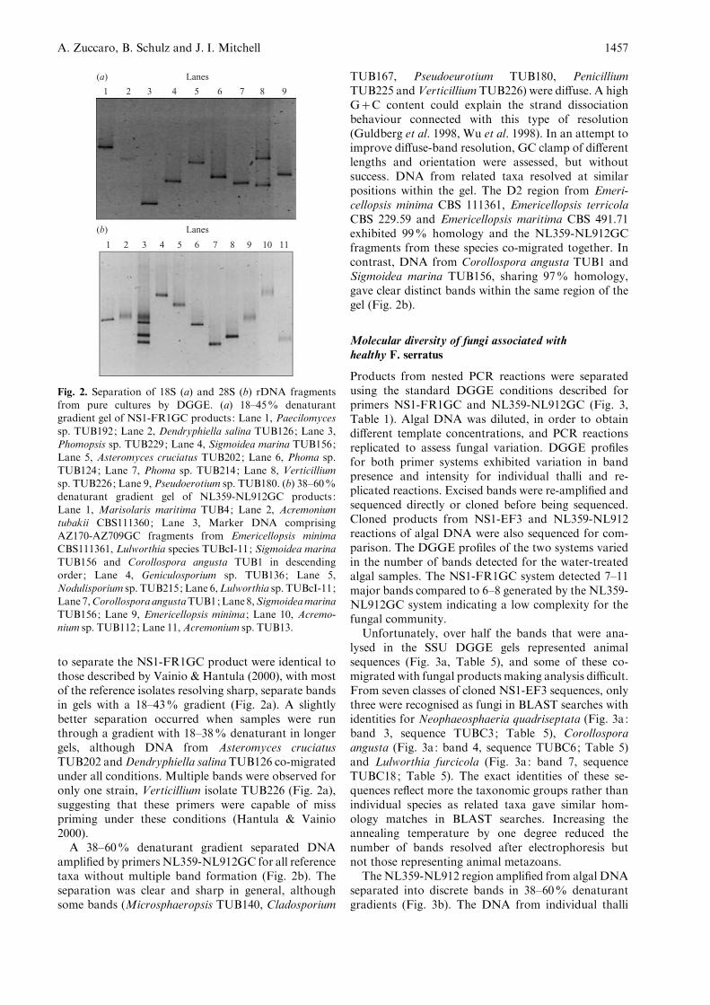
to separate the NS1-FR1GC product were identical tothose described by Vainio & Hantula (2000), with mostof the reference isolates resolving sharp, separate bandsin gels with a 18–43% gradient (Fig. 2a). A slightlybetter separation occurred when samples were runthrough a gradient with 18–38% denaturant in longergels, although DNA from Asteromyces cruciatusTUB202 andDendryphiella salina TUB126 co-migratedunder all conditions. Multiple bands were observed foronly one strain, Verticillium isolate TUB226 (Fig. 2a),suggesting that these primers were capable of misspriming under these conditions (Hantula & Vainio2000).
A 38–60% denaturant gradient separated DNAamplified by primers NL359-NL912GC for all referencetaxa without multiple band formation (Fig. 2b). Theseparation was clear and sharp in general, althoughsome bands (Microsphaeropsis TUB140, Cladosporium
TUB167, Pseudoeurotium TUB180, PenicilliumTUB225 andVerticillium TUB226) were diffuse. A highG+C content could explain the strand dissociationbehaviour connected with this type of resolution(Guldberg et al. 1998, Wu et al. 1998). In an attempt toimprove diffuse-band resolution, GC clamp of differentlengths and orientation were assessed, but withoutsuccess. DNA from related taxa resolved at similarpositions within the gel. The D2 region from Emeri-cellopsis minima CBS 111361, Emericellopsis terricolaCBS 229.59 and Emericellopsis maritima CBS 491.71exhibited 99% homology and the NL359-NL912GCfragments from these species co-migrated together. Incontrast, DNA from Corollospora angusta TUB1 andSigmoidea marina TUB156, sharing 97% homology,gave clear distinct bands within the same region of thegel (Fig. 2b).
Molecular diversity of fungi associated withhealthy F. serratus
Products from nested PCR reactions were separatedusing the standard DGGE conditions described forprimers NS1-FR1GC and NL359-NL912GC (Fig. 3,Table 1). Algal DNA was diluted, in order to obtaindifferent template concentrations, and PCR reactionsreplicated to assess fungal variation. DGGE profilesfor both primer systems exhibited variation in bandpresence and intensity for individual thalli and re-plicated reactions. Excised bands were re-amplified andsequenced directly or cloned before being sequenced.Cloned products from NS1-EF3 and NL359-NL912reactions of algal DNA were also sequenced for com-parison. The DGGE profiles of the two systems variedin the number of bands detected for the water-treatedalgal samples. The NS1-FR1GC system detected 7–11major bands compared to 6–8 generated by the NL359-NL912GC system indicating a low complexity for thefungal community.
Unfortunately, over half the bands that were ana-lysed in the SSU DGGE gels represented animalsequences (Fig. 3a, Table 5), and some of these co-migrated with fungal products making analysis difficult.From seven classes of cloned NS1-EF3 sequences, onlythree were recognised as fungi in BLAST searches withidentities for Neophaeosphaeria quadriseptata (Fig. 3a:band 3, sequence TUBC3; Table 5), Corollosporaangusta (Fig. 3a: band 4, sequence TUBC6; Table 5)and Lulworthia furcicola (Fig. 3a: band 7, sequenceTUBC18; Table 5). The exact identities of these se-quences reflect more the taxonomic groups rather thanindividual species as related taxa gave similar hom-ology matches in BLAST searches. Increasing theannealing temperature by one degree reduced thenumber of bands resolved after electrophoresis butnot those representing animal metazoans.
The NL359-NL912 region amplified from algal DNAseparated into discrete bands in 38–60% denaturantgradients (Fig. 3b). The DNA from individual thalli
1 2 3 4 5 6 7 8 9
Lanes(a)
1
Lanes(b)
2 3 4 5 6 7 8 9 10 11
Fig. 2. Separation of 18S (a) and 28S (b) rDNA fragmentsfrom pure cultures by DGGE. (a) 18–45% denaturantgradient gel of NS1-FR1GC products : Lane 1, Paecilomyces
sp. TUB192; Lane 2, Dendryphiella salina TUB126; Lane 3,Phomopsis sp. TUB229; Lane 4, Sigmoidea marina TUB156;Lane 5, Asteromyces cruciatus TUB202; Lane 6, Phoma sp.
TUB124; Lane 7, Phoma sp. TUB214; Lane 8, Verticilliumsp. TUB226; Lane 9, Pseudoerotium sp. TUB180. (b) 38–60%denaturant gradient gel of NL359-NL912GC products :
Lane 1, Marisolaris maritima TUB4; Lane 2, Acremoniumtubakii CBS111360; Lane 3, Marker DNA comprisingAZ170-AZ709GC fragments from Emericellopsis minimaCBS111361, Lulworthia species TUBcI-11; Sigmoidea marina
TUB156 and Corollospora angusta TUB1 in descendingorder; Lane 4, Geniculosporium sp. TUB136; Lane 5,Nodulisporium sp. TUB215; Lane 6,Lulworthia sp. TUBcI-11;
Lane 7,Corollospora angustaTUB1;Lane 8,SigmoideamarinaTUB156; Lane 9, Emericellopsis minima ; Lane 10, Acremo-nium sp. TUB112; Lane 11,Acremonium sp. TUB13.
A. Zuccaro, B. Schulz and J. I. Mitchell 1457

generated 1–5 bands. Seven groups of bands fromdifferent thalli profiles were successfully isolated, se-quenced, and compared with DGGE profiles from theisolates. BLAST searches allowed an initial identity butmore exact matches were made from precise alignmentsof the sequences with related taxa. Two prominentbands (Fig. 3b: bands f–g), observed from a number ofthalli, co-migrated with those from Corollospora angu-sta TUB1 (band g; sequence TUBS9) and Sigmoideamarina TUB156 (band f; sequence TUBSF22) and ex-hibited a sequence homology of 99% for both matches(Table 5). Sigmoidea marina was not isolated in culture
from the experimental material, although it was presenton previous sampling occasions. The sequence TUBSF3from a diffuse band (Fig. 3b: band c) had 100% se-quence homology with Emericellopsis terricola accord-ing to a BLAST search, but it also co-migrated withDNA from isolates of Emericellopsis minima andAcremonium tubakii that were isolated from algaltissue. An accurate match for this band could not bemade because of the similar sequence homology dis-played (99%) between all the Emericellopsis species(Table 5) ; it did not, however, completely match theA. tubakii sequence (96% homology) and inspection of
Table 4. PCR amplification of 18S or 28S rRNA gene fragments from a variety of taxa. +/x, slightly reduced amplification; n.t., not
tested. See Table 3 for collection acronyms.
Fungal isolates
Primer combinations
28S rRNA gene 18S rRNA gene
NL209-NL912 NL359-NL912 NS1-FR1
Diaporthales
Phomopsis sp. TUB229 + + +Phomopsis sp. TUB191 + + +
Dothideales
Cladosporium sp. TUB194 + + n.t.
Cladosporium sp. TUB167 + + n.t.
Coniothyrium sp. TUB198 + + +Microsphaeropsis sp. TUB140 + + n.t.
Phoma sp. TUB124 + + +Phoma sp. TUB214 + + +
Eurotiales
Penicillium sp. TUB225 + +/x +Pseudeurotium sp. TUB180 + + n.t.
Halosphaeriales
Corollospora angusta TUB1 + + n.t.
Hypocreales
Emericellopsis minima CBS 111361 + + n.t.
Emericellopsis terricola CBS 229.59 + + n.t.
Emericellopsis maritima CBS 491.71 + + n.t.
Fusarium sp. TUB216 + + n.t.
Trichoderma sp. TUB72 + + n.t.
Trichoderma sp. TUB32 + + n.t.
Helotiales
Cryptosporiopsis sp. TUBCr + + +
Lulworthiales
Lulworthia sp. TUBcI-11 + + n.t.
Xylariales
Nodulisporium sp. TUB215 + + +
Mitosporic fungi
Acremonium sp. TUB13 + + n.t.
Acremonium sp. TUB112 + + n.t.
Acremonium tubakii CBS 111360 + + n.t.
Asteromyces cruciatus TUB202 + + +Dendryphiella salina TUB126 + +/x +Geniculosporium sp. TUB136 + + +Marisolaris maritima TUB4 + + n.t.
Paecilomyces sp. TUB192 + + +Phialocephala sp. TUB4197 + + n.t.
Sigmoidea marina TUB156 + + +Scolecobasidium constrictum TUB199 + + +Verticillium cinnabarino TUB78 + + n.t.
Verticillium sp. TUB226 + + +
PCR-DGGE systems for detecting fungi from algae 1458

the sequences suggested a closer, tentative match toE. maritima. Two bands (Fig. 3b: bands d–e) exhibitedsequence homologies with Lulworthia medusa (Table 5,
BLAST results of 93% for band d, sequence TUBS11;and a sequence similarity of 85% for band e, sequenceTUBSF52). The remaining bands analysed (Fig. 3b:bands a–b) exhibited sequence homologies within theHypocreales, with BLAST results for Hypocrea lutea(band b – sequence TUBSFo, 97% homology, Table 5)and the Clavicipitaceae, with a match for Engyodontiumaranearum (band a – sequence TUBSF3n, 96% hom-ology; Table 5). No isolates from Lulworthia,Hypocreaor Engyodontium were recovered from algal tissue,making further identification difficult. DNA extractedfrom surface sterilised material amplified NL359-NL912 products that gave similar DGGE profilesto those obtained from water-treated tissue with noloss of bands, and sequence identities for C. angusta,S. marina, and the Emericellopsis.
Phylogenetic analysis of the D2 region sequencesfrom algae with other ascomycetes confirmed the taxo-nomic positions of the fungi detected molecularly.Maximum parsimony analysis of the 18S and 28SrRNA genes (1757 characters ; 554 parsimony informa-tive sites) generated six trees of 2892 steps from onetree-island. The topologies of these trees were similar,with differences only amongst groups containing shortbranch lengths (Fig. 4). The tree topology reflectedaccurately the sequences that could or could not beidentified unambiguously, and showed clearly a novellineage within the Lulworthiales ; an unresolved Emeri-cellopsis clade within the Bionectriaceae, and a poten-tial link between C. angusta and S. marina. A secondanalysis was performed with additional Corollosporaspecies to confirm the link between C. angusta andS. marina. The second data matrix (2073 characters ;375 parsimony informative sites) resolved three mostparsimonious trees of 1748 steps with Microascustrignonosporus as the outgroup (Fig. 5). The shortbranch length within theC. angusta and S. marina cladeindicate that these species may be conspecific.
Comparison of DGGE profiles between livingand dead Fucus serratus
DGGE profiles from healthy and dead Fucus serratuswere made using the 18S and 28S rDNA systems inorder to compare the molecular diversity of the hostwith and without intact protection mechanisms. LessDNA was recovered from the dead Fucus cast, pre-sumably due to the decomposition of algal tissue. Bothdetection systems recorded fewer bands amplified fromthe dead material ; nevertheless the prominent bandsobserved from the healthy material were still present inprofiles from the dead cast (Fig. 3). Fungal sequencesfor Corollospora angusta (Fig. 3a: band 4) and Neo-phaeosphaeria quadriseptata (Fig. 3a: band 3) wereobserved with the SSU system. Using the LSU rDNAsystem, it was observed that the bands were presentwith greater intensity, indicating a higher concentrationof fungal DNA. Three bands were sequenced andidentified as Sigmoidea marina (Fig. 3b: band f,
Lanes(a)
1 2 3 4 5 6
3
4
8
5
6
1 2
7
d
g
f
c
e
a
b
MDead Water treated
Lanes
1 2 3 4 5 6 7 8 9
(b)
M1 DSurface-sterilised
Water-treated
M2
Fig. 3. (a) Separation of PCR products, generated byNS1-FR1GC amplifications from algal DNA, in an 18–45%
denaturant gradient DGGE gel. Lane 1, Marker DNA (M)consisting of NS1-FR1GC amplicons from Dendryphiellasalina TUB126, Sigmoidea marina TUB156, Phoma sp.
TUB214, Coniothyrium sp. TUB198, Phomopsis sp. 229 indescending order; Lanes 2 and 3, Replicate reactions of DNAextracted from the thallus of dead Fucus serratus ; Lanes 4, 5
and 6, PCR products from the DNA of water-treated algalthalli. Eight independent clones were recovered from thesereactions and sequenced. The positions of the clones (1–8)
are shown in the gel and identities given in Table 4.(b) DGGE profiles of amplified 28S rDNA fragments(NL359-NL912GC) from algal DNA in a 38–60% denatur-ant gradient gel. Lane 1, Marker DNA (M1) comprising
NL359-NL912GC fragments from Lulworthia sp. TUBcI-11and Corollospora angusta TUB1 in descending order; Lane 2,Profile from a dead thallus of F. serratus ; Lanes 3 and 4,
Fragments amplified from surface-sterilised, healthy thalli ofF. serratus ; Lanes 5, 6, 7 and 8, Profiles from water treated,healthy thalli. Lanes 2–7 represents the profiles from indi-
vidual thalli. Lane 9, Marker DNA (M2) comprising theNL359-NL912GC fragment from Emericellopsis minimaCBS111361, Lulworthia sp. TUBcI-11, Sigmoidea marinaTUB156 and Corollospora angusta TUB1 in descending
order. Bands were labelled from a to g, excised and sequenceddirectly or from cloned fragments. Sequence matches for eachband are given in Table 4.
A. Zuccaro, B. Schulz and J. I. Mitchell 1459
sequence TUBSFt2), Corollospora angusta (Fig. 3b:band g, sequence TUBS15) and an unidentified speciesfrom the Lulworthiaceae (Fig. 3b: band d, sequenceTUBS5a).
DISCUSSION
The range of fungal diversity obtained using molecularand traditional techniques varied. Many fungal isolateswere recovered on different media, but few were de-tected molecularly suggesting that colonisation by mostof them was limited. The reference cultures used inthis study came from one sampling occasion, but thediversity observed was in broad agreement with otherreports where potential endophytes (Andrews et al.1982, Petrini 1991, Shearer 2001), saprobes (Kohlmeyer& Kohlmeyer 1979, Haythorn, Jones & Harrison1980, Kohlmeyer & Volkmann-Kohlmeyer 1991) andpathogens (Domsch, Gams & Anderson 1980) wererecovered.
The primers NL209, NL359 and NL912 generatedascomycete and related deuteromycete products thatwere suitable for DGGE analysis allowing a broad-based molecular assessment of diversity. It is possiblethat the actual fungal diversity may be greater than thatdetected with these primers, as a result of some un-discovered bias in amplification (Reysenbach et al.1992) or sampling effects, particularly when consideringother taxonomic groups. For this reason, the moregeneric primers NS1, FR1 and EF3 were used, but theapplication of these primers proved problematic. Theamplification of animal sequences and the generationof products difficult to resolve unambiguously, despite
being well separated in DGGE (something that mightbe more related to variation in product length than thesequence), made this system more reliant on cloningdirectly from algal DNA. The non-fungal specificity ofthese primers observed in this report is in agreementwith suggestions of Borneman & Hartin (2000) andVainio & Hantula (2000). Regardless of the limitationsassociated with the use of 18S rDNA sequences, bothmolecular systems detected similar fungal groups al-lowing the identification of isolates that might havean intimate relationship with Fucus serratus. In thisrespect, the availability of cultures isolated from algaltissue was indispensable for accurate molecular identi-fication.
A number of issues of mycological interest are raisedby the results presented in this paper. The fungal se-quences recovered from the DNA of Fucus serratusbelonged primarily to three main groups: (1) theHalosphaeriales (Fig. 4: group I) ; (2) the Hypocreales(Fig. 4: group II) and (3) the Lulworthiales (Fig. 4:group III). The majority of these sequences were notrecovered as isolates from the duplicate algal tissue,with the exception of Corollospora angusta, and poss-ibly the Emericellopsis minima and Acremonium tubakiiisolates. The remaining fungal isolates were presumablyinactive or present in small, undetectable amounts,such as single spores or fragments of hyphae that couldnot be amplified with the sampling strategy used.
Corollospora angusta/Sigmoidea marina Sequences:Corollospora angusta is considered to be an arenicolousfungus found in sea-foam and on sandy beaches,growing on calcareous shells (Kohlmeyer & Volkmann-Kohlmeyer 1987, Nakagiri & Tokura 1987). It has notbeen reported as being recovered from living brown
Table 5. (A) BLAST search results for cloned NS1-EF3 sequences (Fig. 3a: bands 1, 2, 3, 5, 6, 7 and 8) retrieved from algal DNA. The
number of each sequence represents the band identified in Fig. 3 with a numbered arrow. The highest sequence match is shown. The
sequence match for band 4 was made from an alignment of sequences from different taxa representing the Halosphaeriales. (B) Blast search
results (Fig. 3b: bands a, b, and d) or isolate matches (Fig. 3b: bands c, e, f and g) for the 28S environmental bands re-amplified with
primers NL359-NL912 directly from excised bands (living Fucus serratus) or after cloning of the NL209-NL912 PCR products (dead
F. serratus).
Band Phylum/class or order Closest sequence match (species) % Similarity
(A) 18S rDNA sequences recovered from Fucus serratus (Fig. 3a)
1 Hydrozoa Selaginopsis cornigera 98
2 Hydrozoa Selaginopsis cornigera 98
3 Ascomycota : Pleosporales Neophaeosphaeria quadriseptata 99
4 Ascomycota : Halosphaeriales Corollospora angusta 98
5 Arthropoda Allonothrus russeolus 98
6 Arthropoda Allonothrus russeolus 98
7 Ascomycota : Lulworthiales Lulworthia fucicola 96
8 Annelida Buchholozia fallax 97
(B) 28S rDNA sequences recovered from Fucus serratus (Fig. 3b)
a Mitosporic fungi: hyphomycete Engyodontium aranearum 96
b Ascomycota : Hypocreales Hypocrea lutea 98
c Ascomycota : Hypocreales Emericellopsis maritima ; E. terricola ; E. minima 99
d Ascomycota : Lulworthiales Lulworthia medusa 93
e Ascomycota : Lulworthiales Lulworthia medusa 85
f Mitosporic fungi: hyphomycete Sigmoidea marina 99
g Ascomycota : Halosphaeriales Corollospora angusta 99
PCR-DGGE systems for detecting fungi from algae 1460

Fig. 4. Phylogram showing the relationships of the 28S rRNA gene sequences recovered from Fucus serratus. Maximumparsimony analysis generated six most parsimonious trees of 2892 steps (CI=0462; RI=0.655; RC=0.303; HI=0.538)from 555 pics. Comparison of the log likelihood for each tree using the Kishino–Hasegawa test revealed that the tree
depicted had the lowest –InL (17895.12021) at P=0.0001, P=0.01 and P=0.65.
A. Zuccaro, B. Schulz and J. I. Mitchell 1461

algae, although other Corollospora species, such asC. intermedia, are considered to be active in the de-composition of algae (Kohlmeyer & Kohlmeyer 1979).Corollospora angusta and C. intermedia are morpho-logically very similar and often confused (Nakagiri &Tokura 1987), but they share only 78–79% sequencehomology for the D2 region and formed separateclades in parsimony analysis (Fig. 5) indicating thatthey are distinct. Haythorn, Jones & Harrison (1980)reported the isolation of Sigmoidea marina from thedecaying brown algae Fuccus serratus and Laminariasaccharina, and suggested that this hyphomycete was
saprophytic. The detection of Sigmoidea marina andCorollospora angusta sequences from dead algae sup-port the notion that these fungi are involved in thedecomposition of algal material, whilst their presencefrom healthy algae suggest that the fungal–algal re-lationship might be more complicated. Sand and shellparticles were removed from the algae prior to analysis,leaving calcareous worm tubes as a possible source ofattachment for these fungi, from which hyphae couldextend into the thallus.
Nakagiri & Tubaki (1985) described a range ofanamorphic forms within the genus Corollospora
Fig. 5. Relationship of Corollospora angusta, Sigmoidea marina and environmental sequences recovered from Fucus serratus.Maximum parsimony analysis generated three trees of 1748 steps (CI=0.609; RI=0.501; RC=0.305; HI=0.391) from375 pics. Bootstrap values over 80% are shown above the branches.
PCR-DGGE systems for detecting fungi from algae 1462

including Clavariopsis bulbosa (Corollospora pulchella),Varicosporina sp. (Corollospora intermedia) and Sig-moidea luteola (Corollospora luteola), but there areno reports on the anamorphic stage of C. angusta. Inparsimony analysis S. marina and C. angusta formed asupported clade (82%) connected by short branchlengths (Fig. 5), suggesting that they might be con-specific. Sequence comparison between C. angusta andS. marina for the 28S rRNA region revealed that therewas a 3% difference in homology between them,whereas other Corollospora species varied between58 and 90% from each other, supporting the con-clusion that they are closely related. Both C. angustaand S. marina contain the Q10 H2 ubiquinone system(Nakagiri 1991), indicating further biochemical simi-larities between them.
The Hypocreales : Three of the sequence groupsrecovered from algal DNA were identified as belongingto the Hypocreales. The first matched sequences fromEmericellopsis minima or E. maritima or E. terricola.The genus Emericellopsis comprises 14 species with anAcremonium anamorph stage. It was established by vanBeyma (1940) and originally placed in the Eurotiaceae.Rossman et al. (1999) re-assigned Emericellopsis to theBionectriaceae, as a sister group of Stanjemonium afteranalysis of LSU rRNA sequences. Most species appearprimarily as soil representatives, however Tubaki (1969)described two species from marine mud (E. humicolaand E. microspora) and Davidson & Christensen (1971)isolated another from saline lake muds (E. stolkiae).E. minima has also been recovered several times frommarine and brackish environments (Holler et al. 2000)underlining the idea that this genus might have mem-bers active in marine environments (Tubaki 1973). It isdifficult to assess whether the environmental band re-covered during this study belongs to E. maritima, orE. minima or to some novel taxon, as these sequenceswere very similar in the D2 region. Interestingly, theEmericellopsis/Acremonium sequence was amplifiedfrom more than one healthy thallus, and Acremoniumtubakii was isolated from surface sterilised tissue.Shearer (2001) isolated A. tubakii and E. minima fromMyriophyllum spicatum, an invasive Eurasian water-milfoil, describing them as potential endophyteswithin the host tissue. This poses the question whetherEmericellopsis and Acremonium play similar roles inthe marine environment. In this respect, the recovery ofan Emericellopsis/Acremonium sequence from surfacesterilised material cannot be taken as evidence thatthese fungi exist endophytically within Fucus serratus.There is no information to assess whether damagedDNA, resulting from limited sodium hypochloridetreatment, would be successfully amplified from algalsurfaces or not, making tenuous any conclusions on theepi- or endophytic nature of these fungi from thismolecular data.
The second hypocrealean sequence recovered wasidentified from a BLAST search as Engyodontiumaranearum (96% homology), a pathogen of spiders.
The low sequence homology match questions the exactidentity of this sequence, although it was placed withinthe Clavicipitaceae in the phylogenetic analysis. Fungibelonging to this family are pathogens of arthropods,fungi and grasses. Animal metazoan sequences wereamplified from algal DNA indicating their presenceassociated with Fucus serratus. Some of these sequenceswere derived from an arthropod and it is conceivablethat a pathogen of some invertebrate associated withFucus was amplified. Duncan et al. (2002) identified avariety of Claviceps purpurea adapted to marine en-vironments, and there is no reason why other membersof this family might not be adapted to other marinesubstrates/hosts. According to a BLAST search thethird sequence matched Hypocrea lutea (98% hom-ology). Species of Hypocrea are saprophytic on decay-ing plant material and many are parasites of fleshyfungi, with anamorphs in Trichoderma or Gliocladium-like. Two isolates of Trichoderma were obtained fromFucus but they showed different DGGE profiles incomparison to the environmental sequence (data notshown).
The Lulworthiales : No species of Lulworthia, Lindraor Kohlmeyeriella were isolated from Fucus serratusduring the sampling. The taxonomy of these marinefungi has recently been revised by Kohlmeyer,Spatafora&Volkmann-Kohlmeyer (2000), who createda separate order Lulworthiales to house representativesof Lindra and Lulworthia within the Lulworthiaceae.Campbell et al. (2002) used 28S rDNA sequences toshow that Kohlmeyeriella tubulata also belonged tothis order. The delineation of Lulworthia within theLulworthiaceae is incomplete. The majority of membersrepresenting this order are active in the marine en-vironment and have been implicated in the degradationof wood, and algae in coastal regions (Kohlmeyer &Kohlmeyer 1979). Two sets of sequences, TUBS11,TUBS14 and TUBSF52, TUBS5a (bands d and e re-spectively, Fig. 3b and Table 4) were recovered fromliving and dead algae that exhibited only limitedsequence homology with other representatives in theNCBI database. The closest match found was withLulworthia medusa (93% exact similarity with band d,and 85% exact similarity with band e). To investigatethe origin of this sequence ten species that were rep-resentative of the order were included in the phylogen-etic analysis. Fig. 4 shows one of six most parsimonioustrees. The sequences TUBS11, TUBS14 (Fig. 3b: bandd) and TUBSF52, TUBSFt5a (Fig. 3b: band e) formeda strongly supported clade (100% BS), separated fromother groups, within the Lulworthiaceae but of uncer-tain position. These sequences, therefore, represent adistinct and separate lineage of organisms within theLulworthiaceae. The identity of this group, however,remains unknown. It is possible that it may represent anovel group of organisms; conversely it could consistof species known morphologically but not molecularly.Species from the Lulworthiaceae exhibit parasitic orsaprophytic associations with brown algae, infecting
A. Zuccaro, B. Schulz and J. I. Mitchell 1463

thalli of Sargassum (Lindra crassa ; Tubaki 1969), Fucus(Lulworthia furcicola ; Sutherland 1916) and Litho-phyllum (Lulworthia kniepii ; Kohlmeyer 1967, 1969).Sequence information is not available for many of thesespecies, making identification of the environmentalsequences difficult, even to the genus level.
One other sequence was detected that was not re-lated to the groups described above. Band 3 (Fig. 3a)was amplified using NS1-FR1GC and identified asNeophaeosphaeria quadriseptata with a sequence simi-larity of 99% in a BLAST search. This represents theonly bitunicate ascomycete detected molecularly inthis study. Neophaeosphaeria comprises membersthat are pathogens of plants such as Agave andYucca. However, Kohlmeyer, Volkmann-Kohlmeyer &Eriksson (1995) recorded a perhaps somewhat relatedspecies, Paraphaeosphaeria pilleata, from saltmarshJuncus roemerianus indicating some members ofthis group may be capable of inhabiting marineenvironments.
The majority of fungi we identified molecularly wereobligate marine ascomycetes, with the exception ofthe hypocrealean species. The absence of molecularsequences representing other terrestrial groups is ingeneral agreement with the comments of Kohlmeyer& Volkmann-Kohlmeyer (2003b), who made a cleardistinction between the activities of marine-derivedand authentic marine obligate fungi when determiningassociations with coral reefs. The detection of hypo-crealean groups normally considered terrestrial suggeststhat the diversity of fungi associated with brown algaemight be more extensive than previously thought, withsome terrestrial species exhibiting an adaptation tothe marine environment similar to those described forAspergillus sydowii (Geiser et al. 1998, Alker, Smith &Kim 2001) and Claviceps purpurea (Duncan et al.2002). A more detailed assessment of fungi as-sociated with Fucus serratus throughout the year, andthe nature of these associations, is currently underinvestigation.
ACKNOWLEDGEMENTS
We thank Jack Fisher, Nigel Hywel-Jones, Martina Jahn and Nick
Talbot for taking the time to read and comment on the manuscript
before submission. For help with the taxonomy of many fungi, we are
indebted and thankful to Siegfried Draeger, Richard Summerbell
and Walter Gams. Miruna Oros-Sichler, Newton C. M. Gomes and
Kornelia Smalla are acknowledged with gratitude for assistance with
the SSU DGGE system. A.Z. extends her thanks to Jorn Petersen for
assistance in the laboratory, and to Dieter Jahn and Hans-Jurgen
Aust for financial support.
REFERENCES
Alker, A. P., Smith, G. W. & Kim, K. (2001) Characterisation of
Aspergillus sydowii (Thom et Church), a fungal pathogen of
Caribbean sea fan corals. Hydrobiologia 460 : 105–111.
Andrews, J. H., Hecht, E. P. & Bashirian, S. (1982) Association
between the fungus Acremonium curvulum and Eurasian water
milfoil, Myriophyllum spicatum. Canadian Journal of Botany 60 :
1216–1221.
Armstrong, E., Yan, L., Boyd, K. G., Wright, P. C. & Burges, J. G.
(2001) The symbiotic role of marine microbes on living surfaces.
Hydrobiologia 461 : 27–40.
Borneman, J. & Hartin, R. J. (2000) PCR primers that amplify fungal
rRNA genes from Environmental samples. Applied and Environ-
mental Microbiology 66 : 4356–4360.
Buchan, A., Newell, S. Y., Moreta, J. I. J. & Moran, M. A. (2002)
Analysis of internal transcribed spacer (ITS) regions of rRNA
genes in fungal communities in a southeastern U.S. salt marsh.
Microbial Ecology 43 : 329–340.
Campbell, J., Shearer, C. A., Mitchell, J. I. & Eaton, R. A. (2002)
Corollospora revisited: a molecular approach. In Fungi in Marine
Environments (K. D. Hyde & L. L. P. Vrijmoed, eds): 15–33.
Fungal Diversity Press, Hong Kong.
Davidson, D. E. and Christensen, M. (1971) Emericellopsis stolkiae
sp. nov. from saline soils in Wyoming. Transactions of the British
Mycological Society 57 : 385–391.
Domsch, K. H., Gams, W. & Anderson, T. H. (1980) Compendium of
Soil Fungi. Vol. 1. Academic Press, London.
Dreyfuss, M. M. (1986) Neue Erkenntnisse aus einem pharmakolo-
gischen pilzscreening. Sydowia 39 : 22–36.
Duncan, R. A. jr, Sullivan, R., Alderman, S. C., Spatafora, J. W.
&White, J. F. jr (2002) Claviceps purpurea var. spartinae var. nov.:
an ergot adapted to the aquatic environment. Mycotaxon 81 :
11–25.
van Elsas, J. D., Duarte, G. F., Keijzer-Wolters, A. & Smit, E. (2000)
Analysis of the dynamics of fungal communities in soil via
fungal-specific PCR of soil DNA followed by denaturing
gradient electrophoresis. Journal of Microbiological Methods 43 :
133–151.
Fell, J. W. (1993) Rapid identification of yeast species using three
primers in a polymerase chain reaction. Molecular Marine Biology
and Biotechnology 2 : 174–180.
Fell, J. W., Scorzetti, G., Boekhout, T., Fonseca, A. & Statzell-
Tallman, A. (2000) Biodiversity and Systematics of basisiomyce-
tous yeasts as determined by D1/D2 large subunit rDNA sequence
analysis. International Journal of Systematic and Evolutionary
Microbiology 50 : 1351–1371.
Geiser, D. M., Taylor, J. W., Ritchie, K. B. & Smith, G. W.
(1998) Cause of sea fan death in the West Indies. Nature 394 :
137–138.
Guldberg, P., Grønbæk, K., Aggerholm, A., Platz, A., Straten thor,
P., Ahrenkiel, V., Hokland, P. & Zeuthen, J. (1998) Detection
of mutations in GC-rich DNA by bisulphite denaturing
gradient gel electrophoresis. Nucleic Acids Research 26 : 1548–
1549.
Hawksworth, D. L. (1988) The variety of fungal–algal associations,
their evolutionary significance and the nature of lichens. Botanical
Journal of the Linnean Society 96 : 3–20.
Hawksworth, D. L. (1991) The fungal dimension of biodiversity:
magnitude, significance, and conservation. Mycological Research
95 : 641–655.
Hawksworth, D. L. (2001) The magnitude of fungal diversity: the 1.5
million species estimate revisited. Mycological Research 105 :
1422–1431.
Haythorn, J. M., Jones, E. B. G. & Harrison, J. L. (1980) Obser-
vations on marine algicolous fungi, including the Hyphomycete
Sigmoidea marina sp. Nov. Transactions of the British Mycological
Society 74 : 615–623.
Holler, U., Wright, A. D., Matthee, G. F., Konig, G. M., Draeger, S.,
Aust, J. & Schulz, B. (2000) Fungi from marine sponges: diversity,
biological activity and secondary metabolites. Mycological
Research 104 : 1354–1365.
Hopple, J. S. & Vilgalys, R. (1999) Phylogenetic relationships in the
mushroom genus Coprinus and dark-spored allies on sequence data
from the nuclear gene coding for the Large Ribosomal Subunit
PCR-DGGE systems for detecting fungi from algae 1464
RNA: divergent domains, outgroups and monophyly. Molecular
Phylogenetics and Evolution 13 : 1–19.
Hyde, K. D., Jones, E. B. G., Leano, E., Pointing, S. B., Poonyth,
A. D. & Vrijmoed, L. L. P. (1998) Role of fungi in marine eco-
systems. Biodiversity and Conservation 7 : 1147–1161.
Hyde, K. D., Sarma, V. V. & Jones, E. B. G. (2000) Morphology and
taxonomy of higher marine fungi. InMarine Mycology: a practical
approach (K. D. Hyde & S. B. Pointing, eds) : 172–204. [Fungal
Diversity Research Series. Vol. 1.] Fungal Diversity Press, Hong
Kong.
Jensen, P. R. & Fenical, W. (2002) Secondary metabolites from
marine fungi. In Fungi in Marine Environments (K. D. Hyde, ed.) :
293–315. [Fungal Diversity Research Series. Vol. 7.] Fungal
Diversity Press, Hong Kong.
Jones, E. B. G. (1976) Lignicolous and algicolous fungi. In Recent
Advances in Aquatic Mycology (E. B. G. Jones, ed.) : 1–51. Wiley,
New York.
Kohlmeyer, J. (1967) Intertidal and phycophilous fungi from Tenerife
(Canary Islands). Transactions of the British Mycological Society
50 : 137–147.
Kohlmeyer, J. (1969) The role of marine fungi in the penetration of
calcareous substances. American Zoologist 9 : 741–746.
Kohlmeyer, J. & Kohlmeyer, E. (1979) Marine Mycology: the higher
fungi. Academic Press, New York.
Kohlmeyer, J., Spatafora, J. W. & Volkmann-Kohlmeyer, B. (2000)
Lulworthiales, a new order of marine ascomycetes. Mycologia 92 :
453–458.
Kohlmeyer, J. & Volkmann-Kohlmeyer, B. (1987) Reflections on
the genus Corollospora (ascomycetes). Transactions of British
Mycological Society 88 : 181–188.
Kohlmeyer, J. & Volkmann-Kohlmeyer, B. (1991) Illustrated key
to the filamentous marine fungi. Botanica Marina 4 : 1–61.
Kohlmeyer, J. & Volkmann-Kohlmeyer, B. (2003a) Marine
ascomycetes from algae and animal hosts. Botanica Marina 46 :
285–306.
Kohlmeyer, J. & Volkmann-Kohlmeyer, B. (2003b) Fungi
from coral reefs : a commentary. Mycological Research 107 :
386–387.
Kohlmeyer, J., Volkmann-Kohlmeyer, B. & Eriksson, O. E. (1995)
Fungi on Juncus roemerianus. New marine and terrestrial
ascomycetes. Mycological Research 100 : 393–404.
Kowalchuk, G. A. (1998) Fungal community analysis using dena-
turing gradient gel electrophoresis (DGGE). In Molecular
Microbial Ecology (A. D. L. Akkermans, J. D. van Elas, F. J. de
Bruijn, eds) 3.4.6 : 1–16. Kluwer, Dordrecht.
Landeweert, R., Leeflang, P., Kuyper, T. W., Hoffland, E., Rosling,
A., Wernars, K. & Smit, E. (2003) Molecular identification of
ectomycorrhizal mycelium in soil horizons. Applied and Environ-
mental Microbiology 69 : 327–333.
Lee, S. B., Milgroom, M. G. & Taylor, J. W. (1988) A rapid, high
yield mini-prep method for isolation of total genome DNA from
fungi. Fungal Genetics Newsletter 35 : 23–24.
Lee, S. B. & Taylor, J. W. (1990) Isolation of DNA from
fungal mycelia and single spores. In PCR Protocols: a guide
to methods and applications (M. A. Innes, D. H. Gelfand,
J. J. Sninsky & T. J. White, eds) : 282–287. Academic Press,
San Diego.
Lerman, L. S. & Beldjord, C. (1998) Comprehensive mutation
detection with denaturing gradient gel electrophoresis. InMutation
Detection (R. G. H. Cotton, E. Edkins & S. Forrest, eds): 35–61.
IRL Press, Oxford.
Liesack, W., Janssen, P. H., Rainey, F. A., Ward-Rainey, N. L. &
Stackebrandt, E. (1997) Microbial diversity in soil : the need
for a combined approach using molecular and cultivation techni-
ques. In Modern Soil Microbiology (J. D. van Elsas, E. M. H.
Wellington & J. T. Trevors, eds) : 375–439. Marcel Dekker,
New York.
Mearns-Spragg, A., Boyd, K. G., Hubble, M. O. & Burgess, J. G.
(1997) Cross-species induction and enhancement of anti-microbial
activity produced by epiphytic bacteria from marine algae and
invertebrates after exposure to terrestrial bacteria. Letters Applied
Microbiology 27 : 142–146.
Muyzer, G. (1999) DGGE/TGGE a method for identifying genes
from natural ecosystems. Current Opinion in Microbiology 2 :
317–322.
Nakagiri, A. (1991) Coenzyme Q systems in the genus Corollospora
and allied marine fungi. IFO Research Communications 15 :
97–104.
Nakagiri, A. & Tokura, R. (1987) Taxonomic studies of the genus
Corollospora (Halosphaeriaceae, Ascomycotina) with descriptions
of seven new species. Transactions of the Mycological Society of
Japan 28 : 413–436.
Nakagiri, A. & Tubaki, K. (1985) Teleomorph and anamorph
relationships in marine ascomycetes (Halosphaeriaceae, Asco-
mycotina). Botanica Marina 28 : 485–500.
Nicholas, K. B., Nicholas, H. B. jr & Deerfield, D. W. II. (1997)
GeneDoc: analysis and visualization of genetic variation.
EMBNEW NEWS 4 : 14.
Petrini, O. (1991) Fungal endophytes of tree leaves. In Microbial
Ecology of Leaves (J. H. Andrews & S. S. Hirano, eds) : 179–187.
Springer-Verlag, New York.
Rossman, A. Y., Samuels, G. J., Rogerson, C. T. & Lowen, R. (1999)
Genera of Bionectriaceae, Hypocreaceae and Nectriaceae (Hypo-
creales, ascomycetes). Studies in Mycology 42 : 1–248.
Reysenbach, A.-L., Giver, L. J., Wickham, G. S. & Pace,
N. R. (1992) Differential amplification of rRNA genes by poly-
merase chain reaction.Applied and Environmental Microbiology 58 :
3417–3418.
Rychlik, W. & Rhodes, R. E. (1989) A computer programme for
choosing optimal oligonucleotides for filter hybridisation, sequen-
cing and in vitro amplification of DNA. Nucleic Acids Research 17 :
8543–8551.
Schultz, B., Guske, S., Dammann, U. & Boyle, C. (1998) Endophyte-
host interactions II. Defining symbiosis of the endophyte-host
interaction. Symbiosis 25 : 213–227.
Schultz, B., Boyle, C., Draeger, S., Rommert, A.-K. & Krohn, K.
(2002) Endophytic fungi: a source of novel biologically
active secondary metabolites. Mycological Research 106 : 996–
1004.
Shearer, J. F. (2001) Recovery of endophytic fungi from Myrio-
phyllum spicatum. ERDC TN-APCRP-BC-03.
Smit, E., Leeflang, P., Glandorf, B. & van Elsas, J. D. (1999) Analysis
of fungal diversity in wheat rhizosphere by sequencing of cloned
PCR-amplified genes encoding 18S rRNA and temperature gradi-
ent gel electrophoresis. Applied and Environmental Microbiology
65 : 2614–2621.
Stanley, S. (1991) The autecology and ultrastructral interactions be-
tween Mycosphaerella ascophylli Cotton, Lautitia danica (Berlese)
Schatz, Mycaureola dilseae Maire et Chemin and their respective
marine algal hosts. PhD thesis, University of Portsmouth.
Sutherland, G. K. (1916) Additional notes on marine pyre-
nomycetes. Transactions of the British Mycological Society 5 :
257–263.
Swofford, D. L. (1999) PAUP*: phylogenetic analysis using parsi-
mony (and other methods). Version 4.0. Sinauer Associates,
Sunderland, MA.
Stolk, A. C. (1955) Emericellopsis minima sp. nov. and Westerdykella
ornata gen. nov. sp. nov. Transactions of the British Mycological
Society 38 : 419–424.
Tubaki, K. (1969) Studies on the Japanese marine fungi: lignicolous
group (III), algicolous group and a general consideration. Annual
Review of the Institute of Fermentation, Osaka 4 : 12–41.
Tubaki, K. (1973) Aquatic sediment as a habitat of Emericellopsis,
with a description of an undescribed species of Cephalosporium.
Mycologia 65 : 938–941.
Vainio, E. J. & Hantula, J. (2000) Direct analysis of wood-inhabiting
fungi using denaturing gradient gel electrophoresis of amplified
ribosomal DNA. Mycological Research 104 : 927–936.
van Beyma, T. F. H. (1940) Ueber einige formen von Verticillium
dahliae klebahn. Antonie Van Leeuwenhoek 6 : 33–47.
A. Zuccaro, B. Schulz and J. I. Mitchell 1465

Vandenkoornhuyse, P., Baldauf, S. L., Leyval, J. & Young, J. P. W.
(2002) Extensive fungal diversity in plant roots. Science 295 : 2051.
White, T. J., Bruns, T., Lee, S. & Taylor, J. (1990) Amplification
and direct sequencing of fungal ribosomal RNA genes for phylo-
genetics. In PCR Protocols: a guide to methods and applications
(M. A. Innes, D. H. Gefand, J. J. Sninsky & T. J. White, eds):
315–322. Academic Press, San Diego.
Wu, Y., Hayes, V. M., Osinga, J., Mulder, I. M., Looman, W. G.,
Buys, H. M. C. & Hofstra, M. W. (1998) Improvement of fragment
and primer selection for mutation detection by denaturing gradient
gel electrophoresis. Nucleic Acids Research 26 : 5432–5440.
Corresponding Editor: D. L. Hawksworth
PCR-DGGE systems for detecting fungi from algae 1466
Related Documents