A world-monograph of the genera Ascobolus and Saccobolus (Ascomycetes, Pezizales) PROEFSCHRIFT TER VERKRIJGING VAN DE GRAAD VAN DOCTOR IN DE WISKUNDE EN NATUURWETENSCHAPPEN AAN DE RIJKSUNIVERSITEIT TE LEIDEN, OP GEZAG VAN DE RECTOR MAGNIFICUS DR P. MUNTENDAM, HOOG- LERAAR IN DE FACULTEIT DER GENEESKUNDE, TEN OVERSTAAN VAN EEN COMMISSIE UIT DE SENAAT TE VERDEDIGEN OP WOENSDAG 13 SEPTEMBER 1967 TE 16 UUR DOOR Johannes van Brummelen geboren te Haarlem in 1932 PRINTED BY J. J. GROEN EN ZOON - LEIDEN 1967

Welcome message from author
This document is posted to help you gain knowledge. Please leave a comment to let me know what you think about it! Share it to your friends and learn new things together.
Transcript
A world-monograph of
the genera Ascobolus and Saccobolus
(Ascomycetes, Pezizales)
PROEFSCHRIFT
TER VERKRIJGING VAN DE GRAAD VAN DOCTOR IN
DE WISKUNDE EN NATUURWETENSCHAPPEN AAN DE
RIJKSUNIVERSITEIT TE LEIDEN, OP GEZAG VAN DE
RECTOR MAGNIFICUS DR P. MUNTENDAM, HOOG-
LERAAR IN DE FACULTEIT DER GENEESKUNDE,TEN
OVERSTAAN VAN EEN COMMISSIE UIT DE SENAAT
TE VERDEDIGEN OP
WOENSDAG 13 SEPTEMBER 1967 TE 16 UUR
DOOR
Johannes van Brummelen
geboren te Haarlem in 1932
PRINTED BY
J. J. GROEN EN ZOON - LEIDEN
1967
PROMOTOR: PROF. DR. C. G. G. J. VAN STEENIS
This book was printed with financial support from the Netherlands Organisationfor the Advancement of Pure Research (Z.W.O.).

Aan mijn ouders
Aan mijn vrouw
Curriculum vitae
JOHANNES VAN BRUMMMELEN, geboren 19 februari 1932,behaalde in 1951 zijn eind-
diploma H.B.S.-B aan het Lorentz Lyceum te Haarlem en werd in hetzelfde jaar
ingeschreven als student aan de Universiteit van Amsterdam. Zijn candidaats-
examen (letter K) legde hij af in januari 1955, zijn doctoraal examen biologie
(hoofdvak mycologie, bijvakken dierkunde en microbiologic) in maart 1959 aan
de Rijksuniversiteit te Leiden (cum laude).
Tijdens zijn doctorale studie bewerkte hij de volgende onderwerpen en wel bij:
PROF. DR IR T. Y. KINGMA BOLTJES: onderzoek naar assimilatie en levenscyclus
van stammen van het Actinomyceten geslacht Streptosporangium, gepubliceerd in
Anthony van Leeuwenhoek 23 (1957): 385-392.
PROF. DR W. K. H. KARSTENS, metingen aan bladeren van appelbomen in verband
met ras, groeiplaats en resistentie tegen bladmijten.
DR W. VERVOORT: cytogenetisch onderzoek van twee soorten sprinkhanen
(Chorthyppus) en enkele interspecifische bastaarden.
PROF. DR C. J. VAN DER KLAAUW: over histochemische fixatie en kleurings-
methoden in verband met bepalingen van mucopolysacchariden in de hypophyse
van stekelbaarzen.
DR M. A. DONK: de Nederlandse mestbewonende fungi.
Voorts schreef hij voor PROF. KINGMA BOLTJES een scriptie over de genetica van
bacterien.
Van 1954 tot 1955 was hij als assistent werkzaam bij het practicum morphologie
van de cryptogamen. Van 1956 tot 1958 was hij onderzoek-assistent bij PROF, VAN
DER KLAAUW, in dienst van Z.W.O. Van maart 1959 tot december 1961 was hij in
dienst van Z.W.O. als wetenschappelijk ambtenaar werkzaam op de afdeling
mycologie van het Rijksherbarium te Leiden.
Vanafjanuari 1961 was hij aanvankelijk in dezelfde rang, daarnaals wetenschap-
pelijk ambtenaar eerste klasse verbonden aan het Rijksherbarium, waar hij naast
zijn onderzoekingen op taxonomisch gebied assisteerde bij de opleiding van studenten.
Tevens was hij van maart 1961 tot februari 1965 belast met het toezicht op de voor-
bereiding tot de bouw en de inrichting van het huidige Rijksherbarium.
Artikelen in deze tijd bewerkt zijn:
Types of species ofAscobolus and Saccobolus in Spegazzini's herbarium. In Persoonia
2 (1962): 195-199-
On four species ofFimaria.In Persoonia 2 (1962): 321-330.
5
Contents
INTRODUCTION 7
ACKNOWLEDGEMENTS 7
A. GENERAL PART g
Chapter I. Historical survey 9
Chapter II. Materials and methods 13
Living material 13
Culture procedures 14
Culture media 16
Microscopic examination of fresh material 18
Herbarium material 19
Chapter III. The development of the fruit-body 22
The development 22
Developmental types of fruit-bodies 24
Chapter IV. The structure of the fungus 29
Mycelium and conidial stages 29
Fruit-body 30
Excipulum 30
Flesh 34
Hypothecium 34
Hymenium > 35
Paraphyses 35
Asci 36
Ascospores 38
Arrangement of ascospores 40
Pigmentation of ascospores 42
Ornamentation of ascospores 44
Anisosporv 45
Chapter V. Cytology 47
Chapter VI. Sexuality and compatibility 48
Chapter VII. Genetics 50
Chapter VIII. Ecology 51
Chapter IX. Distribution 53
B. SPECIAL PART 56
Chapter X. Ascobolaceae 56
Key to the subfamilies 57
Ascoboloideae, with key to the generaof the Ascoboloideae....
58
Ascodesmidoideae 59
Theleboloideae 59
6 CONTENTS
Chapter XI. The genera Ascobolus and Saccobolus 61
Ascobolus 61
Synoptic key to the sections of Ascobolus 63Artificial key to the sections of Ascobolus 64
Section Dasyobolus 66
Section Sphaeridiobolus 89
Section Ascobolus 94
Section Pseudascodesmis 153
Section Pseudosaccobolus 155
Section Heimerlia 157
Section Gymnascobolus 158
Saccobolus 166
Key to the sections of Saccobolus 167
Section Saccobolus 167
Section Eriobolus 181
Chapter XII. Insufficiently known and excluded species 206
REFERENCES 243
INDEX 255
PLATES AND EXPLANATIONS following 260
7
Introduction
For these reasons it has appeared that many specific names have to go into
synonymy.
As most of the descriptions are insufficient to recognize the species, a study of
the original material was necessary. Only in recent times a beginning has been made
with the revision of such material.
In the present work a revision is given of the taxa belonging to the genera
Ascobolus and Saccobolus, two genera forming a sharply delimited, natural group in
the Ascobolaceae. Special attention has been paid to the development and the
microscopic structures of these fungi in connection with the relationships within
thegenera.
In many cases authentic specimens have been seen by the author. In cases where
obviously no type specimens are in existence illustrations and descriptions sometimes
have helped in recognizing the species. Several species, however, are only known
from short descriptions in which essential characters are lacking. Their names are
included in the list of nomina dubia.
ACKNOWLEDGEMENTS
Thewriter wishes to express his gratitude to Dr. M. A. Donk for the generous way
!n which he made his wide knowledge of the Fungi available to me. He is much
indebted to Professor Dr. C. G. G. J. van Steenis and to Professor Dr. H. J. Lam,
present and former Directors respectively of the Rijksherbarium at Leiden, and to
those members of the staff of this institute, who assisted him with this monograph.
A great debtofgratitude is due to Miss H. P. Wilkinson for reading the manuscript,
and whose advice has greatly improved the English text. Warmest thanks are also
extended to Dr. R. A. Maas Geesteranus for discussing many problems; to Dr. R.
F. Cain for sending many interesting specimens from his own collections; to Dr. H.
O. Sleumer for preparation of the Latin diagnoses of the new taxa and for sending
Since the genus Ascobolus was established in 179 1, several more genera of Asco-
bolaceae with coloured ascospores became known, each with a fair number of
species.
Some species previously described in other genera were laterrecognized to belong
to the Ascobolaceae and a large number of new species, subspecies, varieties and
forms have been described through the years. Numerous species of Ascobolus have
been described as new simply because the same fungus was found on a different
substratum or because its asci opened by an operculum. Others were described sev-
eral times under different names because of the lack of a complete survey and
inadequate knowledge of specific variability. This has also led to many erroneous
interpretations and identifications.
8 ACKNOWLEDGEMENTS
samples of dung from New Guinea, Hawaii, and the United States; to Mr. H. J.T. Tammel for preparing several of the drawings for publication; and to the
Directors and Curatorsof the institutes mentionedon p. 19—20 for the loan of material
or for permission to examine specimens. Living material has also been gratefully
received fromDr. G. N. Bistis, Dr. R. W. G. Dennis, Dr. H. Dissing, Dr. I. Gamundl de
Amos, Dr. J. A. Harper, Dr. N. Lundqvist, Mr. J. T. Palmer, Mr. H. Romagnesi
and Dr. J. Webster.
The writer would also like to express his indebtedness to the "Netherlands
Organisation for the Advancement of Pure Research (Z.W.O.)" for the generous
grants which enabled him to visit the "Herbarium of the Royal Botanic Gardens"
at Kew, the "British Museum (Natural History)" in London, and the "Narodni
Museum" in Praha.
The "Centre National de la Recherche Scientifique" is gratefully acknowledged
for a grant enabling him to visit the "Museum National d'Histoire Naturelle" in
Paris and the "Laboratoire de Biologie Marine du College de France" in
Concarneau.
The "NetherlandsOrganisation for the Advancement of Pure Research (Z.W.O.)"
supported the publication of this monograph.
9
A. GENERAL PART
CHAPTER I
HISTORICAL SURVEY
The genus Ascobolus was established by Persoon in 179 1 for Ascobolus pezizoides, a
dung-inhabiting cup-fungus, which differed from species of Peziza in possessing
clearly visible and far protruding coloured tips of ripe asci. The generic name
Ascobolus was given by Persoon, because he was of the opinion that the asci were
shot away in toto.
In 1794 Persoon added two other species, while two years later he described
Ascobolus carneus, a species with hyaline ascospores. Further species were incorporated
in the genus by other workers such as von Albertini & von Schweinitz (1805),
Bivona Bernardi (1816), and Schmidt (1817).In 1822 Persoon included seven species in Ascobolus and in the same year Fries
enlarged the number to eleven. Gradually the genus was extended with some more
species by Fries (1828), Wallroth (1833), Berkeley (1836), and Preuss (1851).
Mycological interest was focused on the genus Ascobolus in 1857 when the Crouan
brothers discovered species of this genus in which the opening mechanism of the
ascus was by means of a terminal operculum. The numerous investigations in
different countries that followed after this discovery greatly increased the number
of species described in this genus.
The Crouan brothers (1857, 1858), who contributed so much to the knowledge
of the genus Ascobolus, considered the presence of an operculum at the apex of the
ascus to be the distinguishing character of this genus. All species which they found
possessing asci with an operculum were put into the genusAscobolus. Owing to this,
among the species of Ascobolus they described from Finistere, several elements were
introduced which did not show a direct relationship with the original species.
Karsten (1861) gave a briefsurvey of the species found in Finland.
Coemans (1862) made a study of the species found in Belgium. Fie was the first
to describe the development of the fruit-bodies and the staining of the ascus wall
■with iodine in a number of species.Cooke (1864) listed fifteen species for Britain, and divided the genus in three
"subdivisions" according to the nature of the surface of the fruit-bodies.
He Notaris (1864) described three new species from Italy.
Berkeley & Broome (1865) reported seventeen species from the British Isles
among which two were new to the genus.
Among the twenty-four species recorded by Fuckel (1866b) fromthe surroundings
A WORLD-MONOGRAPH OF THE GENERA ASCOBOLUS AND SACCOBOLUS10
of Oestrich in Germany seven were new to the genus. Most of his species had been
distributed earlier in his "Fungi rhenani exsiccati".
In a flora ofFinistere the Grouan brothers (1867) again described five new species.From the surroundings of Leipzig in Germany Auerswald (1868) described three
species from the dung of dogs.
By far the best monograph on these fungi has been written by Boudier (1869).His "Memoire sur les Ascoboles" has been the base for most of the later studies in
this group. It is still a most valuable source of information, especially on account
of the accurate illustrations.
Boudier succeeded in isolating a natural group from the heterogeneous agglomer-ation ofspecies that had been classified in Ascobolus. He also grouped the species that
seemed to be intermediate between the 'true Ascobolei' and the "Pezizes".
The "Ascobolei" were characterized by the possession of large operculate asci the
tips of which protrude above the hymenium. He divided the "Ascobolei" into two
groups: the "Ascobolei genuini" with coloured ascospores and the "Ascobolei spurii"with hyaline ascospores. Under the "Ascobolei genuini" he placed the genera
Angelina, Ascobolus, and Saccobolus. Under the "Ascobolei spurii" he placed the genera
Thecotheus, Rhyparobius, and Ascophanus.The genus Angelina was known to Boudier only from literature and he rightly
doubted the correctness of its classification in this group. He restricted the genus
Ascobolus to those species which have free ascospores. Sometimes these ascospores
become glued together but they are never surrounded by a common membrane.
The genus Saccobolus was introduced for those species in which the ripe ascospores
are surrounded by such a membrane. Besides, the paraphyses in Saccobolus are
shorter than those in Ascobolus. In the genera Ascobolus and Saccobolus several new
species were described by Boudier.
In his "Symbolae mycologicae", Fuckel (1870), ignorant ofBoudier's work, placedthe genus Ascobolus in the family "Bulgariacei", distinguishing its species according
to the colour of the ascospores and the structure of the outside of the fruit-bodies.
Shortly after Boudier's monograph Karsten (1870) published a monograph of
the species of Ascobolus from Finland, in which twenty-two species and five sub-
species were described. Most of his new species were already published and
distributed in his "Fungi Fenniae exsiccati" (Karsten, 1866-1868). Although un-
aware of the work of Boudier and Fuckel, he also classified the species according to
the colour of the ascospores. The genus was restricted to a rather sharply delimited
naturalgroupof fungi. In his "Mycologia fennica" Karsten (1871) restricted Ascobolus
to the species with coloured ascospores, while the species that Boudier placedunder Ascophanus and Rhyparobius were brought to Peziza and Pezizula respectively.
Not until many years later did Karsten (1885) adopt the generic names of
Boudier.
Under the name Peziza cunicularia Boudier (1869: 258) described a species which
differs from all other "Ascobolei" in the mechanism of dehiscence of the asci.
Instead of an operculum, in this species the ascus possesses a bilabiate slit at the tip.
11HISTORICAL SURVEY
A number of species with a similar bilabiate fissure at the apex of the asci were
found by Renny (1871, 1873). He classified these fungi in a special section Ascozonus
of the genus Ascobolus.
Besides numerous small contributions to the knowledge of the coloured-spored
Ascobolaceae, the work of the following authors deserves special mention because
they gave a more or less complete survey of the species occurring in a restricted
area: Hansen (1877) for Denmark; Spegazzini (1878) for Italy; Oudemans (1882,
1886) and Boedijn (1918) for the Netherlands; Quelet (1873, 1878, 1880, 1881,
1886), Boudier (1881, 1888, 1896, 1904-1911, 19 13) and Grelet (1944) for France;
Cooke (1871), Phillips (1887), Massee (1895), and Salmon & Massee (1901, 1902)for Great Britain; Heimerl (1889) for Austria; Schroeter (1893) and Rehm (1896)for Germany; Velenovsky (1934, 1939, 1947) and Svrcek (1957, 1959, 1963) for
Czechoslovakia; Seaver (1911, 1916, 1928) for the United States.
Boudier (1885) introduced the generic name Sphaeridiobolus for species that differ
from species of Ascobolus in the spherical shape of the ascospores and raised his
Ascobolei to the rank of a family.
Saccardo (1889) put species of Ascobolus in which hairs had been recorded into
a special subgenus, Dasyobolus. Later Saccardo (1895) raised this subgenus to the
rank of a genus.
One of the great merits of Boudier was that he recognized the importance of the
dehiscence-mechanism ofasci in a classification of the Discomycetes and thus (1879,1 885, 1907) laid the basis for a natural classification of these fungi.
In his classification of the European Discomycetes Boudier (1907) subdivided the
family of the Ascobolaceae into two tribes. In the tribe with the coloured-spored
species he placed the genera Ascobolus, Dasyobolus, Sphaeridiobolus, Saccobolus and after
Saccardo (1889) also the genus Boudiera.
Seaver (1927, 1928) gave a classification which was mainly based on artificial
characters and in which, too often, related species were placed in taxonomicallyfar remote groups.
Velenovsky (1934) introduced the genera Ornithascus and Anserina and later
(Velenovsky, 1947) also the genus Leporina in the coloured-spored Ascobolaceae.
The criteria for the introduction of these genera, however, are quite obscure.
Le Gal (1947) excluded the genus Boudiera from the Ascobolaceae and transferred
it to the Humariaceae.
Until recently no attempt has beenmade to revise the numerous coloured-spored
species of Ascobolaceae. Le Gal (1953b, 1961) examined many original collections
in the herbariumof the Crouans, while thepresent author (van Brummelen, 1962a)
investigated the collections and types in the herbarium of Spegazzini.
Several species of the genus Ascobolus were studied both morphologically and
karyologically. Studies of the last type have led to many contradictory observations
and hypotheses, and the problems in this field are still far from being solved.
As a result of systematic examinationof optimum germination of the ascospores
(Dodge, 1912a; Yu, 1954) and of optimum fructification and vegetative growth
12 A WORLD-MONOGRAPH OF THE GENERA ASCOBOLUS AND SACCOBOLUS
(Yu-Sun, 1964) some species of Ascobolus became promising objects for genetical
investigation.
In recent time the problem of the origin of the pigment of the ascospores was
carefully studied with microscopical and simple histochemical methods. There are
two theories on the origin of this pigment, which seem to be strongly contradictory.
Le Gal (1942, 1947, 1963a) maintains that the layer of pigment is formed by precipi-
tation on the ascospore-wall of two types of extremely fine granules from the asco-
plasma. Chadefaud (1942b) and Ma.lengon (1962) on the other hand assume that
the pigment is present on the wall of the ascospores in the form of a leucoderivative
which is converted into the violet form by simple oxidation.
13
CHAPTER II
MATERIALS AND METHODS
Living material was studied whenever possible, as this gives the most reliable
image of the development and of the variation of the species. In some cases fruit-
bodies were conserved in liquid or as microscopical preparations. The major
part of the material studied consisted of dried specimens. Each kind of material
required its own methods in order to obtain the maximum amount of information
from it.
LIVING MATERIAL.—Since most species of the genera studied are coprophilous,a great number of samples of dung of differentorigin were collected and incubated
to isolate fungi growing on them. Samples were taken from animals living under
natural condititions, from domesticated ones as well as from those living in zoological
gardens. Dung ofherbivorous animals proved to be a substratum especially favoured
by species of Ascobolus and Saccobolus. Fruit-bodies of a number of species were col-
lected on the substratum in the field and taken to the laboratory for further in-
vestigation. This is in fact the only way to obtain living material of species on soil,
leaves or wood.
The best method for obtaining coprophilous species is to take samples of dung to
the laboratory and there watch the different organisms in culture. A layer of fresh
or humidified dung, 1-4 cm thick was put into petri-dishes containing several sheets
of wet filter paper. To eliminate drying up of the substratum in longlasting cultures
the filter paper was placed on a layer of wet quartz-sand.
Precaution against contamination with soil-inhabiting organisms was taken by
heat-sterilization of the materials used. In most of these cultures freshly collected
samples of dung were used, but moderately dried samples collected during ex-
peditions were also used after moistening. Samples not too intensely dried at tem-
peratures not exceeding 30 °C showed a good development of coprophilous fungi
tvhen moistened, even after a period of three or four months. If the samples were
preserved dry for still longer periods thenumber ofspecies appearing after moisteningdecreased with time. After preservation in the dried state for more than a year, in
general, no fruit-bodiesof Discomycetes appeared after moistening. The petri-dishes■with substratum were placed at temperatures between 15 and 20 °C in a room or at
a temperature of23
°C in an incubator. The dishes were so placed that they were
exposed to daylight, but protected against direct sunlight.The development of organisms in the cultures was followed from day to day. If
dishes with fresh dung were incubated, the initial stages of fruit-bodies of species
A WORLD-MONOGRAPH OF THE GENERA ASCOBOLUS AND SACCOBOLUS
14
of Ascobolus and Saccobolus became visible with a magnifying glass between the fifth
and the eighth days.
The sequence in which the first fruit-bodiesof the differentspecies appear is always
the same. Often fruit-bodies of different species were found growing intermingled,
but sometimes rather large parts of the surface of the substratum were found covered
with fruit-bodies ofa single species.
In mixed cultures apothecia can often be found of which the hymenium and the
excipulum are covered with spores shot on to them from surrounding fruit-bodies.
In the past this led in some cases to confusion, as spores were ascribed to fruit-bodies
of an alien species. The outside of apothecia may also be covered with hyphae and
conidiophores of Hyphomycetes and, in species of which the initial stages of the
fruit-bodies grow immersedin the substratum, the excipulum ofthe mature apothecia
may still be covered with small parts of the substratum. This may sometimes give
these fruit-bodies a hairy or warted appearance.
It is almost impossible to obtain homogeneous collections of species of Ascobolus
and Saccobolus without the assistance of a binocular dissecting microscope with a
magnification of 30 to 60 times. For the isolation of the very small fruit-bodies
found in many species of these genera, microscopic investigation at even higher
magnification is very desirable. If fruit-bodies of different species with closely
resembling macroscopic appearances are growing together, it is essential to isolate
the fruit-bodies one by one and check their identity under a microscope.
To preserve the specimens, fruit-bodies attached to a piece of substratum were
dried in ascending dry air at temperatures varying between 30 and 45 °C. After
drying the material was fumigated for at least twenty-four hours with carbon treta-
chloride to kill insects which might be present.
CULTURE PROCEDURES.—For various aspects of the study of these fungiitwas desirable to havepure cultures available. For the study of the morphology and
the variation of species it was not necessary to use pure cultures. In most cases a
moderate purification was sufficient. A simple purification of the material was
obtained by isolating a fragment of a superficially clean young fruit-body and by
placing it on the sterilized substratum. Under favourable conditions mycelium
developed on which young fruit-bodies were formed. Such isolates were often still
contaminated with other fungi and bacteria. Often fruit-bodies of Ascobolaceae
developed abundantly after such a first isolation.
The Flyphomycetes, Mucorales, and bacteria, which also grew in these cultures,
rarely interfered with the structural studies of apothecia. After repeated isolation
by means of a part of a fruit-body, fructification remained good in most species.
With this method it was also possible to get pure cultures, ifantibiotics like terramycin,
streptomycin and penicillin were added to the substratum to reduce the growth of
bacteria.
Another method used to isolate these fungi is based on the mechanism by which
the ascospores are shot away over relatively long distances. A petri-dish with a layer
15MATERIALS AND METHODS
of a suitable sterilizednutrient medium was placed upside down over an apothecium
or a groupof apothecia with mature asci. At regular intervals the dish was turned, so
thatsome ascospores were shot to differentsectors of the dish. Sometimes the asco-
spores were first shot against sterilized coverglasses and from these transported one
by one or in groups to the nutrient medium. After incubation of these petri-disheswith
ascospores only in a few cases apothecia were formed directly. In most speciesof Ascobolus and Saccobolus the ascospores did not germinate without special treatment.
Janczewski (1871: 261) had already realised that for the germination of ascospores
of coprophilous Discomycetes a special combination of factors is needed, i.e. those
existing in the intestinal canal of animals such as a rabbit. He knew that during the
passage through the intestine the ascospores swelled greatly and the exospore
disappeared. He also found that sporesisolated from dung germinated easily.
Zukal (1889: 570) did not succeed in bringing to germination the ascospores of
Ascobolus immersus. Even ifascospores were given with bread to a rabbit, which was
kept isolated and was fed with bread for a long time, theascospores did not germinate.Ramlow (1915) also failed in bringing the ascospores of the same species to
germination. Many other investigators (e.g. Boudier, 1869; Dodge, i9i2a;Welsford,1 907) were unsuccesful with the germination of the ascospores of this and other
species. Contrasting in this respect were the results of Green (1931). In his cultures
the ascospores of four different species, among which was also Ascobolus immersus,
germinated promptly without any preliminary treatment at 22 °C on the lids of
petri-dishes.
Experiments by Salmon & Massee (1901, 1902) demonstrated that ascospores
of a great number of coprophilous fungi pass through the intestinal canal of rabbits
and sheep without any harm. Many ascospores of Ascobolus perplexans and A. albidus
germinated during incubation for twenty hours at 80 °F (= 26.7 °C) in hanging
drops of tap-water or dung decoction At 60 °F (= 15.6 °C) no germination could
be observed. The percentage of germinating ascospores at 80 °F was considerably
higher when 1 % pepsin was added to the medium.
In Ascobolus carbonarius, a species growing in burnt places, Dodge (1912a) found
that the most favourable temperature for the induction ofascospore-germination was
between 65 and 75 °C. After a treatment for three minutes at those temperatures
the percentage of germinating ascospores was close to one hundred per cent. The
result of experiments by Betts (1926) with the same species mainly agreed with
those of Dodge, except that the optimum temperature was found to be slightly
higher, at 80 °C, and the treatment was continued for a much longer period—
"overnight".
According to Schweizer (1923) the optimum temperature for the induction of
spore-germination of coprophilous species is between 38 and 40 °C. The treatment
Was continued for five or six hours. These conditions are quite similar to those
existing during the passage through the intestinal canal of a rabbit, where the
spores are exposed to temperatures from 39 to 40 °C during five or six hours.
In a series of more systematically planned experiments Yu (1954) studied the
16A WORLD-MONOGRAPH OF THE GENERA ASCOBOLUS AND SACCOBOLUS
most favourable conditions for ascospore-germination in some coprophilous speciesof Ascobolus. If ascospores of Ascobolus magnificus and A. stercorarius were first immersed
for twenty minutes in a dilute solution of sodium-hydroxide and then incubated
for twenty-four hours at 37 °C, it was found that 80-99 % germination occurred. In
In Ascobolus viridulus, A. immersus, and A. winteri 80-99 % of the ascospores germinatedat 37 °G without a preliminary treatment in an alkaline solution.
Once a pure culture was obtained it was maintained by making subcultures by
means of inoculae consisting of a piece of the substratum with mycelium or with
a part of a young fruit-body.
CULTURE MEDIA.—The knowledge of the different nutrients required in culture
media for Ascobolaceae is still rather restricted and fragmentary. Apart form the
work of Yu-Sun (1964) most of the culture work with Ascobolus and Saccobolus was
carried out upon so-called natural media. Since the culture work in these genera
was almost restricted to the coprophilous species, most workwas donewith decoctions
of horse and sheep dung variously diluted and stiffened with agar.
The primary aim of my studies was to obtain a rich fructification of the species
under observation rather than to analyse the factors for optimum growth. In most
cases good fructification of coprophilous species was obtained on sterilized dung of
horse, sheep, and rabbit. In order to kill spore-forming bacteria, 'discontinuous
sterilization' according to Tyndall was applied to these substrata.
On agar media, fructification generally was rather poor, only on very thin layers
and near the margin fruit-bodies were produced more frequently. The production
of fruit-bodies on agar media containing decoctions of dung was greatly stimulated
by placing a disk or pieces of sterile cellulose (filter-paper) on the surface (cf. Gwynne
Vaughan & Williamson, 1932). When a 3 % solution of yeast extract (Difco) was
added to the medium still better fructification resulted. About the same result was
reached by pouring diluted decoctions of dung, enriched with yeast extract (understerile conditions) over a thick layer of filter-paper or a beer-spill in a petri-dish.
The form and the mode of growth of fruit-bodies developed on such solid surfaces
show great similarity with those of fruit-bodies found in the field. In petri-dishes
having an atmosphere saturated with water vapour, air-mycelium and superficial
hyphae of fruit-bodies develop more strongly. In species that possess pigment in the
excipulum and among the tips of paraphyses in the hymenium, more pigment was
formed in fruit-bodies collected from exposed places in the field than in fruit-bodies
of the same species cultivated in the laboratory. This phenomenon was most evident
in Ascobolus roseopurpurascens and Saccobolus versicolor.
Fruit-bodies developed on the semi-solid surface of agar media often deviated
still more from the forms found in the field. Deficiency of certain nutrients may also
cause some of the aberrations. Besides a reduced production of fruit-bodies, the size
of the fruit-bodies was generally smaller also. In cases where they grew immersed in
the agar medium the orientation was sometimes very irregular.For a good fructification the acidity of the media should remain between certain
17MATERIALS AND METHODS
limits. On clearly acid media less fruit-bodies were produced than on neutral or
weakly alkaline media. In most media the pH was between 6.0 and 7.5.
Although fruit-bodies were formed at a rather wide range of temperatures, the
optimum temperature for many coprophilous species seemed to be at about23 °G.
The formation of fruit-bodies was stimulated by light and for the production of
the violet pigment on the ascospores an exposure to light during the maturation
of the asci was necessary. Even in a rather late phase ofmaturation of the asci, when
the episporia were already precipitated on the walls of the ascospores, a short
exposure to light induced a change in colour of the episporial layer from greenish
hyaline, through pink, to violet.
Molliard (iQ03a-b) found that fructification of Ascobolus furfuraceus was better in
cultures infected by bacterial growth than in pure culture.
On most media for fructification, used thus far, the number of fruit-bodies formed
in each generation gradually diminished after a certain number of isolations. A
method by which the formation of fruit-bodies in species of Ascobolus could be
maintained, made use of roll-cultures, as described by Doyer & van Luyk (1918).A thin layer of dung decoction cherry-agar was applied to the inner wall of small
cylindrical culture vessels. These vessels were then inoculatedwith rather large parts
of a fruit-body. When apothecia had formed on the inner wall of these roll-cultures
a sterilized piece of the stem of lupin covered with dung decoction cherry-agar was
introduced in the vessel. After some time the stem was densely covered with fruit-
bodies. Further inoculation occurred by putting the stem with fruit-bodies in
another vessel with an agar layer. With this method of roll-cultures a rich produc-tion of fruit-bodies could be maintained for unlimited time.
Schweizer (1923) found that in cultures of Ascobolus citrinus on rabbit dung decoc-
tion-agar the number and the size of the fruit-bodies was greatly increased by adding
a very small quantity of egg-albumin to the medium. On places where drops of
albumin were placed after about four days giant fruit-bodies appeared. Addition
of asparagine or inulin to the medium (Schweizer, 1932) also strongly increased the
fructification.
Another method of obtaining rich fructifications of coprophilous fungi was
described by Schweizer (1929). Roughly ground dung tablets were pressed, so that
they neatly fitted into a petri-dish. These tablets were covered by a layer of mull
and sterilized dry. Before inoculation the tablets were moistened with water or with
a nutrient solution.
Of great importance for the study of species of Ascobolus is the recent description
by Yu-Sun (1964) of a synthetic nutrient medium for optimum vegetative growthand formation of fruit-bodies of two compatible strains of Ascobolus immersus. Both
strains she used were deficient in the synthesis of biotine and thiamine. She was able
to show that dextrine, soluble starch, glucose and mannose were satisfactory carbon
sources for both vegetative growth and apothecial formation.
Neither of the strains could utilize lactose, sucrose, sorbose, mannitol, sorbitol,
18A WORLD-MONOGRAPH OF THE GENERA ASCOBOLUS AND SACCOBOLUS
and inulin. As a source of nitrogen potassium nitrate could be used, but better
results were obtained with asparagine, aspartic acid, glutamic acid, and urea.
The nitrogen-carbon ratio proved to be of importance for both the vegatative
growth and the formation of fruit-bodies. Provided the proper nitrogen source of
correct concentration was used, the formation of fruit-bodies in Ascobolus immersus
was favoured by a relatively high concentrationof carbohydrate.If the fructification on this medium does not diminish after many generations,
then Yu's formula may form a basis for biochemical and genetic analysis of
Ascobolus immersus.
MICROSCOPIC EXAMINATION OF FRESH MATERIAL.—For microscopic ex-
amination small fragments of living fruit-bodies were isolated, mounted in a dropof water on a slide, and the elements were spread out by gentle pressure. A i %solution of glucose in tap-water allowed the preparations to be studied for at least
five minutes without any serious structural changes. Cell-structures and cell-
inclusions of suitable objects were studied with phase-contrast microscopy. The
development of asci was studied with supravital stains. Very small quantities of
neutral red and brilliant cresyl blue, dissolved in an isotonic solution of glucose,
were especially valuable in this respect. The staining with these dyes was easier
when the solution was made very weakly alkaline with sodium hydroxide.
For anatomical and morphological examinations fruit-bodies were fixed in
Hollande's fluid of the following composition: —
picric acid 4 g
cupric acetate 2.5 g
formalin (formaldehyde 40%) 10 ml
acetic acid, glacial 1.5 ml
distilledwater 100 ml
The material was fixed for twenty-four hours in this fluid and then washed in 70 %alcohol until most of the yellow coloration, due to picric acid, was extracted.
Awaiting further processing the material was preserved in 70 % alcohol. Afterwards
dehydrating, clearing, embedding, and sectioning took place in the usualway.
For the study of cytological details living material was fixed for about twenty-fourhours inFlemming's weak solution, freshly prepared according to Taylor's formula:
—
10 % chromium troxide in water 1.5 ml
2 % osmium tetroxide in 2 % aquous chromium trioxide 5.0 ml
10 % aquous acetic acid 1.0 ml
distilled water 96.5 ml
Sections of 7 or 10 [x thick were made with a rotary microtome. For morphologicalexaminations the sections were generally stained in a 0.1-0.5 % solution of methylblue (R.A.L.) or trypan blue (E. Gurr). Methyl blue was used instead of cotton
19MATERIALS AND METHODS
blue C4B of Poirrier. 1 Besides their affinity for callose, methyl blue and trypan blue
stained the cytoplasm and in some degree the hyphal walls also. Trypan blue, used
as recommended by Boedijn (1956), gave a slightly more intense staining of the
hyphal walls, as compared with methyl blue.
As a general stain for the study of cytological detail, Heidenhain's haematoxylin
method as recommended for fine structures (Conn & al., 1962: 210) was used. In
the case of microtome sections designed to show early stages in the development of
fruit-bodies, saffranin followed by fast green gave very satisfactory preparations.
HERBARIUM MATERIAL.—The major part of the material studied in this work-
consisted of dried specimens.
Herbaria, from which material was examined, are indicated in the text by the
following abbreviations, as far as possible borrowed from Lanjouw & Stafleu (1959)-
Foreign institutes which I visited personally have been marked with an asterisk (*).
B, Botanisches Museum, Berlin-Dahlem, Germany *.
BAFC, Universidad de Buenos Aires, Departamento de Biologia, Buenos Aires,
Argentina.
BM, British Museum (Natural History), London, Great Britain *.
BPI, The National Fungus Collections, Beltsville, Maryland, U.S.A.
BR, Jardin Botanique de l'Etat, Bruxelles, Belgium.
BRSL, Instytut Botaniczny, Wroclaw, Poland.
C, Institut for Sporeplanter, Botanisk Laboratorium, Kobenhavn, Denmark.
CMI, Commonwealth Mycological Institute, Kew, Great Britain *.
CONC, Laboratoire de Biologie Marine du College de France, Concarneau,
France *.
CP, Herbariumof the Department ofPlant Pathology, Kobenhavn, Denmark.
F)AOM, Mycological Herbarium, Division of Botany and Plant Pathology, Ottawa,
Canada.
E, Royal Botanic Gardens, Edinburgh, Scotland, Great Britain.
FH, Farlow Library and Herbarium of Cryptogamic Botany, Harvard
University, Cambridge, Massachusetts, U.S.A.
G, Conservatoire et Jardin Botaniques, Geneve, Switzerland.
GRO, Botanisch Laboratorium, Afdeling Systematische Botanie, Groningen,
Netherlands.
H, Botanical Museum, Helsinki, Finland.
HBG, Staatsinstitut fur allgemeine Botanik, Hamburg, Germany.K, The Herbarium, Royal Botanic Gardens, Kew, Great Britain *.
L, Rijksherbarium, Leiden, Netherlands.
1The production of cotton blue C4B of Poirrier, especially indicated for staining of
ornamentation ofascospores in Discomycetes, was discontinued. "Bleu de Methyle", R.A.L.
actually proved to be the brand of dye used for many years by Dr. M. Le Gal for this purpose.Of the many dyes tested this French brand gave the best results.
A WORLD-MONOGRAPH OF THE GENERA ASCOBOLUS AND SACCOBOLUS20
LE, Herbariumof the Department of Systematics and Plant Geography of the
Botanical Instutute of the Academy of Sciences of the U.S.S.R., Lenin-
grad, U.S.S.R.
LPS, Instituto de Botanica "C. Spegazzini", La Plata, Argentina.
M, Botanische Staatssammlung, Miinchen, Germany.
MEL, National Herbarium of Victoria, Melbourne, Australia.
Melbourne University, Department of Botany, Melbourne, Australia.
NY, The Herbariumof the New York Botanical Garden, New York, U.S.A.
PAD, Istituto e Orto Botanico dell'Universita, Padova, Italy.
PG, Museum National d'Histoire Naturelle, Laboratoire de Cryptogamie,
Paris, France *.
PR, Botanicke oddelenf Narodnl Museum, Praha, Czechoslovakia *.
PRC, Botanicky ustav university Karlovy, Praha, Czechoslovakia.
S, Naturhistoriska Riksmuseet, Botaniska Avdelningen, Stockholm, Sweden.
TRTC, Cryptogamie Herbarium, University of Toronto, Toronto, Canada.
UPS, Universitets Institution for Systematisk Botanik, Uppsala, Sweden.
URM, Instituto de Micologia, Universidade do Recife, Recife, Pernambuco,Brasil.
W, Naturhistorisches Museum, Wien, Austria.
ZT, Institut fur spezielle Botanik der Eidgenossischen Technischen Hochschule,
Zurich, Switzerland.
Dried fleshy apothecia show a brown colour and are considerably shrunken and
deformed.For microscopic examination dried apothecia were humidifiedand as far
as possible swollen to the original shape and size. The usual procedure of swellingin a 5 % solution of potassium hydroxide had several great disadvantages in the
Ascoboloideae. With this technique the pigments of the ascospores and of the
excipulum, which furnish very important characters for the identification, are
dissolved. An additional disadvantage is caused by the very strong swelling of tissues,
which leads to wrong measurements. However, very old, brittle or poorly dried
material could only be studied in this way. Also delicate structures like tips of para-
physes are often revealed better in diluted solutions of potasium hydroxide or
ammonia.To those alkaline solutions i % Congo red or I % erythrosin were added
for staining.
A very suitable medium for the fungi concerned, proved to be lacto-phenol,
prepared according to Amman. After heating of the dried specimens in this fluid a
swelling was reached up to the sizes found in the original fresh material. In difficult
cases previous heating of the specimens in water with some synthetic detergent
(e.g. 'Teepol') facilitated the swelling.
To simplify the procedures for obtaining good permanent microscopic preparationsof dried specimens, lacto-phenol was used in a modified composition to which
polyvinyl alcohol (P.V.A.) was added. Lubkin & Carsten (1942) described some of
the properties of P.V.A. for use in microtechnique. Under the name 'polyvinyl
21MATERIALS AND METHODS
lacto-phenol' Downs (1943) introduced a medium which consisted of P.V.A. and
lactophenol.The formula for polyvinyl lacto-phenol is:—
P.V.A. stock solution in water 56 % by volume
lactic acid 22 % by volume
phenol 22 % by volume
The stock solution of P.V.A. is prepared by adding 15 grams of P.V.A. (grade
RH-349) powder slowly to 100 ml of cold water in a glass beaker. The mixture is
then continuously stirred and heated in a water bath at a temperature of about
80 °C. This is continueduntil the solution becomes clear and attains the viscosity of
thick molasses. If necessary the solution is filtered through a double layer of cheese
cloth. In mixing, the lactic acid must be added to the P.V.A. stock solution before
phenol is added, otherwise the P.V.A. will turn into a soft white sticky mass. Huber
& Caplin (1947) recommended the use of polyvinyl lacto-phenol as a permanent
mounting medium for fungi and small arthropods in dermatology.
Some of the good properties of polyvinyl lacto-phenol can be summarized as
follows: (1) it quickly penetrates the tissues; (2) under careful heating it gives good
swelling and clearing; (3) pigments of Ascobolaceae are not dissolved in it; (4) it is
miscible with water in all proportions; (5) it is a very good embedding medium to
make 5-20 p, thick sections with a freezing microtome; (6) with 0.02 to 0.05 %
methyl blue or trypan blue good staining of sections and squash preparations is
obtained; (7) it has still rather good optical properties (Nd = 1.410, v = 47.1); (8)!t is a good mounting medium for sections and thin squashes.
The material should be mounted in an excessive amount of this medium. After
one or two days the medium has plastified so far that the coverglass is firmly fixed.
If it recedes under the coverglass during drying, more medium is added. For per-
manent mounts it is advisable to seal the edges of the coverglass with colourless nail
lacquer (Cutex) to prevent the evaporation of phenol and water.
22
CHAPTER III
THE DEVELOPMENT OF THE FRUIT-BODY
THE DEVELOPMENT.—It is as a rule rather easy to follow the development of
fruit-bodies in coprophilous fungi. Fruit-bodies can be isolated and fixed or studied
alive in all stages of the development.Coemans (1862: 79) was the first author to give a description of the development
of the fruit-body in a species of Ascobolus. Janczewski (1871) studied very accurately
the development of Ascobolusfurfuraceus. His pertinent diagram has been reproducedin most handbooks.
The development of the fruit-bodiesof species of Ascobolus and Saccobolus has been
subjected to a special study only in very rare cases. Most informationon the morpho-
logical development of the ascocarp in these genera can be gathered form some of
the more extensive, published studies on the sexuality, compatibility and cytologyof these fungi: the type of development can sometimes be determined from descrip-tions and pictures of different stages, accompanying these studies.
The following species of Ascobolus are more or less well known in this respect:
Ascobolus immersus (Zukal, 1889; Ramlow, 1915); A. strobolinus (Schweizer, 1923);
A. furfuraceus (Janczewski, 1871: Zukal, 1888; Dangeard, 1907; Gaumann & Dodge,
1928; Gamundi & Ranalli, 1962); A. citrinus (Schweizer, 1923); and A. magnificus
(Dodge, 1920; Gaumann & Dodge, 1928; Gwynne Vaughan & Williamson, 1932).
Knowledge of the development of the fruit-bodies in species of Saccobolus
is restricted to the observations on Saccobolus violascens made by Dangeard
( 1 9°7) •
From published studies and from my own in this field it may be concluded that in
the Ascobolaceae the most diverging types of development occur and that Ascobolus
in particular contains species with clearly distinct developmental types. In this
respect the developmental phase in which the hymenium becomes exposed is of
great importance.
The development of the fruit-body in Ascobolus may be summarized as follows.
After a sexual process, whether or not parthenogenetic (cf. Chapter VI), asco-
genous hyphae grow from the fertile part of a naked or sheathed ascogonium,toward the base of the paraphyses. (In most cases paraphyses are already differen-
tiated before this stage.) With the exception of Ascobolus scatigenus, A. castaneus, A.
aglaosporus, and probably A. reticulatus and A. pusillus where it develops unsheathed,
the ascogonium is covered by investing hyphae which originate from tissues near
the base of the ascogonium in all species studied thus far. These investing hyphae
THE DEVELOPMENT OF THE FRUIT-BODY 23
first form a prosenchyma which upon further growth forms some pseudoparen-chymatous layers at the outside.
The paraphyses also develop from these investing hyphae. From some of the
thicker ones around the ascogonium other wide and plasm-rich hyphae branch off
and form a pseudoparenchymatous layer at some distance above the ascogonium.From this layer, which is very variable in thickness, paraphyses with free ends growout upward. At this stage a closed, subglobular or elongated body has been formed
or the ascogonium is situated uncovered on the substratum. When the ascogenous
hyphae are about to reach the groundfloor of the layer of paraphyses they producemany horizontal branches and form a dense layer of interweaving hyphae from
Which the croziers develop. The latter then develop into young asci with largefusion- nuclei. The growth ofparaphyses is monopodial and distalwhilethe formation
of crozier initials proceeds sympodially and centrifugally followed by the subsequentmaturation of the asci in the same mode.
In a very early phase of the development a differentiation between the flesh and
the excipulum sets in. The flesh surrounds the remains of the ascogonium and fills
thespace between excipulum and hymenium. Normally it consists of intermingled
hyphae with relatively thin-walled cells with strongly vacuolated protoplasm. It also
includes the ascogenous hyphae and the elements from which the paraphyses were
formed. Sometimes the flesh is restricted to a very narrow zone near the base of
the fruit-body. The excipulum, which forms the outer protection of the fruit-body,consists of more or less radially growing, intermingled hyphae with rather large,thick-walled cells showing large vacuoles. The older cells on the outside are often
dead.
In species with a closed type of development the excipulum persists for some time
as a many-layered roof over the hymenium.
Under certain conditions superficial excipular cells may proliferate and grow
downwards into the substratum and form a so-calledsecondary mycelium. Especiallym cases that fruit-bodies grow immersed or under very humid conditions secondarytnycelium may be rather abundant.
When all elements of the fruit-bodies are differentiated further development is
mainly an increase in volume of the flesh, the excipulum, and the hymenium. The
hymenium greatly expands, as a result of intercalation of new elements and the sub-
sequent strong inflation of the ripening asci.
From the margin of the subhymenium new elements may be formed in the
direction of both the hymenium and the excipulum by means of the activity of a
marginal growing-zone (Corner, 1929a). In some species of Ascobolus sect. Ascobolus
this marginal growing zone may have a very short period of activity. In species of
Ascobolus sect. Dasyobolus and Ascobolus sect. Sphaeridiobolus there is no indication at
aU of any activity of this zone. Only in Ascobolus sect. Gymnascobolus there is clearly a
longer period of activity.
During this period the asci in the hymenium continue to ripen and generationsoccur in waves. Especially the last phase of maturation of the asci of one wave is
A WORLD-MONOGRAPH OF THE GENERA ASCOBOLUS AND SACCOBOLUS24
synchronous. During the last phase the asci greatly increase in volume, the ascospores
become covered by a layer of pigment which is here called episporium and the asci
finally protrude above the level of the paraphyses. The mature asci of a wave shoot
away their contents simultaneously or individually.
In some, probably abnormal, cases asci ripen without the ascospores being shot
away.
Within a single species of Ascobolus the development of the fruit-bodies may vary
and abnormalitiescan be found in almost any culture. Dangeard (1907, pi. 62 f. 4)
found in cultures of Ascobolusfurfuraceus a fruit-body that had developed laterally of
the ascogonium, while I found twin fruit-bodies with a common base and two
separate hymenia in Ascobolus albidus, A. immersus, and A. stictoideus that had developedfrom a single ascogonium.
The development of fruit-bodies of species of Saccobolus mainly agrees with that
of Ascobolus. In Saccobolus it occurs rather often that one fruit-body contains more
than one ascogonium. Also the individuality of the fruit-bodies is more easily lost
and thus smaller or larger complexes are formed. Some species may even form
crusts. In general the flesh and the excipulum are less developed, while there never
is a marginal growing-zone. The paraphyses are comparatively shorter than in
Ascobolus and the asci are relatively broad. Consequently in Saccobolus the asci
protrude above the hymenium in an earlier phase than in Ascobolus.
A striking phenomenon found during many developmental studies of species of
Ascobolus, is sudden inhibitionof the development. Inhibition may occur in cultures
in almost every phase of development. Especially when in the same culture more
than one generation of fruit-bodies is formed, the second generation often shows
certain inhibitions in the development.
It occurs sometimes that in species ofAscobolus with a 'closed' type of development
the fruit-bodies do not open, even when asci and ascospores have ripened. The
ascospores in these cleistothecia are set free by decay only. Another phase in which
inhibition is very frequent, although less conspicuous, is in the ascogonium phase.
A great number of ascogonia are formed but no or only a very few fruit-bodies
develop. Also in normal cases only a small part of the many ascogonia formed will
develop into a fruit-body. The ascogonia that do not develop further are often
surrounded by some investing hyphae and can be found on the substratum after-
wards without any structural change.
TYPES OF DEVELOPMENT.—The developmental types in Pezizales are mainly
characterized by the development of the hymenium. The developmental phase in
which the hymenium becomes exposed is of great importance. In this connection
strongly diverging types of development occur in the Ascobolaceae.
Hitherto it has been the custom to distinguish two extreme types of development.When the hymenium of the fruit-body develops superficially from the beginning
(Corner, 1929b; Reijnders, 1948; Snell & Dick, 1957) or when it is exposed at least
during the ripening of the spores (Jackson, 1949; Ainsworth, 1961) the development
THE DEVELOPMENT OF THE FRUIT-BODY 25
is called 'gymnocarpic'. The development of a fruit-body is called 'angiocarpic'when the hymenium starts its development in a closed space. The precise phase in
which the hymenium would become exposed in an 'angiocarpic' fruit-body greatlydiffers in the definitions of various authors (cf. Corner, 1929b; Jackson, 1949;
Reijnders, 1948; Snell & Dick, 1957; Ainsworth, 1961).A few authors (Singer, 1951; Jackson, 1949; Gamundl & Ranalli, 1963) use the
term 'hemi-angiocarpic' to indicate the type of development in which the hymeniumis first enclosed but becomes exposed before the spores reach maturity. Other
authors (Corner, 1929b; Reijnders, 1948) include this type of development in the
angiocarpic', while the term 'hemi-angiocarpic' as applied by Corner (1929b)
refers to that early state in which hyphae oflimited growth arch over the ascogonium
Without forming a closed sheath during the further development.In the past the value of these different developmental types was often overrated,
Until Dodge (1912a) proved that both 'angiocarpic' and 'gymnocarpic' developmentoccurred within Ascobolus, a genus generally considered a homogeneous natural
taxon. This has led to the conclusion that the type of development is only of
•mportance at the species level. In relation to apothecial development and form
certain series can be recognized within some natural groups of the Pezizales, as
shown in comparative studies by Corner (1929b, 1930).A more detailed distinctionbetween the different types is desirable in comparative
studies of developmental types in the Pezizales.
Apart from the different applications of each of the terms 'angiocarpic', 'hemi-
angiocarpic', and 'gymnocarpic', the use of these terms in the Ascomycetes is fun-
damentally incorrect. They do not relate to the fruit-body as a whole, as their
etymology would suggest, but to the hymenium in certain phases of fruit-body
development.To avoid further confusion, a new set of descriptive terms for the development of
the fruit-bodies of Discomycetes is suggested. It leaves room for extension and for
aPplication to other groups.
As an unambiguous term for any sporocarp producing asci Wallroth's term
ascoma' is here taken up.
The following phases can be distinguished in chronological order in respect to the
hymenium during the development of an ascoma.
1. The archihymenial phase: before the initialsof the hymenium (which normally
are the paraphyses) are being formed.
2. The prohymenial phase: paraphyses are present but no croziers are as yet
formed. — In cases where paraphyses are formed rather late, this phase may
be very short or may even be omitted.
3- The mesohymenial phase: the hymenium is in progress of ripening, but no
asci have as yet ripened. — As this may be a relatively long phase covering
many important processes, it is further divided into:
a. The earlv mesohymenial phase, characterized by the formation of croziers.
26 A WORLD-MONOGRAPH OF THE GENERA ASCOBOLUS AND SACCOBOLUS
b. The mid-mesohymenial phase, in which the croziers proliferate, nuclear
divisions take place in the asci, and ascospores are being formed in the asci
that are most advanced.
c. The late mesohymenial phase, in which in the most advanced asci the
ascospores are ripening. — As in hymenia with more than one ascus the
growth and the development of the asci in the hymenium is sympodial and
in centrifugal direction; the ripening of all asci is never simultaneous.
Therefore the hymenial phases of the ascomata are distinguishable by the
development of the most advanced asci.
4. The telohymenial phase: mature asci are present. Normally it is in this phasethat the ascospores are discharged.
5. The posthymenial phase: the hymenium becomes overripe or obsolete and
decomposes.
With regard to the hymenial development two main types of ascomata can be
distinguished.
I. Cleistohymenial ascoma (ascoma cleistohymeniale): the hymenium is enclosed,
at least during its early development. — The cleistohymenial ascomata may
again be subdivided according to the hymenial phase when they open to
expose the hymenium.II. Gymnohymenial ascoma (ascoma gymnohymeniale): the hymenium exposed
fromthe first until thematurationof theasci. — The gymnohymenial ascomata
may be subdivided according to the degree of investment of the ascogonium
by investing hyphae.
a. Paragymnohymenial ascoma (ascoma paragymnohymeniale): the asco-
gonium over-arched by hyphae of limited growth, not forming a closed
sheath during further development.b. Eugymnohymenial ascoma (ascoma eugymnohymeniale): the ascogonium
not over-arched. —• As to the eugymnohymenial ascomata two different
types can be distinguished, one in which an excipulum is formed and
another lacking an excipulum altogether.
The types of ascomata that can be distinguished accordingly are summarized in
the following scheme for the Ascobolaceae only (see Plate 17).
I. Cleistohymenial ascoma
a. Remaining permanently closed
b. Opening
1. Opening during the telohymenial phase
2. Opening during the mesohymenial phase
3. Opening during the prohymenial phase
II. Gymnohymenial ascoma
a. Paragymnohymenial ascoma
27THE DEVELOPMENT OF THE FRUIT-BODY
b. Eugymnohymenial ascoma
1. With an excipulum2. Without excipulum
The following is a Latin translation of the above
I. Ascoma cleistohymeniale
a. Semper clausum
b. Aperiens1. Tempore telohymeniali aperiens
2. Tempore mesohymeniali aperiens
3. Tempore prohymeniali aperiensII. Ascoma gymnohymeniale
a. Ascoma paragymnohymenialeb. Ascoma eugymnohymeniale
1. Excipulatum
2. Abexcipulatum
Gleistohymenial ascomata that remain closed, do not release their ascospores until
the decomposition of their wall tissues. This type of ascomata is only occasionally
found in cultures of Ascobolus immersus, A. siamensis and A. stictoideus. As alreadymentioned (p. 24) sometimes inhibition of the development may also result in the
Production of ascomata of this type.
Cleistohymenial ascomata that open in the telohymenial phase normally occur
to all species of Ascobolus sect. Dasyobolus and also in the genera Thelebolus Tode per
IT. and Rhyparobius Boud. As a rare exception they are also produced in Ascobolus
albidus and A. furfuraceus, in which especially the smallest ascomata with only a few
asci may be of this type.
Gleistohymenial ascomata that open in the mesohymenial phase occur as a rule
ln all species studied thus far of Ascobolus sect. Sphaeridiobolus and Ascobolus sect.
Sphaeridiobolus and Ascobolus sect. Ascobolus. By intercalationofnew hymenial elements
and subsequent growth of asci and paraphyses, the excipular rooforiginally coveringthe incipient hymenium becomes thus torn up. There is some variation between the
different species of Ascobolus sect. Ascobolus as to the moment of this rupture. The rule
Is>
the larger the ascomata in a certain species, the earlier the rupture will happen.This normally takes place in the late mesohymenial phase in species of Ascobolus
s ect. Dasyobolus.
Cleistohymenial ascomata that open in the prohymenial phase are rather difficult
to recognize as such, because in this phase the paraphyses are often only visible
With difficulty and few in number. Such ascomata belong to the potentials of Sacco-
bolus versicolor. They might also occur in S. obscurus and S. beckii.
I'aragymnohymenial ascomata probably occur in some species of Saccobolus sect.
Saccobolus and Saccobolus sect. Eriobolus. Such ascomata are already open in the
Prehymenial phase.
28 A WORLD-MONOGRAPH OF THE GENERA ASCOBOLUS AND SACCOBOLUS
Eugymnohymenial ascomata with an excipulum are found in Ascobolus sect.
Gymnascobolus, sect. Heimerlia, sect. Pseudosaccobolus and in many species of Saccobolus.
In Ascobolus sect. Gymnascobolus excipulum and flesh are well developed, but in
Ascobolus sect. Heimerlia and sect. Pseudosaccobolus and in species of Saccobolus the
development of these tissues is often very restricted. Their remains may be difficult
to find in mature ascomata (e.g. in Saccobolus saccoboloides).
Eugymnohymenial ascomata without an excipulum do not occur in Ascobolus and
Saccobolus. They are found in the related genus Ascodesmis Tiegh. and in PyronemaCarus.

29
CHAPTER IV
THE STRUCTURE OF THE FUNGUS
In this chapter species of both Ascobolus and Saccobolus are dealt with, because of
the great similarity in structure.
MYCELIUM AND CONIDIAL STAGES.—In the Ascoboloideae the hyphae of the
niycelium are septate and branched. In cultures with rich fructification the mycelium's usually of restricted growth and sometimes rather inconspicuous. Soon after the
germination of the ascospores anastomoses may often be found between branches
of the mycelium and ascogonia are also often formed.
The segments of the mycelium are coenocytic. Ten or more nuclei may occur in
each segment, while the terminal ones usually contain a smaller number (Dangeard,
■907; Berthet, 1964).
The colour of the mycelium is very pale or almost white. Berthet described in
Ascobolus carbonarius a yellow-green prostrate mycelium and pale yellowish air-
mycelium.
The septa between the segments are perforated by a central pore. A thin ring-
shaped zone in the septa stains intensely with trypan blue. In thick hyphae of the
mycelium and in hyphae of the fruit-body this zone is clearly visible. Near the pores
small, rounded, light-refractive bodies are located. Of these so-called Woronin
bodies (Buller, 1933: 127) 1-5 may be found per septum. This type of septum is
clearly visible also in many other Discomycetes.Recent electron microscope studies of the septum in fungi (Moore & McAlear,
■962) indicated that in Ascobolus, as well as in other Ascomycetes, the septum is a
S1 mple uniporous disk. Here the evidences of the light and electron microscope fullyagree.
Intrahyphal mycelium was found by Dodge in cultures of Ascobolus scatigenus( 1 9 1 5) and A. carbonarius (1920). In A. scatigenus intrahyphal mycelium was found
lr> cultures in which the Papulospora- stage was present. This was found connected
With the mycelium of the species of Ascobolus by a complicated system of intrahyphalmycelium ("Durchwachsungen"). Dodge (1915, 1920) thought that these papulo-spores might even belong to an intrahyphal parasite of the species of Ascobolus
concerned. Since these papulospores often develop directly from branches of the
byphae of A. scatigenus Dodge's supposition is apparently not correct. Especiallymedia rich in starch (like oatmeal agar and potato agar) are favourable for an
abundant development of mycelium and papulospores. These media are not appro-
priate for the development of fruit-bodies.
A WORLD-MONOGRAPH OF THE GENERA ASCOBOLUS AND SACCOBOLUS30
According to Lohwag (1927: 729; 1941: 342) the papulospores might very well
be archicarps that are inhibited in further development. Such archicarps are also
found in cultures of other species, where their structure is strikingly similar to that
of the papulospores.Zukal (1889: 571) described Stemphylium-like gemmae for Ascobolus immersus. This
observation, however, has not been repeated.Oidia were found in Ascobolus furfuraceus (Green, 1931)) A. denudatus, and A.
citrinus (Gaumann & Dodge, 1928: 338). Of A. carbonarius small conidia on hyphalbranches are known (Gaumann & Dodge, 1928: 338).
FRUIT-BODY.—In the Ascoboloideae different types of ascomata may be formed
dependent on the type of development. In Ascobolus sect. Dasyobolus and in a few
rare and abnormal cases in some species of sections Sphaeridiobolus and Ascobolus
perithecioid fruit-bodies are formed. These fruit-bodies might equally well be called
perithecia or apothecia.Normal apothecia, with exposed hymenia, are formed in most species of Ascobolus
sect. Sphaeridiobolus and sect. Ascobolus; they always occur in Saccobolus and in
Ascobolus sect. Pseudosaccobolus, sect. Heimerlia, sect. Pseudascodesmis, and sect. Gymnasco-bolus. As may be concluded from the chapter on the development of the ascomata
(p. 22) in the Ascobolaceae rather gradual transitions occur between cleistothecia,
perithecioid forms, and apothecia. Different developmental types of apothecia may
also be distinguished. These fruit-bodies are referred to as apothecia in the descriptive
text, except for the relatively rare cleistothecia. Their shape may vary from perithe-cioid via turbinate, pyriform, cylindrical, cupulate, scutellate, discoid, and lenticular
to pulvinate.
The fruit-bodies of only a few species of Ascobolus sect. Ascobolus are stalked. In
A. crenulatus, A. epimyces, A. lineolatus, A. singeri, and A. foliicola the stalk is sometimes
rather short and may be seen only as a narrow base. In such species as A. lignatilis,A. costantinii, and A. michaudii the stalk is always clearly differentiated.
Functionally and morphologically the fruit-body consists of two main parts: the
disk and the receptacle. The disk is the spore-producing part of the fruit-body,
viz. the hymenium. The receptacle is the hymenium supporting structure, in which
usually two different tissues have been differentiated, the excipulum (or cortex)and the flesh (or medulla).
Immediately beneath the hymenium a layer of hyphal tissue is differentiated
in which the ascogenous hyphae branch strongly before the formation of croziers.
This is called the hypothecium.
A median section of the fruit-body is necessary for the study of the different layers
of tissue.
EXCIPULUM.—- This term is applied to the outer part of the receptacle. In the
Ascoboloideae this layer is always clearly differentiatedfrom the flesh and usually
consists of rather large, thick-walled elements.
THE STRUCTURE OF THE FUNGUS31
In cleistohymenial ascomata the excipulum covers the initials of the hymeniumas a roof. Normally this excipular roof sooner or later ruptures. In species of Ascobolus
sect. Ascobolus, fragments of the roof may accidentally be found covering the
hymenium in the telohymenial phase. Fruit-bodies of species of this section and of
section Sphaeridiobolus show a membranous, irregularly denticulate margin in the
late mesohymenial phase. This thin margin, which consists of the remains of the
excipular roof, often disappears during the telohymenial phase; this is the rule in
Ascobolus denudatus and most of the terrestrial and pyrophilous species of Ascobolus
section Ascobolus. An entire margin is present in species with gymnohymenial
ascomata; especially in Ascobolus scatigenus it is often very conspicuous.The excipulum is usually much thicker towards the base of the fruit-body than
near the margin. The size of the cells may differ considerably in both these regions.When a stalk is present the delimitation of excipulum and flesh may be rather
irregular in this part. Closely compacted relatively narrow hyphae sometimes form
an enlarged basal plate on the substratum.
Radiating hyphae may extend into the substratum from all living parts of the
excipulum thatare in contact with the substratum. Those rhizoid attachments which
form the so-called secondary mycelium are normally restricted to the base of
the receptacle, but in some species of Ascobolus section Dasyobolus, which developand grow partly or fully immersed in the substratum, they occur over the whole
surface. In some of the latter species the secondary mycelium forms a thin layer of
appressed hyphae, which may be rather wide and thick-walled.
Excipular hairs, as illustrated for Ascobolus immersus by Boudier (1869: pi. 8 f.
XVII 14) and Le Gal (1961: f 6 D), could not be found in collections or cultures of
any species of Ascobolus.
In species of Saccobolus the excipulum is rather restricted and sometimes not
traceable in the mature fruit-bodies. The excipulum of Saccobolus caesariatus is
covered with flexuous bundles of septate hyphae.
Excipular pigments occur in at least four different forms. In Ascobolus and
Saccobolus sect. Eriobolus a purplish or brownish, intercellular, amorphous pigment'a the outer parts of the excipulum is most common. Only in Ascobolus carbonarius a
second deeper layer of this pigment often occurs near the margin of the fruit-body.When this type of pigment is found in the excipulum, it is usually also present
among the tips of the paraphyses in the hymenium.The amount ofpigment and the intensity ofits colour were, in certain cases, found
to depend on the exposure to light and the kind of substratum.
An intracellular reddish-brown to purplish-brown pigment was only found in the
excipular cells of Ascobolus castaneus.
In Ascobolus siamensis, besides reddish-brown intercellularamorphous pigment in
the excipular scales, a yellow one is found in the cell-walls of the deeper excipular
layers. Also in other species ofAscobolus with yellow receptacles such a yellow pigmentoccurs in the cell-walls of the excipulum (e.g. A. michaudii, A. crenulatus, A. lignatilis,and A. costantinii).
A WORLD-MONOGRAPH OF THE GENERA ASCOBOLUS AND SACCO BOLUS32
A rusty brown pigment is often formed in the cell walls in furfuraceous particles
on the excipulum of some species of Ascobolus sect. Ascobolus (e.g. A. denudatus, A.
epimyces, A. foliicola, and A. behnitziensis).
Many species of Ascobolus are more or less rough on the outside of the receptacle
due to the presence of warts or scales. These structures arise in an early stage of
development from the outer layers of the excipulum as a result of differences in
velocity of growth and stretching capacity of the different layers. Usually the outer
layers break into warts or scales in a regular or irregular manner. The furfuraceous
particles largely consist of rather thick-walled subglobular cells. The larger cells are
finally mostly filled with air, which causes the particles to be white coloured.
Especially in species with more regular particles, in the beginning some growth
is found within them. Regular or pyramid-shaped particles are found in e.g. A.
boudieri, A. siamensis, A. denudatus, A. foliicola, and A. singeri. Irregular and scaly
particles occur as a rule in A. furfuraceus, A. michaudii, A. lignatilis, A. costantinii,
and A. epimyces.
In species of Saccobolus the excipulum is always very thin and its surface never
rough.
Excipular texture or the arrangement and the shape of cells of the excipulum
proved to be a constant and valuable character in species of Ascobolus and Saccobolus.
Especially in Ascobolus sect. Ascobolus some species can easily be distinguished by a
particular hyphal structure of the excipulum.Starback's terminology (1895: 11), as emended by Korf (1951: 137; 1958:
13), has proved to be very efficient in indicating the various types of hyphal tissues.
Although originally intended to designate hyphal tissues in Helotiales, it appears
to be also useful in Pezizales.
Lagarde's criticism (1906: 135) against the terminology of Starback with its
possibilities of subtle descrimination, is mainly based on his dislike of a special
terminology for fungi. Lagarde prefers the use of more general, but consequentlymore controversial, terms, like pseudo-parenchyma (de Bary), prosemchyma
(Rehm), etc. "Les distinctions subfiles etablies par [Starback] ne paraissent pas
suffisammentjustifiees et je n'ai pas cru devoir le suivre dans cette voie. Les resultats
essentiels de l'etude anatomique, destines a figurer dans la diagnose des Unites
Systematiques peuvent etre exprimes dans un langage simple, sans avoir recours a
une terminologie speciale, toujours susceptible d'etre mise en defaut par quelqu'une
des innombrables combinaisons realisees dans la Nature."
The following is Korf's emendation of Starback's key to hyphal tissue types. For
a diagram see Figure 1.
I. Short-celled tissue: the separate hyphae not easily distinguishable.
A. Cells round to polyhedral, almost isodiametric.
1. Cells rounding up, with intercellular spaces: texlura globulosa.
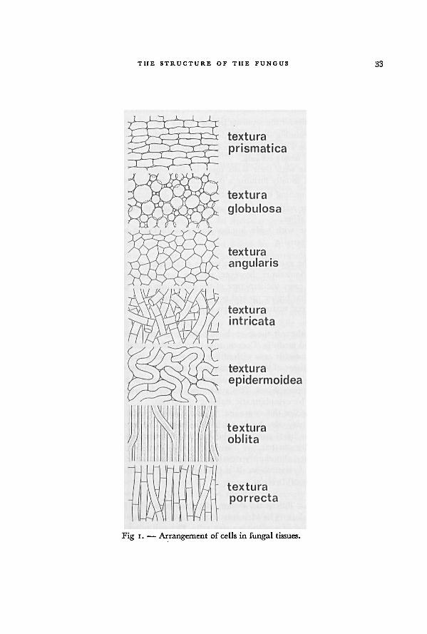
THE STRUCTURE OF THE FUNGUS 33
Fig 1. — Arrangement of cells in fungal tissues.
34 A WORLD-MONOGRAPH OF THE GENERA ASCOBOLUS AND SACCOBOLUS
2. Cells polyhedral by mutual pressure, no intercellular spaces: textura
angularis.
B. Cells more or less rectangular in section, not isodiametric: texturaprismatica.
II. Long-celled tissue: the separate hyphae easily distinguishable.
G. Hyphae running in all directions, not parallel.
3. Hyphae with their walls not cohering, usually with distinct interhyphal
spaces: textura intricata.
4- Hyphae with their walls more or less cohering, without interhyphal
spaces, usually forming a membranaceous tissue: textura epidermoidea.
D. Hyphae running in one direction, more or less parallel.
5. Hyphae with narrow lumina and strongly thickened walls, cohering:textura oblita.
6. Hyphae with wide lumina and non-thickened walls, not cohering:
textura porrecta.
The texture of the excipulum can often be established merely after a glance at
the outer surface. Sometimes, however, sections perpendicular to the surface are
necessary. In some cases the structure of the excipulum near the base of the fruit-
body differs from that higher up and near the margin.
Transitions between textura angularis and globularis occur rather frequently.
FLESH.—This is the soft tissue enclosed between the excipulum and the hypothe-
cium; it is also called medulla (Corner, 1929). In Ascoboloideae it consists of colour-
less, oblong or isodiametric cells with rather thin walls and large intercellularspaces.
Sometimes hyphal tissue of textura intricata is formed. Among the rather large cells
with large vacuoles, a system of hyphae rich in cytoplasm occurs, which is
connected with the paraphyses. This system can easily be traced in species with a
thick layer of flesh by cytoplasmatic staining of sections.
In young fruit-bodies the remains of the ascogonium and of some asco-
genous hyphae may also be found in this layer. The ascogonial remains are
usually found in the basal part near the axis, but accidentally they may be found
strongly excentrically situated.
The flesh is rather abundantly developed in Ascobolus sect. Sphaeridiobolus, sect.
Ascobolus, and sect. Gymnascobolus. It is thin or scarcely deleloped in Saccobolus and
in the other sections of Ascobolus.
HYPOTHECIUM.—This is the system of tightly entangled hyphae immediatelybeneath the hymenium. The elements of this layer give rise to the asci and the
paraphyses. It consists of small plasm-rich cells. These cells are isodiametric or only
slightly elongated in section.
In most species the hypothecium is a rather sharply delimited layer. Sometimes,
however, it is very thin or discontinuous.
35THE STRUCTURE OF THE FUNGUS
HYMENIUM.—The hymenium of the Ascobolaceae varies strongly in shape and
composition, depending on the number of ripening asci and the increase in volume
during this ripening. Hymenia with only a few asci, like they may occur in Ascobolus
immersus,
A. stictoideus, A. albidus, and A. furfuraceus, are restricted to cleistohymenial
ascomata that do not open before the telohymenial phase. The hymenia of most
species of Ascobolus and Saccobolus, however, contain numerous asci.
The asci strongly increase in volume, especially after meiosis. In hymenia with
many asci this results in a considerable enlargement. At first the surface of these
hymenia is concave, but soon it becomes flat or convex. Consequently in Saccobolus
and Ascobolus sect. Pseudascodesmis and sect. Pseudosaccobolus the shape of mature
fruit-bodies is lenticular or pulvinate.Characteristic of the Ascobolaceae is the strong protrusion of the tips of
r'pe asci above the level of paraphyses. In Ascobolus sect. Dasyobolus and in Sacco-
bolus sect. Eriobolus ripe asci may protrude for more than 50 % of their length. This
less in the other sections of Ascobolus and Saccobolus. In species of Saccobolus the
asci with still colourless spores are often already clearly protruding.The thickness of the hymenium is measured from the bases of the asci and the
Paraphyses to the tips of the paraphyses in ripe hymenia.In
many species of Ascobolus sect. Dasyobolus and sect. Ascobolus a mucilaginous
substance is found in the hymenium besides asci and paraphyses. This substance,which sometimes is very abundant, is of a yellowish or greenish-yellow colour.
However, in some species colourless mucus also occurs. The coloured mucus may
contain crystals of pigment in species of Ascobolus sect. Ascobolus. Occasionallythese crystals show a characteristic shape and size (e.g. in A. denudatus).
Under certain circumstances asci in hymenia may reach complete maturity
Without discharging their contents. The episporia of the ascospores in such asci are
always brownish and the colour of the hymenial mucus also changes into brownish.
Consequently these over-ripe hymenia are brownish or olive coloured.
PARAPHYSES.—These sterile hymenial elements develop before the asci and
sooner or later during the development of the fruit-body the tips become exposed.They are mostly filiform or cylindrical, simple or branched, always septate with a
varying number of nuclei per segment (Berthet, 1964). In Saccobolus the paraphysesare comparatively shorter and slightly broader than in Ascobolus.
The shape of the tips of the paraphyses has been considered a valuable
distinguishing character in some species. It proved to be of littleor no value.
Experiments showed that the shape of the tip is strongly influenced by the
developmental type of the fruit-body and by environmental conditions during the
development. The humidity of the air appeared to be ofgreat influence.
No enlargement of the tips ever occurs in species in which the hymenium does
n«t become exposed before the telohymenial phase, as is the case in species of
Ascobolus sect. Dasyobolus. Hymenia that become exposed in an earlier phase may
show paraphyses with more or less enlarged tips.
36 A WORLD-MONOGRAPH OF THE GENERA ASCOBOLUS AND SACCOBOH'S
In cultures of Ascobolus denuatus, A. furfuraceus, A. michaudii, A. crenulatus, and A.
roseopurpurascens thickened tips were formed in a part of the paraphyses under dry
conditions. When in a later phase hymenial mucus was formed or the humidity of
the air was raised until saturated, many of the clavate tips, formed during the dry
period, grew out with very slender, filiform protuberances (Figs. 26g, 27 i).
The most unusual types of paraphyses may be found in over-ripe or alkaline
hymenia, recognizable by the brownish colour of the episporia and the occurrence
of many swollen ascospores. Paraphyses with ellipsoid or subglobular cells are some-
times rather frequent.
Of Saccobolus thaxteri and certain developmental forms of S. versicolor, ascomata
were found that looked as if closed in the prohymenial and early mesohymenial
phase. Further investigation ofthese stages showed that the swollen tips of paraphyses
formed what was seemingly a continuation of the excipular layer. Both excipular
cells and paraphysal tips were covered with a thin layer of purplish or pinkish
amorphous pigment. Also in both species paraphyses without swollen tips were
formed afterwards.
The same phenomenon was observed in Ascophanus microsporus (B. & Br.) E. G.
Hansen and A. coemansii Boud.
Amorphous, purplish or brownish pigment is found between the tips of the
paraphyses in many species of Ascobolus and Saccobolus sect. Eriobolus. In Saccobolus
versicolor it is commonly found surrounding the swollen tips.
Paraphyses with coloured contents are restricted to Saccobolus sect. Saccobolus
and Ascobolus sect. Pseudosaccobolus. Here the contents of terminal and subterminal
segments are coloured yellowish or amber.
Asci.—The asci of the Ascobolaceae are characterized by an extraordinary
increase in volume of the asci, mainly during the last phase of ripening. The wall
of the ascus then is stretched enormously. At maturity the asci strongly protrude
beyond the surface of the hymenium.
Broadly clavate to saccate asci are found in Ascobolus sect. Dasyobolus and sect.
Sphaeridiobolus. More cylindrical-clavate asci occur in Ascobolus sect. Ascobolus, sect.
Heimerlia, and sect. Gymnascobolus, while they are relatively short and broadly clavate
in Saccobolus and Ascobolus sect. Pseudascodesmis and Pseudosaccobolus.
The ripe asci become more or less curved as a response to positive photo-
tropism of the upper part of the ascus (Zopf, 1880: 33; Zopf, 1890: 205; Buller,
1934: 264). In a few species ofAscobolus the asci have already begun to curve towards
the light during an early phase of the process of stretching. Sometimes as much as
the upper third part or even more of each ripe ascus becomes curved.
The blue staining of the wall of the ascus with iodine was already
observed by Coemans (1862) in Ascobolus immersus and Saccobolus glaber and by de
Bary (1866: 108) in A. furfuraceus. The outer layer of the wall stains blue especially
in the young asci. The staining is less intensive or may even be absent in the ripe
asci. The measure of intensity of staining is constant for each species although great
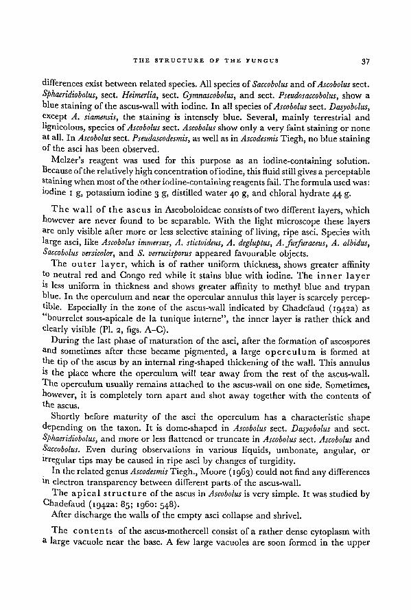
THE STRUCTURE OF THE FUNGUS 37
differences exist between related species. All species of Saccobolus and of Ascobolus sect.
Sphaeridiobolus, sect. Heimerlia, sect. Gymnascobolus, and sect. Pseudosaccobolus, show a
blue staining of the ascus-wall with iodine. In all species of Ascobolus sect. Dasyobolus,except A. siamensis, the staining is intensely blue. Several, mainly terrestrial and
lignicolous, species of Ascobolus sect. Ascobolus show only a very faint staining or none
at all. In Ascobolus sect. Pseudascodesmis, as well as in Ascodesmis Tiegh, no blue stainingof the asci has been observed.
Melzer's reagent was used for this purpose as an iodine-containing solution.
Because oftherelatively high concentrationofiodine, this fluidstill gives a percep table
staining when most of the other iodine-containing reagents fail. The formulaused was:iodine i g, potassium iodine 3 g, distilled water 40 g, and chloral hydrate 44 g.
The wall of the ascus in Ascoboloideae consists of two different layers, which
however are never found to be separable. With the light microscope these layersare only visible after more or less selective staining of living, ripe asci. Species with
large asci, like Ascobolus immersus, A. stictoideus, A. degluptus, A. furfuraceus, A. albidus,Saccobolus versicolor, and S. verrucisporus appeared favourable objects.
The outer layer, which is of rather uniform thickness, shows greater affinityto neutral red and Congo red while it stains blue with iodine. The inner layeris less uniform in thickness and shows greater affinity to methyl blue and trypanBlue. In the operculum and near the opercular annulus this layer is scarcely percep-tible. Especially in the zone of the ascus-wall indicated by Chadefaud (1942a) as
"bourrelet sous-apicale de la tunique interne", the inner layer is rather thick and
clearly visible (PI. 2, figs. A-C).
During the last phase of maturation of the asci, after the formation of ascospores
an d sometimes after these became pigmented, a large operculum is formed at
the tip of the ascus by an internal ring-shaped thickening of the wall. This annulus
ls the place where the operculum will tear away from the rest of the ascus-wall.
The operculum usually remains attached to the ascus-wall on one side. Sometimes,
however, it is completely torn apart and shot away together with the contents of
the ascus.
Shortly before maturity of the asci the operculum has a characteristic shape
depending on the taxon. It is dome-shaped in Ascobolus sect. Dasyobolus and sect.
Sphaeridiobolus, and more or less flattenedor truncate in Ascobolus sect. Ascobolus and
Saccobolus. Even during observations in various liquids, umbonate, angular, or
irregular tips may be caused in ripe asci by changes of turgidity.In therelated genusAscodesmis Tiegh., Moore (1963) could not find any differences
m electron transparency between different parts of the ascus-wall.
The apical structure of the ascus in Ascobolus is very simple. It was studied by
Chadefaud (1942a: 85; i960: 548).After discharge the walls of the empty asci collapse and shrivel.
The contents of the ascus-mothercell consist of a rather dense cytoplasm with
a large vacuole near the base. A few large vacuoles are soon formed in the upper
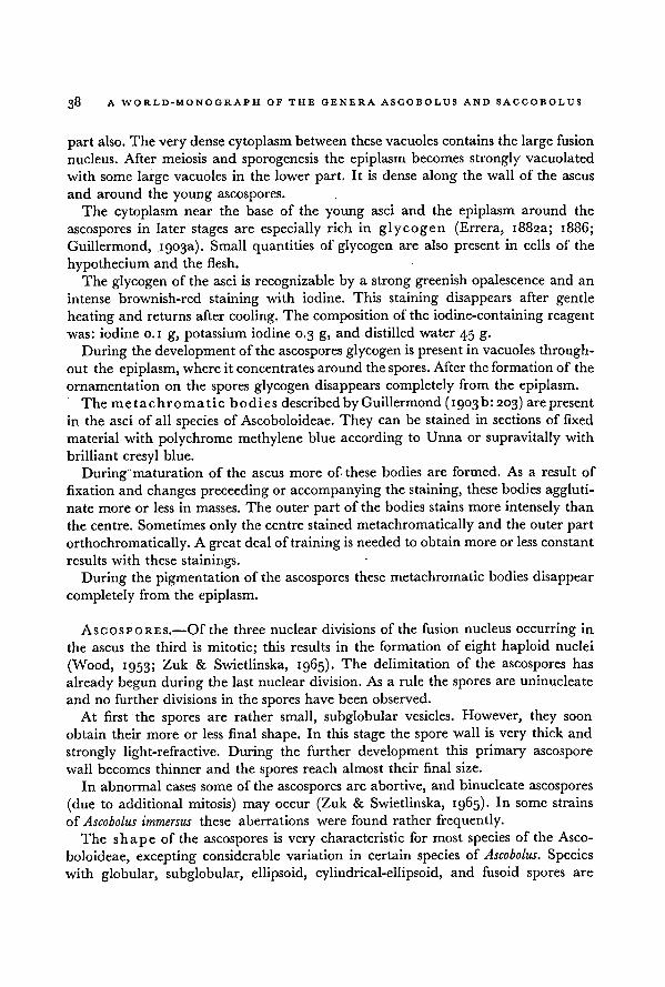
part also. The very dense cytoplasm between these vacuoles contains the large fusion
nucleus. After meiosis and sporogenesis the epiplasm becomes strongly vacuolated
with some large vacuoles in the lower part. It is dense along the wall of the ascus
and around the young ascospores.
The cytoplasm near the base of the young asci and the epiplasm around the
ascospores in later stages are especially rich in glycogen (Errera, 1882a; 1886;
Guillermond, 1903a). Small quantities of glycogen are also present in cells of the
hypothecium and the flesh.
The glycogen of the asci is recognizable by a strong greenish opalescence and an
intense brownish-red staining with iodine. This staining disappears after gentle
heating and returns after cooling. The composition of the iodine-containing reagent
was: iodine o. 1 g, potassium iodine0.3 g, and distilled water 45 g.
During the development of the ascospores glycogen is present in vacuoles through-
out the epiplasm, where it concentrates around the spores. After the formationof the
ornamentation on the spores glycogen disappears completely from the epiplasm.
The metachromatic bodies describedby Guillermond(1903b: 203) are present
in the asci of all species of Ascoboloideae. They can be stained in sections of fixed
material with polychrome methylene blue according to Unna or supravitally with
brilliant cresyl blue.
During maturation of the ascus more of these bodies are formed. As a result of
fixation and changes preceeding or accompanying the staining, these bodies aggluti-
nate more or less in masses. The outer part of the bodies stains more intensely than
the centre. Sometimes only the centre stained metachromatically and the outer part
orthochromatically. A great deal of training is needed to obtain more or less constant
results with these stainings.
During the pigmentation of the ascospores these metachromatic bodies disappear
completely from the epiplasm.
Ascospores.—Of the three nuclear divisions of the fusion nucleus occurring in
the ascus the third is mitotic; this results in the formation of eight haploid nuclei
(Wood, 1953; Zuk & Swietlinska, 1965). The delimitation of the ascospores has
already begun during the last nuclear division. As a rule the spores are uninucleate
and no further divisions in the spores have been observed.
At first the spores are rather small, subglobular vesicles. However, they soon
obtain their more or less final shape. In this stage the spore wall is very thick and
strongly light-refractive. During the further development this primary ascospore
wall becomes thinner and the spores reach almost their final size.
In abnormal cases some of the ascospores are abortive, and binucleate ascospores
(due to additional mitosis) may occur (Zuk & Swietlinska, 1965). In some strains
of Ascobolus immersus these aberrations were found rather frequently.
The shape of the ascospores is very characteristic for most species of the Asco-
boloideae, excepting considerable variation in certain species of Ascobolus. Species
with globular, subglobular, ellipsoid, cylindrical-ellipsoid, and fusoid spores are
38 A WORLD-MONOGRAPH OF THE GENERA ASCOBOLUS AND SACCOBOLUS
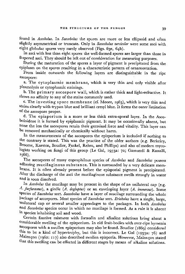
found in Ascobolus. In Saccobolus the spores are more or less ellipsoid and often
slightly asymmetrical or truncate. Only in Saccobolus versicolor were some asci with
eight globular spores very rarely observed (Figs. 63e, 64b).In asci with less than eight spores the well-formed spores are larger than those in
8-spored asci. They should be left out of considerationfor measuring purposes.
During the maturation of the spores a layer of pigment is precipitated from the
cpiplasm on the spores resulting in a characteristic pattern of ornamentation.
From inside outwards the following layers are distinguishable in the ripe
ascosporc:a. The cytoplasmic membrane, which is very thin and only visible after
plasmolysis or cytoplasmic stainings.b. The primary ascospore wall, which is rather thick and light-refractive. It
shows no affinity to any of the stains commonly used.
c. The investing spore membrane (cf. Moore, 1963), which isvery thin and
stains clearly with trypan blue and brilliant cresyl blue. It forms the outer limitation°f the ascospore proper.
d. The episporium is a more or less thick extra-sporal layer. In the Asco-
boloideae it is formed by epiplasmic pigment. It may be occasionally absent, but
n°ne the less the ascospores retain their germinal force and vitality. This layer can
he removed mechanically or chemically without harm.
In the measurements of the ascospores the episporium is included if nothing to
thecontrary is stated. This was the practice of the older authors (e.g. Berkeley,
Ih'oome, Karsten, Boudier, Fuckel, Rehm, and Phillips) and also of modern myco-
logists working on fungi of this group (Le Gal, 1953a: 72; Gamundf & Ranalli,1966).
The ascospores of many coprophilous species of Ascobolus and Saccobolus possess
adhering mucilaginous substance. This is surrounded by a very delicate mem-
brane. It is often already present before the episporial pigment is precipitated.After the discharge of the asci the mucilaginous substance swells strongly in water
and is soon dissolved.
In Ascobolus the mucilage may be present in the shape of an unilateral cap (e.g.A. furfuraceus), a girdle (A. degluptus) or an enveloping layer (A. immersus). Some
sPecies of Saccobolus sect. Saccobolus have a layer of mucilage surrounding the whole
Package of ascospores. Most species of Saccobolus sect. Eriobolus have a single, large,Unilateral cap or several smaller appendages to the packages. In both Ascobolusand Saccobolus species occur in which no mucilage is formed. As a rule it is absent
ln species inhabiting soil and wood.
Certain fixative mixtures with formalin and alkaline solutions bring about a
•Considerable swelling of the episporium. In old fruit-bodies with over-ripe hymeniaascospores with a swollen episporium may also be found. Boudier (1869) considered
'his to be a kind of hypertrophy, but this is incorrect. Le Gal (1953a: 76) and
hlalen5on (1962: 117) also described swollen episporia. However, Malen$on stated
that this swelling can be effected in differentstages by means of alkaline solutions.
THE STRUCTURE OF THE FUNGUS 39
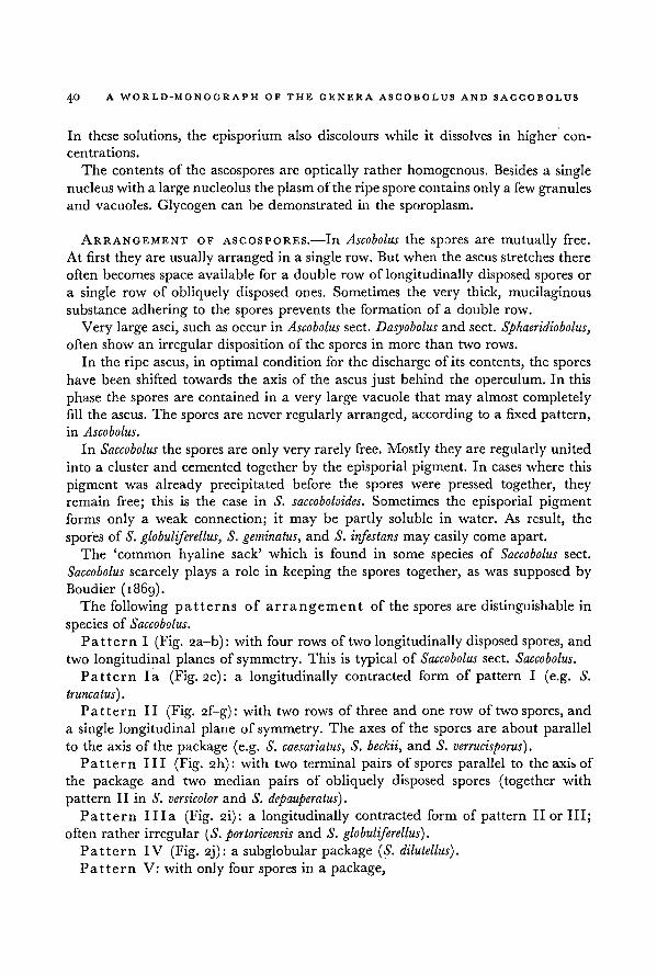
A WORLD-MONOGRAPH OF THE GENERA ASCOBOLUS AND SACCOBOLUS40
In these solutions, the episporium also discolours while it dissolves in higher con-
centrations.
The contents of the ascospores are optically rather homogenous. Besides a single
nucleus with a large nucleolus the plasm of the ripe spore contains only a few granulesand vacuoles. Glycogen can be demonstrated in the sporoplasm.
ARRANGEMENT OF ASCOSPORES.—In Ascobolus the spores are mutually free.
At first they are usually arranged in a single row. But when the ascus stretches there
often becomes space available for a double row of longitudinally disposed spores or
a single row of obliquely disposed ones. Sometimes the very thick, mucilaginous
substance adhering to the spores prevents the formation of a double row.
Very large asci, such as occur in Ascobolus sect. Dasyobolus and sect. Sphaeridiobolus,
often show an irregular disposition of the spores in more than two rows.
In the ripe ascus, in optimal condition for the discharge of its contents, the spores
have been shifted towards the axis of the ascus just behind the operculum. In this
phase the spores are contained in a very large vacuole that may almost completely
fill the ascus. The spores are never regularly arranged, according to a fixed pattern,
in Ascobolus.
In Saccobolus the spores are only very rarely free. Mostly they are regularly united
into a cluster and cemented together by the episporial pigment. In cases where this
pigment was already precipitated before the spores were pressed together, theyremain free; this is the case in S. saccoboloides. Sometimes the episporial pigment
forms only a weak connection; it may be partly soluble in water. As result, the
sporesof S. globuliferellus, S. geminatus, and S. infestans may easily come apart.
The 'common hyaline sack' which is found in some species of Saccobolus sect.
Saccobolus scarcely plays a role in keeping the spores together, as was supposed by
Boudier (1869).
The following patterns of arrangement of the spores are distinguishable in
species of Saccobolus.
Pattern I (Fig. 2a-b): with four rows of two longitudinally disposed spores, and
two longitudinal planes of symmetry. This is typical of Saccobolus sect. Saccobolus.
Pattern la (Fig. 2c): a longitudinally contracted form of pattern I (e.g. S.
truncatus).
Pattern II (Fig. 2f-g): with two rows of three and one row of two spores, and
a single longitudinal plane of symmetry. The axes of the spores are about parallelto the axis of the package (e.g. S. caesariatus, S. beckii, and S. verrucisporus).
Pattern III (Fig. 2h): with two terminal pairs of spores parallel to the axis of
the package and two median pairs of obliquely disposed spores (together with
pattern II in S. versicolor and S. depauperatus).
Pattern Ilia (Fig. 2i): a longitudinally contracted form of pattern II or III;
often rather irregular ( S. portoricensis and S. globuliferellus).
Pattern IV (Fig. 2j): a subglobular package (S. dilutellus).
Pattern V: with only four spores in a package,
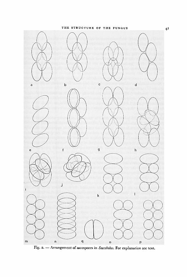
THE STRUCTURE OF THE FUNGUS41
For explanation see text.Saccobolus.Fig. 2. — Arrangement of ascospores in
A WORLD-MONOGRAPH OF THE GENERA ASCOBOLUS AND SACCOBOLUS42
Va: (Fig. 2d): with two rows of two spores with their axes parallel to the
axis of the package ( S. quadrisporus).
Vb : (Fig. 2el: with a single row ofobliquely disposed spores ( S. quadrisporus).Pattern VI (Fig. 2k-p): with the axes of the spores at right angles to the axis
of the package. Of this type only a few of the possibilities are drawn (S. infestans and
S. geminatus). In S. geminatus the spores are firmly united into pairs (Fig. 2q) which
themselves are rather loosely united into a cylindrical pile (Fig. 2I).
PIGMENTATION OF ASCOSPORES.—AS far as known, the episporial pigment of
the Ascoboloideae is unique among the Ascomycetes. It forms an easily observable
character of the fungi of this subfamily.
Already Boudier (1869: 203I and Janczewsky (1872: 202) studied the origin of
this pigment. Of importance in this connection are especially the recent studies of
Ghadefaud (1942b), Le Gal (1942, 1947, 1963a) and Malengon (1962).At the moment there are two strongly competing theories on the origin of the
episporial pigment.Le Gal studied this problem in detail in three species, amongwhich were Ascobolus
furfuraceus and A. carbonarius. According to her at first a greenish substance is deposited
on the spores. This substance is more or less crystalloid in appearance and is sub-
sequently coloured superficially by a violet pigment. Both the greenish substance
and the violetpigment are ofvacuolar origin and both are precipitated on the spores.
After contact with the spore-wall the solid violet pigment becomes diffluent, running
over the greenish substance. After dehydration, the episporial pigment becomes
solid finally.
According to Ghadefaud and Malengon the young asci are free of any pigment
and young ascospores are colourless and smooth. Occasionally they found a colour-
less layer around the spores during a very short period. The surface of the spores
becomes covered by a violet, sculptured layer. They assume that only a single
pigment is present, which is at first in the leuco-form, but after oxidation passes
into the violet form. This change in colour can be brought about with several natural
or artificial oxidators. Malengon could reobtain the leuco-form with a reductor.
In my opinion both theories are less controversial than they look at first sight. The
most important discrepancies can be fully explained by the different methods of
observation used.
Le Gal studied mostly living material with supravital stainings in water using
day-light in microscopy (Le Gal, 1957: 244, 253).
Malengon (personal communication), and probably also Ghadefaud used artificial
light in microscopy and especially studied the influence of some chemicals on the
episporial substance.
The greenish substance, which Le Gal described as "une matiere liquide refringen-
te verdatre, se presentant sous forme de globules plus ou moins gros", is undoubtedlyidentical with glycogen. In strongly diffused light, as is the case in microscopy with
day-light, the green opalescence is very conspicuous. The crystalloid appearance is
THE STRUCTURE OF THE FUNGUS
43
accounted for by the structure ofthe "pseudo-solutions" of the glycogen (Clautriau,
!^95: 234). These colloidal suspensions strongly refract and polarize light and
consequently they look like small, solid bodies.
This substance stains brownish-red with iodine, which fully agrees with the obser-
vations of Errera (i882a-b, 1886) and Guillermond (1903a). As a result of the
influence of the reagent, however, the fine structure is strongly disturbed, and the
vacuoles containing glycogen unite to form large masses. The opalescence ofglycogen
cannot be observed in beams of tungsten light directed along the optical axis of
the microscope, as produced by the usual systems of illumination.
The violet particles which Le Gal found surrounding the globules of the first
substance appear after only a short time (Le Gal, 1947: 244, 249). They are not
always formed during the short period, in which reliable vital observations are
possible. The particles were visible after a short time, both after supravital stainingswith neutral red or brilliant cresyl blue and directly with phase contrast micro-
scopy. Their colour was only rarely violet, mostly they were seen as colourless
bodies.
A change from colourless to violet can be observed after spontaneously or
artificially produced oxidation, as described by Chadefaud (1942b) and Malengon
(1962). Such oxidation also causes some coagulation of the pigment. Especially with
an artificial oxidator, like 1 % chromium trioxide in water, big masses of violet
pigment coagulate. The colourless or opalescent layer on the ripening spores also
becomes violet after oxidation. In the large vacuoles and the mucilaginous substance
surrounding the spores the violet pigment is present in a dissolved state. With a
reductor (e.g. 5 % sodium dithionite in water) the violet colour disappears imme-
diately.
In some species of Ascobolus (e.g. A. furfuraceus, A. stictoideus, and A. immersus)
globules of glycogen in the epiplasm of ripening asci stain brownish-red in a solution
containing strongly diluted iodine, while the particles surrounding these globulesassume their violet colour. In other cases the violetpigment was found to be present111 the outer layer of the globules.
By supravital staining with brilliant cresyl blue the glycogen stains meta-
chromically red, while other small granules and the outer layer of the glycogen
globules stained purplish-blue. This agrees with the results of Guillermond's (1903b:
203) metachromatic staining ofsections of fixed material with polychrome methyleneblue according to Unna, if allowances are made for the methods of fixation.
With the precipitation of the episporial pigment both glycogen and Guillermond's
metachromatic bodies completely disappear from the epiplasm. Possibly the bodies
shown by Guillermond correspond with the violet pigment.
Usually the episporial substance is at first colourless then it becomes pinkish and
gradually this changes into dark violet. Different shades of this colour occur in
different species. It may be almost blue ( S. globuliferellus), greyish-violet (form of
S. versicolor), or opaque blackish-violet, in species with very thick episporia.The violet spores are fully mature and ready to be discharged. Often however,
A WORLD-MONOGRAPH OF THE GENERA ASCOBOLUS AND SACCOBOLUS
44
the violet colour of the episporium changes into purplish-brown in the ascus. As
noticed by Boudier (1869) and Malengon (1962) this final change of colour can
also be brought about by heating the spores. This discolourationis accompanied bya chemical change of the pigment.
The violetform immediately dissolves and discolours in diluted alkali (e.g. 0.5 %
sodium hydroxide), leaving behind a colourless or greenish opalescent layer, which
stains red with iodine. This layer also dissolves gradually in alkali.
The brown form is resistant against dilutedalkali and reductors.
Conclusion.—From vital observations and the progress of chemical reactions
in ten species of Ascobolus and six species of Saccobolus it may be concluded that in
the pigmentation of the ascospores of Ascoboloideae at least two components are
involved. One of these is glycogen, which precipitates in great quantities on the
spores.
At the same time the other component precipitates on the spores in the form of
very small particles or in a soluble state. On the surface of the glycogen this compo-
nent becomes diffluent or dissolves. Its colour and aspect depend on the redox-
potential and the acidity. Usually the change of colour into violet occurs imme-
diately after the precipitation. By further and slower oxidation the episporium
becomes purplish-brown.
The course of the pigment-formation is subject to many variable environmental
factors.
No evidence could be found for Malen$on's (1962: 114) hypothesis that in a
single species in addition to 'chromogenous' races also 'leucogenous' races might
occur.
A detailed chemical analysis of the process ofpigmentation is urgently needed.
ORNAMENTATION OF ASCOSPORES.—The episporial pigment is precipitated
on the spores as very small granules. The form in which it precipitates is theprimary
pattern of ornamentation. As a rule it is determined genetically and is rather
constant for each species.Thoseparts of the episporium that during pigmentation are covered with mucilag-
inous substance or are in contact with other spores may show a thinner layer of
pigment or a disturbed pattern of ornamentation.
The following primary patterns of ornamentation may be distinguished in the
Ascoboloideae: (i) completely smooth ( Ascobolus castaneus); (ii) smooth with only a
single or a few crevices ( Ascobolus scatigenus, A. immersus); (iii) with more or less
longitudinal, anastomosing crevices, sometimes accompanied by small pits or
irregular short crevices (e.g. A. furfuraceus, A. crenulatus); (iv) with a net-work of
crevices (A. behnitziensis, A. subglobulosus, Saccobolus portoricensis, S. quadrisporus, and
S. beckii); (v) with coarse warts (Ascobolus carbonarius, A. archeri, A. stictoideus, Saccobolus
obscurus, and S. verrucisporus); (vi) with small warts or spines ( Ascobolus xylophilus,
A. hawaiiensis, A. brassicae, A. aglaosporus, Saccobolus citrinus, S. globuliferellus, and S.
THE STRUCTURE OF THE FUNGUS 45
geminatus); (vii) with very fine granules (e.g. Ascobolus amoenus, A. elegans, and A.
cainii); (viii) with a net-work of pigment ( Ascobolus reticulatus).The primary pattern ofornamentationmay become more complicated afterwards
by sec ondary crevices, as a result of stretching, growth, orswelling (cf. Figs. 140,
23b, 24c, 1, 34e-g, 5ie, f). Especially in over-ripe hymenia secondary crevices may
bevery frequent in the swollen episporia. Episporia may separate from the primary
ascospore wall and show irregular swellings as a result of the over-ripe hymenium
becoming alkaline (Malen5on, 1962).The thickness of the episporial pigment is rather variable. It varies from about
0.5 fx in Ascobolus scatigenus and A. castaneus to more than 2 (i in A. carbonarius, A.
demangei, and Saccobolus beckii. In Ascobolus carbonarius the pigment is considerably
thicker at the poles than at the sides, while in A. viridis and a form of A. denudatus
irregular thickenings may occur everywhere in the layer. In Saccobolus beckii the
pigment is very thick and irregular and often the individual spores are not easily
observed.
In asci with less than eight spores the pigment is thicker than in 8-spored asci.
Especially in 1- or 2-spored asci the pigment may be very thick and the pattern of
ornamentationabnormal.
ANISOSPORY. —In almost all species of Ascobolus cases of anisospory may be
observed. Boudier figured some of these cases as early as 1869. So far anisospory
within one and the same ascus has not been found in Saccobolus.
The types of anisospory most frequently met in Ascobolus are as follows,
(a) Atrophy or abortion of some of the spores within an ascus. The degeneratedremains are found in the ripe ascus. These are not very conspicuous because of
their small size and lack of pigment (cf. Boudier, 1869: pi. 6 ƒ VI; pi. 7f XII 4).In A. immersus this type of anisospory is sometimes rather frequent (cf. Rizet, 194 1).
(b) The episporium is absent in some of the spores. The spores remain colourless
and smooth. Most colourless spores in ripe asci are of this type (cf. PI. 4, fig. F).
(c) The episporium is present but it remains colourless, while other spores in the
same ascus are violet (cf. Boudier, 1869: pi. 6f. VI18). This type is very rare. After
more powerful artificial oxidation these spores sometimes turn pinkish. With a
diluted iodine-containing solution the colourless episporia stain brownish-red.
Cases of anisospory in which the pigmentation of some of the spores of an ascus
is retarded with respect to the other part, probably also belong to this type.
(d) The pigment is precipitated on some spores in the form of small or large,
irregular lumps (cf. Figs. i2g, 2oe, 24m, 53q; PI. 6, fig. c, PI. 8, fig. E). The characteris-
tic pattern of ornamentation is absent.
(e) Within the same fruit-body or even within a single ascus with normally
pigmented ascospores, different patterns of ornamentation occur (cf. Boudier, 1869:
pi. 7 f. XIII; Figs. 28b-i, 15O. Especially in A. albidus and A. denudatus this type of
anisospory is rather frequent.
46 A WORLD-MONOGRAPH OF THE GENERA ASCOBOLUS AND SACCOBOLUS
The frequency of anisospory is usually relatively low. When anisospory occurs,
segregations of two or more types of spores are often encountered in the contents of
a single ascus. The various ripe asci in a fruit-body were never found to be constant
with respect to their contents in such cases.
In a species ofAscobolus Bistis (1956a) ascertained thatboth ascospore-abortion and
tan spore colour are inherited by a single pair of Mendelianallelomorphs. Also in
A. immersus several cases of anisospory were found to depend on a single geneticfactor (Rizet, 194. 1; Rizet & al., ig6oa-b; Lissouba & al. 1962; Makarewicz, 1964).
Owing to the complexity of the pigmentation, the great number of different types
of abnormal ascospores in Ascobolus is scarcely astonishing (Lissouba & al., 1962).More detailed study of the ascospore-mutants may help solving the problems of the
pigmentation.
Because of the mostly low frequency of anisospory, ascospore characters can be
used in Ascobolus to distinguish between species. In some cases, however, certain
strains or collections show an accumulation of abnormal types. Ascobolus cubensis is
probably based on such material.
47
CHAPTER V
CYTOLOGY
Several species of Ascobolus have been used in karyologic studies. Since the fusion-
nucleus in the ascus is relatively large for this group of fungi, they are very suitable
for studying meiosis.
The existence of a double nuclear fusion in Ascobolus and some allied genera has
been a matter of debate for many decades.
According to Harper (1895), Welsford (1907), Fraser (1909), Fraser & Brooks
(1909), and Gwynne Vaughan & Williamson (1932) the first nuclear fusion occurs
>n the ascogonium of species of Ascobolus studied by them. 2 This is followed by the
generally accepted karyogamy in the ascus.
The first nuclear division in the ascus is meiotic, but according to the authors
cited also the second or the third division would be meiotic (brachymeiosis).Other investigators, like Dangeard (1907), Ramlow (1915), Moreau (1930I, and
Schweizer (1923, 193 1, 1932I, who studied the same and alsoother species 2
,strongly
deny the existence of a double nuclear fusion and of brachymeiosis. Some of them
observed pairing of nuclei (karyonymphy) in the ascogonium of these fungi, but
never karyogamy.
Cytological observations of these stages are difficult because of the very small
nuclei and chromosomes. The results may be influenced by fixation (Ramlow, 1915).Recent cytological investigations by Olive (1949b, 1950), Hirsch (1950), Wood
( [ 953a-b), Bistis (1956b!, and Zuk & Swietlinska (1965) are all strongly in favour
°f Dangeard's point of view. Also from a genetical point of view there are no
•ndications of the existence of brachymeiosis in these fungi. In A. scatigenus (Wood,be.) and A. furfuraceus (Zuk & Swietlinska, I.e.) meiosis was studied in detail.
The number of chromosomes (n) in several species of Ascobolus was found to
be eight, e.g. in A. immersus (8-9 according to Zuk & Swietlinska, 1965), A.
scatigenus (Wood, 1953a), A. furfuraceus (Harper, 1895; Fraser & Brooks, 1909).A review of literature concerning the nucleus in fungi (e.g. in Pezizales) was givenby Olive (1953: 513).
The karyology of the mycelium and the vegetative parts of the fruit-body is
scarcely known. Some work has been done in this respect by Zuk & Swietlinska
(1965) on A. immersus, by Dangeard (1907) on A. furfuraceus and Saccobolus versicolor,aud by Berthet (1964) 011 Ascobolus carbonarius and A. furfuraceus.
2 Asa result of taxonomic confusion in the genus Ascobolus, it is mostly impossible to
ascertain on which species the preceding authors made their observations. They did not
Sive adequate descriptions and figures of their material.Only very rarely material has been
Preserved to enable later investigators to verify the identifications.
48
CHAPTER VI
SEXUALITY AND COMPATIBILITY
Species of Ascobolus are favourable objects for the study of developmental cycli
(ontogeny) and problems of sexuality and compatibility. Several species have been
studied. As these problems are rather complicated and beyond the scope of this
taxonomic monograph, they are merely fleetingly mentioned here.
Ascobolus scatigenus (Dodge, 1920; 1936!, A. carbonarius (Betts, 1926), A. furfuraceus
(Dowding, 1931; Bistis, 1956a!, A. immersus (Rizet, 1939I, and A. geophilus (Betts &
Meyer, 1939) are heterothallic. Saccobolus saccoboloides (Dodge & Seaver, 194.6) and
some other species 3 of Ascobolus and Saccobolus (Dodge, 1912a; Schweizer, 1931;
Gwynne Vaughan & Williamson, 1933) are homothallic.
For the earlier work in this field, concerning species of Ascobolus or Saccobolus, the
following publications are of importance: Dodge (1912a), Lohwag (1927), Kniep
(1928), Gaumann & Dodge (1928), Moreau (1930), Gwynne Vaughan & William-
son (1932), Drayton (1932), Dodge (1936), Dodge & Seaver (1946), Whitehouse
(i949). Langeron (1952), Olive (1954b).
For modern research in this field the work of Bistis (1956a, 1956b, 1957).Bistis & Raper (1963), Esser & Kuenen (1965), and Esser (1967) are particularly
important.The earliest ontogeny of only a very few species is rather well known. In Ascobolus
carbonarius Dodge (1912a) described that a long trichogyne grows into the direction
of an antheridial conidiumfrom a different mycelium and coils around it.
In Ascobolus scatigenus (Gwynne Vaughan & Williamson, 1932) ascospores of types
A and B are formed, fromwhich mycelia develop of types A and B. The only difference
between the two types of mycelia is in their behaviour with respect to the other
itrain. Male branches are formed on the younger hyphae and female branches on
the older hyphae of both strains, A and B. Consequently, there is no difference of
sex between both strains. They are monoecious but not homothallic. A-antheridia
only unite with B-oogonia, and B-antheridia only with A-oogonia. After plasmogamythe male nuclei move through the many-celled trichogyne towards the ascogonium.This is called physiological heterothallism.
Among Ascobolaceae, sexual behaviour has been most extensively studied in
Ascobolus furfuraceus. 4 This is a heterothallic species in which the mating type is
established by a single locus of probably two alleles. The individual strains are
3 See foot-note 2 on p. 47.4 Identified as Ascobolus stercorarius (Bull, per St-Amans) Schroet. by the authors concerned.
SEXUALITY AND COMPATIBILITY 49
hermaphrodite, but self-incompatible and reciprocal cross-compatible (Bistis, 1956a).In the sexual process of this species a donor or fertilizing agent and a receptor
element are involved. Bistis (1956b, 1957) proved that non-germinated oidia can
function as fertilizing elements after undergoing a physiological change, which is
called "sexual activation".
Experiment by Bistis & Raper (1963) with growing hyphal fragments demonstrated
that "sexual activation" also involves a minor morphological change, viz. a dilation
of the apical regions of the hypha. As in the case of activated not germinated oidia,the oogonia develop only in the direct surroundings of the dilated apices.
The activated, hyphal tips are both morphologically and functionally antheridia
and should be so considered. In a sexual reaction the non-germinated oidia function
as antheridia. However, they lack the morphological differentiation and are not
attached to the hyphal system from which they originate. "Sexual activation" is
the same as "antheridial differentiation". In reality the sexual mechanism in this
species is a gametangial-gametangial system.
Olive (1954b) pointed out, that many investigations indicated that the sexual
Process in the fungi is much more labile and variable than was presumed by early
'nvestigators. Heterokaryosis, heterozygosity, and segregation may occur freely in
homothallic as well as in heterothallic fungi.
50
CHAPTER VII
GENETICS
When B. O. Dodge (1936: 40), the later geneticist, started monographing the
Ascobolaceae, he was so fascinated by the reproductive and genetic problems of
this group, that he was soon deeply involved in these problems and abandoned his
taxonomic interests for them.
Some species of Ascobolus have proven to be favourable objects for genetic in-
vestigations. Rizet (1939, 1941) showed the suitability of A. immersus for this type of
work. The ontogeny and cytology of this species are rather well known (Ramlow,
1915; Zuk & Swietlinska, 1965). At present it is an important genetic object. For
a review of the modern genetic literatureconcerning this species the following authors
should be consulted: Rizet & al. (ig6oa-c), Lissouba & al. (1962), Gajewski & al.
(1963), Rizet & Rossignol (1963), Rossignol (1964), Yu-Sun (1964), Paszewski &
Surzyski (1964), Makarewicz (1964, 1966), and Paszewski & al. (1966).
Among ihe species of Ascobolus studied, Dodge & Seaver (1946) considered A.
magnificus and A. furfuraceus to be the most favourable objects for genetic work. Of
both species the life cycles and the cytology are well known (Dangeard, 1907; Dodge,
1920; Dowding, 1931; Gwynne Vaughan & Williamson, 1932; Schweizer, 1932;
Wood, i953a-b; Bistis, ig56a-b, 1957; Bistis & Raper, 1963), while the technique
of inducing the germination of the ascospores has also been considerably improved
(p. 15). Of the two species only A. furfuraceus has since been used in some genetic
investigations concerning sexuality and compatibility by Bistis & Olive (1954) and
Bistis (1956a).Ascobolus albidus might also be a suitable object in my opinion for genetic studies
in relation to the pigmentation and ornamentationof the ascospores.
51
CHAPTER VIII
ECOLOGY
Most species of the Ascoboloideae are coprophilous. The ascospores of the co-
prophilous species pass through the intestinal canal of animals without harm, as
vasproven by Massee & Salmon (1902: 57) who opened the intestines of rabbits
and sheep under aseptic conditions and isolated the contents in a sterile bell-jar.
They published a list of the species developing from these contents.
On the other hand it has been found that ascospores often do not germinate
Without passing through the intestinal tract of an animal or without having been
exposcd to conditions found in the intestines. Treatment with diluted pepsin or
alkaly combined with a raised temperature (38-40 °C) has proved to be veryelfective (p. 00).
In Ascobolus carbonarius, a pyrophilous species, the germination of the ascosporesWas greatly improved after a treatment at temperatures between 65 and 80 °C
(Dodge, 1912a; Betts, 1926).All normal ascospores of Ascobolus and Saccobolus are covered with a layer of dark
P'gment. In many other genera with predominantly coprophilous species, a dark
layer in the walls of spores or sporangia occurs too.
Durrell (1964) studies the composition and structure of walls of dark spores in
sPecies found in desert soil. Under certain conditions colourless spores are formed.
These spores are much more easily killed by ultra-violet radiation. It is possibletnat the layer of pigment surrounding the ascospores in Ascoboloideae protects the
c°ntents against ultra-violet radiationwhen the spores are exposed.The shooting away of the spores is another characteristic of many coprophilous
'nngi. Coemans (1862) already noticed a very regular daily periodicity in the dis-
charge of the asci of Ascobolus.
During the last phase of the maturation of the ascus light is necessary. At first the
Saturation of the asci is quite different from the final phase: the most advanced
asci are more or less synchronous in development by the periodicity of the light,lender
very constant conditions the ripe asci are accumulated in a hymenium until
a stimulus causes their simultaneous discharge. Under certain other conditions thethc asci
may be discharged one after another. Especially in species with large
simultaneous discharge is observed frequently. In species with small
yntenia, like those of Ascobolus sect. Dasyobolus and of Saccobolus, the discharge is
0rdy rarely simultaneous.
During the final and enormous stretching of the asci their tips curve towards the
Maximal light intensity: they are positively phototropic (Zopl, 1880; Falck, 1923).
A WORLD-MONOGRAPH OF THE GENERA ASCOBOLUS AND SACCOBOLUS52
The violent discharge of the contents of the ascus has attracted the attention of
several investigators. The ballistics of the ascospores were studied by Buller (1909:
25 :> t933 : 359); Ingold (1961), and Walkey & Harvey (1966).
The eight spores in each ascus of Ascobolus immersus are relatively very large. They
are somewhat glued together by the thick mucilaginous layer covering them.
Together with the contents of vacuoles and some epiplasm they form a large pro-
jectile. This can be shot away over a horizontal distance of 30 cm and vertically
over 35 cm,which is about 500 times the length of the stretched ascus.
In species of Saccobolus the spores are also firmly united, but in many species of
Ascobolus the spores do not stick together during the discharge ofthe ascus.
Buller (1933: 365) divided the coprophilous fungi with regard to the dissemination
of the spores into two groups. In the first, which contains for instance Ascobolus
furfuraceus and A. scatigenus, the spores are shot away one by one over a relatively
short distance, just sufficient to enable air currents to carry them away. Many of
these spores are thus transported to dry and unfavourable places, but some of them
will settle on herbage in pastures and are then likely to be swallowed by herbivorous
animals. After passing through the intestinal canal they are finally deposited in fresh
dung where they can germinate, produce mycelia, and fructify.
In the species of the second group, like Ascobolus immersus and all species of
Saccobolus, air currents do not play a role as an agent of dispersal of the spores. Here
the spores ofan ascus stick together to form a large projectile, which is very violently
discharged and shot over relatively long distances directly against the surrounding
vegetation. Such places are even more favourable than those in the first group
because herbivorous animals were there before and are likely to return.
Walkey & Harvey (1 966) calculated that in Ascobolusfurfuraceus the mean number
of spores in a projectile is 1.05 and that these are shot over distances from 1 to
16 cm, most shots being over a distance between 3 and 7 cm.
The species of Ascobolus and Saccobolus are mainly found on dung of herbivorous
and omnivorous animals. Among the coprophilous species studied no strong correla-
tion or specialization toward a specific substratum could be established. Only a
few species appeared to be more frequent on dung of birds or small rodents than on
other excrements. The season during which the substratumwas collectedproved to be
also of little or no importance to the composition of the yield of coprophilous fungi.
Several species ofAscobolus are found growing on rotten leaves, rotten wood, moist
paper, dirty walls, soil, burnt soil and charcoal, while some species of Saccobolus may
occur on rotten textile fabrics and other dirtied substrata. In none of these cases
can pollution with animal excreta be fully excluded. The terrestrial and pyrophilous
species in particular are never found on dung. These species certainly belong to the
group of fungi in which the ascospores are disseminatedby air currents. The dispersalof the spores in these species by rodents was suggested by Boudier (1869), but could
not be proven.
In the genera studied, the mucilaginous substance to the ascospores is restricted
to the coprophilous species, although it does not occur in all of them.
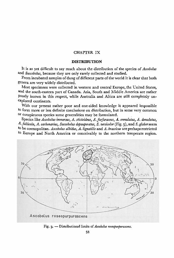
53
CHAPTER IX
DISTRIBUTION
It is as yet difficult to say much about the distribution of the species of Ascobolus
and Saccobolus, because they are only rarely collected and studied.
From incubatedsamples of dung of differentparts ofthe world it is clear that both
genera are very widely distributed.
Most specimens were collected in western and central Europe, the United States,and the south-eastern part of Canada. Asia, South and Middle America are rather
Poorly known in this respect, while Australia and Africa are still completely un-
explored continents.
With our present rather poor and one-sided knowledge it appeared impossibleto form more or less definite conclusions on distribution, but in some very common
0r conspicuous species some generalities may be formulated.
Species like Ascobolus immersus, A. stictoideus, A. furfuraceus, A. crenulatus, A. denudatus,A. foliicola, A. carbonarius, Saccobolus depauperatus, S. versicolor (Fig. 5), and S. glaber seem
to be cosmopolitan. Ascobolus albidus, A. lignatilis and A. brassicae are perhaps restricted
to Europe and North America or conceivably to the northern temperate region.
Fig. 3.— Distributional limitsof Ascobolus roseopurpurascens.
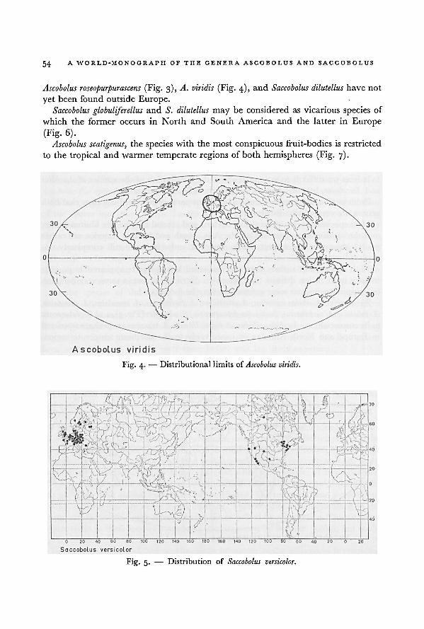
A WORLD-MONOGRAPH OF THE GENERA ASCOBOLUS AND SACCOBOLUS54
Ascobolus roseopurpurascens (Fig. 3), A. viridis (Fig. 4), and Saccobolus dilutellushave not
yet been found outside Europe.
Saccobolus globuliferellus and S. dilutellus may be considered as vicarious species of
which the former occurs in North and South America and the latter in Europe
(Fig. 6).
Ascobolus scatigenus, the species with the most conspicuous fruit-bodies is restricted
to the tropical and warmer temperate regions of both hemispheres (Fig. 7).
Fig. 4. — Distributionallimits of
Saccobolus versicolor.
Ascobolus viridis.
Fig. 5. — Distribution of
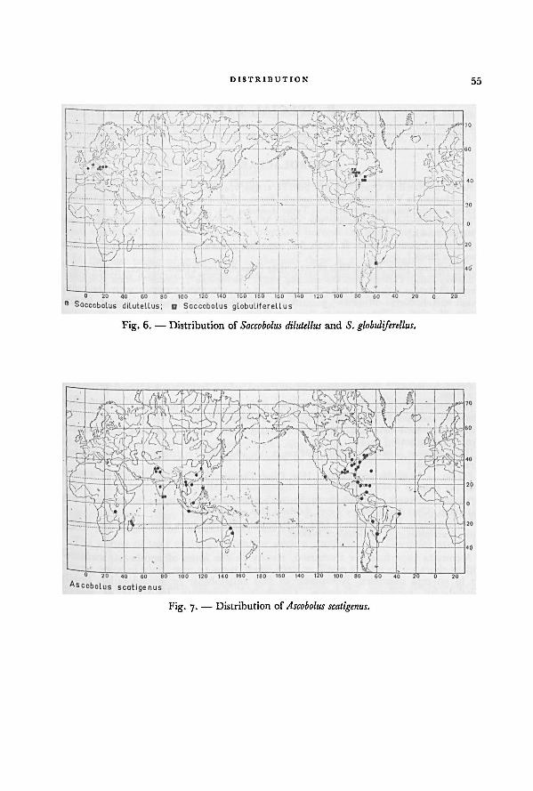
DISTRIBUTION 55
Fig. 7. — Distribution of Ascobolus scatigenus.
andSaccobolus dilutellusFig. 6. — Distribution of S. globuliferellus.
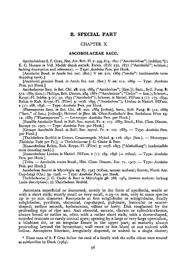
56
B. SPECIAL PART
CHAPTER X
ASCOBOLACEAE SACC.
AscoboloideaeS. F. Gray, Nat. Arr. Brit. PI. i: 599, 674. 1821 ("Ascobolideae"; [subfam.6]);E. C. Hansen in Vid. Meddr dansk naturk. Foren. 1876: 272. 1877 ("Ascobolei"; subfam.;
lacking description and reference). — Type: Ascobolus Pers. per Hook.
[Ascobolei Boud. in Annls Sci. nat. (Bot.) V 10: 210. 1869 ("sectio": inadmissible term
denoting rank).]
[Ascobolei genuini Boud. in Annls Sci. nat. (Bot.) V 10: 212. i86g. — Type: Ascobolus
Pers. per Hook.]Ascobolaceae Sacc. in Bot. Cbl. 18: 219. 1884 ("Ascoboleae"; [fam.]); Sacc., Syll. Fung. 8:
512. 1889 (fam.); Phillips, Brit. Discom. 284. 1887 ("Ascoboleae"; "Order" = fam.); Schroet.,
Krypt.-Fl. Schles. 3 (2): 50. 1893 ("Ascobolei"); Schroet. in Natiirl. PflFam r (1): 175. 1894;Rehm in Rab. Krypt.-Fl. (Pilze) 3: 1078. 1895 ("Ascoboleae"); Lindau in Natiirl. PflFam.
1 (1): 188. i8g6. — Type: Ascobolus Pers. per Hook.
[Phaeosporae Sacc. in Bot. Cbl. 18: 220. 1884 [tribus]; Sacc., Syll. Fung. 8: 512. 1889
("Sect." of fam.; [tribus]); Heimerl in Jber. kk. Ober-Realschule Bez. Sechshaus Wien 15:
12. 1889 ("Phaeosporeae"). — Lectotype: Ascobolus Pers. per Hook.]
[Famille Ascoboles Boud. in Bull. Soc. mycol. Fr. 1: 107. 1885; Boud., Hist. Class. Discom.
Europe 70. 1907. — Type: Ascobolus Pers. per Hook.]
[Groupe Ascoboles Boud. in Bull. Soc. mycol. Fr. 1: 107. 1885. — Type: Ascobolus Pers.
per Hook.]
[Theleboleen Brefeld in Unters. Gesammtgeb. Mykol. 9: 116. 1891 (fam.). — Monotype:Thelebolus Tode per Fr.]; -* ThelebolaceaeJ. C. Cooke & Barr.
[Euascoboleae Rehm, Rab. Krypt.-Fl. (Pilze) 3: 1078. 1895 ("Abtheilung"; inadmissible
term denotingrank).][Euascoboleae Lindau in Natiirl. PflFam. 1 (1): 189. 1896 (= tribus). — Type: Ascobolus
Pers. per Hook.]
[Tribu — Ascoboles vraies Boud., Hist. Class. Discom. Eur. 71. 1907. — Type: Ascobolus
Pers. per Hook.]Ascoboleae Seaver in Mycologia 19: 87. 1927 (tribus; nomen nudum); Seaver, North Am.
Cup-fungi (Op.) 39. 1928. — Type: Ascobolus Pers. per Hook.
Thelebolaceae J. C. Cooke & Barr in Mycologia 56: 768. 1964 (nomen nudum: lacking
Latin description); = TheleboleenBrefeld.
Ascomata superficial or immersed, mostly in the form of apothecia, sessile or
with a short stalk; mostly small or very small, 0.03-10 mm, only in some species
up to 30 mm diameter. Receptacle at first subglobular or semiglobular, finallysubglobular, pyriform, obconical, cup-shaped, pulvinate, lenticular
or saucer-
shaped; surface smooth, lurfuraceous, villose or hairy. Disk roughened by the
protruding tips of ripe asci. Asci obovoid, saccate, clavate or cylindric-clavate,always broad or rather so, often with a rather short stalk; with a dome-shaped,rounded truncate or rarely conical apex; opening by a large or very large operculum,a bilabiate slit, or an irregular fissure in the upper part; at maturity always
protruding beyond the' hymenium; wall more or less blued or not stained with
iodine. Ascospores biseriate, irregularly disposed, or united in a single cluster;
5 These taxa of S. F Gray below the rank of a family with the suffix -ideae were treated
as subfamilies by Donk (1964).
ASCOBOLACEAF 57
ellipsoid, fusoid, globular, or subglobular; hyaline, with weakly coloured contents
or with extrasporal pigment; smooth or ornamented; rarely with small oil-drops.Predominantly fimicolous, more rarely lignicolous, foliicolous, terrestrial, or
Pyrophilous.TYPE.—Ascobolus Pers. per Hook.
GENERA.—See under the subfamilies.
EXCLUDED GENERA.-—Pyronema Carus, Aphanoascus Zukal, Ramsbottomia Buckley,Boudiera Cooke.
This family is characterized among the other families of the Pezizales by the
small apothecia, the broad asci which protrude beyond the hymenium at maturity,and the predominantly fimicolous species.
In the last phase of maturation of the asci there is a considerable increase in
volume during a period of several hours. The wall of the ascus which is very elastic
stretches sometimes enormously.
The development of the fruit-bodies is extremely variable. On the one hand
ln some species of Ascobolus sect. Dasyobolus, cleistohymenial ascomata may be found
that remain closed, while on the other hand in Ascodesmis Tiegh. eugymnohymenial
ascomata without excipulum occur. All possible developmental types (cf. scheme
PI- 17) between those extremes are represented in this family.
The family was subdivided by Boudier (1869) into "Ascobolei genuini", with
strongly protruding asci and violet coloured episporium, and "Ascobolei spurii",vvith generally less protruding asci and hyaline ascospores with a membranous,
colourless episporium. This subdivision of the Ascobolaceae into two tribes was
followed by Saccardo (1884, 1889) and Heimerl (1889), who used the names
Phaeosporae Sacc. and Hyalosporae Sacc. Also Rehm (1895) and Lindau (1896)
recognized the same taxa but in their case as Euascoboleae Rehm and Pseudo-
ascoboleae Rehm respectively.The "Ascoboles" of Boudier's monograph (1869) represented a very natural
group. Gradually many unrelated taxa were introduced, e.g. by Cooke (1877),Saccardo (1889, i8g2, 1899I, Boudier (1907), and Le Gal (1947I. Especially the
genus Ascophanus Boud. became very heterogeneous (cf. Boudier, 1907).
The names of taxa excluded from the genera Ascobolus and Saccobolus are to be
found in the appendix on p. 206.
After the exclusion of unrelated elements the Ascobolaceae may be subdivided
'nto threegroups, which are treated as subfamilies. These are the Ascoboloideae,
Ascodesmidoideae, and Theleboloideae; they can be separated on the basis of the
Presence or absence of flesh and excipulum in the ascomata, the colour and the
formation of episporial pigment, and the shape of the ascogonium.
KEY TO THE SUBFAMILIES
Episporium thick and pigmented. Episporial pigment pinkish, violet, purplishor brownish:
2- Flesh and excipulum present. Episporial pigment at first pinkish, violet or
58 A WORLD-MONOGRAPH OF THE GENERA ASCOBOLUS AND SACCOBOLUS
purplish, finally often passing into purplish-brown or brown; precipitatedon the outsideof the ascospores in the formof a crust that may be discontinuous
in various ways Ascoboloideae, p. 58
2. Flesh and excipulum absent. Episporial pigment permanently brownish or
purplish-brown; formed on the outside of the ascospores in the form of an
elevated reticulum that may be reduced to a pattern of crests, spines or even
warts Ascodesmidoideae, p. 59
1. Episporium membranous and colourless. 6 Flesh and excipulum present
Theleboloideae. p. 59
ASCOBOLOIDEAE
Ascoboloideae S. F. Gray, Nat. Arr. Brit. PI. 1: 599, 674. 1821 ("Ascobolideae"; [subfam.]);E. C. Hansen in Vid. Meddr dansk naturk. Foren. 1876: 272. 1877 ("Ascobolei"; subfam.;
lacking description and reference). — Type: Ascobolus Pers. per Hook.
For furthersynonymy see under the family.
Receptacle smooth, furfuraceousor villose, without true hairs. Flesh and excipulumpresent, although sometimes rather reduced. Asci saccate-clavate to cylindric-clavate, with a large operculum; at maturity the tips strongly protruding above the
hymenium. Ascospores normally 8, rarely 7—1; biseriate, irregularly disposed or
united in a single cluster; ellipsoid, fusoid, globular or subglobular; with a thick,coloured episporium; smooth or ornamented. Episporial pigment of vacuolar
origin, at first sometimes colourless (or greenish opalescent in diffuse light), then
violet or purplish (rarely pinkish or bluish), finally often passing to purplish-brownor brown.
Predominantly fimicolous, more rarely lignicolous, foliicolous, terrestrial, or
pyrophilous.TYPE.
—Ascobolus Pers. per Hook.
GENERA.—Ascobolus Pers. per Hook., Saccobolus Boud. —-
Of uncertain position:Anserina Vel.
This subfamily is the principal subject of the present study. It is a very natural
and sharply delimited taxon. Characteristic of it is the violet coloured episporial
pigment. This pigment is precipitated from small vacuoles in the extrasporal
ascoplasm on the ascospore wall in the form of a more or less discontinuous crust.
KEY TO THE GENERA OF THE ASCOBOLOIDEAE
i. Ascospores free, not regularly arranged in the form of a package during any
phase of the maturation Ascobolus, p. 61
1. Ascospores firmly united into a cluster, cemented into a more or less regular
pattern (fig. 00) by the episporial pigment, or more rarely at first loosely united
according to such a pattern and finally free or partly free. . Saccobolus, p. 166
6 Colourless ascospore-mutants of Ascoboloideae are left out of consideration. Such
mutants were never found homozygous in nature. They can only be maintained in culture
after isolation.
ASCOBOLACEAE 59
Ascodesmidoideae Brumm., subfam. nov.
Ascodesmidaceae Schroet. in Krypt.-Fl. Schles. 3 (2): 31. 1893 ("Ascodesmidacei"; fam.).— Type: Ascodesmis Tiegh.
Ascomata eugymnohymenalia, minutissima. Receptaculum (caro et excipulum) nullum.Asci obovoidei, saccati vel late clavati, operculo latissimo, maturitate manifeste protrusi.Ascosporae breviter ellipsoideae usque globulares. Episporium brunneum, sporigenum,echinulatum, verrucosum vel reticulum irregulare sat elevatum formans. Fimicola. Typus:Ascodesmis Tiegh.
Receptacle (flesh and excipulum) absent. Asci obovoid, oblong-obovoid, saccate
°f broadly clavate; with a very large operculum; at maturity strongly protrudingabove the hymenium. Ascospores 8 or rarely a reduced number; multiseriate or
"regularly disposed; mutually free; shortly ellipsoid, subglobular or globular; with
an irregularly thickened, coloured episporium. Episporial pigment of sporal origin;brownish or purplish-brown; partially filling up the interstices of more or less
irregularly disposed vacuoles around the ascospores, thus forming an irregular,father elevated reticulum or a pattern of crests, spines or warts.
Fimicolous.Type.
—Ascodesmis Tiegh.Genus. —Ascodesmis Tiegh. (inclusive of Cubonia Sacc.).
The ascogonia of Ascodesmis Tiegh. are considerably narrower than in the Asco-
boloideaeor Theleboloideae. The septation of the ascogonium occurs in a rather
'ate phase.
Theleboloideae Brumm,, subfam. nov.
(Ascobolei spurii Boud. in Annls Sci. nat. (Bot.) V 10: 235. 1869. — Lectotype: Ascophanusnoud.]
(Hyalosporae Sacc. in Bot. Cbl. 18: 219. 1884 [= tribus]; Sacc., Syll. Fung. 8: 512. 1889;"nerl in .Jber. kk. Ober-Realschule Bez. Sechshaus Wien 15: 12. 1889 ("Hyalosporeae").
~
Lectotype: Ascophanus,.
Boud.][Groupe Pseudoascobolés Boud. in Buil. Soc. mycol. Fr. i: 108. 1885. — Lectotype:
Ascophanus Boud.][I heleboleen Brefeld in Unters. Gesammtgeb. Mycol. 9: 116. 1891 (fam.). Monotype:
Thelebolus Tode per Fr.]: -> ThelebolaceaeJ. C. Cooke & Barr.
[Pseudoascoboleae Rehm in Rab. Krypt.-FI. (Pilze) 3: 1078. 1895 ("Abtheilung" [=bibus]); Lindau in Natiirl. PilFam. 1 (1): 188. 1896. — Lectotype: Ascophanus Boud.]
[Tribu Pseudo-ascoboles Boud., Hist. Class. Discom. Europe 75. 1907. — Lectotype:Ascophanus Boud.]
1 helebolaceae J. C. Cooke & Barr in Mycologia 56: 768. 1964 (nomen nudum: lackingatin description) = TheleboleenBrefeld.
. sc > obovoidei, saccati, clavati vel cylindrico-clavati, operculo vel fissura bilabiali vel" legulariapicali instructi, maturitate hymenium superantes. Ascosporae 8 vel multipliciter 8,
plus quam mille, laeves vel ornamentaceae. Episporium membranaceum, sine colore,
""icola, lignicola vel terrestris. Typus: Thelebolus Tode per Fr.
a|,^cce Ptacle smooth, furfuraceous, villosc or hairy. Flesh and excipulum present,hough sometimes rather reduced. Asci obovoid, saccate, clavate or cylindric-
avate; with an operculum, a bilabiate slit or an irregular fissure near the apex;
60A WORLD-MONOGRAPH OF THE GENERA ASCOBOLUS AND SACCOBOLUS
at maturity the tips more or less strongly protruding above the hymenium. Asco-
spores 8 or a multiple of 8, up to over a thousand; biseriate, irregularly disposed
or more or less united in a loose cluster; ellipsoid, fusoid, ovoid, globular or sub-
globular; with a thin, membranous, colourless episporium; smooth or ornamented;sometimes with yellowish or pale brownish contents.
Especially fimicolous, but also foliicolous, lignicolous and terrestrial.
TYPE.— Thelebolus Tode per Fr.
GENERA.—Ascophanus Boud. (inclusive of Leporina Vel.), Thecotheus Bond., Rhyparo-
bius Boud. inclusive of Pezizula P. Karst. and Zukalina O. Kuntze sensu Vel.),Lasiobolus Sacc., Thelebolus Tode per Fr. (inclusive of Trichobolus Kimbr.), Ascozonus
(Renny) E. G. Hansen (inclusive of Streptotheca Vuill.). Of uncertain position:Boudierella Sacc. apud March, (not Boudierella Cost.), Comesia Sacc., Zukalina O.
Kuntze, Selenaspora Heim & Le Gal.
This subfamily badly needs a thorough revision. 7
7 Recently this subfamily has been the subject of a study by Kimbrough (ig66a-b). A
synopsis of the genera and species has been given by Kimbrough & Korf (1967).
61
CHAPTER XI
THE GENERA ASCOBOLUS AND SACCOBOLUS
ASCOBOLUS Pers. per Hook.
Ascobolus Pers. apud Gmel., C. Linn. Syst. Nat. 2: 1461. 1791 ("Pers. Fungi ined."); Pers.ln Neues Mag. Bot. x: 115. 1794 [= Tent. 35. 1797]; Pers., Obs. mycol. 1: 33. 1796; (devali-dated name). —
Ascobolus Pers. per Hook. 8
,Fl. scot. 2: 33, May 1821; Fries, Syst. mycol.
2 (0: 161. 1822. — Monotype: Ascobolus pezizoides Pers. apud Gmel.
Phaeopezia subg. Crouaniella Sacc. in Bot. Cbl. x8: 218. 1884. — Crouaniella (Sacc.) Lamb.,Fl. mycol. Belg., Suppl. 1: 320. 1887. — Monotype: Phaeopezia murina (Fuck.) Sacc.
Sphaeridiobolus Boud. in Bull. Soc. mycol. Fr. 1: 108. 1885. —Ascobolus subg. Sphaeridiobolus
(Boud.) Quel., Ench. Fung. 295. 1886; Boud. in Bull. Soc. mycol. Fr. 14: 126. 1898. —
Monotype: Ascobolus hyperboreus P. Karst.
[Libérispores Boud. in Bull. Soc. mycol. Fr. i: 108. 1885. — Representative species:
Ascobolus furfuraceus Pers. per Hook.][Junctispores Boud. in Bull. Soc mycol. Fr. 1: 108. 1885. — Representative species:
Ascobolus immersus Pers. per Pers.]
[Phaeopezia sect. Eu-Phaeopezia Sacc., Syll. Fung. 8: 471. 1889 (type of Phaeopezia Sacc.
not included). — Lectotype: Phaeopezia murina (Fuck.) Sacc.][Ascobolus subg. Eu-Ascobolus Sacc., Syll. Fung. 8: 514. 1889. — Lectotype: Ascobolus furfura-
ceus Pers. per Hook].Ascobolus subg. Dasyobulus Sacc., Syll. Fung. 8: 523. 1889. — Dasyobolus (Sacc.) Sacc.,
S y|}- Fung. 11: 421. 1895. — Type: Ascobolus immersus Pers. per Pers.
Phaeopezia Sacc. & Syd. in Syll. Fung. 16: 738. 1902. [with exclusion of the type ofPhaeopezia(oacc.) Sacc.]; not Phaeopezia (Sacc.) Sacc. in Bot. Cbl. 18: 218. 1884. — Lectotype: PhaeopeziaPurina (Fuck.) Sacc.
Seliniella Arx & Müll. in Acta bot. neerl. 4: 118. 1955.— Monotype: Seliniella macrospora
Arx & Muller.
Apothecia superficial or immersed, sometimes with a short stalk; mostly small,0
j3-io mm, in one species up to 30 mm diameter. Receptacle subglobular, pyriform,°bconical, cup- or saucer-shaped, rarely lenticular; surface smooth, furfuraceousVlllose or downy. Asci saccate-clavate or cylindric-clavate; at maturity the tipsstrongly protruding above the hymenium; with a rounded, dome-shaped or slightlyr uncate apex. Ascospores biseriate or irregularly disposed; free, neither clustered
nor arranged according to a regular pattern (as illustrated in fig. 00) during anyPhase of the maturation. Paraphyses slender, cylindrical; often embedded inc °lourless, yellowish or yellowish-green mucus.
Predominantly fimicolous, but also loliicolous, lignicolous, terrestrial and pyro-Philous.
ETYMOLOGY.—From Greek acocoa, leather sack and pocXXw, to throw.F YPE.—Ascobolus pezizoides Pers.
For the use of "per "see Donk in Taxon 6: 255. 1957.
62 A WORLD-MONOGRAPH OF THE GENERA ASCOBOLUS AND SACCOBOLUS
Within the Ascoboloideae this genus is characterized by its mutually free asco-
spores, viz. they are never cemented together by episporial pigment. Sometimes,
however, in Ascobolus immersus the gelatinous sheaths around the ascospores may
cause them to stick together to a slight degree. This has probably been the reason
for the introduction of Boudier's taxon "Junctispores" and for the fact that this
species has even been described as a species of Saccobolus.
Ascobolus pusillus, which occupies an isolated position in the genus, shows consider-
able resemblance to Saccobolus, except that the ascospores are neither cemented
together nor arranged according to patterns characteristic of that genus. Together
with Saccobolus saccoboloides, in which ripe ascospores are free, it might represent
a transition between Ascobolus and Saccobolus.
In artificial classifications species with spherical and ellipsoid ascospores are
often placed in different genera, only because of the shape of the ascospores. Thus
the genus Sphaeridiobolus was separated from Ascobolus by Boudier ( 1885). However,
when Boudier (1898) described A. crosslandii, he considered the spherical shape
of the ascospores less important. In my opinion the possession of spherical ascospores
alone is not sufficiently important to separate Sphaeridiobolus from Ascobolus. Even
the maintenanceof a subgenus or a section only on the basis ofthis character appears
not justified in this case. In some ellipsoid-spored species spherospory was found
as a very rare mutation.
Ascobolus crosslandii is closely related to such species as A. furfuraceus and A. crenulatus,
with ellipsoid ascospores, and must be placed in their neighbourhood. Ascobolus
brassicae and A. nodulosporus, with spherical ascospores, are mutually related and
do not suggest direct relationship with any of the other species of Ascobolus. These
are retained here in Ascobolus sect. Sphaeridiobolus.
Ascobolus reticulatus has ascospores which are spherical or subglobular in the
young state only and it recalls species of Ascodesmis in more ways than one. It differs
from that genus in the episporial pigment, which is of the type characteristic of
Ascoboloideae, and in the possession of an excipulum. Ascobolus sect. Pseudascodesmis
is proposed for this species.Globular and subglobular ascospores may also occur in A. immersus. Such variations
were described as A. globularis Roll, and A. immersus var. brevisporus Oud.
Another group was artificially separated from Ascobolus as Dasyobolus (Sacc.)Sacc. The taxa united in this genus were described as having hairs on their recep-
tacles. It was extremely heterogeneous (Saccardo, 1895; Boudier, 1907; Le Gal,
1961). The species with true hairs proved to be based on material belonging to
species of Lasiobolus Sacc., Cheilymenia Boud., and Scutellinia (Cooke) Lamb.,
sometimes covered with ascospores of species of Ascobolus (cf. list on p. 206). In the
remaining species no hairs have been found. Probably secondary mycelium and
superficial hyphae of orginally immersed fruit-bodies were described as hairs.
Ascobolus sect. Dasyobolus is maintained here for a strongly emended group, which
has only the type in common with Dasyobolus.
In my opinion a very natural subdivision of Ascobolus is obtained if the species
63ASCOBOLUS
are arranged according to the types of development, described in chapter III. Such
a subdivision is supported by other characters, like the type of ascospore ornamen-
tation, the shape of the receptacle, the activity of a secondary marginal growingzone, the shape of the ascus, and the type of hymenial pigment. The followingsections are distinguished: Ascobolus sect. Dasyobolus, Ascobolus sect. Sphaeridiobolus,Ascobolus sect. Ascobolus, Ascobolus sect. Pseudascodesmis, Ascobolus sect. Pseudosaccobolus,Ascobolus sect. Heimerlia, and Ascobolus sect. Gymnascobolus.
In most species the developmental type can be easily established, but in a few
species this has not yet been found because of the lack of appropriate material.
Species that consequently cannot be placed with certainty are: A. candidus, A.
asininus, A. masseei, A. moellerianus and A. xylophilus. Material of A. xylophilus was
Mailable but unfortunately without the necessary young fruit-bodies.
Below two keys are given for determining the sections of Ascobolus. Key A makes
Use of the taxonomic criteria of the sections and can be used in all cases where
young fruit-bodies are available to establish the developmental type. Key B makes
us e ol other taxonomic characters and can be used in cases where the developmentaltype of the fruit-bodies cannot be established.
A. SYNOPTIC KEY TO THE SECTIONS OF ASCOBOLUS
1 • Ascomata cleistohymenial. Excipulum of textura angularis, globulosa, or epider-
moidea when seen from outside:
2. Ascomata opening in the telohymenial phase or rarely not opening at all;
immersed or superficial, never stalked, the surface often covered with appressed,
thick hyphae; diameter rarely exceeding i mm. Ascospores ellipsoid or rarely
subglobular. Asci saccate to cylindric-clavate, with large dome-shaped apex.
Episporium smooth, granular, or warted, sometimes with irregular cracks,
never with a pattern of anastomosing striae. On dung
I. Ascobolus sect. Dasyobolus, p. 66
2 - Ascomata opening in the mesohymenial phase; superficial; smooth or fur-
furaceous, sometimes covered with fine divaricating hyphae in the lower
part or with secondary mycelium near the base; diameter 0.3-12 mm.
Ascospores ellipsoid, subglobular or globular. Asci cylindric-clavate. Epispo-rium with anastomosing striae, with a net-work of striae, or warted, rarelysmooth or granular:
3- Ascomata opening in the late mesohymenial phase; diameter0.3-1.5 mm.
Ascospores globular. Episporium with rounded warts. Asci clavate-
cylindrical or cylindric-clavate. . II. Ascobolus sect. Sphaeridiobolus, p. 89
3- Ascomata opening in the early or mid-mesohymenial phase; diameter
0.3-12 mm. Ascospores ellipsoid, fusiform, subglobular or globular.
Episporium with anastomosing striae, with a net-work of striae or warted,
rarely smooth or granular. If ascospores globular then episporium not with
rounded warts. Asci cylindric-clavate III. Ascobolus sect. Ascobolus, p. 94
64A WORLD-MONOGRAPH OF THE GENERA ASCOBOLUS AND SACCOBOLUS
i. Ascomata eugymnohymenial or paragymnohymenial. Excipulum often with
other types of textures:
4. Ascomata paragymnohymenial, with the habit of Ascodesmis; very small,
0.1-0.5 111111 diameter. Asci relatively short; the wall not blue with iodine.
Ascospores globular or subglobular. Episporium consisting of a reticulum
of pigment. On dung IV. Ascobolus sect. Pseudascodesmis, p. 153
4. Ascomata paragymnohymenial or eugymnohymenial; small or large,
0.3-30 mm diameter. Ascus-wall stains blue with iodine. Ascospores ellipsoid,
not with a reticulum of pigment:
5. Ascomata paragymnohymenial or eugymnohymenial, with the habit of
Saccobolus. Asci short with a broad base. Ascospores fusiform-ellipsoid,often with blunt ends and asymmetrical. Episporium at first smooth or
granulated, then with a net-work of fine crevices or warted. Paraphyseswith yellowish contents. On burnt places
V. Ascobolus sect. Pseudosaccobolus, p. 155
5. Ascomata eugymnohymenial, not with the habit of Saccobolus. Asci oblong-
clavate to cylindric-clavate. Ascospores symmetrical, ellipsoid, without any
tendency to aggregate. Paraphyses without coloured contents:
6. Apothecia small, up to about 1 mm diameter; without marginal growing
zone. Excipulum scarcely developed. Episporium echinulate or warted
VI. Ascobolus sect. Heimerlia, p. 157
6. Apothecia large or medium-sized; 0.8-30 mm diameter; a marginal
growing zone with distinct activity. Excipulum well-developed. Epispo-
rium smooth or finely punctate
VII. Ascobolus sect. Gymnascobolus, p. 158
B. ARTIFICIAL KEY TO THE SECTIONS OF ASCOBOLUS
i. Ascospores globular or subglobular:
2. Ascospores exactly globular:
3a. Ascospores warted II. Ascobolus sect. Sphaeridiobolus, p. 89
3b. Ascospores smooth, sometimes with some fine irregular crevices
I. Ascobolus sect. Dasyobolus, p. 66
3c. Ascospores with subparallel anastomosing crevices
23. Ascobolus crosslandii, p. 121
3d. Ascospores with a net-work of pigment
IV. Ascobolus sect. Pseudascodesmis, p. 153
2. Ascospores subglobular:
4a. Ascospores with a net-work of pigment
IV. Ascobolus sect. Pseudascodesmis, p. 153
4b. Ascospores smooth, sometimes with some fine irregular crevices
I. Ascobolus sect. Dasyobolus, p. 66
| 4c. Ascospores with isolated fine warts. . I. Ascobolus sect. Dasyobolus, p. 66
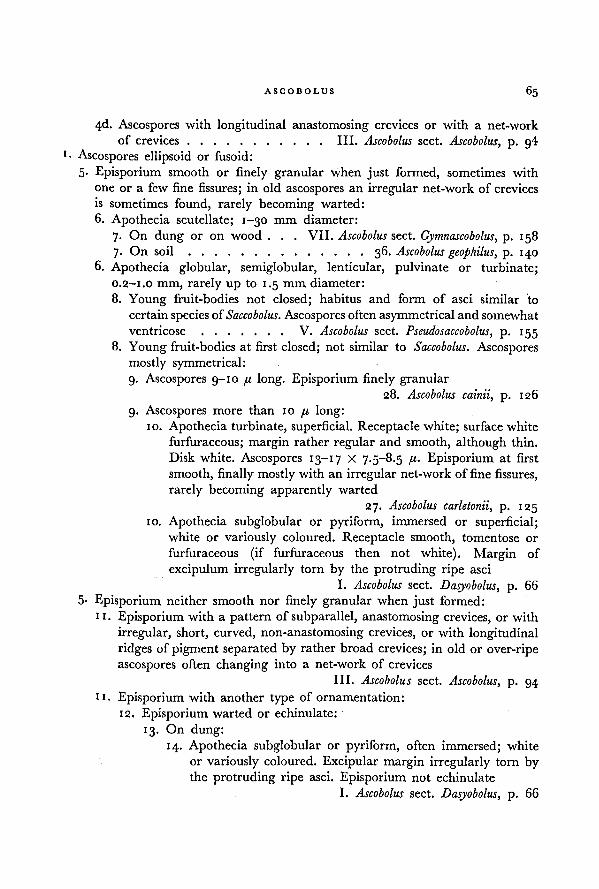
65ASCOBOLUS
4d. Ascospores with longitudinal anastomosing crevices or with a net-work
of crevices III. Ascobolus sect. Ascobolus, p. g41
• Ascospores ellipsoid or fusoid:
5- Episporium smooth or finely granular when just formed, sometimes with
one or a few fine fissures; in old ascospores an irregular net-work of crevices
is sometimes found, rarely becoming warted:
6. Apothecia scutellate; 1-30 mm diameter:
7. On dung or on wood . . . VII. Ascobolus sect. Gymnascobolus, p. 158
7. On soil 36. Ascobolus geophilus, p. 140
6. Apothecia globular, semiglobular, lenticular, pulvinate or turbinate;
o.2-i.o mm, rarely up to 1.5 mm diameter:
8. Young fruit-bodies not closed; habitus and form of asci similar to
certainspecies ofSaccobolus. Ascospores often asymmetrical and somewhat
ventricose V. Ascobolus sect. Pseudosaccobolus, p. 155
8. Young fruit-bodies at first closed; not similar to Saccobolus. Ascospores
mostly symmetrical:
9. Ascospores 9-10 /t long. Episporium finely granular28. Ascobolus cainii, p. 126
9. Ascospores more than 10 /./. long:10. Apothecia turbinate, superficial. Receptacle white; surface white
furfuraceous; margin rather regular and smooth, although thin.
Disk white. Ascospores 13-17 X 7-5-8-5 ft- Episporium at first
smooth, finally mostly with an irregular net-work of fine fissures,
rarely becoming apparently warted
27. Ascobolus carletonii, p. 125
io. Apothecia subglobular or pyriform, immersed or superficial;white or variously coloured. Receptacle smooth, tomentose or
furfuraceous (if furfuraceous then not white). Margin of
excipulum irregularly torn by the protruding ripe asci
I. Ascobolus sect. Dasyobolus, p. 66
5- Episporium neither smooth nor finely granular when just formed:
11. Episporium with a pattern of subparallel, anastomosing crevices, or with
irregular, short, curved, non-anastomosing crevices, or with longitudinal
ridges of pigment separated by rather broad crevices; in old or over-ripe
ascospores often changing into a net-work of crevices
III. Ascobolus sect. Ascobolus, p. 94
11. Episporium with another type of ornamentation:
12. Episporium warted or echinulate:
13. On dung:
14. Apothecia subglobular or pyriform, often immersed; white
or variously coloured. Excipular margin irregularly torn bythe protruding ripe asci. Episporium not echinulate
I. Ascobolus sect. Dasyobolus, p. 66
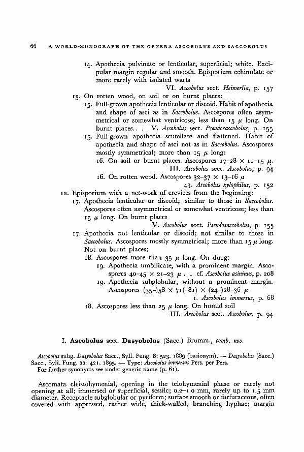
66 A WORLD-MONOGRAPH OF THE GENERA ASCOBOLUS AND SACCOBOLUS
14- Apothecia pulvinate or lenticular, superficial; white. Exci-
pular margin regular and smooth. Episporium echinulate or
more rarely with isolated warts
VI. Ascobolus sect. Heimerlia, p. 157
13. On rotten wood, on soil or on burnt places:
15. Full-grown apothecia lenticular or discoid. Habit of apothecia
and shape of asci as in Saccobolus. Ascospores often asym-
metrical or somewhat ventricose; less than 15 p long. On
burnt places.. .
V. Ascobolus sect. Pseudosaccobolus, p. 155
15. Full-grown apothecia scutellate and flattened. Habit of
apothecia and shape of asci not as in Saccobolus. Ascospores
mostly symmetrical; more than 15 p long:
16. On soil or burnt places. Ascospores 17-28 X 11 — 15 p.
III. Ascobolus sect. Ascobolus, p. 94
16. On rotten wood. Ascospores 32-37 X 13-16 p
43. Ascobolus xylophilus, p. 152
12. Episporium with a net-work of crevices from the beginning:
17. Apothecia lenticular or discoid; similar to those in Saccobolus.
Ascospores often asymmetrical or somewhat ventricose; less than
15 p long. On burnt places
V. Ascobolus sect. Pseudosaccobolus, p. 155
17. Apothecia not lenticular or discoid; not similar to those in
Saccobolus. Ascospores mostly symmetrical; more than 15 fi long.
Not on burnt places:
18. Ascospores more than 35 p long. On dung:
19. Apothecia umbilicate, with a prominent margin. Asco-
spores 40-45 X 21-23 fi . .
cf. Ascobolus asininus, p. 208
19. Apothecia subglobular, without a prominent margin.
Ascospores (35—) 58 X 71 (-81) X (24-) 28-36 p
1 Ascobolus immersus, p. 68
18. Ascospores less than 25 p long. On humid soil
III. Ascobolus sect. Ascobolus, p. 94
I. Ascobolus sect. Dasyobolus (Sacc.) Brumm., comb. nov.
Ascobolus subg. Dasyobolus Sacc., Syll. Fung. 8: 523. 1889 (basionym). — Dasyobolus (Sacc.)
Sacc., Syll. Fung. 11: 421. 1895. — Type: Ascobolus immersus Pers. per Pers.
For further synonyms see under generic name (p. 61).
Ascomata cleistohymenial, opening in the telohymenial phase or rarely not
opening at all; immersed or superficial, sessile; 0.2-1.0 mm, rarely up to 1.5 mm
diameter.Receptacle subglobular or pyriform; surface smooth or furfuraceous, often
covered with appressed, rather wide, thick-walled, branching hyphae; margin
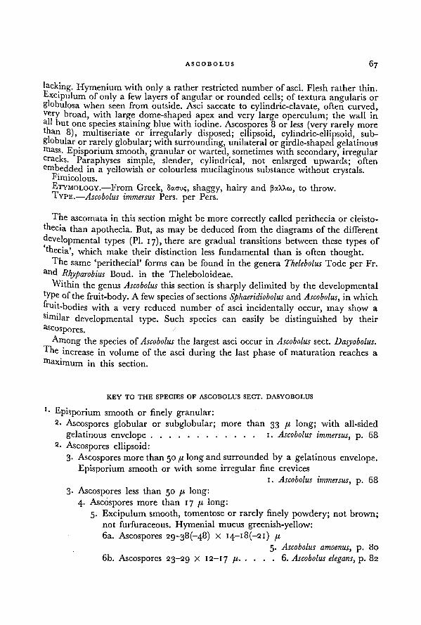
67ASCOBOLUS
lacking. Hymenium with only a rather restricted number of asci. Flesh rather thin.
Fxcipulum of only a few layers of angular or rounded cells; of textura angularis or
globulosa when seen from outside. Asci saccate to cylindric-clavate, often curved,very broad, with large dome-shaped apex and very large operculum; the wall inall but one species staining blue with iodine. Ascospores 8 or less (very rarely more
than 8), multiseriate or irregularly disposed; ellipsoid, cylindric-ellipsoid, sub-
globular or rarely globular; with surrounding, unilateralor girdle-shaped gelatinousmass. Episporium smooth, granular or warted, sometimes with secondary, irregularcracks. Paraphyses simple, slender, cylindrical, not enlarged upwards; often
embedded in a yellowish or colourless mucilaginous substance without crystals.Fimicolous.ETYMOLOGY.—From Greek, SAMJ;, shaggy, hairy and fiaXXco, to throw.
TYPE.—Ascobolus immersus Pers. per Pers.
The ascomata in this section might be more correctly called perithecia or cleisto-
thecia than apothecia. But, as may be deduced from the diagrams of the different
developmental types (PI. 17), there are gradual transitions between these types of
thecia', which make their distinction less fundamental than is often thought.
The same 'perithecial' forms can be found in the genera Thelebolus Tode per Fr.
and Rhyparobius Boud. in the Theleboloideae.
Within the genus Ascobolus this section is sharply delimited by the developmentaltype ofthe fruit-body. A few species ofsections Sphaeridiobolus and Ascobolus, in which
fruit-bodies with a very reduced number of asci incidentally occur, may show a
S1milar developmental type. Such species can easily be distinguished by their
Zoospores.
Among the species of Ascobolus the largest asci occur in Ascobolus sect. Dasyobolus.The increase in volume of the asci during the last phase of maturation reaches a
maximum in this section.
KEY TO THE SPECIES OF ASCOBOLUS SECT. DASYOBOLUS
'• Episporium smooth or finely granular:2. Ascospores globular or subglobular; more than 33 p long; with all-sided
gelatinous envelope 1. Ascobolus immersus, p. 68
2. Ascospores ellipsoid:
3- Ascospores more than 50 long and surrounded by a gelatinous envelope.Episporium smooth or with some irregular fine crevices
1. Ascobolus immersus, p. 68
3- Ascospores less than 50 p long:
4. Ascospores more than 17 p long:
5. Excipulum smooth, tomentose or rarely finely powdery; not brown;
not furfuraceous. Flymenial mucus greenish-yellow:6a. Ascospores 29-38 (-48) X 14-18 (-21) p
5. Ascobolus amoenus, p. 80
6b. Ascospores 23-29 X 12-17 p 6. Ascobolus elegans, p. 82
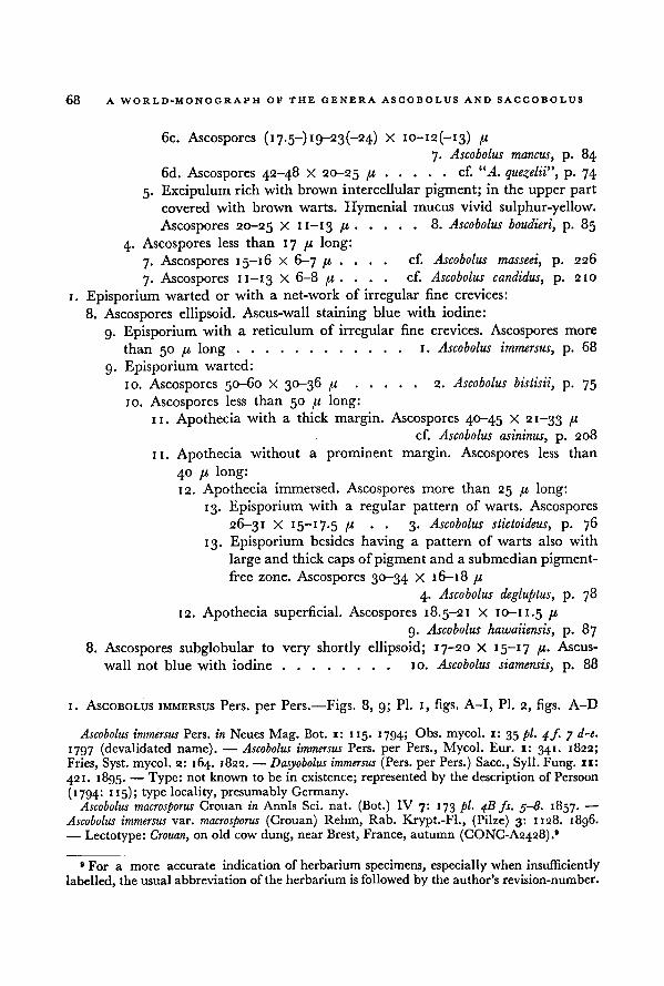
68 A WORLD-MONOGRAPH OF THE GENERA ASCOBOLUS AND SACCOBOLUS
6c. Ascospores (17.5-) 19-23(-24) X io-i2(-i3) //.
7. Ascobolus mancus, p. 84
6d. Ascospores 42-48 X 20-25 /x cf. “A. quezelii”, p. 74
5. Excipulum rich with brown intercellular pigment; in the upper partcovered with brown warts. Hymenial mucus vivid sulphur-yellow.
Ascospores 20-25 X n-13 [X 8. Ascobolus boudieri, p. 85
4. Ascospores less than 17 71 long:
7. Ascospores 15-16 X 6-7 [I ... .
cf. Ascobolus masseei, p. 226
7. Ascospores 11-13 X 6-8 /x . . . .
cf. Ascobolus candidus, p. 210
1. Episporium warted or with a net-work of irregular fine crevices:
8. Ascospores ellipsoid. Ascus-wall staining blue with iodine:
9. Episporium with a reticulum of irregular fine crevices. Ascospores more
than 50 [i long 1. Ascobolus immersus, p. 68
9. Episporium warted:
10. Ascospores 50-60 X 30-36 u 2. Ascobolus bistisii, p. 75
10. Ascospores less than 50 fx long:
11. Apothecia with a thick margin. Ascospores 40-45 x 21-33 fx
cf. Ascobolus asininus, p. 208
11. Apothecia without a prominent margin. Ascospores less than
40 [X long:
12. Apothecia immersed. Ascospores more than 25 fx long:
13. Episporium with a regular pattern of warts. Ascospores
26-31 X 15-17.5 (X . . 3. Ascobolus stictoideus, p. 76
13. Episporium besides having a pattern of warts also with
large and thick caps of pigment and a submedian pigment-
free zone. Ascospores 30-34 X 16-18 /x
4. Ascobolus degluptus, p. 78
12. Apothecia superficial. Ascospores 18.5-21 X 10-11.5 /.t
9. Ascobolus hawaiiensis, p. 87
8. Ascospores subglobular to very shortly ellipsoid; 17-20 X 15-17 I1-
Ascus-
wall not blue with iodine 10. Ascobolus siamensis, p. 88
1. ASCOBOLUS IMMERSUS Pers. per Pers.—Figs. 8, 9; PI. 1, figs. A-I, PI. 2, figs. A-D
Ascobolus immersus Pers. in Neues Mag. Bot. 1: 115. 1794; Obs. mycol. 1: 35 pi. 4ƒ. 7 d-e.
1797 (devalidated name). — Ascobolus immersus Pers. per Pers., Mycol. Eur. 1: 341. 1822;
Fries, Syst. mycol. 2: 164. 1822. — Dasyobolus immersus (Pers. per Pers.) Sacc., Syll. Fung. 11:
421. 1895. — Type: not known to be in existence; represented by the description of Persoon
(1794: 115); type locality, presumably Germany.Ascobolus macrosporus Crouan in Annls Sci. nat. (Bot.) IV 7: 173 pi. 4B fs. 3-8. 1857. —
Ascobolus immersus var. macrosporus (Crouan) Rehm, Rab. Krypt.-Fl., (Pilze) 3: 1128. 1896.— Lectotype: Crouan, on old cow dung, near Brest, France, autumn (CONC-A2428). 9
9 For a more accurate indication of herbarium specimens, especially when insufficiently
labelled, the usual abbreviation of the herbarium is followed by the author's revision-number.
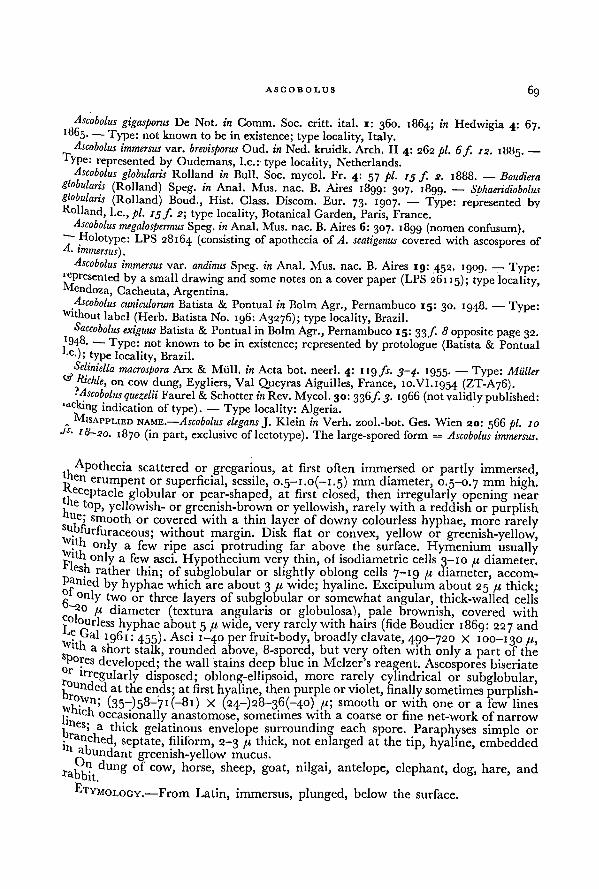
69ASCOBOLUS
Ascobolus gigasporus De Not. in Comm. Soc. critt. ital. i: 360. 1864; in Hedwigia 4: 67.1865. — Type: not known to be in existence; type locality, Italy.
Ascobolus immersus var. brevisporus Oud. in Ned. kruidk. Arch. II 4: 262 pi. 6f. 12.T „ - i88 5-
—
ypc: represented by Oudemans, I.e.: type locality, Netherlands.Ascobolus globularis Rolland in Bull. Soc. mycol. Fr. 4: 57 pi. 13 f. 2. 1888.
— Boudieraglobularis (Rolland) Speg. in Anal. Mus. nac. B. Aires 1899: 307. 1899. —
Sphaeridiobolus
globularis (Rolland) Boud., Hist. Class. Discom. Eur. 73. 1907. — Type: represented byRolland, I.e., pi. 15 f. 2; type locality, Botanical Garden, Paris, France.
Ascobolus megalospermusSpeg. in Anal. Mus. nac. B. Aires 6: 307. 1899 (nomen confusum).~~ Holotype: LPS 28164 (consisting of apothecia of A. scatigenus covered with ascospores ofA. immersus).
Ascobolus immersus var. andinus Speg. in Anal. Mus. nac. B. Aires 19: 452. 1909. — Type:by a small drawing and some notes on a cover paper (LPS 26115); type locality,
Mendoza, Cacheuta, Argentina.Ascobolus cuniculorum Batista & Pontual in Bolm Agr., Pernambuco
15: 30. 1948. — Type:Without label (Herb. Batista No. 196: A3276); type locality, Brazil.
Saccobolus exiguus Batista & Pontual in BolmAgr., Pernambuco 15: 33 f 8 opposite page 32.'948. — Type: not known to be in existence; represented by protologue (Batista & Pontual,c 0; type locality, Brazil.
Seliniella macrospora Arx & Miill. in Acta bot. neerl. 4: 119 fs. 5-4. 1955.— Type:
& Richie' -*• -w -jr-—
Muller
on cow dung, Eygliers, Val Queyras Aiguilles, France, 10.VI. 1954 (ZT-A76).? Ascobolus quezelii Faurel& Schotter in Rev. Mycol. 30: 336ƒ. 3._. 1966 (not validly published:
lacking indication of type). — Type locality: Algeria.MISAPPLIED NAME.—.Ascobolus elegans J. Klein in Verh. zool.-bot. Ges. Wien 20: 566
w. . pi. 10
Js. 18-20. ~ " ■ • F
1870 (in part, exclusive of lectotype). The large-spored form = Ascobolus immersus.
Apothecia scattered or gregarious, at first often immersed or partly immersed,E N erumpent or superficial, sessile, O.5~I.O(-I.5) mm diameter, 0.5-0.7 mm high.
Receptacle globular or pear-shaped, at first closed, then irregularly opening near
1
toP> yellowish- or greenish-brown or yellowish, rarely with a reddish or purplishue; smooth or covered with a thin layer of downy colourless hyphae, more rarely
n. 'urfuraceous; without margin. Disk flat or convex, yellow or greenish-yellow,.
h only a few ripe asci protruding far above the surface. Hymenium usuallytth only a few asci. Hypothecium very thin, of isodiametric cells 3-10 fx diameter.esh rather thin; of subglobular or slightly oblong cells 7-19 (x diameter, accom-
panied by hyphae which are about 3 fx wide; hyaline. Excipulum about 25 fx thick;
g°nly two or three layers of subglobular or somewhat angular, thick-walled cells
A20 ,u diameter (textura angularis or globulosa), pale brownish, covered with
jOIourless hyphae about 5 /« wide, very rarely with hairs (fide Boudier 1869: 227 and
? Dal 1961: 455). Asci 1-40 per fruit-body, broadly clavate, 490-720 x 100-130 fx,
"-h a short stalk, rounded above, 8-spored, but very often with only a part of the
;h°res developed; the wall stains deep blue in Melzer's reagent. Ascospores biseriate
irregularly disposed; oblong-ellipsoid, more rarely cylindrical or subglobular,undedat the ends; at first hyaline, then purple or violet, finally sometimes purplish-
(35—)5 8~7 1 (—81) X (24-)28-36(-40) fx; smooth or with one or a few lines
j.Hen occasionally anastomose, sometimes with a coarse or fine net-work of narrow
brCS
' a thick gelatinous envelope surrounding each spore. Paraphyses simple or
septate, filiform, 2-3 fi thick, not enlarged at the tip, hyaline, embeddedabundant greenish-yellow mucus,
rabbet dUng °' COW'
horse>
sheeP> goat, nilgai, antelope, elephant, dog, hare, and
ki'YMOLOGY.—From Latin, immersus, plunged, below the surface.
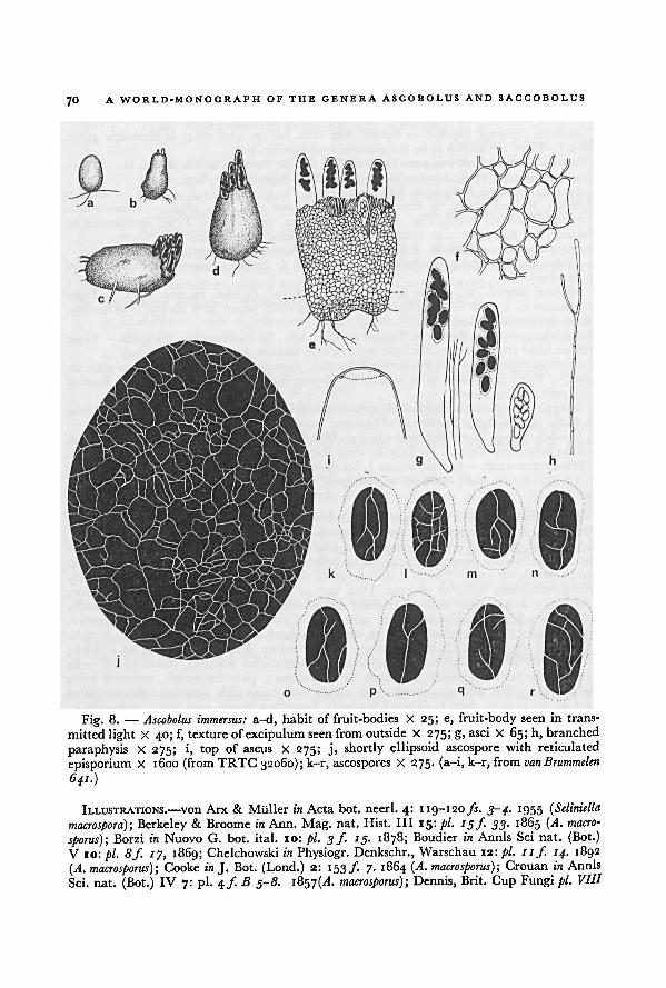
A WORLD-MONOGRAPH OF THE GENERA ASCOBOLUS AND SACCOBOLUS70
ILLUSTRATIONS.—von Arx & Mtiller in Acta bot. neerl. 4: 1 19-120 fs. 3-4. 1955 ( Seliniella
macrospora); Berkeley & Broome in Ann. Mag. nat. Hist. ILL 15: pi. 15f 33. 1865 (A. macro-
sporus); Borzi in Nuovo G. bot. ital. 10: pi. 3/. 15. 1878; Boudier in Annls Sci nat. (Bot.)
V ro: pi. 8f. 17, 1869; Chelchowski in Physiogr. Denkschr., Warschau 12: pi. ii.f. 14. 1892
(A. macrosporus); Cooke in J. Bot. (Lond.) 2: 153/. 7. 1864 (A. macrosporus) ; Crouan in Annls
Sci. nat. (Bot.) IV 7: pi. 4f.B 3-8. iÖ57(-(A. macrosporus) ; Dennis, Brit. Cup Fungi pi. VIII
a-d, habit of fruit-bodies X 25; e, fruit-body seen in trans-
mitted light X 40; f, texture of excipulum seen fromoutside X 275; g, asci X 65; h, branched
paraphysis X 275; i, top of ascus X 275; j, shortly ellipsoid ascospore with reticulated
episporium x 1600 (from TRTC 32060); k-r, ascospores X 275. (a-i, k-r, from
Ascobolus immersus:Fig. 8.—
van Brummelen
641.)
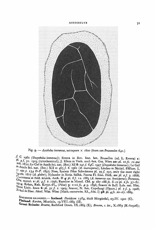
ASCOBOLUS71
C. I g6o (Dasyobolus immersus);,,
Errera in Rec. Inst. bot. Bruxelles (ed. L. Errera) i:
'9°5 (cytochemical); J. Klein in Verh. zool.-bot. Ges. Wien 20: pi. 10 fs. ig and
1870; Le Gal in Annls Sci. nat. (Bot.) XI 8: 2 4 1/- 64C. 1947 (.Dasyobolus immersus);in \ t A .
v ' 4 " j \—"v —-— ~/jLc Gal
Annls Sci. nat. (Bot.) XII i: 465/. 6. 1961 (A. macrosporus);,
Lindau in Natiirl. PflFam. I,>:
Ig2f- -•
154 D-F. 1897; Nees, System Pilze Schwamme pi. 39 f. 297, only the most rightr^ u ' e- 1817 (A. gibber); Nylander in Notis. Sallsk. Fauna Fl. fenn. Förh. 10: pi. 2f. 3. 1868;
OKin Xed- kruidk. Arch. II 4: pi. 6f. 12. 1885 (A. immersus var. brevisporus); Persoon'
s. mycol. 1: pi. '4 f 7. 1796; Ramlow in Mycol. Cbl. 5: 187-188fs. 6-10 pi. 2 fs. 37-61.*9 x 4> Rehm, Rab. Krypt.-Fl., (Pilze) 3: 1112fs. 4-5. 1896; Seaver in Bull. Lab. nat. Hist.
2.1,
Univ'
Iowa 6: t>L. 31 f '■ I9°5 'Seaver
>N
-Am
- Cup-fungi (Operc.) pi. 7 f. j. 1928;ka l in Sber. Akad. Wiss. Wien (Math.-naturwiss. Kl., Abt. I) 98: pi. 4fs. 20-27. 1889.
SPECIMENSEXAMINED.— Iceland: Davidsson 1564, Hole Hörgardal, 23.IV. 1901 (C).
inland: Karsten, Mustiala, 19.VIII. 1869 (H).
re at Britain: Broome, Bathford Down. IX.1863 (E); Broome, s. loc., X.1863 (K-A2526);
Fig. 9. — Ascobolus immersus, ascospore X 1600 (from van Brummelen 641.)
A WORLD-MONOGRAPH OF THE GENERA ASCOBOLUS AND SACCOBOLUS72
Broome, Hanham near Bristol, XI. 1863 (UPS); Broome, near Bath, X.1864, in Rabenhorst
Fungi eur. 779 (BRSL, FH, G, HBG, K, LE, PRC, W, ZT); Broome, Bathford Down, Somer-
set-Wiltshire Border, 22.X. 1864 (K); Broome, Bathford, 15.XI.1864 (K); Broome, s. loc., s. dat.
(K-A2527); Grassland, Hardcastle near Hebden Bridge, 6.VII.1899 (K); Cooke, Forden, s. dat.,
in Fungi brit. ed. 2, 397 (E, K, PAD), in Fungi brit. ed. 2, 399 ( Ascobolus kerverni, PAD);
Grove, Clent Hills, Worcestershire, 16.VI.1888 (BM); Hawkins, Richmond Park, IX.1957
(CMI 70911); Phillips, Shrewsbury, r. dat., in Elv. brit. 46 (A. furfuraceus, PAD); unknown
collector, Shrewsbury, s. dat., in Cooke Fungi exs. sel., (K-A2524).Netherlands: van Brummelen 641, Elspeet, 25.VII. 1958 (L); van Brummelen 652, Elspeet,
10.IV.1959 (L); van Brummelen 68g, Elspeet, 19.VI.1959 (L); van Brummelen 6g4, Zoological
Garden, Amsterdam, 1.VII.1959 (L); vanBrummelen 1445, Elspeet, 23.III. 1962 (L); Oudemans,
s. loc. V. 1882. (GRO).
Belgium: Marchal, Laiken, II.1884 (BR); Mouton, Beaufays, July (BR).
Luxemburg: Marchal, Ebly, VI.1884 (PAD).
France: Boudier, Bois de Beauchamp, IX. 1884 (PC); Boudier, Bois de Beauchamp, XI. 1884
(PC); Breviere, Ambert, Puy-de-Dome, 17.VIII.1900 (S); de Croculs, Dardennes, near Toulon,
III. 1927 (PC); Crouan, on cow dung, near Brest, 29.VI.1857 (PC); Crouan, on cow dung,
near Brest, autumn (lectotype of Ascobolus macrosporus, CONC-A2428); Crouan, on dog-dung,in the dunes of Plovan, 10. IX.1865 (CONC); Gaillard, Angers, VI. 1900 (PC); Miiller &
Richie, Eygliers, Val Queyras, Aiguilles, 10.VI.1954 (type of Seliniella macrospora, ZT); Rizet,
culture on agar., Cluny, Saone & Loire, s. dat. (PC); unknown collector, Ecouen, VIII.i8go
(PC-A2224, PC-A2226, PC-A2227).Denmark: Hansen, s. loc., 11.VI.1874 (C-A64); Hansen, s. loc., s. dat., (BM-A2838).
Germany: Ade, Schammendorfnear Weismain, IX.1907 (S); Ade, in Allgau, 4.VII.1909,
(S); Ade, near Obersdorf in Allgau, 5.VII.1909 (S); Fuckel, Oestrich, s. dat. (G-A1014);
Fuckel, Oestricher Wald, autumn, in Fungi rhen. 1847 (GRO, LE, M, W), in Herb. Barbey-
Boissier 131 1 (BPI, FH, S, W); Fuckel, Oestrich, XI (G-A1016); Fuckel, Oestricher Wald,
autumn, in Fungi rhen. 1848 (A. macrosporus, GRO, LE, M, W), in Herbier Barbey-Boissier
1312 (A. immersus var. macrosporus, BPI, FH, S, W); Fuckel, Oestricher Wald, autumn, in
Fungi rhen. 1852 (A. vinosus, BM); Rehm, near Obernesselbach, Sugenheim, III. 1869 (S);
Rehm, near Obernesselbach, XI.1870, in Ascom. 102b (E, L, M, NY, PAD, S, W); Rehm,
Obernesselbach, 1870 (S-A620); Rehm, Obernesselbach, II.1871 (S-A563); Rehm, near Planegg,
VIII. 1900 (S); Rehm, between Griesen and Eibsee under the Zugspitz, X.1900 (S); Rehm,
Flinterstein, Allgau, IX.1909 (S); Rehm, Obernesselbach, s. dat. (S-A495); Rehm, Hinterstein,
Allgauer Alpen, s. dat. (S-A485); Schroeter, Rastatt, 10.V.1876 (BRSL); Sydow, Zehlendorf
near Berlin, VII.1882 (S), in Myc. march. 361 (HBG, S, W); Wagner, on dung of Antilope
bubalis, zoo, Dresden, IX. 1892 (S); Winter, Halle a. Saale, VI. 1873 (E); Winter, Ilmenau,
Thüringen, VII.1873 (BM, GRO); Winter, Halle a. Saale, IX.1873 (HBG); Winter, Halle a.
Saaie, VI.1875 (S); unknown collector, Nymphenburg, München, II.1927 (M).
Poland: Lojka, Galizien, 1869 (S); Schroeter, Oswitz, V.1870 (BRSL); Schroeter, Rrpslau
(= Wroclaw), VII.1880 (CRSL); Schroeter, Madelkamm in Riesen-Gebirge (= Krknose),
4.VI. 1882 (BRSL); Schroeter, on elephant dung, s. loc. XI. 1885 (BRSL); Schroeter, zoo (=
Wroclaw), 22.V.1886 (BRSL).
Czechoslovakia: Niessl, near Brunn (= Brno), s. dat. (M 15800, M 15802); Niessl, Schreib-
wald near Briinn (= Brno), s. dat. (M. 15801); Niessl, Gr. Niemtschitz, s. dat. (M 41418);
Petrak, M. Weisskirchen (= Hranice), Bartelsdorf, V. 1923 (ZT); Vacek, Cernosice, Bohemia,
I.XII.1947 (PR); Velenovsky, Mnichovice, Bozkov, VIII. 1924 (PR).
Switzerland: Miiller, Aufstieg zur Pierre a Voir, Verbier, Wallis, 30.V. 1955 (ZT); Miiller
& Schnepp, Speergebiet, St. Gallen, 25.V.1955. (ZT).Austria: Demelius nog, Unterwaltersdorf, 7.IX.1916 (W); Niessl, near Wienerbruclc,
VIII.1914 (M 15799); Niessl, near Wienerbruck, VII.1915, (M); Niessl, near Voitsberg a.
Kainach, s. dat. (M 41416); Rechinger, near Aussee, Steiermark, 1921, in Weese, Eumyc.
ASCOBOLUS 73
sel. 592 (M); Rehm,, near Kühstei, Oetz, Tyrol, VIII. 1872, in Ascom. 102a (E, L, M, NY,PAD, S, W); Sauter, near Mittersill (according to Keissler), s. dat.
, „ „(W 982).
Italy: Cavara, Mombolone near Papia, s. dat., in Fungi Long. 163 (A. macrosporus, BPI, L,PAD, S, W); Rehm, Monte Pellegrino near Palermo, Sicilia, X. 1894, in Ascom. 1169 (A.globularis, FH, HBG, M, PAD, PC, S, W, ZT); Rehm, near Palermo, X.1894 (S-A623);Saccardo, Selva, 30.VIII. 1877 (PAD); Saccardo, Giavera, VIII. 1877 (PAD).
U.S.S.R.: Ivanitskaja, Katun near Biysk, 29.VII.1911 (only ascospores) LE; Tiryveko 278,s- loc., 11.VIII. 1903 (LE).
Pakistan: Ahmad 2948,j-r-,
Changla gali, 26.IX.1949 (BPI).ïndia: Lacy, Patna, X.1937 (TRTC).New Guinea: van Brummelen 1287,
,,
Ifar near Hollandia, 26.VI.1961 (L).Australia: Queensland: Simmonds,
__,
Brisbane River, Brisbane, VII. 1912 (K).Hawaii: van Brummelen 1459, 1451,
,
Puuhookomo, Mauna Kea, 7-10.V.1962 (L).Canada: Cain, Lucknow, Bruce Co., Ontario, 10.VII.1930 (TRTC); Cain,. .
_ _
._
North Bav.
Nipissing Co., Ontario, 3.IX. 1931 (TRTC); Cain,, ,
,,, _
, New Durham, Brant Co., Ontario,O x
_ ' ' kJ «y\J \ / ' > 3 3 3
°' X->933 (TRTC); Cain 6948, Pine River, Lac. St. Joseph. Quebec, 26.VIII.1938 (TRTC);Cain
12199, S.E. of Camillo, Dufferin Co., Ontario, 25.X.1948 (TRTC); Cain 12213,
Nashville, York Co., Ontario, 1.XI.1948 (TRTC); Cain,_____ _ , Nashville, York Co., Ontario,
28.ÏX. 1952 (TRTC, ZT); Cain, Nashville, York Co., Ontario, g.V.1954 (TRTC); Jackson,Bear Island, Lake Timagami, Ontario, IX.1929 (NY, TRTG); unknown collector, Melissa,
Muskoka, Ontario, 2.'VIII. 1932 (TRTC 34682).U.S.A.: Oregon:
"
Kienholz K 82, Hood River, 10.XI.1932 (NY). Arizona: Cain,TT
* W X v "* V-, xw.xx-x..yjx \4 "-/* --x x~~xx«.. wv.».,
Mt.
Flagstaf, 21.VI. 1955 (TRTC, small-spored form). Colorado: Seaver & Bethel,Geneva Creek Canyon, IX.1910 (NY). Iowa: Seaver 61 and 61-b,. Iowa City, 17.III. IQO<Ï
(BPI, KY, S). Maine: Thaxter 518, Kittery Point, 18.VI.1886 (FH). New York: Cook,Glyde, X. 1887 (NY-A1346, NY-A1353); Gerard, Po'keepsie, I.1872 (NY). Massachusetts:
Sturgis, Cambridge, III. i8gi (NY). Connecticut: Sturgis,,, „ ,
New Haven, 1.1893 (NY).New Jersey: Ellis, Newfield, VIII. 1890, in Ellis & Everhardt N. Am. fungi ser. 2, 2620(4. vinosus,a \ r
-— > L). Virginia: Meyer 14727,,MountainLake Biological Station (in moist chamber),
b -VlII.i94 2 (BPI, TRTC).
'
Bermuda: Brown et al. 1363, XI-XII. igi2(NY); Brown et al.1546 (culture), XI-XII-IQ12
(NY);" v "
Brown et al. 1599 (culture), XI-XII.1912 (NY); Dodge, 1911 (NY-A1204); Seaver &
Whetzel,, Morgan's Island, 9. 11.1926 (NY).
Puerto Rico: Fink, Mayaguez, VIII. 1920 (NY-A1355); Seaver, s. loc., s. dat., (NY-A1203);Wilson
237,, Luquillo Mountains, 14.VII.1902 (NY).
Brazil) Theobald,,
Sao Leopoldo, Rio Grande do Sul, 11.III.1936 (TRTC).Argentina: Calviello, Capital Federal, 9.V.1962 (BAFC 21293); Spegazzini, Recolcta.
Buenos Aires 10.III.1880 (LPS 26124); Spegazzini,,
Colonia Resistencia, Chaco, 1.1887 (inrftir ° nly ascospores; type of A. megalospermus, LPS 28164); Spegazzini,, La Plata, 17.VI.1g08
26127).
This species is probably one of the commonest members of the genus and its
distribution seems to be limited only by the occurrence of herbivorous mammals.
1can be easily recognized by the very large ascospores with a thick gelatinous
'Tvelope surrounding each spore. The fruit-bodies grow more or less immersed,dte
depth depending on the structure of the substratum. On semi-solid or soft
surfaces and on substrata with superficial, small cavities they will grow fully immersed
only the ripe asci protruding. On very dense, solid surfaces they may developals
° superficially.1 he immersed parts ofthe walls especially are clothed with a thin layer ofappressed
74 A WORLD-MONOGRAPH OF THE GENERA ASCOBOLUS AND SACCOBOLUS
hyphae. These hyphae may be rather wide and show rigid walls. Similar clothing
occurs in all species of section Dasyobolus.
Some authors (e.g. Boudier, 1869; Phillips, 1887; Saccardo, 1889; Heimerl,
1889; Le Gal, 1961) described hairs from the surface of the excipulum of Ascobolus
immersus. However, in the many thousands of fruit-bodies of this species studied by
me, among which were also specimens studied by these authors, no excipular hairs,like those illustrated by Boudier and Le Gal could be found.
The ascospores of A. immersus are very variable. Besides the normal oblong-
ellipsoid ascospores, cylindrical and subglobular ascospores also occur. The form
with subglobular ascospores has been described as A. immersus var. brevisporus Oud.
and as A. globularis Rolland. Of this form excellent material was distributed in
Rehm's "Ascomycetes exsiccati", No. 1169.Rather frequent mutant-forms with only four ascospores per ascus have been
described under the names A. immersus var. andinus Speg. and Saccobolus exiguusBatista & Pontual. Ascobolus gigasporus De Not. with 2-5 ascospores is perhapsanother such mutant-form.
Occasionally the number of ascospores in an ascus varies even in the same fruit-
body from eight to one.
Asci with more than eight ascospores occur only very rarely. Ramlow (1915)found an ascus with sixteen ascospores, while Zuk & Swietlinska (1965) illustrated
an ascus with fifteen ascospores. Here an additional mitosis took place in the ascus.
Of A. immersus more than 2000 ascospore-mutants are known only differing from
the wild-type strain by a single gene (Lissouba & al., 1962). Segregations for
ascospore colour or other episporium phenotypes can often be observed in the asci.
The species is heterothallic and a favourable object for genetical and cytologicalstudies (Rizet, 1939, 1941; Rizet & al., ig6oa-c; Lissouba & aL> 1962; Zuk &
Swietlinska, 1965; Paszewski & al., 1966; Makarewicz, 1966).
In some cultures of A. immersus 'cleistothecia' were formed in which the ascospores
were only set free by decay. These cleistothecial forms are probably inhibited in
their development and not comparable with cleistothecial mutants like those
described by Maniotis (1965) for Gelasinospora calospora (Mouton) C. & M. Moreau.
Rizet (1939) also described sclerotia in cultures of A. immersus, which resembled
immature fruit-bodies. Unfortunately the microscopic structure of these sclerotia
was not described.
Zukal (1889) described for this species gemmae resemblin conidia of the hypho-
mycetous genus Stemphylium Wallr. At the ends of short, often sickle-shaped branches
(consisting of 4—6 cells), are the apical cells which are surrounded by a brownish
membrane and finally become separated as conidia. This observation has never
been repeated.Ascobolus quezelii Faurel & Schotter, of which the type could not yet be studied,
probably represents a form of A. immersus with reddish to purplish receptacles,small asci ["250-35o(-40o) X 50-70 74"], and small ascospores ("42-48 X
20-50 p").
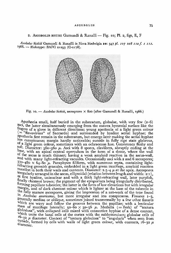
ASCOBO LUS 75
2. ASCOBOLUS BISTISII Gamundi & Ranalli — Fig. 10; PL 2, figs, E, F
Ascobolus bistisii Gamundi & Ranalli in Nova Hedwigia 10: 347pi. ioy 108 nof. 1 in.
1966. — Holotype: BAFC 21293 (C-2178).
Apothecia small, half buried in the substratum, globular, with very few (2-6)jlSci
)the latter simultaneously emerging from the convex hymenial surface like the
lingers of a glove in different directions; young apothecia of a light green colour
"flavovirens" of Saccardo) and surrounded by hyaline aerial hyphae; the
apothecia first remain in the substratum, but emerge later making the aerial hyphae
conspicuous; margin hardly noticeable; outside in fully ripe state glabrous,°f a light green colour, sometimes with an ochraceous hue. Consistency fleshy andsoft. Diameter: 560-960 11. Asci with 8 spores, claviform, abruptly ending at the
oase, with an apical central operculum in the form of a dome, where the wall°f the ascus is much thinner; having a weak amyloid reaction in the ascus-wall,and with
many light-refracting vacuoles. Occasionally asci with 2 and 6 ascospores;370-480 x 64-80 ju. Paraphyses filiform, with numerous septa, containing light-refracting- greenish granules, embedded in a light green mucilage, amyloid reaction
Manifest in both their walls and contents. Diameter:2.5-4 /« at the apex. Ascosporeslrregularly arranged in the ascus, ellipsoidal (relation betweenlength and width: 2:1),
first hyaline, uninuclear and with a thick light-refracting wall, later purplish,onally chestnut brown; the pigment of the episporium being irregularly distributed,orming papillate tubercles; the latter in the form of lowelevations but with irregular
rnargiti, and of dark chestnut colour which is lighter at the base of the tubercle in
._e hi11y mature ascospores, giving the impression of a net-work of the type found
ln Ascobolus americanus, but more irregular and less conspicuous. Fissures 1-3,
generally median or oblique, sometimes joined transversally by a few other fissureswhich are wavy and follow the grooves between the papillae; with a lenticularhtass of mucilage laterally; 50-60 X 30-36 /<. Medulla (= flesh) of "textura
S'obulosa", with collapsed cells mixed with connective hyphae of a dense content,vhich unite the basal cells of the cortex with the subhymenium; globular cells of
32 /.t diameter. Context of "textura globulosa" to "angularis" when seen from
Outside, formed by cells with walls of light green colour, with contents, 16-32 /rai
ameter.
Fig. 10. — Ascobolus bistisii, ascospores X 800 (after Gamundí & Ranalli, 1966.)
7 6 A WORLD-MONOGRAPH OF THE GENERA ASCOBOLUS AND SACCOBOLUS
Oidia present in the aerial mycelium, hyaline, cylindrical, 12-15 X 4 /x.
Only known from dung of horse.
ETYMOLOGY.—After Dr. G. Bistis, the American geneticist.
ILLUSTRATIONS.—Gamundi & Ranalli, I.e.
The paper in which this species was published reached me when the manuscriptof this monograph was already finished. Awaiting the study of authentic material,
above a translation is given of the detailed original Spanish description.This species is closely related to Ascobolus immersus, but there are some differences
in the spores. In the latter the spores are oblong ellipsoid or subcylindrical with
rounded or obtuse ends. The episporium is smooth or with some irregular crevices,
and a gelatinous mass surrounds each spore. In A. bistisii the spores are more
regularly ellipsoid. The pigment of the episporium is irregularly distributed forming
papillate tubercles with irregular margin. The episporium also shows some irregular
crevices and there is a unilateral gelatinous cap to each ascospore.
These differences seem sufficiently important to recognize two distinct species.10
3. ASCOBOLUS STICTOIDEUS Speg. —Fig. II; Pl. 2, figs. G, H
Ascobolus stictoideus Speg. in Michelia 1: 474. 1879. — Ascobolus brunneus var. stictoideus (Speg.)Heimerl in Jber. kk. Ober-Realschule Bez. Sechshaus Wien 15: 14. 1889. — Holotype:LPS 26119.
Apothecia scattered, gregarious or closely crowded, completely immersed or
rarely superficial, 350-600 u diameter, 400-650 u, high. Receptacle at first closed
and globular or pyriform, then opening by irregular rupturing of the wall, watery-white, greenish-yellow or olive in colour; thinly tomentose or rarely smooth; without
a margin. Disk flat or slightly concave, pale olivaceous to almost colourless, with
a few almost black, ripe asci strongly protruding. Hymenium 230-280 [i thick,
containing up to 40 asci. Hypothecium very thin, of isodiametric cells 6-12 71
diameter. Flesh not sharply differentiatedor very thin. Excipulum about 15 11 thick,of more or less isodiametric, polygonal, thick-walled cells 9-25 71 diameter (textura
angularis), with pale yellowish cell-walls, often covered with a thin layer of inter-
woven, cylindrical, irregularly branched, hyaline, 4-9 /t wide hyphae. Asci clavate
or clavate-saccate, gradually tapering downwards into a rather thick base, rounded
above, 150-400 X 39-55 fx, 8-spored; the wall deep blue in Melzer's reagent.Ascospores biseriate or irregularly disposed, ellipsoid; at first hyaline, then paleviolet, finally dark violet, (25-5-)26.5-30.5(-32) X (14.5 —) 16—17.5 fx, ornamented
with rathercoarse, rounded warts; pigment often rather thick
up to 1.5 11; with
10 Through the kindness of Dr. I. Gamundi de Amos, the type specimen of A. bistisii could
recently be studied. This material agrees with the above description. The size of most
ascospores was found to fallwithin 55-60 X 30-33 fi. The shape of the spores is not significantlydifferent from that in A. immersus. Generally the episporium is clearly rough, but in some
fruit-bodies it was found to be almost smooth. Sometimes spores were rough on one side and
smooth on the opposite side. The presence of an unilateral gelatinous cap to each spore
remains a very outstanding character of this fungus, the constancy of which cannot be studied
in dried material, however.
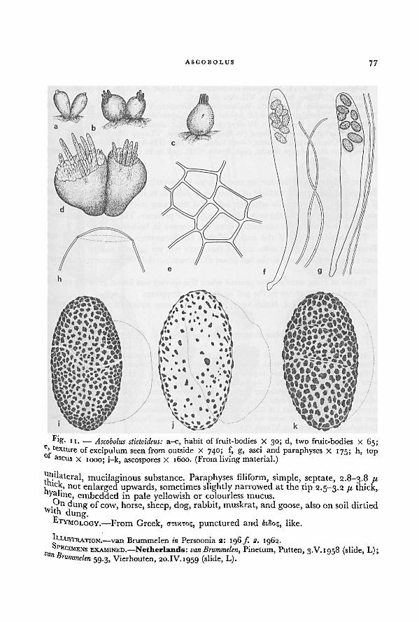
ASCOBOLUS 77
..l ateral, mucilaginous substance. Paraphyses filiform, simple, septate, 2.8-3.8 /xaick, not enlarged upwards, sometimes slightly narrowed at the tip 2.5-3.2 //. thick,yahne, embedded in pale yellowish or colourless mucus.
Gn dung of cow, horse, sheep, dog, rabbit, muskrat, and goose, also on soil dirtied
pduns-
ETYMOLOGY. —From Greek, A-nx-ro;, punctured and etSo;, like.
LLUSTRATION.—van Brummelen in Persoonia 2: 196 f. 2. 1962.P
ECIMENS EXAMINED.- -Netherlands: vanBrummelen, Pinetum, Putten, 3.V. 1958 (slide, L);a" Brummelen
59.3, Vierhouten, 20. IV. 1959 (slide, L).
Fig. 11. — a-c, habit of fruit-bodies X 30; d, two fruit-bodies X 65;e, texture of excipulum seen from outside X 740; f, g, asci and paraphyses X 175; h, top
of ascus x 1000; i-k, ascospores X 1600. (From living material.)
Ascobolus stictoideus:
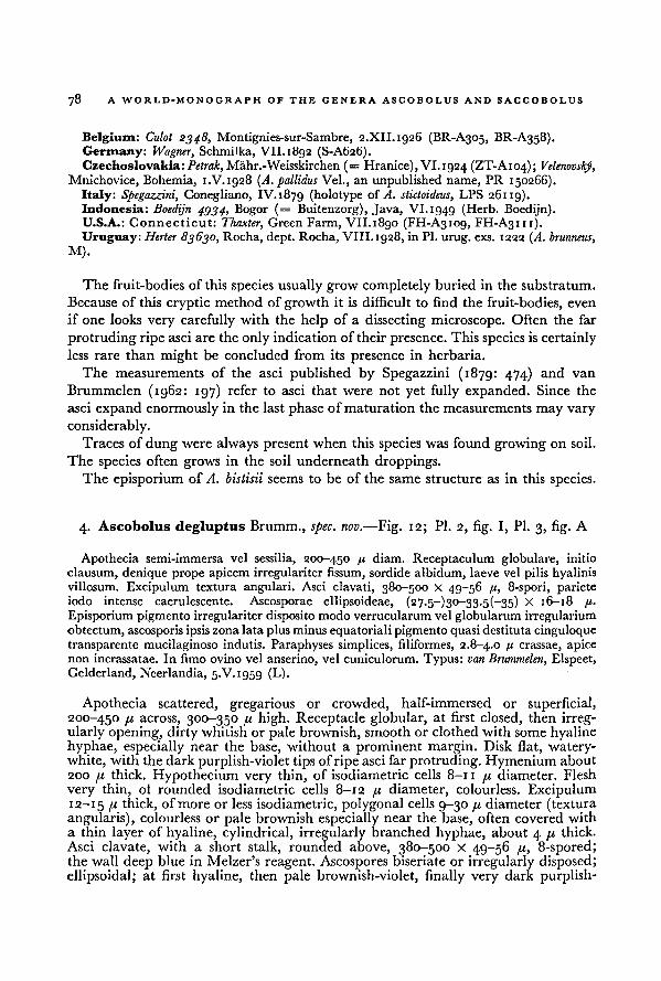
78 A WORLD-MONOGRAPH OF THE GENERA ASCOBOLUS AND SACCOBOLUS
Belgium: Culot 2348, Montignies-sur-Sambre, 2.XII.1926 (BR-A305, BR-A358).
Germany: Wagner, Schmilka, VII. 1892 (S-A626).
Czechoslovakia: Petrak, Mahr.-Weisskirchen (= Hranice), VI.1924 (ZT-A104); Velenovsky,Mnichovice, Bohemia, 1.V.1928 (A. pallidus Vel., an unpublished name, PR 150266).
Italy: Spegazzini, Conegliano, IV.1879 (holotype of A. stictoideus, LPS 26119).Indonesia: Boedijn 4934, Bogor (= Buitenzorg), Java, VI.1949 (Herb. Boedijn).U.S.A.: C onnecticut: Thaxter, Green Farm, VII.1890 (FH-A3109, FH-A3111).Uruguay: Herter 83630, Rocha, dept. Rocha, VIII.1928, in PI.
urug. exs. 1222 (A. brunneus,
M).
The fruit-bodies of this species usually grow completely buried in the substratum.
Because of this cryptic method of growth it is difficult to find the fruit-bodies, even
if one looks very carefully with the help of a dissecting microscope. Often the far
protruding ripe asci are the only indicationof their presence. This species is certainly
less rare than might be concluded from its presence in herbaria.
The measurements of the asci published by Spegazzini (1879: 474) and van
Brummelen (1962: 197) refer to asci that were not yet fully expanded. Since the
asci expand enormously in the last phase of maturation the measurements may vary
considerably.Traces of dung were always present when this species was found growing on soil.
The species often grows in the soil underneath droppings.The episporium of A. bistisii seems to be of the same structure as in this species.
4. Ascobolus degluptus Brumm., spec. nov. —Fig. 12; Pl. 2, fig. I, Pl. 3, fig. A
Apothecia semi-immersa vel sessilia, 200-450 /j diam. Receptaculum globulare, initio
clausum, denique prope apicem irregulariter fissum, sordide albidum, laeve vel pilis hyalinisvillosum. Excipulum textura angulari. Asci clavati, 380-500 x 49-56 ft, 8-spori, parieteiodo intense caerulescente. Ascosporae ellipsoideae, (27-5-)30-33.5(-35) X 16-18 p.
Episporium pigmento irregulariter disposito modoverrucularum vel globularum irregularium
obtectum, ascosporis ipsis zonalata plus minus equatoriali pigmento quasi destitutacinguloquetransparente mucilaginoso indutis. Paraphyses simplices, filiformes, 2.8-4.0 /< crassae, apicenon incrassatae. In fimo ovino vel anserino, vel cuniculorum. Typus: van Brummelen, Elspeet,Gelderland, Neerlandia, 5.V. 1959 (L).
Apothecia scattered, gregarious or crowded, half-immersed or superficial,
200-450 [i across, 300-350 // high. Receptacle globular, at first closed, then irreg-ularly opening, dirty whitish or pale brownish, smooth or clothed with some hyalinehyphae, especially near the base, without a prominent margin. Disk flat, watery-white, with the dark purplish-violet tips of ripe asci far protruding. Hymenium about
200 fi thick. Hypothecium very thin, of isodiametric cells 8-11 /i diameter. Flesh
very thin, ol rounded isodiametric cells 8-12 41 diameter, colourless. Excipulum12-15 fi thick, of more or less isodiametric, polygonal cells 9-30 41 diameter (texturaangularis), colourless or pale brownish especially near the base, often covered with
a thin layer of hyaline, cylindrical, irregularly branched hyphae, about 4 41 thick.
Asci clavate, with a short stalk, rounded above, 380-500 X 49-56 41, 8-spored;the wall deep blue in Melzer's reagent. Ascospores biseriate or irregularly disposed;ellipsoidal; at first hyaline, then pale brownish-violet, finally very dark purplish-
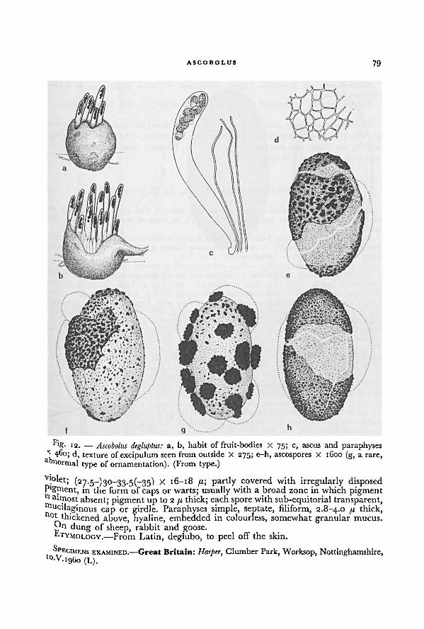
79ASCOBOLUS
Vl.°l et; (27.5^)30-33.5(-35) X 16-18 //; partly covered with irregularly disposedP'gment, in the form of caps or warts; usually with a broad zone in which pigmentls almost
absent; pigment up to 2 //. thick; each spore with sub-equitorial transparent,Mucilaginous cap or girdle. Paraphyses simple, septate, filiform, 2.8-4.0 pr thick,n°* thickened above, hyaline, embedded in colourless, somewhat granular mucus.
n dung of sheep, rabbit and goose.ETYMOLOGY.
—From Latin, deglubo, to peel off the skin.
SP ECIMENS EXAMINED.—Great Britain: Harper, Clumber Park, Worksop, Nottinghamshire,,0-V.I 96O (L).
Fig. 12. — a, b, habit of fruit-bodies X 75; c, ascus and paraphyses
x 460; d, texture of excipulum seen from outside X 275; e-h, ascospores X 1600 (g, a rare,a normal type of ornamentation). (From type.)
Ascobolus degluptus:
80 A WORLD-MONOGRAPH OF THE GENERA ASCOBOLUS AND SACCOIiOLUS
Netherlands: van Brummelen, Elspeet, Gelderland, 5.V.1959 (slide, type of Ascobolus
degluptus, L).Poland: Schroeter, Lehmdamm near Breslau (= Wroclaw), VI.1880 (BRSL-A1674).
This species is closely related to A. stictoideus and shows the same mode of cryptic
growth. It can be distinguished by the irregular disposition of the episporial pigment
and by the larger asci and ascospores. Dr. J. A. Harper's cited collection is a fine
and rich sample of this species.
5. ASCOBOLUS AMOENUS Oud.—Fig. 13; Pl. 3, figs. B-D
Ascobolus amoenus Oudemans in Hedwigia 21: 165. 1882; in Ned. kruidk. Arch. II 4: 262
t>l. 6 f / 9. 1886.— Type: van Ledden Hulsebosch, on dung of camel, Amsterdam, 1852
(slide, GRO-A2000).Ascobolus leveillei Boud. [var.] americanus Cooke & Ellis in Grevillea 5: 52 pi. 80 f. 20. 1876
(without indicationof rank). — Ascobolus americanus (Cooke & Ellis) Seaver, N. Am. Cup-
fungi (Operc.) 85. 1928. — Lectotype: Ellis iog6 (NY).
Apothecia solitary, gregarious or in small coherent groups, superficial or partly
immersed, sessile (150-)250-600 74 diameter, 300-550 //, high. Receptacle sub-
globular, at first closed, then irregularly opening at the top, yellowish or pale ochra-
ceous, smooth or somewhat powdery; sometimes with hyphae growing out from
the base towards the substratum, rarely forming a very loose kind of subiculum;without a margin. Disk flat to convex, greenish-yellow, dotted with the dark violet,far protruding tips of ripe asci. Hymenium 200-300 74 thick. Hypothecium very thin,
discontinuous, of small isodiametric or slightly oblong cells 4.5-6 74 diameter.
Flesh 45-60 74 thick, of isodiametric or slightly irregular cells i3-2o(-32) 74 diameter,
hyaline. Excipulum in the upper part 22-29 B thick of one or two layers of sub-
globular or angular, rather thick-walled cells 14-20 74 diameter (textura globulosaor angularis), near the base up to 80 74 thick, hyaline, sometimes covered with
protruding superficial, excipular cells or with small patches of substratum, adhering
to the surface by the presence of more or less appressed, cylindrical, 2-4 74 wide
hyphae. Asci broadly clavate with a short stalk, rounded above, 1 X
35-40 74, 8-spored; the wall blue in Melzer's reagent. Ascospores at first uniseriate,
finally biseriate, ellipsoid or elongated-ellipsoid, at first hyaline, then violet, finallybecoming brownish, 2g~38(-48) X i4-i8(-2i) 74, smooth to finely punctate or
densely granular; pigment in a very regular layer about 1.2 74 thick. Paraphyses
simple, with only a few septae, filiform, about 2.5 74 thick, not enlarged upwards,hyaline, embedded in greenish-yellow mucus.
On dung of cow, horse, camel, goat, rabbit and muskrat.
ETYMOLOGY.—From Latin, amoenus, charming, lovely.
ILLUSTRATIONS.—Cooke & Ellis in Grevillea 5: pi. 80f. 20. 1876 (A. leveillei Boud. var.
americanus); Oudemans in Ned. kruidk. Arch. II 4: pi. 6f. 13. 1886.
SPECIMENS EXAMINED: Netherlands: van Ledden Hulsebosch, Amsterdam, 1882 (slide, type
of Ascobolus amoenus, GRO-A2000).Canada: Dearness 3330, S. E. shore of Lake Huron, Ipperwash, Ontario, 13.VIII.1911
(DAOM, S).U.S.A.: K ansas-.Bartholomew iy6y, Kansas, 20.VII.1895 (NY); Bartholomew 1882, Kansas,
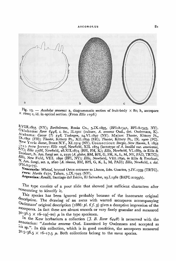
81ASCOBOLUS
NY);
S-yin.'ögs (NY); Bartholomew,.... __ . ,
Rooks Co., 5.IX.1895, (BPI-A1541, BPI-A1543, NY),yklahoma: "
Rorer 6448, s. loc., Il.igoi (culture, A. amoenus Oud., det. Oudemans, K).Carvar (?) 248, Tuskegee, 24.VI.18g7 (NY). Maine: Thaxter,
O-, -1 „ , Kittery Pt.,l8
?_i (FH); Thaxter, Kittery Pt., XII.1899 (FH); Thaxter, Kittery Pt., IX. 1900 (PC).York: ~ ~
'
"""
"
' - -----
Seaver, Bronx N.Y., XI.1914 (NY). Connecticut: Sturgis, New Haven, I. 1893New Jersey: Ellis 1o)6, Newfield, XII. 1874 (lectotype of A. leveillei var. americanus,
Ellis 2386, Newfield, 28.XII.1874 (BPI, FH, K); Ellis,p
'
_ .
, Newfield, VI.i88g, in Ellis &Vet'hart, N. Am. fungi ser. 2, 2333 (A. glaber,
P,, , BM, BPI, G, FH, K, L, M, NY, PAD, TRTC);£"u, New Field, VIII. 1890 (BPI, NY);
"" "" " " ~ * """"
Ellis, Newfield, VIII.1890, in Ellis & Everhart,,fungi, ser. 2, 2620 (A. vinosus, BM, BPI, G, K, L, M, PAD); Ellis, Newfield, s. dat.( p H-A
3i73).Yenezuela: Whetzel,
,beyond Ortez entrance to Llanos, Edo. Guarico, 7.IV. 1939 (TRTG).
**eru: Martin 6270, Talara, 9.IX. 194.5 (NY).Argentina:Ranalli, Santiago del Estero, El Salvador, 23.1.1962 (BAFC 21299b).
Thetype consists of a poor slide that showed just sufficient characters after
to identify it.
T his species has been ignored probably because of the inaccurate originalascription. The drawing of an ascus with warted ascospores accompanyingndemans' original description (1886: pi. 6 f. 3) gives a deceptive impression of the
ascosP°res. In fact these are almost smooth or very finely granular and measured
30-36.5 x i6-ig(-2o) /< in the type specimen.' n l he Kew herbarium a collection (J. B. Rarer 6448) is preserved with the
f"notation: “Ascobolus amoenus Oud. Examined by Oudemans and accepted as
s sP-'\ In this collection, which is in good condition, the ascospores measured3o-5-36.5 X 16-17.5 41. Both collections belong to the same species.
Fig. 13. — Ascobolus amoenus: a, diagrammatic section of fruit-body X 80; h, ascosporeX 1600; c, id. in optical section. (From Ellis 1096.)
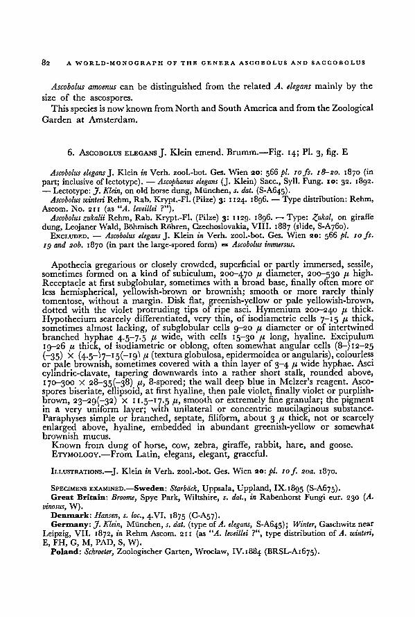
82A WORLD-MONOGRAPH OF THE GENERA ASCOBOLUS AND SACCOBOLUS
Ascobolus amoenus can be distinguished from the related A. elegans mainly by the
size of the ascospores.
This species is now known fromNorth and South America and from the Zoological
Garden at Amsterdam.
6. ASCOBOLUS ELEGANS J. Klein emend. Brumm.—Fig. 14; Pl. 3, fig. E
Ascobolus elegans J. Klein in Verh. zool.-bot. Ges. Wien 20: 566 pi. 10 fs. 18-20. 1870 (in
part; inclusive of lectotype). — Ascophanus elegans (J. Klein) Sacc., Syll. Fung. 10: 32. 1892.
— Lectotype: J. Klein, on old horse dung, Miinchen, s. dat. (S-A645).Ascobolus winteri Rehm, Rab. Krypt.-Fl. (Pilze) 3: 1124. 1896. — Type distribution: Rehm,
Ascom. No. 211 (as “A. leveillei ?”).Ascobolus zukalii Rehm, Rab. Krypt.-Fl. (Pilze) 3: 1129. 1896. — Type: on giraffe
dung, Leojaner Wald, Bohmisch Rohren, Czechoslovakia, VIII. 1887 (slide, S-A760).EXCLUDED. — Ascobolus elegans J. Klein in Verh. zool.-bot. Ges. Wien 20: 566 pi. 10 fs.
ig and 20b. 1870 (in part the large-spored form) = Ascobolus immersus.
Apothecia gregarious or closely crowded, superficial or partly immersed, sessile,sometimes formed on a kind of subiculum, 200-470 [i diameter, 200-530 /< high.Receptacle at first subglobular, sometimes with a broad base, finally often more or
less hemispherical, yellowish-brown or brownish; smooth or more rarely thinly
tomentose, without a margin. Disk flat, greenish-yellow or pale yellowish-brown,dotted with the violet protruding tips of ripe asci. Hymenium 200-240 /u thick.
Hypothecium scarcely differentiated, very thin, of isodiametric cells 7-15 fx thick,sometimes almost lacking, of subglobular cells 9-20 fx diameter or of intertwined
branched hyphae 4.5-7.5 fx wide, with cells 15-30 fx long, hyaline. Excipulum
19-26 fx thick, of isodiametric or oblong, often somewhat angular cells (8-) 12-25
(—35) x (4.5 —"i 7—15(—19"! fx (textura globulosa, epidermoidca or angularis), colourless
or pale brownish, sometimes covered with a thin layer of 3-4 fx wide hyphae. Asci
cylindric-clavate, tapering downwards into a rather short stalk, rounded above,
170-300 X 28-35(~38) fx, 8-spored; the wall deep blue in Melzer's reagent. Asco-
spores biseriate, ellipsoid, at first hyaline, then pale violet, finally violet or purplish-brown, X 11.5-17.5 /<, smooth or extremely fine granular; the pigmentin a very uniform layer; with unilateral or concentric mucilaginous substance.
Paraphyses simple or branched, septate, filiform, about 3 fx thick, not or scarcely
enlarged above, hyaline, embedded in abundant greenish-yellow or somewhat
brownish mucus.
Known from dung of horse, cow, zebra, giraffe, rabbit, hare, and goose.
ETYMOLOGY. —From Latin, elegans, elegant, graceful.
ILLUSTRATIONS.— J. Klein in Verh. zool.-bot. Ges. Wien 20: pi. 10f. 20a. 1870.
SPECIMENS EXAMINED.—Sweden: Starback, Uppsala, Uppland, IX.1895 (S-A675).Great Britain: Broome, Spye Park, Wiltshire, s. dat., in Rabenhorst Fungi eur. 230 (A.
vinosus, W).Denmark: Hansen, s. loc., 4.VI. 1875 (C-A57).Germany: J. Klein, Miinchen, s. dat. (type ofA. elegans, S-A645); Winter, Gaschwitz near
Leipzig, VII. 1872, in Rehm Ascom. 211 (as “A. leveillei ?", type distribution of A. winteri,
E, FH, G, M, PAD, S, W).
Poland: Schroeter, Zoologischer Garten, Wroclaw, IV. 1884 (BRSL-A1675).
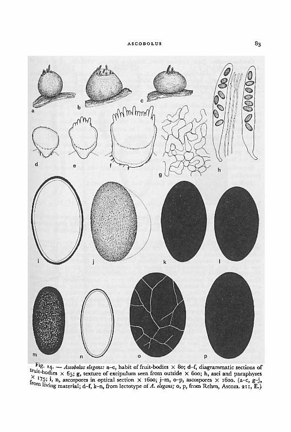
83ASCOBOLUS
o, p, from Rehm, Ascom. 211, E.)A. elegans;
Ascobolus elegans: a-c, habit of fruit-bodies X 80; d-f, diagrammatic sections offruit-bodies x 65; g, texture of excipulum seen from outside X 600; h, asci and paraphyses
x
175; i, n, ascospores in optical section X 1600; j-m, o-p, ascospores X 1600. (a-c, g-j,from living material; d-f, k-n, from lectotype of
Fig. 14.—
84 A WORLD-MONOGRAPH OF THE GENERA AaCOBOLUS AND SACCOBOLUS
Czechoslovakia: Zukal, Leojaner Wald, Bohmisch Rohren, VIII. 1887 (slide, type of
A. zukalii, S-A760).U.S.A.: New York: Dodge, s. loc., 21.X.1911 (culture, NY-A1088); Seaver, Yonkers,
XI.1915 (NY-A1087). Maryland: Toung, Middeltown, XI. 1932 (BPI-A1554). Virginia:
Meyer 14.J26, Mountain Lake Biological Station, 5.VIII.ig42 (in moist chamber,
TRTC)."
From the original description by Klein (I.e.) it is clear that two species were
involved, that appeared successively in his cultures. The first was a small-spored
species, which he described extensively and of which material has been preserved.This material is considered the lectotype. The second was a large-spored species,
which is identifiable as A. immersus as Heimerl (1889) already concluded.
Ascobolus elegans can be distinguished from A. amoenus and A. mancus by the size
of the ascospores, and from A. boudieri by the smooth, faintly coloured receptacle
and the lack of sulphur yellow hymenial mucus.
According to Dodge (1920) in this species ascogonia can be formed without
preceeding formation of antheridia.
Ascobolus elegans is known from Europe and North America.
7. Ascobolus mancus (Rehm) Brumm., stat. & comb. nov. —Fig. 15; Pl. 3, fig. F
Ascobolus mancusRehm in Sydow, Mycoth. March. Cent. 37, No. 3676. 1892 (nomen nudum).— Ascobolus winteri var. mancus Rehm, Rab. Krypt.-Fl. (Pilze) 3: 1124. 1896 (basionym). —
Holotype: P. Sydow, on dungof rabbit, near Berlin, Germany, 3.X.1892 (S-A678).
Apothecia closely crowded or gregarious, superficial, sessile, 1 50-400 11 diameter,about 300 f.i high. Receptacle at first closed and globular, then opening, finallyoften hemispherical, olive-green, brownish-green or sometimes blackish-brown,
smooth, without margin. Disk flat to convex, yellowish-green, dotted with the violet
tips of ripe asci. Hymenium filling almost the entire fruit-body, about 230 /i thick.
Plypothecium very thin, not continuous, of isodiametric cells 3.5-7.5 /< diameter.
Flesh very thin, of subglobular or oblong cells 7-17 it long, hyaline. Excipulumabout 15 fi thick, of more or less angular, isodiametric or oblong, rather thick-
walled cells 10-30 x 6-15 11 (textura angularisl with pale brownish or greenish-yellow walls. Asci clavate, slightly curved, tapering downwards into a short base,rounded above, 230-320 X 29-32 //, 8-spored, the wall deep blue in Melzer's
reagent. Ascospores biseriate, ellipsoid, at first hyaline, then violet, finally often
brownish, (17.5 —) 19-23(-24) X io-I2(—13) //., smooth or extremely fine granular,with unilateral mucilaginous substance. Paraphyses simple or branched, with few
septa, filiform, 2-3 f.i thick, not enlarged upwards, hyaline, embedded in abundant
yellowish-green mucus.
On dung of horse, hare and rabbit, also on rotting leaves of hop.ETYMOLOGY.—From Latin, mancus, defective, imperfect.
SPECIMENS EXAMINED.—France: Meslin, Caen, Calvados, 4.IX.1938 (PC-A2327,PC-A2364).
Germany: Staritz, near Ziebigk - Dessau, IX. 1907 (S-A548); Sydow, Lichterfelde near
Berlin, VIII.1887, in Myc. march. 1578 (A. glaber, BM, HBG, K, S); Sydow, Lichterfeldenear
Berlin, IX.1892, in Myc. march. 3676 (A. mancus, BM, HBG, S); Sydow, s. loc., 3.X.1892
(lectotype of A. mancus, S-A678).
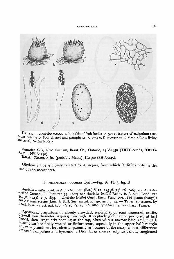
85ASCOBOLUS
Canada: Cain,,
New Durham, Brant Co., Ontario, 24.V.1930 (TRTC-A2162, TRTC-
2i73> NY-A1340).U.S.A.: Thaxter, s. loc. (probably Maine), II. 1900 (FH-A3145).
Obviously this is closely related to A. elegans, from which it differs only in the
Slz c of the ascospores.
8. ASCOBOLUS BOUDIERI Quél. —Fig. 16; Pl. 5, fig. B
Ascobolus leveillei Boud. in Annls Sci. nat. (Bot.) V 10: 225 pl. 7 ƒ. 16. i86g; notAscobolus
leveillei Crouan, Fl. Finistere 57. 1867; nor Ascobolus leveillei Renny in J. Bot., Lond. 12:
356 pi. 1 /j. /-j. 1874. —Ascobolus boudieri Quel., Ench. Fung. 293. 1886 (name change);
not Ascobolus boudieri,,
Lort. in Bull. Soc. mycol. Fr. 30: 223. 1914. — Type: represented by°ud. in Annls Sci. nat. (Bot.) V 10: pi. jf. 16. 1869; type locality, near Paris, France.
Apothecia gregarious or closely crowded, superficial or semi-immersed, sessile,•3-0.6 mm diameter, 0.3-0.5 mm high. Receptacle globular or pyriform, at first
k°sed, then irregularly opening at the top, often with a narrow base, rather darkr
own; surface finely warted or furfuraceous, especially in the upper half; margin
bot Very prominent but often apparently so because of the sharp colour-differencesehveen excipulum and hymenium. Disk flat or convex, sulphur yellow, roughened
Fig. 15. — a, b, habit of fruit-bodies X 50; c, texture of excipulum seen
fr om outside X 600; d, asci and paraphyses X 175; e, f, ascospores X 1600. (From livingmaterial, Netherlands.)
Ascobolus mancus:
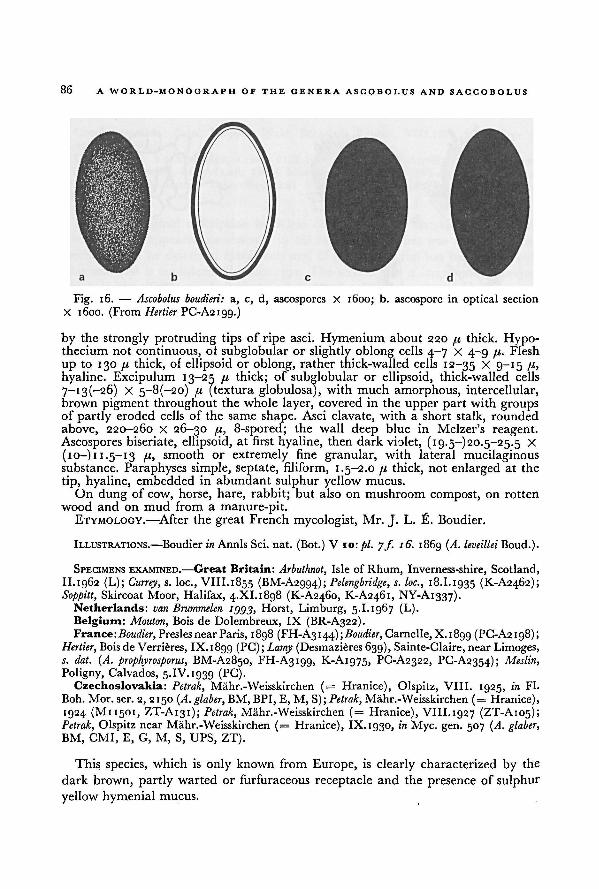
86 A WORLD-MONOGRAPH OF THE GENERA ASCOBOLUS AND SACCOBOLUS
by the strongly protruding tips of ripe asci. Hymenium about 220 /x thick. Hypo-thecium not continuous, of subglobular or slightly oblong cells 4-7 X 4-9 /x. Flesh
up to 130 pi thick, of ellipsoid or oblong, rather thick-walled cells 12-35 X 9-15 79
hyaline. Excipulum 13-25 fx thick; of subglobular or ellipsoid, thick-walled cells
7-i3(-26) X 5-8(-20) fx (textura globulosa), with much amorphous, intercellular,brown pigment throughout the whole layer, covered in the upper part with groupsof partly eroded cells of the same shape. Asci clavate, with a short stalk, rounded
above, 220-260 X 26-30 /x, 8-spored; the wall deep blue in Melzer's reagent.Ascospores biseriate, ellipsoid, at first hyaline, then dark violet, (19.5-) 20.5-25.5 X
(10-111.5-13 ix, smooth or extremely fine granular, with lateral mucilaginoussubstance. Paraphyses simple, septate, filiform, 1.5-2.0 px thick, not enlarged at the
tip, hyaline, embedded in abundant sulphur yellow mucus.
On dung of cow, horse, hare, rabbit; but also on mushroom compost, on rotten
wood and on mud from a manure-pit.ETYMOLOGY.—After the great French mycologist, Mr. J. L. FI. Boudier.
ILLUSTRATIONS.—Boudier in Annls Sci. nat. (Bot.) V 10: pi. 7 ƒ. 16. 1869 (A. leveillei Boud.).
SPECIMENS EXAMINED.—Great Britain: Arbuthnot, Isle of Rhum, Inverness-shire, Scotland,
II.1962 (L); Currey, s. loc., VIII.1855 (BM-A2994); Pelengbridge, s. loc., 18.1.1935 (K-AQ462);Soppitt, Skircoat Moor, Halifax, 4.XI.1898 (K-AQ460, K-A2461, NY-A1337).
Netherlands: van Brummelen t'j'j'j, Horst, Limburg, 5.1.1967 (L).
Belgium: Mouton, Bois de Dolembreux, IX (BR-A322).France: Boudier, Presles nearParis, 1898 (FH-A3144); Boudier, Carnelle, X. 1899 (PC-A2198);
Hertier, Bois de Verrieres, IX. 1899 (PC); Lamy (Desmazieres 639), Sainte-Claire, nearLimoges,s. dat. (A. prophyrosporus, BM-A2850, FH-A3199, K-A1975, PC-A2322, PC-A2354); Meslin,
Poligny, Calvados, 5.IV. 1939 (PC).Czechoslovakia: Petrak, Mahr.-Weisskirchen (= Hranice), Olspitz, VIII. 1925, in Fl.
Boh. Mor. ser. 2, 2150 (A. glaber,BM, BPI, E, M, S); Petrak, Mahr.-Weisskirchen (= Hranice),
1924 (M11501, ZT-A131); Petrak, Mahr.-Weisskirchen (= Hranice), VIII. 1927 (ZT-A105);Petrak, Olspitz near Mahr.-Weisskirchen (= Hranice), IX. 1930, in Myc. gen. 507 (A. glaber,
BM, CMI, E, G, M, S, UPS, ZT).
This species, which is only known from Europe, is clearly characterized by the
dark brown, partly warted or furfuraceous receptacle and the presence of sulphur
yellow hymenial mucus.
a, c, d, ascospores X 1600; b. ascospore in optical section
X 1600. (FromFig. 16. — Ascobolus boudieri:
Hertier PC-A2199.)
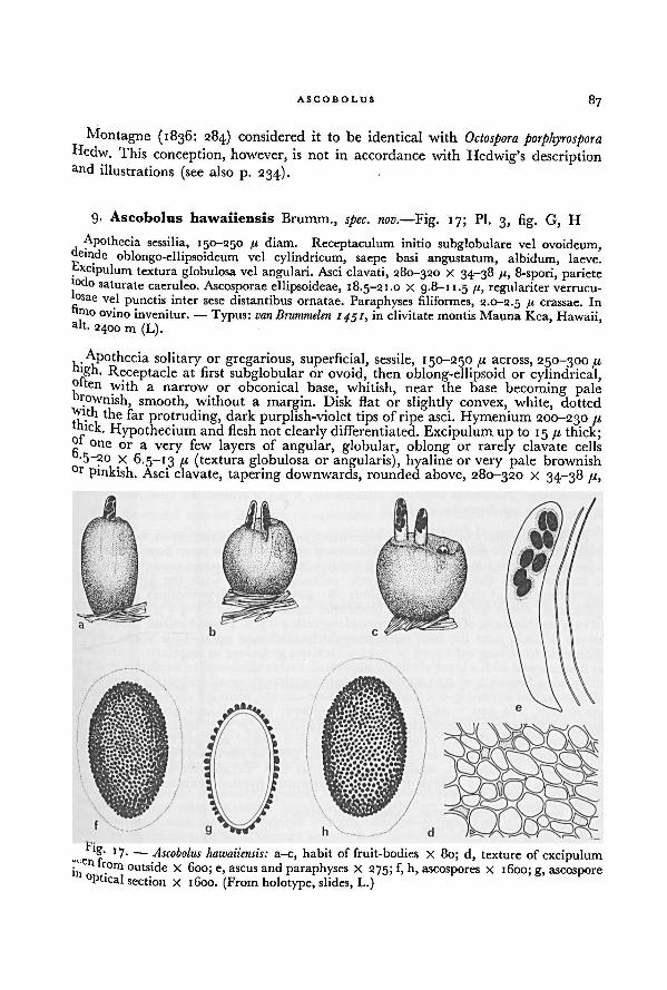
87ASCOBOLUS
Montagne (1836: 284) considered it to be identical with Octospora porphyrosporaHedw. This conception, however, is not in accordance with Hedwig's descriptionand illustrations (see also p. 234).
9. Ascobolus hawaiiensis Brumm., spec. now.—Fig. 17; Pl. 3, fig. G, H
Apothecia sessilia, 150-250 fi diam. Receptaculum initio subglobulare vel ovoideum,«einde oblongo-ellipsoideum vel cylindricum, saepe basi angustatum, albidum, laeve.
. cipulum textura globulosa vel angulari. Asci clavati, 280-320 X 34-38 /<, 8-spori, pariete
j°do saturate caeruleo. Ascosporae ellipsoideae, 18.5-21.0 X 9.8-11.5 /«, regulariter verrucu-
osae vel punctis inter sese distantibus ornatae. Paraphyses filiformes, 2.0-2.5 /1 crassae. Intoo ovino invenitur.— Typus: van Brummelen 1451, in clivitatemontis Mauna Kea, Hawaii,a't. 2400 m (L).
Apothecia solitary or gregarious, superficial, sessile, 150-250 [jl across, 250-300 41!Sh. Receptacle at first subglobular or ovoid, then oblong-ellipsoid or cylindrical,
olten with a narrow or obconical base, whitish, near the base becoming palerownish, smooth, without a margin. Disk flat or slightly convex, white, dotted
With the far protruding, dark purplish-violet tips of ripe asci. Hymenium 200-230 /.ib'ek. Hypothecium and flesh not clearly differentiated. Excipulum up to 15 /.«thick;
one or a very few layers of angular, globular, oblong or rarely clavate cells
•5~ao X 6.5-13 ft (textura globulosa or angularis), hyaline or very pale brownish0r pinkish. Asci clavate, tapering downwards, rounded above, 280-320 x 34-38 41,
Fig. 17. —
a-c, habit of fruit-bodies X 80; d, texture of excipulumseen from outside X 600; e, ascusand paraphyses X 275; f, h, ascospores X 1600; g, ascospore
in optical section X 1600. (From holotype, slides, L.)
Ascobolus hawaiiensis:
88 A WORLD-MONOGRAPH OF THE GENERA ASCOBOLUS AND SACCOBOLUS
8-spored; the wall deep blue in Melzer's reagent. Ascospores biseriate, ellipsoid, at
first hyaline, then purplish-violet, finally purplish-brown, 18.5-21 X 10-11.5 71,
ornamented with a regular pattern of very fine warts or isolated punctae; pigmentin a layer ofuniformthickness; surrounded by a thick gelatinous envelope. Paraphyses
simple or branched, filiform, with few septae, 2.0-2.5 B thick, not enlarged above,
hyaline, without mucus.
Known only from dung of sheep.ETYMOLOGY.—After Hawaii.
SPECIMEN EXAMINED. — Hawaii: van Brummelen 1451, onsheep dung, Puuhookomo, Mauna
Kea, io.V.i962, substratum sent by Dr. H. O. Sleumer (type of A. hawaiiensis, L).
Apparently this is a very rare species. It is sufficiently characterized by the very
small fruit-bodies and the warted or punctate ascospores. Like in A. immersus the
ascospores are surrounded on all sides by a thick gelatinous layer.
10. Ascobolus siamensis Brumm., spec. nov. —Fig. 18; Pl. 3, fig. I, Pl. 4, figs. A-G,Pl. 5 , fig. A
Apothecia sessilia, 0.3-0.9 mm diam. Receptaculum subglobulare, primo clausum et rubro-
brunneum, denique prope apicem irregulariter fissum, luteum, particulis rubro-brunneis
furfuraceis adspersum, margine nvdlo. Discus luteus. Excipulum textura globulosa usque
angulari. Asci clavato-saccati, 180-200 X 29-32 ft, usque ad 8-spori, pariete iodo haud
caerulescente. Ascosporae subglobulares, 17-19.5 X 15— 16-5(— 17) ft, verrucis rotundis
ornamentatae. Paraphyses simplices, cylindraceae, c. 2.5 fi crassae. In fimo Naemorhedi
grisei. Typus: van Brummelen 1776 (L).
Apothecia solitary or gregarious, superficial, sessile 0.3-0.9 mm across, 0.3-0.6 mm
high. Receptacle subglobular, at first closed and reddish-brown, then irregularlyopening near the top and yellow with reddish-brown furfuraceous particles, without
a margin. Disk flat to slighly convex, yellowish, dotted with the almost black
protruding tips of ripe asci. Hymenium 180-200 fx thick. Hypothecium not clearly
delimited, about 30 fi thick, with branched, septate hyphae 2.5-4.5 thick. Flesh
ofvarying thickness, ofisodiametricor oblong cells 4-17 X 4-9 fx, hyaline. Excipulum
20-29 [x thick, near the base often irregularly thickened up to 45 fx thick, of iso-
diametric to oblong cells 4-12 X 7-21 fx (textura globulosa to angularis) yellowish,at first covered with a layer of closely appressed, rather thick-walled hyphae 3.0-4.5 fx
thick, soon breaking up into a great number ofscattered, small scales and a net-work
of thin-walled hyphae 5-9 71 thick. Asci clavate-saccate, rounded above, with a large
operculum, 180-200 X 29-32 fx, up to 8-spored; the wall not blue in Melzer's
reagent. Ascospores irregularly biseriate, subglobular, at first hyaline, then violet
to almost black, 17-19.5 x 15-16.5(—17) fx, in i-spored asci up to 31.5 X 29 fx,
without oil drops or granules, ornamentedwith more or less closely placed, rounded
warts, with transparent, unilateral, gelatinous cap. Paraphyses simple, septate,cylindrical, about 2.5 fx thick, not enlarged upwards, hyaline, without mucilaginoussubstance.
On dung of Burmese goat (Naemorhedus griseus).ETYMOLOGY.—From Siam.
SPECIMEN EXAMINED.—Thailand: van Brummelen 1776, cultured on dung of Burmese goat
(collected by Dr. H. O. Sleumer), limestone massif of the Pha Nok Khao, ca. 800 m, South
of Loey, Prov. Udawn, 13.XI. 1963 (type of A. siamensis, L).
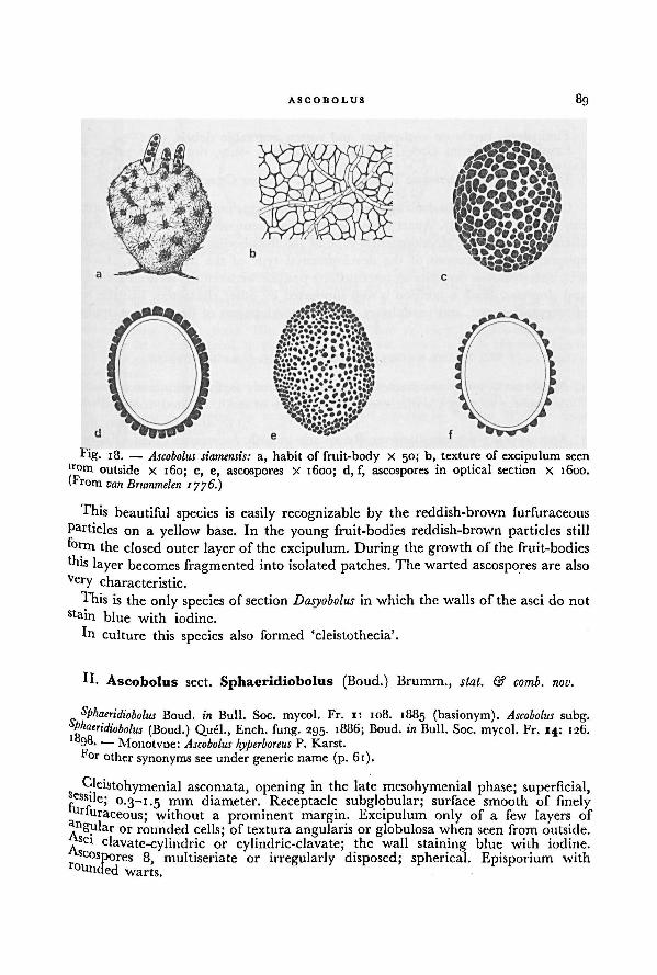
89ASCOBOLUS
This beautiful species is easily recognizable by the reddish-brown furfuraceous
Particles on a yellow base. In the young fruit-bodies reddish-brown particles still
form the closed outer layer of the excipulum. During the growth of the fruit-bodies
'his layer becomes fragmented into isolated patches. The warted ascospores are also
Very characteristic.
This is the only species of section Dasyobolus in which the walls of the asci do not
stain blue with iodine.
in culture this species also formed 'cleistothecia'.
II. Ascobolus sect. Sphaeridiobolus (Boud.) Brumm., stat. & comb. nov.
Sphaeridiobolus Boud. in Bull. Soc. mycol. Fr. i: 108. 1885 (basionym). Ascobolus subg.Sphaeridiobolus (Boud.) Quel., Ench. fung. 295. x886; Boud. in Buil. Soc. mycol. Fr. 14: 126.
'898. — Monotype: Ascobolus hyperboreus P. Karst.
For other synonyms see under generic name (p. 61).
Gleistohymenial ascomata, opening in the late mesohymenial phase; superficial,■ essxle; 0.3-1.5 mm diameter. Receptacle subglobular; surface smooth of finelymiuraceous; without a prominent margin. Excipulum only of a few layers of
ngular or rounded cells; of textura angularis or globulosa when seen from outside,set clavate-cylindric or cylindric-clavate; the wall staining blue with iodine.
Oospores 8, multiseriate or irregularly disposed; spherical. Episporium withr °unded
warts.
Fig. 18.— a, habit of fruit-body X 50; b, texture of excipulum seen
from outside X 160; c, e, ascospores X 1600; d, f, ascospores in optical section X 1600.
(From van Brummelen 1776.)
Ascobolus siamensis:
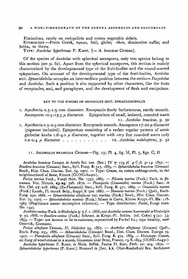
90 A WORLD-MONOGRAPH OF THE GENERA ASCOBOLUS AND SACCOBOLUS
Fimicolous, rarely on owl-pellets and rotten vegetable debris.
ETYMOLOGY.—
From Greek, ocpatpa, ball, globe; -iSiov, diminutive suffix; and
PXA/.CO, to throw.
TYPE: Ascobolus hyperboreus P. Karst. [= A. brassicae Crouan],
Of the species of Ascobolus with spherical ascospores, only two species belong to
this section (see p. 62). Apart from the spherical ascospores, this section is mainly
characterized by the developmental type ol the fruit-bodies and the round-warted
episporium. On account of the developmental type of the fruit-bodies, Ascobolus
sect. Sphaeridiobolus occupies an intermediateposition between the sections Dasyobolusand Ascobolus.
.
Such a position is also supported by other characters, like the form
of receptacles, asci, and paraphyses, and the development of flesh and excipulum.
KEY TO THE SPECIES OF ASCOBOLUS SECT. SPHAERIDIOBOLUS
i. Apothecia 0.5-1.5 mm diameter. Receptacle finely furfuraceous, rarely smooth.
Ascospores 10.5—13.5 u diameter. Episporium of small, isolated, rounded warts
11. Ascobolus brassicae, p. go
i. Apothecia 0.3-0.5mm diameter. Receptacle smooth. Ascospores 17-20 /< diameter
(pigment included). Episporium consisting of a rather regular pattern of semi-
globular knobs 1.8-4.0 n diameter, together with very fine rounded warts only
0.2-0.4 n diameter 12. Ascobolus nodulosporus, p. 92
II. ASCOBOLUS BRASSICAE Crouan—Fig. 19; Pl. 4, fig. H, Pl. 5, figs. C, D
Ascobolus brassicae Crouan in Annls Sci. nat. (Bot.) IV 7: 174/)/. 4 C fs. 9-14. 1857. —
Boudiera brassicae (Crouan) Sacc., Syll. Fung. 8: 513. 1889.— Sphaeridiobolus brassicae (Crouan)
Boud., Hist. Class. Discom. Eur. 74. 1907. — Type: Crouan, on rotten cabbage-stem, in the
neighbourhoodof Brest, France (CONC-A2401).Peziza murina Fuck., Fungi rhen. No. 1597. 1865. — Plicaria murina (Fuck.) Fuck, in Jb.
nassau. Ver. Naturk. 23-24: 326. 1870. ■— Phaeopezia (Crouaniella) murina (Fuck.) Sacc. in
Bot. Cbl. 17: 218. 1884; (Eu-Phaeopezia) Sacc., Syll. Fung. 8: 471. 1889. — Crouaniella murina
(Fuck.) Lamb., Fl. mycol. Belg., Suppl. I: 320. 1887. — Humaria murina (Fuck.) Quel., Ench.
Fung. 2go. 1886. — Sphaeridiobolus albofuscus var. murinus (Fuck.) Boud., Hist. Class. Discom.
Eur. 74. 1907. — Sphaeridiobolus murinus (Fuck.) Moser in Gams, Kleine Krypt.-Fl. Ha: 116.
1963 (illegitimate name: incomplete reference). — Type distribution: Fuckel, Fungi rhen.
No. 1597.
Ascobolus caninusFuck. in Hedwigia 5:3 f. 1. 1866;not Ascobolus caninus Auerswald in Hedwigia
7: 52. 1868.-—Boudiera canina (Fuck.) Schroet. in Krypt.-Fl. Schles. (ed. Cohn) 3(2): 55-
t893- —Type: not known to be in existence, represented by Fuckel I.e.; type locality, near
Oestrich, Germany.Peziza albofusca Crouan, Fl. Finistere 54. 1867. —
Ascobolus albofuscus (Crouan) Quel.,Ench. Fung. 295. 1886. — Sphaeridiobolus (Crouan) Boud., Hist. Glass. Discom. Europe 74-
1907. — Phaeopezia albofusca (Crouan) Sacc., Syll. Fung. 8: 472. 1889. — Holotype: Crouan,
on dung of wood-mousein a marsh, Gouesnou nearBrest, France, 13.X. 1854 (CONC-A243 1) ■Ascobolus hyperboreus P. Karst. in Notis. Sallsk. Fauna Fl. fenn. Forh. 11: 204. 1870. —•
Sphaeridiobolus hyperboreus (P. Karst.) Heimerl in Jber. k.k. Ober-Realschule Bez. Sechshaus
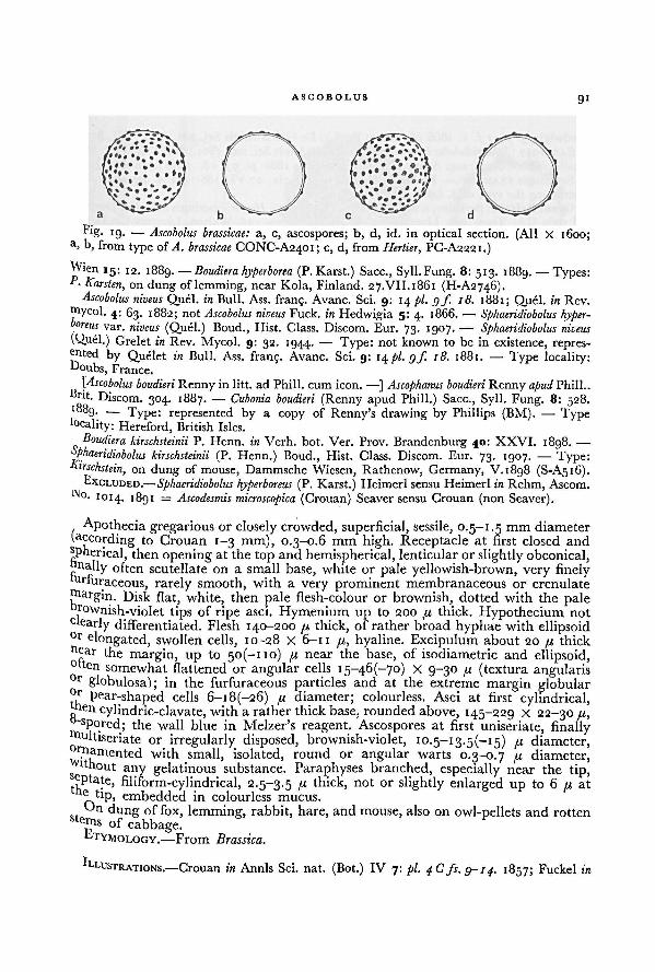
ASCOIiOLUS 9 1
Wien15: 12. 1889. —
Boudiera hyperborea (P. Karst.) Sacc., Syll.Fung. 8: 513. 1889. —Types:P- Karsten, on dung of lemming, near Kola, Finland. 27.VII.1861 (H-A2746).
Ascobolus niveus Quel, in Bull. Ass. frang. Avanc. Sci. 9: 14 pi. gf. 18. 1881; Quel, in Rev.
Oiycol. 4: 63. 1882; not Ascobolus niveus Fuck, in Hedwigia 5: 4. 1866. — Sphaeridiobolus hyper-boreus var. niveus (Quel.) Boud., Hist. Class. Discom. Eur. 73. 1907. —• Sphaeridiobolus niveus
(Quel.) Grelet in Rev. Mycol. 9: 32. 1944. — Type: not known to be in existence, repres-ented by Quelet in Bull. Ass. franj. Avanc. Sci. 9: 14 pi. gf. 18. 1881. — Type locality:IJ°ubs, France.
[Ascobolus boudieri Renny in litt. ad Phill. cum icon. —] Ascophanus boudieri Renny apudBrit. Discom. 304. 1887. — Cubonia boudieri (Renny apud Phill.) Sacc., Syll. Fung. 8: 528.1889. — Type: represented by a copy of Renny's drawing by Phillips (BM). — Type
locality: Hereford, British Isles.Boudiera kirschsteinii P. Henn. in Verh. bot. Ver. Prov. Brandenburg 40: XXVI. 1898. —•
Sphaeridiobolus kirschsteinii (P. Henn.) Boud., Hist. Class. Discom. Eur. 73. 1907. — Type:on dung of mouse, Dammsche Wiesen, Rathenow, Germany, V.1898 (S-A516).
Kirschstein,
EXCLUDED.— Sphaeridiobolushyperboreus- r 'vi
(I1- Karst.) Heimerl sensuHeimerl in Rehm, Ascom.
1014. 18gI = Ascodesmis microscopica (Crouan) Seaver sensu Crouan (non Seaver).
Apothecia gregarious or closely crowded, superficial, sessile, 0.5-1.5 mm diameter
(.according to Crouan 1-3 mm), 0.3-0.6 mm high. Receptacle at first closed and
Pherical, then opening at the top and hemispherical, lenticularor slightly obconical,nnally often scutellate on a small base, white or pale yellowish-brown, very finelyfirfuraceous, rarely smooth, with a very prominent membranaceous or crenulate
ntargin. Disk flat, white, then pale flesh-colour or brownish, dotted with the palefi'°Wnish-vi°let s r ipe asci. Hymenium up to 200 u thick. Hypothecium not
clearly differentiated. Flesh 140-200 n thick, of rather broad hyphae with ellipsoid0r elongated, swollen cells, 10-28 X 6-11 /,<, hyaline. Excipulum about 20 /.i thick
near the margin, up to 5O(-IIO) 41 near the base, of isodiametric and ellipsoid,°nen somewhat flattened or angular cells 15-46(-70) X 9-30 /t (textura angularis°r globulosa); in the furfuraceous particles and at the extreme margin globularfir
pear-shaped cells 6-i8(-26) fi diameter; colourless. Asci at first cylindrical,ten cylindric-clavate, with a rather thick base, rounded above, 145-229 X 22-30 fi," spored; the wall blue in Melzer's reagent. Ascospores at first uniseriate, finallyfiltiseriate or irregularly disposed, brownish-violet, 10.5-13.5(-15) 11 diameter,
fi'namented with small, isolated, round or angular warts 0.3-0.7 /j, diameter,vl thout
any gelatinous substance. Paraphyses branched, especially near the tip,cptate, filiform-cylindrical, 2.5-3.5 ,u thick, not or slightly enlarged up to 6 /r at
tip, embedded in colourless mucus.
tJn dung of fox, lemming, rabbit, hare, and mouse, also on owl-pellets and rottenstems of cabbage.
b'i'YMOLOGY.—From Brassica.
ILLUSTRATIONS.—Crouan in Annls Sci. nat. (Bot.) IV 7: pi. 4C fs. 9-14. 1857; Fuckel in
Fig. 19. — Ascobolus brassicae: a, c, ascospores; b, d, id. in optical section. (All X 1600;a, b, from type of CONC-A2401; c, d, from Hertier, PC-A2221.)A. brassicae
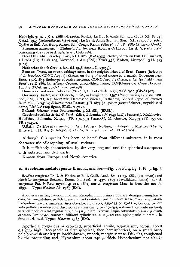
A WORLD-MONOGRAPH OF THE GENERA ASCOBOLUS AND SACCOBOLUS92
Hedwigia 5: pi. if. 1. 1866 (A. caninus Fuck.); Le Gal in Annls Sci. nat. (Bot.) XI 8: 241
f. 64A. 1947 (Sphaeridiobolus hyperboreus); Le Gal in Annls Sci. nat. (Bot.) XII r: 466 f.j. 1961;
Quelet in Bull. Ass. frang. Avanc. Sci., Congr. Reims 1880: pi. gf. 18. 1880 (A. niveus Quel.).SPECIMENS EXAMINED.— Finland: Karsten, near Kola, 27.VII.1861 (as A. lapponicus, also
containing the type of A. hyperboreus, H-A2746).Great Britain: Berkeley, s. loc., 9.III. 1874 (K-A1993); Harper, Sherburn Hill, Co. Durham,
1.I.1962 (L); Travis sog, Liverpool, s. dat. (BM); Travis 356, Walton, Liverpool, 3.11.1929
(BMj.Netherlands: de Groot, s. loc., 6.I.1946 (icon., L-A3253).France: Crouan, on rotten cabbage-stem, in the neighbourhood of Brest, France (holotype
of A. brassicae, CONC-A2401); Crouan, on dung of wood-mouse in a marsh, Gouesnou near
Brest, 13.X. 1854 (holotype of Peziza albofusca, CONC-A2431); Crouan, s. loc. (probably near
Brest), 18.II.1864 ( A. vulpinus Crouan, unpublished name, CONC-A2377); Hertier, Ecouen,
II.1895 (PC-A2221, PC-A2222, S-A558).Denmark: unknown collector ("E.R."), S. Tokkckob Hegn, 7.IV.1919 (CP-A1407).Germany: Fuckel, near Oestrich, s. dat., in Fungi rhen. 1597 ( Peziza murina, type distribu-
tion, BM, GRO, K); Kirschstein, Dammsche Wiesen, Rathenow, V.1898 (type of Boudiera
kirschsteinii, S-A516); Schroeter, nearRastatt, 3.II.1873 (A. sphaerospermus Schroet., unpublished
name, BRSL-A1709 figure, BRSL-A1712).
Poland: Schroeter, near Frankenstein, 4.XI.1883 (BRSL).Czechoslovakia: Svrcek & Vacek, Zdice, Bohemia, i.V. 1949 (PR); Velenovsky, Mnichovice,
Hubachov, Bohemia, X.1927 (PR 150453); Velenovsky, Mnichovice, X. 1933 (PR 153000,
PR 150452).U.S.A.: California: Smiley, s. loc., IV.1915 (culture, FH-A3ogg). Maine: Thaxter,
Kittery Pt., II.1894 (FH-A3098); Thaxter, Kittery Pt., .r. dat. (FH-A3I00).
Although this species has been collected from different substrata it is most
characteristic of droppings of small rodents.
It is sufficiently characterized by the very long asci and the spherical ascospores
with isolated, rounded warts.
Known from Europe and North America.
12. Ascobolus nodulosporus Brumm., nom. nov. —Fig. 20; Pl. 4, fig. I, Pl. 5, fig. E
Boudiera marginata Phill. & Harkn. in Bull. Calif. Acad. Sci. 1: 25. 1884 (basionym); not
Ascobolus marginatus Schum., Enum. PI. Saell. 2: 437. 1803 (devalidated name); nor A.
marginatus Pat. in Rev. mycol. 4: 211. 1882; nor A. marginatus Mass. in Grevillea 22: 98.
1893. — Type: Harkness No. 2383 (BM).
Apothecia sessilia, 0.3-0.5 mm diam. Receptaculum primo globulare, denique hemisphaeri-
cum, basi angustatum,pallide brunneum vel sordide luteo-brunneum,laeve, margine serratum.
Excipulum textura angulari. Asci clavato-cylindracei, 235-275 X 23-32 fi, 8-spori, parieteiodo pallide caerulescente. Ascosporae sphaericae, (16-) 17-19.5 diam. (pigmento incluso),
verrucis nodulosis sat regularibus, 1.8-4.0 fj, diam., verruculisque rotundatis 0.2-0.4 /1 diam.
ornatae. Paraphyses ramosae, filiformi-cylindricae, c. 2 11 crassae, apice paulo dilatatae. In
fimo muris ratti. Typus: Harkness 2g8g (BM).
Apothecia gregarious or crowded, superficial, sessile, 0.3-0.5 mm across, about
0.3 mm high. Receptacle at first spherical, then hemispherical, on a small base,
pale brownish or dirty yellowish-brown, smooth, margin serrate. Disk flat, roughenedby the protruding asci. Hymenium about 240 pt thick. Hypothecium not clearly
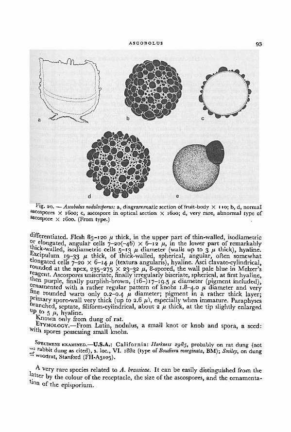
ASCOBOLUS 93
differentiated. Flesh 85- 120 fx thick, in the upper part of thin-walled, isodiametricor elongated, angular cells 7-20(-46) x 6-12 fi, in the lower part of remarkably
p1(:k-walled, isodiametric cells 5-13 fx diameter (walls up to 3 fx thick), hyaline,
'-xcipulum19-33 //. thick, of thick-walled, spherical, angular, often somewhat
( ° n gated cells 7-20 X 6-14 ft (textura angularis), hyaline. Asci clavate-cylindrical,rounded at the apex, 235-275 X 23-32 fi, 8-spored, the wall pale blue in Melzer's
reagent. Ascospores uniseriate, finally irregularly biseriate, spherical, at first hyaline,oen purple, finally purplish-brown, ( 16—) 17—19.5 fx diameter (pigment included),
ornamented with a rather regular pattern of knobs 1.8-4.0 // diameter and very"ne rounded warts only 0.2-0.4 pi diameter; pigment in a rather thick layer;Primary spore-wall very thick (up to 2.6 fx), especially when immature. Paraphyses
ranched, septate, filiform-cylindrical, about 2 /x thick, at the tip slightly enlargedU
P to 5 fx, hyaline.Known only from dung of rat.
ETYMOLOGY.—From Latin, nodulus, a small knot or knob and spora, a seed:Vlth spores possessing small knobs.
SPECIMENS EXAMINED.—U.S.A.: California: Harkness sg85,_, probably on rat dung (notrabbit dung as cited), s. loc., VI. 1882 (type of Boudiera marginata, BM); Smiley,
,
on dungut Woodrat, Stanford (FH-A3105).
Avery rare species related to A. brassicae. It can be easily distinguished from the
ter by the colour of the receptacle, the size of the ascospores, and the ornamenta-
*lf)n of the episporium.
Fig. 20. — Ascobolus nodulosporus: a, diagrammatic section of fruit-body X 110; b, d, normal
ascospores x 1600; c, ascospore in optical section x 1600; d, very rare, abnormal type of
ascospore x 1600. (From type.)
94 A WORLD-MONOGRAPH OF THE GENERA ASCOBOLUS AND SACCOBOLUS
III. ASCOBOLUS sect. ASCOBOLUS
For synonymy see under generic name (p. 61).
Cleistohymenial ascomata, opening in the early or mid-mesohymenial phase;superficial, sessile or stalked; 0.3-12 mm diameter. Receptacle at first subglobularor pyriform, then obconical, cup- or saucer-shaped, finally often flattened; surface
smooth, furfuraceous, rarely with divaricating hyphae or secondary mycelium.Asci cylindric-clavate; with rounded or slightly truncate apex. Ascospores 8; at
first often uniseriate, finally biseriate; ellipsoid, fusoid, subglobular or globular;often with unilateral gelatinous mass. Episporium with a pattern of subparallelanastomosing crevices, with a pattern of irregular, short, curved non-anastomosing
crevices, with a net-work of crevices, with longitudinal ridges of pigment, coarsely
warted, granular or almost smooth. Paraphyses slender, cylindrical, often withswollen ends; often embedded in a colourless, yellowish or yellowish-green, muci-
laginous substance, often containing crystals.
Especially fimicolous, but also foliicolous, lignicolous, terrestrial, and pyrophilous.
The fruit-bodies of most species in this section reach a diameterof one to several
millimeters.These dimensionsare reached by interstitial growth of both the hymenialelements and the flesh as well as theexcipulum. An appreciable activity ofa marginal
growing zone could not be established in any of the species.
In some cultures of Ascobolus albidus, A. furfuraceus, and A. roseopurpurascens sometimes
fruit-bodies were found with only a very few (5-10) ripening asci. In such fruit-
bodies the opening of the hymenium was retarded and occasionally even took
place in the telohymenial phase. Also certain narrow forms of fruit-bodies of A.
albidus may remind one of the fruit-bodies in Ascobolus sect. Dasyobolus. However,
such rare, extremely small forms can be easily recognized as belonging to section
Ascobolus by the form of the asci and the ornamentationof the episporium.
Within this section the species form a very gradual, continuous series. Beginningwith A. albidus this series may be followed via a number of obligatory coprophilous,
and a group of mainly foliicolousand xylophilous to exclusively terrestrial or pyro-
philous species. In this series the components show a constant sequence in the
degree of expression of certain characters. In the last members of the series the
fruit-bodies tend to open earlier in the mesohymenial phase and the ascus-wall
tends to stain less intensively blue with iodine than in the first members; while
the episporial ornamentationgradually varies from a pattern of subparallel anasto-
mosing striae, via a net-work of striae to a pattern of coarse warts. The terrestrial
and pyrophilous species especially show a thick or very thick episporium. The
series is too gradual to make a satisfactory subdivision of this section into subsections
or series.
The position ofonly Ascobolus carletonii and A. cainii is still rather uncertain because
of the diverging type of episporial ornamentation.
ASCOBOLUS 95
KEY TO THE SPECIES OF ASCOBOLUS SECT. ASCOBOLUS
1 • Ascospores globular or subglobular:2. Ascospores globular; 11.5-13.5 /i diameter. Episporium with a pattern of
subparallel, rarely anastomosing crevices. 23. Ascobolus crosslandii, p. 121
2. Ascospores subglobular:
3. Receptacle greenish-yellow, finely furfuraceous. Ascospores 16-19 X 13—16 fi
(pigment included). Episporium very thick (2.5-3.0 /«); with a patternof longitudinal, repeatedly anastomosing or reticulating crevices; with very
prominent projections. On soil.... 42. Ascobolus subglobosus, p. 150
3. Receptacle grass-green, 'hairy'. Ascospores 18-19 X 16-18 11. Episporiumofnormal thickness; with fine, longitudinal anastomosing crevices. On dung
cf. Ascobolus strobolinus, p. 239'• Ascospores ellipsoid, fusiform-ellipsoid or fusoid:
4- Episporium with a pattern of subparallel, anastomosing crevices, or with
a pattern of irregular, short, non-anastomosing crevices; in old or over-ripeascospores sometimes changing into a net-work of crevices (young episporiumnever smooth or granular):
5- Episporium with very densely placed subparallel crevices (more than
12 crevices are visible at each view of the ascospore):6. Ascospores less than 15 11 long. Episporium with relatively broad trans-
verse or oblique crevices 22. Ascobolus lineolatus, p. 120
6. Ascospores more than 20 /< long. Episporium with relatively narrow,
about longitudinal crevices:
7. Ascus-wall clearly blue in Melzer's reagent. Episporium not more
than 1 .0 /x thick. On dung ... 17. Ascobolus laevisporus, p. 112
7. Ascus-wall not or scarcely blue in Melzer's reagent. Episporiumrather thick, up to 2.2 fi. On soil covered with algae
35. Ascobolus demangei, p. 139
5- Episporium not with such a very dense pattern of crevices:
8. Receptacle and disk white or whitish:
9. Receptacle smooth, without a prominent margin. Ascospores 20-36 X
11-14 fx. Episporium with longitudinal occasionally anastomosingcrevices or sometimes with short, more or less curved crevices
13. Ascobolus albidus, p. 100
9. Receptacle finely furfuraceous, with a prominent margin. Ascospores16-20 X 9-10 jx. Episporium with longitudinal anastomosingcrevices 24. Ascobolus sacchariferus, p. 122
8. Receptacle and disk yellowish, greenish, brownish, vinaceous or purplish:10. Receptacle and disk yellowish-brown, brownish, vinaceous or
purplish (beware of a covering of coloured ascospores!). Apothecia
up to 2 mm diameter:
11. Receptacle and disk vinaceous, pinkish-red, purplish or purplish-
96 A WORLD-MONOGRAPH OF THE GENERA ASCOBOLUS AND SACCOBOLUS
brown; smooth or finely furfuraceous; with a clearly differen-
tiated margin. Excipulum, when seen from outside, of textura
epidermoidea. Vinaceous or purplish intercellular, amorphous
pigment in the outer layers of the excipulum and among the
tips of paraphyses present. Ascospores ellipsoid with blunt ends,18-28 X 10-12 fi . . 14. Ascobolus roseopurpurascens, p. 103
11. Receptacle and disk yellowish-brown, brownish or blackish-
brown; smooth; without a prominent margin. Excipulum, when
seen from outside of textura angularis, only rarely in the lower
parts of textura globulosa or epidermoidea. Intercellularamor-
phous pigment brownish if present:
12. Receptacle brownish-yellow to brown. Disk brownish-
yellow. Accumulations of intercellular pigment lacking.
Ascospores ellipsoid, 12.5-14.5 X 7-8.5 //. Hymenial mucus
yellow 21. Ascobolus minutus, p. 118
12. Receptacle and disk brown or blackish-brown. Depositsof intercellular pigment in excipulum and hymenium.
Ascospores oblong-ellipsoid, (18-) 20-36(-39) X (9—) 11—14
(-16) /<. Hymenial mucus colourless or brownish
13. Ascobolus albidus, p. 100
10. Receptacle and disk yellowish, greenish-yellow, greenish, or olive-
coloured:
13. Receptacle in the upper part smooth, yellowish-green, in the
lower part densely white-villose. Ascospores normally 25-30 X
13-14 fi, but also considerably larger 45-50 X 18-24 I1
25. Ascobolus semivestitus, p. 124
13. Receptacle furfuraceous or smooth, not white-villose in the
lower part:
14. Excipulum covered with small regular groups of sub-
globular or pyriform cells with rust-brown walls:
15. Episporium with a pattern of irregular, short, curved
non-anastomosing crevices 32. Ascobolus singeri, p. 133
15. Episporium with subparallel, anastomosing crevices:
16. Ascospores fusoid or fusiform-ellipsoid:
17. Ascospores fusoid or ellipsoid with pointed ends,
I 7-5_I9-5 x 7.0-9.0//. Episporium with rather
narrow longitudinal anastomosing crevices. On
rotten wood, leaves, or paper
30. Ascobolus epimyces, p. 130
17. Ascospores fusoid, 28.5-37.5 X 11-14 //. Epi-
sporium with longitudinal ridges of pigment,
separated by very broad crevices. On humid
soil 37. Ascobolus viridis, p. 142
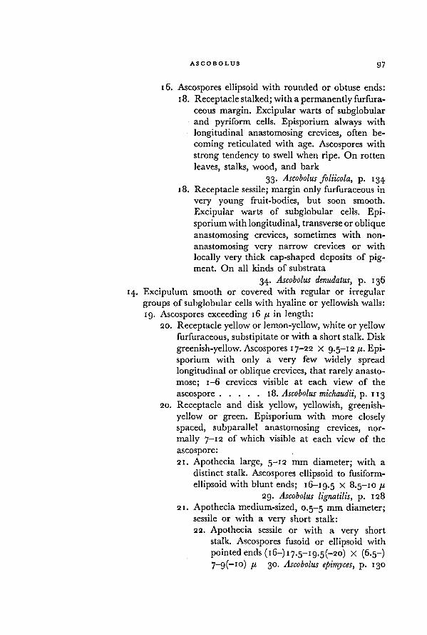
ASCOBOLUS 97
16. Ascospores ellipsoid with rounded or obtuse ends:
18. Receptacle stalked; with a permanently furfura-
ceous margin. Excipular warts of subglobularand pyriform cells. Episporium always with
longitudinal anastomosing crevices, often be-
coming reticulated with age. Ascospores with
strong tendency to swell when ripe. On rotten
leaves, stalks, wood, and bark
33. Ascobolus foliicola, p. 134
18. Receptacle sessile; margin only furfuraceous in
very young fruit-bodies, but soon smooth.
Excipular warts of subglobular cells. Epi-
sporium with longitudinal, transverse or oblique
anastomosing crevices, sometimes with non-
anastomosing very narrow crevices or with
locally very thick cap-shapcd deposits of pig-
ment. On all kinds of substrata
34. Ascobolus denudatus, p. 136
14. Excipulum smooth or covered with regular or irregular
groups of subglobular cells with hyaline or yellowish walls:
19. Ascospores exceeding 16 /j, in length:
20. Receptacle yellow or lemon-yellow, white or yellow
furfuraceous, substipitate or with a short stalk. Disk
greenish-yellow. Ascospores 17-22 X 9.5-12 /r. Epi-
sporium with only a very few widely spread
longitudinal or oblique crevices, that rarely anasto-
mose; 1-6 crevices visible at each view of the
ascospore 18. Ascobolus michaudii, p. 113
20. Receptacle and disk yellow, yellowish, greenish-
yellow or green. Episporium with more closely
spaced, subparallel anastomosing crevices, nor-
mally 7-12 of which visible at each view of the
ascospore:
21. Apothecia large, 5-12 mm diameter; with a
distinct stalk. Ascospores ellipsoid to fusiform-
ellipsoid with blunt ends; 16-19.5 X 8.5-10 [j,
29. Ascobolus lignatilis, p. 128
21. Apothecia medium-sized, 0.5-5 mm diameter;sessile or with a very short stalk:
22. Apothecia sessile or with a very short
stalk. Ascospores fusoid or ellipsoid with
pointed ends (16-) 17.5-19.5(-20) X (6.5-)
7-g(-io) /( 30. Ascobolus epimyces, p. 130
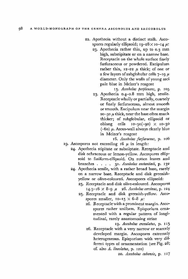
98 A WORLD-MONOGRAPH OF THE GENERA ASCOBOLUS AND SACCOBOLUS
22. Apothecia without a distinct stalk. Asco-
spores regularly ellipsoid; 19-28 X 10-14I1'
23. Apothecia rather thin, up to 0.3 mm
high, substipitate or on a narrow base.
Receptacle on the whole surface finely
furfuraceous or powdered. Excipulum
rather thin, 12-22 fi thick; of one or
a few layers of subglobular cells 7— r g
diameter. Only the walls of youngasci
pale blue in Melzer's reagent
15. Ascobolus perplexans, p. 105
23. Apothecia 0.4-0.8 mm high, sessile.
Receptacle wholly or partially, coarsely
or finely furfuraceous, almost smooth
or smooth. Excipulum nearthe margin
20—50 fi thick, near the base often much
thicker; of subglobular, ellipsoid or
oblong cells 10-50(-90) X 10-30
(-60) fi. Ascus-wall always clearly blue
in Melzer's reagent
16. Ascobolus furfuraceus, p. 106
19. Ascospores not exceeding 16 /t in length:
24. Apothecia stipitate or substiptate. Receptacle and
disk ochraceous or lemon-yellow. Ascospores ellip-
soid to fusiform-ellipsoid. On rotten leaves and
branches.... 31. Ascobolus costantinii, p. 132
24. Apothecia sessile, with a rather broad base, rarely
on a narrow base. Receptacle and disk greenish-
yellow or olive-coloured. Ascospores ellipsoid:
25. Receptacle and disk olive-coloured. Ascospores
14.5-16 X 8-9 n 26. Ascobolus cervinus, p. 124
25. Receptacle and disk greenish-yellow. Asco-
spores smaller, 10-15 X 6-8 /<:
26. Receptacle with a prominent margin. Asco-
spores rather uniform. Episporium orna-
mented with a regular pattern of longi-
tudinal, rarely anastomosing striae
19. Ascobolus crenulatus, p. 115
26. Receptacle with a very narrow or scarcely
developed margin. Ascospores extremely
heterogeneous. Episporium with verydif-
ferent types of ornamentation (see Fig. 28;
cf. also A. lineolatus, p. 120)
20. Ascobolus cubensis, p. 117
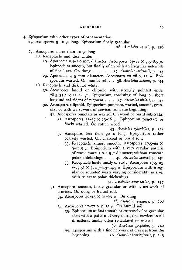
ASCOBOLUS 99
4- Episporium with other types of ornamentation:
27. Ascospores 9-10 p long. Episporium finely granular28. Ascobolus cainii, p. 126
27. Ascospores more than 10 p long:28. Receptacle and disk white:
29. Apothecia 0.4—1.0 mm diameter. Ascospores 13-17 X 7.5-8.5 p.
Episporium smooth, but finally often with an irregular net-work
of fine lines. On dung 27. Ascobolus carletonii, p. 125
29. Apothecia 4-5 mm diameter. Ascospores 20-26 X 12 p. Epi-
sporium warted. On humid soil . . 38. Ascobolus albinus, p. 144
28. Receptacle and disk not white:
30. Ascospores fusoid or ellipsoid with strongly pointed ends;
28.5-37.5 X 11—14 p. Episporium consisting of long or short
longitudinal ridges of pigment . . . 37. Ascobolus viridis, p. 142
30. Ascospores ellipsoid. Episporium punctate, warted, smooth, gran-
ular or with a net-work of crevices from the beginning:
31. Ascospores punctate or warted. On wood or burnt substrata:
32. Ascospores 32-37 X 13-16 p. Episporium punctate or
finely warted. On rotten wood
43. Ascobolus xylophilus, p. 152
32. Ascospores less than 30 p long. Episporium rather
coarsely warted. On charcoal or burnt soil:
33. Receptacle almost smooth. Ascospores 17.5-22 x
9-1 1.5 p. Episporium with a very regular pattern
of round warts 1.0-1.5 p diameter; without truncate
polar thickenings . . . 40. Ascobolus archeri, p. 146
33. Receptacle finely mealy or scaly. Ascospores 17.5-25
(-27. 5I X (11.5-) 13-14.5 p. Episporium with irreg-ular or rounded warts varying considerably in size;
with truncate polar thickenings
41. Ascobolus carbonarius, p. 147
31. Ascospores smooth, finely granular or with a net-work of
crevices. On dung or humid soil:
34. Ascospores 40-45 X 21-23 p. On dungcf. Ascobolus asininus, p. 208
34. Ascospores 17-27 X 9-15 p- On humid soil:
35. Episporium at first smooth or extremely fine granular
then with a pattern of very short, fine crevices in all
directions, finally often reticulated or warted
36. Ascobolus geophilus, p. 140
35. Episporium with a fine net-work of crevices from the
beginning .... 39. Ascobolus behnitziensis, p. 145
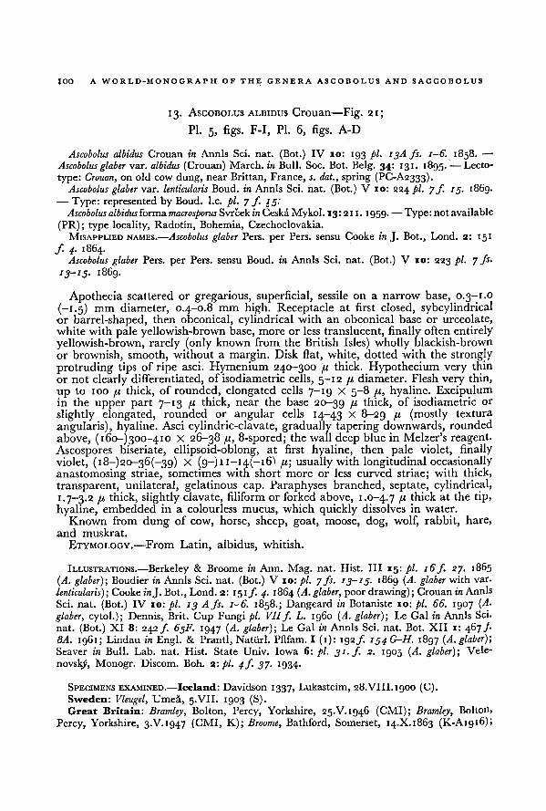
100A WORLD-MONOGRAPH OF THE GENERA ASCOBOLUS AND SACCOBOLUS
13. ASCOBOLUS ALBIDUS Crouan—Fig. 21;
Pl. 5, figs. F-I, Pl. 6, figs. A-D
Ascobolus albidus Crouan in Annls Sci. nat. (Bot.) IV 10: 193 pi. 13A fs. 1-6. 1858. —■
Ascobolus glaber var. albidus (Crouan) March, in Bull. Soc. Bot. Belg. 34: 131. 1895. — Lecto-
type: Crouan, on old cow dung, nearBrittan, France, s. dat., spring (PC-A2333).Ascobolus glaber var. lenticularis Boud. in Annls Sci. nat. (Bot.) V 10: 224 pi. 7 f. 15. i86g.
— Type: represented by Boud. I.e. pi. 7 ƒ. 15:
Ascobolusalbidusforma macrosporus Svrček in Ceska Mykol. 13: 211. 1959. —Type: not available
(PR); type locality, Radotin, Bohemia, Czechoclovakia.
MISAPPLIED NAMES.—Ascobolus glaber Pers. per Pers. sensu Cooke in J. Bot., Lond. 2: 151
f. 4. 1864.Ascobolus glaber Pers. per Pers. sensu Boud. in Annls Sci. nat. (Bot.) V 10: 223 pi. 7 fs.
13-15 . 1869.
Apothecia scattered or gregarious, superficial, sessile on a narrow base, 0.3-1.0
{-1.5) mm diameter, 0.4-0.8 mm high. Receptacle at first closed, sybcylindricalor barrel-shaped, then obconical, cylindrical with an obconical base or urceolate,
white with pale yellowish-brown base, more or less translucent, finally often entirely
yellowish-brown, rarely (only known from the British Isles) wholly blackish-brown
or brownish, smooth, without a margin. Disk flat, white, dotted with the strongly
protruding tips of ripe asci. Hymenium 240-300 fx thick. Hypothecium very thin
or not clearly differentiated, of isodiametric cells, 5—12 /.« diameter. Flesh very thin,
up to 100 fx thick, of rounded, elongated cells 7-19 X 5-8 fx, hyaline. Excipulumin the upper part 7-13 fx thick, near the base 20-39 fx thick, of isodiametric or
slightly elongated, rounded or angular cells 14-43 x 8-29 fi (mostly textura
angularis), hyaline. Asci cylindric-clavate, gradually tapering downwards, rounded
above, (i6o-)300~4io X 26-38 fx, 8-spored; the wall deep blue in Melzer's reagent.
Ascospores biseriate, ellipsoid-oblong, at first hyaline, then pale violet, finally
violet, (i8-)20-36(-39) X (9—) 11—14(—i fx\ usually with longitudinal occasionally
anastomosing striae, sometimes with short more or less curved striae; with thick,
transparent, unilateral, gelatinous cap. Paraphyses branched, septate, cylindrical,
1.7-3.2 fx thick, slightly clavate, filiform or forked above, 1.0-4.7 I1 thick at the tip,
hyaline, embedded in a colourless mucus, which quickly dissolves in water.
Known from dung of cow, horse, sheep, goat, moose, dog, wolf, rabbit, hare,
and muskrat.
ETYMOLOGY.—From Latin, albidus, whitish.
ILLUSTRATIONS.—Berkeley & Broome in Ann. Mag. nat. Hist. ILL 15: pi. 16f. 27. 1865
(A. glaber); Boudier in Annls Sci. nat. (Bot.) V 10: pi. 7 fs. 13-15. 1869 (A. glaber with var.
lenticularis);Cooke in J. Bot., Lond. 2: 151 f. 4. 1864 (A. glaber, poor drawing); Crouan in Annls
Sci. nat. (Bot.) IV 10: pi. 13 A fs. 1-6. 1858.; Dangeard in Botaniste 10: pi. 66. 1907 (A.
glaber, cytol.); Dennis, Brit. Cup Fungi pi. VIIf. L. i960 (A. glaber); Le Gal in Annls Sci.
nat. (Bot.) XI 8: 242 f. 65F. 1947 (A. glaber); Le Gal in Annls Sci. nat. Bot. XII 1: 467 f8A. 1961; Lindau in Engl. & Prantl, Natiirl. Pflfam. I (1): 192 /. 154 G-H. 1897 (A. glaber);Seaver in Bull. Lab. nat. Hist. State Univ. Iowa 6: pi. 31. f. 2. 1905 (A. glaber); Vele-
novslyi, Monogr. Discom. Boh. 2: pi. 4f. 37. 1934.
SPECIMENS EXAMINED.—Iceland: Davidson 1337, Lukasteim, 28. VIII. 1900 (C).Sweden: Vleugel, Umea, 5.VII. 1903 (S).Great Britain: Dramley, Bolton, Percy, Yorkshire, 25.V. 1946 (CMI); Bramley, Bolton,
Percy, Yorkshire, 3-V. 1947 (CMI, K); Broome, Bathford, Somerset, 14.X.1863 (K-A1916);
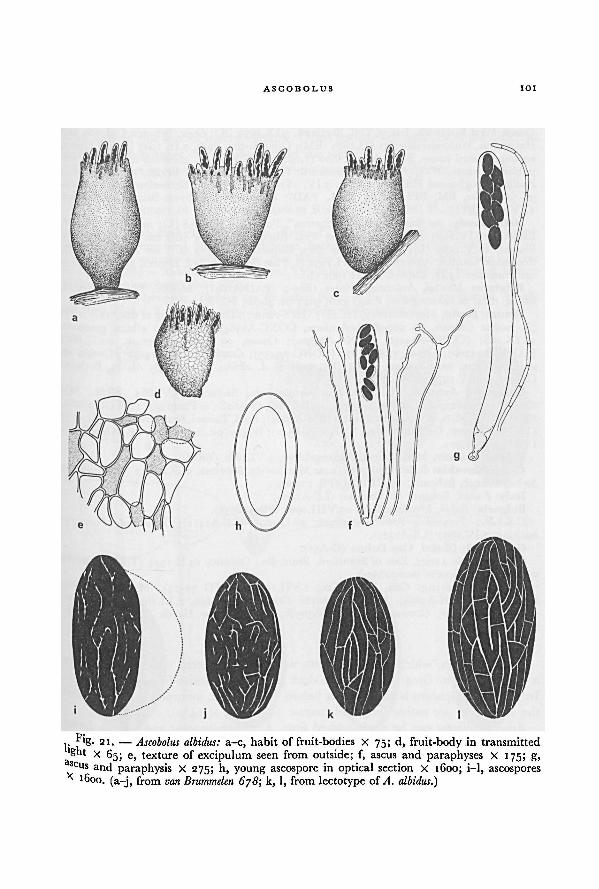
101ASCOBOLUS
van Brummelen 678; A. albidus.)k, l, from lectotype of
Ascobolus albidus: a-c, habit of fruit-bodies X 75; d, fruit-body in transmitted
light X 65; e, texture of excipulum seen from outside; f, ascus and paraphyses X 175; g,ascu
s and paraphysis X 275; h, young ascospore in optical section X 1600; i-l, ascospores
x 1600. (a-j, from
Fig. 21. —
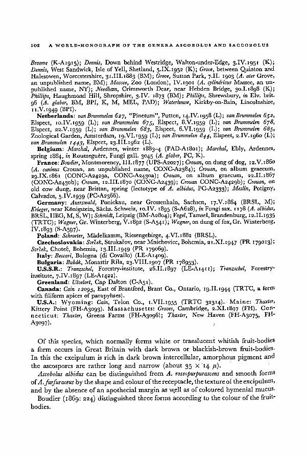
A WORLD-MONOGRAPH OF THE GENERA ASCOBOLUS AND SACCOBOLUS102
Broome (K-A1915); Dennis, Down behind Westridge, Walton-under-Edge, 3.IV.1951 (K);
Dennis, West Sandwick, Isle of Yell, Shetland, 5.IX. 1952 (K); Grove, between Quinton and
Halesowen, Worcestershire, 31.III.1883 (BM); Grove, Sutton Park, 7.II. 1903 (A. ater Grove,
an unpublished name, BM); Massee, Zoo (London), IV. 1901 (A. cylindricus Massee, an un-
published name, NY); Needham, Crimsworth Dear, near Hebden Bridge, 30.1.1898 (K);
Phillips, Haughmond Hill, Shropshire, 3.IV. 1873 (BM); Phillips, Shrewsbury, in Elv. brit.
96 (A. glaber, BM, BPI, K, M, MEL, PAD); Waterhouse, Kirkby-on-Bain, Lincolnshire,
Ï1.V.1949 (BPI).Netherlands: van Brummelen 627, "Pinetum", Putten, 14.IV.1958 (L); van Brummelen 652,
Elspeet, 10.IV.1959 (L); van Brummelen 673, Elspeet, 8.V.1959 (L); van Brummelen 678,
Elspeet, 22.V.ig59 (L); van Brummelen 683, Elspeet, 6.VI.1959 (L); van Brummelen 685,
Zoological Garden, Amsterdam, 19.VI.1959 (L); van Brummelen 844, Elspeet, 2.IV.1960 (L);
van Brummelen 1443, Elspeet, 23.III.1962 (L).
Belgium: Marchal, Ardennes, winter 1883-4 (PAD-A1801); Marchal, Ebly, Ardennes,
spring 1884, in Roumeguère, Fungi gall. 3045 (A. glaber, pC, K).
France: Boudier, Montmorency, III.1877 (UPS-A2027); Crouan, on dung of dog, 12.V.1860
(A. caninus Crouan, an unpublished name, CONC-A2384); Crouan, on album graecum,
29.IX.1861 (CONC-A242ga, CONC-A243oa); Crouan, on album graecum, 22.II. 1867
(CONC-A243ob); Crouan, 12.III.1870 (CONC-A2439); Crouan CONC-A242gb); Crouan, on
old cow dung, near Brlttan, spring (lectotype of A. albidus, PC-A2333); Meslin, Potigny,
Calvados, 5.IV. 1939 (PC-A2366).Germany: Auerswald, Ponickau, near Grossenhain, Sachsen, 17.V.1864 (BRSL, M);
Krieger, near Konigstein, Sachs. Schweiz, 10.IV. 1895 (S-A628), in Fungi sax. 1178 (A. albidus,
BRSL, HBG, M, S, W); Schmidt, Leipzig (BM-A2804); Vogel, Tamsel, Brandenburg, 12.II.1935
(TRTC); Wagner, Gr. Winterberg, V.1892 (S-A541); Wagner, on dung of fox, Gr. Winterberg,
IV.1893 (S-A597).Poland: Schroeter, Madelkamm, Riesengebirge, 4.VI.1882 (BRSL).
Czechoslovakia: Svrcek, Strukarov, near Mnichovice, Bohemia, 21.XI.1947 (PR 179013);
Svrcek, Chotec, Bohemia, 13.III.1949 (PR 179069).
Italy: Beccari, Bologna (di Covallo) (LE-A1409).
Bulgaria: Bubak, Monastir Rila, 23.VIII.1907 (PR 178953).U.S.S.R.: Tranzschel, Forestry-institute, 26.II.1897 (LE-A1411); Tranzschel, Forestry-
institute, 7.IV.1897 (LE-A1422).Greenland: Ulvelort, Cap Dalton (C-A51).Canada: Cain 12033, East of Brantford, Brant Co., Ontario, 19.II. 1944 (TRTC, a form
with filiform apices of parapyhses).U.S.A.: Wyoming: Cain, Telon Co., 1.VII.1955 (TRTC 32314). Maine: Thaxter,
Kittery Point (FH-A3093). Massachusetts: Groves, Cambridge, 2.XI.I8C)7 (FH). Con-
necticut: Thaxter, Greens Farms (FH-A3096); Thaxter, New Haven (FH-A3075, FH-
A3097).
Of this species, which normally forms white or translucent whitish fruit-bodies
a form occurs in Great Britain with dark brown or blackish-brown fruit-bodies.
In this the excipulum is rich in dark brown intercellular, amorphous pigment and
the ascospores are rather long and narrow (about 35 X 14 /J,).
Ascobolus albidus can be distinguished from A. roseopurpurascens and smooth forms
ofA. furfuraceus by the shape and colourofthereceptacle, the texture ol theexcipulum,
and by the absence of an apothecial margin as w.ell as of coloured hymenial mucus.
Boudier (1869: 224) distinguished three forms according to the colour of the fruit-
bodies.
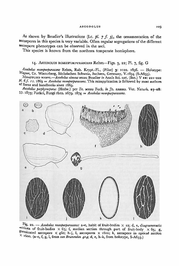
ASCOBOLUS 103
As shown by Boudier's illustrations (I.e. pi. 7 /. 3), the ornamentation of the
Zoospores in this species is very variable. Often regular segregations of the different
ascospore phenotypes can be observed in the asci.
This species is known from the northern temperate hemisphere.
14. ASCOBOLUS ROSEOPURPURASCENS Rehm—Figs. 3, 22; Pl. 7, fig. G
Ascobolus roseopurpurascens Rehm, Rab. Krypt.-Fl., (Pilze) 3: 1122. 1896. — Holotype:Wagner, Gr. Winterberg, Sachsischen Schweiz, Sachsen, Germany, V.1894 (S-A655).
MISAPPLIED NAMES.—Ascobolus vinosus sensu Boudier in Annls Sci. nat. (Bot.) V io: 221-222
pl. 6f 11, 1869 = Ascobolus roseopurpurascens. This misapplication is followed by most authors
°f floras and handbooks since 1869.Ascobolus porphyrosporus (Hedw.) per Fr. sensu Fuck, in Jb. nassau. Ver. Naturk. 27-28:
57- 1873; Fuckel, Fungi rhen. 2679. 1874 = Ascobolus roseopurpurascens.
Fig. 22. — a-c, habit of fruit-bodies X 12; d, e, diagrammaticsec tions of fruit-bodies X 65; f, median section through part of fruit-body X 65; g,
germinatedascospore X 460; h-j, l, ascospores X 1600; k, ascospore in optical section
x 1600. (a-c, f, g, l, from van Brummelen 424; d, e, h-k, from holotype, S-A655.)
Ascobolus roseopurpurascens:
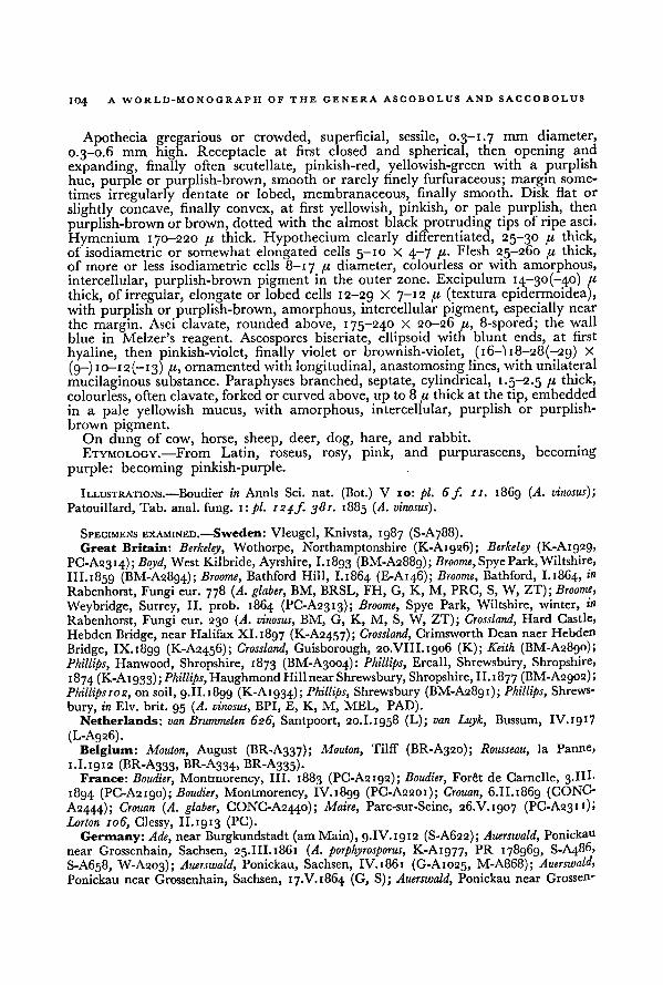
A WORLD-MONOGRAPH OF THE GENERA ASCOBOLUS AND SACCOBOLUS104
Apothecia gregarious or crowded, superficial, sessile, 0.3—1.7 mm diameter,
0.3-0.6 mm high. Receptacle at first closed and spherical, then opening and
expanding, finally often scutellate, pinkish-red, yellowish-green with a purplishhue, purple or purplish-brown, smooth or rarely finely furfuraceous; margin some-
times irregularly dentate or lobed, membranaceous, finally smooth. Disk flat or
slightly concave, finally convex, at first yellowish, pinkish, or pale purplish, then
purplish-brown or brown, dotted with the almost black protruding tips of ripe asci.
Hymenium 170-220 « thick. Hypothecium clearly differentiated, 25-30 fj, thick,
of isodiametric or somewhat elongated cells 5—10 X 4-7 p- Flesh 25-260 /< thick,of more or less isodiametric cells 8—1 7 //, diameter, colourless or with amorphous,
intercellular, purplish-brown pigment in the outer zone. Excipulum 14-30(-40) a
thick, of irregular, elongate or lobed cells 12-29 X 7~ 12 /< (textura epidermoidea),with purplish or purplish-brown, amorphous, intercellular pigment, especially near
the margin. Asci clavate, rounded above, 175-240 X 20-26 pi, 8-spored; the wall
blue in Melzer's reagent. Ascospores biseriate, ellipsoid with blunt ends, at first
hyaline, then pinkish-violet, finally violet or brownish-violet, (16—) 18-28(-29) X
(9-) io-i2(-i3) pi, ornamentedwith longitudinal, anastomosing lines, with unilateral
mucilaginous substance. Paraphyses branched, septate, cylindrical, 1.5-2.5 u thick,
colourless, often clavate, forked or curved above, up to 8 pi thick at the tip, embedded
in a pale yellowish mucus, with amorphous, intercellular, purplish or purplish-brown pigment.
On dung of cow, horse, sheep, deer, dog, hare, and rabbit.
ETYMOLOGY.—From Latin, roseus, rosy, pink, and purpurascens, becoming
purple: becoming pinkish-purple.
ILLUSTRATIONS.—Boudier in Annls Sci. nat. (Bot.) V 10: pi. 6 f 11. 1869 (A. vinosus);
Patouillard, Tab. anal. fung. 1: pi. 124/. 381. 1885 (A. vinosus).
SPECIMENS EXAMINED.—Sweden: Vleugel, Knivsta, 1987 (S-A788).Great Britain: Berkeley, Wothorpe, Northamptonshire (K-A1926); Berkeley (K-Algsg,
PC-A23I4); Boyd, West Kilbride, Ayrshire, 1.1893 (BM-A2889); Broome, Spye Park, Wiltshire,
III.1859 (BM-A2894); Broome, Bathford Hill, 1.1864 (E-A146); Broome, Bathford, I.1864, tn
Rabenhorst, Fungi eur. 778 (A. glaber, BM, BRSL, EH, G, K, M, PRC, S, W, ZT); Broome,
Weybridge, Surrey, II. prob. 1864 (PC-A2313); Broome, Spye Park, Wiltshire, winter, in
Rabenhorst, Fungi eur. 230 (A. vinosus, BM, G, K, M, S, W, ZT); Crossland, Hard Castle,
Hebden Bridge, near Halifax XI. 1897 (K-A2457); Crossland, Crimsworth Dean naer Hebden
Bridge, IX. 1899 (K-A2456); Crossland, Guisborough, 20.VIII.1906 (K); Keith (BM-A28go);
Phillips, Hanwood, Shropshire, 1873 (BM-A3004): Phillips, Ercall, Shrewsbury, Shropshire,
1874 (K-A1933);Phillips, Haughmond HillnearShrewsbury, Shropshire, II. 1877 (BM-A2902);
Phillipsi02, on soil, 9.II. 1899 (K-A1934); Phillips, Shrewsbury (BM-A2891); Phillips, Shrews-
bury, in Elv. brit. 95 (A. vinosus, BPI, E, K, M, MEL, PAD).Netherlands: van Brummelen 626, Santpoort, 20.I.1958 (L); van Luyk, Bussum, IV. 1917
(L-A926).Belgium: Mouton, August (BR-A337); Mouton, Tilff (BR-A320); Rousseau, la Panne,
1.I.1912 (BR-A333, BR-A334, BR-A335).France: Boudier, Montmorency, III. 1883 (PC-A2192); Boudier, Forêt de Carnelle, 3.III-
1894 (PC-A2190); Boudier, Montmorency, IV.i8g9 (PC-A2201); Crouan, 6.11.1869 (CONC-
A2444); Crouan (A. glaber, CONC-A2440); Maire, Parc-sur-Seine, 26.V.1907 (PC-A231 0'
Lorton 106, Clessy, II.1913 (PC).
Germany: Ade, near Burgkundstadt (am Main), 9.IV.1912 (S-A622); Auerswald, Ponickau
near Grossenham, Sachsen, 25.III.1861 (A. porphyrosporus, K-Ai977, PR 178969, S-A486,
S-A658, W-A203); Auerswald, Ponickau, Sachsen, IV. 1861 (G-A1025, M-A868); Auerswald,
Ponickau near Grossenhain, Sachsen, 17.V.1864 (G, S); Auerswald, Ponickau near Grossen-
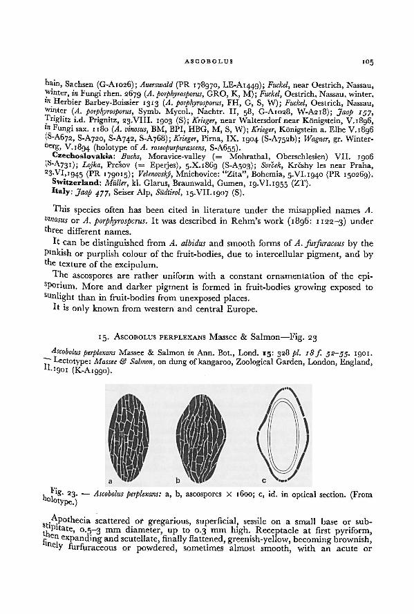
ASCOBOLUS 105
hain, Sachsen (G-A1026); Auerswald (PR 178970, LE-A1449); Fuckel, near Oestrich, Nassau,winter, in Fungi rhen. 2679 (A. porphyrosporus, GRO, K, M); Fuckel, Oestrich, Nassau, winter,tn Herbier Barbey-Boissier 1313 (A. porphyrosporus, FH, G, S, W); Fuckel, Oestrich, Nassau,winter (A. porphyrosporus, Symb. Mycol., Nachtr. II, 58, G-A1028, W-A218); Jaap ryy,Triglitz i.d. Prignitz, 23.VIII. 1903 (S); Krieger,
_ . _ ,
near Waltersdorfnear Königstein, V. 1896,to Fungi sax. 1180 (A. vinosus, BM, BPI, HBG, M, S, W); Krieger,j Königstein a. Elbe V.1896(S-A672, S-A720, S-A742, S-A768); Krieger, Pirna, IX. 1904 (S-A.752b); Wagner, gr. Winter-
berg, V.1894 (holotype of A. roseopurpurascens,......
. . ,S-A655).
Czechoslovakia: Buchs,, Moravice-valley (= Mohrathal, Oberschlesien) VII. 1906(S-A731); Lojka, Presov (= Eperjes), 5.X.1869 (S-A503); Svrcek,
......,
, „ ,, „ . , „ ,Krcshy les near Praha,
23;Vi
.»1945 (PR 179015); Velenovsky, Mnichovice: "Zita", Bohemia, 5.VI. 1940 (PR 150269).
Switzerland: Miller,,
kl. Glarus, Braunwald, Gumen, 19.VI.1955 (ZT).Italy: Jaap 4JJ, Seiser Alp, Sudtirol, 15.VII.1907 (S).
This species often has been cited in literature under the misapplied names A.
vinosus or A. porphyrosporus. It was described in Rehm's work (1896: 1122-3) under
three different names.
It can be distinguished from A. albidus and smooth forms of A. furfuraceus by the
Pmkish or purplish colour of the fruit-bodies, due to intercellular pigment, and bythe texture of the excipulum.
The ascospores are rather uniform with a constant ornamentation of the epi-sPorium. More and darker pigment is formed in fruit-bodies growing exposed to
sunlight than in fruit-bodies from unexposed places.It is only known from western and central Europe.
15. ASCOBOLUS PERPLEXANS Massee & Salmon—Fig. 23
Ascobolus perplexans Massee & Salmon in Ann. Bot., Lond. 15: 328 pi. 18f. 52-55. 1901.
.
~ Lectotype: Massee & Salmon,,
on dung of kangaroo, Zoological Garden, London, England,*•1901 (K-A1990).
Apothecia scattered or gregarious, superficial, sessile on a small base or sub-
'Pitate, 0.5-3 mm diameter, up to 0.3 mm high. Receptacle at first pyriform,e
ei? expanding and scutellate, finally flattened, greenish-yellow, becoming brownish,ely furfuraceous or powdered, sometimes almost smooth, with an acute or
Fig. 2 3. — Ascobolus perplexans: a, b, ascospores X 1600; c, id. in optical section. (From
holotype.)
106 A WORLD-MONOGRAPH OF THE GENERA ASCOBOLUS AND SACCOBOLUS
irregularly dentate margin; stipe up to 170 pi high and 450-700 diameter, stronglyenlarged at the base. Disk at first slightly concave or flat, finally convex, dirtygreenish-yellow, dottedwith the almost black protruding tips ofripe asci. Hymenium140-160 pi
thick. Hypothecium not clearly differentiated.Flesh of varying thickness,in the upper part pseudoparenchymatous, in the basal part —- especially in the
stipe — plectenchymatous, hyaline. Excipulum 12-22 //, thick, of one or a very few
layers of subglobular cells 7-19 pi diameter, pale brownish or almost colourless,covered with very small isolated groups of subglobular cells
10-ig pi diameter.
Asci clavate-cylindrical, with a narrow base, rounded above, 185-240 X 20-27 /'>
8-spored; only when young the wall pale blue in Melzer's reagent. Ascosporesuniseriate to biseriate, ellipsoid, at first hyaline, then violet, finally purplish-brown,
19-22(-25) X 9.5-13 ju;often swollen and showing a loose episporium, ornamented
with longitudinal, anastomosing striae, finally often also with short transverse
crevices. Paraphyses simple or branched, septate, filiform, 2.5-3.0 ft thick, slightlyclavate at the tip, hyaline embedded in greenish-yellow mucus.
On dung of sheep and kangaroo. According to Massee & Salmon (1901: 328)also "on dung of Raphiceri melanotidis (Grijs-bok), Elephantis africani (Elephant), Cervi
elaphi (Red Deer), Ovis vignei (Ural Wild Sheep) and horse."
ETYMOLOGY.—From Latin, perplexans, causing confusion.
ILLUSTRATION—Massee & Salmon in Ann. Bot., Lond. 15: pi. 8fs. 52-55. 1901.
SPECIMENS EXAMINED.—Great Britain: Cooke, Ascot, Berkshire, XI.1863 (K-A1917);Massee & Salmon, on dungof kangaroo, Zoological Garden, London, II. 1901 (holotype of
A. perplexans, K-Aiggo).Netherlands: van Brummelen, on sheep dung, Elspeet, VI. 1956 (L-A922).
This species is closely related to A.furfuraceus, from which it can be distinguished
by the rather thin substipitate apothecia, the structure of the excipulum, and the
weak blue staining of the ascus-wall with Melzer's reagent.
16. ASCOBOLUS FURFURACEUS Pers. per Hook.—Fig. 24;
Pl. 6, figs. E-I, Pl. 7, figs. A-F
Elvellafimetaria Scop., Fungi Hung, in Ann. Hist. nat. 4: 149 pi. i f 6. 1770 (devalidatedname). — Type: represented by Scopoli I.e. pi. 1 f. 6; type locality, "Hungary". —■ Fide
Fries (1822: 163).Peziza atra Huds., Fl. angl. (ed. 2) 637. 1778 (devalidated name). — Peziza atra Huds.
per With., Arr. Brit. PI. (ed. 7) 4: 313. 1830. — Type: represented by the description of
Hudson I.e.; type locality, England. — Fide Fries (1822: 163).Peziza fusca Bolt., Hist. Fung. Halifax 3: pi. log f 2. 1789 (devalidated name). — Peziza
fusca Bolt, per With., Arr. Brit. PI. (ed. 7) 4: 309. 1830. -— Type: represented by Bolton I.e.
pi. log f. 2; type locality, near Halifax, England.Peziza stercoraria Bull., Herb. Fr. pi. 376f. 1. 1787; Bull., Hist. Champ. Fr. 256. 1791
(devalidated name). — Peziza stercoraria Bull,per St. Amans, Fl. agen. 532. 1821. -— Ascobolus
stercorarius (Bull, per St. Amans) Schroet. in Krypt.-Fl. Schles. (ed. Cohn) 3 (2): 56. 1893-— Type: represented by Bull., Herb. Fr. pi. 376 f. 1. 1787; type locality, France. -> Peziza
stercoraria var. lutea Bull.
Peziza stercoraria var. lutea Bull., Hist. Champ. Fr. 256. 1791 (devalidated name). —Peziza
stercoraria var. lutea Bull, per St. Amans, Fl. agen. 532. 1821. = Peziza stercoraria Bull.
Peziza stercoraria var. violacea Bull., Hist. Champ. Fr. 256. 1791 (devalidated name). —
ASCOBOLUS
107
Peziza violacea (Bull.) Relh., Fl. Catabrigiensis, Suppl. 3: 31. 1793 (devalidated name); not
Peziza violacea Pers. in Neues Mag. Bot. 1: 113. 1794. — Peziza stercoraria var. violaceaper
..
Bull.St. Amans. Fl. ae-en. ZQQ. IRC>I.
— Peziza violacea- (Bull, per St. Amans) With., Arr. Brit.
PI. (ed. 7) 4: 312. 1830. — Type: represented by Bull., Herb. Fr. pi. 438 f.4. 1789 ( Pezizastercoraria)',
* "
type locality, France.
Ascobolus pezizoides Pers. apud Gmel., C. Linn. Syst. Nat. a: 1461. 17g 1 (devalidated name).= Ascobolusfurfuraceus Pers.
Ascobolusfurfuraceus Pers. in Neues Mag. Bot. r: 115. 1794 (devalidated name); Pers., Obs.mycol. x: 33,pi. 4,/. 3a, 4-61 1796.-— Ascobolus furfuraceus Pers. per Hook., Fl. scot. 2: 33. May1821; Fries, Syst. mycol. a (1): 163. 1822.— Lectotype: L 910.262.143;type locality,Germany.
[Ascobolusfurfuraceus var. a. Dilute viridis Pers. in Neues Mag. Bot. 1: 115. 1794 (unnamedvariety). —] Ascobolus furfuraceusvar. ß. flavo-virens Pers., Obs. mycol. 1: 33. 1796 (devalidated
name). — Type: not known to be in existence; type locality, probably Germany.[Ascobolus furfuraceus var. ß. fuscus Pers. in Neues Mag. Bot. 1: 115. 1794 (unnamed variety).
-] Ascobolus furfuraceus var. a fuscus Pers., Obs. mycol. 1: 33. 1796 (devalidated name). —
Ascobolus furfuraceus Pers.
Ascobolus marginatus Schum., Enum. PI. Saell. 2: 437. 1803 (devalidated name); not Ascobolus
marginatus Pat. in Rev. mycol. 4: 211. 1882; nor Ascobolus marginatus Mass. in Grevillea 22:
98. 1893. — Type: not known to be in existence; type locality, Sjaelland, Denmark.Ascobolus aerugineus Fries, Syst. mycol. 2 (1): 165. 1822. —- Type: represented by the
description of Fries, I.e.; type locality, probably Sweden.
Peziza fimiputris Fr. apud Weinm., Hym.- Gastro- m. ross. 426. 1836. — Ascobolus fimiputrisU(r. apud Weinm.) Quel., Ench. Fung. 293. 1886. —
Ascobolus stercorarius var. fimiputrisIvr. apud Weinm.) Boud., Hist. Class. Discom. Eur. 72. 1907. —
Ascobolus furfuraceus var.
fimiputris (Fr. apud Weinm.) Grelet in Rev. Mycol. 9: 22. 1944. — Type: not known to beln existence, represented by the description of Fries apud Weinm. I.e.
Peziza subrugulosa P. Karst., Syn. Pez. Ascob. 7. 1861.— Type: P. A. Karsten, near Abo,
• inland, 10.V.1860 (H-A2750).Ascobolus furfuraceus var. nudus Kickx, Fl. Crypt. Flanders 1: 479. 1867; Boud. in Annls Sci.
?aL (Bot.) V 10: 220 pi. 6 f. 10. 1869. — Ascobolus stercorarius var. nudus (Kickx) Boud.,Hist. Class. Discom. Eur., 72. 1907. — Type: not known to be in existence; type locality,•'landers, Belgium.
Ascobolusfurfuraceus var. coronatus Boud. in Annls Sci. nat. (Bot.) V 10: 220 pi. 6f. g. 1869.—Ascobolus stercorarius var. coronatus (Boud.) Boud., Hist. Class. Discom. Eur., 72. 1907. —
•ype: represented by Boud. in Annls Sci. nat. (Bot.) V 10: pi. 6f. g. 1869; type locality, near
aris, France.
Ascobolus furfuraceus var. fallens_
Heimerl. in Jber. k.k. Ober-Realschule Bezirke Sechshaus
15: ig, 1889. — Type: not known to be in existence, represented by the description of'eimerl I.e.; type locality, Austria.
Ascobolus stercorarius retisporus Clements in Bot. Surv. Nebraska 5: 9. 1901. — Type: not
no\vn to be in existence; type locality, Nebraska, U.S.A. — Fide Seaver (1928: 82).Ascobolus glaber var. caprea Beeli in Bull. Soc. roy. Bot. Belg. 54: 61. 1924. — Holotype:
Reeliy 30 (BR).
Ascobolus stercorarius var. pusillusp .
Vel., Monogr. Discom. Boh. 365. 1934. — Lectotype:150303.
Ascobolus minor Vel., Monogr. Discom. Boh. 365. 1934. —Type: not known to be in existence;
ni?e Iocality, near Mnichovice, Bohemia, Czechoslovakia. — Representative specimen:148318 (A. minor).
ORTHOGRAPHIC VARIANT. — Ascobolus purpurascens Calkins in J. Mycol. a: 106. 1886; Seaver,• Am. Cup-fungi (Operc.) 92. 1928. (derived from incorrect spelling of 'furfuraceus' on
k'bel).EXCLUDED.—„Ascobolus fimiputris (Fr. apud Weinm.) Quel, sensu Rehm, Rab. Krypt.-Fl.v*"uze) 3: 1130. 1896 = Ascobolus denudatusFr.
108 A WORLD-MONOGRAPH OF THE GENERA ASCOBOLUS AND SACCOBOLUS
Apothecia solitary or gregarious, superficial, sessile, 0.5-5 mra across, 0.4-0.8 mm
high. Receptacle at first closed and subglobular, then opening and hemisphericalor cup-shaped, finally saucer-shaped, yellowish-green or yellowish, often becomingolive-green, ochraceous or brownish; wholly or partially, coarsely or finely, whitish
furfuraceous, almost smooth or smooth; margin furfuraceous, denticulated, mem-
branaceous or smooth. Disk slightly concave or flat, at first yellowish, greenishor yellowish-green, dotted with the almost black protruding tips of ripe asci, finallyoften brownish or blackish-brown. Hymenium (120-)i5o-20o(-220) //, thick. Hypo-thecium 20-50 fi thick, of isodiametric, rounded cells 7—12 fi diameter. Flesh up
to 500 /< thick, ofsubglobular or slightly ellipsoid cells 10-40 //. diameter, accompaniedby irregularly undulating, branched hyphae which are connected with the para-
physes, hyaline. Excipulum near the margin 20-50 /u thick, near the base sometimes
reaching 170 /i; of globular, ellipsoid or oblong cells io-5o(-go) X IO-3O(-6O) /(
(textura globulosa); hyaline or sometimes with some brownish, amorphous, inter-
cellular pigment; the external zone usually roughened by protruding groups of
globular or oblong cells, often with irregular fissures giving rise to eroded groups
of cells; sometimes almost smooth. Asci clavate, tapering downwards into a rather
short stalk, rounded above, (120-1180-250 X (20-)24-3o(-32) /<, 8-spored; the
wall blue in Melzer's reagent. Ascospores at maturity biseriate or irregularlydisposed, ellipsoid, at first hyaline, then violet, finally often purplish-brown, (16-)ig-28(-32) X (g-i IO-I4(-I61 //; ornamented with more or less longitudinal,occasionally anastomosing striae, rarely swollen and reticulated, with unilateral
mucilaginous substance. Paraphyses simple or branched, septate, filiform, 2-4
thick, usually scarcely thickened above, only very rarely with ellipsoid, stronglyswollen cells up to 21 /< thick, embedded in yellowish or yellowish-green mucus.
On dung of cow, horse, deer, goat, yak, bear, pig, fox, rabbit, and hare, on
manured soil, on rotten refuse, and on rotten stems of cabbage.ETYMOLOGY.—From Latin, furfuraceus, scurfy.
ILLUSTRATIONS.—Bolton, Hist. Fung. Halifax 3: pi. iogf 2. 1789 (.Peziza fusca); Boudier in
Annls Sci. nat. (Bot.) V 10: pi. 6fs. 6-10. 1869 (with var. coronatus and var. nudus); Boudier
in Annls Sci. nat. (Bot.) V 10: pi. 7 f. 12. 1869 (A. ‘aeruginosus’); Bulliard, Herb. Fr. pi. 376f
. 1, pi. 438/. 4. 1787-9 ( Peziza stercoraria); Cooke in J. Bot., Lond. 2: 150f. 1. 1864; Cooke,
Handb. Brit. Fung, a: 725 f. 338. 1871; Corda, Anleit. Stud. Mykol. pi. G 64 fs. 24-29.
1842; Currey in Trans. Linn. Soc. Lond. (Bot.) 24: pi. 25 fs. 9-10. 1863; De Bary, Vergl.
Morph. Biol. Pilze/. 45,f. 85, f. 95. 1884; Delacroix, Malad. PI. cultiv. Pays chauds PI.33J. 4-
1911; Dennis, Brit. Cup Fungi pi. VII, f. H. I960; Gillet, Champ. Fr., Discom. pi. 84 f. 2.
1883; Greville, Scot, crypt. FI. 6: pi. 307. 1827; Harper in Ber. deutsch. bot. Ges. 13: pi. 12
fs. 26-29. '895; H. Hoffmann, Ic. anal. Fung. (4:) pi. 24 f. 2. 1865; Hornemann in FI.
danica, Fasc. 31: pi. 1856f. 2. 1825; Janczewski in Bot. Ztg. 29: pi. 4. 1871; Janczewski in
Annls Sci. nat. (Bot.) V 15 : pi. 8. 1872; Krombholz, Naturgetr. Abbild. Beschr. Schwamme
1: pi. 5 fs. 45-48. 1831; Le Gal in Annls Sci. nat. (Bot.) XI 8: 254/. 70, 255/. 71. 1947;
Le Gal, Discom. Madagascar fs. 26-27. *953 (d. stercorarius) ; Lindau in Engl. & Prantl,
Nat. PffFam. 1 (1): 192 f. 154 J-L. 1896 (A. stercorarius); Linsbauer, Schneiders illustr.
Handworterb. Bot. 59f. 30. 19 17; Luerssen, Handb. system. Bot. 1: 167 f. 51 C-G. 1879;
Massee, Brit. Fungus-Fl. 4: 162 fs. 1-7. 1895; Payer, Bot. crypt.: 5ƒ. 12, 8 fs. 24-26 ((Pezizafurfuracea). 1850; Persoon, Obs. mycol. 1: pi. 4 fs. 3a, 4> 5■ 1796; Rehm, Rab. Krypt.-Fl-(Pilze) 3: 1112fs. 1-3, 6. 1896 (A. stercorarius); Schneider, Illustr. Handworterb. Bot. 50f 31-
1905; Scopoli in Ann. Hist.-nat. 4: pi. 1 f. 6. 1770 (Elvella fimitaria); Seaver in Bull. Lab.
nat. Hist. State Univ. Iowa 6: pi. 29f. 2. 1905 (A. stercorarius) ; Seaver, N. Am. Cup-fungi
(Operc.) pi. 45f. 21. 1928 (A. stercorarius); Sowerby, Colour. Fig. Engl. Fung. pi. 18, pi. 389
fs. 3-6. 1796 and 1803 ( Peziza stercoraria); Zopf, Pilze/. 59 IV, f 64 IV. 1890.
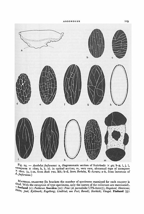
ASCOBOLUS 109
MATERIAL EXAMINED (In brackets the number of specimens examined for each country isc 'ted. With the exception of type specimens, only the names of the collectors are mentioned)."Mceland (i): Fedderstn. Sweden (22): Fries (A. parmeloides UPS-A2007); Hagelund; Hemicson-,
°'"i> Juel; Kjellmark-, Kugelberg; Lindblad; von Post; Romell; Starback; Vleugel. Finland (3):
Fig. 24. —
a, diagrammatic section of fruit-body X 40; b-g, i, j, l,as cospores X 1600; h, k, id. in optical section; m, very rare, abnormal type of ascosporex 1600. (a, i—m, from Beeli 730, BR; b-d, from Berkeley, K-A1905; e—h, from lectotype of
Ascobolus furfuraceus:
A. furfuraceus.)
110A WORLD-MONOGRAPH OF THE GENERA ASCOBOLUS AND SACCOBOLUS
Karsten; Tigerstedt. Great Britain (44): Berkeley, Broome; Cooke; Crossland; Currey; Dennis;
J. W. Ellis; Greville; Hughes; Limminghe; Massee; Phillips; Redhead; Salmon. Netherlands (18):van Brummelen; Buse; Daams; Destre'e; van Luyk; Molkenboer; Oudemans; W. J. Reijnders; Rick.
Belgium (15): Beeli 730, Woluwe-St. Pierre, 11.VIII.1918 (Type of A. glaber var. caprea)(BR); Bemmer; Coemans; Libert; Mouton; Rousseau; Wallays; Westendorp. France (67): Bernard;
Boudier; de Brebisson; Bueison; Crouan; Desmazieres; Fautrey; Gaillard; Huijsman; Leveille; Louvet;
Meslin; Montague; Penis; Quelet, Herimoncourt, (A. fimiputris) (PC-A2200); Richon; Riel;
Roberge; Roussel; Therry; Tulasne. Monaco (1): Renner. Portugal (1): Torrend. Denmark (9):E. C. Hansen; Lind; Mullen; Rostrup. Germany (103): Ade; Auerswald; Britzelmayr; Feurich;
Fuckel; Haller; Jaap; Jahn; Junghuhn;Krieger; Kunze; Lojka; Ludwig; Persoon, s. loc., s. dat. (lecto-type of A. furfuraceus, L 910.262.143); Puis; Rehm; Staritz; Strassen; Sydow; Wagner; Wilms;
Winter. Poland (25): van Brummelen; Dressier; Falck; Lasch; Lojka; Schroeter. Czechoslovakia
(46): Hruby; Klika; JViessl; Petrak; Picbauer; Svrcek; Vacek, Velenovsky. Switzerland (5): Obritz;
Regel; Volkart. Austria (28): Beck; Britzelmayr; Demelius; von Keissler; Lojka; Rechinger; Rehm;
Rick; Sauter; Strasser. Italy (7): Bresadola; Spegazzini. Hungary (1): Hollos. Romania (14):
Lojka. U.S.S.R. (7): Dietrich; Girzitska; Newodowski; Satuna; Tranzschel.
Algeria (2): Durieu de Maisonneuve. Madagascar (1): Heim.
Pakistan (1): Ahmad. India (1): Ahmad.
Australia (3): Simmonds.
Canada (36): Bisby; Cain; Jackson; Macoun; A. T. T. Smith.
Greenland (1): Ulvelort.
U.S.A.: Oregon (3 ): Kienholz. California(5): Coon; Copeland; Harkness; McClatchie. Idaho
(3): W. B. & V. G. Cooke. Montana (2): Cummins. Wyoming (4): Cain. Colorado (3):Seaver & Bethel. Nebraska (2): Moore; Saunders & Clements. Oklahoma (1): Ray. Iowa (3):
Holway; Martin; A. H. Smith et al. Missouri (2): Linder. Ohio (1): Morgan. Tennessee (4):Hesler. Alabama (2): Baker; Earle. Maine (1): Thaxter. New Hampshire (i): unknown
collector. New York (13): Burnham; Cain; Clinton; Cook; Dodge; Peck; Seaver; Warne.
Massachusetts (1): Sturgis. Connecticut (1): unknown collector. Pennsylvania (3):
Michener; Orton. District of Columbia (1): Shear. North Carolina (1): Diehl. South
Carolina (14): Ravenel. Georgia (2): Ravenel. Florida (2): Sturgis; West. Bermuda (3):Brown et al.
Venzuela (2): Chardon; Dennis. Chile (4): Gay. Argentina (1): Singer.
EXSICCATI.—Cooke, Fungi brit., ed. 2, i8g (K) ;Desmazières, PL crypt., ed. II ser. 1, 715
(BM); Desmazières, PI. crypt, ed. I, ser. 1, 1315 (BM, G, K, L, PC); Fuckel, Fungi rhen.
1132 (BM, G, GRO, K, LE, M, W); Fuckel, Fungi rhen. 1 134 (A. glaber; K, GRO); Herbier
Barbey-Boissier 1309 (FH, K, S, UPS); HerbierBarbey-Boissier 1310 (A. glaber; K); Karsten,
Fungi fenn. 170 (BM, K); Krieger, Fungi sax. 1179 (A. stercorarius; BM, BPI, HBG, M, W);
Libert, Reliquiae Libertianae, ser. 3, 14 (ined.; BR); Linhart, Fungi hung. 1869 (S); Niessl,
Fl. Austr.-Hung. 393 (BM, BRSL, DAOM, E, FH, G, HBG, K, L, LE, M, MEL, PRC, S,
W, ZT); Patouillard & Doassans, Champ, fig. des. 72 (M); Petrak, Myc. carp. 204 (A.
stercorarius; BM, BPI, K, S); Petrak, Fl. Boh. Mor., ser. 2, 255 (A. stercorarius; BM, E,
FH, LE, S); Petrak, Fl. Boh. Mor., ser. 2, 1995 (A. glaber; BM, BPI, E, M, S); Petrak, Myc.
gen. 507 (A. glaber; BPI); Phillips, Elv. brit. 46 (BPI, K, M, MEL, S); Rabenhorst, Klotssch.
Herb. viv. mycol., ed. 2, 522 (BRSL, L, LE, M, PAD); Rabenhorst & Winter, Fungi eur.
3069b (BM, G, K, L, LE, M, S, W, ZT); Rabenhorst & Winter, Fungi eur. 3069a (Bhl>G, HBG, K, L, LE, M, S, W, ZT); Ravenel, Fungi am. 312 (BM, BPI, E, K, NY, PAD, W)i
Rehm, Ascom. 1 (BM, E, K, L, M, PAD, S, W); Rehm Ascom. ib (BM, BRSL, E, HBG,
K, M, PAD, S); Roumegufere, Fungi gall. 2178 (G, K, L, NY); Sydow, Myc. germ. 1175
(A. stercorareus; BM, BPI, E, K, L, LE, M, PAD, S, W, ZT); Sydow, Myc. march. 362 (BM>HBG, K, S, W); Sydow, Myc. march. 587 (HBG, K, PRC, S, W); Sydow, Myc. march.
887 (BM, K, W); von Thiimen, Myc. univ. 2178 (BM, BPI, BRSL, G, K, L, LE, M, MEL,
ASCOBOLUS 111
PAD, S, W, ZT); Therry, Crypt. Lyonn. 6531 (G, PAD); Therry, Crypt. Lyonn. 6697 (PAD);Vize, Micro-fungi brit. 371 (G, K, MEL, PAD).
This is an extremely variable species. Several forms, varieties and even species
have been distinguished on the ground of certain more or less conspicuous characters.
AH these forms, however, are linked by transitions, as could be established throughthe study of several hundreds of rich collections.
Some of the noticeable forms that have been given names are the following.
(A) The typical form, which is coarsely furfuraceous over the whole surface
of the receptacle (A. furfuraceus, A. furfuraceus var . fuscus Pers., Peziza stercoraria var.
violacea Bull.).
(B) A form with only the marginal zone of the receptacle coarsely furfuraceous
and dentate; the lower regions of the surface smooth (A. furfuraceus var. coronatus
Boud., A. stercorarius var. pusillus Vel.).
(G) A form with the receptacle entirely smooth and a membranous marerin
(A. furfuraceus var. nudus Kickx).
(D) A formwith yellowish disk and finely furfuraceous receptacle (Peziza stercoraria
var. lutea Bull., A.furfuraceus var. flavo-virens Pers.).(El A formwith greenish or green disk and smooth or finely furfuraceous receptacle
(A. aerugineus Fr, A. marginatus Schum.).
(F) A form with small fruit-bodies (A. furfuraceus var. fallens Heimerl, A. minor Vel.).The attempts of Olive (1954a) to separate A. stercorarius from A. furfuraceus on
the ground of intersterility cannot be sustained on morphological or structural
grounds.
Because of the wide range of variation in this species it may sometimes be rather
difficult to delimit it lrom related ones.
Smooth forms of A. furfuraceus draw their limits against A. roseopurpurascens byffie
excipular texture and the absence of pinkish or purplish intercellular pigment,it differs from A. perplexans by the structure of the excipulum, the more intense
Blue staining of the ascus-wall with iodine, and the absence of a substipitate base
°f the receptacle.
Prom the related A. laevisporus it can be separated by the smaller apothecia and
ffie less dense disposition of the striae of the episporium.Ascobolus furfuraceus is a heterothallic species (Dowding, 193 1; Bistis, 1956a)which the development was studied by Janczewski (1871), Dangeard (1907),
aud Gamundf & Ranalli (1963). In experiments designed to find the most favourable
c °nditions for ascospore germination Yu (1954) registrated a strong NaOH-effect.
The origin of the episporial pigment was studied in this species by Chadefaud
Le Gal (1947) and MalenQon (1962).A
very common cosmopolitan species.
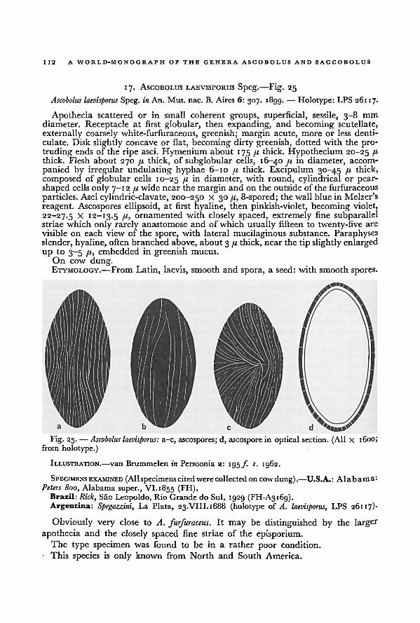
A WORLD-MONOGRAPH OF THE GENERA ASCOBOLUS AND SACCOBOLUS112
17. ASCOBOLUS LAEVISPORUS Speg. —Fig. 25
Ascobolus laevisporus Speg. in An. Mus. nac. B. Aires 6: 307. 1899. — Holotype: LPS 26117.
Apothecia scattered or in small coherent groups, superficial, sessile, 3-8 mm
diameter. Receptacle at first globular, then expanding, and becoming scutellate,
externally coarsely white-furfuraceous, greenish; margin acute, more or less denti-
culate. Disk slightly concave or flat, becoming dirty greenish, dotted with the pro-
truding ends of the ripe asci. Hymenium about 175 pi thick. Hypothecium 20-25 /t
thick. Flesh about 270 pi thick, of subglobular cells, 16-40 /< in diameter, accom-
panied by irregular undulating hyphae 6-10 pi thick. Excipulum 30-45 pi thick,
composed of globular cells 10-25 I1 m diameter, with round, cylindrical or pear-shaped cells only 7-12 pi wide near the margin and on the outside ofthe furfuraceous
particles. Asci cylindric-clavate, 200-250 x 30 pi, 8-spored; thewall blue in Melzer's
reagent. Ascospores ellipsoid, at first hyaline, then pinkish-violet, becoming violet,
22-27.5 X 12-13.5 pi, ornamented with closely spaced, extremely fine subparallelstriae which only rarely anastomose and of which usually fifteen to twenty-five are
visible on each view of the spore, with lateral mucilaginous substance. Paraphyses
slender, hyaline, often branched above, about 3 pi thick, near the tip slightly enlarged
up to 3-5 pi, embedded in greenish mucus.
On cow dung.ETYMOLOGY.—From Latin, laevis, smooth and spora, a seed: with smooth spores.
ILLUSTRATION.—van Brummelen in Persoonia 2: 195 ƒ. 1. 1962.
SPECIMENSEXAMINED (Allspecimens cited were collectedon cow dung).—U.S.A.: ALABAMA:
Peters 800, Alabama super., VI.1855 (FH).Brazil: Rick, Sao Leopoldo, Rio Grande do Sul, 1929 (FH-A3169).Argentina: Spegazzini, La Plata, 23.VIII.1888 (holotype of A. laevisporus, LPS 26117)-
Obviously very close to A. furfuraceus. It may be distinguished by the larger
apothecia and the closely spaced fine striae of the episporium.The type specimen was found to be in a rather poor condition.
This species is only known from North and South America.
Fig. 25. — a-c, ascospores; d, ascospore in optical section. (All X 1600;
from holotype.)
Ascobolus laevisporus:
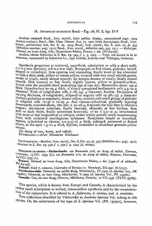
ASCOBOLUS
113
18. ASCOBOLUS MICHAUDII Boud.—Fig. 26; Pl. 8, figs. D-F
Ascobolus michaudii Boud., Icon, mycol., Liste prelim, champ., unnumbered page. 1904(nomen nudum); Boud., Hist. Class. Discom. Eur. 71. 1907 (with description); Boud., Icon,
mycol., preliminary text, Ser. 6: 22. 1909; Boud., Icon, mycol., Ser. 6. Livr. 27, pi. 593
(definitive number: 409). 1910; Boud., Icon, mycol., definitive text, 232. 1911. — Holotype:Michaud, on horse dung, Alix, Department Rhone, France, s. dat. (PC-A2196).
Ascobolus citrinus Schweizer in Z. Bot. 15: 529,/. 1, 3. 1923. — Type: not known to be in
existence, represented by Schweizer I.e.; type locality, Lustnau near Tubingen, Germany.
Apothecia gregarious or scattered, superficial, substipitate or with a short stalk,°-7~2.5 mm diameter, 0.8-2.0 mm high. Receptacle at first closed, globular, barrel-
shaped or cylindrical, then opening and expanding, finally more or less obconical°r with a short stalk, yellow or lemon-yellow, covered with very small whitish grains,mealy or scurfy, rarely almost smooth; the margin dentate or mealy, finally almost
smooth. Disk concave or flat, finally slightly convex, yellow or greenish-yellow,dotted with the purplish-black protruding tips of ripe asci. Hymenium about 140 pxthick. Hypothecium 20-25 fi thick, of closely compacted isodiametric cells 4.5-10 pidiameter. Flesh of subglobular cells 7-18 (-44) pi diameter, hyaline. Excipulum of
varying thickness, of subglobular, ellipsoid or angular cells 19-38 (-55) px diameter
(textura globulosa or angularis), lemon-yellow, covered with small groups ofglobularellipsoid cells 10-32 X 10-23 ll- Asci clavate-cylindrical, gradually tapering
downwards, rounded above, 180-280 X 22-26 px, 8-spored; the wall blue in Melzer'sreagent. Ascospores uniseriate, finally biseriate, ellipsoid; at first hyaline, then
pale or dark violet, finally sometimes brownish; 17-22 X 9-5~i2 px; ornamented
lyth more or less longitudinal or oblique, rather widely spread, rarely anastomosingtines; with unilateral mucilaginous substance. Paraphyses simple or branched,s<T>tate, cylindrical or clavate, 2.2-3-5(-7) px thick, enlarged, narrowed or forked
above, at the apex 1.5- 12 px thick, hyaline, embedded in abundant greenish-yellowmucus.
On dung of cow, horse, and rabbit.
ETYMOLOGY.—After Monsieur Michaud.
ILLUSTRATIONS. —Boudier, Icon, mycol., Ser. 6, livr. 27, pi. 593 (definitive no.: 409). 1910;c hweizer in Z. Bot. 15: 530 /. 1, 532 f. 3. 1923 ( A. citrinus).
SPECIMENS EXAMINED.—Netherlands: van Brummelen 706, on dung of rabbit, Elswout,Dverveen, 13.VII. 1959 (L); van Brummelen 710, on dung of rabbit, Elswout, Overveen,HATlI.igsg (L ).
'ranee: Michaud, on horse dung, Alix, Department Rhone, s. dat. (type of A. michaudii,"C-A2ig6).
Poland: Falck 7, cultures, University of Breslau (= Wroclaw), Ill.igoi (S-A757).Czechoslovakia: VelenovskJ, on rabbit dung, Mnichovice, IV.ig2g (A. leporinus Vel., PR
149480); Vtlenovshy, on hare dung, Mnichovice, V.ig2g (A. leporinus Vel., PR i4g362).Canada: Cain, on cow dung, Glencoe, Middlesex, Ontario, 12.VII.1g32 (TRTC 35285).
This species, which is known from Europe and Canada, is characterized by therat her small substipitate or stalked, lemon-yellow apothecia and by the ornamenta-
b°n of the episporium. It is related to A. furfuraceus, A. cervinus, and A. crenulatus.
Two collections identified by Velenovsky as Ascobolus leporinus Vel. belong to thissPecies. On the substratum of the type of A. leporinus Vel. (PR 150301), however,
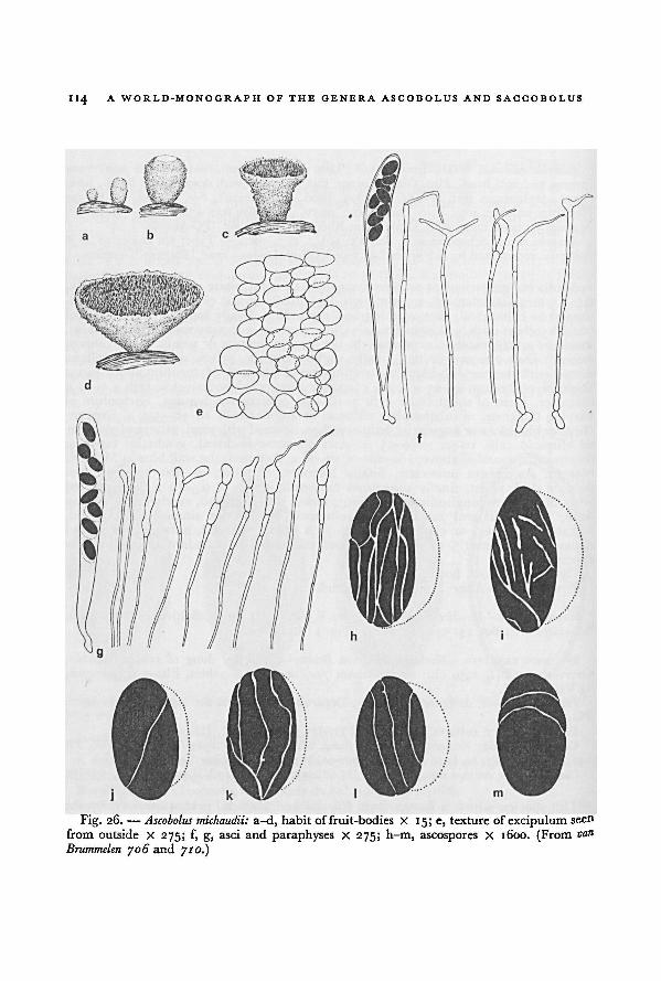
114 A WORLD-MONOGRAPH OF THE GENERA ASCOBOLUS AND SACCOBOLUS
710.)and
van
Brummelen 706
Ascobolus michaudii:Fig. 26. — a-d, habit offruit-bodies X 15; e, texture of excipulum seen
from outside X 275; f, g, asci and paraphyses X 275; h-m, ascospores X 1600. (From
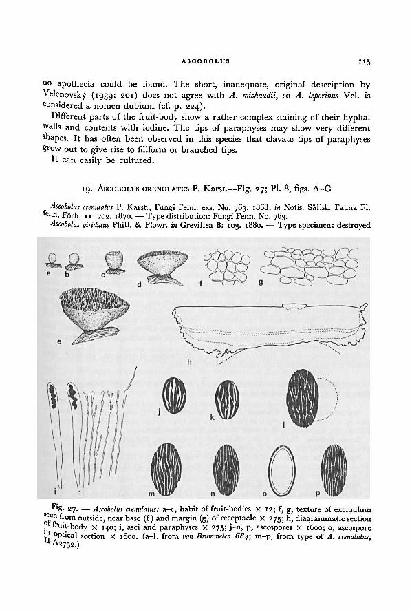
ASCODOLUS 115
no apothecia could be found. The short, inadequate, original description by
Velenovsky (1939: 201) does not agree with A. michaudii, so A. leporinus Vel. is
considered a nomen dubium (cf. p. 224).
Different parts of the fruit-body show a rather complex staining of their hyphalWalls and contents with iodine. The tips of paraphyses may show very different
shapes. It has often been observed in this species that clavate tips of paraphysesgrow out to give rise to filiform or branched tips.
It can easily be cultured.
19. ASCOBOLUS CRENULATUS P. Karst.—Fig. 27; Pl. 8, figs. A-C
Ascobolus crenulatus P. Karst., Fungi Fenn. exs. No. 763. 1868; in Notis. Sallsk. Fauna Fl.tain. Förh. 11: 202. 1870. —Type distribution:Fungi Fenn. No. 763.
Ascobolus viridulus Phill. & Plowr. in Grevillea 8: 103. 1880.— Type specimen: destroyed
Fig. 27. — Ascobolus crenulatus:
A. crenulatus,
H-A2752.)
a-e, habit of fruit-bodies X 12; f, g, texture of excipulumseen from outside, near base (f) and margin (g) ofreceptacle X 275; h, diagrammatic section
of fruit-body x 140; i, asci and paraphyses X 275; j-n, p, ascospores X 1600; o, ascospore
in optical section x 1600. (a-l, from m-p, from type ofvan Brummelen 684;
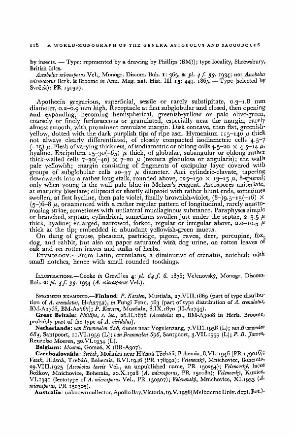
116A WORLD-MONOGRAPH OF THE GENERA ASCOBOLUS AND SACCOBOLUS
by insects. — Type: represented by a drawing by Phillips (BM)); type locality, Shrewsbury,
British Isles.
Ascobolus microsporus Vel., Monogr. Discom. Boh. i: 365, 2:pi. 4/. 33. 1934; not Ascobolus
microsporus Berk. & Broome in Ann. Mag. nat. Hist. ILL 15: 449. 1865. —Type (selected by
Svrcek): PR 150307.
Apothecia gregarious, superficial, sessile or rarely substipitate, 0.3-1.8 mm
diameter, 0.2-0.9 mm high. Receptacle at first subglobular and closed, then openingand expanding, becoming hemispherical, greenish-yellow or pale olive-green,
coarsely or finely furfuraceous or granulated, especially near the margin, rarelyalmost smooth, with prominent crenulate margin. Disk concave, then flat, greenish-yellow, dotted with the dark purplish tips of ripe asci. Hymenium 115-140 fi
thick
not always clearly differentiated, of closely compacted isodiametric cells 4.5-7( _I 5) <"• Flesh ofvarying thickness, ofisodiametricor oblong cells 4.5-20 X 4.5-14 p,
hyaline. Excipulum 15-30(-65) p thick, of globular, subangular or oblong rather
thick-walled cells 7~3o(-4o) X 7-20 p (textura globulosa or angularis); the walls
pale yellowish; margin consisting of fragments of excipular layer covered with
groups of subglobular cells 20-37 p diameter. Asci cylindric-clavate, taperingdownwards into a rather long stalk, rounded above, 125-150 X 13-15 p, 8-spored;
only when young is the wall pale blue in Melzer's reagent. Ascospores uniseriate,
at maturity biseriate; ellipsoid or shortly ellipsoid with rather blunt ends, sometimes
swollen, at first hyaline, then pale violet, finally brownish-violet, (8-)g.5-i5(-i6) X
(5-) 6-8 p, ornamentedwith a rather regular pattern of longitudinal, rarely anasto-
mosing striae, sometimes with unilateralmucilaginous substance. Paraphyses simpleor branched, septate, cylindrical, sometimes swollen just under the septae, 2—3.5 I1
thick, hyaline; enlarged, narrowed, forked, regular or irregular above, 2.0-10.5 pthick at the tip; embedded in abundant yellowish-green mucus.
On dung of grouse, pheasant, partridge, pigeon, raven, deer, porcupine, fox,
dog, and rabbit, but also on paper saturated with dog urine, on rotten leaves of
oak and on rotten leaves and stalks of herbs.
ETYMOLOGY.—From Latin, crenulatus, a diminutive of crenatus, notched: with
small notches, hence with small rounded toothings.
ILLUSTRATIONS.—Cooke in Grevillea 4: pi. 64 f. 6. 1876; Velenovsky, Monogr. Discorm
Boh. 2: pi. 4/. 33. 1934 (A. microsporus Vel.).
SPECIMENS EXAMINED.—Finland: P. Karslen, Mustiala, 27.VIII.1869 (part of type distribu-
tion of A. crenulatus, H-A2752), in Fungi Fenn. 763 (part of type distribution ofA. crenulatus,
BM-A2766, BM-A2767); P. Karsten, Mustiala, 8.IX.1870 (H-A2744).Great Britain: Phillips, s. loc., 26.II.1878 ( Ascobolus sp., BM-A3008 in Herb. Broome,
probably part of the type of A. viridulus).Netherlands: vanBrummelen 628, dunesnear Vogelenzang, 7.VIII.1958 (L); vanBrummelen
684, Santpoort, 11.VI.1959 (L); vanBrummelen 6g6, Santpoort, 3.VII.1959 (L); P. B. Jansen,Reuzelse Moeren, 30.VI.1954 (L).
Belgium: Mouton, Gomzé, X (BR-A307).
Czechoslovakia: Svrcek, Morinka nearHlasna Treban, Bohemia,8.VI. 1946 (PR 179016);Vacek, Hlasna, Treban, Bohemia, 8.VI.1946 (PR 178992); Velenovsky, Mnichovice, Bohemia»
29.VIII.1925 ( Ascobolus laevis Vel., an unpublished name, PR 150254); Velenovsky, lacus
Bozkov, Mnichovice, Bohemia, 20.X. 1928 (A. microsporus, PR 150180); Velenovsky, Kunice,
VI. 193 1 (lectotype of A. microsporus Vel., PR 150307); Velenovský, Mnichovice, XI. 1933 (A.
microsporus, PR 150305).Australia: unknown collector, Apollo Bay, Victoria, 19. V.1936(Melbourne Univ. dept.Bot.)-
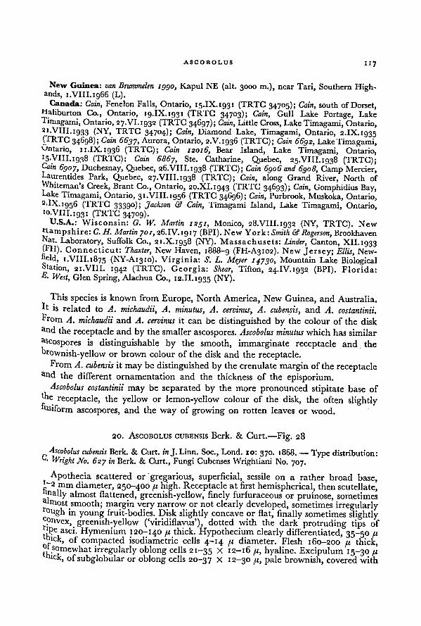
ASCOBOLUS117
New Guinea: van Brummelen 1990, Kapul NE (alt. 3000 m.), near Tari, Southern Highl-ands, 1.VIII.1966 (L).
Canada: Cain, Fenelon Falls, Ontario, 15.IX.1931 (TRTC 34705); Cain,, south of Dorset,Haliburton Co., Ontario, 19.IX.1931 (TRTC 34703); Cain,
_ __ w,, ,Gull Lake Portage, Lake
Timagami, Ontario, 27.VI.1932 (TRTG 34697); Cain,.
Little Cross, Lake Timagami, Ontario,ai-ym. 1933 (NY, TRTC 34704);
"
Cain,, Diamond Lake, Timagami, Ontario, 2.IX. 1035CTRTC 34698);
— ■ - -
Cain 6637, Aurora, Ontario, 2.V.1936 (TRTC); Cain 6692,j Lake Tima^ami,Ontario, 11.IX.1936 (TRTC);
~ ~------
—
Cain 12016, Bear Island, Lake Timagami, Ontario,15.VIII.1938 (TRTC);
"" ""
Cain 6867, Ste. Catharine, Quebec, 25.VIII.1938 (TRTC);Cain 6907, Duchesnay, Quebec, 26.VIII.1938 (TRTC); Cain 6906 and 6908,, Camp Mercier,Caurentides Park, Quebec, 27.VIII.1938 (TRTC); Cain,, along Grand River, North ofWhiteman's Creek, Brant Co., Ontario, 20.XI.1943 (TRTC 34693); Cain,, Gomphidius Bay,Lake Timagami, Ontario, 31.VIII.1956 (TRTG 34696); Cain,
,Purbrook, Muskoka, Ontario,
2.IX.i95 6 (TRTC 33390);~
'Jackson & Cain,_ ,
Timagami Island, Lake Timagami, Ontario,10.VIII.1931 (TRTC 34709).
U.S.A.: Wisconsin: G. IV. Martin 1251,, Monico, 28.VIII.1932 (NY, TRTC). New
Hampshire: C. H. Martin 701, 26.IV. 1917 (BPI). New York: Smith & Rogerson,. * --u-, \ / a ,
BrookhavenNat. Laboratory, Suffolk Co., 21.X.1958 (NY). Massachusets: Linder,fPm _
. _r- •' Canton, XII. 1933
Connecticut:.
\7TTT-0_
_/TVT1
Thaxter, New Haven, 1888-9 (FH-A3102). New Jersey; Ellis,, , - — —j j x - —• J • j '-"««'JNew-
eld, 1.VIII.1875 (NY-A1310). Virginia: S. L. Meyer 14730,, Mountain Lake BiologicalStation, 21.VIII. 1942 (TRTC). Georgia: Shear, Tifton, 24.IV. 1932 (BPI). Florida:£• West, Glen Spring, Alachua Co., 12.II.1935 (NY).
This species is known from Europe, North America, New Guinea, and Australia.H is related to A. michaudii, A. minutus, A. cervinus, A. cubensis, and A. costantinii.
From A. michaudii and A. cervinus it can be distinguished by the colour of the disk
and the receptacle and by the smaller ascospores. Ascobolus minutus which has similar
Zoospores is distinguishable by the smooth, immarginate receptacle and the
brownish-yellow or brown colour of the disk and the receptacle.From A. cubensis it may be distinguished by the crenulate margin of the receptacle
a"d the different ornamentation and the thickness of the episporium.Ascobolus costantinii may be separated by the more pronounced stipitate base of
the receptacle, the yellow or lemon-yellow colour of the disk, the often slightlyfusiform
ascospores, and the way of growing on rotten leaves or wood.
20. ASCOBOLUS CUBENSIS Berk. & Curt.—Fig. 28
Ascobolus cubensis Berk. & Curt, in J. Linn. Soc., Lond. 10: 370. 1868. — Type distribution:C- Wright No. 627 in Berk. & Curt., Fungi Cubenses Wrightiani No. 707.
Apothecia scattered or gregarious, superficial, sessileon a rather broad base,
'-2 mm diameter, 250-400 41 high. Receptacle at first hemispherical, then scutellate,'nally almost flattened, greenish-yellow, finely furfuraceous or pruinose, sometimesa|rnost smooth; margin very narrow or not clearly developed, sometimes irregularly
inyoung fruit-bodies. Disk slightly concave or flat, finally sometimes slightly
onvex, greenish-yellow ('viridiflavus'), dotted with the dark protruding tips of'pe asci. Hymenium 120-140 41 thick. Hypothecium clearly differentiated, 35-50 41bick, of compacted isodiametric cells 4-14 41 diameter. Flesh 160-200 41 thick,l somewhat irregularly oblong cells 21-35 X 12-16 41, hyaline. Excipulum 15-30 41
k, of subglobular or oblong cells 20-37 X 12-30 41, pale brownish, covered with
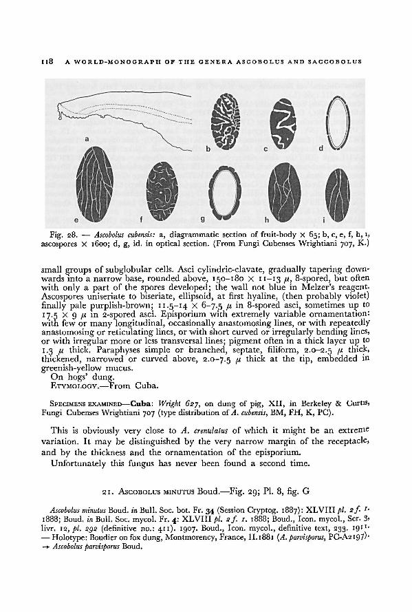
118A WORLD-MONOGRAPH OF THE GENERA ASCOBOLUS AND SACCOBOLUS
small groups of subglobular cells. Asci cylindric-clavate, gradually tapering down-
wards into a narrow base, rounded above, 150-180 X 11—13 /,<, 8-spored, but often
with only a part of the spores developed; the wall not blue in Melzer's reagent.
Ascospores uniseriate to biseriate, ellipsoid, at first hyaline, (then probably violet)
finally pale purplish-brown; 11.5-14 X 6-7.5 I1 in 8-spored asci, sometimes up to
1 7.5 X 9 /< in 2-spored asci. Episporium with extremely variable ornamentation:
with few or many longitudinal, occasionally anastomosing lines, or with repeatedly
anastomosing or reticulating lines, or with short curved or irregularly bending lines,
or with irregular more or less transversal lines; pigment often in a thick layer upto
1.3 /j, thick. Paraphyses simple or branched, septate, filiform, 2.0-2.5 u thick,
thickened, narrowed or curved above, 2.0-7.5 f,i thick at the tip, embedded in
greenish-yellow mucus.
On hogs' dung.ETYMOLOGY.—From Cuba.
SPECIMENS EXAMINED—Cuba: Wright 627, on dung of pig, XII, in Berkeley & Curtis,
Fungi Cubenses Wrightiani 707 (type distributionof A. cubensis, BM, FH, K, PC).
This is obviously very close to A. crenulatus of which it might be an extreme
variation. It may be distinguished by the very narrow margin of the receptacle,and by the thickness and the ornamentation of the episporium.
Unfortunately this fungus has never been found a second time.
21. ASCOBOLUS MINUTUS Boud.—Fig. 29; Pl. 8, fig. G
Ascobolus minutus Boud. in Bull. Soc. bot. Fr. 34 (Session Cryptog. 1887): XLVIII pi. sf. '■
1888; Boud. in Bull. Soc. mycol. Fr. 4: XLVIIIpi. 2f. 1. 1888; Boud., Icon, mycol., Ser. 3'
livr. 12, pi. 292 (definitive no.: 411). 1907. Boud., Icon, mycol., definitive text, 233. 19"'
— Holotype: Boudieron fox dung, Montmorency, France, II. 1881 (A. parvisporus, PG-A2197)"
—> Ascobolus parvisporus Boud.
a, diagrammatic section of fruit-body X 65; b, c, e, f, h, i,
ascospores X 1600; d, g, id. in optical section. (From Fungi Cubenses Wrightiani 707, K.)
Fig. 28. —Ascobolus cubensis:
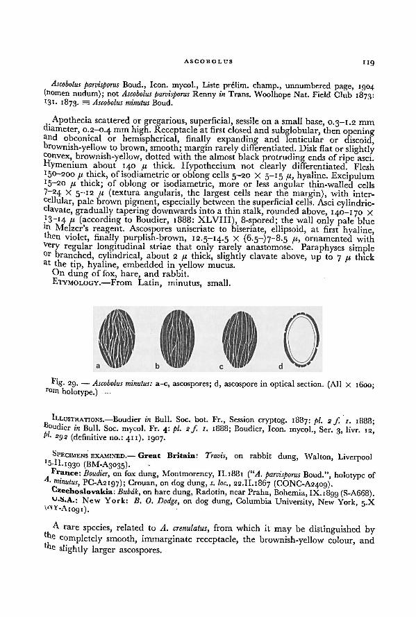
ASCOBOLUS119
Ascobolus parvisporus Boud., Icon, mycol., Liste prelim, champ., unnumbered page, 1904(nomen nudum); not Ascobolus parvisporus Renny in Trans. Woolhope Nat. Field Club 1873:*3 1 • 1873. = Ascobolus minutus Boud.
Apothecia scattered or gregarious, superficial, sessile on a small base, 0.3-1.2 mm
diameter, 0.2-0.4 mm high. Receptacle at first closed and subglobular, then openingand obconical or hemispherical, finally expanding and lenticular or discoid,brownish-yellow to brown, smooth; margin rarely differentiated.Disk flat or slightlyconvex, brownish-yellow, dotted with the almost black protruding ends of ripe asci.
flymenium about 140 //, thick. Hypothecium not clearly differentiated. Flesht5°-200 /j, thick, of isodiametric
or oblong cells 5-20 X 5-15 /.1, hyaline. ExcipulumJ 5~20 n thick; of oblong or isodiametric, more or less angular thin-walled cells
7-24 x 5-12 /{ (textura angularis, the largest cells near the margin), with inter-
cellular, pale brown pigment, especially between the superficial cells. Asci cylindric-clavate, gradually tapering downwards into a thin stalk, rounded above, 140-170 x
,
I 3~i4 a (according to Boudier, 1888: XLVIII), 8-spored; the wall only pale bluelr' Melzer's reagent. Ascospores uniseriate to biseriate, ellipsoid, at first hyaline,then violet, finally purplish-brown, 12.5-14.5 X (6.5-)7-8.5 fi, ornamented withvery regular longitudinal striae that only rarely anastomose. Paraphyses simpleor branched, cylindrical, about 2 /.i thick, slightly clavate above, up to 7 /u thickat the tip, hyaline, embedded in yellow mucus.
On dung of fox, hare, and rabbit.
ETYMOLOGY.—From Latin, minutus, small.
ILLUSTRATIONS.—Boudier in Bull. Soc. bot. Fr., Session cryptog. 1887: pi. 2 f. 1.p 1888;"oudier in Bull. Soc. mycol. Fr. 4: pi. 2 f. 1. 1888; Boudier, Icon, mycol., Ser. 3, livr. 12,pl. 2g2 (definitive no.: 411). 1907.
SPECIMENS EXAMINED.— Great Britain: Travis, on rabbit dung, Walton, Liverpool
15-11.1930 (BM-A3035).France: Boudier, on fox dung, Montmorency, II.1881 (“A. parvisporus Boud.", holotype of
A. minutus, PC-A2197); Crouan, on dog dung, s. loc.,w
_
w , , 22.11.1867 (CONC-A2409).Czechoslovakia: Bubak, on hare dung, Radotin, nearPraha, Bohemia, IX.1800 (S-A668).U.S.A.: New York: IS. 0. Dodge, on dog dung, Columbia University, New York, 5.X
vNY-Aiogi).
A rare species, related to A. crenulatus,,
from which it may be distinguished bye completely smooth, immarginate receptacle, the brownish-yellow colour, ande slightly larger ascospores.
Fig. 29. — a-c, ascospores; d, ascospore in optical section. (All X 1600;from holotype.)
Ascobolus minutus:
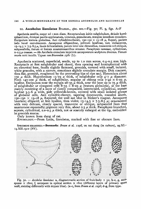
120 A WORLD-MONOGRAPH OF THE GENERA ASCOBOLUS AND SACCOBOLUS
22. Ascobolus lineolatus Brumm., spec. nov. —Fig. 30; Pl. 9, figs. A-F
Apothecia sessilia, usque ad 1 mm diam. Receptaculum initio subglobulare, deinde hemi-
sphaericum, denique paullo applanatum, virescens, granulatum, margine interdum crenulato.
Excipulum textura globulosa. Asci cylindrico-clavati, 130-150 X 15-18 /(, 8-spori, parieteiodo haud caerulescente. Ascosporae ellipsoideae, primum hyalinae, tum violascentes,
I 3_I 4-5 x 7.5-8.5 fi, lineis delicatissimis, paruminter sese distantibus, transversis vel obliquis,
subparallelis, iterum et iterum anastomosantibus ornatae. Paraphyses ramosae, cylindricae,
2-2.5 /' crassae. —Ab Ascobolo crenulato imprimis ascospororum sculptura diversus. Fimum
muris ratti incolit. Typus: van Brummelen 1981 (L).
Apothecia scattered, superficial, sessile, up to 1.0 mm across, 0.4-0.5 mm high.Receptacle at first subglobular and closed, then opening and hemispherical with
an obconical base, finally slightly flattened, greenish, covered with small, isolated,white granules, with a narrow, sometimes slightly crenulate margin. Disk concave,
then flat, greenish, roughened by the protruding tips of ripe asci. Hymenium about
130 /< thick. Hypothecium 15-23 // thick, of subglobular cells 4-7 /i diameter.
Flesh 140-200 fi thick, of subglobular, angular or oblong cells 7-42 X 6-23 //,
hyaline. Excipulum near the margin 28-35 fx thick, near the base up to 75 /<thick,
of subglobular or elongated cells 8-35 x 8-23 /i (textura globulosa), at the base
mainly consisting of a layer of closely compacted, intertwined, cylindrical, septate
hyphae 3.5-6 /t wide, pale yellowish-brown, covered with small isolated groupsof globular cells. Asci cylindric-clavate, tapering downwards, rounded above,
130-150 X 15—18 p, 8-spored; the wall not blue in Melzer's reagent. Ascospores
biseriate; ellipsoid; at first hyaline, then violet; 13-14.5 X 7.5-8.5 /<;ornamented
with very delicate, closely spaced, transverse or oblique, subparallel lines that
anastomose repeatedly; pigment very thin, about 0.3 fi thick. Paraphyses branched,
septate, cylindrical, 2.0-2.5 fx thick, not or scarcely enlarged at the tip, embeddedin greenish mucus.
Only known from dung of rat.
ETYMOLOGY.—From Latin, lineolatus, marked with fine or obscure lines.
SPECIMENS EXAMINED.—Bermuda: Brown et al. 1296, on rat dung (in culture), 29. XI.-
14.XII.1912 (NY).
a, diagrammatic section of fruit-body X 50; b-e, g,asco-
spores X 1600; f, ascospore in optical section X 1600 (different layers of primary spore
wall, staining differently with trypan blue). (a-c, from
Ascobolus lineolatus:
Brown et al. 1296; d-g, from type.
Fig. 30. —
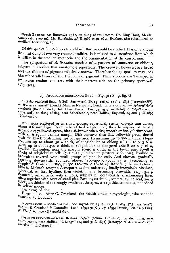
ASCOBOLUS 121
North Borneo: van Brummelen 1981, on dung of rat (comm. Dr. Ding Hou), Mesilau
Camp (alt. 1500 m). Mt. Kinabalu, 4.VII. 1966 (type of A. lineolatus, also subcultured on
sterilized horse dung, L).
Of this species fine cultures from North Borneo could be studied. It is only known
from rat dung of two very remote localities. It is related to A. crenulatus, from which
it differs in the smaller apothecia and the ornamentationof the episporium.The episporium of A. lineolatus consists of a pattern of transverse or oblique,
subparallel crevices that anastomose repeatedly. The crevices, however, are broad
and the ribbons of pigment relatively narrow. Therefore the episporium may look
like subparallel rows of short ribbons of pigment. These ribbons are T-shaped in
transverse section and rest with their narrow side on the primary spore-wall
(Fig. 3of).
23. ASCOBOLUS CROSSLANDII Boud.—Fig. 31; Pl. 9, fig. G
Ascobolus crosslandii Boud. in Bull. Soc. mycol. Fr. 14: 126pi. 11f 2. 1898 ("crowslandi").—Boudiera crosslandii (Boud.) Mass. in Naturalist, Lond. 1901: 179. 1901.
— Sphaeridioboluscrosslandii (Boud.) Boud., Hist. Class. Discom. Eur. 73. 1907. — Holotype: Soppitt (comm.Crossland), on dung of dog, near Salterhebble, near Halifax, England, 24 and 31.X. 1897(PC-A2218).
Apothecia scattered or in small groups, superficial, sessile, 0.5-2.0 mm across,about 0.6 mm high. Receptacle at first subglobular, then hemispherical, finallyexpanding; yellowish-green, blackish-brown when dry, smooth or finely furfuraceous,With an irregular dentate margin. Disk concave, then flat, yellowish-green, dotted
With the black protruding tips of ripe asci. Hymenium up to 200 u thick. Hypo-thecium
up to about 30 /x thick, of subglobular or oblong cells 4-10 X 3-6 pi.blesh
up to about 400 pi thick, of subglobular or elongated cells 8-20 X 7-16 /1,
hyaline. Excipulum near the margin 19-25 11 thick, in the lower part 26-38 pxthick; of subglobular cells (7-) 12-24 !l diameter (textura globulosa), hyaline or
brownish; covered with small groups of globular cells. Asci clavate, graduallytapering downwards, rounded above, '170-200 X about 25 71' (according to
boppitt & Crossland 1899, p. 30: 130-170 X 16-20 pi), 8-spored; the wall clearlyblue in Melzer's reagent. Ascospores at first uniseriate, finally irregularly biseriate,spherical, at first hyaline, then violet, finally becoming brownish, 11.5-13.4 pidiameter, ornamented with sinuous, subparallel, occasionally anastomosing lines,°ften together with rows of small pits. Paraphyses simple, septate, cylindrical, 2-4 fithick, not thickened to strongly swollen at the apex, 2-11 pi thick at the tip, embeddedIn yellow mucus.
tin dung of dog.ETYMOI.OGY.
—Alter G. Crossland, the British amateur mycologist, who sent the
ttaterial to Boudier.
ILLUSTRATIONS.—Boudier in Bull. Soc. mycol. Fr. 14: pi. 11 ƒ. 2. 1898 (“A. crowslandi”);& Crossland in Naturalist, Lond. 1899: 31 ƒ. 9-13. 1899; Dennis, Brit. Cup Fungi
pi. VIII f E. i960 ( Sphaeridiobolus).
SPECIMEN EXAMINED.—Great Britain: Soppitt„ ... (comm. Crossland), on dog dung, near
terhebble, near Halifax, "XI. 1897" (24 and 31.X. 1897) [lectotype of A. crosslandii (“A.cr
owslandi”), PC-A22 I8],
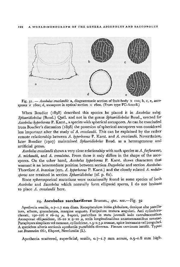
A WORLD-MONOGRAPH OF THE GENERA ASCOBOLUS AND SACCOBOLUS122
When Boudier (1898) described this species he placed it in Ascobolus subg.
Sphaeridiobolus (Boud.) Quel, and not in the genus Sphaeridiobolus Boud., erected for
Ascobolus hyperboreus P. Karst., a species with spherical ascospores. As can be concluded
irom Boudier's discussion (1898) the possesion of spherical ascospores was considered
less important after the study of A. crosslandii. This can be explained by the rather
remote relationship between A. hyperboreus P. Karst. and A. crosslandii. Nevertheless,
later Boudier (1907) maintained Sphaeridiobolus Boud. as a heterogeneous and
artificial genus.
Ascobolus crosslandii shows a very close relationship with such species as A. furfuraceus,
A. michaudii, and A. crenulatus. From these it only differs in the shape of the asco-
spores. On the other hand, Ascobolus hyperboreus P. Karst. shows characters that
warrant it an intermediateposition between section Dasyobolus and section Ascobolus.
Therefore A. brassicae (syn. A. hyperboreus P. Karst.) and the closely related A. nodulo-
sporus are retained in section Sphaeridiobolus (cf. p. 62).
Since spherosporical mutations were occasionally found in some species of both
Ascobolus and Saccobolus which normally form ellipsoid spores, I do not hesitate
to place A. crosslandii here.
24. Ascobolus sacchariferus Brumm., spec. nov. —Fig. 32
Apothecia sessilia, 0.7-1.7 mm diam. Receptaculum initio globulare, deniquealte patella-
turn, album, granulatum, margine angusto. Excipulum textura angulari. Asci cylindrico-
clavati, 190-226 X 16-24 fh 8-spori, parietibus in statu juvenili iodo caerulescentibus.
Ascosporae ellipsoideae, 16-20 X 9-10 /t,striis longitudinalibus anastomosantibus ornatae.
Paraphyses simplices vel ramosae, cylindricae, 1.5-2.5 /« crassae, apice incrassato vel irregularis
A speciebus alteris sectionis apotheciis puralbidis diversus. Fimum cervinum incolit. Typus:
van Brummelen 661, Elspeet, Necrlandia (L).
Apothecia scattered, superficial, sessile, 0.7-1.7 mm across, 0.5-0.8 mm high-
a, diagrammatic section of fruit-body X 110; b, c, e, asco-
spores X 1600; d, ascospore in optical section X 1600. (From type PC-A2218.)
Ascobolus crosslandii:Fig. 31. —
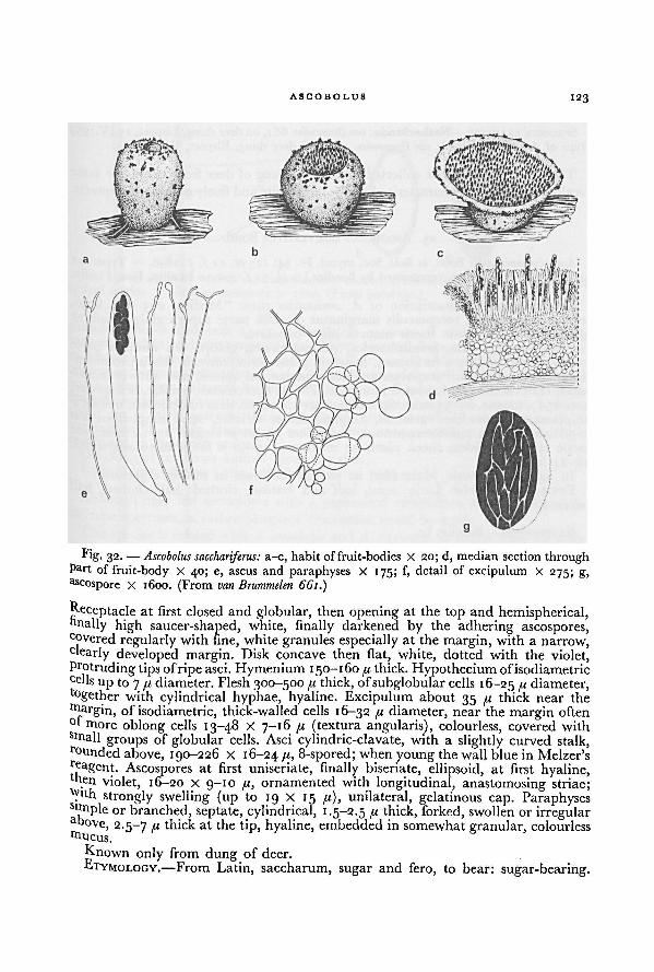
ASCOBOLUS 123
Receptacle at first closed and globular, then opening at the top and hemispherical,finally high saucer-shaped, white, finally darkened by the adhering ascospores,covered regularly with fine, white granules especially at the margin, with a narrow,clearly developed margin. Disk concave then flat, white, dotted with the violet,Protruding tips ofripe asci. Hymenium 150-160 f1 thick. Hypotheciumofisodiametriccells
up to 7 fx diameter. Flesh 300-500fi thick, ofsubglobular cells 16-25 [i diameter,together with cylindrical hyphae, hyaline. Excipulum about 35 fi thick near the
tfiargin, of isodiametric, thick-walled cells 16-32 fi diameter, near the margin often°t more oblong cells 13-48 X 7-16 it (textura angularis), colourless, covered withsmall groups of globular cells. Asci cylindric-clavate, with a slightly curved stalk,founded above, 190-226 X 16-24 /6 8-spored; when young thewall blue in Melzer'sre
agent. Ascospores at first uniseriate, finally biseriate, ellipsoid, at first hyaline,nen violet, 16-20 x 9-10 fi, ornamented with longitudinal, anastomosing striae;
tyuh strongly swelling (up to 19 X 15 fi), unilateral, gelatinous cap. ParaphysesSlfiiple or branched, septate, cylindrical, 1.5-2.5 fi thick, forked, swollen or irregularabove, 2.5-7 B thick at the tip, hyaline, embedded in somewhat granular, colourlesstfiucus.
Known only from dung of deer.
ETYMOLOGY.—From Latin, saccharum, sugar and fero, to bear: sugar-bearing.
Fig. 32. —
a-c, habit of fruit-bodies X 20; d, median section throughpart of fruit-body X 40; e, ascus and paraphyses X 175; f, detail of excipulum X 275; g,
ascospore X 1600. (From van Brummelen 661.)
Ascobolus sacchariferus:
124 A WORLD-MONOGRAPH OF THE GENERA ASCOBOLUS AND SACCOBOLUS
SPECIMEN EXAMINED.—Netherlands: van Brummelen 66 1, on deer dung, Elspeet, 13.IV.1959(type of A. sacchariferus, L); van Brummelen 1438, on deer dung, Elspeet, V.1960 (L).
This species, which was collected twice from dung of deer from about the same
locality, is sufficiently characterized by the pure white and finely granular receptacle.
25. ASCOBOLUS SEMIVESTITUS Boud.
Ascobolus semivestitus Boud. in Bull. Soc. mycol. Fr. 14: 125 pi. 11 f. /. 1898. — Type: not
known to be in existence, represented by Boudier I.e. pi. 11f. /; type locality, Jura, France.
Boudier's specific description of A. semivestitus runs: "Medius, 2-3 mm latus,
sessilis, luteo-virens, receptaculis marginatus dimidia parte supera glabra, inferne
albido-villosa, hymenio thecis maturis nigro-punctato.
Receptacula sessilia, hemisphaerica, sub-marginata, glabra, sed dimidia parteinfera albo-villosa, non furfuracea, hymenio piano, dein convexo, thecis prominen-tibus nigro-punctato. Paraphyses graciles, septatae, ad apicem non aut vix incras-
satae, et ut thecae gelatina lutea immersae. Thecae operculatae, amplae, octosporae,
200-250 [x longae, 25-35 latae. Sporae pulchre violaceae, dein fuscescentes, oblongo-ellipticae, magnitudine variantes, longitudinaliter striatae, striis simplicibus aut
2-3 divisis, rarius anastomosantibus, normaliter 25-30 fx longae, 13-14 latae, sed
saepe etiam in eadem theca plurimae maximae, 45-50 /.i longitudine aequantes,18-24 latitudine.
In montibus juranis, Maio 1896 ad stercus vaccinum in turfosis dejectum."ETYMOLOGY.—From Latin, semi, half and vestitus, clothed: half clothed, half
covered.
ILLUSTRATION.—Boudier, I.e.
Of this species no material could be studied. Boudier's description and illustrations,
however, are sufficiently detailed to ensure future recognition. Undoubtedly, it is
related to A. furfuraceus, from which it can be distinguished by the villose inferior
part of the receptacle and the larger ascospores. No explanation has been found
for the occurrence of two different types of ascospores.
26. ASCOBOLUS CERVINUS Berk. & Broome—Fig. 33
Ascobolus cervinus Berk. & Broome in J. Linn. Soc. Lond. (Bot.) 15: 85. 1876. — Holotype ;Thwaites No. 1122 (K), in literature cited by error as "Thwaites 422".
Apothecia gregarious or crowded, superficial, sessile on a broad base, up to
1.5 mm across, up to 0.5 mm high. Receptacle at first closed and globular, then
opening at the top and hemispherical, finally expanding and scutellate, olive-green,
finely furfuraceous, with a prominent margin. Disk concave to flat, olive-green.Hymenium about 130 74 thick. Hypothecium clearly differentiated, about 30 «
thick, of closely compacted subglobular cells 6-12 /,< diameter. Flesh up to 300 /<
thick, of isodiametric cells 12-22 /,< diameter, hyaline. Excipulum 20-30 ji,thick,
of irregular or subglobular cells 12-15 fi diameter (the largest cells at the surface),near the base with 3.0-3.5 41 wide hyphae which connect the fruit-body with the
substratum; covered with furfuraceous particles consisting of loose groups of sub-
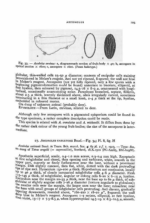
ASCOBOLUS 125
globular, thin-walled cells 25-50 fx diameter; contents of excipular cells stainingbrownish-red in Melzer's reagent. Asci not yet ripened, 8-spored; the wall not bluem Melzer's reagent. Ascospores (not yet fully ripened, only a few spores with a
beginning pigment-formation could be found) uniseriate to biseriate, ellipsoid, at
hrst hyaline, then coloured by pigment, 14.5-16 X 8-9 //, ornamented with longi-tudinal, occasionally anastomosing striae. Paraphyses branched, septate, filiform,about
2.5 thick, scarcely thickened above, often irregularly curved, sometimes
terminating in a thin filament or a small knob, 2-4 /< thick at the tip, hyaline,embedded in coloured mucus.
On dung of unknown animal (probably deer).ETYMOLOGY.— From Latin, cervinus, related to deer.
Although only few ascospores with a pigmented episporium could be found inthe type specimen, a rather complete description could be made.
This species is related with A. crenulatus and A. michaudii. It differs from these bythe rather dark colour of the young fruit-bodies; the size of the ascospores is inter-
mediate.
27. ASCOBOLUS CARLETONII Boud.—Fig. 34; Pl. 8, fig. H
Ascobolus carletonii Boud. in Trans. Brit. mycol. Soc. 4: 62 pl. s ƒ. 1. 1913. — Type: Rea,0tl dung of Tetrao urogalli (= capercaillie), Scotland, 18.X.1912 (PC-A2265, BM-A2988).
Apothecia superficial, sessile, 0.4-1.0 mm across, 0.3-0.4 mm high. Receptacleat first subglobular and closed, then opening and turbinate, white, smooth in theuPper part, coarsely or finely furfuraceous near the base, without a prominent
Disk slightly concave, then flat, white, dotted with the dark protrudinglPs of ripe asci. Hymenium 150-200 u thick. Hypothecium not continuous, locallyP to 40 thick, of closely compacted subglobular cells 4-8 fi diameter. Flesh
!l thick, of subglobular, angular or oblong cells 6-20 x 6-13 /LI, hyaline.Xcipulum near the margin 20-33 /< thick, near the base up to 60 fi thick, of sub-
s °bular or slightly angular cells 7-26 //, diameter (textura angularis to globulosa);fte smaller cells near the margin, the larger ones near the base; colourless; near
( : base with small groups of subglobular cells protruding. Asci clavate, graduallyaPering downwards, rounded above, "160-220 X 18-20 /<", 8-spored; the wall
t
eeP biue in Melzer's reagent. Ascospores 1-2 seriate, ellipsoid, at first hyaline,
en violet, 13-17 X 7.5-8.5 when hypertrophied 14.5-19 X 8.5-10.5/(., smooth.
Fig. 33. — Ascobolus cervinus:a, diagrammatic section of fruit-body X 40; b, ascospore in
optical section X 1600; c, ascospore x 1600. (From holotype.)
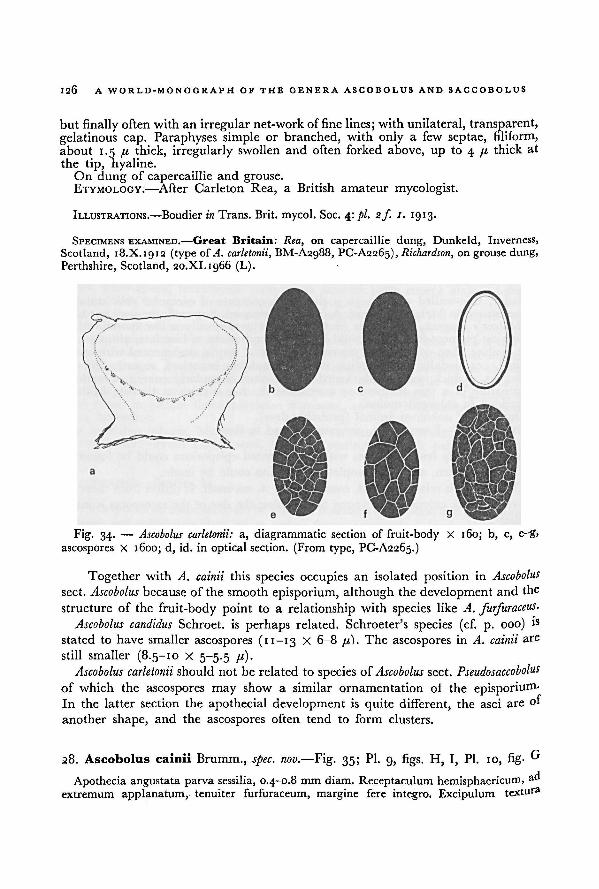
126 A WORLD-MONOGRAPH OF THE GENERA ASCOBOLUS AND SACCOBOLUS
but finally often with an irregular net-workof fine lines; with unilateral, transparent,
gelatinous cap. Paraphyses simple or branched, with only a few septae, filiform,
about 1.5 /< thick, irregularly swollen and often forked above, up to 4 (i thick at
the tip, hyaline.On dung of capercaillie and grouse.
ETYMOLOGY.—After Carleton Rea, a British amateur mycologist.
ILLUSTRATIONS.—Boudier in Trans. Brit, mycol. Soc. 4: pi. 2f 1. 19 13.
SPECIMENS EXAMINED.—Great Britain: Rea, on capercaillie dung, Dunkeld, Inverness,
Scotland, 18.X.1912 (type ofA. carletonii, BM-A2g88, PC-A2265), Richardson, on grouse dung,
Perthshire, Scotland, 20.XI.1966 (L).
Together with A. cainii this species occupies an isolated position in Ascobolus
sect. Ascobolus because of the smooth episporium, although the development and the
structure of the fruit-body point to a relationship with species like A. furfuraceus.Ascobolus candidus Schroet. is perhaps related. Schroeter's species (cf. p. 000) is
stated to have smaller ascospores (11-13 X 6-8 it). The ascospores in A. cainii are
still smaller (8.5-10 X 5-5.5 /<).Ascobolus carletonii should not be related to species of Ascobolus sect. Pseudosaccobolus
of which the ascospores may show a similar ornamentation of the episporium-
In the latter section the apothecial development is quite different, the asci areof
another shape, and the ascospores often tend to form clusters.
28. Ascobolus cainii Brumm., spec. nov. —Fig. 35; Pl. 9, figs. H, I, Pl. 10, fig. G
Apothecia angustata parva sessilia, 0.4-0.8 mm diam. Receptaculum hemisphaericum,ad
extremum applanatum, tenuiter furfuraceum, margine fere integro. Excipulum textura
a, diagrammatic section of fruit-body X 160; b, c, e-g,
ascospores X 1600; d, id. in optical section. (From type, PC-A2265.)
Ascobolus carletonii:Fig. 34.—
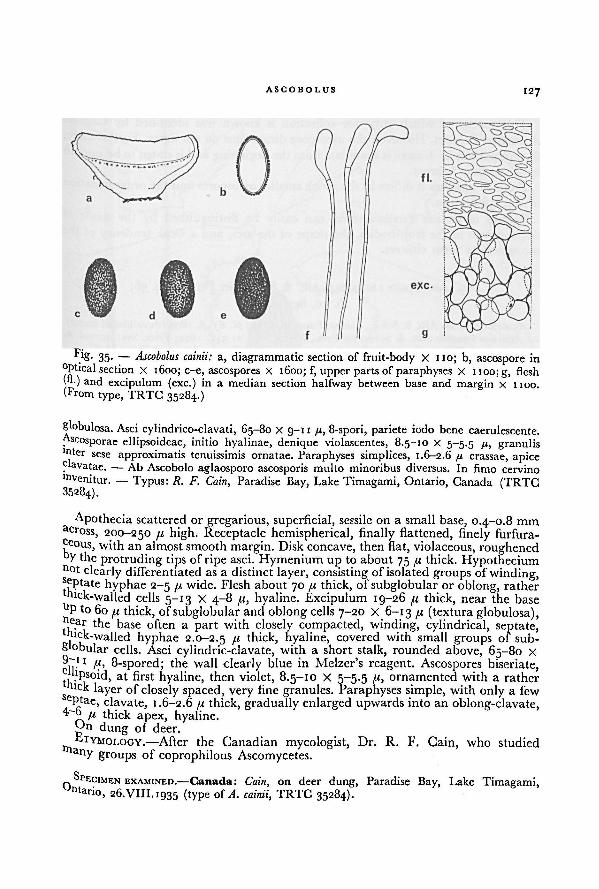
ASCOBOLUS 127
Slobulosa. Asci cylindrico-clavati, 65-80 X 9-11 ft, 8-spori, pariete iodo bene caerulescente.
«cosporae ellipsoideae, initio hyalinae, denique violascentes, 8.5-10 X 5-5.5 ft, granulis"iter
sese approximatis tenuissimis ornatae. Paraphyses simplices, 1.6-2.6 ft crassae, apice. avatae.
—Ab Ascobolo aglaosporo ascosporis multo minoribus diversus. In fimo cervino
"tvenitur. — Typus: R. F. Cain, Paradise Bay, Lake Timagami, Ontario, Canada (TRTC35284).
Apothecia scattered or gregarious, superficial, sessile on a small base, 0.4-0.8 mmacross, 200-250 ft high. Receptacle hemispherical, finally flattened, finely furfura-ceous, with an almost smooth margin. Disk concave, then flat, violaceous, roughenedDV the protruding tips of ripe asci. Hymenium up to about 75 ft thick. Hypotheciumn°t clearly differentiatedas a distinct layer, consisting of isolated groups of winding,septate hyphae 2-5 ft wide. Flesh about 70 /« thick, of subglobular or oblong, rather
hick-walled cells 5-13 X 4-8 fi, hyaline. Excipulum 19-26 fi thick, near the baseU P to 60
ft thick, of subglobular and oblong cells 7-20 X 6-13 ft (textura globulosa),Pear the base often a part with closely compacted, winding, cylindrical, septate,mck-walled hyphae 2.0-2.5 ft thick, hyaline, covered with small groups of sub-
globular cells. Asci cylindric-clavate, with a short stalk, rounded above, 65-80 x
up
1 8-spored; the wall clearly blue in Melzer's reagent. Ascospores biseriate,hpsoid, at first hyaline, then violet, 8.5-10 X 5-5.5 R, ornamented with a rathertick layer of closely spaced, very fine granules. Paraphyses simple, with only a few
cptae, clavate, 1.6-2.6 //. thick, gradually enlarged upwards into an oblong-clavate,R thick apex, hyaline,
dung of deer.
Ltymology.— After the Canadian mycologist, Dr. R. F. Cain, who studiedany groups of coprophilous Ascomycetes.
SPECIMEN EXAMINED.—Canada: Cain, on deer dung, Paradise Bay, Lake Timagami,°ntario, 26.VIII. 1935 (type of A. cainii, TRTC 35284).
Fig. 35. — Ascobolus cainii: a, diagrammatic section of fruit-body X 110; b, ascospore in
optical section X 1600; c-e, ascospores X 1600; f, upper parts of paraphyses X 1100; g, flesh(fl.) and excipulum (exc.) in a median section halfway between base and margin X 1100.
(From type, TRTC 35284.)
128 A WORLD-MONOGRAPH OF THE GENERA ASCOBOLUS AND SACCOBOLUS
This species of which only one collection is known was identified by Cain as
A. candidus Schroet. However, the ascospore dimensions do not agree. Furthermore,
the episporium in A. cainii is granulate from the beginning and is stated to be smooth
in A. candidus Schroet.
From A. carletonii it differs in the much smaller ascospores and the ornamentation
of the episporium.
Species of section Pseudosaccobolus can easily be distinguished by the mode of
development of the fruit-bodies, the shape of the asci, and a weak tendency of the
ascospores to form clusters.
29. ASCOBOLUS LIGNATILIS Alb. & Schw. per Pers.—Fig. 36;
Pl. 10, figs. A-D
Ascobolus lignatilis Alb. & Schw., Consp. Fung. nisk. 347pi. 6ƒ. 6. 1805 (devalidated name).— Ascobolus lignatilis Alb. & Schw. per Pers., Mycol. eur. 1: 342. 1822; Fries, Syst. mycol. 2:
162. 1822. — Type: Albertini & Schweinitz, s. loc., s. dat. (L 910.262.142, MEL-A1636).Peziza psittacina Quel, in Mem. Soc. Emulation Montbeliard Ser. II 5: 395 pi. gf. g. 1873
(= Champ. Jura Vosges 390 pi. 5 f. g. 1873). — Ascobolus psittacinus (Quel.) Quel., Ench.
Fung. 293. 1886. — Type: Quelet, in a flower-hothouse, Valentigney, Doubs, France, VI.1872
(UPS-A2005).Ascobolus marchalii Bomm. & Rouss. in Bull. Soc. roy. Bot. Belg. 23: 143. 1884.— Lectotype:
Marchal, on cow dung, Bruxelles, Belgium, 1883 (BR-A353).
Apothecia scattered or gregarious, superficial, usually with a very distinct stalk,
( 2_)5~ 12 mm diameter, up to 5 mm high. Receptacle at first closedand subglobularor cylindrical, then opening at the top and becoming scutellate or cyathiform,finally flattened, with a distinct furrowed or furfuraceous stipe, which often broadens
at the base, dingy yellow to greenish-yellow, furfuraceous; the margin at first often
slightly crenulate or fimbriate, prominent, rather thin, finally often slightly curved
outward. Disk flat, finally sometimes slightly convex, dingy yellowish-green to
yellowish, dotted with the almost black protruding tips of ripe asci. Hymeniurn
150-190 pi thick. Hypothecium about 30 pi thick, of closely compacted, isodiametriccells 7-1 1 pi diameter. Flesh of varying thickness, up to more than 4000 pi thickin the central part, of subglobular or rounded cells 7-12 pi diameter, hyaline.Excipulum 24-50(-65) pi thick, of subglobular cells 12-20 pi diameter, hyaline,covered with irregularly formed groups of globular cells. Asci clavate, graduallytapering downwards, rounded above, 160-230 X 16-20 pi, 8-spored; the wall blue in
Melzer's reagent. Ascospores biseriate; ellipsoid to fusiform-ellipsoid, with blunt
ends, at first hyaline, then violet, finally sometimes purplish-brown, 16-19.5 X
8.5-10 pi, when swollen reaching 22 X 13 //, ornamented with subparallel, usually
longitudinal lines, that only occasionally anastomose, more or less reticulated when
swollen. Paraphyses simple or branched, septate, cylindrical, 1.5-2.5 pi thick, clavate
or irregularly swollen to 6 pi thick at the tip, embedded in greenish-yellow mucus.
On rotten wood, branches or leaves, on manure heap, on rotten straw and old
paper, on humus in a hot-house, on owl-pellets, on dung of cow and man, and on
all kinds of substrata which have been dirtied or mixed with dung or urine.
ETYMOLOGY.—From Latin, lignatilis, pertaining to wood.
ILLUSTRATIONS.—Albertini & Schweinitz, Consp. Fung. nisk. pi. 6 f. 6. 1805; Boudier i"
Annls Sci. nat. (Bot.) V 10: pi. 5 f. 1. 1869; Falck in Beitr. Biol. Pfl. (ed. Cohn) 8: pi. ,2
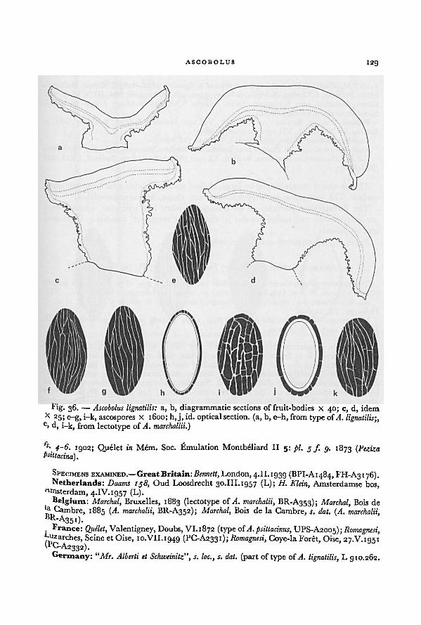
ASCOBOLUS 129
fs- 4-6. 1902; Quelet in Mem. Soc. Emulation Montbeliard II 5: pi. 5f g. 1873 ((Peziza
psittacina).
SPECIMENS EXAMINED.—Great Britain-.Bennett, London,4.II.1939 (BPI-A1484, FH-A3176).Netherlands: Daams 158, Oud Loosdrecht 30.III.1957 (L); H. Klein, Amsterdamse bos,
Amsterdam, 4.IV.1957 (L).Belgium: Marchal, Bruxelles, 1883 (lectotype of A. marchalii, BR-A353); Marchal, Bois de
' a Cambre, 1885 (A. marchalii, BR-A352); Marchal, Bois de la Cambre, s. dat. (A. marchalii,
BR-A351).France: Quelet, Valentigney, Doubs, VI.1872 (type ofA. psittacinus, UPS-A2005); Romagnesi,
~uzarches, Seine et Oise, 10.VII.1949 (PC-A2331); Romagnesi,_ , Coye-la Forêt, Oise, 27.V. 1951
\rC-A2332).Germany: Mr. Alberti et Schweinitz", s. loc., s. dat. (part of type of A. lignatilis, L 910.262.
Fig. 36. — a, b, diagrammatic sections of fruit-bodies X 40; c, d, idem
x 25; e-g, i-k, ascospores X 1600; h, j, id. optical section. (a, b, e-h, from type of
Ascobolus lignatilis:A. lignatilis;
c, d, i-k, from lectotype of A. marchallii.)
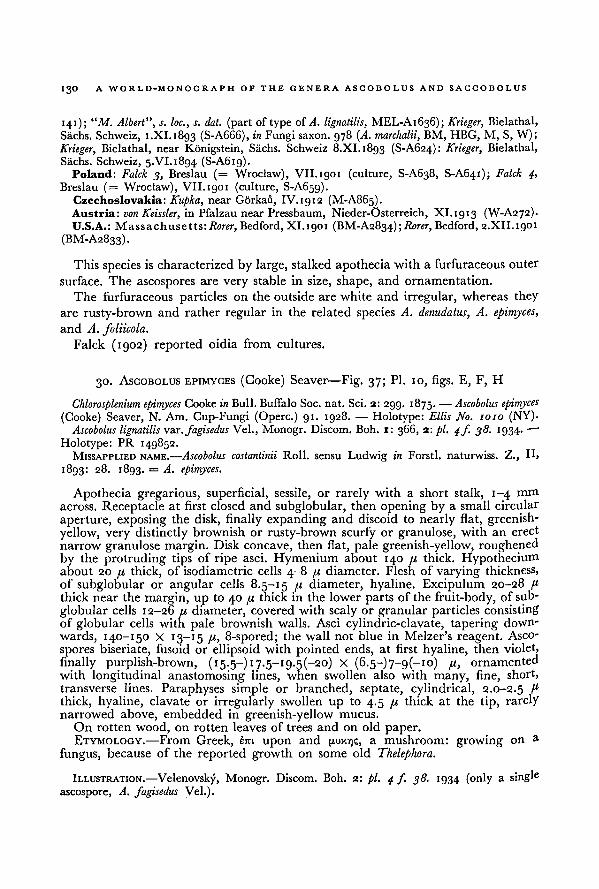
130A WORLD-MONOGRAPH OF THE GENERA ASCOBOLUS AND SACCOBOLUS
141); "M. Albert", s. loc., s. dat. (part of type of A. lignatilis, MEL-A1636); Krieger, Bielathal,
Sachs. Schweiz, i.XI. 1893 (S-A666), in Fungi saxon. 978 (A. marchalii, BM, HBG, M, S, W);
Krieger, Bielathal, near Konigstein, Sachs. Schweiz 8.XI.1893 (S-A624): Krieger, Bielathal,
Sachs. Schweiz, 5.VI.1894 (S-A6rg).Poland: Falck 3, Breslau (= Wroclaw), VII. 1901 (culture, S-A638, S-A641); Falck 4,
Breslau (= Wroclaw), VII.1901 (culture, S-A659).Czechoslovakia: Kupka, near Gorkaii, IV.1912 (M-A865).Austria: von Keissler, in Pfalzau near Pressbaum, Nieder-Osterreich, XI.1913 (W-A272).U.S.A.: Massachusetts: Rorer, Bedford, XI.1901 (BM-A2834); Rorer, Bedford, 2.XII.1901
(BM-A2833).
This species is characterized by large, stalked apothecia with a furfuraceous outer
surface. The ascospores are very stable in size, shape, and ornamentation.
The furfuraceous particles on the outside are white and irregular, whereas they
are rusty-brown and rather regular in the related species A. denudatus, A. epimyces,
and A. foliicola.
Falck (1902) reported oidia from cultures.
30. ASCOBOLUS EPIMYCES (Cooke) Seaver—Fig. 37; Pl. 10, figs. E, F, H
Chlorosplenium epimyces Cooke in Bull. Buffalo Soc. nat. Sci. 2: 299. 1875. — Ascobolus epimyces
(Cooke) Seaver, N. Am. Cup-Fungi (Operc.) 91. 1928. — Holotype: Ellis No. 1010 (NY).Ascobolus lignatilis var. fagisedus Vel., Monogr. Discom. Boh. 1: 366, 2: pi. 4f. 38. 1934- —
Holotype: PR 149852.MISAPPLIED NAME.—Ascobolus costantinii Roll, sensu Ludwig in Forstl. naturwiss. Z., IE
1893: 28. 1893. = A. epimyces.
Apothecia gregarious, superficial, sessile, or rarely with a short stalk, 1-4 mm
across. Receptacle at first closed and subglobular, then opening by a small circular
aperture, exposing the disk, finally expanding and discoid to nearly flat, greenish-yellow, very distinctly brownish or rusty-brown scurfy or granulose, with an erect
narrow granulose margin. Disk concave, then flat, pale greenish-yellow, roughenedby the protruding tips of ripe asci. Hymenium about 140 fx thick. Hypotheciumabout 20 fi thick, of isodiametric cells 4-8 fi diameter. Flesh of varying thickness,
of subglobular or angular cells 8.5-15 fi diameter, hyaline. Excipulum 20-28 fx
thick near the margin, up to 40 fi thick in the lower parts of the fruit-body, of sub-
globular cells 12-26 /< diameter, covered with scaly or granular particles consisting
of globular cells with pale brownish walls. Asci cylindric-clavate, tapering down-
wards, 140-150 X 13-15 fx, 8-spored; the wall not blue in Melzer's reagent. Asco-
spores biseriate, fusoid or ellipsoid with pointed ends, at first hyaline, then violet,
finally purplish-brown, (15. 5-) 17.5—19. 5(—20) X (6.5-)7-g(-io) fi,ornamented
with longitudinal anastomosing lines, when swollen also with many, fine, short,
transverse lines. Paraphyses simple or branched, septate, cylindrical, 2.0-2.5 f 1
thick, hyaline, clavate or irregularly swollen up to 4.5 fi thick at the tip, rarelynarrowed above, embedded in greenish-yellow mucus.
On rotten wood, on rotten leaves of trees and on old paper.Etymology.—From Greek, km upon and (ruxv)?, a mushroom: growing on a
fungus, because of the reported growth on some old Thelephora.
ILLUSTRATION.—Velenovsky, Monogr. Discom. Boh. 2: pi. 4 f. 38. 1934 (only a single
ascospore, A. fagisedus Vel.).
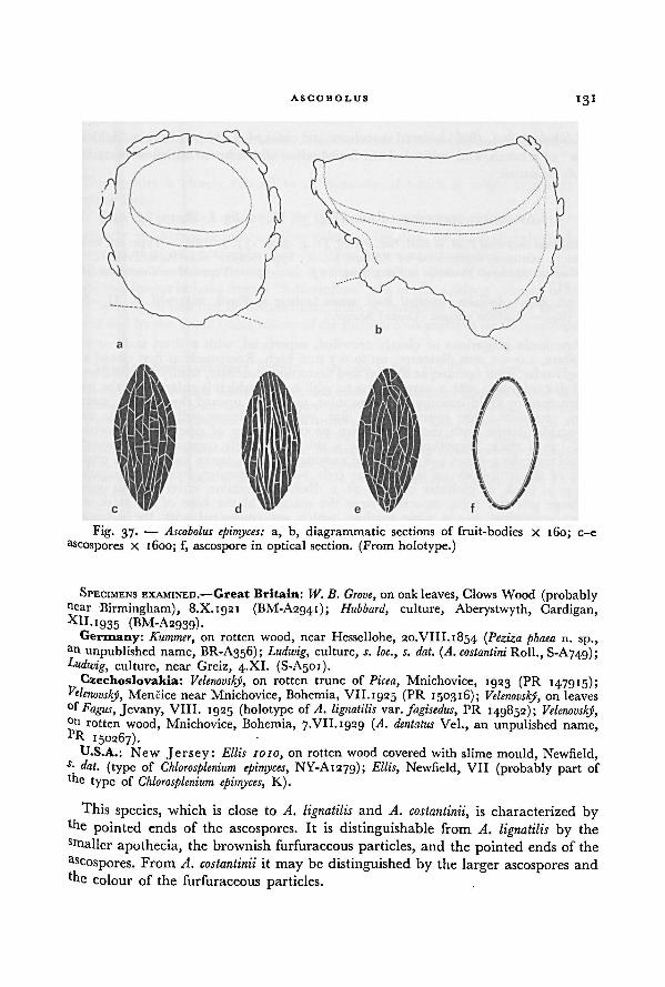
ASCOBOLUS131
SPECIMENS EXAMINED.—Great Britain: W. B. Grove, on oak leaves, Clows Wood (probablyfear Birmingham), 8.X.1921 (BM-A2941); Hubbard, culture, Aberystwyth, Cardigan,XlI
-'935 (BM-A2939).Germany: Kummer, on rotten wood, near Hessellohe, 20.VIII.1854 ( Peziza phaea n. sp.,
af unpublished name, BR-A356); Ludwig, culture, s. loc., s. dat. (A. costantini Roll., S-A74g);Ludwig, culture, near Greiz, 4.XI. (S-A501).
Czechoslovakia: Velenovsky, on rotten trunc of Picea, Mnichovice, 1923 (PR 147915);Velenovsky, Mencice near Mnichovice, Bohemia, VII.1925 (PR 150316); Velenovsky, on leaves
Fagus, Jevany, VIII. 1925 (holotype of A. lignatilis var. fagisedus, PR 149852); Velenovsky,°f
rotten wood, Mnichovice, Bohemia, 7.VII. 1929 (A. dentatus Vel., an unpulished name,pR 150267).
U.S.A.: New Jersey: Ellis 1010, on rotten wood covered with slime mould, Newfield,s
- dat. (type of Chlorosplenium epimyces, NY-A1279); Ellis, Newfield, VII (probably part ofthe type of Chlorosplenium epimyces, K).
This species, which is close to A. lignatilis and A. costantinii, is characterized by'he pointed ends of the ascospores. It is distinguishable from A. lignatilis by the
srnallcr apothecia, the brownish furfuraceous particles, and the pointed ends of the
Zoospores. From A. costantinii it may be distinguished by the larger ascospores and
'he colour of the furfuraceous particles.
Fig. 37.—
a, b, diagrammatic sections of fruit-bodies X 160; c-e
ascospores X 1600; f, ascospore in optical section. (From holotype.)
Ascobolus epimyces:
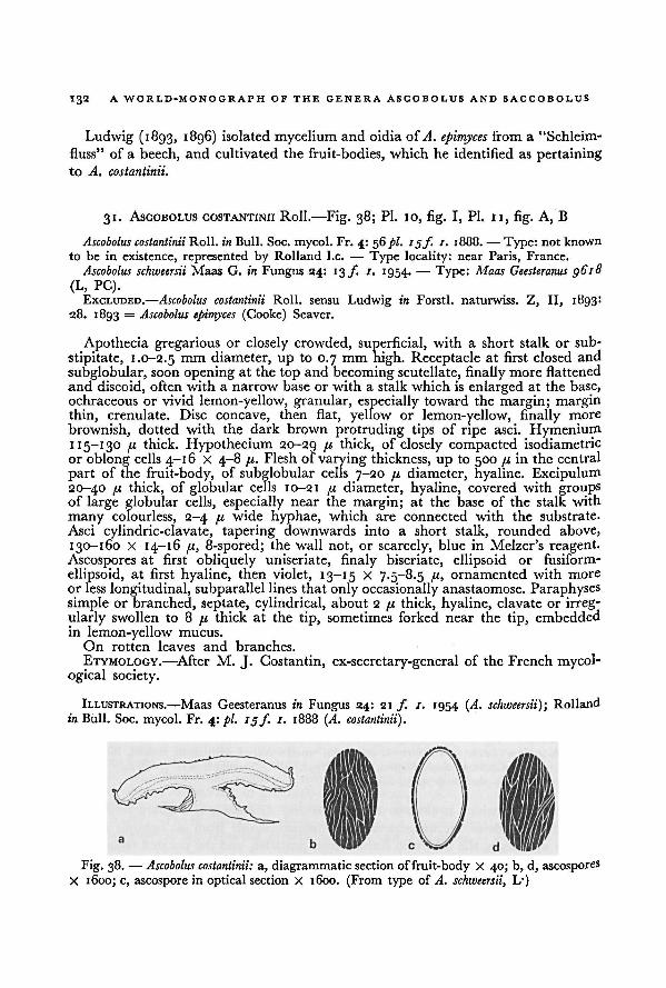
132 A WORLD-MONOGRAPH OF THE GENERA ASCOBOLUS AND SACCOBOLUS
Ludwig (1893, 1896) isolated mycelium and oidia of A. epimyces from a "Schleim-
fluss" of a beech, and cultivated the fruit-bodies, which he identified as pertaining
to A. costantinii.
31. ASCOBOLUS COSTANTINII Roll.—Fig. 38; Pl. 10, fig. I, Pl. II, fig. A, B
Ascobolus costantinii Roll, in Bull. Soc. mycol. Fr. 4: 56 pi. 15f. 1. 1888. — Type: not known
to be in existence, represented by Rolland I.e. — Type locality: near Paris, France.
Ascobolus schweersii Maas G. in Fungus 24: 13/. 1. 1954. — Type: Maas Geesteranus 9618
(L, PC).
EXCLUDED.—Ascobolus costantinii Roll, sensu Ludwig in Forstl. naturwiss. Z, II, 1893:28. 1893 = Ascobolus epimyces (Cooke) Seaver.
Apothecia gregarious or closely crowded, superficial, with a short stalk or sub-
stipitate, 1.0-2.5 mm diameter, up to 0.7 mm high. Receptacle at first closed and
subglobular, soon opening at the top and becoming scutellate, finally more flattenedand discoid, often with a narrow base or with a stalk which is enlarged at the base,
ochraceous or vivid lemon-yellow, granular, especially toward the margin; marginthin, crenulate. Disc concave, then flat, yellow or lemon-yellow, finally more
brownish, dotted with the dark brown protruding tips of ripe asci. Hymenium115-130 p thick. Hypothecium 20-29 pi thick, of closely compacted isodiametric
or oblong cells 4-16 X 4-8 pi. Flesh of varying thickness, up to 500 pi in the central
part of the fruit-body, of subglobular cells 7-20 pi diameter, hyaline. Excipulum20-40 u thick, of globular cells 10-21 pi diameter, hyaline, covered with groupsof large globular cells, especially near the margin; at the base of the stalk with
many colourless, 2-4 pi wide hyphae, which are connected with the substrate.
Asci cylindric-clavate, tapering downwards into a short stalk, rounded above,
130-160 X 14-16 pi, 8-spored; the wall not, or scarcely, blue in Melzer's reagent.Ascospores at first obliquely uniseriate, finaly biseriate, ellipsoid or fusiform-
ellipsoid, at first hyaline, then violet, 13-15 X 7.5-8.5 pi, ornamented with more
or less longitudinal, subparallel lines that only occasionally anastaomose. Paraphysessimple or branched, septate, cylindrical, about 2 pi thick, hyaline, clavate or irreg-ularly swollen to 8 pi thick at the tip, sometimes forked near the tip, embeddedin lemon-yellow mucus.
On rotten leaves and branches.
ETYMOLOGY. —After M. J. Costantin, ex-secretary-general of the French mycol-ogical society.
ILLUSTRATIONS.—Maas Geesteranus in Fungus 24: 21 f. /. 1954 (A. schweersii); Rolland
in Bull. Soc. mycol. Fr. 4: pi. 19f. 1. 1888 (A. costantinii).
a, diagrammatic section of fruit-body X 40; b, d, ascospores
X 1600; c, ascospore in optical section X 1600. (From type of
Ascobolus costantinii:
L.)A. schweersii,
Fig. 38. —
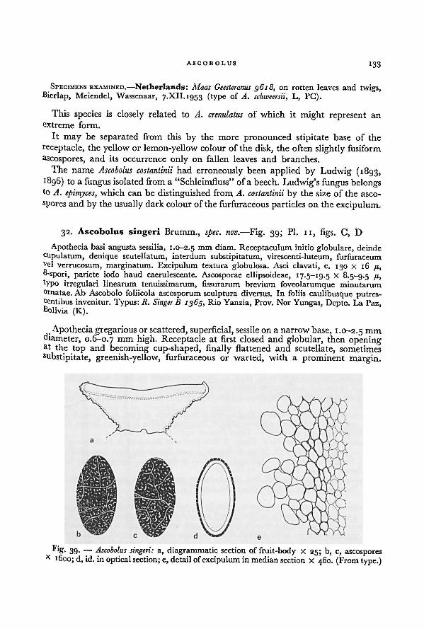
ASCOBOLUS 133
SPECIMENS EXAMINED.—Netherlands: Maas Geesteranus g6i8, on rotten leaves and twigs,
Bierlap, Meiendel, Wassenaar, 7.XII. 1953 (type of A. schweersii, L, PC).
This species is closely related to A. crenulatus of which it might represent an
extreme form.
It may be separated from this by the more pronounced stipitate base of the
receptacle, the yellow or lemon-yellow colour of the disk, the often slightly fusiform
ascospores, and its occurrence only on fallen leaves and branches.
The name Ascobolus costantinii had erroneously been applied by Ludwig (1893,
1896) to a fungus isolated from a "Schleimfluss" of a beech. Ludwig's fungus belongsto A. epimyces, which can be distinguished from A. costantinii by the size of the asco-
spores and by the usually dark colourof the furfuraceous particles on the excipulum.
32. Ascobolus singeri Brumm., spec. nov. —Fig. 39; Pl. II, figs. C, D
Apothecia basi angusta sessilia, 1.0-2.5 111111 diam. Receptaculum initio globulare, deinde
cupulatum, denique scutellatum, interdum substipitatum, virescenti-luteum, furfuraceumvel
verrucosum, marginatum. Excipulum textura globulosa. Asci clavati, c. 130 X 16 71,
8-spori, pariete iodo haud caerulescente. Ascosporae ellipsoideae, 1 7.5—19.5 X 8.5-9.5 1u >
typo irregulari linearum tenuissimarum, fissurarum brevium foveolarumque minutarum°rnatae. Ab Ascobolo foliicola ascosporum sculptura diversus. In foliis caulibusque putres-centibus invenitur. Typus: It. Singer B 1365, Rio Yanzia, Prov. Nor Yungas, Depto. La Paz,Bolivia (K).
Apothecia gregarious or scattered, superficial, sessile on a narrow base, 1.0-2.5 mm
diameter, 0.6-0.7 mm high- Receptacle at first closed and globular, then openingat the top and becoming cup-shaped, finally flattened and scutellate, sometimes
substipitate, greenish-yellow, furfuraceous or warted, with a prominent margin.
Fig. 39.— a, diagrammatic section of fruit-body X 25; b, c, ascospores
x 1600; d, id. in optical section; e, detailof excipulum in median section X 460. (From type.)
Ascobolus singeri:
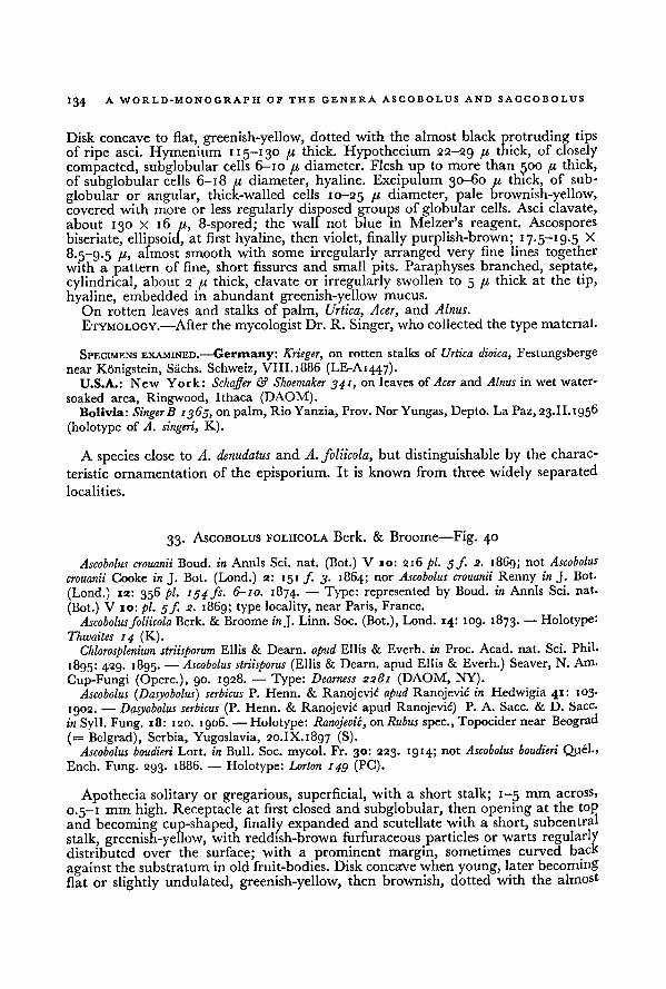
134 A WORLD-MONOGRAPH OF THE GENERA ASCOBOLUS AND SACCOBOLUS
Disk concave to flat, greenish-yellow, dotted with the almost black protruding tips
of ripe asci. Hymenium 115-130 /.< thick. Hypothecium 22-29 /'- thick, of closely
compacted, subglobular cells 6-10 /.i diameter. Flesh up to more than 500 /< thick,of subglobular cells 6-18 /i diameter, hyaline. Excipulum 30-60 /< thick, of sub-
globular or angular, thick-walled cells 10-25 B diameter, pale brownish-yellow,covered with more or less regularly disposed groups of globular cells. Asci clavate,
about 130 X 16 /(, 8-spored; the wall not blue in Melzer's reagent. Ascosporesbiseriate, ellipsoid, at first hyaline, then violet, finally purplish-brown; 17.5- 19.5 X
8.5-9.5 !l>almost smooth with some irregularly arranged very fine lines together
with a pattern of fine, short fissures and small pits. Paraphyses branched, septate,
cylindrical, about 2 pt thick, clavate or irregularly swollen to 5 [x thick at the tip,
hyaline, embedded in abundant greenish-yellow mucus.
On rotten leaves and stalks of palm, Urtica, Acer, and Alnus.
ETYMOLOGY. —After the mycologist Dr. R. Singer, who collected the type material.
SPECIMENS EXAMINED.—Germany: Krieger, on rotten stalks of Urtica dioica, Festungsberge
near Konigstein, Sachs. Schweiz, VIII. 1886 (LE-A1447).
U.S.A.: New York: Schaffer & Shoemaker 341, on leaves of Acer and Alnus in wet water-
soaked area, Ringwood, Ithaca (DAOM).Bolivia: SingerB 1365, on palm, Rio Yanzia, Prov. Nor Yungas, Depto. La Paz, 23.II.1956
(holotype of A. singeri, K).
A species close to A. denudatus and A. foliicola, but distinguishable by the charac-
teristic ornamentationof the episporium. It is known from three widely separated
localities.
33. ASCOBOLUS FOLIICOLA Berk. & Broome—Fig. 40
Ascobolus crouanii Boud. in Annls Sci. nat. (Bot.) V 10: 216 pi. 5/. 2. 1869; not Ascobolus
crouanii Cooke in J. Bot. (Lond.) 2: 151 /. 3. 1864; nor Ascobolus crouanii Renny in J. Bot.
(Lond.) 12: 356 pi. 154 fs. 6-10. 1874. — Type: represented by Boud. in Annls Sci. nat.
(Bot.) V 10: pi. j/. 2. 1869; type locality, near Paris, France.
Ascobolusfoliicola Berk. & Broome in J. Linn. Soc. (Bot.), Lond. 14: 109. 1873. — Holotype:
Thwaites 14 (K).
Chlorosplenium striisporum Ellis & Dearn. apud Ellis & Everh. in Proc. Acad. nat. Sci. Phil-
1895: 429. 1895. — Ascobolus striisporus (Ellis & Dearn. apud Ellis & Everh.) Seaver, N. Am.
Cup-Fungi (Operc.), 90. 1928. — Type: Dearness 2281 (DAOM, NY).
Ascobolus (Dasyobolus) serbicus P. Henn. & Ranojevid apud Ranojevic in Hedwigia 41: 103.
1902. — Dasyobolus serbicus (P. Henn. & Ranojevic apud Ranojevic) P. A. Sacc. & D. Sacc.
in Syll. Fung. 18: 120. 1906. — Holotype: Ranojevic, on Rubus spec., Topocider near Beograd
(= Belgrad), Serbia, Yugoslavia, 20. IX. 1897 (S).Ascobolus boudieri Lort. in Bull. Soc. mycol. Fr. 30: 223. 1914; not Ascobolus boudieri Quel.,
Ench. Fung. 293. 1886. — Holotype: Lorton 143 (PC).
Apothecia solitary or gregarious, superficial, with a short stalk; 1-5 mm across,
0.5-1 mm high. Receptacle at first closed and subglobular, then opening at the top
and becoming cup-shaped, finally expanded and scutellate with a short, subcentral
stalk, greenish-yellow, witb reddish-brown furfuraceous particles or warts regularlydistributed over the surface; with a prominent margin, sometimes curved back
against the substratum in old fruit-bodies. Disk concave wben young, later becoming
flat or slightly undulated, greenish-yellow, then brownish, dotted with the almost
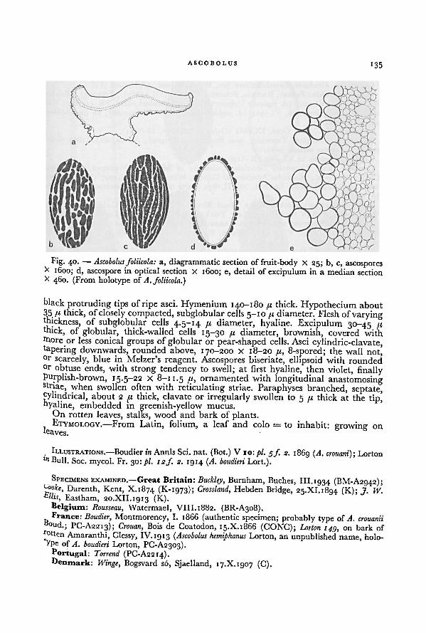
ASGOBOLUS135
black protruding tips of ripe asci. Hymenium 140-180 fi thick. Hypothecium about
35 /< thick, of closely compacted, subglobular cells 5-10pi diameter. Flesh of varyingthickness, of subglobular cells 4.5-14 pi diameter, hyaline. Excipulum 30-45 pithick, of globular, thick-walled cells 15-30 pi diameter, brownish, covered with
more or less conical groups of globular or pear-shaped cells. Asci cylindric-clavate,tapering downwards, rounded above, 170-200 X 18-20 fi, 8-spored; the wall not,°r scarcely, blue in Melzer's reagent. Ascospores biseriate, ellipsoid with rounded°r obtuse ends, with strong tendency to swell; at first hyaline, then violet, finallyPurplish-brown, 15.5-22 X 8-11.5 fi, ornamented with longitudinal anastomosingstriae, when swollen often with reticulating striae. Paraphyses branched, septate,cylindrical, about 2 pi thick, clavate or irregularly swollen to 5 it thick at the tip'hyaline, embedded in greenish-yellow mucus.
On rotten leaves, stalks, wood and bark of plants.ETYMOLOGY.—From Latin, folium, a leaf and colo = to inhabit: growing on
, ILLUSTRATIONS.—Boudier in Annls Sci. nat. (Bot.) V 10: pi. 5/. 2. 1869 (A. crouani); Lortonln Bull. Soc. mycol. Fr. 30: pi. 12/. 2. 1914 (A. boudieri Lort.).
SPECIMENS EXAMINED.—Great Britain: Buckley, Burnham, Buches, 111.1934 (BM-A2942);uoofe, Darenth, Kent, X.1874 (K-ig73); Crossland, Hebden Bridge, 25.XI.1894 (K); j. W.Ellis, Eastham, 20.XII.1913 (K).
Belgium: Rousseau, Watermael, VIII.1882. (BR-A308).France: Boudier, Montmorency, I. 1866 (authentic specimen; probably type of A. crouanii
B°ud.; PC-A2213); Crouan, Bois de Coatodon, 15.X.1866 (CONC); Lorton 149, on bark ofrotten Amaranthi, Clessy, IV. 1913 ( Ascobolus hemiphanus Lorton, an unpublished name, holo-VPe of A. boudieri Lorton, PC-A2303).
Portugal: Torrend (PC-A2214).Denmark: Winge, Bogsvard so, Sjaelland, 17.X. 1907 (C).
Fig. 40. — Ascobolus foliicola: a, diagrammatic section of fruit-body X 25; b, c, ascosporesX 1600; d, ascospore in optical section X 1600; e, detail of excipulum in a median sectionX 460. (From holotype of A. foliicola.)
136A WORLD-MONOGRAPH OF THE GENERA ASCOBOLUS AND SACCOBOLUS
Germany: Kirschstein, Grossbehnitzer See, Westhavelland, 4.VII.1909 (FH).
Czechoslovakia: Fechtner, Radotin, Bohemia, IX. 1924 (PR 148859); Velenovsky, Mnicho-
vice, Bohemia, VI. 1923 (A. juglandis Velenovsky, an unpublished name, PR 147789); Vele-
novsky, Radotin, Bohemia, VI. 1924 (A. juglandis var. corticola Velenovsky, an unpublished
name, PR 149700).Austria: JViessl, near Graz, IX.1861 (G-A978, M-A830, M-A831).Yugoslavia: Ranojevic, Topocider, near Beograd (= Belgrad), Serbia, 20.IX. 1897 (holo-
type of Dasyobolus serbicus, S-A610).Ceylon: Thwaites 12.XI.1867 (K-A1961).Canada: Bisby, University Grounds, University of Manitoba, Winnipeg, 30.VI. 1933
(CMI); Bisby et al., Manitoba Agricultural College, Winnipeg, 4.VIII. 1927 (NY); Dearness
2281, Mc Grady's Bank, London, 20.VII.1895 (parts of type of Chlorosplenium striisporum,DAOM, NY).
U.S.A.: Oregon; Kienholz KD12, Hood River, 9.V.1943 (BPI). California: Toole,
Vista Nurseries, 18.IV.r930 (BPI). New York: Latham 577, Orient, Long Island, 28.VI.
1922 (A. lathamiDearness, an unpublished name, DAOM). Massachusetts: Rarer, Bedford,
X. 1901 (BPI-A1534). Virginia: Lefebvre, Blacksburg, V. 1947 (BPI-A1535).
The growth of this species is restricted to rotten vegetable debris. The fruit-
bodies always show a clearly developed stalk and a prominent furfuraceous margin.
In the related A. denudatus the fruit-bodies are sessile and the margin is smooth.
It can be distinguished from A. singeri by the ornamentation of the episporium.
34. ASCOBOLUS DENUDATUS Fr.—Fig. 41; Pl. II, figs. E, F
Ascobolus denudatus Fr., Syst. mycol. 2 (1): 162. 1822; sensu Cooke in J. Bot. (Lond.) 2: 150.
1864; sensu Berk. & Broome in Ann. Mag. nat. Hist. Ill15: 448 pi. 16f 24. 1865; sensu
Fuckel, Fungi rhen. No. 1849. 1866; inJb. nassau. Ver. naturk. 23-24: 287. 1870. — Type:
represented by the description ofFries, I.e.; type locality, probably Sweden.
Ascobolus immarginatus Becc. in Erb. critt. ital. Ser. I, fasc. 16, No. 775. 1862; De Not. in
Comm. Soc. critt. ital. 1: 360. 1864. — Type distribution: Erb. critt. ital. No. 775.
Ascobolus angulisporus Boud. in Bull. Soc. bot. Fr. 28: 92 pi. 3 f. 2. 1881. — Ascobolus
furfuraceus var. angulisporus (Boud.) Quel., Ench. Fung. 293. 1886.— Holotype: Boudier, on
rotten wood, Montmorency, France, June 1879 (PC-A2217).Ascobolus fimiputris var. lindaviana P. Henn. in Verh. bot. Ver. Prov. Brandenburg 40: 150
pi. 2 f. 2. 1898. — Ascobolus stercorarius var. lindaviana (P. Henn.) Boud., Hist, class. Discom.
Eur. 72: 1907. — Type: represented by P. Henn. I.e.; type locality, Greenhouse, Botanic
Gardens, Berlin, Germany.
Ascobolus perdicinus Vel., Monogr. Discom. Boh. 1: 365. 1934. — Holotype: PR 150305.
Ascobolus pani Vel., Monogr. Discom. Boh. 1: 367, 2: pi. 4 f. 41. 1934. — Holotype:
PR 149851.Ascobolus transverse-rimosus Svrcek in Ceska Mykol. 11: 106, 117 pi. 2f. 2. 1957. — Holotype:Svfcek, onrotten wood, Nemysl near Tabor,Bohemia, Czechoslovakia, 1.IX.1946 (PR-A2723)-
Apothecia gregarious or more rarely crowded, superficial, sessile, often on a
small base, 2-10 mm diameter, up to 1 mm high. Receptacle at first closed and
subglobular, then opening at the top and cup-shaped, finally expanding and
scutellate, sometimes undulate, greenish-yellow to yellowish-green, finally more
brownish, covered with rusty-brown powder, finally sometimes more or less smooth,
especially near the margin; with a prominent, often somewhat crenulate or dentate
margin, almost disappearing with age. Disk at first concave, then flat, greenish-
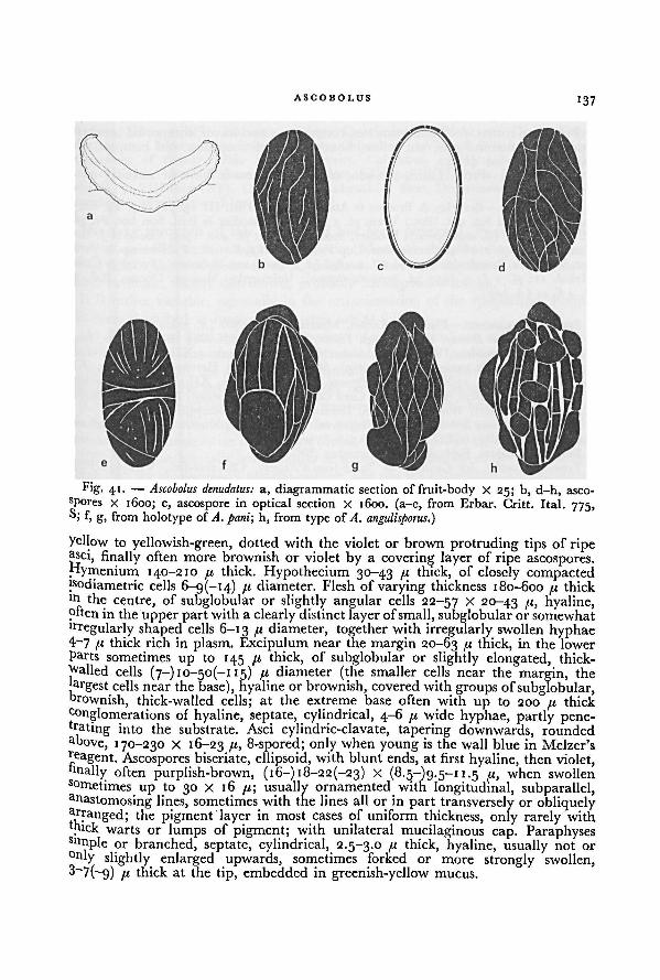
ASCOBOLUS 137
yellow to yellowish-green, dotted with the violet or brown protruding tips of ripeasci, finally often more brownish or violet by a covering layer of ripe ascospores.yfymeniurn 140-210 74 thick. Hypothecium 30-43 74 thick, of closely compactedjsodiametric cells 6-9(-14) 74 diameter. Flesh of varying thickness 180-600 /< thickln the centre, of subglobular or slightly angular cells 22-57 X 2°~43 7b hyaline,often in the upperpart with a clearly distinct layer of small, subglobular or somewhatlrregularly shaped cells 6-13 74 diameter, together with irregularly swollen hyphae4-7 /'- thick rich in plasm. Excipulum near the margin 20-63 /i thick, in the lower
Parts sometimesup to 145 74 thick, of subglobular or slightly elongated, thick-
called cells (7-) IO-5O(-I 15) 74 diameter (the smaller cells near the margin, the
|argest cells near the base), hyaline or brownish, covered with groups of subglobular,brownish, thick-walled cells; at the extreme base often with up to 200 74 thick
conglomerations of hyaline, septate, cylindrical, 4-6 74 wide hyphae, partly pene-trating into the substrate. Asci cylindric-clavate, tapering downwards, roundedaoove, 170-230 X 16-23 ("> 8-spored; only when young is the wall blue in Melzer's
Reagent. Ascospores biseriate, ellipsoid, with blunt ends, at first hyaline, then violet,bnally often purplish-brown, (16-) 18-22(-23) X (8.5-)g.5-i 1.5 74, when swollen
sometimes up to 30 x 16 74; usually ornamented with longitudinal, subparallel,a nastomosing lines, sometimes with the lines all or in part transversely or obliquelyRanged; the pigment layer in most cases of uniform thickness, only rarely with
mick warts or lumps of pigment; with unilateral mucilaginous cap. ParaphysesSlmple or branched, septate, cylindrical, 2.5-3.0 74 thick, hyaline, usually not or° nly slightly enlarged upwards, sometimes forked or more strongly swollen,3—7(—9) 74 thick at the tip, embedded in greenish-yellow mucus.
Fig. 41. — a, diagrammatic section of fruit-body X 25; b, d-h, asco-
spores x 1600; c, ascospore in optical section X 1600. (a-e, from Erbar. Critt. Ital. 775,S; f, g, from holotype of
Ascobolus denudatus:
A. pani; h, from type of A. angulisporus.)
138A WORLD-MONOGRAPH OF THE GENERA ASCOBOLUS AND SACCOBOLUS
Found on rotten wood and branches, rotten straw and leaves, composted bracken,humid soil, manure pile, tan refuse, honey comb of wasp nest, old compost, old
carpet, rarely on dung.ETYMOLOGY.—From Latin, denudo, to lay bare: made bare or naked.
ILLUSTRATIONS.—Berkeley & Broome in Ann. Mag. nat. Hist. Ill 15: pi. 16f 24. 1865;
Boudier in Annls Sci. nat. (Bot.) V 10: pi. 5f. 3. 1869; Boudier in Bull. Soc. bot. Fr. 28:
pi. 3f. 2. 1881 (A. angulisporus); Boudier, Icon, mycol. 2: pi. 408. ï9°9; Cooke in J. Bot.
(Lond.) 2: 150 f. 2. 1864; Dennis, Brit. Cup Fungi pi. VIIf. K. i960; P. Hennings in Verh.
bot. Ver. Prov. Brandenburg 40: pi. 2f. 2. 1898 (A. fimiputris var. lindaviana); Svrcek in Geska
Mykol. ii: pi. 2 f. 2. 1957 (A. transverse-rimosus); Velenovsky, Monogr. Discom. Boh. 2:
pi. 4f. 41 (A. pani).
SPECIMENS EXAMINED..—Finland: Karsten, Mustiala, 8.IX.1866 (A. viridis, H).Great Britain: Broome, Marlborough Forest, Wiltshire, 15.X. 1863 (BM); Foister, near
Cleghorn, Lamarkshire, IV.ig4i (E); Massee & Crossland, Leeds, g.XII.i8g3 (type of A.
barbatus in part, only ascospores, NY-A1259); Moore, Harpenden, Hertford, 2.VII.1953 (K);Wilson, Nottingham, 25.XI.1939 (K); Perceval, Stopham, Sussex, XII.1876 (BM); Phillips,
Norton Camp, XI.1876 (BM); Phillips, Card Cock, 1880 (BM-A2999); Plowright, Downton,
X.1886 (BM-A2881); Wright, Braunton, Devon, 22.1. 1950 (K-A2475).Netherlands: van Brummelen 1398, Singraven, Denekamp, 14.X.1961 (L); Rick, Valken-
burg, Limburg, 1899 (GRO-Ag, GRO-Ai, S-A750).
Belgium: Mouton, Foret de Pery, November (BR-A311).France: Bouchet, les Granges de Saint-Jean-d'Angely, Charente-Inf., VI.1937 (PC);
Bouchet, Bois d'Essouverts, near Loulay, Charente Inf., XI. 1939 (PC); Boudier, Montmorency,
VIII.1889 (PC-A2206, PC-A2207); Boudier, Ham, VIII.1893 (PC-A2208); Boudier, Mont-
morency, VII.1899 (PC-A2205); Boudier (PC-A2186); Boudier, Montmorency, June 1879
(type of A. angulisporus, PC-A2217); Grelet, Cirray, Vienne, 17.V.1916 (PC); Grelet, Savigne
Vienne, VI.1916 (PC-A2271); Louvet, IV.1899 (PC-A2209).Portugal: Torrend, Mafra, XII.1907 (URM 9011); Torrend 638 (S-A774); Torrend, Lisbon
(PC-A2210).Germany: von Brefeld, near Minister, Westfalen, IV.1896 (S-A729); Britzelmayr, near
Augsburg, IX. 1876 (S-A535); Fuckel, Grünau near Hattenheim, autumn, in Fungi rhen.
1849 (A. denudatus, BM, M); Fuckel, Altrhein near Hattenheim, Nassau, August (G-Ag7g);Fuckel, Oestrich, Nassau, autumn, in Herbier Barbey-Boissier 1309 (A. furfuraceus, TRTC);Staritz, inundated grounds of the river Elbe, Worlitz, 1890 (S-A761); P. Sydow, Tiergarten
near Berlin, VII.1885 (S-A759), in Myc. march. 786 (BM, HBG, K, PRC, S, W), in Rehm,
Ascom. 823 (BM, HBG, K, LE, M, PAD, S).Poland: Schroeter, Botanical Garden, Wroclaw (= Breslau), III.1888 (BRSL-A1726).
Czechoslovakia: Baumler 217, near Bratislava (= Pressburg), X.1884 (S); Svrcek, NemySlnear Tabor,Bohemia, i .IX. 1946 (holotype of A. transverse-rimosus, PR-A2723); Svrcek, Modrany
(Libussky potok), Bohemia, 3.V.1947 (PR 179010); Svrcek, Roblin, Bohemia, 2.X.1949
(PR 179011); Tyn, road to the water-fall, 26.VI.1923 (PR 178939); Vacek, Hrusice near
Mnichovice, 2.VIII.1940 (PR 150234); Vacek, Cernosice, Bohemia, 17.X.1948 (PR 178948);
Velenovsky, Karliche udoli, near Roblin, V.1927 (holotype of A. pani, PR 149851); Velenovsky,
Mnichovice, Bozkov-lacus, 19.IX.1928 (holotype of A. perdicinus, PR 150304); Velenovsky,
Mnichovice, V. 1931 (PR 150128); Velenovsky, Mnichovice, Kozenyvich, X. 1933 (PR 150302).
Italy: Beccari, Orto Botanico, Pisa, II-III.1862, in Erbar. Critt. Ital. 775 (type distribution
of A. immarginatus, BM, G, S); Bresadola, Magra, 1.1883 (S-A778).Pakistan: Ahmad 2262, Lahore, 29.II. 1948 (BPI).India: Thind 3037, Narkanda, Mahasu, 22.VIII.1965. (K).
U.S.A.: California: Gardner 349, near pumping station, Golden Gate Park, San
ASCOBOLUS 139
Francisco, III.1 9 15 (BPI). Indiana: Anderson, Crawfordsville (NY-A1125). Ohio: Koshy,Department of Botany, Ohio State University, Columbus, 21.VI. 1948 (NY-A1104); Koshy,Department of Botany, Ohio State University, Columbus, 4.VIII. 1948 (NY-A1102).Pennsylvania: Sinden & Reese 1, State College, 3.IV.1946 (NY). New Jersey: Seaver,Palisade, 31.VIII.1916 (NY). District of Columbia: Shear, Department of AgricultureGreenhouse, Washington, 1.1900 (BPI-A1601).
The type specimen of this species has not been preserved. Fries' description,however, is sufficient to recognize the species. Moreover there has been no contro-
versies about the interpretation of A. denudatus. Therefore the name is used for a
rather common, widely distributed, probably cosmopolitan species.It is rather variable, especially in the ornamentation of the episporium. Forms
that were described as species or varieties are as follows:—-
(A) The typical form, with the episporium of uniform thickness and ornamented
With longitudinal anastomosing striae (A. denudatus Fr., A. perdicinus Vel.).
(B) A form with the episporium ornamented with very narrow longitudinal,oblique or transverse striae, which are only rarely parallel (A. immarginatus Becc .).
(C) A form with the episporium with transverse, subparallel, anastomosing striae
(A. transverse-rimosus Svrcek). Since in the same fruit-bodies ascospores with longi-tudinal anastomosing striae may also be found, this is probably only a mutant-form.
(D) A form with locally semiglobular or irregular thickenings of the episporium,Which give the ascospores an angular outline. The rest the episporium is ornamented
With longitudinal anastomosing striae (A. angulisporus Boud., A. fimiputris var.
lindaviana P. Henn., A. pani Vel.).
Ascobolus demangei is distinguishable from A. denudatus by the thick episporium and
thevery closely spaced longitudinal anastomosing striae.
The related A. foliicola differs in the stalked apothecia, the furfuraceous margin,and the presence of pyriform cells in the excipular warts. Further studies will be
Uceded to establish their relationship.
35. ASCOBOLUS DEMANGEI Pat.—Fig. 42
Ascobolus demangei Pat. in Bull. Soc. mycol. Fr. 29: 222. 1913.— Holotype: Demanee
3sr (FH).Ascobolus nigricans Vel., Nov. mycol. novissimae 153. 1947. — Holotype: PR 150235.
Apothecia gregarious or scattered, superficial, sessile on a broad base, 2-8 mm
diameter, about 0.6 mm high. Receptacle at first closed and subglobular, then
°Pening and cup-shaped, finally expanding and scutellate or discoid, yellowish or
greenish-yellow, becoming dark brown with age, smooth near the margin, finelyjurl'uraceous at the underside; margin often rather broad, rounded, soon smooth,hhsk concave, then flat, greenish, then more brownish, finally almost black,r°ughened by the protruding asci. Hymenium 170-220 /t thick. Hypothecium39~5° /r thick, of closely compacted isodiametric or elongated cells 4-12 X 4-8 /<.flesh 200-260 fi thick, of subglobular, angular or oblong cells 8-34 x 7-25 /t,he larger cells near the outside, hyaline. Excipulum 37-62 /< thick near the margin,
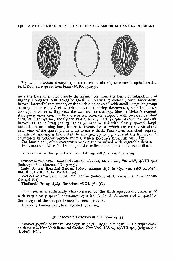
140 A WORLD-MONOGRAPH OF THE GENERA ASCOBOLUS AND SACCOBOLUS
near the base often not clearly distinguishable from the flesh, of subglobular or
slightly elongated cells 13-33 X 13-26 11, (textura globulosa), with amorphous,brown, intercellular pigment, at the underside covered with small, irregular groupsof subglobular cells. Asci cylindric-clavate, tapering downwards, rounded above,
200-250 X 20-22 /i, 8-spored; the wall not, or scarcely, blue in Melzer's reagent.Ascospores uniseriate, finally more or less biseriate, ellipsoid with rounded or blunt
ends, at first hyaline, then dark violet, finally dark purplish-brown to blackish-
brown, 21-25 X (10.5—)i2-i3(— 15.5) /t; ornamented with closely spaced, longi-tudinal, anastomosing lines, fifteen to twenty-five of which are usually visible on
each view of the spore; pigment up to 2.2 /.« thick. Paraphyses branched, septate,cylindrical, 2.0-2.5 fi thick, slightly enlarged up to 5 /( thick at the tip, hyaline,embedded in yellowish-green mucus, which becomes brownish with age.
On humid soil, often overgrown with algae or mixed with vegetable debris.
ETYMOLOGY.—After V. Demange, who collected in Tonkin for Patouillard.
ILLUSTRATIONS.—Dissing in Dansk bot. Ark. 23: 118 f 1, 119/. 2. 1963.
SPECIMENS EXAMINED.—Czechoslovakia: Velenovsky, Mnichovice, "Bzozek", 4.VIII.1941
(holotype of A. nigricans, PR 150235).Italy: Saccardo, Botanical Garden, Padova, autumn 1878, in Myc. ven. 1386 (A. viridis,
BM, BPI, BRSL, K, W, PAD-A1849).Viet-Nam: Demange 321, La Pho, Tonkin (holotype of A. demangei, as A. viridis var.
demangei, FH).Thailand: Dissing, 8384, Rachaburi 26.XI.1961 (C).
This species is sufficiently characterized by the thick episporium ornamented
with very closely spaced anastomosing striae. As in A. denudatus and A. geophilus,the margin of the receptacle soon becomes smooth.
It is only known from four isolated localities.
36. ASCOBOLUS GEOPHILUS Seaver—Fig. 43
Ascobolus geophilus Seaver in Mycologia 8: 96 pi. 184 fs. i-s. 1916. — Holotype: Seaver,
on damp soil, New York Botanical Garden, New York, U.S.A., 14.VIII.1914 (originally aS
A. viridis, NY).
a, c, ascospores X 1600; b, ascospore in optical section.
(a, b, from holotype; c, from
Fig. 42. — Ascobolus demangei:
Velenovský, PR 150235).
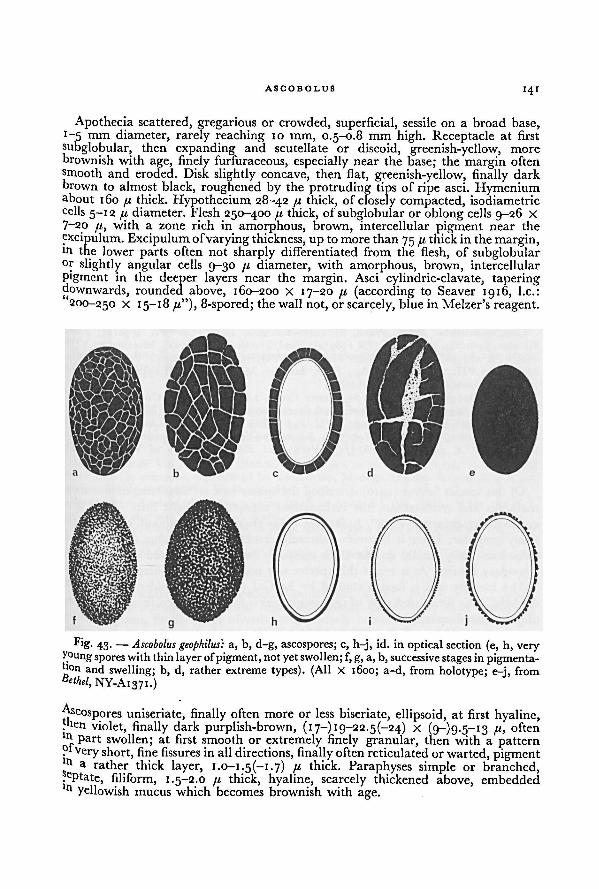
ASCOBOLUS 141
Apothecia scattered, gregarious or crowded, superficial, sessile on a broad base,1-5 mm diameter, rarely reaching 10 mm, 0.5-0.8 mm high. Receptacle at first
subglobular, then expanding and scutellate or discoid, greenish-yellow, more
brownish with age, finely furfuraceous, especially near the base; the margin oftensmooth and eroded. Disk slightly concave, then flat, greenish-yellow, finally dark
brown to almost black, roughened by the protruding tips of ripe asci. Hymeniumabout 160 74 thick. Hypothecium 28-42 74 thick, of closely compacted, isodiametric
cells 5-12 /t diameter. Flesh 250-400 74 thick, of subglobular or oblong cells 9-26 X
7-20 /i>with a zone rich in amorphous, brown, intercellular pigment near the
excipulum. Excipulum ofvarying thickness, up to more than 75 74 thick in the margin,in the lower parts often not sharply differentiated from the flesh, of subglobular°r slightly angular cells 9-30 74 diameter, with amorphous, brown, intercellular
Pigment in the deeper layers near the margin. Asci cylindric-clavate, taperingdownwards, rounded above, 160-200 X 17-20 74 (according to Seaver 1916, I.e.:
200-250 X 15-18 74"), 8-spored; thewall not, or scarcely, blue in Melzer's reagent.
uniseriate, finally often more or less biseriate, ellipsoid, at first hyaline,men violet, finally dark purplish-brown, (17-) 19-22.5(-24) x (9-)9-5~i3 79
often
part swollen; at first smooth or extremely finely granular, then with a pattern?i very short, fine fissures in all directions, finally often reticulated or warted, pigment' n A rather thick layer, I.O-I.5(-I.7) 74 thick. Paraphyses simple or branched,, ePtate, filiform, 1.5-2.0 74 thick, hyaline, scarcely thickened above, embeddedn yellowish mucus which becomes brownish with age.
Fig. 43. —a, b, d-g, ascospores; c, h-j, id. in optical section (e, h, very
young spores with thin layer ofpigment, not yet swollen; f, g, a, b, successive stagesin pigmenta-tion and swelling; b, d, rather extreme types). (All X 1600; a-d, from holotype; e-j, from
Ascobolus geophilus:
Bethel, NY-A137 1.)
A WO R LD-M ONOGRAPH OF THE GENERA ASCOBOLUS AND SACCOBOLUS142
On humid soil, especially when covered with algae, also on bark ofa tree covered
with mud.
ETYMOLOGY.—From Greek, yi], the earth and cpiXeco, to love: earth-loving, soil-
loving.
ILLUSTRATIONS—Seaver in Bull. Lab. nat. Hist. State Univ. Iowa 6: pi. 30 f. 1. 1905
(A. viridis); Scaver in Mycologia 8: pi. 184/5. 1-2. ig16; Seaver, N. Am. Cup-fungi (Operc.)
pi. 7 ƒ. 2. 1928.
SPECIMENS EXAMINED.—Great Britain: Soppitt, Midge hole road, Hebden Bridge, 12.VI.
1898 (K); Soppitt, Hebden Bridge, VII. 1898 (K-A2484); Wright, Braunton, Devon, 18.XI.
'95 1 (K).Belgium: Mouton, Beaufays (BR-A312); Mouton, Wandre, on the bank of the Meuse
{BR-A313); Rousseau, Boitsfort, VIII.1881 (BR-A334).France: Romagnesi, Coye-la-Forêt, Oise, 22.VI. 1952 (PC).Poland: Bucks, near Zülz, VI.1907 (S-A488).Czechoslovakia: Svrcek, Praha, Slivenec, Bohemia, 10.VI.1946 (PR 179012); Vacek,
Slivenec, Bohemia, 10.VI.1946 (PR 178952); Vacek, zarosice, "Gregovna\, Moravia, 31.VIII.
1946 (PR 178951); Vacek, Jtarosice, Moravia, 3.VIII.1947 (PR 178949); Vacek, zarosice,
Moravia, 5.VIII.1947 (PR 178984); Vacek, Zarosice, Moravia, 19.VIII. 1947 (PR 178950).
Italy: Bresadola, IV.i8g3 (PC-A2185).U.S.A.: Oregon: Kienholz K170, Mosier Creek, 1.IV.1935 (BPI). Colorado: Bethel,
Denver (NY-A1371). Seaver & Shope, Coal Creek Canon, 23.VIII.1929 (NY). Nebraska:
Clements, Island of Platte, Gering, 7.VII.1897 (BPI). New York: Seaver, New York
Botanical Garden, VIII.1912 (NY-A1366); Seaver, New York Botanical Garden, 14.VIII.1914
(holotype of A. geophilus, NY-A1374). Virginia: Betts, University of Virginia, 2.VI.
1938 (NY).
Of this species Seaver (1916) described the episporium as "consisting of verrucose
markings and reticulations, the reticulations consisting of light lines which give
rise to an irregular net-work". In fact the episporium is at first smooth or extremely
finely granular. Later it becomes delicately cracked by short, fine fissures in all
directions. Only finally do the thick episporia become reticulated or warted by
secondary fissures. As a result this species was only rarely recognized.
The identity of the fungus studied by Le Gal (1947: 249) under this name is
doubtful. The illustrated type of ornamentation is not characteristic of this or any
of the related species.
Ascobolus geophilus can be distinguished from A. denudatus by the ornamentation
and the development of the episporium.Of this species material was studied from North America and Europe. Dr. L. R-
Batra collected it in the Mussoorie Hills in India, where it is said to be rather
common. Of the Indian material only a description with drawings could be studied.
37. ASCOBOLUS VIRIDIS Curr.—Figs. 4, 44; Pl. II, fig. G
Ascobolus viridis Curr. in Trans. Linn. Soc. Lond. (Bot.) 24: 154. 1863. — Holotype: Currey,
on clay ground, Hanham Woods near Bristol, England, 15.X. 1861 (K-A1958).Ascobolus striato-punctatus Boud., Icon, mycol., Ser. 1, livr. 1, preliminary text: 3. i9£>4:
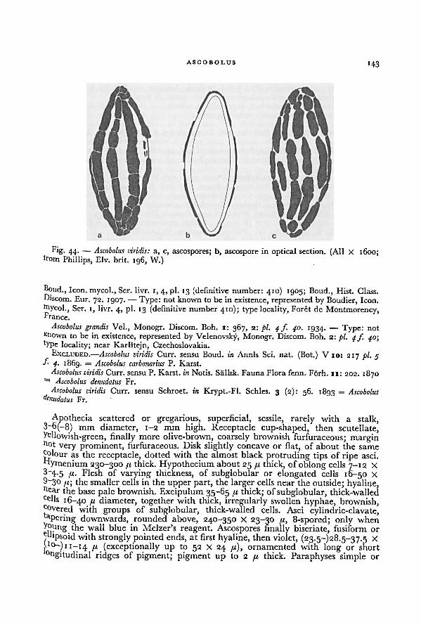
ASCOBOLUS143
Boud., Icon, mycol., Ser. livr. I, 4, pi. 13 (definitive number: 410) 1905; Boud., Hist. Class.
Ffiscom. Eur. 72. 1907. — Type: not known to be in existence, represented by Boudier, Icon.
Wycol., Ser. 1, livr. 4, pi. 13 (definitive number 410); type locality, Foret de Montmorency,France.
Ascobolus grandis Vel., Monogr. Discom. Boh. 1: 367, 2: pi. 4 f. 40. 1934. — Type: not
known to be in existence, represented by Vclcnovsky, Monogr. Discom. Boh. 2: pi. 4/. 40;
type locality; near Karlstejn, Czechoslovakia.
EXCLUDED.—Ascobolus viridis Curr. sensu Boud. in Annls Sci. nat. (Bot.) V 10: 217 pi. 5f-
4. 1869. — Ascobolus carbonarius P. Karst.
Ascobolus viridis Curr. sensu P. Karst. in Notis. Sallsk. FaunaFlora fenn. Forh. 11: 202. 1870= Ascobolus denudatus Fr.
Ascobolus viridis Curr. sensu Schroet. in Krypt.-Fl. Schles. 3 (2): 56. 1893 = Ascobolusdenudatus Fr.
Apothecia scattered or gregarious, superficial, sessile, rarely with a stalk,3-6(-8) mm diameter, 1-2 mm high. Receptacle cup-shaped, then scutellate,yellowish-green, finally more olive-brown, coarsely brownish furfuraceous; marginn°t very prominent, furfuraceous. Disk slightly concave or flat, of about the same
colour as the receptacle, dotted with the almost black protruding tips of ripe asci.
Fiymenium 230-300 pi thick. Hypothecium about 25 pi thick, of oblong cells 7-12 X
3~4-5 ft- Flesh of varying thickness, of subglobular or elongated cells 16-50 X
9~3° ft; the smaller cells in the upper part, the larger cells near the outside; hyaline,ne ar the base pale brownish. Excipulum 35-65 pi thick; of subglobular, thick-walledCe lls 16-40 11 diameter, together with thick, irregularly swollen hyphae, brownish,covered with groups of subglobular, thick-walled cells. Asci cylindric-clavate,aPering downwards, rounded above, 240-350 X 23-30 pi, 8-spored; only when
Ajpng the wall blue in Melzer's reagent. Ascospores finally biseriate, fusiform or
p'psoid with strongly pointed ends, at first hyaline, then violet, (23.5-)28.5-37.5 x
jjo—) 1 1—14 fi (exceptionally up to 52 X 24 pi), ornamented with long or short° ngitudinal ridges of pigment; pigment up to 2 // thick. Paraphyses simple or
Fig. 44. — Ascobolus viridis: a, c, ascospores; b, ascospore in optical section. (All X 1600;from Phillips, Elv. brit. 196, W.)
144 A WORLD-MONOGRAPH OF THE GENERA ASCOBOLUS AND SACCOBOLUS
branched, septate, cylindrical, 2.5-3.x B thick, hyaline, swollen to 7 // thick at
the tip, embedded in yellowish-green or pale greenish mucus.
Only known from humid soil, especially in woods.
ETYMOLOGY.—From Latin, viridis, green.
ILLUSTRATION.—Boudier in Bull. Soc. bot. Fr. 24: pi. 4 f. 6. 1877; Boudier, Icon, mycol.,
Ser. 1, livr. 4, pi. 13 (definitive number: 410). 1905 (A. striato-punctatus); Dennis, Brit. Cup
Fungi pi. VIIf J. U)6o; Phillips, Brit. Discom. pi. g ƒ. 54. 1887; Seaver, N. Am. Cup-fungi
pi. 7 f. 1. 1928; Velenovsky, Monogr. Discom. Boh. 2: pi. 4/. 40. 1934 (A. grandis).
SPECIMENS EXAMINED.—Great Britain: Broome 393, Leigh Wood, 4.IX. 1848 (BM); Broome,
Dinmore, Hereford, X.1878 (BM-A2944); Broome, Hereford (K-A1956); Crossland, Norland,
near Halifax 8.X. 1898 (K-A2455); Currey, Hanham Woods, near Bristol, 15.X.1861 (holotypeof A. viridis, K-A1958); Perceval, Henbury, Bristol, Gloucestershire 21. VI.1878 (BM); Phillips,
14.VII.1873 (BM-AQ945); Phillips, Whitfield and Dinmore, Hereford, V.1875 (BM-A2949);
Phillips, near Shrewsbury, V.1877 (K.Aigsg); Phillips, Shrewsbury, spring, in Elv. brit. 196
(BM, BPI, E, K, M, MEL, W); Phillips, Shropshire (PC-A2188).
Belgium: Mouton, Gomze, VI.1898 (BR-A329); Mouton, Beaufays, September (BR-A310);Mouton, Beaufays, October (BR-A328).
France: Bouchet, nearLes Ouilleres-lez-Nouillers, Charente inferieure, V. 1936 (PC-A2309);Bouchet, Les Ouilleres-lez-Nouillers, Charente inferieure, V.ig37 (PC-A2316); Bouchet, Foret
de Mervent, Vendée, VIII. 1939 (PC-A2308); Clere, Ain, VI.1902 (PC-A2189); Quelet,
Herimoncourt, Doubs, 10.VII.1878 (UPS); Quelet, Jura, 10.VI.1879 (A. chlorinus Quel.
unpublished name, UPS-A2013); Quelet, 1879 (A. chlorinus Quel. K-A1960); Romagnesi,
Coye-la-Forêt, Oise, 18.V.1948 (PC).Czechoslovakia: Vacek, Karlstejn, Bohemia, 28.VI. 1942 (PR 178991); Velenovsky, Mnicho-
vice, Bohemia, IX. 1922 (PR 150070); Velenovsky, Mnichovice, Hubackov, Bohemia, 1.VI.1932
(PR 150309).Austria: Rick, near Feldkirch, Vorarlberg, VIII. 1897 (S-A790); Rick, near Feldkirch,
Vorarlberg, IX. 1897 (S-A736).
This species, which is known only from Europe, is sufficiently characterized by
the long-fusiform ascospores with longitudinal ridges of pigment.
38. ASCOBOLUS ALBINUS Seaver
Ascobolus albinus Seaver in Mycologia 8: 95. 1916. — Type not known to be in existence.
Type locality: Woods, near Yonkers, New York, U.S.A.
"Apothecia gregarious, at first subglobose, expanding and becoming scutellate,
reaching a diameterof 4-5 mm, externally pure white, minutely rough; hymeniurn
plane or nearly plane, at first whitish, becoming darkened by the maturing spores,"asci cylindric or subcylindric, 8-spored, reaching a length of about 200 fi
and a
diameter of about 25 41; spores at first obliquely 1-seriate, becoming 2-seriate or
irregularly crowded, ellipsoid, becoming purple then brown, 20-26 X 12 41, rough!
spore roughenings consisting of minute warts; paraphyses very slender, hyaline."On damp soil in woods."
ETYMOLOGY.—From Latin, albinus, somewhat white.
DISTRIBUTION.—Known only from the type locality.
This species can easily be distinguished from the related A. behnitziensis by the
pure white colour of the fruit-body and the finely warted episporium.
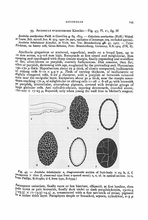
ASCODOLUS145
39. ASCOBOLUS BEHNITZIENSIS Kirschst.—Fig. 45; Pl. II, fig. H
Ascobolus amethystinus Phill. in Grevillea 4: 84. 1875. — Galactinia amethystina. . „
(Phill.) Wakef.lrl Trans. Brit, mycol. Soc. 6: 375. 1920 (in part, exclusive oflectotype; seq. excluded species).
Ascobolus behnitziensis Kirschst. in Verh. bot. Ver. Brandenburg 48: 47. 1907. — Type:Kirchstein, on loamy soil, Gross-Behnitz, Prov. Brandenburg, Germany, 8.X.1904 (FH, S).
Apothecia gregarious or scattered, superficial, sessile on a broad base, up to
10 fflm across, 0.3-0.6 mm high. Receptacle at first closed and subglobular, then
opening and cup-shaped with sharp sinuate margin, finally expanding and scutellateor flat, olive-brown or purplish, coarsely furfuraceous. Disk concave, then flat,olive or purplish, darkening with age, roughened by the protruding asci. Hymenium
[i thick. Hypothecium about 22 fi thick, of closely compacted, isodiametricor oblong cells 6-12 X 4.5-7 Flesh of varying thickness, of isodiametric orshghtly elongated cells, 6-20 /.t diameter, with a purplish or brownish colouredzpne near the excipular layer. Excipulum about 30 pt thick, near the margin some-
times reaching 1 70 fi, ofsubglobular or oblong cells 11-28 X 8-28 fx; with brownishor purplish, intercellular, amorphous pigment, covered with irregular groups of
large globular cells. Asci cylindric-clavate, tapering downwards, rounded above,160-200 X 17—23 /(, 8-spored; only when young the wall blue in Melzer's reagent.
A-scospores uniseriate, finally more or less biseriate, ellipsoid; at first hyaline, thenPale violet or pale brownish, finally dark violet or dark purplish-brown, 19-22.5
|~" 23-5) X 11—13-5(—14-5) /z, ornamented with a fine net-work of striae; pigmentn a rather thick layer. Paraphyses simple or branched, septate, cylindrical, 2-3 [I
Fig. 45.—
a, diagrammatic section of fruit-body X 25; b, d, f,
ascospores X 1600 (f, abnormal type from 2-spored ascus); c, e, id. in optical section, (a-c,from
Ascobolus behnitziensis:
Phillips, K-A1980; d-f, from type, S-A534.)
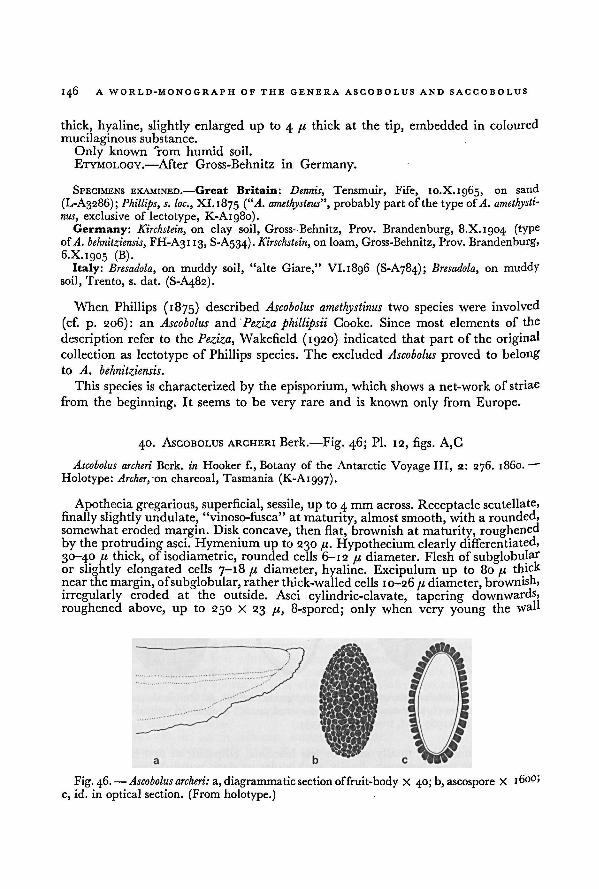
146 A WORLD-MONOGRAPH OF THE GENERA ASCOBOLUS AND SACCOBOLUS
thick, hyaline, slightly enlarged up to 4 /< thick at the tip, embedded in coloured
mucilaginous substance.
Only known °rom humid soil.
ETYMOLOGY.—After Gross-Behnitz in Germany.
SPECIMENS EXAMINED.—Great Britain: Dennis, Tensmuir, Fife, 10.X. 1965, on sand
(L-A3286); Phillips, s. loc., XI. 1875 (“A. amethysteus”, probably part ofthe type of.A. amethysti-
nus, exclusive of lectotype, K-A1980).
Germany: Kirchstein, on clay soil, Gross-Behnitz, Prov. Brandenburg, 8.X. 1904 (typeofA. behnitziensis, FH-A3113, S-A534). Kirschsttin, on loam, Gross-Behnitz, Prov. Brandenburg,
6.X. 1905 (B).Italy: Bresadola, on muddy soil, "alte Giare," VI. 1896 (S-A784); Bresadola, on muddy
soil, Trento, s. dat. (S-A482).
When Phillips (1875) described Ascobolus amethystinus two species were involved
(cf. p. 206): an Ascobolus and Peziza phillipsii Cooke. Since most elements of the
description refer to the Peziza, Wakefield (1920) indicated that part of the originalcollection as lectotype of Phillips species. The excluded Ascobolus proved to belong
to A. behnitziensis.
This species is characterized by the episporium, which shows a net-work of striae
from the beginning. It seems to be very rare and is known only from Europe.
40. ASCOBOLUS ARCHERI Berk.—Fig. 46; Pl. 12, figs. A, C
Ascobolus archeri Berk, in Hooker f., Botany of the Antarctic Voyage III, 2: 276. i860. —
Holotype: Archer, on charcoal, Tasmania (K-Aigg7).
Apothecia gregarious, superficial, sessile, up to 4 mm across. Receptacle scutellate,
finally slightly undulate, "vinoso-fusca" at maturity, almost smooth, with a rounded,somewhat eroded margin. Disk concave, then flat, brownish at maturity, roughenedby the protruding asci. Hymenium up to 230 pi. Hypothecium clearly differentiated,
30-4.0 /u thick, of isodiametric, rounded cells 6-12 pi diameter. Flesh of subglobulai"or slightly elongated cells 7-18 pi diameter, hyaline. Excipulum up to 80 pi thick
near the margin, ofsubglobular, rather thick-walled cells 10-26 pi diameter, brownish,
irregularly eroded at the outside. Asci cylindric-clavate, tapering downwards,
roughened above, up to 250 X 23 pi, 8-spored; only when very young the wall
a, diagrammatic section of fruit-body X 40; b, ascospore X 1600;
c, id. in optical section. (From holotype.)
Fig. 46. — Ascobolus archeri:
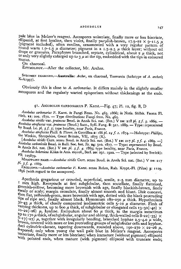
ASCOBOLUS147
pale blue in Melzer's reagent. Ascospores uniseriate, finally more or less biseriate,ellipsoid, at first hyaline, then violet, finally purplish-brown, 17.5-22 X 9-11.5 fi(pigment included), often swollen, ornamented with a very regular pattern ofround warts 1.0-1.3 P diameter; pigment in a 1.5-2.5 /z thick layer; without oil
drops or granules. Paraphyses branched, septate, cylindrical, about 2 f.i thick, notor only very slightly enlarged up to 3 /z at the tip, embeddedwith the tips in colouredmucus.
On charcoal.
ETYMOLOGY. —After the collector, Mr. Archer.
SPECIMEN EXAMINED.—Australia: Archer, on charcoal, Tasmania (holotype of A. archeri;K-A1997).
Obviously this is close to A. carbonarius. It differs mainly in the slightly smaller
ascospores and the regularly warted episporium without thickenings at the ends.
41. ASCOBOLUS CARBONARIUS P. Karst.—Fig. 47; Pl. 12, fig. B, D
Ascobolus carbonariusp..
P. Karst. in Fungi Fenn. No. 463. 1866; in Notis. Sallsk. Fauna Fl.örh. ir; 202. 1870. — Type distribution: Fungi fenn. No. 463.Ascobolus viridis var. pruinosus Boud. in Annls Sci. nat. (Bot.) V 10: 218 pi. 5/. 5. 1869. —
Ascobolus atrofuscus var. pruinosus (Boud.) Sacc., Syll. Fung. 8: 521. 1889. — Type: representedoy Boud. I.e. pl. 5/. 5; type locality, near Paris, France.
Ascobolus atrofuscus Phill. & Plowr. in Grevillea 2: 186pi. 24 f 1. 1874. —Holotype: Phillips,the Wrekin, Shropshire, Great Britain, VII, 1873 (K).
[Ascobolus viridis Curr. sensu Boud. in Annls Sci. nat. (Bot.) V 10: 217 pi. 3/. 4. 1869. —]Ascobolus carbonicolaBoud. in Bull. Soc. bot. Fr. 24: 310. 1877. — Type: represented by Boud.l" Annls Sci. nat. (Bot.) V ro: pi. 5 f. 4. 1869; type locality, near Paris, France.
Ascobolus bohemicus Klika in Ann. mycol., Berl. 20: 291. 1922. — Type (selected by Svrcek):"R 129879.
MISAPPLIED NAME.—Ascobolus viridis Curr. sensu Boud. in Annls Sci. nat. (Bot.) V 10: 217PI-
5f 4• 1869.EXCLUDED.— Ascobolus carbonarius P. Karst. sensu Rehm, Rab. Krypt.-Fl. (Pilze) 3: 1129.
■896 (with regard to the ascospores).
Apothecia gregarious or crowded, superficial, sessile, 2-5 mm diameter, up to1
mm high. Receptacle at first subglobular, then scutellate, finally flattened,greenish-yellow, becoming more brownish with age, finally blackish-brown, finelymealy or scaly; margin crenulate, finally almost smooth and blunt. Disk concave,thenflat, yellowish-green, more brownish with age, dottedwith theblack protrudingtps of ripe asci, finally almost black. Hymenium 180-250 /j, thick. Hypothecium
3°-43 /I thick, of closely compacted isodiametric cells 5-10 fi diameter. Flesh of
Varying thickness, up to 600 ft thick, ofsubglobular or elongated cells 13-30(-40) X10-22(-26) /r, hyaline. Excipulum about 60 ft thick, in the margin sometimes
hp to 1 70 fi thick, ofsubglobular, angular and oblong, thick-walled cells 8-22 (-35) X
1
~1 7(—23) /(, together with irregularly bending, branched hyphae 2.5-4.0 /« wide,r°wn, covered with more or less protruding groups ofsubglobular cells and hyphae.SC1 cylindric-clavate, tapering downwards, rounded above, 190-270 X 22-26 ju,-spored; only when young the wall pale blue in Melzer's reagent. Ascosporeshiseriate, finally more or less biseriate; when immature (without pigment) ellipsoidfin pointed ends, when mature (with pigment) ellipsoid with truncate ends;
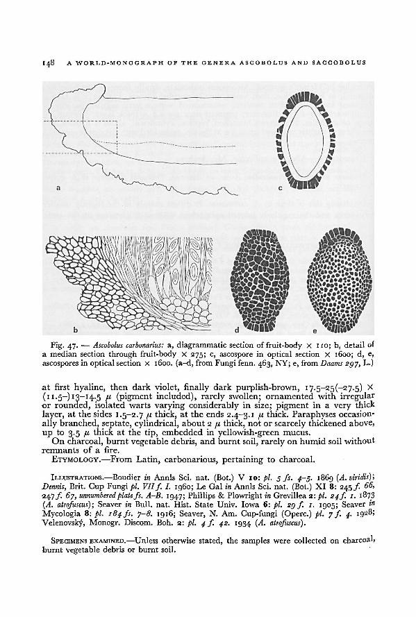
148 A WORLD-MONOGRAPH OF THE GENERA ASCOBOLUS AND SACCOBOLUS
at first hyaline, then dark violet, finally dark purplish-brown, 17-5~-25(—27.5) X
(11.5-) 13-14.5 ji (pigment included), rarely swollen; ornamented with irregularor rounded, isolated warts varying considerably in size; pigment in a very
thick
layer, at the sides 1.5-2.7 //, thick, at the ends 2.4-3.1 /< thick. Paraphyses occasion-
ally branched, septate, cylindrical, about 2 /,< thick, not or scarcely thickened above,
up to 3.5 /u thick at the tip, embedded in yellowish-green mucus.
On charcoal, burnt vegetable debris, and burnt soil, rarely on humid soil without
remnants of a fire.
ETYMOLOGY.—From Latin, carbonarious, pertaining to charcoal.
ILLUSTRATIONS.—Boudier in Annls Sci. nat. (Bot.) V 10: pi. 3 fs. 4-9. 1869 (A. viridis);
Dennis,Brit. Cup Fungi pi. VIIf. I. i960; Le Gal in Annls Sci. nat. (Bot.) XI 8: 245f 66,
247/". 67, unnumberedplate fs. A-B. 1947; Phillips & Plowright in Grevillea 2 : pi. 24 J1 1. 1873
(A. atrofuscus); Seaver in Bull. nat. Hist. State Univ. Iowa 6: pi. 2gf. 1. 1905; Seaver 1»
Mycologia 8: pi. 184fs. 7-8. 1916; Seaver, N. Am. Cup-fungi (Operc.) pi. 7 f 4. 1928;Velenovsky, Monogr. Discom. Boh. 2: pi. 4 f. 42. 1934 (A. atrofuscus).
SPECIMENS EXAMINED.—Unless otherwise stated, the samples were collected on charcoal,
burnt vegetable debris or burnt soil.
a, diagrammatic section of fruit-body X 110; b, detailof
a median section through fruit-body X 275; c, ascospore in optical section X 1600; d, e,
ascospores in optical section X 1600. (a-d, from Fungi fenn. 463, NY; e, from Daams 297, L.)
Fig. 47. — Ascobolus carbonarius:
ASCOBOLUS
149
Sweden: Starback,,
Knivsta, Uppland, VII.1895 (S-A738, S-A753).f inland: Karsten, Mustiala, VIII.1866 (S-A744); Karsten, Mustiala, 2s.VIII.1866
(H-A2756);"
Karsten, Mustiala, IX.1866, in Karsten, Fungi Fenn. 463 (type distribution ofA. carbonarius, BM-A2827, BM-A2828, NY-A1285); Karsten,
,Mustiala, 4.VIII.1871 (S-A767).
Ureat Britain:" '
Dennis, Norbury Park, Wickham, Surrey, 20.VI.1948 (K); Dennis & Ellis,Wheatfen, Norfolk, 5.VI. 1954 (K); Eyre, Abresford, Swarralow, 14.VI.1887 (BM); Phillips,the Wrekin, Shropshire, VII.1873 (holotype of A. atrofuscus, K); Phillips, Excal Hill near
Shrewsbury, VII. 1873 (BM-A2955, BM-A2959); Phillips, Arkollhill, Wellington, Shropshire,VII.1873 (BM); Phillips, s. loc., VII. 1873 (BM-A2960); Phillips, Shrewsbury, 1873 (S-A763);Phillips, Shrewsbury, 5. dat., in Phillips, Elv. brit. 47 (A. atrofuscus, BM, BPI, K, M, PAD);Phillips, CravenArmes, 6. X.1875 (BM); Phillips, road nearSallow Coppice, Upper Millichope,6.VIII. 1888 (BM); Phillips, Salop. (= Shropshire), s. dat. (Ascobolus fuscoatra, unpublishedname, BM-A2963); Phillips, Shrewsbury, r. dat. (BM-A2951, S-A532); Phillips, f. loc., s. dat.(BM-A2958); Plowright, Ringstead, Norfolk, X.1876 (BM); Ilea, near Perth; 11.VIII.1910(BM); Rea, Weybridge, 22. XI. 1920 (BM); Rea, New Parks, Wyreforest, Worcestershire,30.VII.1g25 (BM); Reid, Keswick, Cumberland, 4.VI.1956 (K).
Netherlands: Daams 297, "Over-Holland", Nieuwersluis, 21.VIII.1957 (L); Rick, Valken-burg, Limburg, XII. 1899 (S).
Belgium: Bommer & Rousseau, Groenendael, VII-IX. 1886, in Roumeguere, Fungi gall.3935 (A. atrofuscus, G); Mouton, s. loc., s. dat. (BR-A306); Rousseau, Florenville, IX.1884 (BR);Rousseau, Groenendael, VIII.1887 (BR); Rousseau, Groenendael, VII. 1890 (BR); unknowncollector, near Liège, s. dat. (S-A601).
France: Bertrand, Malzeville, Meurthe, VI. 1914 (PC); Bouchet,t
Bois des Essouverts, near
Eoulay, Charente-Maritime, V. 1942 (PC); Boudier, s. loc., VI(?).I882 (S); Boudier,... ... , Blois,
VU897 (PC);~ ~ " -
Cooke, Villers Cotterets, s. dat. (K-A1968); Costantin, Montmorency, Seine-et-Oise, 29.V.1886 (PC); Grelet, Savigne, Vienne, 26.V.1916 (PC); Grelet, same locality,VI. 1916 (PC); Le Gal, Pare de St. Cloud, Seine-et-Oise, VI. 1942 (PC); Patouillard, Boise Menrois, Jura, VIII. 1903 (FH); Quelet, Herimoncourt, s. dat. (K-A1969).Germany: P. Sydow, Schlossgarten, Charlottenburg, VIII.1888 (S); Wagner, gr.-Winter-
berg, VII-VIII. 1895, 1897 and 1898, in Krieger, Fungi sax. 1982 (A. atrofuscus,HBG.M, S);*"
------ - - -
>BM, BPI,Wagner, gr.-Winterberg, 17.VI.1897 (HBG).
Poland: Bucks, near Schammelwitz, VI.1905 (S); Bucks, Neisse-Ufer, near Ottmachau,VIII. 1906 (S); Bucks, Silberberggrund, VII. 1907 (S); Schroeter, Kreuzberg, near Striegau
(BRSL).
"Chechoslovakia: Klika, Vsetaty, VII. 1922 (PR 129837); Klika-Vlach, Kyselky privorske,K. Mikovu", IX.ig22 (lectotype of A. bohemicus, PR 129879); Klika, Larnov, 20.VII.1924
(PR 129787); Klika, D. Stubnia, 26.VII.1924 (PR 129784); Klika, Cesta pod Vosvuym,bhedohou, Bohemia, VII.1925 (BPI); Melzer, Smolov, Domazlice, 1.VII.1919 (PR 129841);
etrak, Mahr.-Weisskirchen (= Hranice), Chorin, VIII.1922 (ZT); Vacek, Ricany, Bohemia!
.A11-'939 (PR 149850); Vacek, Krc near Praha, 18.VIII.1946 (PR 178924); Vacek, zarosice,
Vloravia, 18.VIII.1948 (PR 178927); Vacek, Karlstejn, Bohemia, j. dat. (PR 178925);'clenovskf Mnichovice, VIII. 1923 (PR 150225); Velenovsky, Jevany, 3.VIII.1925 (PR 148950);*elenovsky
9 Ondrejov, 27.IX.t927 (PR 149848); Velenovsky,-
.„
. ...... .,
Mnichovice, Menêice, XI.1028
Velenovsky, Mnichovice, "Zburany", 3.VI.1929 (PR 150314); Lany, VIII.1929(PR 149847); Velenovsky, Mnichovice, Hrusice; Velenovsky, VIII. 1930 (PR 150313); Velenovsky,IX.1931 (PR 149845); Velenovsky, Mnichovice, Kunice, VII. 1932 (PR 150312)!
Switzerland: von Tavel, Zurichberg, near Zurich, 17.VI.1893 (S, ZT); Winter,near Zürich, V. 1877, in Winter, Fungi helv. 209 (A. viridis, BRSL, HBG, PAD, S).
Austria: Rick, near Feldkirch, Vorarlberg, IX. 1893 (S); von Keissler,u.,,.. , „• - \-/> > near the Kummer-
rucke, Gesause, Steiermark, VI.igi2 (W).Italy: Bresadola, Andalo, 30.VIII.18g6 (S).U.S.S.R. : Tranzschel, Bologoje, Nowgorod, 29.VI.18g7 (S).
150 A WORLD-MONOGRAPH OF THE GENERA ASCOBOLUS AND SACCOBOLUS
Uganda: Hansford 1839, on damp soil, Kampala, VI. 1934 (CMI).Canada: Bisby et al. 3640, on damp soil, Manitoba Agr. Coll., Winnipeg, 29.VII.1927
(NY); Seaver, Duchesnay, Quebec, 23-25. VIII. 1938 (NY).U.S.A.: Oregon: Kienholz K 84, on damp, mossy soil, Hood River, 16.XI. 1932 (NY);
Kienholz KD 84, Tony Mohr ranch, Hood River, ii.V.ig43 (BPI). Iowa: Seaver 63, Iowa
City, X.1904 (BPI); Seaver 63b, Iowa City, X.1904 (NY, S). Wisconsin: Dodge, Kewaunee
County, s. dat. (NY-A1295). Michigan: Fitzpatrick, Ann Arbor, s. dat. (NY-A1296).Indiana: Cummins
, Lafayette, 27.VI.1930 (NY). Ohio: Lloyd 0236, Linwood, V.1902
(NY); Lloyd, s. loc., 1903 (S). Tennessee: Underwood 11981, on sand in greenhouse,
Knoxville, 16.III.1939 (L). Maine: Thaxter, Kittery Point, VII. 1897 (FH). New York:
Dodge & Seaver, Hollis, Long Island, 25.VI.1910 (NY); unknown collector, Hollis, Long
Island, 10.VI.1911 (NY-A1264); Seaver, N.Y. Bot. Gardens, VI.1910 (NY-A1282, NY-A1291,
NY-A1302); Seaver, N.Y. Bot. Gardens, 27.V.1912 (NY); Seaver, near Yonkers, 19.VII.1912
(NY); Seaver, on soil, near Yonkers, VIII.1912 (NY); Seaver, New York City, summer 1912
(NY); Seaver, Yonkers woods, 26.V.1913 (NY); Seaver, New York City, 3.VI.1914 (NY);
Seaver, New York City, s. dat. (NY-A1292); Whetzel & White 1744, White Lake, near
Syracuse, 13.VI.1935 (FH). Connecticut: Thaxter, Cheshire, VI.i8go (FH); Thaxter,
West Rock, 20.VI.1891 (FFI). Pennsylvania: Dodge, Bethlehem, 7.VI. 1922 (BPI). West
Virginia: Nuttall, Nuttallburg, V.1893 (Phaeopeziza Nuttallii, BPI, NY-A1306, NY-A1308);
Nuttall, Nuttallburg, VI. 1893, in Ellis & Everhart, N. Am. fungi Ser. 2, 2908 (PhaeopezizaNuttallii Ell. & Ev., n. sp., type distribution, BM, BPI, NY); Nuttall, Fayette Country, s. dat.
(BPI). Virginia: Shear, Radnor Hts, 23.V. 1920 (BPI); Shear, Arlington Cemetery, 14.V. 1933
(BPI). North Carolina: Olive, near Buck Creek Ranch, Jackson County, 16.VIII.
1955 (NY).Costa Rica: Martin 8747, hills above Palmer Norte, Prov. Puntarenas, 8.VIII.1952
(developed in moist chamber, BPI).
This common, cosmopolitan species occurs rather frequently on burnt places,
especially when these are on loamy soil. It is characterized by the irregularly warted
episporium with polar thickenings.
There is some variationwith respect to the coarseness of the furfuraceous particles
on the outside of the receptacle. Also the colour of the receptacle may vary.Often
this depends on the age of the fruit-body and on environmental conditions during
the development.Chadefaud (1942b) and Le Gal (1947) studied the development of the episporial
ornamentation. The pigment is abundantly formed and deposited in the form of
thick warts.
The species is heterothallic (Betts, 1926). The germination of the ascospores can
be induced by a heat-treatment at temperatures between 65 and 8o° C. (Dodge,
1912a; Betts, 1926).
42. ASCOBOLUS SUBGLOBOSUS Seaver.—Fig. 48; Pl. 12, figs. F, G
Ascobolus subglobosus Seaver in Mycologia 8: 96. 1916. — Holotype: Seaver, on damp soil
among mosses in woods, near Yonkers, New York, U.S.A., 13.VIII.1914 (NY).
Apothecia scattered, gregarious or crowded, superficial, sessile, 2-5 mm diameter,
about 0.5 mm high. Receptacle at first closed and subglobular, then opening at
the top and cup-shaped, finally expanding and discoid, "greenish-yellow", more
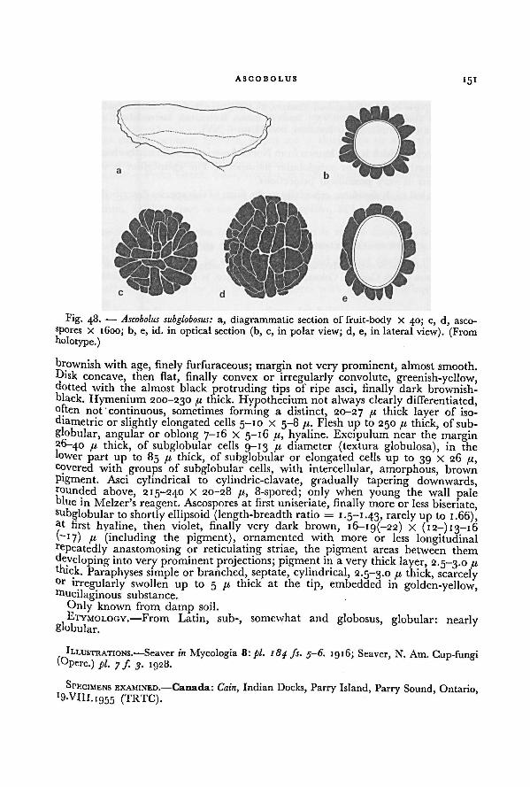
ASCOBOLUS 151
brownish with age, finely furfuraceous; margin not very prominent, almost smooth.Disk
concave, then flat, finally convex or irregularly convolute, greenish-yellow,dotted with the almost black protruding tips of ripe asci, finally dark brownish-
black. Hymenium 200-230 /x thick. Hypothecium not always clearly differentiated,often not continuous, sometimes forming a distinct, 20-27 /x thick layer of iso-diametric or slightly elongated cells 5-10 X 5-8 /.x. Flesh up to 250 /x thick, of sub-
globular, angular or oblong 7-16 X 5-16 JLI, hyaline. Excipulum near the margin26-40 [j, thick, of subglobular cells 9-13 /< diameter (textura globulosa), in the
lower part up to 85 [A, thick, of subglobular or elongated cells up to 39 x 26 /x,covered with groups of subglobular cells, with intercellular, amorphous, brown
pigment. Asci cylindrical to cylindric-clavate, gradually tapering downwards,rounded above, 215-240 X 20-28 fi, 8-spored; only when young the wall paleblue in Melzer's reagent. Ascospores at first uniseriate, finally more or less biseriate,subglobular to shortly ellipsoid (length-breadth ratio = 1.5-1.43, rarely up to 1.66),ut first hyaline, then violet, finally very dark brown, i6-ig(-22) X (12-) 13-16(~i7)
n (including the pigment), ornamented with more or less longitudinalrepeatedly anastomosing or reticulating striae, the pigment areas between them
developing into very prominent projections; pigment in a very thick layer, 2.5-3.0 (xbuck. Paraphyses simple or branched, septate, cylindrical, 2.5-3.0 fx thick, scarcely°r irregularly swollen up to 5 [X thick at the tip, embedded in golden-yellow,rrtucilaginous substance.
Only known from damp soil.
ETYMOLOGY.—From Latin, sub-, somewhat and globosus, globular: nearlyglobular.
ILLUSTRATIONS. —Seaver in Mycologia 8: pi. 184 fs. 5-6. 1916; Seaver, N. Am. Cup-fungi(Operc.) pi. 7 ƒ 3. 1928.
SPECIMENS EXAMINED.—Canada: Cain, Indian Docks, Parry Island, Parry Sound, Ontario, 9.VIH.I
955 (TRTC).
Fig. 48. — Ascobolus subglobosus: a, diagrammatic section of fruit-body X 40; c, d, asco-
spores x 1600; b, e, id. in optical section (b, c, in polar view; d, e, in lateral view). (Fromholotype.)
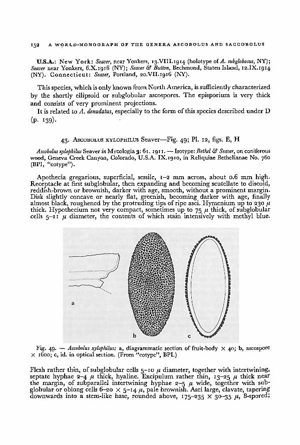
A WORLD-MONOGRAPH OF THE GENERA ASCOBOLUS AND SACCOBOLUS
152
U.S.A.: New York: Seaver, nearYonkers, 13.VIII.1914 (holotype of A. subglobosus, NY);
Seaver near Yonkers, 6.X.1916 (NY); Seaver & Button, Bechmond, Staten Island, 12.IX.1914
(NY). Connecticut: Seaver, Portland, 20.VII.1916 (NY).
This species, which is only known from NorthAmerica, is sufficiently characterized
by the shortly ellipsoid or subglobular ascospores. The episporium is very thick
and consists of very prominent projections.It is related to A. denudatus, especially to the form of this species described under D
(P- 139)-
43. ASCOBOLUS XYLOPHILUS Seaver—Fig. 49; Pl. 12, figs. E, H
Ascobolus xylophilus Seaver in Mycologia 3: 61. 191 1. — Isotype: Bethel & Seaver, on coniferous
wood, Geneva Creek Canyon, Colorado, U.S.A. IX.1910, in Reliquiae BethelianaeNo. 760(BPI, "cotype").
Apothecia gregarious, superficial, sessile, 1-2 mm across, about 0.6 mm high.
Receptacle at first subglobular, then expanding and becoming scutellate to discoid,reddish-brown or brownish, darker with age, smooth, without a prominent margin.Disk slightly concave or nearly flat, greenish, becoming darker with age, finallyalmost black, roughened by the protruding tips of ripe asci. Hymenium up to 230 71
thick. Hypothecium not very compact, sometimes up to 75 71 thick, of subglobularcells 5-1 1 ft diameter, the contents of which stain intensively with methyl blue.
Flesh rather thin, of subglobular cells 5-10 /< diameter, together with intertwining,
septate hyphae 2-4 pt thick, hyaline. Excipulum rather thin, 13-25 u thick near
the margin, of subparallel intertwining hyphae 2-5 a wide, together with sub-
globular or oblong cells 6-20 X 5-14 /r, pale brownish. Asci large, clavate, taperingdownwards into a stem-like base, rounded above, 175-235 X 30-35 /z, 8-spored;
Fig. 49. — a, diagrammatic section of fruit-body X 40; b, ascospore
X 1600; c, id. in optical section. (From “cotype”, BPI.)
Ascobolus xylophilus:
ASCOBOLUS 153
the wall deep blue in Melzer's reagent. Ascospores finally biseriate; oblong-ellipsoid,rarely with somewhat narrowed ends, at first hyaline, then beautifully purple,finally brown, 32-37 X 13-16 fj; ornamentedwith minute warts or finely punctate.Paraphyses simple, septate, filiform, about 2.0 fx thick, not or scarcely enlargedabove, hyaline.
Only known from rotten coniferous wood.
ETYMOLOGY.—From Greek, £uXov, wood and tpiAsw, to love: wood-loving.
SPECIMEN EXAMINED.—U.S.A.: Colorado: Bethel & Seaver, on coniferous wood, Geneva
Creek Canyon, IX.1910 (part of the type of A. xylophilus, BPI-A1483).
The position of this species in section Ascobolus is rather doubtful because the
development of the fruit-bodies could not yet be established. The fruit-bodies in the
single collection that could be studied were too old to find clues of the type of
development.
It is well-characterized by the large ascospores with finely warted or punctate
episporium.
IV. Ascobolus sect. Pseudascodesmis Brumm., sect. nov.
Ascomata paragymnohymenialia, iis generis Ascodesmis similia, minutissima. Excipulum
superficie textura prismatica vel porrecta. Asci sat breves latique, clavati, pariete iodo haud
caerulescente. Ascosporae subglobulares vel breviter ellipsoideae. Episporium forma reticuli
continui, depressi, pigmentacei. Fimicola. Typus: Ascobolus reticulatus Brumm.
Ascomata paragymnohymenial, resembling those of an Ascodesmis; superficial,sessile; very small. Receptacle at first subglobular, then lenticular or discoid.
Excipulum of textura prismatica or porrecta when seen on the outside. Asci relativelyshort and broad, with rounded apex; the wall not blue with iodine. Ascospores 8;at first biseriate and spherical, finally multiseriate and usually short-ellipsoid or
subglobular. Episporium in the form of a continuous, low net-work of pigment.Fimicolous.
ETYMOLOGY.—From IJ/EUSV)?, false, and the generic name Ascodesmis.
Of this section only a single species is known. It shows a relationship with both
Ascobolus sect. Sphaeridiobolus and with Ascodesmis Tiegh.
The distribution of episporal pigment and the spherical young ascospores suggest
relationship with Ascobolus sect. Sphaeridiobolus. The pattern of episporal orna-
mentation, the shape of asci and ripe ascospores, and the absence of a blue
stain with iodine in the ascus-wall suggest relationship with Ascodesmis Tiegh.The developmental type of the fruit-body might represent an intermediateposition.
44. Ascobolus reticulatus Brumm., spec. nov. —Fig. 50; Pl. 13, figs. A-C
Apothecia sessilia, usque ad 0.5 mm diam. Receptaculum initioglobulare,deinde lenticularevel discoideum, lutescens vel lutescenti-brunneum, laeve vel imprimis ad basin dilatatam
typhis sat rigidis obtectum. Excipulum textura prismatica vel porrecta. Asci clavati, 75- 90 X
2 5 f, 8-spori, pariete iodo haud caerulescente. Ascosporae primum sphaericae, maturitate
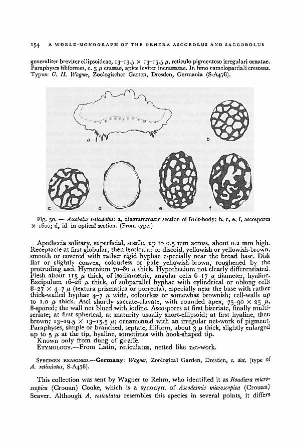
154 A WORLD-MONOGRAPH OF THE GENERA ASCOBOLUS AND SACCOBOLUS
generaliter breviter ellipsoideae, 13-19.5 X 13-15.5/r, reticulo pigmentoso irregulari ornatae.
Paraphyses filiformes, c. 3 /j crassae, apice leviter incrassatae. In fimo camelopardali crescens.
Typus: G. H. Wagner, Zoologischer Garten, Dresden, Germania (S-A478).
Apothecia solitary, superficial, sessile, up to 0.5 mm across, about 0.2 mm high.
Receptacle at first globular, then lenticularor discoid, yellowish or yellowish-brown,smooth or covered with rather rigid hyphae especially near the broad base. Disk
flat or slightly convex, colourless or pale yellowish-brown, roughened by the
protruding asci. Hymenium 70-80 u thick. Hypothecium not clearly differentiated.
Flesh about 115 /t thick, of isodiametric, angular cells 6-17 /t diameter, hyaline.
Excipulum 16-26 /t thick, of subparallel hyphae with cylindrical or oblong cells
8-27 X 4-7 [i (textura prismatica or porrecta), especially near the base with rather
thick-walled hyphae 4-7 /.i wide, colourless or somewhat brownish; cell-walls up
to 1.0 thick. Asci shortly saccate-clavate, with rounded apex, 75-90 X 25 /1>
8-spored; the wall not blued with iodine. Ascospores at first biseriate, finally multi-
seriate; at first spherical, at maturity usually short-ellipsoid; at first hyaline, then
brown; 13—19.5 X 13—15.5 fi', ornamented with an irregular net-work of pigment.Paraphyses, simple or branched, septate, filiform, about 3 /< thick, slightly enlarged
up to 5 [i at the tip, hyaline, sometimes with hook-shaped tip.Known only from dung of giraffe.ETYMOLOGY.—From Latin, reticulatus, netted like net-work.
SPECIMEN EXAMINED.—Germany: Wagner, Zoological Garden, Dresden, s. dat. (type of
A. reticulatus, S-A478).
This collection was sent by Wagner to Rehm, who identified it as Boudiera micro-
scopica (Crouan) Cooke, which is a synonym of Ascodesmis microscopica (Crouan)Seaver. Although A. reticulatus resembles this species in several points, it differs
Ascobolus reticulatus: a, diagrammatic section of fruit-body; b, c, e, f, ascospores
X 1600; d, id. in optical section. (From type.)Fig. 50.
—
ACCOBOLUS 155
fundamentally from this and other species of Ascodesmis Tiegh. in the developmentof the fruit-body and the origin of the episporial pigment.
Since the type consists of only very few fruit-bodies and no other collections
are known, it is to be hoped that this interesting fungus will be collected again.
V. Ascobolus sect. Pseudosaccobolus Brumm., sect. nov.
Ascomata para- vel eugymnohymenialia, iis generis Saccobolus simillima, primo hemisphaer->ca, denique discoidea vel lenticularia. Asci late clavati, pariete iodo intense caerulescente.
Ascosporae fusiformi-ellipsoideae, saepe leviter asymmetricae. Episporium primo laeve vel
granulatum. Paraphyses intus plus minus luteae. Pyrophila. Typus: Ascobolus pusillus Boud.
Ascomata paragymnohymenial or eugymnohymenial, strongly resembling those
of a Saccobolus; superficial, sessile; small. Receptacle at first hemispherical, then
discoid or lenticular. Excipulum of textura globulosa when seen on the surface.Asci broadly clavate, with a broad base and a rounded apex; the wall deep blue
with iodine. Ascospores 8; biseriate; fusiform-ellipsoid, often slightly asymmetrical.Episporium at first smooth or granulate, then warted or rarely with a net-work
of fine crevices. Paraphyses with yellowish contents.
Pyrophilous.ETYMOLOGY.—From false, and the generic name Saccobolus.
Only a single species is known. It occupies an isolated position within the genus.
As noticed by Boudier (1877 ), A. pusillus shows in several characters a noteworthyresemblance to species of Saccobolus.
Together with species like Saccobolus saccoboloides, with free ascospores, this speciespossibly forms a transition between Ascobolus and Saccobolus.
The absence of compact or regularly arranged clusters of ascospores and the
Way of growth on burnt places of A. pusillus justify its disposition in Ascobolus.
45. ASCOBOLUS PUSILLUS Boud.—Fig. 51
Ascobolus pusillus Boud. in Bull. Soc. bot. Fr. 24: 310 pi. 4 ƒ.. _
• 7- 1 ; Boud., Icon, mycol.,Ser. i, livr. 4
, pl. 8g (definitive number: 412). Jun. 1905; Boud., Icon, mycol., preliminarytext, Ser. i,livr.5: 17. Oct. 1905; Boud., Icon, mycol., definitive text, 234. 19 11. —Holotype:Boudier, on burnt soil, Montmorency, France, 22.XI. 1874 (PC-A2193).
Apothecia scattered or in small groups, superficial, sessile on a broad base,°-4~I.2 mm diameter, about 0.3 mm high. Receptacle hemispherical, then discoidor lenticular, "pale purplish-yellow, darker near the margin", smooth, withouta distinct margin. Disk flat, then convex, "purplish-yellow", roughened by the
Protruding tips of ripe asci. Hymenium 100-170 ji thick. Hypothecium not clearlydifferentiated as a continuous layer, of isolated groups of isodiametric or elongatedcells
4-9 x 4-7 ju. Flesh 100-200 71 thick, of subglobular cells 4-10 /t diameter,together with cylindrical hyphae 2.5-5.0 71 thick, hyaline. Excipulum 20-29 7<
thick, of subglobular or oblong, rather thick-walled cells 6-16 X 4-12 71 (texturaglobulosa or prismatica), with some pale yellowish-brown pigment. Asci broadlyclavate, rounded above, 120-150 X 15 7 1 (according to Boudier, 191 1 p. 234:
'70-200 x 25-27 41"), 8-spored; the wall deep blue in Melzer's reagent. Ascospores
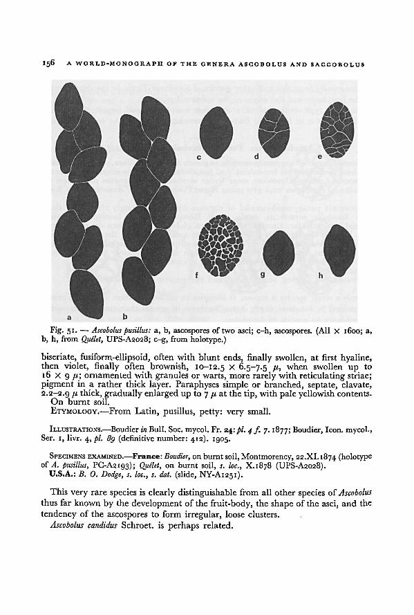
156 A WORLD-MONOGRAPH OF THE GENERA ASCOBOLUS AND SACCOBOLUS
biseriate, fusiform-ellipsoid, often with blunt ends, finally swollen, at first hyaline,then violet, finally often brownish, 10-12.5 X 6.5-7.5 /b when swollen up to
16 X 9 fi\ ornamented with granules or warts, more rarely with reticulating striae;pigment in a rather thick layer. Paraphyses simple or branched, septate, clavate,
2.2-2.9 7' thick, gradually enlarged up to 7 /« at the tip, with pale yellowish contents.
On burnt soil.
ETYMOLOGY.—From Latin, pusillus, petty: very small.
ILLUSTRATIONS.—Boudier in Bull. Soc. mycol. Fr. 24 \ pi. 4 f. 7. 1877; Boudier, Icon, mycol.,Ser. 1, livr. 4 , pi. 8g (definitive number: 412). 1905.
SPECIMENS EXAMINED.—France: Boudier, on burnt soil, Montmorency, 22.XI.1874 (holotypeof A. pusillus, PC-A2193); Quelet, on burnt soil, s. loc., X.1878 (UPS-A2028).
U.S.A.: B. O. Dodge, s. loc., s. dat. (slide, NY-A1251).
This very rare species is clearly distinguishable from all other species of Ascobolus
thus far known by the development of the fruit-body, the shape of the asci, and the
tendency of the ascospores to form irregular, loose clusters.
Ascobolus candidus Schroet. is perhaps related.
Fig. 51. —
a, b, ascospores of two asci; c-h, ascospores. (All X 1600; a,
b, h, from
Ascoboluspusillus:Quélet, UPS-A2028; c-g, from holotype.)
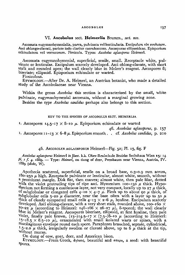
157ASCOBOLUS
VI. Ascobolus sect. Heimerlia Brumm., sect. nov.
Ascomata eugymnohymenialia, parva, pulvinata vel lenticularia.Excipulum vix evolutum.
Asci oblongo-clavati, pariete iodo clariter caerulescente. Ascosporae ellipsoideae. Episporiumechinulatum vel verrucosum. Fimicola. Typus: Ascobolus aglaosporus Heimerl.
Ascomata eugymnohymenial, superficial, sessile, small. Receptacle white, pul-vinate or lenticular. Excipulum scarcely developed. Asci oblong-clavate, with shortstalk and rounded apex; the wall clearly blue in Melzer's reagent. Ascospores 8;biseriate; ellipsoid. Episporium echinulate or warted.
Fimicolous.
ETYMOLOGY.—After Dr. A. Heimerl, an Austrian botanist, who made a detailed
study of the Ascobolaceae near Vienna.
Within the genus Ascobolus this section is characterized by the small, white
pulvinate, eugymnohymenial ascomata, without a marginal growing zone.
Besides the type Ascobolus candidus perhaps also belongs to this section.
KEY TO THE SPECIES OF ASCOBOLUS SECT. HEIMERLIA
I. Ascospores 14.5-17 X 8-10 pi. Episporium echinulate or warted
46. Ascobolus aglaosporus, p. 157
1. Ascospores 11-1 3 X 6-8 ju. Episporium smooth. .
cf. Ascobolus candidus, p. 210
46. ASCOBOLUS AGLAOSPORUS Heimerl—Fig. 52; Pl. 13, fig. F
Ascobolus aglaosporus Heimerl in Jber. k.k. Ober-Realschule Bezirke Sechshaus Wien 15: 14PI. 1 f. 4. i88g. — Type: Heimerl, on dung of deer, Pressbaum near Vienna, Austria, IV.
1889 (slide, W).
Apothecia scattered, superficial, sessile on a broad base, 0.3-0.9 mm across,
180-250 fi high. Receptacle pulvinate or lenticular, almost white, smooth, without
a prominent margin. Disk flat, then convex; almost white, then pale lilac, dotted
With the violet protruding tips of ripe asci. Hymenium 100-150 fx thick. Hypo-thecium not forming a continuous layer, not very compact, locally up to 27 /< thick,°f subglobular or elongated cells 4-10 X 4-7 70 Flesh up to about 90 fx thick, of
subglobular cells 5-20 a diameter, near the base often with a layer up to 50 fxthick of closely compacted small cells 4-13 X 2-6 /x, hyaline. Excipulum scarcely
developed. Asci oblong-clavate, with a very short stalk, rounded above, 100-160 X
20-22 fx (according to Heimerl: 146-166 X 26-27 fi), 8-spored; the wall clearlyolue in Melzer's reagent. Ascospores biseriate, ellipsoid, at first hyaline, then paleVlolet, finally pale brown, ( 13—) 14-5— 17 X (7.5—)8—10 fx (according to Heimerl:
1 5~i8.5 x 8.5-10 /x), ornamented with small isolated warts or spines, with a
Oiucilaginous envelope round each spore. Paraphyses branched, septate, cylindrical,1-3-2.0 pi thick, irregularly swollen or clavate above, up to 6 fx thick at the tip,
Without mucus.
On dung of cow, goat, deer, and American bison.
ETYMOLOGY.—From Greek, ayXao?, beautiful and orcopa, a seed: with beautifuls Pores.
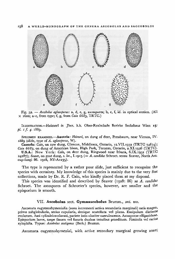
158 A WORLD-MONOGRAPH OF THE GENERA ASCOBOLUS AND SAGCOBOLUS
ILLUSTRATION.—Heimerl in Jber. k.k. Ober-Realschule Bezirke Sechshaus Wien 15:
pi. if. 4. 1889.
SPECIMEN EXAMINED.—Austria: Heimerl, on dung of deer, Pressbaum, near Vienna, IV.
1889 (slide, type of A. aglaosporus, W).Canada: Cain, on cow dung, Glencoe, Middlesex, Ontario, 12.VII.1932 (TRTC 34674);
Cain 6683, on dung of American bison, High Park, Toronto, Ontario, 2.XI.1936 (TRTC).U.S.A.: New York: Cain, on deer dung, Ringwood near Ithaca, 6.IX. 1952 (TRTC
24287); Seaver, on goat dung, s. loc., I.1915 (= A. candidus Schroet. sensu Seaver, North Am.
cup-fungi 86. 1928, NY-A1235).
The type is represented by a rather poor slide, just sufficient to recognize the
species with certainty. My knowledge of this species is mainly due to the very fine
collections, made by Dr. R. F. Gain, who kindly placed them at my disposal.This species was identified and described by Seaver (1928: 86) as A. candidus
Schroet. The ascospores of Schroeter's species, however, are smaller and the
episporium is smooth.
VII. Ascobolus sect. Gymnascobolus Brumm., sect. nov.
Ascomata eugymnohymenialia (zona incrementi activa secundaria marginali) satis magna,
primo subglobularia, serius cupularia, denique scutellata vel plana. Excipulum distincte
evolutum. Asci cylindrico-clavati, pariete iodo clariter caerulescente. Ascosporae ellipsoideae.
Episporium laeve, saepe fissura vel fissuris duabus tenuibus praeditum. Fimicola vel rarius
xylophila. Typus: Ascobolus scatigenus (Berk.) Brumm.
Ascomata eugymnohymenial, with active secondary marginal growing zone;
a, d, e, g, ascospores; b, c, f, id. in optical section. (All
X 1600; a-e, from type; f, g, from
Ascobolus aglaosporus:Fig. 52.—
Cain 6683, TRTC.)
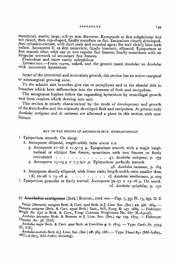
ASCOBOLUS159
superficial, sessile; large, 0.8-30 mm diameter. Receptacle at first subglobular butnot closed, then cup-shaped, finally scutellate or flat. Excipulum clearly developed.Asci cylindric-clavate, with short stalk and rounded apex; the wall clearly blue withiodine. Ascospores 8; at first uniseriate, finally biseriate; ellipsoid. Episporium at
first smooth often with one or two regular fine fissures; finally sometimes with an
irregular net-work of secondary fine fissures.
Fimicolous and more rarely xylophilous.ETYMOLOGY.—From yupvoi;, naked, and the generic name Ascobolus: an Ascobolus
with uncovered hymenium.
Apart of the interstitial and intercalary growth, this section has an active marginalor submarginal growing zone.
To the adaxial side branches give rise to paraphyses and to the abaxial side to
branches which later differentiate into the elements of flesh and excipulum.The ascogenous hyphae follow the expanding hymenium by centrifugal growth
and form croziers which develop into asci.
This section is clearly characterized by the mode of development and growthof the fruit-bodies and the copiously developed flesh and excipulum. At present onlyAscobolus scatigenus and A. castaneus are allocated a place in this section with con-
fidence.
KEY TO THE SPECIES OF ASCOBOLUS SECT. GYMNASCOBOLUS
1 • Episporium smooth. On dung:
2. Ascospores ellipsoid, length-width ratio about 2.0:
3. Ascospores 21-26 x 11.5-13 pi. Episporium smooth with a single longi-tudinal or oblique fine fissure, sometimes with two fissures or finelyreticulated 47. Ascobolus scatigenus, P- 1 59
3. Ascospores 15-19.5 X 7.5-9.0 /.i. Episporium perfectly smooth
48. Ascobolus castaneus, p. 1642. Ascospores shortly ellipsoid, with blunt ends; length-width ratio smaller than
1.6; 20-28 X 15-18 pi cf. Ascobolus moellerianus, p. 229'• Episporium granular or finely warted. Ascospores 32-37 X 13-16 pi. On wood.
cf. Ascobolus xylophilus, p. 152
47. Ascobolus scatigenus (Berk.) Brumm., comb. nov. —Figs. 7, 53; Pl. 13, figs. D. E
Peziza (Humaria) scatigena Berk. & Curt, apud Berk, inJ. Linn. Soc. (Bot.) 10: 366. 1869. —
Humaria scatigena (Berk. & Curt, apud Berk.) Sacc., Syll. Fung. 8: 147. 1889. — Holotype:bright No. 636 in Berk. & Curt., Fungi Cubensis Wrightiana No. 667 (K-A3046).
Ascobolus leiocarpus Berk. & Broome in J. Linn. Soc. (Bot.) 14: iog. 1873. — Holotype:Thwaites No. 56 (BM).
Ascobolus major Berk. & Curt, apud Berk, in Grevillea 4: 6. 1875. — Type: Curtis No. 3794(K-, FH).
Ascobolus australis Berk, in J. Linn. Soc. (Bot.) 18: 389. 1881. —Type: Thozel 831 (BM-A2855,MEL-A1625, BM-A2820 drawing).
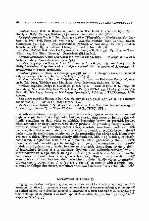
160A WORLD-MONOGRAPH OF THE GENERA ASCOBOLUS AND SACCOBOLUS
Ascobolus baileyi Berk. & Broome in Trans. Linn. Soc. Lond. II (Bot.) 2: 69. 1883. —
Holotype: Bailey No. 232, Brisbane, Queensland, Australia, s. dat. (BM).
Phaeopezia orientalis Pat. in J. Bot., Paris 4: 59. 1890 ('Pheopezia’). — Aleurina orientalis (Pat.)Sacc. & Syd., Syll. Fung. 16: 739. 1902. — Ascobolus orientalis (Pat.) Le Gal, Discom.
Madagascar 75. 1953. — Type distribution: [Delavay], on cow dung, Tu Phap, Tonkin,
Indochina, VII. 1887, in Balansa, Champ, du Tonkin No. 118 (G).Ascobolus phillipsii Berk, apud Cooke, Australian fungi, 268, pi. 20, f. 164. 1892. — Type:
\Thozet\ No. 851 (Herb. Berkeley), Queensland (BM-A2855).Ascobolus sarawacensis Cesati apud Cooke in Grevillea 21: 74. 1893. -— Holotype: Beccari 226,
on buffalo dung, Sarawak, s. dat. (K-A1991).Ascobolus megalospermus Speg. in Anal. Mus. nac. B. Aires 6: 307. 1899. — Holotype: LPS
28164 (consisting of apothecia of A. scatigenus covered with ascospores of A. immersus; A.
megalospermus = nomen confusum).Ascobolus gollanii P. Henn. in Hedwigia 40: 338. 1901. — Holotype: Gollan, on manured
soil, Saharanpur-Garden, India, 14.VII.1900 (S-A517).Ascobolus latus Penz. & Sacc. in Malpighia 15: 218. 1902. — Holotype: Penzig tub. gij,
on buffalo dung, Tjidahoe near Mt. Salak, Java, Indonesia, 22.1.1897 (PAD).Ascobolus magnificus Dodge in Mycologia 4: 218. 1912. —
Parts of type: B. 0. Dodge, on
horse dung, New York City, New York, U.S.A., IV.1912 (BPI-A1555, FH-A3147, K-A1985,K-A1986, NY-A1317 holotype, NY-A1319 "paratype", PC-A2304, TRTC-A2151, UPS-
A2020).
Papulospora magnified Hotson in Bot. Gaz. 64: 277 pi. 22f. 39, pi. 23f. 48-69. 19 17 (nomenanamorphosis). — Fide B. O. Dodge (1920: 123).
Ascobolus notatus Batista & Vital apud Batista & al. in Ann. Soc. Biol. Pernambuco 13: 61
fs. 1-3. 1955 ("notata"). — Type: Vital No. 1976 (L, TRTC, UPS).
Apothecia solitary or gregarious, superficial, sessile, 0.5-3.0 cm diameter, 1-5 mm
high. Receptacle at first subglobular but not closed, then more or less cup-shaped,finally scutellate or flat, white or whitish, becoming brown or greenish-brown,often wrinkled or irregularly curved, finely pruinose or granular; margin white or
brownish, smooth or granular, rather thick, involute, sometimes undulate. Disk
concave, then flat or undulate, greenish-yellow, brownish or reddish-brown, alwaysdarker thanthe excipulum, roughened by the protruding tips of ripe asci. Hymenium170-200 pi thick. Hypothecium clearly differentiated, about 25 pi thick, of closely
compacted, subglobular cells 6-12 pi diameter. Flesh 350-1000 pi thick or even
more, of globular or oblong cells io-25(-65) X 7-17 pi, accompanied by irregularundulating hyphae 5-9 pi wide, hyaline or brownish. Excipulum 50-60 pi
thick,of intertwined hyphae 2-4 pi diameter, hyaline. Asci cylindric-calavate, with a
short, narrow stalk, 140-280 X 16-25 /'■> 8-spored; the wall blue in Melzer's reagent.
Ascospores uniseriate, finally irregularly biseriate, ellipsoid, very rarely slightlyasymmetrical; at first hyaline, then pale pinkish-violet, finally violet or purplish-brown; (20-)2i.5-25-5(-27.5) X ( 11 —) 11
. 5— 13 (— 14) pi, smooth with a single longi-tudinal or oblique fine fissure, sometimes with two fissures or finely reticulated; with
Ascobolus scatigenus:Fig. 53. —
g-j,
from holotype of
A. scatigenus;
G; p-r, from ‘paratype’ ofk-o, from type of
NY-A1319.)
EXPLANATION OF FIGURE 53
d-f, from holotype of
a, diagrammatic section offruit-body X 25; b-e, g-n, p—r,
ascospores X 1600 (q, represents a rare, abnormal type of ornamentation); f, o, ascosporesin optical section. (a-c, from holotype of
A.
magnificus,
A. leiocarpus;A. gollani; A. orientalis,
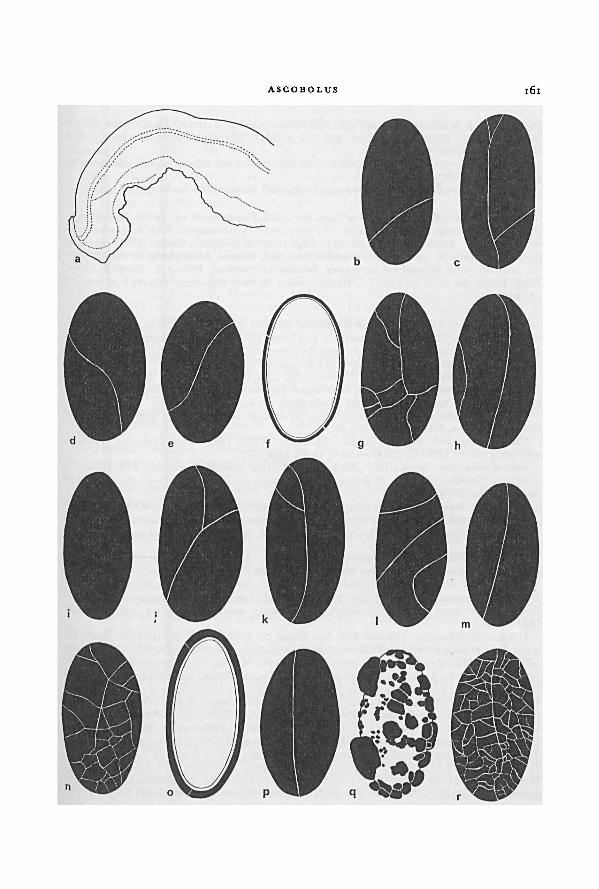
ASCOBOLUS 161
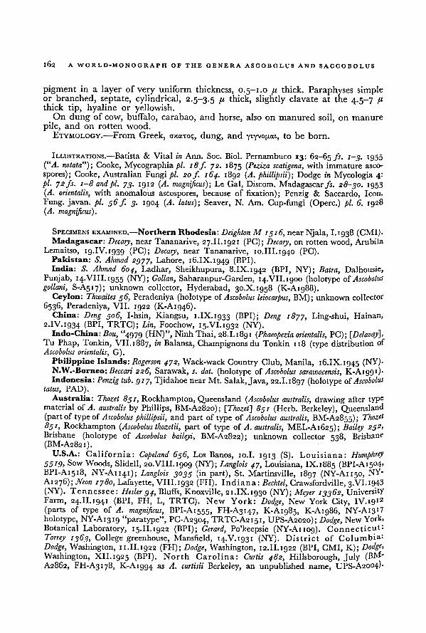
162 A WORLD-MONOGRAPH OF THE GENERA ASCOBOLUS AND SACCOBOLUS
pigment in a layer of very uniform thickness, 0.5-1.0 // thick. Paraphyses simpleor branched, septate, cylindrical, 2.5-3.5 I1 thick, slightly clavate at the 4.5-7 /'-thick tip, hyaline or yellowish.
On dung of cow, buffalo, carabao, and horse, also on manured soil, on manure
pile, and on rotten wood.
ETYMOLOGY.—From Greek, cxa-roq, dung, and YIYVOPOA, to be born.
ILLUSTRATIONS.—
Batista & Vital in Ann. Soc. Biol. Pernambuco 13: 62-65 fs. 1-3. 1955
(“A. notata”); Cooke, Mycographia pi. 18 f. 72. 1875 ( Peziza scatigena, with immature asco-
spores); Cooke, Australian Fungi pi. 20f 164. 1892 (A. phillipsii); Dodge in Mycologia 4:pi. 72 fs. 1-8 and pi. 73. 1912 (A. magnificus)-, Le Gal, Discom. Madagascar fs. 28-30. 1953
(A. orientalis, with anomalous ascospores, because of fixation); Penzig & Saccardo, Icon.
Fung, javan. pi. 56 f. 3. 1904 (A. latus); Seaver, N. Am. Cup-fungi (Operc.) pi. 6. 1928(A. magnificus).
SPECIMENS EXAMINED.—Northern Rhodesia: Deighton M 1316, near Njala, 1.1938 (CMI).Madagascar: Decary,
near Tananarive, 27.II. 1921 (PC); Decary, on rotten wood, Arubila
Lemaitso, 19.IV.1939 (PC); Decary ,near Tananarive, 10.III. 1940 (PC).
Pakistan: S. Ahmad 2977, Lahore, 16.IX. 1949 (BPI).India: S. Ahmad 604, Ladhar, Sheikhupura, 8.IX.1942 (BPI, NY); Batra, Dalhousie,
Punjab, 14.VIII.1955 (NY); Gollan, Saharanpur-Garden, 14.VII.1900 (holotype of Ascobolus
gollani, S-A517); unknown collector, Hyderabad, 30.X.1958 (K-A1988).Ceylon: Thwaites 36, Peradeniya (holotype of Ascobolus leiocarpus, BM); unknown collector
6536, Peradeniya, VII. 1922 (K-A1946).China: Deng 306, I-hsin, Kiangsu, i.IX.ig33 (BPI); Deng 1877, Ling-shui, Hainan,
2.IV.ig34 (BPI, TRTC); Lin, Foochow, 15.VI.1932 (NY).Indo-China:Bou, "4979 (HN)", Ninh Thai, 28.I.1891 (Phaeopezia orientalis, PC); [Delavay],
Tu Phap, Tonkin, VII.1887, in Balansa, Champignons du Tonkin 118 (type distribution of
Ascobolus orientalis, G).
Philippine Islands: Rogerson 472, Wack-wack Country Club, Manila, 16.IX.1945 (NY).N.W.-Borneo: Beccari 226, Sarawak, s. dat. (holotype of Ascobolus sarawacensis, K-Aig9i).Indonesia:Penzig tub. 917, Tjidahoe near Mt. Salak, Java, 22.1.1897 (holotype of.Ascobolus
latus, PAD).
Australia: 7 hozet 831, Rockhampton, Queensland (Ascobolus australis, drawing after typematerial of A. australis by Phillips, BM-A2820); [Thozet] 831 (Herb. Berkeley), Queensland(part of type of Ascobolus phillipsii, and part of type of Ascobolus australis, BM-A2855); ThozA
851, Rockhampton (Ascobolus thozetii, part of type of A. australis, MEL-A1625); Bailey 252,
Brisbane (holotype of Ascobolus baileyi, BM-A2822); unknown collector 538, Brisbane
(BM-A2821).U.S.A.: California: Copeland 636, Los Banos, 10.I. 1913 (S). Louisiana: Humphrey
55!9> Sow Woods, Slidell, 20.VIII.1909 (NY); Langlois 47, Louisiana, IX.1885 (BPI-A1504JBPI-A1518, NY-A1141); Langlois 3033 (in part), St. Martinsville, 1897 (NY-A1150, NY-
A1276); Neon 1780, Lafayette, VIII. 1932 (FH). Indiana: Bechtel, Crawsfordville, 3.VI. 1943(NY). Tennessee: Hesler 94, Bluffs, Knoxville, 21.IX.1930 (NY); Meyer 13362, University
Farm, 24.II.1941 (BPI, FH, L, TRTC). New York: Dodge, New York City, IV.1912
(parts of type of A. magnificus, BPI-A1555, FH-A3147, K-A1985, K-A1986, NY-Ai3!7
holotype, NY-Ai3ig "paratype", PC-A2304, TRTC-A2151, UPS-A2020); Dodge, New York,
Botanical Laboratory, 15.II.1922 (BPI); Gerard, Po'keepsie (NY-A1109). Connecticut:
Torrey 1363, College greenhouse, Mansfield, 14.V.1931 (NY). District of Columbia:
Dodge, Washington, 11.II. 1922 (FH); Dodge, Washington, 12.II. 1922 (BPI, CMI, K); Dodge,
Washington, XII. 1925 (BPI). North Carolina: Curtis 482, Hillsborough, July (BM -
A2862, FH-A3178, K-A1994 as A. curtisii Berkeley, an unpublished name,UPS-A2004).
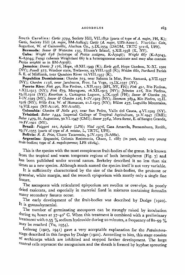
163ASCOBOLUS
South Carolina: Curtis 3794, Society Hill, VII.1852 (parts of type of A. major, FH, K);Curtis, Society Hill (A. major, BM-A2824); Curtis (A. major, UPS-A2021). Florida: Cain,Sugarfoot, W. of Gainesville, Alachua Co., 5.IX.1954 (DAOM, TRTC 31216, UPS).
Bermuda: Seaver & Waterston 135, Hinson's Island, 5.XII. 1938 (K, NY).Cuba: Wright 636 (holotype of Peziza scatigena, K-A3046); Wright 667 (K-A3045,
K-A3044; Fungi cubensis Wrightiani 667 is a heterogeneousexsiccate andmay also contain
Peziza wrightii as in BM-A3036).Jamaica: Dennis J 176, Hope, 18.XII.1949 (K); Earle 498, Hope Gardens, X-XI. 1902
(NY); Powell 377, Windsor state, Trelawny, 23.VIII.1956 (K); Welden 660, Portland ParishS. E. of Millbank, near Quashies River 12.VIII.1957 (K).
Republica Dominicana: Chardon 703, near Sabana la Mar, Prov. Samana, 4.VII.ig37(NY); Chardon 1136, near Jarabacoa, Prov. La Vega, 13.IX.1937 (NY).
Puerto Rico: Fink 490, Rio Piedras, 1.XII. 1915 (BPI, NY, FH); Fink 491, Rio Piedras,i.XlI. 1915 (NY); Fink 879, Mayaguez, 16.XII.1915 (NY); Johnston 216, Rio Piedras,25.II. 1912 (NY); Kevorkian 1, Cartagena Lagoon, 5.IX.1936 (FH); Seaver & Chardon 59,
I-IV.1923 (NY); Seaver & Chardon 126, I-IV.1923 (NY); Stevenson 3894, Rio Piedro, 15.II.>916 (NY); Wille 812, W. of Humacao, 21.I.1915 (NY); Wilson 237, Luguillo Mountains,14.VII.1902 (NY-A1126, NY-A1268).
Colombia: Chardon & Nolla 407, near San Pedro, Valle del Cauca, 4.VI. 1929 (NY).Trinidad: Baker 1444, Imperial College of Tropical Agriculture, 31.V.1947 (CMI);
Baker1479, St. Augustine, 30.VI.1947 (CMI); Seaver 3564, Mora forest, E. of Sangre Grande,
10.IV.1921 (NY).Brazil: Puiggari 101, Apiahy (LPS); Vital 1976, Casa Amarela, Pernambuco, Recife,
29-IV.i955 (parts of type of A. notatus, L, TRTC, UPS).Bolivia: R. E. Fries, Chaco Tatarenda, 9.IV. 1905 (S-A662).Argentina: Spegazzini, Colonia Resistencia, Chaco, I. 1887 (in part, only very young
fruit-bodies; type of A. megalospermus; LPS 28164).
This is the species with the most conspicuous fruit-bodies of the genus. It is known
from the tropical and warm temperate regions of both hemispheres (Fig. 7) and
has been published under several names. Berkeley described it no less than six
frmes as a new species. Although much named the species itself is not very variable.
It is sufficiently characterized by the size of the fruit-bodies, the pruinose or
granular, white margin, and the smooth episporium with mostly only a single fine
fissure.
Theascospores with reticulated episporium are swollen or over-ripe. In poorly
dried exsiccata, and especially in material fixed in mixtures containing formalin
Btany secondary fissures occur.
The early development of the fruit-bodies was described by Dodge (1920).It is gymnohymenial.
The number of germinating ascospores can be strongly raised by incubation
during 24 hours at 37-40° C. When this treatment is combined with a preliminarytreatment with0.55 % sodium hydroxide during 20 minutes, a frequency of8o-gg %"tay be reached (Yu, 1954).
Lohwag (1927, 1941) gave a very acceptable explanation for the Papulospora-
stage described in this fungus by Dodge (1920). According to him, this stage consists
°* archicarps which are inhibited and stopped further development. The largecentral cells represent the ascogonium and the sheath is formed by hyphae sprouting
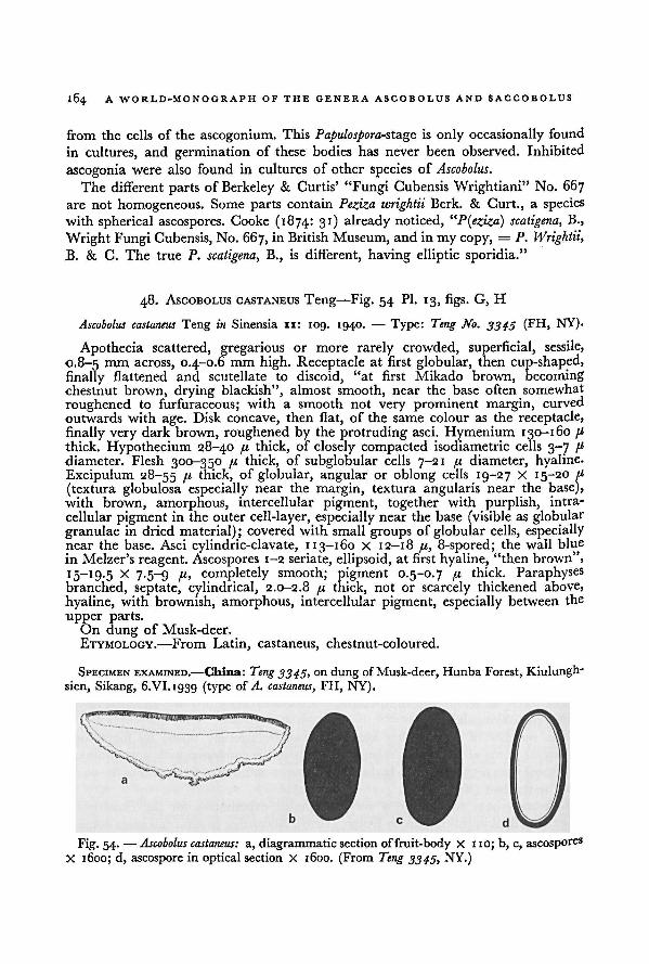
164 A WORLD-MONOGRAPH OF THE GENERA ASCOBOLUS AND SACCOBOLUS
from the cells of the ascogonium. This Papulospora- stage is only occasionally found
in cultures, and germination of these bodies has never been observed. Inhibited
ascogonia were also found in cultures of other species of Ascobolus.
The different parts of Berkeley & Curtis' "Fungi Cubensis Wrightiani" No. 667
are not homogeneous. Some parts contain Peziza wrightii Berk. & Curt., a species
with spherical ascospores. Cooke (1874: 31) already noticed,"P(eziza) scatigena, B.,
Wright Fungi Cubensis, No. 667, in British Museum, and in my copy, = P. Wrightii,
B. & C. The true P. scatigena, B., is different, having elliptic sporidia."
48. ASCOBOLUS CASTANEUS Teng—Fig. 54 Pl. 13, figs. G, H
Ascobolus castaneus Teng in Sinensia 11: 109. 1940.— Type: Teng No. 3345 (FH, NY).
Apothecia scattered, gregarious or more rarely crowded, superficial, sessile,
0.8-5 mm across, 0.4-0.6 mm high. Receptacle at first globular, then cup-shaped,finally flattened and scutellate to discoid, "at first Mikado brown, becomingchestnut brown, drying blackish", almost smooth, near the base often somewhat
roughened to furfuraceous; with a smooth not very prominent margin, curved
outwards with age. Disk concave, then flat, of the same colour as the receptacle,
finally very dark brown, roughened by the protruding asci. Hymenium 130-160 pthick. Hypothecium 28-40 pi thick, of closely compacted isodiametric cells 3-7 pdiameter. Flesh 300-350 pi thick, of subglobular cells 7-21 p diameter, hyaline.Excipulum 28-55 P thick, of globular, angular or oblong cells 19-27 X 15-20 p
(textura globulosa especially near the margin, textura angularis near the base),with brown, amorphous, intercellular pigment, together with purplish, intra-
cellular pigment in the outer cell-layer, especially near the base (visible as globular
granulae in dried material); covered with small groups of globular cells, especiallynear the base. Asci cylindric-clavate, 113-160 X 12-18 pi, 8-spored; the wall blue
in Melzer's reagent. Ascospores 1-2 seriate, ellipsoid, at first hyaline, "then brown",
I5_I9-5 x 7-5~9 P> completely smooth; pigment 0.5-0.7 pi thick. Paraphysesbranched, septate, cylindrical, 2.0-2.8 pi thick, not or scarcely thickened above,
hyaline, with brownish, amorphous, intercellular pigment, especially between the
upper parts.On dung of Musk-deer.
ETYMOLOGY.—From Latin, castaneus, chestnut-coloured.
SPECIMEN EXAMINED.—China: Teng 3345, on dung ofMusk-deer, Hunba Forest, Kiulungh-
sien, Sikang, 6.VI.1939 (type of A. castaneus, FFI, NY).
a, diagrammaticsection of fruit-body X 110; b, c, ascosporesX 1600; d, ascospore in optical section X 1600. (From
Fig. 54.—
NY.)
Ascobolus castaneus:
Teng 3345,
165ASCOBOLUS
This species is characterized by the chestnut brown colour of the receptacle
and disk and by the perfectly smooth episporium without any fissures.
The growth of the fruit-body is as in A. scatigenus, by means of a marginal growing-
zone. Probably as a result of differences in relative speed of growth and in the
structure of the receptacle the fruit-bodies of A. castaneus tend to be more discoid
or flattened, while in A. scatigenus they are cup-shaped with inrolled margin.
166
SAGCOBOLUS Boud.
Saccobolus Boud. in Annls Sci. nat. (Bot.) V 10: 228. 1869. — Ascobolus.. .
Saccobolus (Boud.)Cooke, Handb. Brit. Fung. 2: 729. 1871 (unreferable rank). — Ascobolus subg. Saccobolus
(Boud.) Quel., Ench. Fung. 294. 1886. — Type: Saccobolus kervernii (Crouan) Boud.
[Saccobolus sect. Eu-Saccobolus Sacc., Syll. Fung. 8: 524. 1889. — Type: Saccobolus kervernii
(Crouan) Boud.]
Saccobolus sect. Eriobolus Sacc., Syll. Fung. 8: 527. 1889. — Monotype: Saccobolus caesariatus
Phillips.Ornithascus Vel., Monogr. Discom. Boh. 1: 368. 1934.
— Monotype: Ornithascus corvinus
Vel.
Ascomata paragymnohymenial or eugymnohymenial; superficial, sessile; small
0.1-1.0 mm, rarely up to 2.0 mm diameter. Receptacle at first often subglobularor semiglobular but soon pulvinate or lenticular; surface smooth or sometimes
covered with spreading bunches of flexuous hyphae; without a distinct margin.Asci shortly clavate, with clearly truncate apex; at least at maturity the tips stronglyprotruding above the hymenium; the wall staining blue with iodine. Ascosporesfirmly united in a cluster or package and ejected together, cemented in a more
or less regular pattern (Fig. 2) by the episporial pigment or rarely at first looselyunited according to such a pattern and finally free or partly free. Paraphyses slender
or rather thick, cylindrical or cylindric-clavate; relatively short; sometimes embedded
in colourless mucus; contents colourless or yellow. Sometimes intercellular, amor-
phous, violet or brown pigment between the tips of paraphyses or in the outer
layers of the excipulum.
Fimicolous, more rarely on rotten textiles or on substrata dirtied with dung-ETYMOLOGY.—From Greek, aaxxosack, and |3aXXw, to throw.
Type: Saccobolus kervernii (Crouan) Boud.
Within the Ascoboloideae this genus is characterized by the clustered ascospores,
by the relatively short and broad, clavate, truncate asci, and by the relatively short
paraphyses.
When Boudier (1869) established Saccobolus, the presence of a 'common hyaline
sack' around the ascospores was considered one of the major criteria. Although
this envelope occurs only in a limited number of species, many authors have main-
tained it as the most important distinguishing character (e.g. Saccardo, 1884, 1889;
Phillips, 1887; Massee, 1895; Rehm, 1896; Boudier, 1907; Le Gal, 1947, i953a) -This common envelope is rather constant in Saccobolus sect. Saccobolus. In Saccobolus
sect. Eriobolus, however, the gelatinous mass never surrounds the spores; it may
be unilateral, subdivided, orabsent.
The force by which the ascospores are driven together and united into a package
according to a very constant stereometric pattern is still unknown.
The precipitation of the episporial pigment may occur in different phases of the
aggregative process, while also the degree to which the ascospores are pressed
together differs fromspecies to species. When the precipitation of episporial pigmentis finished or almost finished before the spores are firmly united, the spores remain
loose ( S. saccoboloides) or the connection is lost easily (S. globuliferellus, S. geminatus,
S. infestans). In S. geminatus the ascospores of a pair are more strongly united than
167SACCOBOLUS
the pairs among themselves. The strongest pressing together of the ascospores is
reached in S. dilutellus in which the package is subglobular.
Concerning the episporial ornamentation, it is noteworthy that in species of
Saccobolus the episporia of all spores in a package are always found to be quite
similar, while in Ascobolus the spores of one and the same ascus sometimes differ
greatly in this respect.
The arrangement of the ascospores proved to be a very constant and valuable
character to distinguish groups of species. For the sake of convenience the different
stereometric patterns according to which the spores may be arranged in Saccobous
are indicated by numbers of which the corresponding description and figure are
found in the chapter on ascospores (p. 40 et seq.).The species with yellowish or amber fruit-bodies, where the colour is due to
yellowish contents of the paraphyses, have their spores arranged according to
pattern I. These species are united in Saccobolus sect. Saccobolus.
The species with white, violet or brownish fruit-bodies, where intercellular
pigments may be absent or present, have their spores arranged according to other
patterns. They form together Saccobolus sect. Eriobolus.
In some species of Saccobolus the fruit-bodies show a tendency to aggregate and
form complexes or even crusts of confluent fruit-bodies. Especially in S. saccoboloides,S. versicolor, and S. depauperatus this is of rather frequent occurrence. In these and
other species often more than one ascogonium is found in a single fruit-body. In
complex fruit-bodies with a narrow base the peripheral asci may be considerablylonger than the central ones.
KEY TO THE SECTIONS OF SACCOBOLUS
1 • Receptacle and disk yellow, yellowish or amber. Ascospores ellipsoid or fusiform-
ellipsoid with truncate ends; at first arranged according to pattern I, finallysometimes more or less shortened, irregularly arranged or free. Paraphyses,
especially in the upperparts, with yellow, lemon-yellow, golden-yellow or orange-
yellow contents. Without accumulations of intercellular amorphous pigment in
excipulum or hymenium I. Saccobolus sect. Saccobolus, p. 167'• Receptacle and disk white, pale violet, violet or brown. Ascospores ellipsoid
or fusiform-ellipsoid, often with slightly truncate ends, often asymmetrical, ventri-
culose or somewhat triangular; not arranged according to pattern I. Paraphyseswithout yellowish or orange contents, mostly colourless. If pigmented, then
intercellular and amorphous in excipulum and between the tips of paraphysesII. Saccobolus sect. Eriobolus, p. 181
I. SACCOBOLUS sect. SACCOBOLUS
lorsynonymy see under generic name (p. 166).
168 A WORLD-MONOGRAPH OF THE GENERA ASCOBOLUS AND SACCOBOLUS
Receptacle and disk yellow, yellowish or amber. Ascospores 8; ellipsoid or fusiform-
ellipsoid with truncate ends; at first arranged in a package according to a sym-metrical pattern with 4 rows of 2 ascospores, with their longitudinal axes parallelto the length axis of the package (pattern I, see Fig. 2a, b), finally sometimes more
or less shortened or free. Paraphyses especially in the upper parts with yellow,lemon-yellow, golden-yellow or orange-yellow contents. Without accumulation of
amorphous intercellular pigment in excipulum or hymenium.Fimicolous and rarely on rotten textile fabrics.
TYPE: Saccobolus kervernii (Crouan) Boud.
This section contains six sharply delimited species, which can be easily recognized
by the colour of the fruit-bodies, the shape of the ascospore-packages, the shape
and size of the ascospores, and by the ornamentationof the episporium.
KEY TO THE SPECIES OF SACCOBOLUS SECT. SACCOBOLUS
i. Apothecia tending to form crusts. Spore-clusters very loose and ascospores free
at maturity. Ascospores 16-19.5 x 7-5~9 /'• Episporium smooth or sometimes
finely granular 1. Saccobolus saccoboloides, p. 168
1. Apothecia not forming crusts. Spore-clusters compact and ascospores firmly
cemented:
2. Ascospores at maturity with length-width ratio 2.0-2.5; ellipsoid or fusiform-
ellipsoid, with truncate ends. Episporium up to 0.8 fi thick:
3. Receptacle and disk vividly yellow or lemon-yellow. Ascospores ventricose
with strongly truncate ends; 16-22 X 7.5-9 p- Episporium with isolated
fine warts 3. Saccobolus citrinus, p. 174
3. Receptacle and disk golden-yellow or amber. Ascospores not strongly
truncate and ventricose. Episporium smooth or finely granular, sometimes
with a few irregular crevices:
4. Ascospores 22-29 X 8.5-14.5 p 2. Saccobolus glaber, p. 170
4. Ascospores less than 18 p long:
5. Ascospores 14-17.5 X 7.5-8.5 p. Spore-clusters at maturity shortened
and ascospores arranged according to pattern la (Fig. 2c)
4. Saccobolus truncatus, p. 1 76
5. Ascospores 11.5-13.5 X 5.5-6.5 /a. Spore-clusters not shortened at
maturity 6. Saccobolus minimus, p. 179
2. Ascospores at maturity with length-width ratio about 1.3—1.5; short-ellipsoidor subglobular; 15-17.5 X 10-12.5 p . Episporium more than 1.0 p
thick;
with a net-work of crevices. Spore-clusters rich in extra-episporial pigment
5. Saccobolus portoricensis, p. 17®
1. Saccobolus saccoboloides (Seaver apud Dodge & Seaver) Brumm., comb. nov. —
Fig. 55; Pl. 1 4, fig. A
Ascobolus saccoboloides Seaver apud Dodge & Seaver in Mycologia 38: 640. 1946 (basionym). —'
Holotype: G. W. Martin J 637 (NY, K).
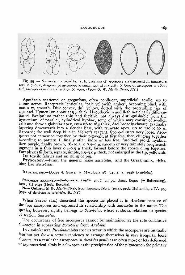
169SACCOBOLUS
Apothecia scattered or gregarious, often confluent, superficial, sessile, up to
i mm across. Receptacle lenticular, 'pale yellowish amber', becoming black with
maturity, smooth. Disk convex, dull yellow, dotted with the protruding tips of
npe asci. Hymenium about 125 [X thick. Hypothecium and flesh not clearly differen-tiated. Excipulum rather thin and fugitive, not always distinguishable from the
hymenium, of parallel, cylindrical hyphae, some of which may consist of swollen
cells and show a globular apex, even up to 16/x thick. Asci broadly clavate, graduallytapering downwards into a slender base, with truncate apex, up to 150 X 20 fx,
8-spored; the wall deep blue in Melzer's reagent. Spore-clusters very loose. Asco-
spores not cemented together by their pigment, at first free, then clinging togetheraccording to pattern I, finally often more or less free, fusoid-ellipsoid; hyaline,then purple, finally brown, 16-19.5 x 7.5 -9 fx, smooth or very minutely roughened;pigment in a thin layer 0.4-0.5 /x thick, formed before the spores cling together.Paraphyses filiform, septate,simple, 2.5-3.0 fx thick, not enlarged at the tip, yellowish.
On textile fabrics and on dung of pig.. ETYMOLOGY.—From the generic name Saccobolus, and the Greek suffix, -èiSo<;,hke: like Saccobolus.
ILLUSTRATION.—Dodge & Seaver in Mycologia 38: 641 ƒ. 1. 1946 (Ascobolus).
SPECIMENS EXAMINED.— Indonesia: Boedijn 4306, on pig dung, Bogor (= Buitenzorg),Java, III. 1949 (Herb. Boedijn).
New Guinea: G. IV. Martin J63J, from Japanese fabric (sock), prob. Hollandia, 2.IV. 1945
(type of Ascobolus saccoboloides, K, NY).
When Seaver (I.e.) described this species he placed it in Ascobolus because of
the free ascospores and expressed its relationship with Saccobolus in the name. The
species, however, rightly belongs to Saccobolus, where it shows relations to species°f section Saccobolus.
The occurrence of free ascospores cannot be maintained as the sole conclusive
character in separating Saccobolus from Ascobolus.
In Ascobolus sect. Pseudosaccobolus species occur in which the ascospores are mutuallyfree but yet show a certain tendency to arrange themselves in very irregular, loose
clusters. As a result the ascospores in Ascoboluspusillus are often more or less deformed
°r asymmetrical. Only in a few species the precipitation ofthe pigment on the primary
Fig. 55. — Saccobolus saccoboloides: a, b, diagram of ascospore arrangement in immature
asci x 740; c, diagram of ascospore arrangement at maturity X 800; d, ascospore X 1600;e, f, ascospores in optical section X 1600. (From G. W. Martin J637, NY.)
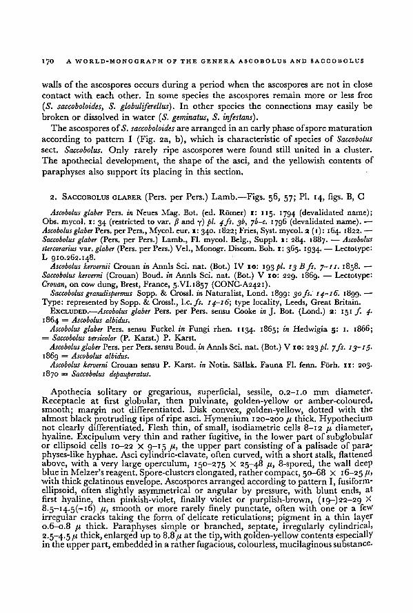
170 A WORLD-MONOGRAPH OF THE GENERA ASCOBOLUS AND SACCOBOLUS
walls of the ascospores occurs during a period when the ascospores are not in close
contact with each other. In some species the ascospores remain more or less free
(S. saccoboloides, S. globuliferellus). In other species the connections may easily be
broken or dissolved in water ( S. geminatus, S. infestans).Theascospores ofS. saccoboloides are arranged in an early phase ofspore maturation
according to pattern I (Fig. 2a, b), which is characteristic of species of Saccobolus
sect. Saccobolus. Only rarely ripe ascospores were found still united in a cluster.
The apothecial development, the shape of the asci, and the yellowish contents of
paraphyses also support its placing in this section.
2. SACCOBOLUS GLABER (Pers. per Pers.) Lamb.—Figs. 56, 57; Pl. 14, figs. B, C
Ascobolus glaber Pers. in Neues Mag. Bot. (ed. Romer) i: 115. 1794 (devalidated name);
Obs. mycol. 1: 34 (restricted to var. /? and y) pi. 4fs. 3b, yb-c. 1796 (devalidated name). —
Ascobolus glaber Pers. per Pers., Mycol. eur. 1: 340. 1822; Fries, Syst. mycol. 2 (1): 164. 1822. —
Saccobolus glaber (Pers. per Pers.) Lamb., Fl. mycol. Belg., Suppl. 1: 284. 1887. — Ascobolus
stercorarius var. glaber (Pers. per Pers.) Vel., Monogr. Discom. Boh. x: 365. 1934. -— Lectotype:L 910.262.148.
Ascobolus kervernii Crouan in Annls Sci. nat. (Bot.) IV 10: 193 pi. 13 B fs. 7-11. 1858. —
Saccobolus kerverni (Crouan) Boud. in Annls Sci. nat. (Bot.) V 10: 229. 1869. — Lectotype:
Crouan, on cow dung, Brest, France, 5.VI.1857 (CONC-A2421).Saccobolus granulispermus Sopp. & Crossl. in Naturalist, Lond. 1899: 30 fs. 14-16. 1899. —
Type: represented by Sopp. & Crossl., I.e. fs. 14-16; type locality, Leeds, Great Britain.
EXCLUDED.—Ascobolus glaber Pers. per Pers. sensu Cooke in J. Bot. (Lond.) 2: 151 f. 4-
1864 = Ascobolus albidus.
Ascobolus glaber Pers. sensu Fuckel in Fungi rhen. 1134. 1865; in Hedwigia 5: 1. 1866;= Saccobolus versicolor (P. Karst.) P. Karst.
Ascobolus glaber Pers. per Pers. sensuBoud. in Annls Sci. nat. (Bot.) V 10: 223 pi. 7fs. 13-15.
1869 = Ascobolus albidus.
Ascobolus kerverni Crouan sensu P. Karst. in Notis. Sallsk. Fauna Fl. fenn. Forh. 11: 203.
1870 = Saccobolus depauperatus.
Apothecia solitary or gregarious, superficial, sessile, 0.2-1.0 mm diameter.
Receptacle at first globular, then pulvinate, golden-yellow or amber-coloured,
smooth; margin not differentiated. Disk convex, golden-yellow, dotted with the
almost black protruding tips of ripe asci. Hymenium 120-200 41 thick. Hypotheciumnot clearly differentiated.Flesh thin, of small, isodiametric cells 8-12 41 diameter,
hyaline. Excipulum very thin and rather fugitive, in the lower part of subglobularor ellipsoid cells 10-22 X 9-15 the upper part consisting of a palisade of para-physes-like hyphae. Asci cylindric-clavate, often curved, with a short stalk, flattened
above, with a very large operculum, 150-275 X 25-48 41, 8-spored, the wall deepblue in Melzer's reagent. Spore-clusters elongated, rather compact, 50-68 X 16-25 46with thick gelatinous envelope. Ascospores arranged according to pattern I, fusiform-
ellipsoid, often slightly asymmetrical or angular by pressure, with blunt ends, at
first hyaline, then pinkish-violet, finally violet or purplish-brown, (19-)22-29 X
8.5-i4.5(-i6) 41, smooth or more rarely finely punctate, often with one or afew
irregular cracks taking the form of delicate reticulations; pigment in a thin layer0.6-0.8 41 thick. Paraphyses simple or branched, septate, irregularly cylindrical,2.5-4.5 thick, enlarged up to 8.841 at the tip, with golden-yellow contents especiallyin the upperpart, embedded in a rather fugacious, colourless, mucilaginous substance.
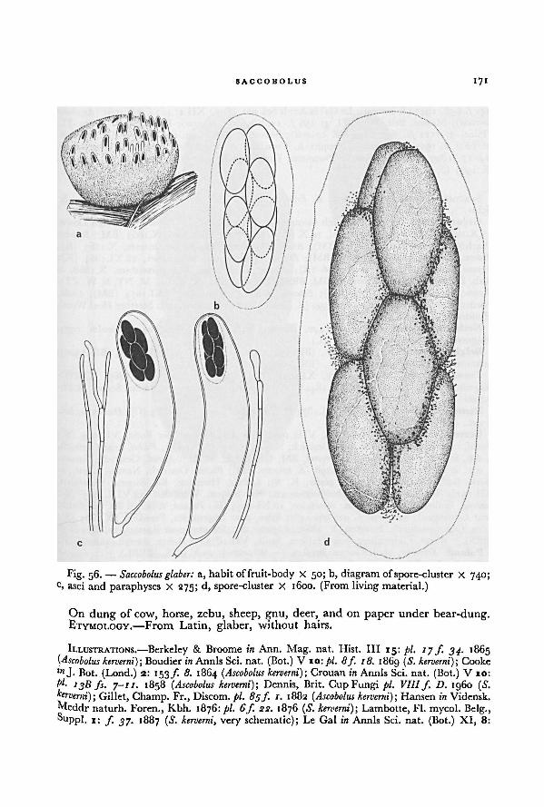
171SACCOBOLUS
On dung of cow, horse, zebu, sheep, gnu, deer, and on paper under bear-dung.ETYMOLOGY.—From Latin, glaber, without hairs.
ILLUSTRATIONS.—Berkeley & Broome in Ann. Mag. nat. Hist. ILL 15: pi. 17f. 34. 1865(Ascobolus kerverni); Boudier in Annls Sci. nat. (Bot.) V 10: pi. 8f. 18. 1869 ( S. kerverni); Cooke! "J- Hot. (Lond.) 2: 153 f. 8. 1864 (Ascobolus kerverni); Crouan in Annls Sci. nat. (Bot.) V 10:
t'l. 13B fs. 7-1 1. 1858 ( Ascobolus kerverni); Dennis, Brit. Cup Fungi pi. VIIIf. D. i960 (S.kerverni) ; Gillet, Champ. Fr., Discom. pi. 85f. 1. 1882 ( Ascobolus kerverni); Hansen in Vidensk.
Meddr naturh. Foren., Kbh. 1876: pi. 6f. 22. 1876 (S. kerverni); Lambotte, H. mycol. Belg.,i; f. 37. 1887 (S. kerverni, very schematic); Le Gal in Annls Sci. nat. (Bot.) XI, 8:
Fig. 56. —
a, habit of fruit-body X 50; b, diagram of spore-cluster X 740;c, asci and paraphyses X 275; d, spore-cluster X 1600. (From living material.)
Saccobolus glaber:
172 A WORLD-MONOGRAPH OF THE GENERA ASCOBOLUS AND SACCOBOLUS
242J 65E. 1947 ( S. kerverni); Le Gal in Annls Sci. nat. (Bot.) XII 1: 467f 8B. 1961 ( Ascobolus
kerverni); Massee, Brit. Fung.-Fl. 4: 156/. 11. 1895 (S. kerverni); Rehm, Rab. Krypt.-Fl.,(Pilze) 3: 1 hi fs. 6-7. 1896 ( S. kerverni) ; Seaver in Bull. Lab. nat. Hist. Univ. Iowa 6:
pi. 18 f 2. 1905 ( S. kerverni); Soppitt & Crossland in Naturalist, Yorkshire 1899: 31 fs.
14-17. i8gg ( S. granulispermus); Swanton, Fungi and how to know them (Lond.) pi. 6 f.8. 1909 ( S. kerverni).
SPECIMENS EXAMINED.—-Sweden: E. Fries, s. loc., s. dat. (Ascobolus glaber, "vidi Alb. &
Schw."), (S-A756).Great Britain: Broome, Warleigh Common, 5.X. 1863 (BM, E); Broome, Warleigh Down,
13.X.1863 (BM); Broome, Bathford, 14.X.1863 (K); Broome, Bathford, X.1863 (BM); Broome,
Warleigh, Sommerset, X.1863 (BM); Broome, Warleigh Down, Sommerset, X.1863 (K);Broome, Banner Down, X.1863 (BM); Broome, Batheaston, Sommerset, 28.XI.1864 (K);Broome, Bathford Down, 22.X.1866 (K); Broome, Batheaston, Sommersetshire, X. 1868, in
Rab. Fungi eur. 1313 (A. kerverni, BM, BRSL, FH, G, HBG, K, L, LE, M, NY, S, W, ZT);
Broome, Bathford, 21.X.1870 (BM); Broome, Warleigh Common, 7.XI.1874 (BM); Cooke,
Forden, s. dat., in Fungi brit. ed. 2, 399 (A. kerverni, E, K); Crossland, near Sterther Heel Wood,
Huddersfield, 24.IX.1906 (K).Netherlands: van Brummelen 1440, Elspeet, 24.VIII.1961 (L); van Brummelen 1399,
Singraven, Denekamp, 14.X. 1961 (L).
Belgium: Coemans, s. loc., s. dat., (BR-A327); Mouton, Beaufays, VII-VIII, (BR-A320);unknown collector, Gomze, s. dat., (BR-A290).
France: Boudier, Montmorency, XI.i8gi (PC); Crouan, s. loc., 20.XI.1869, (CONC-A2426); Roberge, Lebisey, 31.VII. 1842 (G); unknown collector 579, near Sedan (UPS-A2022).
Denmark: Hansen, s. loc., 26.VII.1874 (C); Hansen, s. loc., 7.VI.1875 (C); Hansen, s. loc.,
s. dat., (HBG-A406).Germany: Ade, near Weismain, VIII. 1908 (S); Ade, Weismainer Forst, VI. 1909 (S);
Fuckel, near Reicharthausen, near Oestrich, Nassau, s. dat. (G-A1059); Fuckel, near Oestrich,
s. dat., in Fungi rhen. 1846 (A. kerverni, BM, GRO, LE, M, W); Fuckel, Oestrich, Nassau,
s. dat., in Herb. Barbey-Boissier 1298 (S. kerverni, FH); Fuckel, Oestrich, Nassau, s. dat., in
Herb. Barbey-Boissier 13 10 (A. glaber, K, S); Ludwig, Hausling, Kr. Siegen, Westfalen,
VII.1926 (B); Ludwig, Langenholdinghausen, Kr. Siegen, Westfalen, 24.VIII.1945 (B);
Ludwig, Innkerhees, Kr. Siegen, Westfalen, 18.XI.1945 (B); Persoon, s. loc., s. dat. (probablynear Gottingen), lectotype L 910.262.148); Rehm, near Sugenheim, Franken, IX.1867 (S);Rehm, Obernesselbach, Franken, V. 1869 (A. glaber, S); Schroder, near Rastatt, 22.VIII.1878
(BRSL); Winter, Galgenberg near Halle a. Saale, VII. 1873, in Rehm, Ascom. 166 (B, S)-Poland: Schroder, zool. Garten Breslau (= Wroclaw), 22.V.1886, (BRSL).Czechoslovakia: JViessl, s. loc., s. dat. (M-A815); Petrak, Mahr.-Weisskirchen (= Hranice),
Welka, 21.IX.1912, in Fl. Boh. Mor. ser. 2, 233 (E, FH, S); Petrak 19, Mahr.-Weisskirchen
(= Hranice), IX. 1912 (S-A540); Petrak, Mahr.-Weisskirchen (= Hranice), IX. 1927 (ZT-A73); Svrcek, Brezany, Bohemia, 15.VI. 1946 (PR); Svrcek, Hostice near Nemysl Bohemia,
20.VIII.1946 (PR); Vacek, Brezany, Bohemia, 15.VI. 1946 (PR); Vacek, KarlStejn, Bohemia,
13.VII.1947 (PR).Austria: Britzelmayr, Peischelkopf, W. Tyrol, VII.1878 (S); Heimerl, Pressbaum near Wien,
III.1889, in Rehm, Ascom. 964 (S. kerverni, slide, HBG, M, S); Heimerl, s. loc., 1889, (S-A571)!Rehm, near Kiihtei, Oetz, Tyrol, VIII.1872, in Ascom. 165 (S. violascens, M, NY); Rehm,
near Praxmar and Sellrain, Tyrol, VII. 1872 (S); Rick, near Feldkirch, Vorarlberg, LX.
1897 (S).Italy: Bizzozzero, Hortus botanicus, Padua, VIII. 1886 (PAD); Bresadola, s. loc., V. 1886,
(S-A568); Bresadola, Gocciadoro, VI. 1896 (S); Cesati, Vercilli, s. dat., in Rabenhorst, Klotzsch.
Herb. viv. mycol. ed. 2, 1430 (A. glaber, L, UPS); Cavara, S. Giuseppe near Papia, s. dat.,
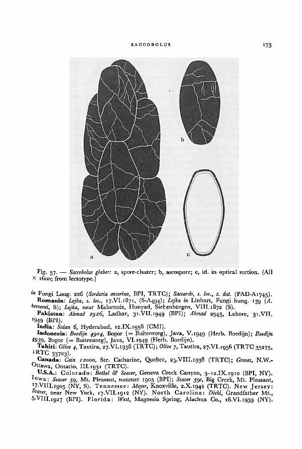
SACCOBOLUS 173
ln Fungi Long. 226 ( Sordaria anserina, BPI, TRTC); Saccardo, s. loc., s. dat. (PAD-A1745).Romania: Lojka, s. loc., 17.VI.1871, (S-A494); Lojka in Linhart, Fungi hung. 139 (A.
kerverni, S); Lojka, near Malornoiz, Hunyad, Sietenburgen, VIII. 1872 (S).Pakistan: Ahmad 2926, Ladhar, 31.VII.1949 (BPI); Ahmad 2943, Lahore, 31.VII.
>949 (BPI).India: Salan 6, Hyderabad, 12.IX.1958 (CMI).Indonesia: Boedijn 4904, Bogor (= Buitenzorg), Java, V.ig4g (Herb. Boedijn); Boedijn
4939, Bogor (= Buitenzorg), Java, VI. 1949 (Herb. Boedijn).Tahiti: Olive 4, Tautira, 27.VI.1956 (TRTC); Olive 7, Tautira, 27.VI.1956 (TRTC 35275,
TRTC 33723).Canada: Cain 12000, Ste. Catharine, Quebec, 25.VIII.1938 (TRTC); Groves, N.W.-
Ottawa, Ontario, III.193 1 (TRTC).U.S.A.: Colorado: Bethel & Seaver, Geneva Creek Canyon, 3-12.IX.1910 (BPI, NY).
' 0vv a: Seaver 55, Mt. Pleasant, summer 1905 (BPI); Seaver 39c, Big Creek, Mt. Pleasant,
'7'VlII.i905 (NY, S). Tennessee: Meyer, Knoxville, 2.X.ig42 (TRTC). New Jersey:Seaver, near New York, 17.VII.1912 (NY). North Carolina: Diehl, Grandfather Mt.,5*VlII.
1927 (BPI). Florida: West, Magnesia Spring, Alachua Co., 18.VI. 1939 (NY).
Fig. 57. — a, spore-cluster; b, ascospore; c, id. in optical section. (AllX 1600; from lectotype.)
Saccobolus glaber:
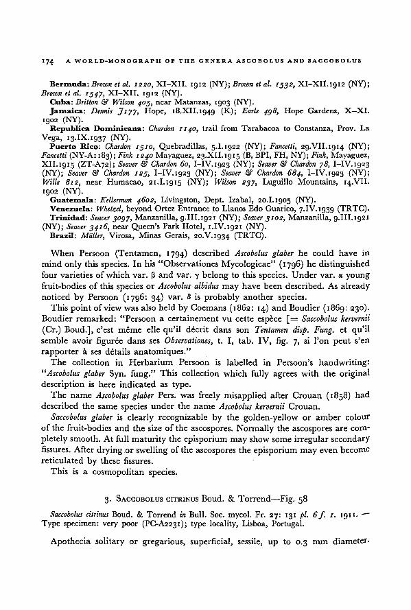
A WORLD-MONOGRAPH OF THE GENERA ASCOBOLUS AND SACCOBOLUS
174
Bermuda: Brown et al. 1220, XI-XII. 1912 (NY); Brown et al. 1532, XI-XII.1912 (NY);Brown et al. 1547, XI-XII. 1912 (NY).
Cuba: Britton & Wilson 403, near Matanzas, 1903 (NY).Jamaica: Dennis J177, Hope, 18.XII.1949 (K); Earle 438, Hope Gardens, X—XI.
1902 (NY).Republica Dominicana: Char don 1140, trail from Tarabacoa to Constanza, Prov. La
Vega, 13.IX.1937 (NY).Puerto Rico: Chardon 1310, Quebradillas, 5.1.1922 (NY); Fancetti, 29.VII.1914 (NY);
Fancetti (NY-A1183); Fink 1240 Mayaguez, 23.XII.1g15 (B, BPI, FH, NY); Fink, Mayaguez,XII.1915 (ZT-A72); Seaver & Chardon 60, I-IV.1923 (NY); Seaver & Chardon 78, I-IV.1923
(NY); Seaver & Chardon 123, I-IV.1923 (NY); Seaver & Chardon 684, I-IV.1923 (NY);Wille 812, near Humacao, 21.1.1915 (NY); Wilson 237, Luguillo Mountains, 14.VII.
1902 (NY).Guatemala: Kellerman 4602, Livingston, Dept. Izabal, 20.1. 1905 (NY).Venezuela: Whetzel, beyond Ortez Entrance to Llanos Edo Guarico, 7.IV.1939 (TRTC).Trinidad: Seaver 3037, Manzanilla, 9.III.1921 (NY); Seaver 3102, Manzanilla, g.III.1921
(NY); Seaver 3416, near Queen's Park Hotel, 1.IV.1921 (NY).Brazil: Miiller, Virosa, Minas Gerais, 20.V.1934 (TRTC).
When Persoon (Tentamen, 1794) described Ascobolus glaber he could have in
mind only this species. In his "Observationes Mycologicae" (1796) he distinguished
four varieties of which var. (3 and var. y belong to this species. Under var. a young
fruit-bodies of this species or Ascobolus albidus may have been described. As already
noticed by Persoon (1796: 34) var. 8 is probably another species.
This point of view was also held by Coemans (1862: 14) and Boudier (1869: 230).
Boudier remarked; "Persoon a certainementvu cette espece [= Saccobolus kervernii
(Gr.) Boud.], c'est meme elle qu'il decrit dans son Tentamen disp. Fung. et qu'ilsemble avoir figuree dans ses Observationes, t. I, tab. IV, fig. 7, si l'on peut s'en
rapporter a ses details anatomiques."The collection in Herbarium Persoon is labelled in Persoon's handwriting:
“Ascobolus glaber Syn. fung." This collection which fully agrees with the original
description is here indicated as type.
The name Ascobolus glaber Pers. was freely misapplied after Crouan (1858) had
described the same species under the name Ascobolus kervernii Crouan.
Saccobolus glaber is clearly recognizable by the golden-yellow or amber colour
of the fruit-bodies and the size of the ascospores. Normally the ascospores are com-
pletely smooth. At full maturity the episporium may show some irregular secondary
fissures. After drying or swelling of the ascospores the episporium may even become
reticulated by these fissures.
This is a cosmopolitan species.
3. SACCOBOLUS CITRINUS Boud. & Torrend—Fig. 58
Saccobolus citrinus Boud. & Torrend in Bull. Soc. mycol. Fr. 27: 131 pi. 6f. 1. 1911.—
*
Type specimen: very poor (PC-A2231); type locality, Lisboa, Portugal.
Apothecia solitary or gregarious, superficial, sessile, up to 0.3 mm diameter.
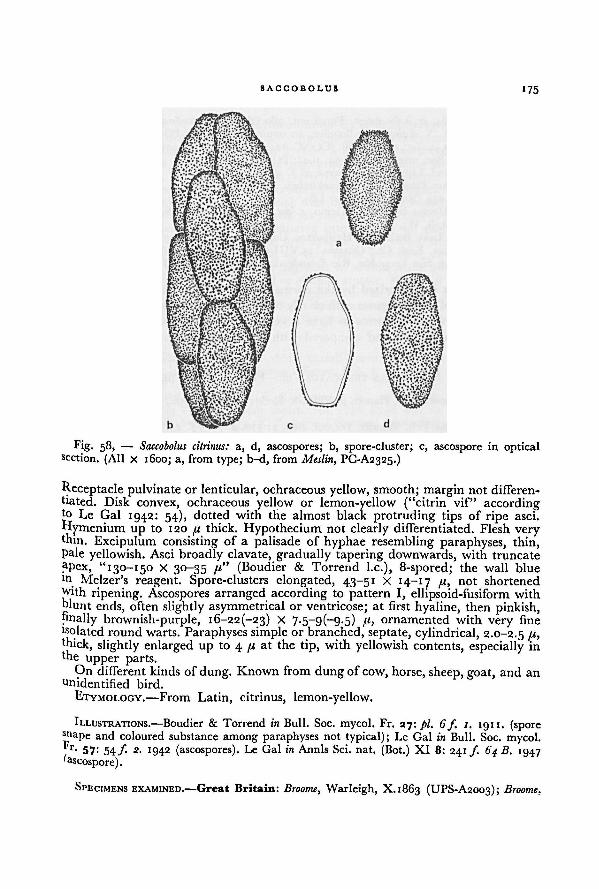
SACCOBOLUS175
Receptacle pulvinate or lenticular, ochraceous yellow, smooth; margin not differen-
tiated. Disk convex, ochraceous yellow or lemon-yellow ("citrin vif" accordingto Le Gal 1942: 54), dotted with the almost black protruding tips of ripe asci.
Hymenium up to 120 ft thick. Hypothecium not clearly differentiated. Flesh verythin. Excipulum consisting of a palisade of hyphae resembling paraphyses, thin,Pale yellowish. Asci broadly clavate, gradually tapering downwards, with truncate
apex, "130-150 X 30-35 /t" (Boudier & Torrend I.e.), 8-spored; the wall blue
m Melzer's reagent. Spore-clusters elongated, 43-51 X 14-17 71, not shortened
tvith ripening. Ascospores arranged according to pattern I, ellipsoid-fusiform with
blunt ends, often slightly asymmetrical or ventricose; at first hyaline, then pinkish,finally brownish-purple, 16-22(-23) X 7-5~9(-9-5) fi, ornamented with very fine
Wolated round warts. Paraphyses simple or branched, septate, cylindrical, 2.0-2.5 /*>thick, slightly enlarged up to 4 p, at the tip, with yellowish contents, especially inthe
upper parts.On differentkinds of dung. Known from dung of cow, horse, sheep, goat, and an
Unidentified bird.
ETYMOLOGY.—From Latin, citrinus, lemon-yellow.
ILLUSTRATIONS.—Boudier & Torrend in Bull. Soc. mycol. Fr. 27: pi. 6f. 1. 1911. (sporeshape and coloured substance among paraphyses not typical); Le Gal in Bull. Soc. mycol.* r- 57: 54/• 2- 1942 (ascospores). Le Gal in Annls Sci. nat. (Bot.) XI 8: 241 f 64 B. 1947'ascospore).
SPECIMENS EXAMINED.—Great Britain: Broome, Warleigh, X.1863 (UPS-A2003); Broome.
Fig. 58, — Saccobolus citrinus: a, d, ascospores; b, spore-cluster; c, ascospore in opticalsection. (All X 1600; a, from type; b-d, from Meslin, PC-A2325.)
176 A WORLD-MONOGRAPH OF THE GENERA ASCOBOLUS AND SACCOBOLUS
Batheaston, X. 1864, in Rabenhorst, Fungi eur. 780 ( Ascobolus depauperatus, W); Crossland,
Halifax, VII.1898 (S. distinguendus Boudier, an unpublished name, PC-A2232).France: Crouan, s. loc., 15.X.1868 (CONC-A2425); Meslin, Potigny, Calvados, s. dat.
(see Le Gal, Bull. Soc. mycol. Fr. 57: 54. 1941; PC-A2325).
Portugal: Torrend, Lisboa, II (holotype of S. citrinus, PC-A2231).Czechoslovakia: Petrak, Mahr.-Weisskirchen (= Hranice), Welka, 21.IX.1912, in Fl.
Boh. Mor., Pilze 233 ( Ascobolus kerverni, LE).Italy: Cavara, Hortus botanicus, Ticino, s. dat., in Fungi Long. 164 (S. neglectus, BM,
BPI, L, NY, PAD, S, W).Canada: Cain, Lake Boshkuny, Haliburton Co., Ontario, 14.IX.1931 (TRTC 34740);
Cain, Cedar Springs, Kent Co., Ontario, 13.VIII.1932 (TRTC 34738).Brazil: Theobald, Sao Leopoldo, Rio Grande do Sul, 11.III.1936 (TRTC 34739).
This species is characterized by the normally lemon-yellow colour of the fruit-
bodies, the size of the ascospores with clearly truncate ends, and the episporium with
isolated, round warts. It seems to have a very wide distribution.
The type was studied and compared with richer material by Le Gal (1942).
4. SACCOBOLUS TRUNCATUS Vel.—Fig. 59; Pl. 14, fig. D
Saccobolus nov. spec. E. C. Hansen in Vidensk. Meddr naturh. Foren., Kbh 1876: 87 pi. 6
f. 23. 1876.
Saccobolus truncatus Vel., Monogr. Discom. Boh. 1: 370, 2: pi. 5/. 23. 1934. —• Holotype:PR 152994-
Apothecia solitary or gregarious, superficial or in small cavities, sessile, o. 1-0.3 mm
diameter, about 0.1 mm high. Receptacle semiglobular or lenticular, pale yellow,
smooth; margin not differentiated. Disk convex, pale yellow, dotted with the
a-c, spore-clusters; d, ascospore in optical section. (AllX 1600; from holotype.)
Fig. 59.— Saccobolus truncatus:
SACCOBOLUS 177
purplish-black tips of ripe asci. Hymenium 50-80 [i thick. Hypothecium thin, of
closely compacted isodiametriccells 4.5-8 jx diameter. Flesh not clearly differentiated.
Excipulum consisting of a palisade of hyphae with subglobular cells 7-10 X 6-8 fx(textura globulosa), hyaline. Asci clavate, gradually tapering downwards into a
rather thick base, with truncate apex, 74-97 X 16-23 8-spored; the wall deepblue in Melzer's reagent. Spore-clusters elongated to very compact, (20-)29-42(-48)X (14—) 16—ig(-22) fx\ the longitudinal axis becoming increasingly shorter with
ripening; surrounded by a thick gelatinous envelope. Ascospores at first arrangedaccording to pattern I, gradually changing into pattern la, ellipsoid, sometimes
fusiform-ellipsoid, with blunt ends, at first hyaline, then violet, finally often violet-
brown, 14-17.5 x 7.5-8.5 fx, smooth or very finely punctate, sometimes with an
occasional fissure in the episporium; pigment in a thin layer about 0.3 fx thick.
Paraphyses slightly clavate, branched or simple, septate, 1.5-3.0 pi thick, the upperpart enlarged up to 4.5 pi, with yellow pigment in the enlarged parts.
On dung of cow, horse, sheep, rabbit, raven, and partridge.ETYMOLOGY.—From Latin, truncatus, shortened, ending abruptly as if cut off
at the tip.
ILLUSTRATIONS.—Hansen in Vidensk. Meddr naturh. Foren., Kbh. 1876: pi. 6 f. 23. 1876(Saccobolus nov. spec.); Velenovsky, Monogr. Discom. Boh. 2: pi. 3f. 23. 1934.
SPECIMENS EXAMINED.—Denmark: E. C. Hansen, s. loc., 29.VI.1874 (= Saccobolus nov.
spec. E. C. Hansen in Vidensk. Meddr naturh. For. 1876: 87 pi. 6 f. 23. 1876; C-A58).Netherlands: van Brummelen 39.18, Zoological Garden, Amsterdam, 24.VII.1959 (L).Germany: Winter, Galgenberg, near Halle a.d. Saale, VII. 1873, in Rehm, Ascom. 166
(S. kerverni, E).Czechoslovakia: Velenovsky, Mnichovice, Bohemia, VI. 1931 (PR 152997); Velenovsky,
on raven dung, Kunice, Mnichovice, Bohemia, VII.1931 (holotype of S. truncatus, PR 152994).Austria: Demelius 1180,
,Schöngrabern, near Unterwaltersdorf, Wiener Neustadt, Nieder-
Oesterreich, 24.IX.1917 (W).Thailand: van Brummelen 1779,
, ,
Pha Nok Khao, S. of Loey, prov. Udawn, 14.XI.1963(from culture; L).
Canada: Bisby, M.A.C., Winnipeg, Manitoba, 2.IX.1935 (TRTC 34716); Cain,- .
, , Sing-harnpton, Grey Co., Ontario, 5.VII.ig30 (TRTC 34735); Cain,
„ ,
North Bay, Ontario, 3.IX. 1931
(TRTC 34733); Cain, Singhampton, Grey Co., Ontario, 11.II. 1932 (NY-A1202); Cain,Uderton, Middlesex Co., Ontario, 12.VII.1932 (TRTC 34737).
U.S.A.: New York: Cain, McLean, near Ithaca, 5.IX. 1952 (TRTC 34750). Virginia:L. Meyer 14J 45, Mountain Lake Biological Station, 8.VIII. 1942 (TRTC).
Peru: G. W. Martin 6280, Talara, 5.IX.1945 (NY-A1229).
This is related to S. citrinus and S. minimus. It is sufficiently characterized by the
Pale yellow colour, the shape and size of the ascospores, and the finely punctate0r smooth episporium.
With maturity the ascospore packages in this species are somewhat shortened,and the arrangement of the ascospores, which is at first very regular, becomes
rather irregular at full maturity. In S. portoricensis the ascospores are pressed togetherstill more tightly.
This widely distributed species is easily overlooked by its hidden and often
solitary manner of growth.
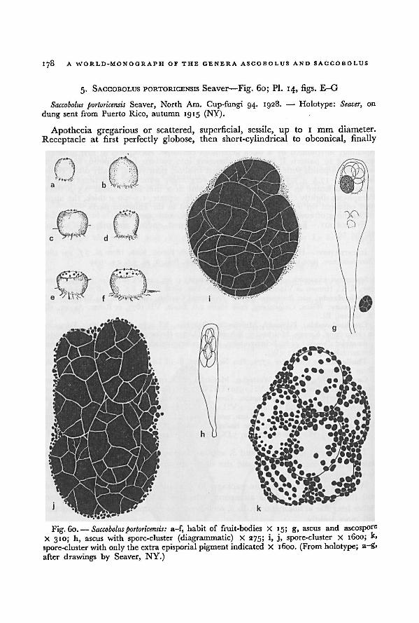
1 7 8 A WORLD-MONOGRAPH OF THE GENERA ASCOBOLUS AND SACCOBOLUS
5. SACCOBOLUS PORTORICENSIS Seaver—Fig. 60; Pl. 14, figs. E-G
Saccobolus portoricensis Seaver, North Am. Cup-fungi 94. 1928. — Holotype: Seaver, on
dung sent from Puerto Rico, autumn 1915 (NY).
Apothecia gregarious or scattered, superficial, sessile, up to 1 rum diameter.
Receptacle at first perfectly globose, then short-cylindrical to obconical, finally
a-f, habit of fruit-bodies X 15; g, ascus and ascospore
X 310; h, ascus with spore-cluster (diagrammatic) X 275; i, j, spore-cluster X 1600; k,
spore-cluster with only the extra episporial pigment indicated X 1600. (From holotype; a-g,
after drawings by Seaver, NY.)
Fig. 60. — Saccobolusportoricensis:
SACCOBOLUS 179
discoid, pale-amber, smooth; margin not differentiated. Disk flat, then convex,
at first similar in colour to the outside of the apothecium, dotted over with the
protruding asci, finally almost entirely black. Hymenium about 200 fx thick. Hypo-thecium not clearly differentiated. Flesh 80-200 //, thick, of rather loosely united
roundish or angular cells, hyaline. Excipulum in the lower part not differentiated
from the flesh; in the upper part of parallel, paraphysoid hyphae, not clearly distin-
guishable from paraphyses. Asci clavate, tapering downwards into a rather long,slender base, with truncate apex, 170-200 X 35-40 fx, 8-spored. Spore-clustersat first elongated, finally subglobular, closely compacted and opaque or nearlyso, surrounded by many purple granules, 34-44 X 22-30 fx. Ascospores arrangedaccording to pattern I, then pressed together in an irregular, subglobular cluster;at first fusiform-ellipsoid, finally subglobular; at first hyaline, then purple, finallypale-brown; mature spores, 15—17.5 X 10-12.5 a, shorter and broader than the
almost mature ones, 19-21 X 9-10 /u; ornamentation consisting of very delicate
reticulations; pigment in a 1.0-1.2 [X thick layer and in granules up to 1 ft diameter
surrounding the spore-clusters. Paraphyses slender, simple, septate, about 3 /x thick,not or scarcely enlarged above, filled with orange granules.
On dung of unidentified animal.
ETYMOLOGY.—From Porto Rico (= Puerto Rico).
SPECIMENS EXAMINED.—Puerto Rico: Seaver, on dung from Puerto Rico, autumn 19 15
(holotype of S. portoricensis, NY-A1185).
This species, which is only known from the type collection, is sufficiently charac-
terized by the short-ellipsoid to subglobular ascospores and the thick episporiumwith a net-work of crevices.
The ascospores are at first regularly arranged in an elongated cluster accordingto pattern I. Soon, however, the cluster is shortened and becomes almost globularat maturity. The shape of the ascospores also changes during this process from
ellipsoid to short-ellipsoid or subglobular.
Apart from a rather thick episporium, granules of extra-episporial pigment are
also formed in the asci and surround the spore-clusters.
6. SACCOBOLUS MINIMUS Vel.—Fig. 61; PI. 14, figs. H, I, PI. 15, figs. A, B
Saccobolus minimus Vel., Monogr. Discom. Boh. 1: 370 2: pi. 5 f 26._ .
1934- — Holotype:PR 150081.
Apothecia solitary or gregarious, superficial, sessile, o. 1-0.2 [X diameter. Receptacleat first globular, then pulvinate, transparent amber-coloured to ochraceous yellow("melina, vitreo-pellucida" according to Velenovsky, 1934: 370), smooth; marginnot differentiated. Disk convex, amber-coloured to golden-yellow, dotted with the
almost black protruding tips of ripe asci. Hymenium 50-60 fx thick. Hypotheciumtot clearly differentiated.Flesh thin, ofsubglobular cells5-12 ft diameter. Excipulumvery thin, and rather fugitive, in the lower part of small subglobular cells, in theuPper part consisting of a palisade of paraphyse-like hyphae. Asci cylindric-clavate,gradually tapering downwards into a rather thick base, with truncate apex, 50-60 XI4y I6 fx, 8-spored, the wall blue in Melzer's reagent. Spore-clusters compact,
pllipsoid to elongated, 29-33 x 12-1 5 /"» surrounded by a common hyaline, mucilag-Jtious envelope. Ascospores arranged according to pattern I, ellipsoid to fusiform-
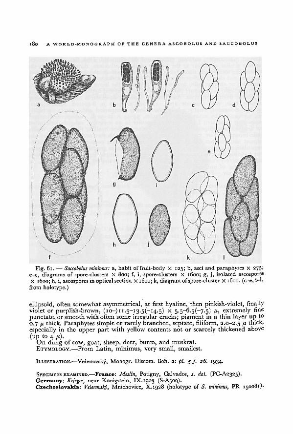
180 A WORLD-MONOGRAPH OF THE GENERA ASCOBOLUS AND SACCOBOLUS
ellipsoid, often somewhat asymmetrical, at first hyaline, then pinkish-violet, finally
violet or purplish-brown, (10-) 11-5— 13-5(—14-5) X 5-5-6.5(-7-5) ft, extremely fine
punctate, or smooth withoften some irregular cracks; pigment in a thin layer upto
0.7 f.i thick. Paraphyses simple or rarely branched, septate, filiform, 2.0-2.5 fi thick,
especially in the upper part with yellow contents not or scarcely thickened above
(up to 4 fl).On dung of cow, goat, sheep, deer, burro, and muskrat.
ETYMOLOGY.—From Latin, minimus, very small, smallest.
ILLUSTRATION.—Velenovsky, Monogr. Discom. Boh. 2: pi. 5 f 26. '934-
SPECIMENS EXAMINED.—France: Meslin, Potigny, Calvados, s. dat. (PC-A2325).
Germany: Krieger, near Konigstein, IX. 1903 (S-A509).Czechoslovakia: Velenovsky, Mnichovice, X.1928 (holotype of S. minimus, PR 150081)-
a, habit of fruit-body X 125; b, asci and paraphyses X 275;
c-e, diagrams of spore-clusters X 800; f, l, spore-clusters X 1600; g, j, isolated ascospores
X 1600; h, i, ascospores in optical section X 1600; k, diagramofspore-cluster X 1600. (c-e, i-l,
from holotype.)
Saccobolus minimus:Fig. 61.—
181SACCOBOLUS
Austria: Demelius 762, Voslau, Nieder-Oesterreich, X.igi2 (W 1808).Poland: Schroeter, near Breslau (= Wroclaw), VII.1880 (BRSL-A1652).Thailand: van Brummelen 1789, Khao Dai Kuad, prov. Surat, 26.XI.1963 (L); van
Brummelen 1783, Doi Chieng Dao, prov. Payap, 22.XI. 1963 (L); van Brummelen 1781, Pha» u ' i / i » J \ —/ > — —■ - / "•?
A lia
Nok Khao, S. of Loey, prov. Udawn, 21.XI. 1963 (L).Hawaii: van Brummelen 1465, Mt. Kaala, Oahu, 3.V.1962 (L); van Brummelen 1468, Mt.
K-aala, Oahu, 9.V.1962 (L); van Brummelen 1462, Mt. Kaala, Oahu, io.V. 1962 (L).Canada:
~
Cain, Storeleigh, Muskoka, Ontario, 18.VIII.1932 (TRTC 34717); Cain, Pickerel
L.P., Lake Timagami, Ontario, 10.VIII.1933 (TRTG 34724); Cain,,
Lake Timagami, Ontario,a7.II.1934 (NY); Cain 6968, Pine River, Lac St. Joseph, Quebec, 25.VIII.1938 (TRTC);Cain 6978, Pine River, Lac St. Joseph, Quebec, 26.VIII.1938 (TRTC); Cain 6930,
, CampMercier, Laurentides Nat. Park, Quebec, 27.VIII.1938 (TRTC).
U.S.A.: Maine: Thaxter, Kittery Point, IX.1891 (FH-A3077).Ecuador: G. W. Martin 6277, Salinas, 3.IX.1945 (NY); G. W. Martin 6281,
... . ,Islet Baltra,
Galapagos, 6.IX. 1945 (NY).
Because of the small fruit-bodies, this fungus has been collected only rarely.Probably it will prove to have a world-wide distribution.
It is easily recognizable by its small ascospores. Saccobolus depauperatus has ascosporesofabout the same size, but the fruit-bodies are white or pale violet instead of amber
or ochraceous yellow. Furthermore the arrangement of the ascospores is different.
Yet these two species show a certain degree of relationship. In S. depauperatus an
ascospore-cluster can occasionally be found in which the spores are arranged
according to pattern I. Sometimes the contents of paraphyses are faintly coloured.
II. SACCOBOLUS sect. ERIOBOLUS Sacc.
Saccobolus sect. Eriobolus Sacc., Syll. Fung. 8: 527. 1889. — Monotype: Saccobolus caesariatus
Benny apud Phillips.
Receptacle and disk white, pale violet, violet or brown. Ascospores 8 or some-
times 4; ellipsoid or fusiform-ellipsoid, very rarely subglobular or globular, oftenthe ends slightly truncate, often asymmetrical, ventriculose or somewhat triangular;arranged differently from pattern I or la (Fig. 2a-c). Paraphyses mostly colourless,Without yellowish or orangecontents. Sometimes with violetor brownish, amorphous,mtercellular pigment in the outer layers of the excipulum and between the tips of
Paraphyses.Fimicolous, more rarely on textiles, rotten vegetable debris, board and plaster
ETYMOLOGY.—From Greek, £piov, wool and (IaXX<o, to throw.
In this section twelve species can be distinguished, among which is Saccobolus
versicolor, an extremely variable species. Formerly, different variations of this specieshad been given different specific names. However, studies on the variation of the
different characters in many cultures and rich collections have shown that they are
tbere variations bridged by very gradual transitions.
The ascospores of the species in this section are arranged according to various
Patterns. Pattern II and III (Fig. 2f-h) occur in at least seven species. Two special
182 A WORLD-MONOGRAPH OF TOE GENERA ASCOBOLUS AND SACCOBOLUS
patterns (Va and Vb, Fig. 2d, e) are found in S. quadrisporus, which has 4-spored asci.
In S. globuliferellus and S. dilutellus the spore-cluster is shortly ellipsoid or sub-
globular. In my opinion, the patterns found in the packages of these species are
only extremely contracted variations of pattern II or III (Fig. 2i, j).
A quite different arrangement is found in the spore-clusters of S. geminatus and
S. infestans. Here the longitudinal axes of the ascospores is perpendicular to the
longitudinal axis of the package (pattern VI). These two very rare species mightdeserve a separate section. At the moment, however, our knowledge of the develop-
ment of their ascospores is insufficient to determine their relationship with other
species of Saccobolus sect. Eriobolus.
KEY TO THE SPECIES OF SACCOBOLUS SECT. ERIOBOLUS
i. Ascus with 4 spores; ellipsoid or slightly triangular; 16.5-19.5 X 9.5-10.5 fi
12. Saccobolus quadrisporus, p. 196
1. Ascus with 8 spores:
2. Ascospores at first arranged according to pattern II or III. Spore-clusters
finally sometimes becoming shortened or somewhat irregular but never loose
or subglobular:
3. Episporium smooth or finely granular, sometimes with small pits or with
an incomplete net-work of fine fissures. Ascospores not shortened at
maturity:
4. Receptacle outside with bunches of hyphae to form tapering squamules.
Excipulum of textura angularis .... 9. Saccobolus caesariatus, p. 191
4. Receptacle completely smooth. Excipulum of textura globulosa or
intricata:
5. Apothecia 0.1-0.3 mm diameter. Excipulum of textura globulosa,
rarely with intercellular, amorphous pigment. Central asci 60-95 -*■
15-20 fi. Ascospores 10-14.5 x 5~7-5 Episporium smooth or
finely granular 7. Saccobolus depauperatus, p. 183
5. Apothecia 0.2-2.0 mm diameter. Excipulum predominantly of
textura intricata, mostly with intercellular, amorphous pigment in
excipulum and between the tips of paraphyses. Central asci 100-145*
22-37 P- Ascospores 13-21.5 X 6.5-9.5 /"• Episporium smooth, some-
times with small pits, with an incomplete net-work of fissures or
finely granular 8. Saccobolus versicolor, p.186
3. Episporium coarsely or finely warted or with a coarse net-work of fissures;
sometimes very thick. Ascospores sometimes shortened at maturity:
6. Episporium very coarsely warted or reticulated, 1—3 /( thick. Ascospores
1 7.5-23 X 8.5-10 qi 10. Saccobolus beckii, p. 193
6. Episporium not so roughened and thick. Ascospores smaller:
7. Apothecia lenticular or pulvinate. Spore-clusters at maturity of the
ascospores shortened, by which the arrangement becomes more or
183SACCOBOLUS
less irregular (pattern Ilia, Fig. 2i). Episporium coarsely or finelywarted:
8. Apothecia 0.3-0.8 mm diameter. Ascospores ellipsoid or fusiform-
ellipsoid; 13.5-18 X 7.5-9.5 fi; firmly united in the clusters. With
intercellular, amorphous, brown pigment in excipulum and
between the tips of paraphyses . .
11. Saccobolus obscurus, p. 194
8. Apothecia 0.1-0.3 mm diameter. Ascospores broadly ellipsoid or
fusiform-ellipsoid; 10.5-14 X 7-9 /(; rather loosely united in the
clusters. Without intercellular pigment in excipulum and
hymenium 13. Saccobolus thaxteri, p. 197
7. Apothecia cylindrical or subglobular. Spore-clusters at maturity not
shortened, always arranged according to pattern II. Episporiumcoarsely warted. Ascospores 14-16 X 8-9 pt. With intercellular,amorphous pigment in excipulum and between the tips of paraphyses.
14. Saccobolus verrucisporus, p. 1982. Ascospores not arranged according to pattern II or III; at maturity according
to pattern IV or VI, or rather loosely in a shortly ellipsoid or subglobularcluster:
9. Spore-cluster at maturity subglobular or shortly ellipsoid. Ascospores
11.5-14.5 X 6-7.5
10. Ascospores loosely united in the cluster, mostly at all sides covered with
fine warts 15. Saccobolus globuliferellus, p. 200
10. Ascospores closely compacted in a subglobular cluster; only the
exposed surfaces covered with fine and coarse warts
16. Saccobolus dilutellus, p. 201
9. Ascospores arranged in a package according to pattern VI (axes of asco-
spores at right angles with axis of package, Fig. 2k-p):11. Ascospores firmly united in pairs (Fig. 2q); 10.5-13 x 5-6.5 pi. The
pairs rather loosely united in a cylindrical pile. Episporium finelywarted 17. Saccobolus geminatus, p. 202
11. Ascospores arranged according to package VI, not firmly united in
pairs; 9-1 1 X 5-6.5 7 1. Episporium granular
18. Saccobolus infestans, p. 204
7. SACCOBOLUS DEPAUPERATUS (Berk. & Broome) E. C. Hansen—Fig. 62
Ascobolus depauparatus Berk. & Broome in Ann. Mag. nat. Hist. Ill 15: 448 pi. 14 J 6.■865; Broome in Rab., Fungi eur. No. 780. 1865. — Saccobolus depauperatus (Berk. & Broome)E. G. Hansen in Vid. Meddr dansk naturh. Foren. 1876: 87. 1876. — Lectotype: Broome(Ao. jig), Hanham, 31.X.1864 (K, BM, E).
Saccobolus neglectus Boud. in Annls Sci. nat. (Bot.) V 10: 231 pi. gf. 20. 1869. — Ascobolusneglectus (Boud.) Phill. & Plowr. in Grevillea 10: 69. 1881; Gillet, Discom. 141. 1883. —
Ascobolus violascens var. neglectus (Boud.) Quel., Ench. Fung. 294. 1886. — Type: representedby Boudier I.e.: pi. gf. 20; type locality, Montmorency, France.
184 A WORLD-MONOGRAPH OF THE GENERA ASCOBOLUS AND SACCOBOLUS
Saccobolus depauperatus forma denigratus Rehm, Ascomyceten exs. No. 1271. 1899 (nomen
nudum); Rehm in Hedwigia 38: (243). 1899 (with diagnosis). — Type distribution: von
Lagerheim in Rehm, Ascom. 1271.
Saccobolus aparaphysatus Speg. in An. Mus. nac. Hist. nat. B. Aires 6: 308. 1899. — Holotype:LPS 26139.
Apothecia scattered or gregarious, superficial, sessile, o.io-o.2o(-o.25) mm
diameter, 0.10-0.30 mm high. Receptacle at first subglobular, then more expandedand becoming pulvinate or turbinate-hemispherical, often with a narrow base,
white, then pale violet, smooth; margin not differentiated. Disk convex, white,then pale vinous to violet, dotted with the dark purplish protruding tips of ripe asci.
Hymenium 7o-go(-ioo) pi thick. Hypothecium thin, of closely compacted iso-
diametric cells 3-6 pi diameter. Flesh not differentiated or very thin, of globularcells 6-14 pi diameter. Excipulum very thin, consisting of subglobular cells 6-12 pi
diameter (textura globulosa), hyaline or with pale purplish, intercellular, amorphouspigment. Asci broadly clavate, gradually tapering downwards into a rather thick
base, with abruptly truncate apex, 60-95 x (12-) 15-20 pi (the longer ones near
the margin of the disk), 8-spored, the wall deep blue in Melzer's reagent. Spore-clusters compact, elongated, 28-37 x I0-I 3 I1- Ascospores arranged according to
pattern II or III, very rarely in a part of the asci according to pattern I, ellipsoidor fusiform-ellipsoid, sometimes somewhat asymmetrical, with blunt ends, at first
hyaline, then pinkish-violet, finally dark violet or purplish-brown, 10-14.5 X
5-7.5 pi, smooth or extremely fine granular; pigment in a thin or a thick layer(0.5-1. 1 pi thick). Paraphyses sometimes rather scarce, simple, septate, filiform,
1.5-3.2 pi thick, colourless or faintly coloured, slightly enlarged up to 4-5(-6) pi
at the tip, often slightly curved above, without mucus.
On dung of horse, cow, sheep, deer, porcupine, elk, chamois, elephant, rabbit,and hare, also on the mud plaster of a wall.
ETYMOLOGY.—From Latin, de-, prefix signifying down, away, off and paupero,
to impoverish: poorly developed.
ILLUSTRATIONS.—Berkeley & Broome in Ann. Mag. nat. Hist. Ill 15: pi. 14 f. 6. 1865
(Ascobolus); Boudier in Annls Sci. nat. (Bot.) V 10: pi. g f. 20. 1869 ( S. neglectus); van
Brummelen in Persoonia 2: 197 f. 3. 1962 (S. aparaphysatus, ascospores); Hansen in Vid.
Meddr. dansk. naturh. Foren. 1876: pi. 6fs. 14-21. 1876; Heimerl in Jber. k.k. Ober-Real-
schule Bezirke Sechshaus Wien 15: pi. 1 f. 6. 1889.
SPECIMENS EXAMINED.—Sweden: Lagerheim, Nacka near Stockholm, in Rehm, Ascom.
1271 (type distribution of S. depauperatus forma denigratus, B, BM, HBG, LE, M, S, W).Finland: Karsten, Wasa, 20.VIII. 1867 (H).Great Britain: Broome, Hanham, 24.X. 1864 ( Ascobolus depauperatus, BM-A3012); Broome,
Bathford-Down, 28.X.1864 ( Ascobolus vinosus, K-A1914); Broome, Hanham, 31.X.1864 (lecto-
type of Ascobolus depauperatus, BM-A3010, BM-A3013, E-A136, K-A2448); Broome, Batheaston,
Somerset, 28.XI. 1864 ( Ascobolus depauperatus, K-A2450); Broome, 7.XII. 1864 ( Ascobolus depau-peratus, K-A2451); Cooke, Forden, in Fungi brit. ed. II, 399 (Ascobolus kerverni, PAD); Crossland,
Stokesley, Cleveland, 8.VIII.1899 (K); Dennis, West Sandwick, Islet Yell, Shetland Islands,
5.IX. 1952 (K); unknown collector, Kew, 1.IX.1916 (K-A2496).Netherlands: van Brummelen 623, Leiden, 6.VIII.1958 (L); van Brummelen 642, Leiden,
20.VIII.1958 (L).France: de Croculs, near Toulon, V. 1927 (PC); Crouan, s. loc., 10.X.1868 (CONC-A2436).Denmark: E. Miiller, Vintre holler, Seeland, 13.IX.1952 (ZT); E. C. Hansen, 15. VIII.1874
(C-A60).Germany: Ludwig, Langenholdinghausen, Kr. Siegen, Prov. Westfalen, 23.VIII. 1945
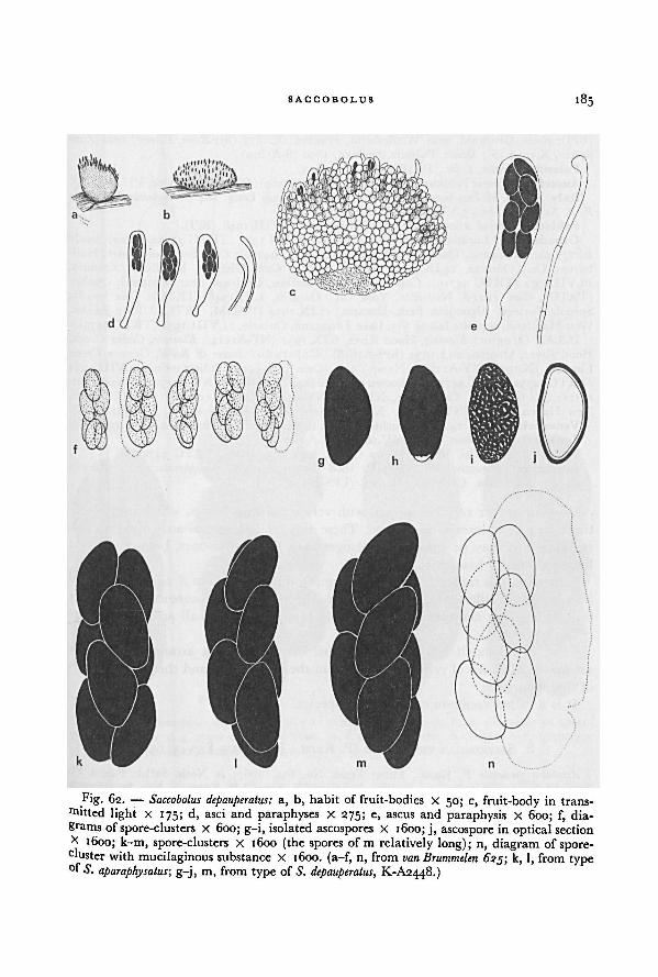
SACCOBOLUS
185
g-j, m, from type of S. depauperatus, K-A2448.)S. aparaphysatus;ofk, l, from typevan Brummelen 625;
Saccobolus depauperatus: a, b, habit of fruit-bodies X 50; c, fruit-body in trans-
mitted light X 175; d, asci and paraphyses X 275; e, ascus and paraphysis X 600; f, dia-grams ofspore-clusters X 600; g-i, isolated ascospores X 1600; j, ascospore in optical section
x 1600; k-m, spore-clusters X 1600 (the spores of m relatively long); n, diagram of spore-cluster with mucilaginous substance X 1600. (a-f, n, from
Fig. 62. —
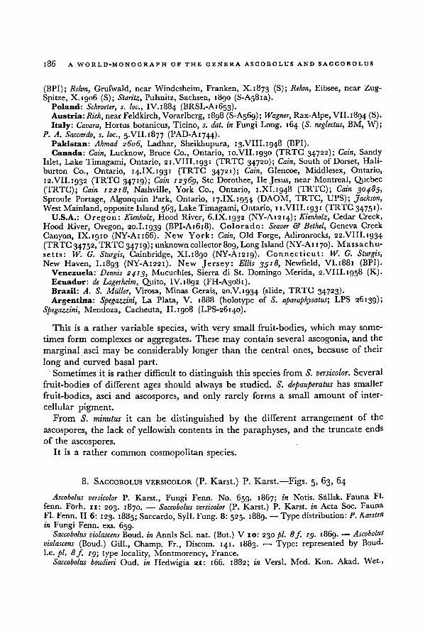
186 A WORLD-MONOGRAPH OF THE GENERA ASCOBOLUS AND SACCOBOLUS
(BPI); Rehm, Grufwald, near Windesheim, Franken, X. 1873 (S); Rehm, Eibsee, near Zug-
Spitze, X. 1906 (S); Staritz, Pulsnitz, Sachsen, 1890 (S-A58ia).Poland: Schroeter, s. loc., IV.I884 (BRSL-A1653).Austria: Rick, near Feldkirch, Vorarlberg, 1898 (S-A569); Wagner, Rax-Alpe, VII. 1894 (S).
Italy: Cavara, Hortus botanicus, Ticino, s. dal. in Fungi Long. 164 ( S. neglectus, BM, W);P. A. Saccardo, s. loc., 5.VII.1877 (PAD-AI744)-
Pakistan: Ahmad 2606, Ladhar, Sheikhupura, 13.VIII. 1948 (BPI).Canada: Cain, Lucknow, Bruce Co., Ontario, 10.VII.1930 (TRTC 34722); Cain, Sandy
Islet, Lake Timagami, Ontario, 21.VIII.1931 (TRTG 34720); Cain, South of Dorset, Hali-
burton Co., Ontario, 14.IX.1931 (TRTC 34721); Cain, Glencoe, Middlesex, Ontario,
12.VII.1932 (TRTC 34719); Cain 12369, Ste Dorothee, lie Jesus, near Montreal, Quebec
(TRTG); Cain 12218, Nashville, York Co., Ontario, 1.XI. 1948 (TRTC); Cain 304.85,
Sproule Portage, Algonquin Park, Ontario, 17.IX. 1954 (DAOM, TRTC, UPS); Jackson,West Mainland, opposite Island563, Lake Timagami, Ontario, 11.VIII.1931 (TRTC 3475')•
U.S.A.: Oregon: Kienholz, Hood River, 6.IX. 1932 (NY-A1214); Kienholz, Cedar Creek,
Hood River, Oregon, 20.1.1939 (BPI-A1618). Colorado: Seaver & Bethel, Geneva Greek
Canyon, IX. 1910 (NY-A1166). New York: Cain, Old Forge, Adironrocks, 22.VIII. 1934
(TRTC 34752, TRTC 34719); unknown collector809, Long Island (NY-Ai 170). Massachu-
setts : W. G. Sturgis, Cambridge, XI.1890 (NY-A1219). Connecticut: W. G. Sturgis,New Haven, I.1893 (NY-A1221). New Jersey: Ellis 3518, Newfield, VI.1881 (BPI).
Venezuela: Dennis 2413, Mucuchies, Sierra di St. Domingo Merida, 2.VIII.1958 (K).Ecuador: de Lagerheim, Quito, IV. 1892 (FH-A3081).Brazil: A. S. Miiller, Virosa, Minas Gerais, 20.V. 1934 (slide, TRTG 34723).
Argentina: Spegazzini, La Plata, V. 1888 (holotype of S. aparaphysatus; LPS 26139);
Spegazzini, Mendoza, Cacheuta, II.1908 (LPS-26140).
This is a rather variable species, with very small fruit-bodies, which may some-
times form complexes or aggregates. These may contain several ascogonia, and the
marginal asci may be considerably longer than the central ones, because of their
long and curved basal part.
Sometimes it is rather difficult to distinguish this species from S. versicolor. Several
fruit-bodies of different ages should always be studied. S. depauperatus has smaller
fruit-bodies, asci and ascospores, and only rarely forms a small amount of inter-
cellular pigment.From S. minutus it can be distinguished by the different arrangement of the
ascospores, the lack of yellowish contents in the paraphyses, and the truncate ends
of the ascospores.
It is a rather common cosmopolitan species.
8. SACCOBOLUS VERSICOLOR (P. Karst.) P. Karst.—Figs. 5, 63, 64
Ascobolus versicolor P. Karst., Fungi Fenn. No. 659. 1867; in Notis. Sallsk. Fauna Fl-
fenn. Forh. 11: 203. 1870. — Saccobolus versicolor (P. Karst.) P. Karst. in Acta Soc. Fauna
Fl. Fenn. II 6: 123. 1885; Saccardo, Syll. Fung. 8: 525. 1889. — Type distribution:P. Karsten
in Fungi Fenn. exs. 659.Saccobolus violascens Boud. in Annls Sci. nat. (Bot.) V 10: 230 pi. 8f. ig. 1869. — Ascobolus
violascens (Boud.) Gill., Champ. Fr., Discom. 141. 1883. — Type: represented by Boud.
I.e. pi. 8 f. ig; type locality, Montmorency, France.
Saccobolus boudieri Oud. in Hedwigia 21: 166. 1882; in Versl. Med. Kon. Akad. Wet.,
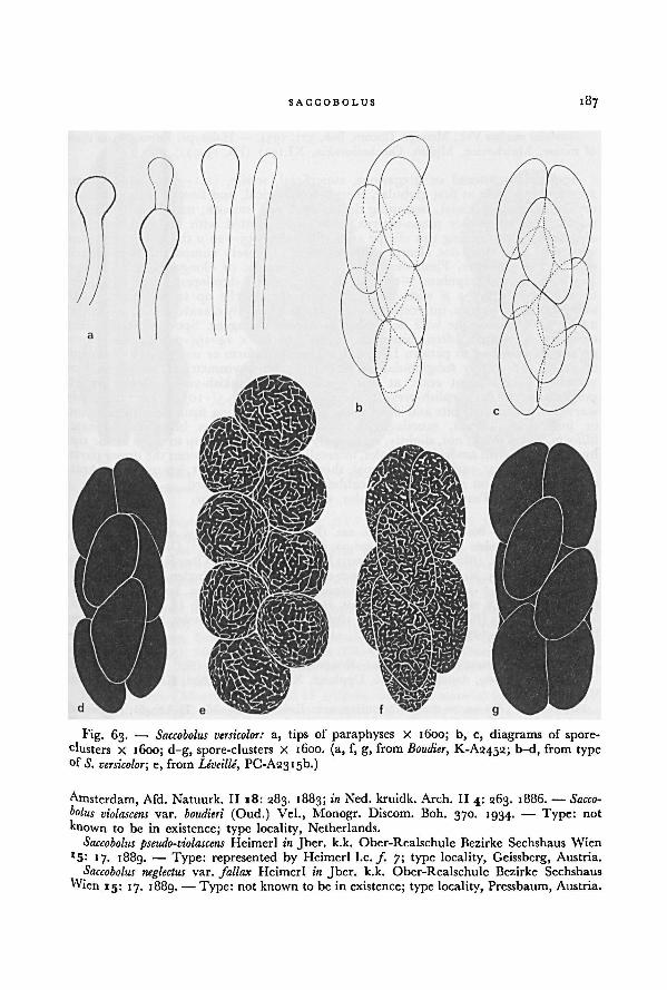
187SACCOBOLUS
Amsterdam, Afd. Natuurk. II 18: 283. 1883; in Ned. kruidk. Arch. II4: 263. 1886. — Sacco-
bolus violascens var. boudieri (Oud.) Vel., Monogr. Discom. Boh. 370. 1934.— Type: not
known to be in existence; type locality, Netherlands.
Saccobolus pseudo-violascens Heimerl in Jber. k.k. Ober-Realschule Bezirke Sechshaus Wien
*5 : 17. 1889. — Type: represented by Heimerl I.e. f 7; type locality, Geissberg, Austria.
Saccobolus neglectus var. fallax Heimerl in Jber. k.k. Ober-Realschule Bezirke Sechshaus
Wien15: 17. 1889. — Type: not known to be in existence; type locality, Pressbaum, Austria.
a, tips of paraphyses X 1600; b, c, diagrams of spore-
clusters X 1600; d-g, spore-clusters X 1600. (a, f, g, from
Fig. 63. — Saccobolus versicolor:
Boudier, K-A2452; b-d, from typeof S. versicolor; PC-A2315b.)e, from Léveillé,
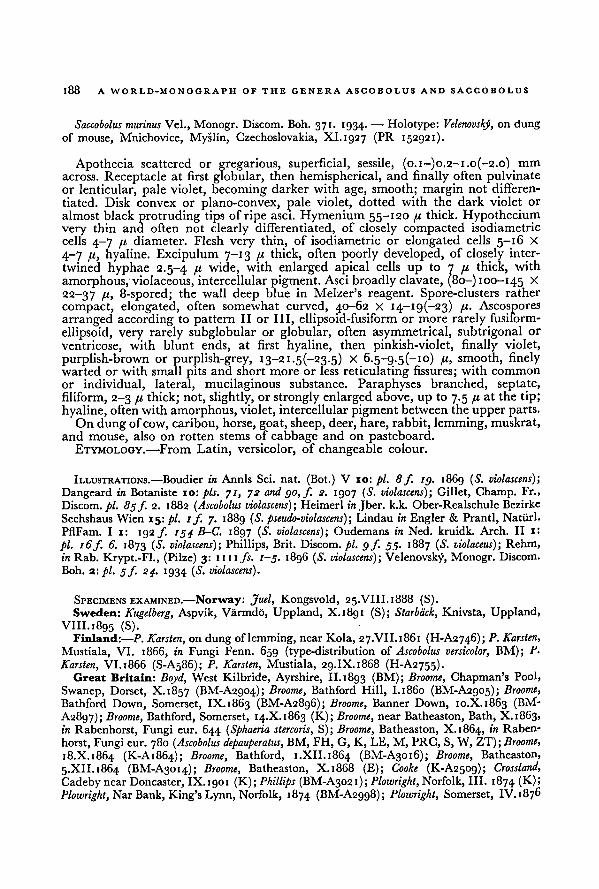
188A WORLD-MONOGRAPH OF THE GENERA ASCOBOLUS AND SACCOBOLUS
Saccobolus murinus Vel., Monogr. Discom. Boh. 371. 1934. — Holotype: Velenovsky, on dung
of mouse, Mnichovice, Myslin, Czechoslovakia, XI.1927 (PR 152921).
Apothecia scattered or gregarious, superficial, sessile, (0.1—)o.2-i.o(-2.o) mm
across. Receptacle at first globular, then hemispherical, and finally often pulvinate
or lenticular, pale violet, becoming darker with age, smooth; margin not differen-
tiated. Disk convex or plano-convex, pale violet, dotted with the dark violet or
almost black protruding tips of ripe asci. Hymenium 55-120 ft thick. Hypothecium
very thin and often not clearly differentiated, of closely compacted isodiametric
cells 4-7 ft diameter. Flesh very thin, of isodiametric or elongated cells 5—16 X
4-7 pi, hyaline. Excipulum 7-13 pi thick, often poorly developed, of closely inter-
twined hyphae 2.5-4 B wide, with enlarged apical cells up to 7 71 thick, with
amorphous, violaceous, intercellularpigment. Asci broadly clavate, (80-) 100-145 *
22-37 B> 8-spored; the wall deep blue in Melzer's reagent. Spore-clusters rather
compact, elongated, often somewhat curved, 40-62 X i4-ig(-23) pi. Ascospores
arranged according to pattern II or III, ellipsoid-fusiform or more rarely fusiform-
ellipsoid, very rarely subglobular or globular, often asymmetrical, subtrigonal or
ventricose, with blunt ends, at first hyaline, then pinkish-violet, finally violet,
purplish-brown or purplish-grey, i3-2i.5(-23-5) X 6.5-g.5(-io) fi, smooth, finelywarted or with small pits and short more or less reticulating fissures; with common
or individual, lateral, mucilaginous substance. Paraphyses branched, septate,
filiform, 2-3 ft thick; not, slightly, or strongly enlarged above, up to 7.5 fi at the tip;
hyaline, often with amorphous, violet, intercellularpigment between the upperparts.On dung of cow, caribou, horse, goat, sheep, deer, hare, rabbit, lemming, muskrat,
and mouse, also on rotten stems of cabbage and on pasteboard.ETYMOLOGY.
—From Latin, versicolor, of changeable colour.
ILLUSTRATIONS.—Boudier in Annls Sci. nat. (Bot.) V 10: pi. 8f. ig. 1869 (S. violascens);
Dangeard in Botaniste 10: pis. 71, 72 and go,f. 2. 1907 (S. violascens)-, Gillet, Champ. Fr.,
Discom. pi. 8gf 2. 1882 (Ascobolus violascens) ; Heimerl in Jber. k.k. Ober-Realschule Bezirke
Sechshaus Wien 15: pi. 1 f. 7. 1889 (S. pseudo-violascens); Lindau in Engler & Prantl, Natürl.
PflFam. I i: 192 ƒ. 154 B-C. 1897 (S. violascens); Oudemans in Ned. kruidk. Arch. II 1:
pi. 16f. 6. 1873 (S. violascens); Phillips, Brit. Discom. pi. g f. 55. 1887 ( S. violaceus); Rehm,
in Rab. Krypt.-Fl., (Pilze) 3: mi fs. 1-5. 1896 ( S. violascens); Velenovsky, Monogr. Discom.
Boh. 2: pi. 5 f. 24. 1934 (S. violascens).
SPECIMENS EXAMINED.—Norway: Juel, Kongsvold, 25.VIII.1888 (S).Sweden: Kugelberg, Aspvik, Varmdd, Uppland, X.1891 (S); Starback, Knivsta, Uppland,
VIII.1895 (S).Finland:—,P. Karsten, on dungof lemming, near Kola, 27.VII.1861 (H-A2746); P. Karsten,
Mustiala, VI. 1866, in Fungi Fenn. 659 (type-distribution of Ascobolus versicolor, BM); p.
Karsten, VI. 1866 (S-A586); P. Karsten, Mustiala, 29.IX. 1868 (H-A2755).
Great Britain: Boyd, West Kilbride, Ayrshire, II.1893 (BM); Broome, Chapman's Pool,
Swanep, Dorset, X.1857 (BM-A2904); Broome, Bathford Hill, 1.1860 (BM-A2905); Broome,
Bathford Down, Somerset, IX. 1863 (BM-A28g6); Broome, Banner Down, 10.X.1863 (BM-
A2897); Broome, Bathford, Somerset, 14.X.1863 (K); Broome, near Batheaston, Bath, X. 1863,
in Rabenhorst, Fungi eur. 644 (Sphaeria stercoris, S); Broome, Batheaston, X.1864, in Raben-
horst, Fungi eur. 780 ( Ascobolus depauperatus, BM, FH, G, K, LE, M, PRC, S, W, ZT); Broome,
18.X.1864 (K-A1864); Broome, Bathford, 1.XII.1864 (BM-A3016); Broome, Batheaston,
5.XII. 1864 (BM-A3014); Broome, Batheaston, X.1868 (E); Cooke (K-A2509); Crossland,
Cadeby near Doncaster, IX. 1901 (K); Phillips (BM-A3021); Plowrighl, Norfolk, III. 1874 (K);
Plowright, Nar Bank, King's Lynn, Norfolk, 1874 (BM-A2998); Plowright, Somerset, IV. 1876
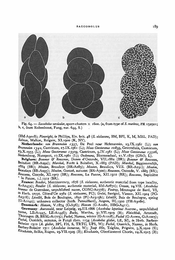
189SACCOBOLUS
(BM-A3018); Plowright, in Phillips, Elv. brit. 48 ( S. violascens, BM, BPI, K, M, MEL PAD);Salmon, Walton, Reigate, XI. 1900 (K, NY).
Netherlands: van Brummelen 1357, De Peel near Helenaveen, 23.IX.1961 (L); van
Brummelen1342, Castricum, 27.IX. 1961 (L); Maas Geesteranus 10833, Geversduin, Castricum,
23.X.1955 (L); Maas Geesteranus 13303, Castricum, 9.IV.1961 (L); Maas Geesteranus 13326,Stakenberg, Nunspeet, 17.IX. 1961 (L); Oudemans, Bloemendaal, 21.V.1872 (GRO, L).
Belgium: Bommer & Rousseau, Dunes d'Ostende, VII. 1882 (BR); Bommer & Rousseau,Boitsfort (BR-A292); Marchal, Foret a Boitsfort, X.1883 (PAD); Marchal,
„ .. . . _ .,
Bigginnendijk,(BR); Mouton, Beaufays (BR-A289); Mouton, Beaufays, VIII. (BR-A291); Mouton,
Beaufays (BR-A295); Mouton, Gomze, autumn (BR-A300); Rousseau, Ostende, V. 1885 (BR);Rousseau, Coxyde, XI.1907 (BR); Rousseau, La Panne, XII.1910 (BR); Rousseau,
_ .
._ ,
Sapinièrela Panne, 1.1.1912 (BR).France: Boudier, Montmorency, 1878 ( S. violascens, authentic material from type locality,
K-A2452); Boudier (S. violascens, authentic material, BM-A2870); Crouan, 24.VII. ( Ascobolus
bruneus de Guernisac, unpublished name, CONC-A2378); Fautrey, Montagne de Bard, VI,ln Herb, crypt. Cote-d'Or 2382 (S. violascens, PC); Grelet, Savigne, Vienne, XII. 1924 (PC-A2318); Leveille, Bois de Boulogne, 1850 (PC-A23i5b); IJveille, Bois de Boulogne, spring(G-A1040); unknown collector (herb. Patouillard), Angers, III.1900 (FH-A3080).
Denmark: Hansen, V.1874 (C-A56); Hansen (G-A1061, HBG-A411).Germany: Auerswald, near Leipzig, 29.III.1866 ( Ascobolus leporinus Auersw., unpublished
))ame, LE-A1445, LE-A1446); Bucks, Wartha, 31.VII.1919 (B); Fleischhak, Arnstadt,Hiiiringen (B, BRSL-A1715); Fuckel, Nassau, winter (G-A1008); Fuckel (G-A1002, G-A1003);F
uckel, Oestrich, autumn, in Fungi rhen. 1134 (Ascobolus glaber,„
LE, M), in Herb. Barbey-IQIO
n . J (A. glaber, BPI, FH, S, TRTC, UPS, W); Fuckel, Oestrich, Nassau, in Herb.
Barbey-Boissier 131 1 ( Ascobolus immersus, W); Jaap 860, Triglitz, Prignitz, 5.X.1910 (S);Kirschstein, Sellin, Rtigen, 29.VII. 1909 (B); Kirschslein, Oberfiirsterei Chorin, 24.X.1915 (B);
Fig. 64. — Saccobolus versicolor, spore-clusters X 1600. (a, fromtype of S. murinus, PR 15292 1;
b, c, from Rabenhorst, Fung, eur. 644, S.)
190A WORLD-MONOGRAPH OF THE GENERA ASCOBOLUS AND SACCOBOLUS
Kirschstein, between Potsdam and Templin a.d. Havel, 12.XII.1915 (B); Kirschstiin, Rodelbergj
Niedcr-Schönhausen, 12.XI.1938 (B, FH); Kirschstiin, Schönholzer Heide, Berlin-Pankow,
4.XI.1939 and 5.XI.1940 (B); Kirschstein, Rathenow (B-A1377); Kirschstein, Stadtforst,
Rathenow (B-A1378); Krieger, near Konigstein, V. 1882 (S-A562); Krieger, near Königstein,
VII-VIII.1881, 23.V.188Q ( S. depauperatus), in Fungi sax. 842 (BM, BPI, BRSL, M, S, W),
in Rehm, Ascom. 661 (BM, E, FH, G, HBG, M, NY, PAD, S), in Roumeguère, Fungisel. 4740 (G), in von Thümen, Myc. univ. 2213 (B, BM, BPI, G, L, LE, M, MEL, PAD,
W, ZT); Ludwig, Breitscheiderwald, Dillkreis, Hessen-Nassau, V.1944 (B); Ludwig, near
Langenholdinghausen, Siegen, Westfalen, 23.VIII.1945 (B-Ai3gi, B-A1397); Magnus, Havel-
ufer near Schildhorn, 2.XII.1876 (HBG); Rehm, Obernesselbach, Franken, 1870 (S-A580);
Rehm, Obernesselbach, Franken, IV. 1871 (S-A537); Rehm, Miinchen, IX.1902 (S-A670).
Schroeter, near Oelze, 16.X.1881 (BRSL); Staritz, near Pulsnitz, Sachsen, 1884 (S); Winter,
Harth, near Leipzig, V. 1874 (S-A591).Poland: Schroeter, 22.V. 1876 (BRSL-A1655); Schroeter, Karlowitz nearBreslau (=Wroclaw),
V.i 882 (BRSL); Schroeter, Ochojetz near Rybnik, X.1882 (BRSL); Schroeter, near Wiersbei,
9.'VII. 1884 (BRSL).Czechoslovakia: Lojka, Linglez, near Eperjes (= Presov), 5.X.1869 (S); Niessl, Schweib-
wald, 28.IV. 1870 (M); Niessl, nearStrutz, Moravia, VI.1876 (M); Niessl, Schweibwald, Vllb
1876 (M); Niessl, near Ratschitz, Moravia, VIII.1886 (M); Niessl, Brünn (= Brno) (PAD-
A175O; Niessl, Briinn (= Brno), autumn (PAD-A1754); Niessl, near Popuwek, Moravia
(M-A805); Petrak, Mahr.-Weisskirchen, near Hrabuvka, 24.II.1912 (S), in Fl. Boh. Mor.
ser. 2, 232 (S. depauperatus, BM, BPI, E, FH, LE, S); Petrak, Mahr.-Weisskirchen: Welka,
21.IX.1912, in Fl. Boh. Mor. ser. 2, 233 (S. kerverni, BM); Petrak, Sternberg, Moravia,
VIII.1930 (BM, ZT); Svrcek, Zbraslav, 15.VI.1946 (S. kerverni f. minima Svrcek, unpublishedname, PR-A267ga); Swcr#, Jiloviste, Bohemia, 18.V. 1947 (PR 179019); Velenovskj), Mnichovice,
VIII.1923 (PR 150061); Velenovskj, Stranicice, XII.1926 (PR 148812); Velenovskj, Mnichovice,
Myslin, XI.1927 (holotype of S. murinus, PR 152921); Velenovskj, Mnichovice, Hubachov,
25.v1.1929 (S. atrofuscus Vel., unpublished name, PR).Switzerland: Volkart, Fiirstenalp, 26.VI. 1903 (ZT).Austria: Britzelmayr, Peischl-Kopf, W. Tyrol, VII.1878 (S); Rehm, near Kiihtei, Oetz,
Tyrol, VIII. 1872, in Ascom. 165 (S. violascens, BM, E, L, M, PAD, S, W).Italy: Bizzozzero, VIII.1884 (PAD-A1757).Romania: Lojka, near Malornoiz, Siebenbiirgen, VIII.1872 (S).U.S.S.R.: Tranzschel, Varsonia near Zobki, 20.X. i8q8 (LE); Tranzschel, Zobki near
Varsonia,
II..899 (LE).Hawaii: van Brummelen 1458, 1470, 1471, 1473, Puuhokomo, Mauna Kea, 3-10.V.
1962 (L).Canada: Cain, S.E.-Aurora, Ontario, 19.X.1930 (TRTC); Cain, Long Point, Lake Erie»
Ontario, 17.VII.1g32 (TRTC); Cain, Sky Lake, Bruce, Ontario, 22.VII.1932 (TRTC);Cain, Melissa, Muskoka, Ontario, 2.VIII.1932 (TRTC); Cain 6674, High Park, 'Ioronto,
Ontario, 2.XI. 1936 (TRTG); Cain 6681, High Park, Toronto, Ontario, 2.XI.1936 (TRTC);Clark, Prain Lake, Thelon Game Preserve, N.W.T., summer 1936 (TRTC).
U.S.A.: Oregon: Kienholz, Winan's pasture, Dee, 21.II.1939 (BPI, NY). California:
Harkness 1362 (BM, K); Martin 653, 14.II.1917 (culture, BPI). Idaho: Bigelow, Payette
Lake, 2.VII.1954 (G); W. B. & V. G. Cooke 13530, near Stites, Idaho Co., iQ.IV.1q47 (BPF
FH, NY). Colorado: F. E. & E. S. Clements 301, Castle Canyon, 1g.VII.1906 (E). Texas:
Ravenel 1863, Houston (BPI). Maine: Thaxter, Kittery Point (FH-A3076). New York:
Kaufman, Ithaca, 24.V.1902 (BPI); Kaufman 838, Coy Glen, Cayuga Lake Basin, 27.IV.i9°4
(TRTC). Connecticut: Thaxter, Greens Farms (FH-A3og6). New Jersey: Ellis 3510'
Newfield, VI.1881 (BPI).
This is without doubt the most variable species of the genus Saccobolus. The fruit-
SACCOBOLUS 191
bodiesmay be rather large with a well developed excipulum which is rich in violet
pigment or else they may be minute without a clearly developed excipulum and
without intercellular pigment. Sometimes the paraphyses show pear-shaped tipssurrounded by violet pigment; this was thought to be characteristic of S. violascens
Boud. (Boudier, 1869: 231). In other fruit-bodies of the same culture or collection
the paraphyses are without thickened tips. Rarely the paraphyses are almost absent.
This variation in the shape and structure of the fruit-bodies finds its origin in
the wide scale of possibilities during the initial development of the fruit-bodies.
This fully agrees with Dangeard's observations (1907: 327): "Nous n'avons jamaisrencontre dans aucune espece d'Ascomycete une aussi grande irregularite dans
la maniere d'etre des filaments recouvrants; il n'existe souvent qu'un peloton
irregulier sans veritable parois; sur ce peloton preeminent les premieres theques
peu nombreuses; on ne voit pas encore le systeme des paraphyses; des hyphespassent d'un peloton a l'autre, reliant les divers peritheces entre eux; la dimension
de ces peritheces est elle-meme des plus variables."
Normally the ascomata are eugymnohymenial or paragymnohymenial (c.f. p. 00).Only rarely also cleistohymenial ascomata were observed that opened in the pro-
hymenial phase.
Apart of the variation due to differences in the development, there is a considerable
variation in pigmentation of the fruit-bodies, the size of the ascospores, and the
ornamentation of the episporium.
Experiments with cultures proved that more and darker intercellular pigmentls formed in fruit-bodies exposed to high intensities of light than in unexposed fruit-
bodies. Irrespective of this, more pigment also seems to be formed when growing
°n dung of cow and rabbit than when growing on other substrata.
I found it impossible to distinguish more or less constant forms within the species.
Only in this species of Saccobolus spherosporical mutations were accidentallyfound (Figs. 63c, 64b).
The related species S. caesariatus, S. beckii, and S. quadrisporus might prove to
beextreme forms or mutant-forms of it.
Saccobolus versicolor is the most common species of Saccobolus and has a world-wide
distribution.
9. SACCOBOLUS CAESARIATUS Renny apud Phill.—Fig. 65
Saccobolus caesariatus Renny apud Phill., Brit. Discom. 297. 1887. — Type: represented bya drawing- after Renny (BM) "Saccobolus caesariatus Renny after Renny, Sussex 1875"; type'ocality, Sussex, England.
Apothecia solitary, superficial, sessile, 150-200 /u diameter, 200-300 pt high.. cePtacle ovoid-cylindrical or hemispherical, white in all parts; surface covered
With white, flexuous bunches of septate hyphae to form tapering squamules; marginn°t differentiated. Disk flat or convex, colourless, roughened by the protrudingasci. Flesh none or very thin. Excipulum thin, of elongated or angular cells (textura
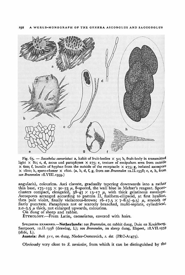
192A WORLD-MONOGRAPH OF THE GENERA ASCOBOLUS AND SACGOBOLUS
angularis), colourless. Asci clavate, gradually tapering downwards into a rather
thin base, 175-193 X 30-33 ft, 8-spored, the wall blue in Melzer's reagent. Spore-clusters compact, elongated, 38-43 X 15-17 fc, with thick gelatinous envelope-
Ascospores arranged according to pattern II, fusiform-ellipsoid, at first hyaline,then pale violet, finally violaceous-brown; 16-17.5 X 7~8.5(-g.5) fi, smooth or
finely punctate. Paraphyses not or scarcely branched, multi-septate, cylindrical,2.0-3.5 B thick, not enlarged upwards, colourless.
On dung of sheep and rabbit.
ETYMOLOGY.—From Latin, caesariatus, covered with hairs.
SPECIMENS EXAMINED.—Netherlands: van Brummelen, on rabbit dung, Duin en Kruidberg>
Santpoort, 12.II.1958 (drawing, L); van Brummelen, on sheep dung, Elspeet, i8.VII.i959
(slide, L).Austria: Beck 511, on dung, Nieder-Oesterreich, r. dat. (PRC-A1473).
Obviously very close to S. versicolor, from which it can be distinguished by the
a, habit of fruit-bodies X 50; b, fruit-body in transmitted
light X 80; c, d, ascus and paraphyses X 275; e, texture of excipulum seen from outside
X 600; f, bundle of hyphae from the outside of the receptacle X 275; g, isolated ascosporeX 1600; h, spore-cluster X 1600. (a, b, d, f, g, from
Fig. 65. — Saccobolus caesariatus:
van Brummelen 12.II. 1958; c, e, h, from
van Brummelen 18.VIII. 1959.)
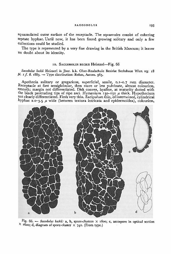
SACCOBOLUS 193
squamulated outer surface of the receptacle. The squamules consist of cohering
septate hyphae. Until now, it has been found growing solitary and only a few
collections could be studied.
The type is represented by a very fine drawing in the British Museum; it leaves
no doubt about its identity.
10. SACCOBOLUS BECKII Heimerl—Fig. 66
Saccobolus beckii Heimerl in Jber. k.k. Ober-Realschule Bezirke Sechshaus Wien 15: 18
pi- 1 f. 8. 1889. — Type distribution: Rehm, Ascom. 965.
Apothecia solitary or gregarious, superficial, sessile, 0.1-0.7 rnm diameter.
Receptacle at first semiglobular, then more or less pulvinate, almost colourless,smooth; margin not differentiated. Disk convex, hyaline, at maturity dotted with
the black protruding tips of ripe asci. Hymenium 130-150 fi thick. Hypotheciumnot clearly differentiated. Flesh very thin. Excipulum thin, of intertwined, cylindricalhyphae 2.0-3.5 /i wide (between textura intricata and epidermoidea), colourless,
Fig. 66. — a, b, spore-clusters X 1600; c, ascospore in optical section
x 1600; d, diagram of spore-cluster x 740. (From type.)
Saccobolus beckii:
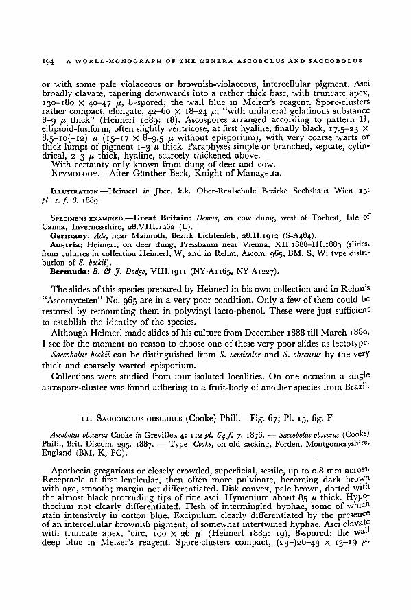
A WORLD-MONOGRAPH OF THE GENERA ASCOBOLUS AND SACCOBOLUS194
or with some pale violaceous or brownish-violaceous, intercellular pigment. Asci
broadly clavate, tapering downwards into a rather thick base, with truncate apex,
130-180 X 40—47 {.1, 8-spored; the wall blue in Melzer's reagent. Spore-clustersrather compact, elongate, 42-60 X 18-24 /"> "with unilateral gelatinous substance
8-9 /t thick" (Heimerl 1889: 18). Ascospores arranged according to pattern II,
ellipsoid-fusiform, often slightly ventricose, at first hyaline, finally black, 17.5-23 X
8.5-io(-i2) [x (15-17 X 8-9.5 B without episporium), with very coarse warts or
thick lumps of pigment 1-3 fj,thick. Paraphyses simple or branched, septate, cylin-
drical, 2-3 /t thick, hyaline, scarcely thickened above.
With certainty only known from dung of deer and cow.
ETYMOLOGY. —After Giinther Beck, Knight of Managetta.
ILLUSTRATION.—Heimerl in Jber. k.k. Ober-Realschule Bezirke Sechshaus Wien 15:
pi. i.f 8. 1889.
SPECIMENS EXAMINED.—Great Britain: Dennis, on cow dung, west of Torbest, Isle of
Canna, Invernessshire, 28.VIII.1962 (L).
Germany: Ade, near Mainroth, Bezirk Lichtenfels, 28.II.1912 (S-A484).Austria: Heimerl, on deer dung, Pressbaum near Vienna, XII. 1888—III. 1889 (slides,
from cultures in collection Heimerl, W, and in Rehm, Ascom. 965, BM, S, W; type distri-
bution of S. beckii).Bermuda: B. & J. Dodge, VIII.1911 (NY-A1165, NY-A1227).
The slides of this species prepared by Heimerl in his own collection and in Rehm's
"Ascomyceten" No. 965 are in a very poor condition. Only a few of them could be
restored by remounting them in polyvinyl lacto-phenol. These were just sufficient
to establish the identity of the species.
Although Heimerl madeslides ofhis culture from December 1888 till March 1889,
I see for the moment no reason to choose one of these very poor slides as lectotype.
Saccobolus beckii can be distinguished from S. versicolor and S. obscurus by the very
thick and coarsely warted episporium.Collections were studied from four isolated localities. On one occasion a single
ascospore-cluster was found adhering to a fruit-body of another species from Brazil-
11. SACCOBOLUS OBSCURUS (Cooke) Phill.—Fig. 67; Pl. 15, fig. F
Ascobolus obscurus Cooke in Grevillea 4: 112 pi. 64ƒ. 7. 1876. —Saccobolus obscurus (Cooke)
Phill., Brit. Discom. 295. 1887. — Type: Cooke, on old sacking, Forden, Montgomeryshire,
England (BM, K, PC).
Apothecia gregarious or closely crowded, superficial, sessile, up to 0.8 mm across.
Receptacle at first lenticular, then often more pulvinate, becoming dark brown
with age, smooth; margin not differentiated.Disk convex, pale brown, dotted with
the almost black protruding tips of ripe asci. Hymenium about 85 [A, thick. Hyp 0'
thecium not clearly differentiated. Flesh of intermingled hyphae, some of which
stain intensively in cotton blue. Excipulum clearly differentiated by the presenceof an intercellular brownish pigment, of somewhat intertwined hyphae. Asci clavate
with truncate apex, 'circ. 100 X 26 /a' (Heimerl 1889: 19), 8-spored; the wah
deep blue in Melzer's reagent. Spore-clusters compact, (23-)26-43 X 13-19 f 1'
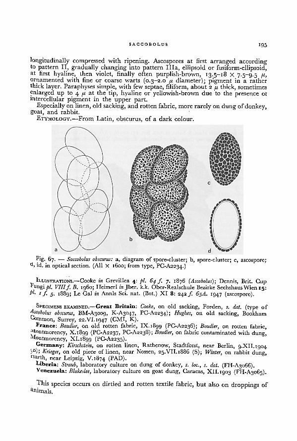
SACCOBOLUS 195
longitudinally compressed with ripening. Ascospores at first arranged accordingto pattern II, gradually changing into pattern Ilia, ellipsoid or fusiform-ellipsoid,at first hyaline, then violet, finally often purplish-brown, 13.5-18 X 7.5-9.5ornamented with fine or coarse warts (0.3-2.0 fi diameter); pigment in a ratherthick layer. Paraphyses simple, with few septae, filiform, about 2 [i thick, sometimes
enlarged up to 4 at the tip, hyaline or yellowish-brown due to the presence 01
intercellular pigment in the upper part.Especially on linen, old sacking, and rotten fabric, more rarely on dung of donkey,
goat, and rabbit.
ETYMOLOGY.—From Latin, obscurus, of a dark colour.
ILLUSTRATIONS.—Cooke in Grevillea 4: pi. 64 f. 7. 1876 (Ascobolus); Dennis, Brit. CupFungi pl. VIIIf. B. i960; Heimerl in Jber. k.k. Ober-Realschule Bezirke Sechshaus Wien 15:t> 1
- if. 5. 1889; Le Gal in Annls Sci. nat. (Bot.) XI 8: 242 f. 65A. 1947 (ascospore).
SPECIMENS EXAMINED.—Great Britain: Cooke, on old sacking, Forden, s. dat. (type of
Ascobolus obscurus, BM-A3009, K-A3047, PC-A2234); Hughes,. _ _
,on old sacking, Bookham
Common, Surrey, 22.VI.1g47 (CMI, K).France: Boudier, on old rotten fabric, IX.1899 (PC-A2236); Boudier,
__.
„ ,on rotten fabric,
X. 1899 (PC-A2237, PC-A2238); Boudier,,
on fabric contaminated with dung,Montmorency, XI.1899 (PC-A2235).
Germany: Kirschstein,,
on rotten linen, Rathenow, Stadtforst, near Berlin, 9.XII. 1904—
.-
... ... - —- —
Krieger, on old piece of linen, near Nossen, 25.VII.1886 (S); Winter,_ . -
. ,. ,on rabbit dung,
near Leipzig, V. 1874 (PAD).Liberia: Straub, laboratory culture on dung of donkey, s. loc., s. dat.
. . _ (FH-A3066).Venezuela: Blakeslee, laboratory culture on goat dung, Caracas, XII.igo3 (FH-A3065).
This species occurs on dirtied and rotten textile fabric, but also on droppings ofa uimals.
Fig. 67. — Saccobolus obscurus: a, diagram of spore-cluster; b, spore-cluster; c, ascospore;
d, id. in optical section. (All X 1600; from type, PC-A2234.)
196A WORLD-MONOGRAPH OF THE GENERA ASCOBOLUS AND SACCOBOLUS
From S. versicolor and S. beckii it can be distinguished by the brown, intercellular
pigment, the shape of ascospores and spore-clusters, and the ornamentationof the
episporium. From S. thaxteri it differs in the size of the fruit-bodies, the size and
shape of the ascospores, the compactness of the spore-clusters, and thepresenceot
intercellular pigment.This widely distributed species has been collected only rarely.
12. SACCOBOLUS QUADRISPORUS Mass. & Salm.—Fig. 68; Pl. 15, fig. C
Saccobolus quadrisporus Mass. & Salm. in Ann. Bot. 15: 329/>/. i8fs. 48-51. 1901.-—Holotype:
Massee & Salmon, on goose dung, Kew Gardens, England, X.1901 (S. tetrasporus, NY).
Apothecia superficial, sessile, 0.5-0.7 mm diameter. Receptacle more orless
pulvinate, at first clear watery violet, gradually becoming darker with age, smooth;
margin not differentiated. Disk at first convex, then flat, clear watery violet, becoming
blackish-violet or black, at maturity dotted with the dark apices of the protrudingasci. Asci clavate, tapering downwards into a rather thick base, with truncate
apex, 95-110 X 18-20 41, 4-spored, the wall staining blue with iodine. Spore-clusters ellipsoid or elongated, 42-48 X 16-17 [X ifascospores in two rows, 46-51
14-16 [X if ascospores in a single row, surrounded by a common mucilaginous
covering. Ascospores arranged in two parallel rows or side by side in a single roW,
ellipsoid, often somewhat trigonous, at first hyaline, then blackish-violet, 16.5-19.5 *
9.5-10.5 fx; with a thick cap or girdle of irregularly reticulated or warted pigment
up to 1.6 [X thick, which is surrounded by a narrow zone of greater translucency>
spore-clusters X 1600 (from type)Saccobolus quadrisporus,Fig. 68. —
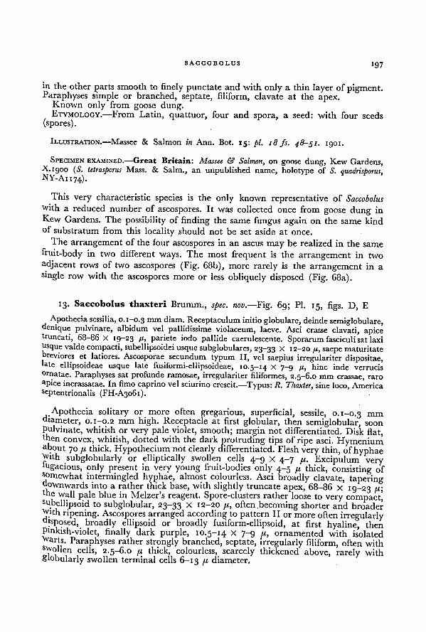
SACCOBOLUS 197
in the other parts smooth to finely punctate and with only a thin layer of pigment.Paraphyses simple or branched, septate, filiform, clavate at the apex.
Known only from goose dung.ETYMOLOGY.—From Latin, quattuor, four and spora, a seed: with four seeds
(spores).
ILLUSTRATION.—Massee & Salmon in Ann. Bot. 15: pi. 18 fs. 48-51. 1901.
SPECIMEN EXAMINED.—Great Britain: Massee & Salmon, on goose dung, Kew Gardens,K.i 900 (S. tetrasporus Mass. & Salm., an unpublished name, holotype of S. quadrisporus,NY-Ai 174).
This very characteristic species is the only known representative of Saccobolus
■with a reduced number of ascospores. It was collected once from goose dung in
Kew Gardens. The possibility of finding the same fungus again on the same kind
of substratum from this locality should not be set aside at once.
The arrangement of the four ascospores in an ascus may be realized in the same
fruit-body in two different ways. The most frequent is the arrangement in two
adjacent rows of two ascospores (Fig. 68b), more rarely is the arrangement in a
single row with the ascospores more or less obliquely disposed (Fig. 68a).
13. Saccobolus thaxteri Brumm., spec. nov.—Fig. 69; Pl. 15, figs. D, E
Apothecia sessilia, 0.1-0.3 mm diam. Receptaculum initioglobulare, deindesemiglobulare,denique pulvinare, albidum vel pallidissime violaceum, laeve. Asci crasse clavati, apiceb'uncati, 68-86 X 19-23 /(, pariete iodo pallide caerulescente. Sporarum fasciculi sat laxi
usque valde compacti, subellipsoidei usque subglobulares, 23-33 X 12-20 //, saepe maturitatereviores et latiores. Ascosporae secundum typum II, vel saepius irregulariter dispositae,
'ate ellipsoideae usque late fusiformi-ellipsoideae, 10.5-14 x 7-9 /i, hinc inde verrucisornatae. Paraphyses sat profunde ramosae, irregulariter filiformes, 2.5-6.0 mm crassae, raro
apice incrassatae. In fimo caprino vel sciurino crescit.—Typus: II. Thaxter,,
sine loco, America
scptentrionalis (FH-A3061).
Apothecia solitary or more often gregarious, superficial, sessile, 0.1-0.3 mm
diameter, 0.1-0.2 mm high. Receptacle at first globular, then semiglobular, soon
Pulvinate, whitish or very pale violet, smooth; margin not differentiated. Disk flat,then
convex, whitish, dotted with the dark protruding tips of ripe asci. Hymeniumabout 70 fi thick. Hypothecium not clearly differentiated. Flesh very thin, of hyphaeWith subglobularly or elliptically swollen cells 4-9 x 4-7 p. Excipulum very'ugacious, only present in very young fruit-bodies only 4-5 p thick, consisting ofsomewhat intermingled hyphae, almost colourless. Asci broadly clavate, taperingdownwards into a rather thick base, with slightly truncate apex, 68-86 X 19-23 p;the wall pale blue in Melzer's reagent. Spore-clusters rather loose to very compact'subellipsoid to subglobular, 23-33 X 12-20 p, often becoming shorter and broaderwith ripening. Ascospores arranged according to pattern II or more often irregularlydisposed, broadly ellipsoid or broadly fusiform-ellipsoid, at first hyaline, then
Pinkish-violet, finally dark purple, 10.5-14 X 7-9 p, ornamented with isolatedwarts. Paraphyses rather strongly branched, septate, irregularly filiform, often withswollen cells, 2.5-6.0 p thick, colourless, scarcely thickened above, rarely with
S'obularly swollen terminal cells 6-13 p. diameter.
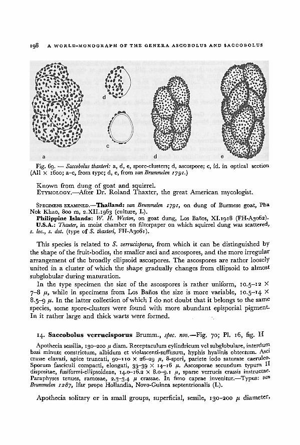
198A WORLD-MONOGRAPH OF THE GENERA ASCOBOLUS AND SACCOBOLUS
Known from dung of goat and squirrel.ETYMOLOGY.
—After Dr. Roland Thaxter, the great American mycologist.
SPECIMENS EXAMINED.—Thailand: van Brummelen 1791, on dung of Burmese goat, Pha
Nok Khao, 800 m, 2.XII. 1963 (culture, L).
Philippine Islands: W. H. Weston, on goat dung, Los Baiios, XI. 1918 (FH-A3062).U.S.A.: Thaxter, in moist chamber on filterpaper on which squirrel dung was scattered,
s. loc., s. dat. (type cf S. thaxteri, FH-A3061).
This species is related to S. verrucisporus, from which it can be distinguished by
the shape of the fruit-bodies, the smaller asci and ascospores, and the more irregular
arrangement of the broadly ellipsoid ascospores. The ascospores are rather loosely
united in a cluster of which the shape gradually changes from ellipsoid to almost
subglobular during maturation.
In the type specimen the size of the ascospores is rather uniform, 10.5-12 X
7-8 fx, while in specimens from Los Banos the size is more variable, 10.5-14 X
8.5-9 I 11 latter collectionof which I do not doubt that it belongs to the same
species, some spore-clusters were found with more abundant episporial pigment.In it rather large and thick warts were formed.
14. Saccobolus verrucisporus Brumm., spec. nov. —Fig. 70; Pl. 16, fig. H
Apothecia sessilia, 130-200 fi diam. Receptaculum cylindricum vel subglobulare, intcrdum
basi minute constrictum, albidum et violascenti-suffusum, hyphis hyalinis obtectum. Asci
crasse clavati, apice truncati, 90-110 X 26-29 /i, 8-spori, pariete iodo saturate caeruleo.
Sporum fasciculi compacti, elongati, 33-39 X 14-16 /t. Ascosporae secundum typumH
dispositae, fusiformi-ellipsoideae, 14.0-16.2 X 8.0-g.i /(, sparse verrucis crassis instructae.
Paraphyses tenues, ramosae, 2.3-3.4 B crassae. In fimo caprae invenitur.—Typus: van
Brummelen 1287, Ifar prope Hollandia, Novo-Guinea septentrionalis (L).
Apothecia solitary or in small groups, superficial, sessile, 130-200 pt diameter,
a, d, e, spore-clusters; d, ascospore; c, id. in optical section
(All X 1600; a-c, from type; d, e,from van Brummelen 1791.)
Saccobolus thaxteri:Fig. 69. —
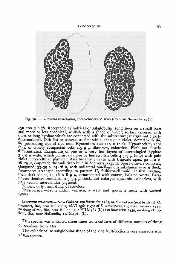
SACCOBOLUS199
150-200 p high. Receptacle cylindrical or subglobular, sometimes on a small base
and more or less obconical, whitish with a shade of violet; surface covered withshort or long hyphae which are connected with the substratum; margin not clearlydifferentiated. Disk flat or convex, at first white, then pale violet, dotted with thefar protruding tips of ripe asci. Hymenium 100-115 thick. Hypothecium verythin, of closely compacted cells 4.5-9 p diameter, colourless. Flesh not clearlydifferentiated. Excipulum of one or a very few layers of intermingled hyphae37-4.5 p wide, which consist of more or less swollen cells 4.5-9 p long; with paleviolet, intercellular pigment. Asci broadly clavate with truncate apex, 90-110 X
26-29 Ab 8-spored; the wall deep blue in Melzer's reagent. Spore-clusters compact,elongated, 33-39 X 14-16 p, with unilateral mucilaginous substance 7-10 p thick.
Ascospores arranged according to pattern II, fusiform-ellipsoid, at first hyaline,then dark violet, 14-16 X 8-9 p, ornamented with coarse, isolated warts. Para-
Physes slender, branched, 2.3-3.4 p thick, not enlarged upwards, colourless, with
Pale violet, intercellular pigment.Known only from dung of roe-deer.
ETYMOLOGY.— From Latin, verruca, a wart and spora, a seed: with wartedsPores.
SPECIMENS EXAMINED.—New Guinea: van Brummelen 1287,, j
on dung ofroe (sent by Dr. H. O.
Sleumer), Ifar, near Hollandia, 26.VI.1961 (type of S. verrucisporus, L); van Brummelen 1442,°n dung of roe, Ifar, near Hollandia, 5.VIII.1961 (L); van Brummelen 1439, on dung of roe-
deer, Ifar, near Hollandia, 11.IX.1961 (L).
This species was collected three times from cultures of different samples of dung°f roe-deer from Ifar.
The cylindrical to subglobular shape of the ripe fruit-bodies is very characteristic
°f this species.
Fig. 70. — Saccobolus verrucisporus, spore-clusters X 1600 (from van Brummelen 1287).
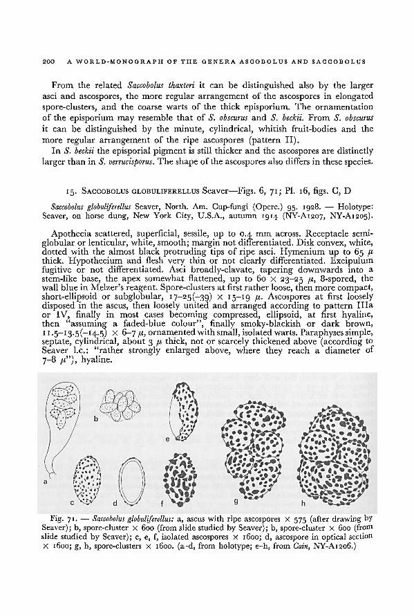
200 A WORLD-MONOGRAPH OF THE GENERA ASCOBOLUS AND SACCOBOLUS
From the related Saccobolus thaxteri it can be distinguished also by the larger
asci and ascospores, the more regular arrangement of the ascospores in elongated
spore-clusters, and the coarse warts of the thick episporium. The ornamentation
of the episporium may resemble that of S. obscurus and S. beckii. From S. obscurus
it can be distinguished by the minute, cylindrical, whitish fruit-bodies and the
more regular arrangement of the ripe ascospores (pattern II).In S. beckii the episporial pigment is still thicker and the ascospores are distinctly
larger than in S. verrucisporus. The shape of the ascospores also differs in these species.
15. SACCOBOLUS GLOBULIFERELLUS Seaver—Figs. 6, 71; Pl. 16, figs. C, D
Saccobolus globuliferellus Seaver, North. Am. Cup-fungi (Operc.) 95. 1928. — Holotype:
Seaver, on horse dung, New York City, U.S.A., autumn 1914 (NY-A1207, NY-A1205).
Apothecia scattered, superficial, sessile, up to 0.4 mm across. Receptacle semi-
globular or lenticular, white, smooth; margin not differentiated. Disk convex, white,dotted with the almost black protruding tips of ripe asci. Hymenium up to 65 p
thick. Hypothecium and flesh very thin or not clearly differentiated. Excipulumfugitive or not differentiated. Asci broadly-clavate, tapering downwards into a
stem-like base, the apex somewhat flattened, up to 60 x 23-25 p, 8-spored, the
wallblue in Melzer's reagent. Spore-clusters at first rather loose, then more compact,short-ellipsoid or subglobular, 17-25(-39) X 15-19 /<• Ascospores at first loosely
disposed in the ascus, then loosely united and arranged according to pattern Ilia
or IV, finally in most cases becoming compressed, ellipsoid, at first hyaline,then "assuming a faded-blue colour", finally smoky-blackish or dark brown,
1 1 -5—13-5(~ I 4-5) X 6-7 p, ornamentedwith small, isolated warts. Paraphyses simple,septate, cylindrical, about 3 p thick, not or scarcely thickened above (according to
Seaver I.e.: "rather strongly enlarged above, where they reach a diameter of
7-8 p"), hyaline.
a, ascus with ripe ascospores X 575 (after drawing by
Seaver); b, spore-cluster x 600 (from slide studied by Seaver); b, spore-cluster X 600 (fromslide studied by Seaver); c, e, f, isolated ascospores X 1600; d, ascospore in optical section
X 1600; g, h, spore-clusters X 1600. (a-d, from holotype; e-h, from
Saccobolus globuliferellus:Fig. 71. —
Cain, NY-A1206.)
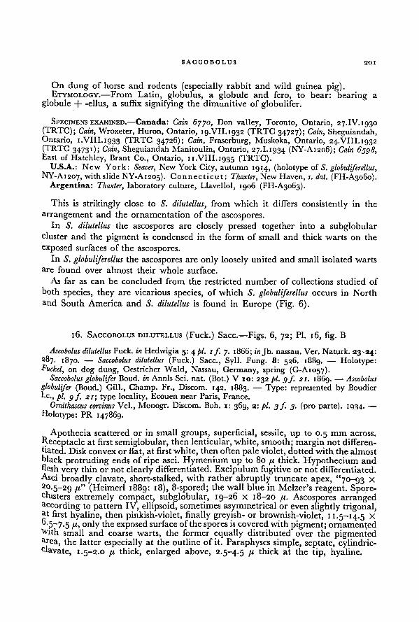
SACCOBOLUS 201
On dung of horse and rodents (especially rabbit and wild guinea pig).ETYMOLOGY.—From Latin, globulus, a globule and fero, to bear: bearing a
globule + -ellus, a suffix signifying the dimunitive of globulifer.
SPECIMENS EXAMINED.—Canada: Cain 6yyo, Don valley, Toronto, Ontario, 27.IV.1930(TRTC);
"
Cain, Wroxeter, Huron, Ontario, 19.VII.1G32 (TRTC 34727); Cain,, ,
Sheguiandah,Ontario, 1.VIII.1933 (TRTC 34726); Cain,
,Fraserburg, Muskoka, Ontario, 24.VIII. 1932
(TRTC 34731); Cain, SheguiandahManitoulin, Ontario, 27.1.1934 (NY-A1206); Cain 6598,East of Hatchley, Brant Co., Ontario, 11.VIII.1g35 (TRTC).
U.S.A.: New York: Seaver, New York City, autumn 1914, (holotype of S. globuliferellus,NY-A1207, with slide NY-A1205). Connecticut: Thaxter, New Haven, s. dat. (FH-A3060).
Argentina: Thaxter, laboratory culture, Llavellol, 1906 (FH-A3063).
This is strikingly close to S. dilutellus, from which it differs consistently in the
arrangement and the ornamentation of the ascospores.
In S. dilutellus the ascospores are closely pressed together into a subglobularcluster and the pigment is condensed in the form of small and thick warts on the
exposed surfaces of the ascospores.
In S. globuliferellus the ascospores are only loosely united and small isolated warts
are found over almost their whole surface.
As far as can be concluded from the restricted number of collections studied of
both species, they are vicarious species, of which S. globuliferellus occurs in North
and South America and S. dilutellus is found in Europe (Fig. 6).
16. SACCOBOLUS DILUTELLUS (Fuck.) Sacc. —Figs. 6, 72; Pl. 16, fig. B
Ascobolus dilutellusFuck, in Hedwigia 5: 4 pi. 1f_ „
7- 1 B66; injb. nassau. Ver. Naturk. 23-24:
287. 1870. — Saccobolus dilutellus (Fuck.) Sacc., Syll. Fung. 8: 526. 1889. — Holotype:Fuckel, on dog dung, Oestricher Wald, Nassau, Germany, spring (G-A1057).
Saccobolus globulifer Boud. in Annls Sci. nat. (Bot.) V 10: 232 pi. 9 f. 21. 1869. — Ascobolus
globulifer (Boud.) Gill., Champ. Fr., Discom. 142. 1883. — Type: represented by Boudier
he., pi. 9 f 21; type locality, Ecouen near Paris, France.
Ornithascus corvinus Vel., Monogr. Discom. Boh. 1: 369, 2: pi. 3f. 3. (pro parte). 1934. —
Holotype: PR 147869.
Apothecia scattered or in small groups, superficial, sessile, up to 0.5 mm across.
Receptacle at first semiglobular, then lenticular, white, smooth; margin not differen-
tiated. Disk convex or flat, at first white, then often pale violet, dottedwith thealmostblack protruding ends of ripe asci. Hymenium up to 80 /t thick. Hypothecium and
flesh very thin or not clearly differentiated.Excipulum fugitive or not differentiated.Asci broadly clavate, short-stalked, with rather abruptly truncate apex, "70-93 X
20.5-29 (Heimerl 1889: 18), 8-spored; the wall blue in Melzer's reagent. Spore-clusters extremely compact, subglobular, 19-26 X 18-20 /i. Ascospores arrangedaccording to pattern IV, ellipsoid, sometimes asymmetrical or even slightly trigonal,at first hyaline, then pinkish-violet, finally greyish- or brownish-violet, 11.5-14.5 X
fl-5~7-5 only the exposed surface of the spores is covered with pigment; ornamented
tvith small and coarse warts, the former equally distributed over the pigmentedarea, the latter especially at the outline of it. Paraphyses simple, septate, cylindric-clavate, 1.5-2.0 fi thick, enlarged above, 2.5-4.5 P thick at the tip, hyaline.
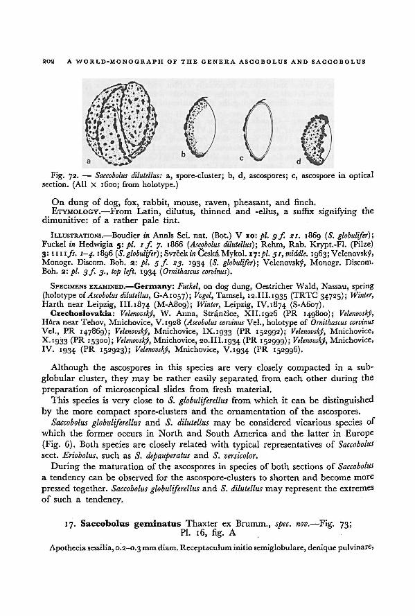
202 A WORLD-MONOGRAPH OF THE GENERA ASCOBOLUS AND SACCOBOLUS
On dung of dog, fox, rabbit, mouse, raven, pheasant, and finch.
ETYMOLOGY.—From Latin, dilutus, thinned and -ellus, a suffix signifying the
dimunitive: of a rather pale tint.
ILLUSTRATIONS.—Boudier in Annls Sci. nat. (Bot.) V 10: pi. gf. si. 1869 ( S. globulifer)',Fuckel in Hedwigia 5: pi. 1 f. 7. 1866 ('Ascobolus dilutellus) ; Rehm, Rab. Krypt.-Fl. (Pilze)
3: mi fs. 1-4. i8g6 (S. globulifer); Svrcekm CeskaMykol. 17: pi. 31, middle. 1963;Velenovsky,
Monogr. Discom. Boh. 2: pi. 5 f. 23. 1934 (S. globulifer)-, Velenovsky, Monogr. Discoro.
Boh. 2: pi. 3 f. 3., top left. 1934 ( Ornithascus corvinus).
SPECIMENS EXAMINED.—Germany: Fuckel, on dog dung, Oestricher Wald, Nassau, spring
(holotype ofAscobolus dilutellus, G-A1057); Vogel, Tamsel, 12.III.1935 (TRTC 34725); Winter,
Harth near Leipzig, III.1874 (M-A809); Winter, Leipzig, IV. 1874 (S-A607).
Czechoslovakia: Velenovsky, W. Anna, Strancice, XII.1926 (PR 149800); Velenovskj,Hura near Tehov, Mnichovice, V.1928 (Ascobolus corvinus Vel., holotype of Ornithascus corvinus
Vel., PR 147869); Velenovskj, Mnichovice, IX.1933 (PR 152992); Velenovsky, Mnichovice,
X.1933 (PR 15300); Velenovsky, Mnichovice, 20.III.1934 (PR 152999); Velenovsky, Mnichovice,
IV. 1934 (PR 152923); Velenovsky, Mnichovice, V.1934 (PR 152996).
Although the ascospores in this species are very closely compacted in a sub-
globular cluster, they may be rather easily separated from each other during the
preparation of microscopical slides from fresh material.
This species is very close to S. globuliferellus from which it can be distinguished
by the more compact spore-clusters and the ornamentation of the ascospores.
Saccobolus globuliferellus and S. dilutellus may be considered vicarious species of
which the former occurs in North and South America and the latter in Europe
(Fig. 6). Both species are closely related with typical representatives of Saccobolus
sect. Eriobolus, such as S. depauperatus and S. versicolor.
During the maturation of the ascospores in species of both sections of Saccobolus
a tendency can be observed for the ascospore-clusters to shorten and become more
pressed together. Saccobolus globuliferellus and S. dilutellusmay represent the extremes
of such a tendency.
17. Saccobolus geminatus Thaxter ex Brumm., spec. nov. —Fig. 73;
Pl. 16, fig. A
Apotheciasessilia, 0.2-0.3 mm diam. Receptaculum initio semiglobulare, denique pulvinare,
a, spore-cluster; b, d, ascospores; c, ascospore in opticalsection. (All X 1600; from holotype.)
Fig. 72. — Saccobolus dilutellus:
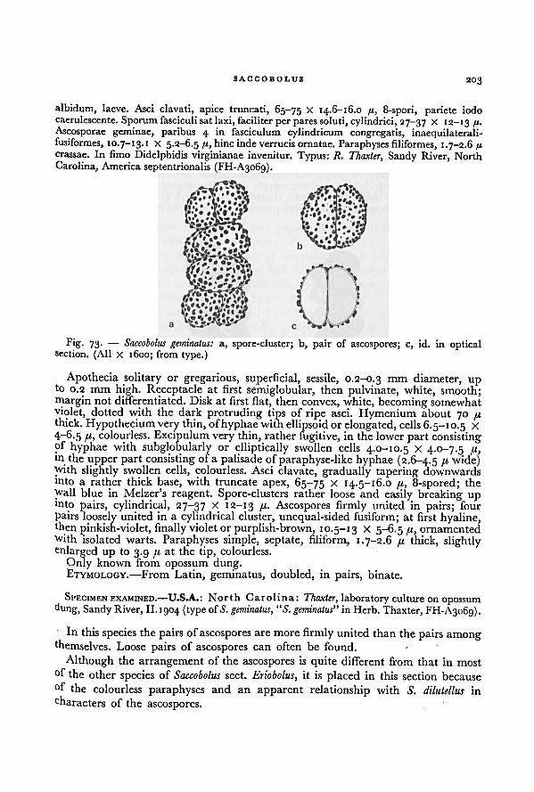
SACCOBOLUS 203
albidum, laeve. Asci clavati, apice truncati, 65-75 x 14.6-16.0 /<, 8-spori, pariete iodo
caerulescente. Sporum fasciculi sat laxi, faciliter per pares soluti, cylindrici, 27-37 x 12-13 fi.
Ascosporae geminae, paribus 4 in fasciculum cylindricum congregatis, inaequilaterali-fusiformes, 10.7-13.1 X 5.2-6.5 /i, hinc inde verrucis ornatae. Paraphyses filiformes, 1.7-2.6 fi
crassae. In fimo Didelphidis virginianae invenitur. Typus: R. Thaxter,,
Sandy River, North
Carolina, America septentrionalis (FH-A3o6g).
Apothecia solitary or gregarious, superficial, sessile, 0.2-0.3 mm diameter, upto 0.2 mm high. Receptacle at first semiglobular, then pulvinate, white, smooth;margin not differentiated. Disk at first flat, then convex, white, becoming somewhat
violet, dotted with the dark protruding tips of ripe asci. Hymenium about 70 /xthick. Hypothecium very thin, ofhyphae with ellipsoid or elongated, cells 6.5-10.5 X
4-6.5 ft, colourless. Excipulum very thin, rather fugitive, in the lower part consistingof hyphae with subglobularly or elliptically swollen cells
4.0-10.5 X 4.0-7.5 /x,in the upper part consisting of a palisade of paraphyse-like hyphae (2.6-4.5 !l wide)with slightly swollen cells, colourless. Asci clavate, gradually tapering downwardsinto a rather thick base, with truncate apex, 65-75 X 14.5-16.0 /x, 8-spored; the
Wall blue in Melzer's reagent. Spore-clusters rather loose and easily breaking upmto pairs, cylindrical, 27-37 X 12-13 [x. Ascospores firmly united in pairs; four
pairs loosely united in a cylindrical cluster, unequal-sided fusiform; at first hyaline,then pinkish-violet, finally violetor purplish-brown, 10.5-13 X 5-6.5 a, ornamented
With isolated warts. Paraphyses simple, septate, filiform, 1.7-2.6 /< thick, slightlyenlarged up to 3.9 jx at the tip, colourless.
Only known from opossum dung.ETYMOLOGY.—From Latin, geminatus, doubled, in pairs, binate.
SPECIMEN EXAMINED.—U.S.A.: North Carolina: Thaxter, laboratory culture on opossum
dung, Sandy River, II.1904 (type of S. geminatus, “S. geminatus” in Herb. Thaxter, FH-A3069).
In this species the pairs ofascospores are more firmly united than the pairs amongthemselves. Loose pairs of ascospores can often be found.
Although the arrangement of the ascospores is quite different from that in most
°f the other species of Saccobolus sect. Eriobolus, it is placed in this section because
°f the colourless paraphyses and an apparent relationship with S. dilutellus in
characters of the ascospores.
Fig. 73. — Saccobolus geminatus: a, spore-cluster; b, pair of ascospores; c, id. in opticalsection. (All X 1600; from type.)
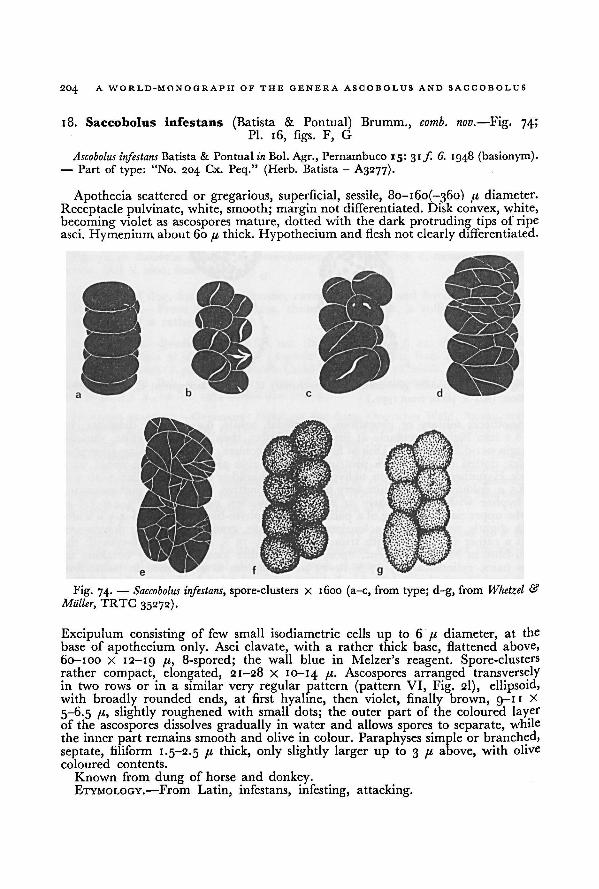
204 A WORLD-MONOGRAPH OF THE GENERA ASCOBOLUS AND SACCOBOLUS
18. Saccobolus infestans (Batista & Pontual) Brumm., comb. nov. —Fig, 74;Pl. 16, figs. F, G
Ascobolus infestans Batista & Pontual in Bol. Agr., Pernambuco 15:31 f. 6. 1948 (basionym).—
Part of type: "No. 204 Cx. Peq." (Herb. Batista- A3277).
Apothecia scattered or gregarious, superficial, sessile, 8O-I6O(-36O) /T diameter.
Receptacle pulvinate, white, smooth; margin not differentiated.Disk convex, white,
becoming violet as ascospores mature, dotted with the dark protruding tips of ripeasci. Hymenium about 60 /i thick. Hypothecium and flesh not clearly differentiated.
Excipulum consisting of few small isodiametric cells up to 6 /.i diameter, at the
base of apothecium only. Asci clavate, witha rather thick base, flattened above,
60-100 x 12-19 /x, 8-spored; the wall blue in Melzer's reagent. Spore-clustersrather compact, elongated, 21-28 X 10-14 fx. Ascospores arranged transverselyin two rows or in a similar very regular pattern (pattern VI, Fig. 2I), ellipsoid,with broadly rounded ends, at first hyaline, then violet, finally brown, 9—1 1 X
5-6.5 /x, slightly roughened with small dots; the outer part of the coloured layerof the ascospores dissolves gradually in water and allows spores to separate, while
the inner part remains smooth and olive in colour. Paraphyses simple or branched,
septate, filiform 1.5-2.5 fx thick, only slightly larger up to 3 [x above, with olive
coloured contents.
Known from dung of horse and donkey.ETYMOLOGY.—From Latin, infestans, infesting, attacking.
spore-clusters X 1600 (a-c, from type; d-g, from Whetzel &
Müller, TRTC 35272).
Saccobolus infestans,Fig. 74. —
SACCOBOLUS 205
ILLUSTRATIONS.—Batista & Pontual in Bol. Agr., Pernambuco 15: 31 f. 6. 1948.
SPECIMENS EXAMINED.—Tahiti: L. S. Olive,
on horse dung, Paea District, 24.IV. 1956(TRTC 35274); L. S. Olive, on horse dung, Tautira, 27.VI.1956 (TRTC 35273).
Panama: S. L. Meyer ij8g2, on horse dung, Salud, 6.IV.1945 (NY).Venezuela: Whelzel & Miiller, on donkey dung, Los Choros, near Caracas, 29.1.1942
(TRTC 35272).Brazil: Batista & Pontual, on horse dung, s. loc., s. dat. ("No. 204 Cx. Peq.", part of type
of Ascobolus infestans, Herb. Batista - A3277).
This species is known from Tahiti and tropical America. It is characterized by
the special arrangement of the ascospores (Fig. 2k-p).It occupies a rather isolated position in Saccobolus sect. Eriobolus because of the
aberrant type of ascospore arrangement and the coloured contents of paraphyses.It is tentatively placed in this section, together with S. geminatus in which the asco-
spores are also transversely arranged in the spore-cluster.
This species could not be recognized from the insufficient original description,
apart from this it had been unexpectedly placed in Ascobolus. My knowledge of
this species in the fresh condition is mainly due to Drs. R. F. Cain and L. S. Olive,who kindly placed at my disposal their material and their description after fresh
material.
206
CHAPTER XII
INSUFFICIENTLY KNOWN AND EXCLUDED SPECIES
The following species have been referred, at some time or other, to the genera
placed here under the Ascoboloideae. Many are excluded from this subfamily
because they do not answer to the accepted character of that group. The remaining
species are insufficiently known on account of the lack of sufficient descriptionswhich are not supported by adaequate material. Among these insufficiently known
species some might still be re-instated when good material is collected: of these
species the descriptions are cited below.
albicans. — Ascobolus albicans Fuck, in Hedwigia 1:3 pi. if. 2. 1866; Fungi rhen.
No. 1855. 1866. — Peziza (Sarcoscypha) albicans (Fuck.) Cooke, Mycographia 135
t>l. 60f. 234. 1877. — Neottiella albicans (Fuck.) Sacc., Syll. Fung. 8: 191. 1889. —-
Lasiobolus albicans (Fuck.) Sacc., Syll. Fung. 8: 538. 1889. — Lachnea albicans (Fuck.)Rehm, Rab. Krypt.-Fl., Pilze 3: 1068. 1895. — Type distribution: Fuckel, Fungi
rhen. No. 1855.
Boudier (1907: 64) listed this as a species of Neottiella (Cooke) Sacc. emend. Boud.
amethysteus. — Ascophanus amethysteus Quel, in Bull. Soc. bot. Fr. 25: 291-
1878. —Ascobolus amethysteus (Quel.) Gill., Champ. Fr., Discom. 143. 1883; Quel-,
Ench. Fung. 297. 1886. — Type locality: Jura, France.
The transfer of this species to Ascobolus by Gillet (I.e.) and Quelet (I.e.) is certainly
due to their very broad conception of the genus. According to its description it
belongs to Ascophanus Boud.
amethystinus. — Ascobolus amethystinus Phill. in Grevillea 4: 84. 1875. —
Galactinia amethystina (Phill.) Wakef. in Trans. Brit, mycol. Soc. 6: 375. 1920.—
Lectotype (selected by Wakefield, I.e.): the fungus described by Cooke (1876a:
48) under the name Peziza phillipsii Cooke.
When Phillips (1875) described Ascobolus amethystinus two species were involved:
a species of Ascobolus and a species described from the same collection by Cooke
(1876a) as Peziza phillipsii Cooke.
Cooke (1876a: 48) did not exclude Ascobolus amethystinus from Ascobolus. But
Wakefield (I.e.) considered the fungus described by Cooke identical with Ascobolus
amethystinus, and excluded it from Ascobolus.
INSUFFICIENTLY KNOWN AND EXCLUDED SPECIES 207
The Ascobolus element belongs to A. behnitziensis Kirschst. According to Wakefield
A. amethystinus must be called Galactinia amethystina (Phill.) Wakef. The positionof this species in Galactinia (Cooke) Boud. is problematic, since no part of the ascus-
Wall stains blue with iodine.
The description by Cooke agrees with my observationof the type. In his ascospore
measurements the ornamentation is included. Normally this ornamentation is
colourless, but it may be stained by the hymenial pigment (Massee & Crossland,
1906; Wakefield, I.e.).
applanatus. — [Peziza applanata (Hedw.) per Fr. sensu Gonn. & Rab., Mycol.eur. pi. 6 f 5. 1869 (without text and lacking authors' citation). —] Phaeopezia
applanata Sacc., Syll. Fung. 8: 472. 1889 [exclusive of type, viz. Peziza applanata
(Hedw.) per Fr., see p. 92; "(Rab. et Gonn.)"]; not Phaeopezia applanata (Hedw.per Fr.) Sacc. in Bot. Cbl. 18: 218. 1884 ["(Rabenh. et Gonn.)"; basionym, Peziza
applanata (Hedw.) per Fr. which was not yet explicitely excluded], — Aleurina
applanata (Sacc.) Sacc. & Syd. in Syll. Fung. 16: 739. 1902. — Ascobolus applanatus(Sacc.) Rehm, Rab. Krypt.-Fl., Pilze 3: 1131. 1896. —
Aleuria applanata (Sacc.)Boud., Hist. Class. Discom. Eur. 46. 1907. — Type: represented by Gonn. & Rab.,Mycol. eur. pi. 6 f. 5; type locality: presumably Germany.
This species with hyaline, ellipsoid ascospores and brown paraphyses is based
on the figure by Gonnermann and Rabenhorst (I.e.) the accompanying text of
Which has never been published. Saccardo (1889: 472) conceived it as a new speciesof his genusPhaeopezia. It is, however, very unlikely that Gonnermann & Rabenhorst
Would have overlooked the name Peziza applanata (Hedw.) per Fr. for a strikinglysimilar species. In my opinion Gonnermann & Rabenhorst intended to illustrate
Hedwig's species.
Boudier (1907: 46, 48) transferred Saccardo's species to Aleuria (Fr.) Boud.,While he referred Hedwig's species to the synonymy of Galactinia castanea Quel.
According to current conception both species belong to Galactinia (Cooke) Boud.
emend. Le Gal, where their position is still rather doubtful.
argenteus Curr. — Ascobolus argenteus Curr. in Trans. Linn. Soc. (Bot.) 24:
496. 1864; not Ascobolus argenteus Renny in Trans. Woolhope Nat. Field Club 1871:46- 1871. — Ascophanus argenteus (Curr.) Boud. in Annls Sci. nat. (Bot.) V 10: 245. —
type locality: Eltham, British Isles.
A species of Ascophanus Boud.
argenteus Renny. — Ascobolus argenteus Rcnny in Trans. Woolhope Nat. Field
1871: 46. 1871; not Ascobolus argenteus Curr. in Trans. Linn. Soc. (Bot.) 24: 496.
208 A WORLD-MONOGRAPH OF THE GENERA ASCOBOLUS AND SACCOBOLUS
1864. — Rhyparobius argenteus (Renny) Berk. & Broome in Ann. Mag. nat. Hist. IV
11: 347 pi. g f 11. Mai 1873. — Ascozonus argenteus (Renny) Boud., Hist. Class.
Discom. Eur. 79. 1907. — Type locality: Hereford, British Isles.
This typical representative of Ascozonus (Renny) E. C. Hansen was reduced to
the synonymy ofAscobolus (Ascozonus) cunicularius (Boud.) Renny by Renny (1874: 356).
asininus. — Ascobolus asininus Cooke & Mass. apud Cooke in Grevillea 21: 72.
1 893. — Type specimen: not known to be in existence.— Type locality: Kew,
England.
This fungus is only known from the original description and has never been
found again. The description is insufficient to place the species definitely in the
genus Ascobolus.
The original description (Cooke, I.e.) runs: —
"Scattered or gregarious, hemispherical or sub-depressed, watery, fleshy, at first
umbilicate, then open, with a thick margin, indistinctly rugulose, pale olive green
or amber, or a combination of both. Asci broadly clavate, octosporous, sporidia
elliptical, at first colourless, at length reddish brown, epispore thick, finally minutelycracked into subhexagonal minute areolae, paraphyses very long, Aliform, flexuous,
much longer than the asci. On asses' dung. Kew."
"Cups 1-2 mm diam., asci 160-170 X 45-50 p, sporidia 40-45 X 21-23 /.«•"
atrovirens.— Peziza atrovirens Pers., Syn. meth. Fung. 2: 635. 1801 (devalidated
name). — Ascobolus atrovirens (Pers.) Nees, Syst. Pilze Schwamme 269. 1816
(devalidated name). — Peziza atrovirens Pers. per Pers., Mycol. eur. 1: 306. 1822;
Fries, Syst. mycol. 2 (1): 141. 1822.— Calloria atrovirens (Pers. per Pers.) Fr., Summa
Veg. Scand. 2: 359. 1849. — Ombrophila atrovirens (Pers. per Pers.) P. Karst. in
Bidr. Kann. Finl. Nat. Folk. 19: 92. 1871. — Mollisiaatrovirens (Pers. per Pers.) Gill-)
Champ. France, Discom. 126. 1882. — Coryne atrovirens (Pers. per Pers.) Sacc.,
Syll. Fung. 8: 641. 1889. — Tympanis atrovirens (Pers. per Pers.) Rehm, Ascom-
exs. Fasc. 13, No. 618. 1882; Rehm in Hedwigia 21: 70. 1882. — Corynella atrovirens
(Pers. per Pers.) Boud., Hist. Class. Discom. Eur. 99. 1907. — Chlorosplenium atrovirens
(Pers. per Pers.) Seaver, N. Am. Cup-fungi (Inoperc.) 106. 195 1.
This species belongs to the genus Corynella Boud. It is one of the two species
mentioned in the original description of the genus (Boudier, 1885: 114) and d
is now considered type of the name Corynella.
aurora. — Peziza aurora Crouan, Fl. Finist. 53. 1867. — Ascophanus aurora (Couan)Boud. in Annls Sci. nat. (Bot.) V 10: 248, pi. 11 f. 36. 1869. — Ascobolus aurora
(Crouan) Cooke in Grevillea 1: 132. 1873. — Ascobolus testaceus var. aurora (Crouan)
INSUFFICIENTLY KNOWN AND EXCLUDED SPECIES209
Quel., Ench. Fung. 297. 1886. ("auroreus"). — Type: Crouan, on very old cow
dung, s. loc., 15.X.1866 (CONC-A2447).
A representative of Ascophanus Boud.
barbatus. —Ascobolus barbatus Mass. & Grossl. apud Mass. in Grevillea 22: 97.
1894. — Type: H. Wagner, on tan refuse, Leeds, British Isles, 6.XII.1893 (NY-A.2
59); 1.XII.1893 (K-A2489); 9.XII.1893 (K-A1982).
Ascobolus barbatus is a nomen confusum, and as such should be rejected. The
parts of the type cited above originally belonged together, although different dates
Were given. They consist of immature fruit-bodies of a species of Scutellinia (Gooke)Lamb, covered with ascospores from neighbouring old apothecia of Ascobolus
denudatus. Massee & Crossland described this heterogeneous combination as a singlespecies.
Denison (1959: 630) also studied type material and came to the same conclusion.
brunneus. — Rhyparobius brunneus Boud. in Annls Sci. nat. (Bot.) V 10: 237
pl- Q f. 23. 1869. — Ascobolus brunneus (Boud.) Gooke in Grevillea 5: 153. 1876;
not Ascobolus brunneus Cooke, Handb. Brit, fungi 728. 1871. — Type locality:
Montmorency near Paris, France.
This fungus with 32 ascospores per ascus belongs to Rhyparobius Boud.
brunneus. — Ascobolus brunneus Cooke in Fung. brit. exs. No. 286. 1867 (nomen
nudum); Cooke in Hedwigia 6: 154. 1867 (nomen nudum); Gooke, Handb. Brit.
Ling. 728. 1871 (with description); not Ascobolus brunneus (Boud.) Cooke in Grevillea
5 : 153. 1876. — Dasyobolus brunneus (Gooke) Sacc., Syll. Fung. 11: 421. 1895. —
Type distribution: Cooke, Fungi brit. exs. No. 286.
This name has been a source of confusion since its publication. It was often
'nterprcted as a species of Dasyobolus or of Ascobolus, related to Ascobolus amoenus.
The writer examined different parts of the type distribution and found Ascobolus
brunneus Cooke to be nothing else but Lasiobolus pilosus (Fr.) Sacc. of which the
contents of theascospores do become pale brownish with age (PI. 16, fig. E). Cooke's
tame thus appears to be a later synonym.
burcardius. — [Burcardia globosa, cortice reticulato Schmidel, Icon. PI. 261 pl. 6gf s
- 1-13. 1797 (“Burkardia” under plate; devalidated name). —] Peziza burcardia
"ers., Syn. Fung. 632. 1801 (first binomium, devalidated name). — Ascobolus
burcardius (Pers.) Martius, Fl. crypt. Erlang. 471. 1817 ("burcardia", devalidated
210 A WORLD-MONOGRAPH OF THE GENERA ASCOBOLUS AND SACCOBOLUS
name). — Peziza (Burcardia) burcardia Pers. per Pers., Mycol. eur. i: 319. 1822. —-
Type: represented by Schmidel I.e., pi. 6g.
As Burcardia globosa is the type of the generic name Bulgaria Fr., the correct name
for this species is Bulgaria globosa (Schmidel) ex Fr. See also under 'inquinans'.
candidus. — Ascobolus candidus Schroet. in Krypt.-Fl. Schles. 3 (2): 55. 1893. —
Type specimen: not known to be in existence. — Type locality: Oswitz, Wroclav,
Poland.
This species is unknown to the writer. Ascobolus candidus Schroet. sensuSeaver
(1928: 86) is Ascobolus aglaosporus Heimerlwhich has larger ascospores and a different
spore-sculpturing.Ascobolus candidus undoubtedly belongs to the genus Ascobolus and is likely to be
found again.
Schroeter's description runs: —
"Fruchtkorper zerstreut, sitzend, flach gewolbt, 0.5-1 mm breit, schneeweiss,
glatt. Scheibe unberandet, zuletzt von den vortretenden Schlauchen schwarz
punktiert. Schlauche keulenformig, am Scheitel abgerundet, nach unten stark
zusammengezogen, bis 120 // lang, 22 fi breit, 8 sporig. Sporen 2 reihig, im oberen
Drittel des Schlauches ruhend, ellipsoidisch, n-13 //, lang, 6-8 11 breit; Membran
lange Zeit farblos, zuletzt hell-violett, glatt. Paraphysen fadenformig, 2-3 fx breit.
"Auf Hasenmist. November, Dezember. — Breslau: Oswitz."
caninus.— Ascobolus caninus Auersw. in Hedwigia 7: 52. 1868; not Ascobolus
caninus Fuckel in Hedwigia 5: 3. 1866. — Rhyparobius caninus (Auersw.) Sacc., Syll-
Fung. 8: 539. 1889. — Rhyparobius crustaceus var. caninus (Auersw.) Boud., Hist.
Glass. Discom. Eur. 78. 1907. — Type locality: Rosenthal, Leipzig, Germany.
A species of Rhyparobius with 24-32 ascospores.
carneus. — Ascobolus carneus Pers., Syn. Fung. 1: 676. 1801 (devalidated name). —'
Ascobolus carneus Pers. per Pers., Mycol. eur. 1: 341. 1822; Fries, Syst. mycol. 2:
165. 1822. —• Ascophanus carneus (Pers. per Pers.) Boud. in Annls Sci. nat. (Bot.)V 10: 250. 1869. — Peziza carnea (Pers. per Pers.) P. Karst. in Notis. Sallsk. F. Fl-
fenn. Forh. 10: 120. 1869. — Pyronema carneum (Pers. per Pers.) Schroet. W
Krypt.-Fl. Schl. 3 (2): 34. 1893. — Type locality: Germany.
One of the most common species of Ascophanus. Ascophanus carneus was chosen as
lectotype of the generic name Ascophanus Boud. by Clements & Shear (1931: 33°)
and Korf (1958). As this agrees with Boudier's conception (1885) and with Heimerl s
introduction (1889: 20) of Ascophanus sect. Eu-Ascophanus, this typification is followed
here. The choice ofA. cinereus (Crouan) Boud. by Saccardo (1884: 219) as represent-
INSUFFICIENTLY KNOWN AND EXCLUDED SPECIES 211
ative species and the indication according to the first species rule of A. subfuscus
(Grouan) Boud., by Seaver (1927: 87) were not adopted thus far.11
carneus forma conglobatus. — Ascobolus carneus forma conglobatus Rehm in
Ascom. No. 425. 1878 (nomen nudum). — Type distribution: Rehm, Ascom.
No. 425.
One of the many variations of Ascophanus carneus. The name is not cited in later
Work by Rehm.
carneus var. cuniculi. — Ascophanus carneus var. cuniculi Boud. in Annls Sci.
nat. (Bot.) V 10: 250 pi. 12 f. jg. 1869. —Ascobolus carneus var. cuniculi (Boud.)
Pim in Grevillea 9: 57. 1880. — Type locality: France.
One of the many variations of Ascophanus carneus.
cenangioides. —Ascobolus cenangioides Ces. in Atti Accad. Sci. fis. mat. Napoli
8 (3): 11. 1879. — Type: not known to be in existence. — Type locality: Sarawak,Borneo.
Judging from the description this species does neither belong to the Ascobolaceae,
ftor to the other families of the operculate Discomycetes. The description is insuf-
ficient to place the species.
cesatii. — Ascobolus cesatii Garest. in Rab., Fungi europ. exs. No. 976a. 1866. —
Pezizula cesatii (Carest.) P. Karst. in Bidr. Kann. Finl. Nat. Folk. 19: 83. 1871. —
Rhyparobius cesatii (Carest.) P. Karst. in Acta Soc. Fauna Fl. fenn. II 6: 122. 1885. —
Ascophanus cesatii (Carest.) Sacc., Syll. Fung. 8: 533. 1889. — Type distribution:
Rabenhorst, Fungi europ. exs. No. 976a.
This name is a synonym of Ascophanus microsporus (Berk. & Broome) E. C. Hansen.
ciliatus. — Ascobolus ciliatusJ. G. Schmidt in Mykol. Hefte 1: 90. 1817 (devalidated
ftame). — Ascobolus ciliatus J. C. Schmidt per Pers., Mycol. europ. 1: 340. 1822;Pries, Syst. mycol. 2: 164. 1822. — Ascobolus papillatus var. ciliatus (J. G. Schmidt
Per Pers.) Kickx, Fl. crypt. Flandres 1: 476. 1867. — Ascophanus ciliatus (J. C.
uHowever, very recently Kimbrough & Korf (1967: 19) supported Seaver's typification,
jHiich according to them makes Ascophanus Boud. a synonym of Thelebolus_ w . ,
Tode per Fr.
A'or ' * ' ' "
A. carneus and its close allies they introduced the generic name Iodophanus, 0
- —r
1 Korf apudKimbr. & Korf.
A WORLD-MONOGRAPH OF THE GENERA ASCOBOLUS AND SACCOBOLUS212
Schmidt per Pers.) Boud. in Annls Sci. nat. (Bot.) V 10: 253. 1869. —Ascobolus
equinus subsp. ciliatus (J. G. Schmidt per Pers.) P. Karst. in Not. Sallsk. Fauna
Flora fenn. Forh. 10: 209. 1870. — Peziza equina var. ciliata (J. C. Schmidt per
Pers.) P. Karst. in Bidr. Kann. Finl. Nat. Folk. 19: 73. 1871. — Ascophanus pilosus var.
ciliatus (J. C. Schmidt per Pers.) Phill., Discom. 312. 1887. — Lasiobolus equinus
subsp. ciliatus (J. C. Schmidt per Pers.) Sacc., Syll. Fung. 8: 537. 1889. — Lasiobolus
ciliatus (J. C. Schmidt per Pers.) Boud., Hist. Glass. Discom. Europ. 78. 1907. —
Type locality: near Schandau, Germany.
This species of Lasiobolus is characterized by a disk with a swollen margin, which
is covered with a white powder and cilia of the same colour. Rehm (1895: 1096)
reduced Ascobolus ciliatus together with many other names to the synonymyof
Lasiobolus equinus. In his remarks to this species he doubts whether Ascophanus ciliatus
sensu Boudier (1869: 253) belongs to it. From Boudier's description, however, it is
clear that his species is the same as Schmidt's. As I have not yet come across this
species, I hesitate to place it into the synonymy of Lasiobolus pilosus.
ciliatus var. citrinus. —Ascobolus ciliatus var. citrinus Crouan, Fl. Finist. 55-
1867. — Streptotheca citrina (Crouan) Le Gal in Annls Sci. nat. (Bot.) XII 1: 451-
i960. — Type: Crouan, on dog dung, s. loc., autumn (A. citrinus Crouan, CONC-
A2424, lectotype; CONC-A2412, probably also part of type).
After studying both specimens and a small authentic drawing in colour, onthe
ground of observations, deviating at some points from those of Le Gal (ig® 1 '
450-451), my conclusion is that Crouan's variety differs in nothing from typicalLasiobolus pilosus but the lemon-yellow colour.
cinerellus.—
Ascobolus cinerellus P. Karst., Fungi Fenn. exs. No. 760. 1868;
P. Karst. in Not. Sallsk. Fauna Fl. fenn. Forh. II:2?>6. 1870. — Peziza cinerella (P-
Karst.) P. Karst. in Bidr. Kann. Finl. Nat. Folk 19: 51. 1871. — Ascophanus cinerellus
(P. Karst.) Speg. in Ann. Soc. cient. argent. 10: 24. 1880. — Type distribution:
P. Karsten, Fungi Fenn. exs. No. 760.
A species of the genus Ascophanus Boud. (fide Spegazzini, I.e.; Karsten, 1885: 12G
Saccardo, 1889: 532; Heimerl, 1889: 25; Rehm, 1895: 1085; Boudier, 1907: 77)'
cinereus.— Ascobolus cinereus Crouan in Annls Sci. nat. (Bot.i IV 10: 194
pi. 13D fs. iy-20. 1858. — Ascophanus cinereus (Crouan) Boud. in Annls Sci. nat.
(Bot.) V 10: 249. i86g. — Peziza cinerea (Crouan) P. Karst. in Bidr. Kann. Fink
Nat. Folk 19: 59. 1871. — Thecotheus cinereus (Crouan) Chenant. in Bull. Soc. mycoh
Fr. 34: 39. 1918. — Thecotheus setisperma Le Gal in Bull. Soc. mycol. Fr. 78: 41'•
1963 (superfluous name change). — Type: Crouan, on old cow dung, near Brest,
Finistere, France, s. dat. (CONC-A2395, Cotype PC-A2336).
INSUFFICIENTLY KNOWN AND EXCLUDED SPECIES 213
Boudier (1869: 250) placed this species with some doubt in the genus Ascophanus.Chenantais (1918: 39) considered Ascobolus cinereus to be closely related to Ascophanus
holmskjoldii and, on account of their ascospores, transferred both of them to the genusThecotheus Boud. Le Gal (1961: 443; 1963b: 405) who studied the type, argued that
it should be kept in the latter genus.
Thecotheus setisperma Le Gal is a superfluous name, since there is a type specimenand even a cotype. Moreover, if Ascophanus holmskjoldii E. C. Hansen is to be
considered a synonym of Grouan's species (Le Gal 1963: 406), then Hansen's name
should have priority. See also under 'holmskjoldii.'
coccineus. — Ascobolus coccineus Crouan in Annls Sci. nat. (Bot.) IV 7: 174pi. 4Dfs. 15-ig. 1857. — Leucoloma coccinea (Crouan) Fuck., Fungi rhen. No. 1854.
1866; Fuck, in Jb. nassau. Ver. Naturk. 23-24: 318. 1870. — Peziza corallina Cooke
in Grevillea 3: 73. 1874 (name change); not Peziza coccinea Jacq., Fl. austr. pi. i6g.1774. — Humaria coccinea (Crouan) Quel., Ench. Fung. 289. 1886.
—Humarina
coccinea (Crouan) Seaver, N. Am. Cup-fungi (Operc.) 137. 1928. — Type: Crouan,on soil among small mosses, under Chateau de Brest, and in the neighbourhoodof Morlaix, Finistere, France, spring, autumn (CONC-A2389); Crouan, on soil
among small mosses, near Brest, Finistere, France, s. dat. (PC-A2337, probablypart of type).
This species is related to Octospora tetraspora (Fuck.) Korf and Octospora fusispora 12
and should likewise be placed in the genus Octospora Hedw. per S.F. Gray emend.
Korf among the species with fusiform ascospores as Octospora coccinea (Crouan)Brumm., comb. nov.
coemansii. — Ascophanus coemansii Boud. in Annls Sci. nat. (Bot.) V 10: 244
pl. 10 f. go. 1869. — Ascobolus coemansii (Boud.) Quel., Ench. Fung. 296. 1886.—
Type locality: near Paris, France.
A species of Ascophanus of the group of Ascophanus microsporus (Berk. & Broome)E. C. Hansen.
conglomeratus. —Ascobolus conglomeratus Schwein. in Trans. Am. phil. Soc. II 4:
j78. 1832. — Angelina conglomerata (Schwein.) Fr. in Nova Acta Soc. Sci. upsal.Hi 1: 121. 1851. — Type locality: Carolina, U.S.A.
This is the type of the name Angelina Fr. According to Duby (1862: 51-52)Ascobolus conglomeratus falls into the synonymy of Angelina rufescens (Schwein.) Duby,a species of Hysteriaceae.
12Octospora fusispora (Berk.) Brumm., comb. nov. — basionym: Peziza fusispora Berk.
ln J. Bot., Lond. 5: 5. 1846.
214 A WORLD-MONOGRAPH OF THE GENERA ASCOBOLUS AND SACCOBOLUS
consociatus.—
Ascobolus consociatus Berk. & Broome in Ann. Mag. nat. Hist.
Ser. IV 15: 39 pi. 2 f 7. 1875. — Ascophanus consociatus (Berk. & Broome) Phill.,
Brit. Discom. 312. 1887. — Type locality: Langridge, British Isles.
According to Phillips (1887: 312) and Boudier (1907: 77) this is a species of
Ascophanus Boud.
cookei.— Ascobolus cookei Crouan, Fl. Finistere 56. 1867. — Rhyparobius cookei
(Grouan) Boud. in Annls Sci. nat. (Bot.) V 10: 238. 1869. — Type: Crouan, on
album graecum, s. loc., 7.IV.1860 (in pencil also: III. 1862) (CONC-A2391).
One of the many synonyms of Rhyparobius crustaceus (Fuck.) Rehm. In the type
collection asci with 32, 48 and 64 ascospores were found. In the same package
Le Gal (1961: 449) also found fruit-bodies with at least 150-180 ascospores in an
ascus.
coronatus. — Ascobolus coronatus Schum., Enum. PI. Saell. 2: 437. 1803 (devalid-
ated name). —- Phacidium coronatum (Schum.) Fr., Obs. mycol. 1: 167. 1815
(devalidated name). — Phacidium coronatum (Schum.) per Fr., Syst. mycol. 2 (2):
577. 1823. — Coccomyces coronatus (Schum. per Fr.) De Not. in Erb. critt. ital.,
Ser. I, fasc. 5, No. 236. 1859. — Type locality: Bagsvaerd, Sjaelland, Denmark.
This species belongs to Coccomyces De Not. of the Phacidiales.
corvinus var. avium.— Ornithascus corvinus var. avium Veh, Monogr. Discom.
Boh. 1: 369. 1934. — Type: not known to be in existence. — Type locality: near
Bozkov, Bohemia, Czechoslovakia.
Velenovsky's description of this variety is too short and too incomplete to reduce
it with certainty to the synonymy of Saccobolus dilutellus. Considered a nomen
dubium.
crec'hqueraultii. — Ascobolus crec’hqueraultii Crouan in Annls Sci. nat. (Bot.)IV 10: 194 pi. 1jofs. 12-16. 1858. — Mollisia crec’hqueraultii (Crouan) Gill., Champ-
Fr., Discom. 118. 1882. — Humaria crec’hqueraultii (Crouan) Quel., Ench. Fung-288. 1886. •— Barlaea crec’hqueraultii (Crouan) Sacc., Syll. Fung. 8: 113. 1889. —
Lamprospora crec’hqueraultii (Crouan) Boud., Icon, mycol., Ser. I, livr. 2, Liste
preliminaire, unnumberedpage. 1904. — Barlaeina crec’hqueraultii (Crouan) Sacc. &
Trav. in Syll. Fung. 19: 138. 1910. — Type: Crouan, on soil on the banks of brooks,
s. loc., IX and X. 1858 (lectotype, PC-A2340); Crouan, on soil on the banks of brooks,
in the neighbourhood of Morlaix and Brest, Finistere, France, September (CONC-A2371, exhausted).
A well-knownspecies ofLamprospora De Not. with beautifully ornamentedascospores.
INSUFFICIENTLY KNOWN AND EXCLUDED SPECIES 215
crouanii. — Ascobolus crouanii Cooke in J. Bot., Lond. 2: 151 f. 3. / May 1864;
not A. crouanii Boud. in Annls Sci. nat. (Bot.) V 10: 216 pi. 3 f. 2. 1869; nor A.
crouanii Renny in J. Bot., Lond. 12: 356 pi. 134 fs. 6-10. 1874. — Peziza crouanii
(Cooke) Cooke in Grevillea 3: 185. 1875; Cooke, Mycographia 13 pi. jf. iy. 1875. —
Aleuria crouanii (Cooke) Gill., Champ. Fr., Discom. 50. 1879. — Crouania crouanii
(Cooke) Lamb., Fl. mycol. Belg., Suppl. I: 319. 1887. — Lamprospora crouanii (Cooke)Seaver in Mycologia 6: 8. 1914; s Ascobolus miniatus Crouan.
crouanii.— Ascobolus crouanii Renny in Trans. Woolhope Nat. Field Club
1872-3: 130 pi. 154fs. 6-10. 1873; Renny in J. Bot., Lond. 12: 356 pi. 154 fs. 6-10.
1874; not A. crouanii Cooke in J. Bot., Lond. 2: 151 f. 3. 1864; nor A. crouanii Boud.
in Annls Sci. nat. (Bot.) V 10: 216 pi. 5 f. 2. 1869. — Rhyparobius crouanii (Renny)Sacc., Syll. Fung. 8: 543. 1889. — Ascozonus crouanii (Renny) Schroet. in Bot. Jb.(ed. Just) 2: 283. 1876 (incidental mention); Boud., Hist. Class. Discom. Eur. 79.
1907. — Streptotheca crouanii (Renny) Seaver, N. Am. Cup-fungi (Operc.) 142.
1928. — Type locality: Hereford, British Isles.
A species of Ascozonus (Renny) Boud.
crustaceus.— Ascobolus crustaceus Fuck., Fungi rhen. No. 1858. 1866; in Hedwigia
5 ; 4- 1866. — Pezizula crustacea (Fuck.) P. Karst. in Bidr. Kann. Finl. Nat. Folk
*9: 81. 1871. — Rhyparobius crustaceus (Fuck.) Rehm, Ascom. exs. No. 52b. 1872. —
Type distribution: Fuckel, Fungi rhen. No. 1858.
A species of Rhyparobius Boud.
cunicularius. — Peziza cunicularia Boud. in Annls Sci. nat. (Bot.) V 10: 258.
1869. — Ascobolus cunicularius (Boud.) Renny in Trans. Woolhope Nat. Field Club
1872-3: 130. 1873; Renny in J. Bot., Lond. 12: 255. 1874; Cooke in Grevillea 3::85. 1875. — Ascozonus cunicularius (Boud.) Schroet. in Bot. Jb. (ed. Just) 2: 283.i876> (incidental mention); E. C. Hansen in Vidensk. Medd. naturh. Foren. Kbh.
1876: 91. 1877. — Type locality: Montmorency, France.
A species of Ascozonus (Renny) Boud. and very probably a synonym of Ascozonusleveillei (Crouan) Brumm.
daldinianus. — Ascobolus daldinianus De Not. in Comm. Soc. critt. ital. 5: 360.1864.. — Type: not known to be in existence. — Type locality: Locarnese, Italy.
From the original description it is not even certain that De Notaris' species
belongs to Ascobolus. As already suggested by Boudier (1869: 234) it might be a
sPecies of Saccobolus related to S. versicolor or S. depauperatus. A nomen dubium.
216 A WORLD-MONOGRAPH OF THE GENERA ASCOBOLUS AND SACCOBOLUS
difformis. — Ascobolus testaceus var. difformis P. Karst., Syn. Pez. Ascob. 43.
1861. — Ascobolus difformis (P. Karst.) P. Karst., Fungi Fenn. exs. No. 325. 1866;
Nyl. in Notis. Sallsk. Fauna Fl. fenn. Forh. 10: 85. 1868; P. Karst. in Notis. Sallsk.
Fauna Fl. fenn. Forh. 11: 207. 1870. — Ascophanus difformis (P. Karst.) Boud. in
Annls Sci. nat. (Bot.) V 10: 252. 1869. — Ascophanus carneus var. difformis (P. Karst.)
Rehm, Rab. Krypt.-Fl. (ed. 2), Pilze 3: 1095. 1895. — Type locality: Finland.
This fungus belongs to Ascophanus Boud. and is closely related to Ascophanus
carneus (Pers. per Pers.) Boud.
diversisporus. — Ascobolus diversisporus Fuck, in Jb. nassau. Ver. Naturk.
23-24: 289. 1870. —- Humaria diversispora (Fuck.) Speg. in Michelia 1: 236. 1878. —
Lasiobolus diversisporus (Fuck.) Sacc., Syll. Fung. 8: 538. 1889. — Type locality:
Frankensteiner Kopf, Germany.
According to the description this species belongs to Lasiobolus Sacc. Seaver (1928:
155) placed it into the synonymy of Lasiobolus equinus (O. F. Miiller per Pers.)
P. Karst. [= L. pilosus (Fr.) Sacc.].
dubius.— Rhyparobius dubius Boud. in Annls Sci. nat. (Bot.) V 10: 240 pi. 10f. 26.
1869. — Ascobolus dubius (Boud.) Quel., Ench. Fung. 296. 1886. — Type locality:
Montmorency, France.
A member of Rhyparobius Boud.
dubius. — Saccobolus dubius Vel., Monogr. Discom. Boh. 414. 1934. — Type
specimen: non-existent. — Type locality: near Mnichovice, Bohemia, Czecho-
slovakia.
Because of the very insufficient description Saccobolus dubius is to be regardedas a nomen dubium.
equinus. — Helvella equina O. F. Müller in Fl. dan. 5, Fasc. 13: 8 pi. yyg f 3-
1778 ("jElvela”, devalidated name). — Peziza equina (O. F. Miiller) Retz., Fl. Scand.
Prodr. (ed. 2): 325. 1795 (devalidated name); Sowerby, Engl. Fung. 3: 44 pi. 372.
1803. — Peziza stercorea var. equina (O. F. Miiller) per Pers., Mycol. europ. 1: 247-
1822. — Peziza equina (O. F. Miiller per Pers.) Sommerf., Plant, crypt, norv. Cent.
I, No. 98. 1826.— Ascobolus equinus (O. F. Miiller per Pers.) P. Karst. in Not.
Sallsk. Fauna Flora fenn. Forh. 11: 209. 1870. — Ascophanus equinus (O. F. Miillerper
Pers.) Speg. in An. Soc. ci. argent. 10: 24. 1880. — Lasiobolus equinus (O. F. Miiller
per Pers.) P. Karst. in Acta Soc. FaunaFl. fenn. II 6: 122. 1885. — Type: represented
by O. F. Miiller in Fl. Dan. 5, fasc. 13: pi. 77g f. 3.— Type locality: Denmark.
INSUFFICIENTLY KNOWN AND EXCLUDED SPECIES 217
It seems impossible to distinguish this fungus from Lasiobolus pilosus (see also
Heimerl, 1889: 23; Schroeter, 1893: 54; Rehm, 1895: 1096; Seaver, 1928: 155.).The distinction of even two varieties or subspecies within one species (Karsten,
1871: 73; Karsten, 1885: 122; and Boudier, 1907: 78) is untenable because of the
wide variation in almost all characters.
Since Lasiobolus pilosus is the earlier of the two names and because of the choice
of Boudier (1869: 254) and Heimerl (1889: 23) of this epithet from a greater series
of synonyms, I propose to use this name for the species in its broad sense.
equinus. — Saccobolus equinus Vel., Novit. mycol. 203. 1939. — Type specimen:non-existent. — Type locality: Mnichovice, Bohemia, Czechoslovakia.
A nomen dubium because of the insufficient description and the lack of a type
specimen.
fallax. — Ascobolus fallax Auersw. in Hedwigia 4: 52. 1868. — Ascobolus crustaceus
subsp. fallax (Auersw.) P. Karst. in Notis. Sallsk. Fauna Fl. fenn. Forh. 11: 208.
1870. — Pezizula crustacea subsp. fallax (Auersw.) P. Karst. in Bidr. Kann. Finl.
Nat. Folk 19: 82. 1871. — Ascophanus fallax (Auersw.) Sacc., Syll. Fung. 8: 532.
1889; Boud., Hist. Class. Discom. Eur. 77. 1907. — Type locality: Rosenthal,
Leipzig, Germany.
Although this species has the appearance of a Rhyparobius it should be placed in
Ascophanus Boud. because of the eight-spored asci.
felinus. — Rhyparobius felinus Boud. in Annls Sci. nat. (Bot.) V 10: 238 pi. 10
f 25. 1869. —Ascobolus felinus (Boud.) Quel., Ench. Fung. 296. 1886. — Type
locality: near Montmorency, France.
A representative of Rhyparobius Boud.
fuckelii.— [Ascobolus testaceus (Moug. apud Fr.) Wallr. sensu Fuck, inJb. nassau.
Ver. Naturk. 27-28: 58. 1873; Fuck., Fungi rhen. fasc. 12, No. 2680. 1874; not
Ascobclus testaceus (Moug. apud Fr.) Wallr. sensu Moug. in Wallr., Fl. crypt. Germ.
2: 513. 1833; nor Ascobolus testaceus P. Henn. in Hedwigia 41: 32. 1902. —] Ascobolus
fuckelii J. Kunze, Fungi sel., Fasc. 3, No. 286. 1879; in Oest. bot. Z. 30: 67. 1880.—
Ascophanus fuckelii (J. Kunze) Rehm, Rab. Krypt.-Fl., Pilze 3: 1090. 1895. — Type:Fuckel, Fungi rhen. No. 2680.
A synonym of Fimaria hepatica (Batsch per Pers.) Brumm. (see van Brummelen,
1962b: 322).
218 A WORLD-MONOGRAPH OF THE GENERA ASCOBOLUS AND SACCOBOLUS
glaber y ruber. — [Ascobolus glaber Pers. sensu Schum., Enum. PL Saell. 2: 436.
1803 (devalidated name). —] Ascobolus glaber γ ruber Pers., Mycol. eur. 1: 341.
1822. — Type: not known to be in existence. — Type locality: Sjaelland, Denmark.
A nomen dubium because of the insufficient description and the lack of a type
specimen.
glaber [i varius. — [Ascobolus glaber Pers. sensu Biv. Bern., Stirp. rar. Sicilia
4: 27 pi. 6f. 4. 1816 (devalidated name). —■] Ascobolus glaber ß varius Pers., Mycol.
eur. 1: 341. 1822. — Type: not known to be in existence. — Type locality: Sicilia,
Italy.
This fungus is only known from the very brief description by Bivona Bernardi.
It is considered a nomen dubium.
globulosus. —Mollisia globulosa Quel, in Bull. Soc. bot. Fr. 24: 328 pi. 6f 6.
1877. — Humaria globulosa (Quel.) Quel., Ench. Fung. 287. 1886. — Barlaea globulosa
(Quel.) Sacc., Syll. Fung. 8: 115. 1889. — Sphaeridiobolus globulosus (Quel.) Boud.,
Hist. Glass. Discom. Eur. 74. 1907. — Barlaeina globulosa (Quel.) Sacc. & Trav.,
Syll. Fung. 19: 139. 1910. — Lectotype: Quelet, on fox dung, Jura, France, spring
(K-A2516, part of type).
This name is a synonym of Pulvinula constellatio (Berk. & Broome) Boud.
granuliformis. — Ascobolus granuliformis Crouan in Annls Sci. nat. (Bot.) IV
10: 195 pi. 13F fs. 27-31. 1858. — Ascophanus granuliformis (Crouan) Boud. in Annls
Sci. nat. (Bot.) V 10: 245. 1869. — Holotype: Crouan, on cow dung, neighbourhood
of Brest, Finistere, France, spring (CONC-A2376).
A species of Ascophanus Boud. Le Gal (1961: 445) gave a description with figures
after specimens in the herbarium of the Crouans.
granulatus. — Peziza granulata Bull., Herb. Fr. pi. 438 f. 3. 1789; Hist. Champ.
258. 179 1 (devalidated name). — Peziza granulosa Pers., Syn. fung. 667. 1801
(devalidated name, name change); not Peziza granulosa Schum., Enum. PI. Saell.
2: 415. 1803. — Peziza granulata Bull. per Fr., Syst. mycol. 2 (1): 67. 1822. — Ascobolus
granulatus (Bull, per Fr.) Fuckel in Jb. nassau. Ver. Naturk. 23-24: 287. 1870. —
Aleuria granulata (Bull, per Fr.) Gill., Champ. Fr., Discom. 56. 1879. — Humaria
granulata (Bull, per Fr.) Quel., Ench. Fung. 290. 1886. — Coprobia granulata (Bull-
per Fr.) Boud., Hist. Class. Discom. Eur. 69. 1907. — Type: represented by Bulliard
I.e. pi. 438 f. 3. — Type locality: France.
INSUFFICIENTLY KNOWN AND EXCLUDED SPECIES219
This species is the type of Coprobia Boud., a small genus that belongs to the
Humariaceae.
guernisacii. —Ascobolus guernisacii Crouan, Fl. Finistere 56. 1867. —
Humaria
guernisacii (Crouan) Quel., Ench. Fung. 291. 1886.— Ascophanus guernisacii (Crouan)
Sacc., Syll. Fung. 8: 536. 1889. — Selenaspora guernisacii (Crouan) FTeim & Le Gal
apud Le Gal in Rev. Mycol. 18: 88. 1953.— Type: Crouan, on old walls and slaty
slopes, among small mosses, s. loc. 4.XI.1866 (CONC-A2419).
This species belongs to the genus Selenaspora Heim & Le Gal. The position of this
genus is still somewhat doubtful. The rather broad asci and large ascospores might
support its admission to the Theleboloideae but other characters are more in
accordance with the Humariaceae.
Although Le Gal (1953b: 87-88) studied this species twice she was not certain
about the position of the genus, which was expressed thus: —
" il se ratta-
cherait a la tribu des Pseudo-Ascobolae." and further on "Mais actuellement, la
position taxonomique exacte d'un tel genre ne nous apparait pas encore bien
clairement."
hansenianus.— Saccobolus hansenianus Speg. in Michelia 1: 234. 1878. — Type:
not known to be in existence. — Type locality: Conegliano, Italy.
"Ascomatibus primo conoideis, durissimis, flavo-viridulis, vertice atrovinoso, dein
applanatis undique flavo-viridulis sed disco ob ascos exsilientes brunneo-punctato;ascis amplis saccatis elliptico-fusoideis, deorsum brevissime stipitatis, apice truncatis,180-210 X 67-75, basidiis cylindraceis, vix clavulatis, 40-45 X 10-12, suffultis,
paraphysibus filiformibus viridulis obvallatis; glomerulis sporidiorum elliptico-ovatis, 85-90 X 30, sacculo crassissimo inclusis; sporidiis ovato-ellipsoideis, inaequi-lateralibus, utrinque rotundato-truncatis, 35-40 X 25, primo opace violaceis, dein
intense fuligineis"."Hab. in fimo vaccino et equino in umbrosis herbidis a Conegliano, Oct.
1877, raro."
Known to the writer only from the description quoted above. The spore-measure-
nients are rather extreme for Saccobolus. Also the colour of the apothecia ("flavo-viridulis") is not characteristic of this genus. Spegazzini's species might even have
been a species of Ascobolus section Dasyobolus. In this section ascospores are often
surrounded by an enveloping hyaline mucilaginous layer, which may sometimes
look like a common hyaline sac, especially when the ascospores are more or less
agglutinated by this substance.
hirsutus. — Ascobolus hirsutus Coem. An unpublished name in Herbarium
Coemans (BR-A321).
l he material shows a species of Scutellinia (Cooke) Lamb.
220 A WORLD-MONOGRAPH OF THE GENERA ASCOBOLUS AND SACCOBOLUS
hirtellus.— Ascobolus hirtellus P. Karst., Fungi Fenn. exs. No. 657. 1867; P.
Karst. in Not. Sallsk. Fauna Fl. fenn. Forh. 11: 210. 1870. — Peziza hirtella (P. Karst.)
P. Karst. tttBidr. Kann. Finl. Nat. Folk 19: 74. 1871. —Lasiobolus hirtellus (P. Karst.)
P. Karst. in Acta Soc. Fauna Fl. fenn. II 6: 122. 1885. — Type distribution: P.
Karsten, Fungi Fenn. exs. No. 657.
This species of Lasiobolus Sacc. is probably one of the many forms of Lasiobolus
pilosus (Fr.) Sacc. (Heimerl 1889: 23).
liolmskjoldii. — Ascophanus holmskjoldii E. C. Hansen in Vidensk. Meddr naturh.
Foren. Kbh. 1876: 84 pi. 6fs. 1-8. Jan. 1877. — Ascobolus holmskjoldii (E. C. Hansen)
Winter in Hedwigia 17: 91. 1878. — Thecotheus holsmkjoldii (E. C. Hansen) Chenant.
in Bull. Soc. mycol. Fr. 34: 39. 1918 (as the variety major of Thecotheus cinereus: not
validly published). — Type locality: Denmark.
Of this rare fungus good descriptions were published by Hansen (I.e.) and
Heimerl (1889: 20). Heimerl founded a special subgenus ( Holmskjoldia) ofAscophanus
Boud. for this species. The most conspicuous character of the fungus is the presence
of a large cap-shaped or semiglobular body at each polar end of the ascospore.
The epispore is covered by a layer of fine granules with the largest granules near
the ends. Both ends of the ascospore bear a bundle of fine, hyaline, brush-shaped,
mucilaginous appendages. The ascospores are surrounded also by a mucilaginous
envelope. All these mucilaginous structures rapidly disappear in water and are
not preserved in dried material.
Ascophanus cinereus (Crouan) Boud. may be a variation of the same species in
which the episporium is only poorly developed or preserved.Ghenantais (1918: 39) considered Hansen's species a variety of Thecotheus cinereus
(Crouanl Chenant. which name he misapplied. Le Gal (1961: 443, 1963: 406I,who studied the type specimen of Thecotheus cinereus, still found some episporialremains and made Ascophanus holmskjoldii a synonym of T. cinereus.
I could not find any episporial granules in Crouan's material.
In the original drawing by Crouan which was made from fresh material, the
ascospores are completely smooth, too.
incanus.— Ascobolus (Ascophanus) incanus Phill. in Grevillea 5: 117 pi. 88 f. 10.
March. 1877. — Ascophanus incanus (Phill.) Sacc., Syll. Fung. 8: 529. 1889. — Type
locality: Blue Canon, Sierra Nevada Mountains, California, U.S.A.
When Philips described Ascobolus incanus, the same species had been described
two months earlier by Hansen (1877: 84) as Ascophanus holmskjoldii. It is, therefore,
a later synonym of that species.
incolor. —Ascobolus incolor Quel, in C. r. Ass. frang. Avanc. Sci., Congr. Reims
INSUFFICIENTLY KNOWN AND EXCLUDED SPECIES 221
i88o: 674 pi. g f. ly. 1881; Quel, in Rev. mycol. 13: 63. 1882. — Type specimen:
not known to be in existence. — Type locality: Doubs, France.
The description leaves no doubt but that this is a species of Ascobolus. Because
of the lack of sufficient characters its position in the genus remains doubtful. No
author has as yet given an interpretation of this fungus. Quelet's original descriptionruns:
"Cupule turbinee (o m 002-3), tendre, epaisse, glabre, blanchatre ou opaline,avec une marginelle crénelée-furfuracée d'un incarnat grisatre. Hymenium concave
puis plan, subtilement voilé d'une cortine fugace, blanchatre puis pointillé de
violet. Spore ellipsoïde oblongue (o mm 025), réticulée-veinée, d'un beau violet."
"Printemps. — Sur les troncs de choux pourrissants. II a l'aspect de helotium
clavus.”
indicus.— Ascobolus indicus Sanwal in Sydowia 7: 202 fs. j-g. 1953. — Type
specimen: not known to be in existence. Type locality: Delhi, India.
Because of the very insufficient description and the lack of an available type
specimen this is a nomen dubium.
Sanwal (I.e.) recorded oidia and chlamydospores for this species of Ascobolus.
inquinans. — Peziza inquinans Pers. in Neues Mag. Bot. (ed. Romer) 1: 113.
1794; Pers., Syn. Fung. 631. 1801 (devalidated name). — Ascobolus inquinans (Pers.)
Nees, Syst. Pilze Schwamme 268 pi. ggf 2g6. 1817 (devalidated name). — Peziza
inquinans Pers. per Hook., Fl. scot. 2: 32. May 1821. — Bulgaria inquinans (Pers. per
Hook.) Fr., Syst. mycol. 2: 167. 1822. — Phaeobulgaria inquinans (Pers. per Hook.:
Fr.) Nannf. in Nova Act. Soc. Sci. upsal. IV 8: 311. 1932. — Type locality: presum-
ably Germany.
There are two different views as to the genus in which this species should be
placed. The problem hinges on the typification of the genus Bulgaria Fr.
According to Brogniart (1824: 572; reprint: 83), Saccardo (1884: 249), Boudier
(1885: 113), Clements & Shear (193 1: 313), and Korf (1957: 105) Peziza inquinans
is the type of the name Bulgaria Fr. and therefore the correct name should be Bulgaria
inquinans (Pers. per Hook.) Fr.
According to Seaver (1932: 253) and Nannfeldt (1932: 310), however, the type
of Bulgaria Fr. is Burcardia globosa Schmidel and Peziza inquinans Pers. the type of
Phaeobulgaria Seaver. In the latter case the name should be Phaeobulgaria inquinans
(Pers. per Hook.) Nannf.
The first point of view was amply discussed in a paper by Korf (1957: 102-5),which is based on Brogniart's choice ofPeziza inquinans Pers. as the type ofBulgaria Fr.
A year before Brogniart, however, Ficinus & Schubert (1823: 463, 465) excluded
222 A WORLD-MONOGRAPH OF THE GENERA ASCOBOLUS AND SACCOBOLUS
Peziza inquinans Pers. from Peziza section Burcardia (Schmidel) Pers., the name of
"which section is an isonym ofBulgaria Fr.
When Fries (1822: 166) published Bulgaria, it was clear that he merely intro-
duced a new name for Burcardia Schmidel (1797), which was preoccupied by
Burcardia Schreber (1789) and Burcardia Gmelin (1791). Fries's name recalls the
sack-shaped form (bulga = leather sack): "Nomen dedi ob formam saccatum, cum
Burcardiae plantae phanerogamae jam impositum." Of the two species ofBurcardia
described by Schmidel, only Burcardia globosa agrees strikingly with this form in
this respect (Schmidel 1797: pi. 6gf. 1-13).
Schmidel appended the discussion on Burcardia as No. 5 of his "observationes"
to Burcardia globosa.
Persoon (1801: 632) changed Peziza burcardia Pers. into Burcardia globosa.
These facts support the typification of Bulgaria by Burcardia globosa.
Since Voeltzkowiella P. Henn. (Pfennings 1908: 31) is a nomen dubium (vonHohnel 19 11: 175; Clements & Shear 193 1: 313; Korf 1957: 104), Peziza inquinansshould be placed in the genus Phaeobulgaria Seaver.
insignis. — Ascobolus insignis Crouan in Annls Sci. nat. (Bot.) IV 10: 196
pi. 13 H f. 38-43. 1858. — Humaria stercorea var. insignis (Crouan) Quel., Ench.
Fung. 286. 1886. — Lachnea insignis (Crouan) Sacc., Syll. Fung. 5: 181. 1887. —■
Cheilymenia insignis (Crouan) Boud., Hist. Class. Disc. Eur. 63. 1907. — Dasyobolus
insignis (Crouan) Le Gal in Annls Sci. nat. (Bot.) XII 1: 455. 1961. — Typ e:
Crouan, on cow dung, s. loc. XII. 1857 (CONC-A2394).
Boudier (1869: 257) excluded this species from his "Ascobolei", because the asci
did not protrude and transferred it to Cheilymenia Boud. (Boudier 1907: 63).Le Gal (1961: 455) studied specimens in Crouan's herbarium where she found
ascospores having a sculpture typical ofAscobolus and placed the species in Dasyobolus
Sacc.
In my opinion this species belongs to the hairy Humariaceae and agrees best
with the generic description of Cheilymenia Boud. although this was strongly disputedby Le Gal. This conclusion is supported by the cylindrical, non-amyloid, and not
protruding asci; the paraphyses with bright yellowish-orange contents; the smooth,
uniseriate ascospores with only the contents coloured; and finally the thick-walled,
rigid, brown hairs with strongly lobed bases. All these characters are shown in the
original material of the Grouans and also illustrated in their coloured drawings
drawn from fresh material.
Some of the apothecia of the specimens cited by Le Gal were found to be covered
externally with ascospores of species of Ascobolus.
jungermanniae. — Peziza jungermanniae Nees ex Fr. Syst. mycol. 2 (1): 144-
1822. —Ascobolus jungermanniae (Nees ex Fr.) Berk. & Broome in Ann. Mag. nat.
INSUFFICIENTLY KNOWN AND EXCLUDED SPECIES 223
Hist. Ser. Ill 15: 447. 1865. — Mollisia jungermanniae (Nees ex Fr.) Rehm, Rab.
Krypt. Fl., Pilze 3, 548. — Mniaeciajungermanniae (Nees ex Fr.) Boud., Icon, mycol.,
Liste des especes figurees dans la serie II (p. 2). 1905. — Type locality: Germany.
This species belongs to Mniaecia Boud., a genus of the Lecanorales (Nannfeldt
1932: 326).
kerverni var. anserinus. — Saccobolus kerverni var. anserinus Vel., Monogr.Discom. Boh. 1: 370. 1934. — Type specimen: non-existent. — Type locality:near Mnochovice, Bohemia, Czechoslovakia.
The very insufficient description and the lack of a type specimen make this a
nomen dubium.
kerverni forma minor. —Saccobolus kerverni forma minor Rayss in Palest. J. Bot.,
J. Ser. 4: 73. 1947 (without Latin diagnosis, a nomen nudum). — Type locality:
Jerusalem, Israel.
According to the "International Code of Botanical Nomenclature" this name
is not validly published and need not be taken into consideration froma nomenclative
point of view. Moreover, judging from the description, the taxonomic position of
this fungus in the genus Saccobolus is doubtful.
kerverni forma vaccae. — Saccobolus kerverni forma vaccae is a name used in the
herbarium of von Thiimen.
lacteus.— Ascobolus (Ascophanus) lacteus Cooke & Phill. apud Cooke, Fungi brit.
exs. No. 560. 1872; Cooke in Grevillea 5: 119. 1877. — Ascophanus lacteus (Cooke &
Phill. apud Cooke) Sacc., Syll. Fung. 8: 528. 1889. — Type locality: Shrewsbury,British Isles.
A species of Ascophanus Boud.
lapponicus. — Ascobolus lapponicus P. Karst. in Notis. Sallsk. Faun. Fl. fenn.
Porh. 11: 204. 1870. — Type locality: near Kola, Finland.
Judging from the very short description by Karsten, this fungus very probably
belongs to Ascobolus.
The substratum of the type specimen in Karsten's herbarium shows only a few
fruit-bodies of Saccobolus versicolor, a species that does not fit in with Karsten's
description of Ascobolus lapponicus. So there is ample reason to consider that Ascobolus
lapponicus should be a nomen dubium.
224 A WORLD-MONOGRAPH OF THE GENERA ASCOBOLUS AND SACCOBOLUS
leporinus. —Ascobolus leporinus Vel., Nov. Mycol. 201. 1939.
— Type: PR 150301.
Since no fruit-bodies could be found on the substratum in the type package and
Velenovsky's description is completely inadequate, this must be regarded as a
nomen dubium.
leporinus. — Saccobolus leporinus Vel., Monogr. Discom. Boh. 370. 1934. —"
Type specimen: non-existent. — Type locality: near Mnichovice, Bohemia, Czecho-
slovakia.
This fungus is probably related to Saccobolus dilutellus, but the description by
Velenovsky is too inadequate to place it. Saccobolus leporinus must be treated as
a nomen dubium.
leporum. —- Peziza granulosa var. ßß, leporum Alb. & Schw., Consp. Fung. 337-
1805 (devalidated name). — Peziza granulosa var. leporum Alb. & Schw. per Pers.,
Mycol. eur. 1: 298. 1822. — Peziza leporum (Alb. & Schw. per Pers.) Fuck., Fungi
rhen. No. 1877. 1866. — Ascobolus leporum (Alb. & Schw. per Pers.) Fuck, in Jb.
nassau. Ver. Naturk. 23-24: 288. 1870. — Humaria leporum (Alb. & Schw. per Pers.)
Quel., Ench. Fung. 291. 1886. — Coprobia leporum (Alb. & Schw. per Pers.) Boud.,
Hist. Class. Discom. Eur. 69. 1907. — Fimaria leporum (Alb. & Schw. per Pers.)
Vel., Monogr. Discom. Boh. 1: 331. 1934. — Type locality: Ober Lausitz, Germany-
This species belongs to Fimaria Vel. A description has been given in a previous
paper (van Brummelen 1962b: 324).
leveillei Crouan. — Ascobolus leveillei Crouan, Fl. Finistere 57 pi. suppl. f. 1
1867; not Ascobolus leveillei Boud. in Annls Sci. nat. (Bot.) V 10: 225 pi. 7 f. id-
1869; nor Ascobolus leveillei Renny in J. Bot. (Lond.) 12: 356 pi. 154fs. 1-5. 1874. —
Thecotheus leveillei (Crouan) Lamb, Fl. mycol. Beige, Suppl. I: 282. 1887. — Comesia
leveillei (Crouan) Sacc., Syll. Fung. 8: 468. 1889. — Type (lectotype): Crouan,
on dung of field-mouse, Kervalon, Finistere, France, 17. 1.1862 (CONC-A238I)-
This fungus is characterized by cigar-shaped, 48-spored asci, showing a ring
near the apex and opening by a bilabiate split. The spindle-shaped ascospores
measure 12-14.5 X 2.5-4 F-
It is a typical representative of Ascozonus (Renny) Boud. In fact it is the first
described species of that genus. The correct name for it is Ascozonus leveillei
(Crouan) Brumm., comb. nov. [not Ascozonus leveillei (Renny) Schroeter in Bot. Jb-
(ed. Just) 2: 283. 1876 (incidental mention, not validly published)].
leveillei Renny. —Ascobolus leveillei Renny in Trans. Woolhope Nat. Field Club
INSUFFICIENTLY KNOWN AND EXCLUDED SPECIES 225
1 873 : I3° plate opposite page 130 fs. 1-3. 1873; Renny in J. Bot. (Lond.) 12: 356
pi. 134 fs. 1-3. 1874; not Ascobolus leveillei Crouan, Fl. Finistere 57 pi. suppl. f. 1.
1867; nor Ascobolus leveillei Boud. in Annls Sci. nat. (Bot.) V 10: 225 pi. 7 f. 16.
1869. — Ascozonus leveillei (Renny) Schroeter in Bot. Jb. (ed. Just) 2: 283. 1876
(incidental mention, not validly published). — Rhyparobius leveilleanus (Renny)
Phill., Brit. Discom. 301. 1887, name change. — Ascozonus leveilleanus (Renny) Boud.,Hist. Class. Discom. Eur. 79. 1907. — Type locality: Hereford, British Isles.
The correct name for this species is Ascozonus leveilleanus (Renny) Boud.
lignatilis var. exiguus. — Ascobolus lignatilis var. exiguus Vel., Novit. Mycol.
201. 1939.— Type specimen: PR 150317 (destroyed by insects). — Type locality:
"Strnad", Bozkov, near Mnichovice, Bohemia, Czechoslovakia.
This fungus is probably related to Ascobolus costantinii, but the lack of a type
specimen and the inadequate description make it impossible to place it. A nomen
dubium.
lignieri. — Ascobolus lignieri L. Corbiere in Mem. Soc. nat. Sci. nat. math.
Cherbourg 45: 91. "1951" (without latin diagnosis, a nomen nudum). — Type
locality: "la Manche", France.
According to the "International Code of Botanical Nomenclature" this name
is not validly published and need not be considered from a nomenclative pointofview. The highly insufficient description, lacking all essential characters, makes it
unpossible even to guess at the genus.
lilacinus. — Ascobolus lilacinus Cooke in Greveillea 21: 74. 1893. — Ascophanus
lilacinus (Cooke) Sacc., Syll. Fung. 11: 421. 1895. — Type locality: Poughkeepsie,New York, U.S.A.
This species belongs to Ascophanus Boud.
marginatus Pat. — Ascobolus marginatus Pat. in Rev. mycol. 4: 211. 1882; not
Ascobolus marginatus Schum., Enum. PI. Saell. 2: 4.37. 1803 (devalidated name);'tor Ascobolus marginatus Mass. in Grevillea 22: 98. 1893. — Type specimen: not
known tobe in existence.—Type: represented by Patouillard, op. cit., pi. 32 f. Fa.—
Type locality: Poligny, France.
Because of the lack of authentic material it is not possible to establish the exact
Position of this species in Ascobolus. However, judging from the description, it is
likely that it is a synonym of Ascobolus lignatilis.
226 A WORLD-MONOGRAPH OF THE GENERA ASCOBOLUS AND SACCOBOLUS
marginatus Mass. — Ascobolus marginatus Mass. = Ascobolus masseei.
masseei. — Ascobolus marginatus Mass. in Grevillea 22: 98. 1893; not Ascobolus
marginatus Schum., Enum. PI. Saell. 2: 437. 1803 (devalidated name); nor Ascobolus
marginatus Pat. in Rev. mycol. 4: 211. 1882. —Ascobolus masseei Sacc. & Syd. in
Syll. Fung. 14: 794. 1899 (name change). — Type: not known to be in existence. —-
Type locality: Kew, England.
Massee's description runs:
"Ascophore sessile, at first almost globose, then becoming narrowed at the base,
apex truncate, disc at length quite plane, bounded by a slightly raised, blunt margin,soft and pellucid, almost hyaline or with a slight tinge of olive, quite glabrous;|-i mm across; excipulum parenchymatous, cells almost regularly hexagonal,10-16
ji diameter; asci clavate, apex slightly narrowed, pedicel short, slender,
8-spored, slightly projecting above the surface of the disc at maturity, 8-spored;spores irregularly 2-seriate, elliptical ends rather acute, continuous, episporepersistently smooth, pale rosy violet, then purple-brown, 15-16 X 6-7 fx\ paraphyseshyaline, septate, about 2 11 thick, apex not thickened; hypothecium minutelyparenchymatous.''
"On dung of ass. Kew."
This fungus is known to me from the cited description only. It may be a goodspecies of Ascobolus
,distinguished by the small, perfectly smooth ascospores, perhaps
to be placed in section Dasyobolus close to Ascobolus mancus. The slightly raised,
blunt margin and the disc, which becomes quite flat, does not fully agreewith
such a disposition.The species needs further study.
megalospermus. — Ascobolus megalospermus Speg. in An. Mus. nac. B. Aires
6: 307. 1899. — Type: Spegazzini, on dung of Hydrochoeris capybaris, near Colonia
Resistencia, Ghaco, Argentina, I. 1887 (LPS 28164, as Ascophanus megaloporus).
Ascobolus megalospermus is a nomen confusum, which should be rejected. The
description is based on two different elements: immature apothecia of Ascobolus
scatigenus and ascospores of Ascobolus immersus.
In the type specimen both species were found mixed. Of Ascobolus immersus the
immersed apothecia measured 0.3-0.6 mm across, while the ascospores were
54-69 X 34-37 H-
melanospermus. —• Peziza melanosperma Crouan, Fl. Finistere 50. 1867. —
Ascobolus melanospermus (Crouan) Gill., Discom. 139. 1883. — Phaeopezia melanosperma
(Crouan) Sacc., Syll. Fung. 8: 471. 1889. —Plicaria melanosperma (Crouan) Boud.,
INSUFFICIENTLY KNOWN AND EXCLUDED SPECIES 227
Hist. Class. Discom. Eur. 50. 1907. — Type: in Herbier Crouan (CONC) (seeLe Gal 1953: 75). —- Type locality: Morlaix, Finistere, France.
Regarded by Le Gal (1953b: 75) as a synonym ofPlicaria trachycarpa (Curr.) Bond.
microscopicus Crouan. —- Ascobolus microscopicus Crouan in Annls Sci. nat.
(Bot.) IV 7: 175 pi. 4 Efs. 20-23. 1 H57; not Ascobolus microscopicus (Wallr.) Sacc.,
Syll. Fung. 8: 524. 1889. — Boudiera microscopica (Crouan) Cooke in Grevillea 6:
76. 1877. — Ascodesmis microscopica (Crouan) Seaver in Mycologia 8: 3. 1916
(misapplied). — Type: Crouan, on album graecum, near Brest, Finistère, France,
•f. dat. (PC-A2351).
This name was misapplied by Seaver (1916: 3). The species, he described as
Ascodesmis microscopica, has spherical ascospores and was described by Obrist (1961:
948) as Ascodesmis sphaerospora Obrist.
Le Gal (1949: 93) published a description of Crouan's species drawn up from
the type specimen. It is one of the most common species of Ascodesmis Tiegh.
microscopicus Wallr.— Peziza microscopica Wallr., Fl. crypt. Germ. 2: 480.
'833; not Peziza microscopica Wallr., Fl. crypt. Germ. 2: 471. 1833. — Ascobolus
microscopicus (Wallr.) Sacc., Syll. Fung. 8: 524. 1889; not Ascobolus microscopicus
Crouan in Annls Sci. nat. (Bot.) IV 7: 175. 1857. —Lasiobolus microscopicus (Wallr.)
Rehm, Rab. Krypt.-Fl., Pilze 3: 1098. 1895. — Type: not known to be in existence.
— Type locality: Thiiringen, Germany.
It is not possible to establish the genus from Wallroth's brief description. Ascobolus
ts a very unlikely genus for it and the arachnoid hairs at the base do not agree
With Lasiobolus Sacc. A nomen dubium.
microspermus. —See under 'viridis subsp. microspermus'.
microsporus. — Ascobolus microsporus Berk. & Broome in Ann. Mag. nat. Hist.
HI 15: 449. 1865; not Ascobolus microsporus Vel., Monogr. Discom. Boh. 365. 1934. —
Ascophanus microsporus (Berk. & Broome) E. C. Hansen in Vidensk. Meddr naturhist.
For. 1876: 287. 1877. — Type distribution (lectotype): Rabenhorst, Fungi eur.
No.977 .
This is a very common species of Ascophanus Boud.
miniatus Preuss. — Ascobolus miniatus Preuss in Linnaea 24: 147. 1851; not
Ascobolus miniatus Crouan in Annls Sci. nat. (Bot.) IV 10: 197. 1858. — Ascophanus
228 A WORLD-MONOGRAPH OF THE GENERA ASCOBOLUS AND SACCOBOLUS
miniatus (Preuss) Sacc., Syll. Fung. 8: 535. 1889. — Type: Preuss 581, on dirtied
pieces of bark of walnut tree ( Juglans), Hoyerswerda, Germany, 1844 (B).
The study ot the type proved that this is a synonym of Ascophanus carneus (Pers.
per Pers.) Boud. The fruit-bodies had grown on a layer of dirt or mud covering
pieces of bark of a walnut tree.
miniatus Grouan. — Ascobolus miniatus Crouan in Annls Sci. nat. (Bot.) IV 10:
T97 pl- '3 Ifs- 44-47■ 1858; not Ascobolus miniatus Preuss in Linnaea 24: 147. 1851. —-
Crouania miniata fCrouan) Fuck, in Jb. nassau. Ver. Naturk. 23-24: 320. 1869. —-
Humaria miniata (Crouan) Quel., Ench. Fung. 288. 1886. — Barlaea miniata (Grouan)
Sacc., Syll. Fung. 8: in. i88g. — Plicariella miniata (Crouan) Lindau in Engler &
Prantl., Natiirl. PHFam. I 1: 180. 1897. — Detonia miniata (Crouan) Rehm apud
Dodge in Trans. Wiscons. Acad. Sci. 17: 1037. 1914. — Type (lectotype): Crouan,
on soil among small mosses, Brest, Finistere, France (PC-A2352). —> Ascobolus
crouanii Cooke.
This species is the type of the generic names Crouania Fuck., Barlaea Sacc. and
Barlaeina Sacc. The generic nameLamprospora De Not., based on Lamprospora miniata
De Not., a closely related if not conspecific fungus, has priority over these names.
The correct name for the species is Lamprospora crouani (Cooke) Seaver.
minutellus. — Ascobolus minutellus P. Karst. in Notis. Sallsk. Fauna Fl. fenn.
Forh. 11: 206. 1870. — Peziza minutella (P. Karst.) P. Karst. in Bidr. Kann. Finl-
Nat. Folk 19: 51. 1871. — Ascophanus minutellus (P. Karst.) P. Karst. in Acta Soc.
Fauna Fl. Fenn. II 6: 121. 1885. — Type locality: Mustiala, Finland.
This species belongs to Ascophanus Boud. (see also Saccardo, 1889: 531; Rehm,
1895: 1090; and Boudier, 1907: 77).
minutissimus.— Ascophanus minutissimus Boud. in Annls Sci. nat. (Bot.) V 10:
243 pl. 10 f. 2g. 1869. —Ascobolus minutissimus (Boud.) Quel., Ench. Fung. 296.
1886.— Montmorency, France.
A species of Ascophanus closely related to or perhaps even conspecific with Asco-
phanus microsporus (Berk. & Broome) E. C. Hansen.
mirabilis. — Ascobolus mirabilis Dangeard in Botanistc 10: 321 pis. 67-70. 1907.—
Type: represented by Dangeard, I.e. — Type locality: Laboratoire Botanique
(culture), Poitiers, France.
This fungus cannot be recognized in Dangeard's description, which was made
from incompletely developed material, without ripe asci and ascospores. A nomen
dubium.

INSUFFICIENTLY KNOWN AND EXCLUDED SPECIES229
moellerianus.—
Ascobolus moellerianus P. Henn. in Hedwigia 41: 31. 1902. —
Type specimen: very probably destroyed with the herbarium in Berlin during the
last world-war.— Type locality: Sta. Catarina., near Blumenau, Brazil.
Hennings's description runs:
". . .; ascomatibus carnosis sessilibus, primo clausis subglobosis, dein cupulatis,extus levibus, brunneis ca. 2 J-3 mm diametro, disco concavo, brunneo, marginecrasso, levi; ascis clavatis apice applanatis vel obtuso-rotundatis, basi attenuatis
curvulis, 170-200 X 18-22 11, 8-sporis; paraphysibus filiformibus apice pauloclavatis, 3-I-4. /< crassis, flavidulis; sporis oblonge ellipsoideis utrinque obtusis, primoflavis dein castaneis, 20-28 X 18-15 levibus."
"St. Gathar. bei Blumenau aufder Strasse auf Kuhmist. 22. Marz 1891. No. 471."
This description leaves no doubt but that this is a species of Ascobolus. If the data
are correct, it may even be a 'good' species. But because of the lack of material it
is impossible to place Henning's species. A nomen inquirendum.
myriadeus. — Ascobolus myriadeus P. Karst., Fungi Fenn. exs. No. 552. 1866
(pro parte: the fungus with 48- to 64-spored ascil. Type locality: near Wasa,Finland.
This fungus was identified with Rhyparobius crustaceus (Fuck.) Rehm by both
Karsten (1871: 81) and Rehm (1895: 1103).
myriadeus. — Ascobolus myriadeus P. Karst., Fungi Fenn. exs. No. 552. 1866
(pro parte: the fungus with 24- to 32-spored asci). — Ascobolus crustaceus subsp.myriadeus P. Karst. in Notis Sallsk. Fauna Fl. fenn. Forh. 11: 208. 1870. — Pezizula
myriadea (P. Karst.) P. Karst. in Bidr. Kann. Finl. Nat. Folk. 19: 81. 1871. —
Rhyparobius crustaceus var. myriadeus (P. Karst.) Sacc., Syll. Fung. 8: 539. 1889. —
Type locality: near Wasa, Finland.
This fungus belongs to Rhyparobius Boud. and differs from R. crustaceus (Fuck.)Rehm only by the number of ascospores in an ascus.
myriosporus. — Nectria myriospora Crouan, Fl. Finistere 37 suppl. pi. f jj.
1867. — Rhyparobius myriosporus (Crouan) Boud. in Annls Sci. nat. (Bot.) V 10:
24°- 1869. — Chilonectria myriospora (Crouan) Sacc. in Michelia i: 279. 1878.Ascobolus myriosporus (Crouan) Quél., Ench. Fung. 296. 1886. — Type locality:Ministère, France.
According to Crouan (I.e.) this species has 100-150 ascospores in an ascus.
Foudier (1869: 241) described it with 200-250 ascospores. It belongs to Rhyparobius"oud. and is related to Thelebolus Tode.
230A WORLD-MONOGRAPH OF THE GENERA ASCOBOLUS AND SACCOBOLUS
nitidus Fuckel.—
Ascobolus nitidus Fuck, in Hedwigia 5: 4 pi. 1 f. 4. 1866; not
Ascobolus nitidus Rodway in Pap. Proc. roy. Soc. Tasmania 1920: 153. 1921.—•
Ascophanus nitidus (Fuck.) Speg. in Michelia 1: 235. 1878. —Type locality: Oestrich,
Germany.
From Fuckel's insufficient description it seems likely that this is a species of
Ascophanus Boud. (Spegazzini, I.e.; Saccardo, 1889: 529; Rehm, 1895: 1095; Boudier,
I 9°7 : 77), though a doubtfulspecies; it is perhaps related to Ascophanus carneus (Pers.
per Pers.) Boud.
nitidus Rodway. —Ascobolus nitidus Rodway in Pap. Proc. roy. Soc. Tasmania
1920: 153. 192 1; not Ascobolus nitidus Fuck, in Hedwigia 5: 4 pi. 1 f 4. 1866. —-
Type: not known to be in existence. — Type locality: Cascades Hobart, Tasmania
Australia.
This fungus "with oblong, uniseptate, smooth, sooty-black ascospores and pyri-
form asci" should be excluded from the Ascobolaceae and even from the operculate
Discomycetes. To be considered a nomen dubium because of the highly insufficient
description.
niveus.— Ascobolus niveus Fuck, in Hedwigia 5: 4 pi. if. 3. 1866; Fuck, in Jb-
nassau. Ver. Naturk. 23-24; 289. 1870; Fuck., Fungi rhen. No. 2375. 1871 (notthe type); not Ascobolus niveus Quel, in G.r. Ass. fran£. Avanc. Sci. (Congr. Reims,
1880) 9: 674 pi. g f. 18. 1881. — Rhyparobius niveus (Fuck.) Sacc., Syll. Fung.
544. 1889. — Ascozonus niveus (Fuck.) Boud., Hist. Class. Discom. Eur. 79. 1907. —
Type locality: Rabenkopf, Germany.
Boudier made an error when he included this species in Ascozonus (Renny) Boud.;
in his figure Fuckel clearly indicated the large operculum of the ascus. This recalls
the genus Rhyparobius Boud.
ochraceus. — Ascobolus ochraceus Crouan, Fl. Finistere 57. 1867. — Ascophanusochraceus (Grouan) Boud. in Annls Sci. nat. (Bot.) V 10: 247. 1869. — Type specimen'-
destroyed by insects (CONC-A2411). — Type locality: Finistere, France.
A species of Ascophanus Boud.
papillatus. — Peziza papillata Pers. in Neues Mag. Bot. 1: 114. 1794 (= Tent-
disp. Fung. 34. 1797); Pers., Syn. Fung. 650. 1801 (devalidated name). — Peziza
papillata Pers. per Pers., Mycol. eur. 1: 254. 1822; Fries, Syst. mycol. 2: 88. 1822.'
Ascobolus papillatus (Pers. per Pers.) Wallr., Fl. crypt. Germ. 2: 514. 1833. — Asco-
phanus papillatus (Pers. per Pers.) Boud. in Annls Sci. nat. (Bot.) V 10: 252. 1869. ""
INSUFFICIENTLY KNOWN AND EXCLUDED SPECIES 231
Lasiobolus papillatus (Pers. per Pers.) Sacc. in Bot. Cbl. 18: 220. 1884. — Ascobolus
pilosus var. papillatus (Pers. per Pers.) Quel., Ench. Fung. 297. 1886.— Type:
not known to be in existence.— Type locality: presumably Germany.
This fungus must be excluded from Ascobolus. There is some doubt about its
identity. Many authors regarded it as a synonym of Lasiobolus equinus (O. F. Miiller
per Pers.) P. Karst. (Karsten, 1870: 209; 1871: 73; 1885: 122; Schroeter, 1893: 54;
Rehm, 1895: 1096; Seaver, 1928: 155).
According to Boudier (1869: 252), however, it is a flesh-coloured fungus covered
with septate hairs. Although the fungus Boudier described under this name seems
to agree better with Persoon's short description, it is impossible to establish the
identity with certainty. I regret, that I do not know the fungus described by Boudier.
parasiticus. — Ascobolus parasiticus Wolk in Mycol. Cbl. 4: 240 unnumberedplate
opposite page 241 fs. J-9. 19 14. — Type: not known to be in existence.— Type
locality: Buitenzorg (= Bogor), Java, Indonesia.
The fungus that van der Wolk studied belongs to Ascobolus. The descriptionand illustrations are extremely poor. "There is nothing very interesting about it
to say here, since it corresponds to the usual type."
To be considered a nomen dubium. According to van der Wolk this fungus has
two "by-fruit forms" (imperfect states): Rhizostilbella rubra Wolk and Sclerotium
omnivorum Wolk.
parvisporus. — Ascobolus parvisporus Renny in Trans. Woolhope Nat. Fid
Club. 1872-3: 131 pi. 3fs. 1-5. 1873; Renny in J. Bot. Lond. 12: 356 pi. 136fs. 1-5.
1874; not Ascobolus parvisporus Boud., Icon, mycol., ser. I, livr. 2, liste prelim, champ.,unumbered page. 1904. — Rhyparobius parvisporus (Renny) Phill., Brit. Discom.
303. 1887. — Ascozonus parvisporus (Renny) Schroet. in Bot. Jber. (ed. Just) 2: 283.
1876 (incidental mention: not validly published); Boud., Hist. Class. Discom. Eur.
79. 1907. — Type locality: Hereford, British Isles.
A representative of Ascozonus (Renny) Boud.
pelletieri. —Ascobolus pelletieri Crouan in Annls Sci. nat. (Bot.) IV 7: 173
pl. 4 A js. 1-4. 1857. — Thecotheus pelletieri (Crouan) Boud. in Annls Sci. nat. (Bot.)V 10: 236. 1869. — Pezizula pelletieri (Grouan) Speg. in Michelia i: 238. 1879. —
Rhyparobius pelletieri (Crouan) Sacc. in Michelia 1: 605. 1879. — Parts of type:Crouan, on cow dung in a marsh, near Brest, Finistere, France, 20.VI.1857 (lectotype,PC-A2353); Crouan, on horse dung and old cow dung in marshes and meadows
near Brest, Finistere, France, spring, autumn (cotype, CONC-A2390C).
For this species Boudier introduced the new genus Thecotheus Boud. Le Gal
A WORLD-MONOGRAPH OF THE GENERA ASCOBOLUS AND SACCOBOLUS232
(ig61: 447), studied two specimens of this species in the herbariumof the Crouans
also and maintained it as a good genus, clearly distinct from the genus Rhyparobius
Boud.
The position of this genus in the Theleboloideae and its relationship to the
other genera of this subfamily are not yet well understood.
persoonii. —Ascobolus persoonii Crouan, Fl. Finistere 56. 1867. — Humaria
persoonii (Crouan) Quel, in C.r. Ass. frang. Avanc. Sci. (Congres Grenoble, 1885)
14: 451. 1886.— Barlaea persoonii (Crouan) Sacc., Syll. Fung. 8: 116. 1889 (mis-
applied). —Plicaria persoonii (Crouan) Boud., Ic. mycol. ser. I, livr. 2, Liste prelim,
champ., unnumbered page. 1904. — Barlaeina persoonii ( Crouan) Sacc. & Trav.
in Syll. Fung. 19: 140. 1910 (misapplied). — Lectotype: Crouan; on soil ofexcavation
among small mosses, "Moulin a poudre", Finistere, France, 12.X.1866 (CONC-
A2413 in part; only the rough-spored fungus).
Under this epithet two closely related fungi are described in literature, a rough-
spored one (Crouan, 1867: 56; Boudier, Ic. mycol. pi. 308-, Velenovsky, 1934-
321; Le Gal, 1947: 108, f. 8 H) and a smooth-spored one (Saccardo, 1889: 166;
Rehm, 1894: 929; Lagarde, 191 1: 41, pi. /; Le Gal, 1953b: 75).The same fungi have also been described as Peziza violascens Cooke (Cooke,
1876: 46, rough-spored; Phillips, 1887: 88, smooth-spored), Humaria violascens (Cooke)
Quel. (Quelet, 1880: 234, rough-spored), Humaria persoonii var. amethystina Quel.
(Quelet, 1886: 451, rough-spored) and Barlaea amethystina (Quel.) Sacc. (Saccardo
1889: 116, rough-spored).
Macroscopically both fungi look strikingly similar, microscopically there is only
significant difference in the ascospore-sculpture. The rough-spored fungus shows
ascospores with an almost complete net-work of ridges with stronger elevations at
the points of intersection, while in the smooth-spored fungus the ascospores are
always completely smooth.
A complication arose when it was found that both fungi grew together in the
Crouan's original specimen. Le Gal (1953b: 75), studied this specimen and described
only the smooth-spored fungus, which she considered to belong to Ascobolus persooniiCrouan sensu Crouan. There can, however, be no doubt that the fungus described
by the Crouans had rough ascospores. In the description they call the ascospores
"granuleuses", while in the small drawing after the original material in their her-
barium the ascospores clearly show a net-work. So the material of the rough-spored
fungus should be considered lectotype.
Peziza violascens Cooke has to be considered a name change for Ascobolus persoonii
Crouan to avoid homonomy with Peziza persoonii Moug. apud Pers. The first part
of Cooke's description is a translation in Latin from the description by the Crouans.
The specimens figured by Cooke were sent by Phillips and represent the smooth-
spored fungus as described by Phillips (1887: 88). I have examined Phillips' original
material preserved at the British Museum.
INSUFFICIENTLY KNOWN AND EXCLUDED SPECIES 233
Humaria persoonii var. amethystina seems also to be a synonym of the Crouans'
species, as was stated by Boudier (1907: 50).As pointed out by Le Gal (1953b: 80), these fungi should be placed in a genus
other than Galactinia (Cooke) Boud. and Plicaria Fuck. She suggests that the genus
Barlaeina Sacc. should be emended to accomodate them. However, if Barlaeina
Sacc. were to be emended thus, then the type species of this genus, Ascobolus miniatus
Crouan, would be excluded. Such action is contrary to the rules ofbotanical nomen-
clature. Since Lamprospora De Not. is an older typonym of Barlaeina Sacc., the
latter name should be regulated to the synonymy of the former.
As for Barlaeina Sacc. sensu Le Gal no generic name is available, the generic name
Marcelleina Brumm., Korf & Rifai, dedicatedto Dr. Marcelle Le Gal, is proposed. 13
The name for the Crouans' species becomes: Marcelleina persoonii (Crouan)
Brumm., comb. nov.u
For the smooth-spored fungus the following names are available: Peziza atroviolacea
Delile ex de Seynes (1886: 84), Plicaria planchonis Boud. (1887: 92), and Barlaea
cookei Massee apud Crossland (1900: g). The name Marcelleina atroviolacea
(Delile ex de Seynes) Brumm., comb. nov.ls is proposed for it.
phasaneus. —Ascobolus phasaneus Vel., Monogr. Discom. Boh. 1: 368. 1934. —
Type: not known to be in existence.— Type locality: near Mnichovice, Bohemia,
Czechoslovakia.
This is very probably a representative of Ascobolus sect. Dasyobolus. Since no
original material is preserved and the description is insufficiently accurate to
establish the identity Ascobolus phasaneus Vel. is considered a nomen dubium.
piceus. — [Peziza picea Limminghe in Herb. —] Ascobolus piceus (Limminghe)ex Cooke in Grevillea 21: 74- !893- — Type: Limminghe, on sandy soil, s. loc.,I. 1818 (“Peziza picea in herb. Limminghe", BM-A2815, K-A1998).
A synonym of Fimaria hepatica (Batsch per Pers.) Brumm., a description of which
Was given in an earlier paper (van Brummelen 1962a: 323).
13 Independently and simultaneously the same conclusions were reached by Drs. R. P. Korf
and M. A. Rifai in a publication by the latter author now also in press. In joining forces
the following new genus is proposed.Marcelleina Brumm., Korf & Rifai, gen. nov.
Apothecia sessilia, interdum ad basin paullo constricta, terricola. Receptaculum carnosum,
molle, sat parvum, demum expansum. Discus e piano convexus, violaceus, brunneo-violaceus,brunnescens aut nigrescens, pigmento carotenoideo miniato aut rubro destitutus. Asci
cylindracei, operculati, pariete iodo non caerulescente. Ascosporae globulosae, ornatae aut
laeves. Paraphyses tenues, apice incrassatae saepe curvatae vel curvulae. Typus: Ascobolus
persoonii Crouan.14 Basionym: Ascobolus persoonii Crouan (1867: 56).15 Basionym Peziza atroviolacea Delile ex de Seynes (1886: 84).
234 A WORLD-MONOGRAPH OF THE GENERA ASCOBOLUS AND SACCOBOLUS
pilosus. —Ascobolus pilosus Fr., Syst. mycol. 2 (1): 164. 1822. — Ascophanus
pilosus (Fr.) Boud. in Annls Sci. nat. (Bot.) V 10: 254. 1869. — Ascobolus equinus
subsp. pilosus (Fr.) P. Karst. in Notis. Sallsk. Fauna Fl. fenn. Forh. 10: 210. 1870. —
Peziza equina O. F. Mull. var. pilosus (Fr.) P. Karst. in Bidr. Kann. Finl. Nat. Folk.
19: 73. 1871. —Lasiobolus pilosus (Fr.) Sacc. in Bot. Cbl. 18: 220. 1884. —
Lasiobolus
equinus subsp. pilosus (Fr.l P. Karst. in Acta Soc. Fauna Fl. fenn. II 6: 122. 1885. —-
Type locality: probably Sweden.
This is one of the names given to a very variable species of the genus Lasiobolus
Sacc. As already noticed by Boudier (1869: 256) this species is best described by
Fries (I.e.) under the name Ascobolus pilosus:"En présence des caractères particuliers de cette espèce, j'ai dü accepter le nom
de Fries qui l'a le mieux décrite sous le nom d'.Asc. pilosus; toutefois l'espèce du
savant cryptogamiste répond seulementa ma variétéA. Quant aux autres synonymes
donnés pour les auteurs anciens, il est presque impossible de les déterminer avec
certitude absolue, privé que l'on est de planches exactes; je ne les indique done
qu'avec une certaine réserve, quoique ces espèces m'aient paru représenter certaines
variétés de 1'.Asc. pilosus.”
Lasiobolus pilosus is accepted by me as the correct name for the species in the broad
sense (see under 'equinus').For further discussion on the often cited synonyms Ascobolus papillatus, A. ciliatus,
and A. equinus, see there.
polysporus P. Karst. — Ascobolus polysporus P. Karst., Fungi Fenn. exs. No. 656.
1867; P. Karst. in Not. Sallsk. Fauna Fl. fenn. Forh. 11: 208. 1870; not Ascobolus
polysporus Auersw. in Hedwigia 7: 51. 1868.— Pezizula polyspora (P. Karst.) P. Karst.
in Bidr. Kann. Finl. Nat. Folk 19: 82. 1871. — Rhyparobius polysporus (P. Karst.) Speg-
in An. Soc. cient. argent. 10: 24. 1880. — Type distribution: P. Karsten, Fungi
Fenn. exs. No. 656.
A species of Rhyparobius Boud. with 150-200 spores in an ascus.
polysporus Auersw. — Ascobolus polysporus Auersw. in Hedwigia 7: 51. 1868;
not Ascobolus polysporus P. Karst., Fungi Fenn. exs. No. 656. 1867. — Type locality:
Rosenthal, Leipzig, Germany.
This species of Rhyparobius, with 60 or more spores in an ascus, is generally con-
sidered a synonym of Rhyparobius crustaceus (Fuckel) Rehm (see Karsten, 1870: 208;
Phillips, 1887: 299; Saccardo, 1889: 539; Schroeter, 1893: 52; Rehm, 1895: 1103)-
porphyrosporus. — Octospora porphyrospora Hedw., Descriptio et adumbratio
microsc.-anal. Muse, frond, etc. . . Stirp. crypt. 2: 25 pi. 7 f. A. 1788 (devalidated
INSUFFICIENTLY KNOWN AND EXCLUDED SPECIES 235
name). — Peziza porphyrospora (Hedw.) Lam., Encycl. meth. Bot. 4: 211. 1796
(devalidated name). — Ascobolus porphyrosporus (Hedw.) per Fr., Syst. mycol. 2 (1):
163. 1822. — Dasyobolus porphyrosporus (Hedw. per Fr.) Sacc., Syll. Fung. 11: 421.
1895. — Dasyobolus immersus var. porphyrosporus (Hedw. per Fr.) Boud., Hist. Class.
Discom. Eur. 73. 1907. — Type; represented by Hedwig I.e.
EXCLUDED: Ascobolus porphyrosporus (Hedw.) per Fr. sensu Montagne in Annls
Sci. nat. (Bot.) II 5: 284. 1836 (without description) = Ascobolus boudieri Quel.
Ascobolus porphyrosporus (Hedw.) per Fr. sensu Fuckel in Jb. nassau. Ver. Naturk.
27-28: 57. 1873 = Ascobolus roseopurpurascens Rehm.
Although Hedwig's description and the accompanying microscopical analysis
are a sample of, for that time, very advanced methods, there is no acceptable,
modern interpretation of his species.Fuckel's interpretation of the species is not acceptable because it lacks the
characteristic soft hairs on the upper part of the excipulum of the fruit-bodies.
Montagne listed Ascobolus porphyrosporus as a species new to France after material
sent by Lamy from Limoges. However, this material belongs to Ascobolus boudieri
Quel, which also lacks soft hairs on the upper part to the excipulum and has an
entirely different colour of the hymenium.
Rehm (1896: 1123), who merely reproduced Fuckel's description, called the
species "Hochst fraglich".
Since there are no sufficient microscopical characters available and no species
of Ascobolus is known with the type of hairs as described, Ascobolus porphyrosporus
should be considered a nomen dubium.
pulcherrimus. — Ascobolus pulcherrimus Crouan in Annls Sci. nat. (Bot.) IV 10:
196 pi. 13 G. f 32-37. 1858. — Peziza ( Sarcoscypha ) pulcherrima (Crouan) Cooke,
Mycographia 84. 1876. — Humaria pulcherrima (Crouan) Speg. in Michelia 1: 37.
1878. — Lachnea pulcherrima (Crouan) Gill., Champ. Fr. 76. 1880. — Humaria stercorea
var. pulcherrima (Crouan) Quel., Ench. Fung. 286. 1886. — Scutellinia pulcherrima
(Crouan) O. Kuntze, Rev. Gen. PI. 2: 869. 1891. — Lasiobolus pulcherrimus (Crouan)Schroet. in Krypt.-Fl. Schles. (ed. Cohn) 3 (2): 54. 1893. — Cheilymenia pulcherrima
(Crouan) Boud., Hist. Class. Discom. Europ. 63. 1907. — Patellapulcherrima (Crouan)
Seaver, North Am. Cup-fungi (Op.) 172. 1928. — Type: Crouan, on cow dung,
Brest, Finistere, France, summer (PC-A2355, CONC-A2388).
As already noticed by Boudier (1869: 257), this species with cylindrical, not
protruding asci should be excluded from the Ascobolaceae and placed close to
Peziza subhirsuta Schum. and P. stercorea Pers. Later Boudier (1885: 105) introduced
his genus Cheilymenia for these three species.
A description drawn up from original material in Herbier Crouan was published
by Le Gal (1961: 451), who, like Schroeter (1893: 54) and Rehm (1895: 1098),
placed the species in Lasiobolus Sacc. She excluded it from Cheilymenia because of the
236 A WORLD-MONOGRAPH OF THE GENERA ASCOBOLUS AND SACCOBOLUS
yellowish or yellowish-brown contents of the ascospores and the hairs which are
more superficial in origin.
The occurrence of yellowish contents in the ascospores is not restricted to the
genus Lasiobolus. It is also rather frequent in certain genera of Humariaceae, for
instance in Fimaria Vel. I could not find any purplish hue in the colour of the
ascospores as mentioned by Le Gal. The relationship with Ascobolus sect. Dasyobolusis not as close as suggested in her paper. The generic limits within the Humariaceae
need a profound revision before this species can be adequately placed.
punctiformis P. Karst.— Ascobolus punctiformis P. Karst., Fungi Fenn. exs.
No. 655. 1867. —Ascobolus polysporus P. Karst. subsp. punctiformis (P. Karst.) P.
Karst. in Notis. Sallsk. Fauna Fl. fenn. Forh. 11: 209. 1870. — Pezizula polyspora
subsp. punctiformis (P. Karst.) P. Karst. in Bidr. Kann. Finl. Nat. Folk 19: 82.
1871. — Ascophanus punctiformis (P. Karst.) Sacc., Syll. Fung. 8: 532. i88g. —
Type distribution: P. Karsten, Fungi Fenn. exs. No. 655.
This fungus belongs to Ascophanus Boud. and is closely related to Ascophanusmicrosporus (Berk. & Broome) E. C. Hansen.
punctiformis Ces.—
Sordaria punctiformis Ces. in Atti Accad. Sci. fis. mat.
Napoli 8 (3): 23. 1879. — Hypocrea punctiformis (Ces.) Sacc., Syll. Fung. 1: 247.
1882.— Saccobolus punctiformis (Ces.) Petch apud Petch & Bisby in Peradeniya
Manual 6: 21. 1950. — Type locality: Pedrotallagalla, Ceylon.
This species should be excluded from the Ascobolaceae. It may be a coprophilouspyrenomycete, but Cesati's description is too short even to guess at the family.
raripilus. — Ascobolus raripilus Phill. in Grevillea 7: 23. 1878. —Lasiobolus
raripilus (Phill.) Sacc., Syll. Fung. 8: 537. 1889. — Patella raripila (Phill.) Seav.,
North Amer. Cup-fungi (Op.) 173. 1928. — Type locality: California, U.S.A.
This is probably a species of Lasiobolus Sacc.
rhizophorus. — Peziza rhizophora Willd., Fl. berol. Prodr. 402. 1787 (devalidatedname). — Octospora rhizophora (Willd.) Hedw., Descr. et adumbr. microsc.-anal.
Muse, frond., Stirp. crypt. 2: 15. 1788 (devalidated name). — Ascobolus rhizophorus
(Willd.) per Spreng., Syst. veg. (C. Linn.) 4 (1): 517. 1827. — TYPe locality: near
Berlin, Germany.
This fungus does not belong to the Ascobolaceae. Fries (1828: 16; 1832: 53)
placed it in the synonymy ofRhizina laevigata Fr. per Pers.
INSUFFICIENTLY KNOWN AND EXCLUDED SPECIES 237
rhododendri. —' Ascobolus rhododendri Ellis & Everh. ex Gash, Record Funginamed by J. B. Ellis 17. 1952 (nomen nudum, incidental mention). — Type:
Nuttall, Flora of Fayette County, W. Va., No. 633 (NY).
The material on which this name was based belongs to Pestalopezia rhododendri
Seaver.
ruber. — Ascophanus ruber Quel, in Grevillea 8:117 />'• I3I f 8- 1880.—
Ascobolus
ruber (Quel.) Quel., Ench. Fung. 297. 1886.— Lasiobolus ruber (Quel.) Sacc., Syll.
Fung. 8: 537. 1889. — Type locality: Jura, France.
This fungus belongs to Lasiobolus Sacc.
rufopallidus. — Ascobolus rufopallidus P. Karst., Syn. Pez. Ascob. fenn. 44. 1861;P. Karst. in Notis. Sallsk. Fauna Fl. fenn. Forh. 11: 205. 1870. — Type specimen:
not known to be in existence. — Type locality: near Kola, Finland.
A species not sufficiently known to Karsten himself. According to the original
diagnosis the ascospores finally became black. In a later description based on the
same material (Karsten, 1870: 205) the ascospores were described as colourless;
no dark spores could be found: "Sporae ex Syn. Pez. Asc. I.e. demum nigrae, sed
in speciminibus denuo examinatis incolores mode invenimus. Species ceteroquinulterius inquirenda."
Karsten (1885: 123) placed this species with doubt in the genus Ascobolus. Since
no original material is preserved. Ascobolus rufopallidus has to be considered a nomen
dubium.
saccharinus. —Ascobolus saccharinus Berk. & Gurr. apud Berk., Outl. Brit.
Fungol. 374. i860 (nomen nudum). — Ascobolus saccharinus Berk. & Curr. ex Cooke
mj. Bot. (ed. Seemann) 2: 154/. 10. 1864 (with description and drawing); Berk. &
Broome in Ann. Mag. Nat. Hist., Ser. Ill 15: 450 pi. 17 f. 36. 1865. — Ascophanussaccharinus (Berk. & Curr. ex Cooke) Boud. in Annls Sci. nat. (Bot.) V 10: 251.1 869. —
Ascobolus carneus var. saccharinus (Berk. & Curr. ex Cooke) Quel., Ench.
Fung. 297. 1886. — Ascophanus carneus var. saccharinus (Berk. & Curr. ex Cooke)Phill., Brit. Discom. 310. 1887. — Type locality: Chiselhurst, Kent, England.
This fungus belongs to Ascophanus Boud. It is closely related to, or even con-
specific with, Ascophanus carneus (Pers. per Pers.) Boud. (Boudier, 1869: 251; Phillips,l887= 3'O).
sarcoides.— Lichen sarcoides Jacq., Misc. austr. 2: 378 pi. 22. 1781 (devalidated
name). — Peziza sarcoides (Jacq.) Pers., Syn. Fung. 633. 1801 (devalidated name). —
238 A WORLD-MONOGRAPH OF THE GENERA ASCOBOLUS AND SACCOBOLUS
Ascobolus sarcoides (Jacq.) Nees, Syst. Pilze Schwamme 269. 1816 (devalidated
name). — Octospora sarcoides (Jacq.) per S. F. Gray, Nat. Arr. Brit. PI. 1: 667. 1821.—
Bulgaria sarcoides (Jacq. per S. F. Gray) Fr., Syst. mycol. 2: 168. 1822. — Corynesarcoides (Jacq. per S. F. Gray) Tul., Sel. Fung. carp. 3: 190. 1865; not Coryne
sarcoides (With, per Hook.) Bonord., Handb. allgem. Mykol. 149. 1851 (= imperfect
state). — Sarcodea sarcoides (Jacq. per S. F. Gray) P. Karst. in Notis. Sallsk. Fauna
Fl. fenn. Forh. 11: 232. 1870. — Ombrophila sarcoides (Jacq. per S. F. Gray) P-
Karst. in Bidr. Kann. Finl. Nat. Folk 19: 86. 1871. — Type: represented by
Jacquin I.e. (= perfect state).
This fungus, of which a shortened synonymy is given, belongs to the genus
Coryne Tul. This generic name should be conserved against Coryne Nees per S. F. Gray.
sexdecimsporus. — Ascobolus sexdecimsporus Crouan in Annls Sci. nat. (Bot.)IV 10: 195 pi. 13 fs. 21-26. 1858. — Ascophanus sexdecimsporus (Crouan) Boud. in
Annls Sci. nat. (Bot.) V 10: 247. 1869. — Rhyparobius sexdecimsporus (Crouan) Sacc.,
Syll. Fung. 8: 541. 1889. — Type specimen: non-existent. — Type locality: Brest,
Finistere, France.
VARIANT SPELLING: Ascobolus 6-10 sporus Crouan in Rabenhorst, Fungi eur. exs.
No. 781. 1865.
This fungus belongs to Ascophanus Boud. It has been redescribed by Le Gal
(1961: 446) after collections in the herbarium of the brothers Crouan.
solms-laubachii. — Ascobolus solms-laubachii Rabenh. in Fungi europ. exs., Cent.
V, No. 420. 1862; Rabenh. in Bot. Ztg. 20: 198. 1862. — Rhyparobius solms-laubachii
(Rabenh.) Rehm, Rab. Krypt.-Fl., Pilze 3: 1101. 1895. — Type distribution:
Rabenhorst, Fungi europ. exs. No. 420.
This fungus belongs to Rhyparobius Boud. and possesses 32-spored asci.
sphaericus. — Ascobolus sphaericus Preuss in Bot. Cbl. 1: 201. 1846. (nomen
nudum). — Ascobolus sphaericus Preuss in Linnaea 24: 147. 1851 (with description). —'
Type specimen: not known to be in existence. — Type locality: near Hoyerswerda,Lusatia, Germany.
This fungus which is only known from the very brief original description i s
considered a nomen dubium (Boudier, 1869: 233; Schroeter, 1893: 56; Rehm,
1896: 1128).
stercoreus. — Myrothecium stercoreum Tode, Fungi Meckl. 1: 26 pi. 5 /■ 4°'
1790 (devalidated name). — Myrothecium stercoreum Tode per Steudel, Nomend-
INSUFFICIENTLY KNOWN AND EXCLUDED SPECIES 239
bot. 294. 1824. — Ascobolus stercoreus (Tode per Steudel) Quel., Ench. Fung. 296.
1886. — Type locality: Mecklenburg, Germany.
This fungus should be excluded from Ascobolus. Boudier (1869: 239) regarded
it as a possible synonym of both Rhyparobius cookei (Crouanl Boud. and Ascophanus
subfuscus (Crouan) Boud. Since both species look macroscopically quite similar and
Tode gave no microscopical details, it is impossible to establish the identity of
Tode's species with certainty.
strobolinus. — Ascobolus strobolinus G. Schweizer in Planta 12: 589, with
10 text-figures. 1931. — Type specimen: not known to be in existence. — Type
locality: Hohenheim, Wiirttemberg, Germany.
"Apothecien: 2-4 mm gross, anfangs tonnenformig, ungestielt, dann schwach
becherfdrmig, mit glattem Rand sich offnend, grasgriin und aussen stark behaart.
Haare ebenfalls griin. Bei der Reife die Fruchtscheibe stark hervorgewolbt und
durch die vielen weit fiber das Hymenium hervorragenden Asci violett punktiert.""Asci: Keulig, 230-250 /x lang, 25-30 fx breit, in grosser Zahl, mit einem deutlich
abgesetzten, bei der Reife sich offnenden Deckel. Jod farbt die jungen Asci in der
Deckelregion intensiv dunkelblau."
"Sporen: Zuerst farblos, dann dunkelviolett, ohne besondere charakteristische
Lagerung im Ascus. 18-19 lang, 16-18 [i breit, elliptisch stumpf derbwandig,mit wenigen feinen, zum Teil zusammenfliessenden Langsstreifen."
"Paraphysen: Fadig septiert, oben schwach zugespitzt, 3 /i breit, grfinlichgefarbt, unverzweigt. Jod farbt die Paraphysen an der Spitze schwach blau."
"Gehause: Parenchymatisch grfinlich, durch Jod schwach blaulich gefarbt.""Fundort. Auf Hammelkot aus der Stallung der Landesversuchsanstalt ffir land-
wirtschaftliche Chemie in Hohenheim (Wiirttemberg)."
This fungus, which is only known from the cited, original description, certainly
belongs to Ascobolus. If the description is correct, it may well be a good species.
Thegreen hairy apothecia and the characteristic ascospores easily characterize
this species. It is to be hoped that future mycologists will find this fungus again to
establish its variation and true relationship.
subfusca. — Peziza subfusca Crouan, Fl. Finistere 53. 1867. — Ascophanus sub-
fuscus (Crouan) Boud. in Annls Sci. nat. (Bot.) V 10: 242. 1869. — Ascobolus sub-
fuscus (Crouan) Rehm in Ascomyceten exs. No. 167. 1873; Cooke in Fungi brit.
exs. (ed. II) No. 657. 1874; Quel., Ench. Fung. 296. 1886. — Type: Crouan, on
dog dung s. loc., 28.II.1864 (CONC-A2432).
This fungus, of which the type has been described by Le Gal (1953b: 89), belongs
to Ascophanus Boud. It is a synonym of Ascophanus microsporus (Berk. & Broome)E. C. Hansen.
subhirtus. — Ascobolus subhirtus Renny in Trans. Woolhope Nat. Field Club
240 A WORLD-MONOGRAPH OF THE GENERA ASCOBOLUS AND SACCOBOLUS
1872-3: \^\ pl.2fs.4~7- 1873; Renny inJ. Bot.,Lond. 12: 357/>/. 155/5. 4~7- 1874.-
Rhyparobius subhirtus (Renny) Phill., Brit. Discom. 302. 1887. — Ascozonus subhirtus
(Renny) Schroet. in Bot. Jb. fed. Just) 2: 283. 1876 (incidental mention); Boud.,
Hist. Class. Discom. Eur. 79. 1907. — Type locality: Hereford, British Isles.
This fungus is a member of Ascozonus (Renny) E. C. Hansen.
testaceus Moug. — Peziza testacea Moug. apud Fr., Elench. Fung. 2: 11. 1828. —•
Ascobolus testaceus (Moug. apud Fr.) Wallr., Fl. crypt. Germ. 2, 4: 513. 1833;Berk. & Broome in Ann. Mag. nat. Hist. Ill, 15: 447. 1865; not Ascobolus testaceus
P. Henn. in Hedwigia 41: 32. 1902. — Helotium testaceum (Moug. apud Fr.) Berk.,
Outl. Brit. Fungol. 372. i860. — Ascophanus testaceus (Moug. apud Fr.) Phill.,
Brit. Discom. 310. 1887. — Humaria testacea (Moug. apud Fr.) Schroet. in Krypt.-Fl.Schles. (ed. Cohn) 3 (2): 36. 1893. — Ascophanus carneus var. testaceus (Moug. apud
Fr.) Mass., Brit. Fungus-Fl. 4: 178. 1895. — Humarina testacea (Moug. apud Fr.)
Seaver, North Am. Cup-fungi (Op.) 125. 1928. — Type specimen: not known
to be in existence. — Type locality: probably France.
This fungus belongs to Ascophanus Boud. It is closely related to, or conspecificwith, Ascophanus carneus (Pers. per Pers.) Boud. Probably there is only a difference
in substratum.
testaceus P. Henn. —Ascobolus testaceus P. Henn. in Hedwigia 41: 32. 1902;
not Ascobolus testaceus (Moug. apud Fr.) Wallr., Fl. crypt. Germ. 2, 4: 513. 1833. —
Type specimen: very probably destroyed with the herbarium of Berlin. — Type
locality: Sta. Catarina, near Blumenau, Brazil.
“A. testaceus P. Henn. n. sp.; ascomatibus carnosis primo hemisphaericis, dein
explanato-scutellatis emarginatis, 2-3 mm diametro, extus levibus testaceis, disco
piano, levi concolori; ascis clavatis apice applanatis, basi attenuato-stipitatis,140-160 x 20-33 [i; paraphysibus filiformibus apice clavatis, 7-9 //, crassis, pluri-guttulatis, flavidis; sporis monostichis subglobosis vel late ellipsoideis, i-guttulatis,hyalino-brunneolis, levibus 15-19 X 14-16 fi."
"St. Cathar., Aipiberg, auf verfaultem Pferdemist. 18 Juli 1892. No. 660."
This fungus, which is only known from the quoted original description, was
placed by Hennings in Ascobolus. In the discussion of his species he remarks that,
according to Saccardo, it would belong to the genus Phaeopezia (Sacc.) Sacc., which
is a conglomeration of unrelated, operculate cup-fungi with brownish ascospores.From the description it is very doubtful ifthis fungus belongs to the genus
Ascobolus.
The colour of the fruit-body, the smooth disk and the hyaline-brownish ascospores
oppose such a disposition. It might well be a species of the Humariaceae. Until this
fungus is found again, Ascobolus testaceus P. Henn. must be considered a nomef
dubium.
INSUFFICIENTLY KNOWN AND EXCLUDED SPECIES 241
tetrasporus. — Ascobolus tetrasporus Fuck, in Hedwigia 5: 4 pi. 1 f. 5. 1866;
Fuck., Fungi rhen. No. 1856. 1866. — Leucoloma tetraspora (Fuck.) Fuck, in Jb.
nassau. Ver. Naturk. 23-24: 317. 1870. — Peziza tetraspora (Fuck.) Cooke in Grevillea
3: 73. 1874. — Aleuria tetraspora (Fuck.) Gill., Champ. Fr., Discom. 207. 1886. —■
Humaria muralis var. tetraspora (Fuck.) Quel., Ench. Fung. 287. 1886. — Humaria
tetraspora (Fuck.) Sacc., Syll. Fung. 8: 121. 1889. — Humarina tetraspora (Fuck.)
Seaver, North Am. Cup-fungi (Op.) 134. 1928. — Octospora tetraspora (Fuck.)
Korf in Mycologia 46: 838. 1954. — Type distribution: Fungi rhen. No. 1856.
This is a member of Octospora Hedw. per S. F. Gray emend. Korf.
tetricum.—
Ascobolus tetricum Carestia in Rabenhorst, Fungi eur. No. 1236.
1869. — Ascophanus tetricum (Carestia) Rehm, Rab. Krypt.-Fl., Pilze 3: 1087.
1895. — Type distribution: Rabenhorst, Fungi eur. No. 1236.
This name is synonymous with Ascophanus microsporus (Berk. & Broome) E. C.
Flansen, as could be concluded from different parts of the type.
thwaitesii.— Ascobolus thwaitesii Berk. & Broome in J. Linn. Soc. (Bot.) 14:
109. 1875. — Type: Thwaites No. iogg, on rotten wood, Central province, Ceylon,
XIE 1868 (K-A1974).
This name must be excluded from Ascobolus and reduced to the synonymy of
Ascophanus carneus (Pers. per Pers.) Boud., as couldbe verified from the type specimen.
trifolii.—
Ascobolus trifolii Biv. Bern., Stirp. rar. minusque cogn. Sicilia 4: 27
pi. 6f. J. 1816 (devalidated name). — Ascobolus trifolii Biv. Bern, per Pers., Mycol.
europ. 1: 342. 1822. — Pseudopeziza trifolii (Biv. Bern, per Pers.) Fuck, in Jb. nassau.
Ver. Naturk. 23-24: 290. 1870. — Type specimen: not known to be in existence. —
Type locality: Sicily.
This is the type species of the name Pseudopeziza Fuck.
vicinus. — Ascophanus vicinus Boud. in Annls Sci. nat. (Bot.) V 10: 246 pi. 1 /f. 33.
1869. — Ascobolus vicinus (Boud.) Cooke & Ellis in Grevillea 6: 8. 1877. —Ascobolus
granuliformis var. vicinus (Boud.) Quel., Ench. Fung. 296. 1886. — Type locality:
Montmorency, France.
This belongs to Ascophanus Boud. and is closely related to Ascophanus granuliformis
(Crouan) Boud.
vinosus. —Ascobolus vinosus Berk, in Hook., Engl. Fl. 5 (2): 209. 1836. —
A WORLD-MONOORAPII OF THE GENERA ASCOBOLUS AND SACCOBOLUS242
Ascophanus vinosus (Berk.) Dennis, Brit. Cup Fungi 41. i960. — Holotype: Berkeley,
"on a mole-hill overgrown with moss, and covered with rabbits' and sheeps' dung",
England (K-Aig27).MISAPPLIED NAMES. — Ascobolus vinosus Berk, sensu Berk. & Broome in Ann. Mag.
nat. Hist. 15: 448 pi. 16f 25. 1865 = Saccobolus versicolor (P. Karst.) P. Karst.
overgrown by a sporulating Hyphomycete (K-A1939).Ascobolus vinosus Berk, sensu Broome in Rabenhorst, Fungi eur. No. 658. 1864 =
Lamprospora miniata (Crouan) De Not. (BM, G, M).Ascobolus vinosus Berk, sensu Fuck., Fungi rhen. No. 1852; in Jb. nassau.
Ver.
Naturk. 23-24: 289. 1870; P. Syd., Myc. march. No. 2168. 1888 = Ascophanus
carneus (Pers. per Pers.) Boud.
Ascobolus vinosus Berk, sensu Boud. in Annls Sci. nat. (Bot.) V 10: 221-222 pi. 6
f. 11. 1869. = Ascobolus roseopurpurascens Rehm. This is the conception of A. vinosus
accepted by most authors of floras and handbooks since 1869.
Ascobolus vinosus Berk, sensu Ellis & Everhart, N. Am. Fungi No. 2620. 1891 =
Ascobolus amoenus Oudemans.
According to the type, Ascobolus vinosus belongs to Fimaria Vel. and goes into the
synonymy of Fimaria hepatica (Batsch per Pers.) Brumm. (van Brummelen, 1962b).
viridis subsp. microspermus. — Ascobolus viridus Curr. subsp. microspermus
Speg. in An. Soc. cient. argent. 12: 88. 1881.— Type locality: Recoleta, Buenos
Aires, Argentina.
When I received on loan the types of Ascobolus from Spegazzini's herbarium the
type specimen could not be found (van Brummelen, 1962: 198). Recently Dr. I-
Gamundi de Amos informed me that she studied the type materialand that a paper
with figures and description is in press.
vitis. — Ascobolus vitis Wallr. ("ined."): Fr., Elench. 2: 9. 1828 (as a synonymof Peziza alboviolascens). — Type locality: presumably Thiiringia, Germany.
A synonym of Lachnella alboviolascens (Alb. & Schwein. per Pers.) Fr. (see Donk,
1959: 101, 103).
woolhopensis. — Ascobolus woolhopensis Renny in Trans. Woolhope Nat. Field
Club 1871: 47. 1871. — Rhyparobius woolhopensis (Renny) Berk. & Broome in Ann-
Mag. nat. Hist. IV 11: 348. Mai 1873. — Ascobolus (Ascozonus) woolhopensis Renny
in Trans. Woolhope Nat. Field Club 1872-3: 130. end 1873; Renny in J- Bot-,
Lond. 12: 356 pi. 1jj. 1874. — Ascozonus woolhopensis (Renny) Schroeter in Bot-
Jb. (ed. Just) 2: 283. 1876 (incidental mention); Boud., Hist. Class. Discom. Eur-
79. 1907 ("wollopensis"). — Streptotheca woolhopensis (Renny) Seaver, N. Am. Cup"
fungi (Op.) 143. 1928. — Type locality: Hereford, England.
A species of Ascozonus (Renny) E. C. Hansen.
243
REFERENCES
AINSWORTH, G. C. (1961). Ainsworth & Bisby's Dictionary of the Fungi, ed. 5. I-VIII,
1-547 '4 ph- Kew.
ALBERTINI, J. B. VON & SCHWEINITZ, L. D. VON (1805). Conspectus fungorum in Lusatiae
superioris agro niskiensi crescentium. 376 pp., 12 pis Lipsiae.AUERSWALD, B. (1868). Die Ascobolus-Arten auf Hundekoth. In Hedwigia 7: 50-52.
BERKELEY, M. J. (1836). Fungi. In W. J. Hooker, The English Flora of Sir James Edward
Smith. Vol. 5 (2): 1-386, 1 *-32*, i-xv. London.
& BROOME, C. E. (1865). Notices of British fungi. In Ann. Mag. nat. Hist. ILL15:
444-452.
& (1873). Enumerationof the Fungi of Ceylon. Part II, containing the remainder
of the Hymenomycetes with the remaining established tribes of Fungi. In J. Linn. Soc.
(Bot.) 14: 29-140pis. 2-10.
BERTHET, P. (1963). Le nombre des noyaux dans la spore et son intérêt pour la systématiquedes Discomycètes operculés. In C.r. Acad. Sci. (Paris) 256: 5185-5186.(1964). Essai biotaxonomique sur les Discomycètes. 158 pp., 13 pis. These, Lyon.
BETTS, E. M. (1926). Heterothallismin Ascobolus carbonarius. In Am. J. Bot. 13: 427-432.
& MEYER, S. L. (1939). Heterothallism and segregation of sexes in Ascobolus geophilus.In Am. J. Bot. 26: 617-619.
BISTIS, G. N. (1956a). Studies on the gentics of Ascobolus stercorarius (Bull.) Schrot. In Bull.
Torrey bot. Club 83: 35-61.■ (1956b). Sexuality in Ascobolus stercorarius. I. Morphology of the ascogonium; plas-
mogamy; evidence for a sexual hormonal mechanism. In Am. J. Bot. 43: 389-394.• (1957). Sexuality in Ascobolus stercorarius. II. Preliminary experiments on various aspects
of the sexual process. In Am. J. Bot. 44: 436-443 fs. 1-13.
■ & OLIVE, L. S. (1954). Ascomycete spore mutants and their use in genetic studies. In
Science 120: 105-106.& RARER, J. R. (1963). Heterothallism and sexuality in Ascobolus stercorarius. In Am.
J. Bot. 50: 880-891 22fs.
BIVONA BERNARDI, A. (1816). Stirpium rariorum minusque cognitarum in Sicilia sponte
provenientium descriptiones nonnullis iconibus auctae. Manipulus IV. 1-39 pis. 1-6.
Panormi.
BJORLING, K. (1941). Zur Kenntnis der Kernverhaltnisse im Ascus von Ascobolus stercorarius.
In Kungl. Fysiogr. Sallsk. Lund Förh. n: 46-62 23fs.
BOEDIJN, K. B. (1918). Mestzwammen. In Meded. Neder1. mycol. Ver. 9: 110-118.
• (1956). Trypan blue as a stain for fungi. In Stain Techn. 31: 115-116..
BOMMER, E. & ROUSSEAU, M. (i8gi). Contributions a la flore mycologique de Belgique. In
Bull. Soc. Bot. Belg. 29: 205-302.
BORZI, A. (1878). Studii sulla sessualita degli Ascomyceti. In Nuovo G. bot. ital. 10: 43-78
pis. 3-4.
BOUDIER, J. L. E. (1869). Mémoire sur les Ascobolés. In Annls Sci. nat. (Bot.) V 10: 191-268
pis. 5-12.
(1877). De quelques espèces nouvelles de champignons. In Buil. Soc. bot. Fr. 24:
307-312 pl. 4.
(1879). On the importance that should be attached to the dehiscence of asci in the
classification of the Discomycetes. In Grevillea 8: 45-49.
(1881). Nouvelles espèces de champignons de France. In Bull. Soc. bot. Fr. 28: 91-
98 pis. 2-3.
■ (1885). Nouvelle classification naturelle des Discomycètes charnus connus généralementsous le nom de Pezizes. In Buil. Soc. mycol. Fr. 1: 91-120.
244 REFERENCES
BOUDIER, J. L. E. (1887). Notice sur les Discomycètes figurés dans les dessins inédits de
Dunal conservés a la Faculté de Mbntpellier. In Buil. Soc. mycol. Fr. 3: 88-96pl. 8.
■ (1888). Description de trois nouvelles espèces d'Ascobolés de France. In Buil. Soc. bot.
Fr., Session cryptog. 1887: XLVIII-L pl. 2; and in Buil. Soc. mycol. Fr. 4: XLVIII-L
pl. 2.
(1896). Description de quelques nouvelles espèces de Discomycètes de France. In Bull.
Soc. mycol. Fr. 12: 11-17 pis. 3-4.
(1898). Sur deux nouvelles espèces d'AscoboIés et observations sur 1'Urnula Craterium
récemment découvert en France. In Bull. Soc. mycol. Fr. 14: 125-129 pi. 11.
(1904-1911). Icones mycologicae, ou iconographie des champignons de France. 1-372
pis 1-600. "1905-1910". Paris.
(1907). Histoire et classification des Discomycètes d'Europe. I-VII, 1-223. P- Klincksick,
Paris [published: 1-7 July 1907].
(1911). Note sur le Plicaria Planchonis (Dun.) Boud. In Bull. Soc. mycol. Fr. 27: 328.
(1913) - Sur deux nouvelles espèces de Discomycètes d'Angleterre. In Trans. Brit.
mycol. Soc. 4: 62-63 1 pi.
BROGNIARD, A. (1824). Essai d'une classification naturelle des champignons. In Diet. Sci.
nat. 33: 492-588 8 pis. (Reprint 1-99, 1825).BRUMMELEN, J. VAN (1962a). Studies on Discomycetes-I. Types of species of Ascobolus and
Saccobolus in Spegazzini's herbarium. In Persoonia 2: 195-199 3fs.(1962b). Studies on Discomycetes-II. On four species of Fimaria. In Persoonia 2:
321-330 5 fi-BULLER, R. (1909-1950). Researches on fungi. London. Vol. 1: 1909; Vol. 2 1922; Vol. 4
1931; Vol. 5 1933; Vol. 7 1950.
CIIADEFAUD, M. (1942a): Etudes d'asques II. Structure et anatomie comparée de l'appareil
apical des asques chez divers Discomycètes et Pyrenomycètes. In Rev. Mycol. 7: 57-8885fi-
(1942b). Etudes d'asques III. Le mécanisme de la pigmentation des ascospores chez
les Ascoboles. In Bull. Soc. bot. Fr. 89: 58-61.(1960). Les végétaux non vasculaires (Cryptogamie). In Chadefaud, M. & Emberger,
L., Traité de botanique systématique I.
CHELCHOWSKI, St. (1893). Pryzyczynek do zoujomosci krajowych gozybów geojowych. In
Physiogr. Denkschr. 12: 9 pp. 1 pl.CHENANTAIS, J. (igi8). Trois Discomycètes. In Buil. Soc. mycol. Fr. 34: 34-40 pl. 3.
CLAUTRIAU, G. (1895). Etude chimique du glycogene chez les champignons et les levures.
In Mem. Acad. r. Sci. Lett. Belg. 52: 99 pp. [also in Rec. Inst. bot. (Univ. Bruxelles)1: 201-295. 1906.]
CLEMENTS, F. E. & SHEAR, C. L. (1931). The genera of fungi. New York.
COEMANS, E. (1862). Spicilege mycologique. I.—Notice sur les Ascobolus de la flore beige.In Bull. Soc. Bot. Belg. 1: 76-91.('863). Spicilege mycologique, nr. 1-4. In Fledwigia 2: 137-140.
COHN, H.J. & AL. (1962). Staining procedures used by the biological stain commission ed.
2 (reprint) I-XII, 1-289. Baltimore.
COOKE, J. C. & BARR, M. E. (1964). The taxonomic position of the genus Thelebolus. In
Mycologia 56: 763-769.COOKE, M. C. (1864). The genus Ascobolus,
,
with descriptions of the British species. In J. Bot.,
Lond. 2: I47~I54 9FI-
■ (1871). Handbook of British Fungi. Vol. 2: 489-981. London.
(1874). Carpology of Peziza. In Grevillea 3: 30-31 pis. 27-30.
(1876a). Mycographia seu icones fungorum. I. Discomycetes. 45-136 pis 21-60. London.
(1876b). New British Fungi. In Grevillea 4: 109-114.
(1877). New British Fungi. In Grevillea 6: 71-76 pl. 37.
REFERENCES 245
CORBIERE, L. (1951). Champignons de la Manche II, Ascomycetes. In Mem. Soc. natn. Sci.
nat. math. Cherbourg 45: 61-117. ('T950-1951").CORNER, E. J. H. (1929a). Studies in the morphology of Discomycetes. I. The marginal
growth of apothecia. In Trans. Brit, mycol. Soc. 14: 263-274 4 fs.
(ig2gb). Studies in the morphology of Discomycetes. II. The structure and developmentof the ascocarp. In Trans. Brit, mycol. Soc. 14: 275-291 7 fs.
(1930). Studies in the morphology of Discomycetes. IV. The evolution of the ascocarp.
In Trans. Brit, mycol. Soc. 15: 121-134 1 f.
CROSSLAND, C. (1900). New and critical British fungi found in West Yorkshire. In Naturalist
1900: 5-10 13 fs.
(1909). Omitted asci measurements of some British Discomycetes. In Trans. Brit, mycol.Soc. 3: 85-91.
CROUAN, P. L. & CROUAN, H. M. (1857). Note sur quelques Ascobolus nouveauxet sur une
espece nouvelle de Vibrissea. In Annls Sci. nat. (Bot.) IV 7: 173-178/)/. 4.
& (1858). Note sur neuf Ascobolus nouveaux. In Annls Sci. nat. (Bot.) IV 10:
i93-!99PL '3■
& (1867). Florule du Finistère 1-262 frontisp. pis. 1-31 pi. suppl. Brest.
DANGEARD, P. A. (1904). Sur le développement du périthèce des Ascobolées. In C.r. Acad.
Sci. Paris 138: 223-225.
(1907). Recherches sur le développement du périthèce chez les Ascomycètes. In Botaniste
10: 1-385 91 pis.DE BARY, A. (1866). Morphologie und Physiologie der Pilze, Flechten und Myxomyceten.
I-XII, 1-316 100fs. 1 pi. Leipzig.DENISON, W. C. (1959). Some species of the genus Scutellinia. In Mycologia 51: 605-635
4fi-
(1964). The genus Cheilymenia in North America. In Mycologia 56: 718-737 21 fs.
DE NOTARIS, G. (1864). Proposte di alcune rettificazione al profilo dei Discomiceti. In Comm.
Soc. critt. ital. r: 357-388.
DISSING, H. (1963). Studies in the Flora of Thailand 25, Discomycetes and Gasteromycetes.In Dansk bot. Ark. 23: 115-130 15 fs.
DODGE, B. O. (1912a). Methods of culture and the morphology of the archicarp in certain
species of the Ascobolaceae. In Bull. Torrey bot. Club 39: 139-197 pis. 10-15.
- (1912b). Artificial cultures of Ascobolus and Aleuria. In Mycologia 4: 218-222 pis. 72-
73-
■ (1915)- The Papulospora question as related to Ascobolus (Abstract). In Science, N.Y.,Ser. II 41: 173.
(1920). The life history of Ascobolus magnificus. Origin of the ascocarp from two strains.
In Mycologia 12: 115-134 pis. 7-8 28 fs.
(1936). Facultative and obligate heterothallism in Ascomycetes. In Mycologia 28:
399-409 2 fs.
• & SEAVER, F.J. (1946). Species of Ascobolus for genetic study. In Mycologia 38: 639-651
'/■DONK, M. A. (1959). Notes on 'Cyphellaceae'.—I. In Persoonia 1: 25-110.
(1964). A conspectus of the families of Aphyllophorales. In Persoonia 3: 199-324.DOWDING, E. S. ( 193 1 ) -
The sexuality of Ascobolus stercorarius and the transportation of oidia
by mites and flies. In Ann. Bot. 45: 621-637 10fs. pi. ig.
DOWNS, W. G. (1943). Polyvinyl alcohol: a medium for mounting and clearing biologicalspecimens. In Science, N.Y., Ser. II 97: 539-540.
DOYER, L. G. & LUYK, A. VAN (1918). lets over de cultuur van een Ascobolus soort en over
de identiteit van Asc. brunneus Cooke en Asc. amoenus Oudem. In Meded. Nederl. mycol.Ver. 9: 119-129.
246 REFERENCES
DRAYTON, F. L. (1932). The sexual function of the microconidia in certain Discomycetes.In Mycologia 24: 345-348.
DUBY, E.J. (1862). Mémoire sur la tribu des Hystérinées de la Familie des Hypoxylées(Pyrenomycètes). In Mém. Soc. Phys. Genève (Estr.) 16: 15-70 2 pis.
DURAND, E.J. (1900). The classification ofthe fleshy Pezizineae with reference to the structural
characters illustrating the bases of their division into families. In Bull. Torrey bot.
Club 27: 463-495pis. 27-32.
■ (1902). The genus Angelina. In]. Mycol. 8: 108-109.DURRELL, L. W. (1964). The composition and structure of walls of dark fungus spores. In
Mycopath. Mycol. appl. 23: 339~345 5 fi-ERRERA, L. (1882a). L'epiplasme des Ascomycetes et le glycogtee des vegetaux. These,
Bruxelles. [also published in Rec. Inst. bot. (Univ. Bruxelles) 1: 1-68. 1906.]
(1882b). L'epiplasme des Ascomycetes et le glycogene desvegetaux. In Bot. Cbl. 12: 5-9.
(1886). Ueber den Nachweis des Glycogens bei Pilzen. In Bot. Ztg 41: 316-320.
ESSER, K. (1967). Die Incompatibilitat. In Handbuch der Pflanzenphysiologie (W. Ruhland-
ed.), Vol. 18.
& KUENEN, R. (1965). Genetik der Pilze. 497 pp.
Falcic, R. (1902). Die Kultur der Oidien und ihre Rückführung in die höhere Fruchtform
bei den Basidiomyceten. In Beitr. Biol. Pfl. VIII 3: 307-346 1 pi.
( I 9 23)- Ueber die Sporenverbreitung bei den Askomyceten. II. Die taktiosensibelen
Diskomyceten. In Mykol. Unters. Ber. I (3): 370-403 18fs.
FICINUS, H. & SCHUBERT, C. (1823). Flora der Gegend um Dresden 2: KryptogamieFRASER, H. C. I. (1909). Recent work on the reproduction of Ascomycetes. In Trans. Brit,
mycol. Soc. 3: 100-107.& BROOKS, J. (1909). Further studies on the cytology of the ascus. In Ann. Bot. 23:
537-549 2 pis.
FAUREL, L. & SCHOTTER, G. (1966). Notes mycologiques V.— Champignons coprophiles du
Tibesti. In Rev. Mycol. 30: 330-351 1 pi. fs. 1-8.
FRIES, E. M. (1822). Systema mycologicum. Vol. 2, Sect. 1: 1-274. Lundae.
(1828). Elenchus fungorum 1: 1-238; 2: 1-154. Gryphiswaldiae.(1832). Systema mycologicum. Vol. 3, Sect. 2: 261-542, index 1-207. Gryphiswaldiae.
FUCKEL, K. W. G. L. (1865). Fungi rhenani exsiccati. Fase. XII: 1101-1200.
■ (1866a). P'ungi rhenani exsiccati. Fase. XIX: 1801-1900.
(1886b). Ueber rheinische Ascobolus-Arten. In Hedwigia 5: 1-5 pi. 1 fs. 1-7.(1870). Symbolae mycologiace. Beitrage zur Kenntniss der rheinischen Pilze. In Jb.nassau. Ver. Naturk. 23-24: 1-459 pl s- l~6-
(1873). Symbolae mycologicae. Beitrage zur Kenntniss der rheinischen Pilze. Zweiter
Nachtrag. In Jb. nassau. Ver. Naturk. 27-28: 1-99 1 pi.
GAJEWSKI, W. & AL. (1963). Conversion and crossing-over as recombination mechanism in
Ascoobolus immersus. In Genetics Today, Proc. XI. Int. Congr. Genet., Vol. r.
GAMUNDI, I.J. & RANALLI, M. E. (1963). Apothecial development in Ascobolus stercorarius.
In Trans. Brit, mycol. Soc. 46: 393-400 pis. 23-24 1 f.& (1966). Estudio sisternatico biologico de las Ascobolaceas de Argentina H-
In Nova Hedwigia 10: 337-366 pis. 106-112.
GAUMANN, E. A. & DODGE, C. W. (1928). Comparative morphology of fungi. I-XLV,
1-701. New York.
GILLET, C. (1883). Les Discomycetes. 6e livraison. Alengon. 1883 p. 141-164.GMELIN, J. F. (1791). Caroli a Linne Systema naturae. Editio decima tertia, aucta, reformata
2: 1-1661. Lipsiae.
REFERENCES 247
GREEN, E. (1931). Observations on certain Ascobolaceae. In Trans. Brit, mycol. Soc. 15
321-332 7 fs.
GRELET, L. J. (1944). Les Discomycetes de France d'apres la classification de Boudier. In
Rev. Mycol. 9: 16-35.Guilliermond, A. (1903a). Contribution a l'étude de l'épiplasme des Ascomycètes. In C.r.
Acad. Sci., Paris 86: 253-255
(1903b) Contribution a l'étude de l'épiplasme des Ascomycètes et recherches sur les
corpuscules métachromatiques des champignons In Ann. Mycol. 1: 201-215 pis. 6-7.GWYNNE-VAUGHAN, H. C. I. & WILLIAMSON, H. S. (1932). The cytology and development
of ' ''
Ascobolus magnificus., In Ann. Bot. 46: 653-670 13fs. pis. 21-23.
& (i933)-Notes on the Ascobolaceae. In Trans. Brit, mycol. Soc. 18: 127-134 20fs.
HANSEN, E. C. (1877). De danske Gjodningssvampe. (Fungi fimicoli danici). In Vid. Meddr
dansk naturh. Foren. 1876: 207-354pis.. 4-9- Jan. 1877.
HARPER, R. A. (1895). Ueber Kerntheilung und Sporenbilding im Ascus der Pilze. In Bot.
Cbl. 64: 206.
(1896). Ueberdas Verhaltender Kerne bei derFruchtentwicklung einigen Ascomyceten.In Jb. wiss. Bot. 29: 655-685.
HEIMERL, A. (1889). Die niederösterreichischen Ascoboleen. In Jber. k.k. Oberrealschule
Bezirke Sechshaus Wien 15: 1-32 pi. 1.
HENNINGS, P. (i8g8a)). Ueber verschiedene neue und interessante markische Pilzarten,besonders aus der Umgebung von Rathenow. In Verh. bot. Ver. Prov. Brandenb. 40:
XXV—XXX.
(i8g8b). Die in den Gewachshausern des Berliner botanischen Gartens beobachteten
Pilze. In Verh. bot. Ver. Prov. Brandenb. 40: iog-177 1 pi. gfs.( 19°8) • Fungi von Madagaskar, den Comoren und Óstafrika. In A. Voeltzkow, Reisein Ostafrika 3: 16-33 2~3-
HIRSCH, H. E. (1950). No brachymeiosis in Pyronema confluens. In Mycologia 42: 301-305 12fs.HÖHNEL, F. VON (1918). Fragmente zur Mykologie. (XXII. Mitteilung, Nr. 1092 bis 1 153).
In Sber. K. Akad. Wiss. Wien, Math.-nat. KI,
I. 127: 549-634.HOTSON, J. W. ( i 9i 7) -
Notes on bulbiferous fungi with a key to described species. In Bot.
Gaz. 64: 265-284pis. 21-23 6fs.HUBER, W. M. & CAPLIN, S. M. (1947). Simple plastic mount for permanent preservation
of fungi and small arthropods. In Arch. Dermatol. Syphol. 56: 763-765.
INGOLD, C. T. (1961). Ballistics in certain Ascomycetes. In New Phytol. 60: 143-149 3FS.
JACKSON, B. D. (1949). A glossary of botanic terms with their derivation and accent, I—XIII,
1-481. Revised 4th edition, London.
JANCZEWSKI, E. DE (1871). Morphologische Untersuchungen uber Ascobolus furfuraceus. In
Bot. Ztg 29: 256 bis-262, 271-278pi. 4
(1872). Recherches morphologiques sur 1 Ascobolus furfuraceus I'ers. In Annls Sci. nat.
(Bot.) V 15: 199-214 pi. 8.
KARSTKN, P. A. (1861). Synopsis Pezizarum et Ascobolorum Fenniae. Öfversigt afi Finland
funna arter af svampslagtena Peziza och Ascobolus. I-III, 1-45. Akademisk afhandling.Helsingfors.(1866 -1868). Fungi Fenniae exsiccati. Sammling of Finska svampar. No. 301-600.1866; No. 601-700. 1867; No. 701-800. 1868.
(1870). Monographia Ascobolorum Fenniae. In Notis. Sallsk. Fauna Fl. fenn. Förh.
11: 197-210.
(1871). Mycologia fennica. Pars prima: Discomycetes. In Bidr. Kann. Finl. Nat. Folk
19: 1-264.
248 REFERENCES
KARSTEN, P. A. (1885). Revisio monographica atque synopsis Ascomycetum in Fennia
husque detectorum. In Acta Soc. Fauna Fl. fenn. II 6: 1 —174.
KIMBROUGH, J. W. (1966a). Studies in the Pseudoascoboleae. In Canad. J. Bot. 44: 685-704
3Pis-
(1966b). The structure and developmentof Trichobolus zukallii. In Mycologia 58: 289-306.& KORF, R. P. (1967). A synopsis of the genera
and species of the tribe Theleboleae
(= Pseudoascoboleae). In Am. J. Bot. 54: 9-23 4fs.KIRSCHSTEIN, W. VON (1907). Neue markische Ascomyceten. In Verh. bot. Ver. Prov.
Brandenb. 48: 39-61 5 fs.
( 1 938) - Ueber neue, seltene und kritische Ascomyceten und Fungi imperfecti. In Annls
mycol. 36: 367-400.KNIEP, H. (1928). Die Sexualitatder niederenPflanzen, Differenzierung, Verteilung, Bestim-
mung und Vererbung des Geschlechts bei den Thallophyten. 544 pp. Jena.
KORF, R. P. (1951). A monograph of the Arachnopezizeae. In Lloydia 14: 129-180fs. 1-20.
('954)- A revision of the classification of operculate Discomycetes (Pezizales). In Rapp.
Comm. Sect. 18-20, Vllle Congr. int. Bot., Paris: 80.
(1957). Nomenclatural notes. II. On Bulgaria, Phaeobulgaria and Sarcosoma. In Mycologia
49 : 102-106.
O 958). Japanese Discomycetes Notes I-VIII. In Sci. Rep. Yokohama natn. Univ.
11 7- 7-35 16fs-
LAGARDE, J. (1906). Contribution a L'étude des Discomycètes charnus. In Annls Mycol. 4:
125-256pis. 3-4.
(191 0-Note sur Ie Plicaria Planchonis (Dunal) Boud. In Bull. Soc. mycol. Fr. 27: 39-43
pi. 1.
LANGERON, M. (1952). Précis de mycologie, mycologie générale, mycologie humaine et
animale, techniques (ed. 2 par R. Vanbreuseghem). 703 pp. Paris.
LANJOUW, J. & STAFLEU, F. A. (1964). Index Herbarium. Part 1. The herbaria of the world
(ed. 5). In Regn. veget. 31: 1-251.
LE GAL, M. (1942). Mode de formation des ornementations sporales chez les Ascobolus. In
C.r. Acad. Sci., Paris 215: 167-168.( 1 947) • Recherches sur les ornamentations sporales des Discomycètes operculés. In Annls
Sci. nat. (Bot.) XI 8: 73-297 73 fs. 1 pi.
( I 949)- Deux Discomycètes mal connus: Ascodesmis nigricans vanTieghem et Ascodesmis
microscopica (Crouan) Le Gal, non Seaver. In Rev. Mycol. 14: 85-99 4 fs -
('953 a)- Les Discomycètes de Madagascar. 1-465 172fs._ _
Paris.
(ï953b). Les Discomycètes de l'herbier Crouan. In Rev. Mycol. 18: 73-132 18fs.
(Ig61). Les Discomycètes de l'herbier Crouan. In Annls Sci. nat. (Bot.) XII x: 4.41-4678fs.
(1963a). Observations sur la pigmentation des spores chez les Ascoboleae vraies. In
Bull. Soc. mycol. Fr. 78: 380-386.
(1963b) Les erreurs d'interprétation concernant 1Ascophanus cinereus (Crouan) Boud.
In Bull. Soc. mycol. Fr. 78: 405-411.
LINDAU, G. (1896). Pezizineae (pp.), Phacidiineae, Hysteriineae. In Natiirl. PflFam. 1 (1) •
177-272.
LISSOUBA, P. & AL. (1962). Fine structure of genes in the Ascomycete Ascobolus immersus. In
Adv. Genetics 11: 343-380 6 fs.
LOHWAG, H. (1927). Das Oogon als Wesensbestandteil der Geschlechtsorgane im Pilzreich.
In Biol. gen. 3: 699-772 51 fs.
(194. i). Anatomie der Asco- und Basidiomyceten. Berlin. I-XI, 1-572 348fs.
LUBKIN, V. & CARSTEN, M. (1942). Elimination of dehydration in histological technique.
In Science, N.Y., Ser. II 95: 633-634.
REFERENCES 249
LUDWIG, F. (1893). Ein neuer Pilzfluss der Waldbaume und der Ascobolus Costantini. In
Forstl.-naturw. Z. 2: 38.
(1896). Sur les organismes des écoulements des arbres. III. In Rev. mycol. 18: 114-123.
MAKAREWICZ, A. (1964). Ascobolus immersus as an object for genetic studies. In Genet. Polon.
5: 25-35-
(1966). Colourless mutants in Ascobolus immersus with alternative phenotypes. In Acta
Soc. Bot. Polon. 35: 175-179.
MALEN(JON, G. (1962). Remarques sur le comportement desspores de VAscobolus furfuraceus.
In Bull. Soc. mycol. Fr. 78: 105-119 14/s.MANIOTIS, J. (1965). A cleistothecial mutant of the perithecial fungus Gelasinospora calospora.
In Mycologia 57: 23-35 8 Is -
MASSEE, G. E. (1895). British fungus-flora. 4: 1-522. London.
& CROSSLAND, C. (1906). New and rare BritishFungi. In Naturalist (Lond.) 1906: 6-10.
& SALMON, E. S. (1901). Researches on coprophilous fungi. In Annls Bot. 15: 313-357pis. 17-18.& (1902). Researches on coprophilous fungi. II. In Annls Bot. 16: 57-93pis.. - . - . - -
4~5-
MOLLIARD, M. (1903a). Róle des bactéries dans la production des périthèces des Ascobolus.
In C.r. Acad. Sci., Paris 86: 899-901.
(1903b). Sur une condition qui favorise la production des périthèces chez les Ascobolus.
In Bull. Soc. mycol. Fr. 19: 150-152.
MONTAGNE, C. (1836). Notice sur les plantes cryptogames récemment découverts en France,contenant aussi l'indication précise des localités de quelques espèces les plus rares de
la Flore In Annls Sci. nat. (Bot.) II 5: 280-291.MOORE, R. T. (1963). Fine structure of mycota. 1. Electron microscopy of the Discomycete
Ascodesmis. In Nova Hedwigia 5: 263-278 pis. 38-43/5. 1-18.
& MCALEAR, J. H. (1962). Fine structure of mycota. 7. Observations on septa of
Ascomycetes and Basidiomycetes. In Am. J. Bot. 49: 86-94 /jr. 1-18.
MOREAU, F. & MME. (1930). Le developement du perithfece chez quelques Ascomycetes.In Rev. gen. Bot. 42: 65-95.
MULLER, E. & ARX, J A. VON (1962). Die Gattungen der didymosporen Pyrenomyceten.In Beitr. Krypt.-Fl. Schweiz 11 (2).
NANNFELDT, J. A. (1932). Studiën über die Morphologie und Systematik der nicht-licheni-
sierten inoperculaten Discomyceten. In Nova Acta R. Soc. Sci. upsal. IV, 8 (2): 1-36820 pis.
OBRIST, W. (1961). The genus Ascodesmis. In Canad. J. Bot.39: 943-953.
OLIVE, L. S. ( I 949). Cytological evidence opposing the theory ofbrachymeiosis in Ascomycetes.In Science, N. Y. no: 185.
■ (1950). A cytological study of ascus development in Patella melaloma (Alb. & Schw.)Seaver. In Am. J. Bot. 37: 757-763.
■ (1953). The structure and behaviour of fungus nuclei. In Bot. Rev. 19: 439-586 7 pis.■ (1954a). Taxonomic differentiation between Ascobolus stercorarius and A. furfuraceus. In
Mycologia 46: 105-109 4 fs.
(1954b). Sexuality of the Ascomycetes. In Rapp. Comm. Sect. 18-20, VHIe Congr.int. Bot., Paris: 67-71.
OUDEMANS, C. A. J. A. (1874). Aanwinsten voor de Flora mycologica van Nederland. InNed. Kruidk. Arch. II, 1 (4): 312-318 pi. 16.
(1882). Notiz über einige neue Fungi Coprophili. In Hedwigia 21: 161—166.
(1886). Aanwinstenvoor de Flora mycologica van Nederland IX en X. In Ned. Kruidk.Arch. II, 4 (3): 203-278pis. 4-6.
250 REFERENCES
OUDEMANS, C. A.J. A. (1905). Catalogue raisonne des Champignons des Pays-Bas. In Verh.
Akad. Amst. II Sect. 11: 558 pp.
PASZEWSKY, A. & SURZYCKI, S. (1964). 'Sellers' and high mutation rate during meiosis in
Ascobolus immersus. In Nature 204: 809.
& AL. (1966). Chromosome maps in Ascobolus immersus (Rizet's strain). In Acta Soc. Bot.
Polon. 35: 181-1 88 1 pi. 2 text-fs.PATOUILLARD, N. (1882). Observations sur quelques Hymenomycetes. In Rev. mycol. 4:
208-211.
OlMS)- Quelques champignons du Tonkin. In Bull. Soc. mycol. Fr. 29: 206-228.
PERSOON, C. H. (1801). Synopsis methodica fungorum. I-XXX, 1-706 pis. 1-5. Göttingae.
(1822). Mycologia europaea I: 1-356 pis. 1-12. 1822. Erlangae.PHILLIPS, W. (1875). On a new British species of Ascobolus. In Grevillea 4: 84.
(1887). A manual of the British Discomycetes. 1-462 pis. 1-12. London.
& PLOWRIGHT, C. B. (1874). New and rare British Fungi. In Grevillea 2: 186-189
pis. 24-25.
PREUSS, G. T. (1851). Uebersicht untersuchter Pilze, besonders aus der Umgegend von
Hoyerswerda. In Linnaea 24: 99-153.
QUÉLET, L. (1873). Les champignons du Jura et des Vosges. He Partie. In Mém. Soc. Émul.
Montbéliard, II 5: 335-427 pis. 1-5.
(1878). Quelques espèces de Champignons nouvellement observées dans le Jura, dans
les Vosges et aux environs de Paris. In Buil. Soc. bot. Fr. 24: 317-332pis. 5-6._
"I877".
(i88o). Diagnoses nouvelles de quelques espèces critiques de Champignons. Deuxième
Partie. In Bull. Soc. bot. Fr. 26: 228-236. "1879".(1881). Quelques espèces critiques ou nouvelles de la Flore Mycologique de France.
In C.r. Ass. fhun;. Av. Sci. (Reims, 1880) 9: 661-675 pis. 8-g.
(1886). Enchiridion fungorum in Europa media et praesertim in Gallia vigentium.
352 pp. Lutetiae.
RABENHORST, L. (1844). Deutschlands Kryptogamen-Flora. Erster Band. Pilze. I-XXII,
1-614. Leipzig.RAMLOW, G. (1915). Beitrage zur Entwicklungsgeschichte der Ascoboleen. In Mycol. Cbl. 5:
177-198 pis. 1-2 20 fs.
RAMSBOTTOM, J. (1914). A list of British species of Discomycetes arranged according to
Boudier's system, with a key to the genera. In Trans. Brit, mycol. Soc. 4: 343-381.
RAYSS, T. (1947). Nouvelle contribution a L'étude de la mycoflore de Palestine (Quatrième
partie). In Palest. J. Bot., Jerusalem Ser., 4: 59-76.
REHM, H. (1887-1896). Ascomyceten: Hysteriaceen und Discomyceten. In Rabenh. Kryptog-.
Fh i (3) : 913-1040. 1894; 1041-1104. 1895; 1105-1275. 1896.
RENNY, J. (1871). A description of some species of the genus Ascobolus new to England. In
Trans. Woolhope Nat. Fid Club 1871: 45-48 [published probably end 1871].(1873). New species of the genus Ascobolus. In Trans. Woolhope Nat. Fid Club 1872-3:
127-131 4 pis. [published probably end 1873].(12 Dec. 1874). New species of the genus Ascobolus. In J. Bot., Lond. 12: 353~357pis. 153-156-
REYNDERS, A. F. M. (1948). Etudes sur le développement et l'organisation histologique des
carpophores dans les Agaricales. In Reel Trav. bot. néerl. 41: 213-396 22 pis.
RIZET, G. (1939). Sur les spores dimorphes et l'heredite de leurs caracteres chez un nouvel
Ascobolus heterothallique. In C.r. Acad. Sci., Paris 208: 1669-1671.
• (1941). Sur des phenomenes d'heredite"non mendelienne"chez quelques Ascomycfetes
heterothalliques. In Bull. Soc. mycol. Fr. 56: 131-133.
REFERENCES
251
RIZET, G. & AL. (1960a). Sur 1'interference negative au sein d'uneserie d'alleles chez Ascobolus
immersus. In C.r. Soc. Biol, ix: 1967-1970.& AL. (1960b). Les mutations d'ascospores chez L'Ascomycète Ascobolus immersus et
l'analyse de la structure fine des gènes. In Bull. Soc. franc;,. Physiol, végét. 6: 175-
193-
& AL. (1960c). Sur un Ascomycète intéressant pour l'étude de certains aspects du
problème de la structure du gêne. In C.r. Acad. Sci., Paris 250: 2050-2052.
& ROSSIGNOL, J. L. (1963). Sur la dissymétrie de certaines conversions et sur la dimension
de l'erreur de copie chez 1Ascobolus immersus. In Revista Biol., Lisb. 3: 261-268.
ROSSIGNOL, J. L. (1964). Phénomènes de recombinaisson intragénique et unité functionelle
d'un locus chez 1'.Ascobolus immersus. Th£se, Paris.
SACCARDO, P. A. (1884). Conspectus generum Discomycetum hucusque cognitorum. In Bot.
Cbl. 18: 213-220, 247-256.
(1882-1931). Sylloge fungorum omnium hucusque cognitarum. Vol. 8: 1-1143. 1889;
vol. 10: 1-964. 1892; vol. 11: [i-vii,] 1-718. 1895. Potavii.
& SYDOW, P. (1899). Supplementum universale, pars iv. In Syll. Fung. (ed. Saccardo)
14: 1-1273.
SANWAL, B. D. (1953). Contributions towards our knowledge of Indian Discomycetes II.
Two new operculate Discomycetes. In Sydowia 7: 200-205 1 f.SCHMIDEL, C. C. (1797). Icones plantarum aeri incisae atque vivis coloribus insignitae (ed. 2)
235-280 pis. 61-75.SCHMIDT, J. C. (1817). Ascobolus ciliatus. In Kunze, G. & Schmidt, J. C. Neue Arten 28.
In Mykol. Hefte 1: 90.
SCHREBER, J. C. D. VON (1789). In C. Linne, Genera plantarum. Ed. 8. Francofurti ad
Moenum.
SCHROETER, J. (1893). Die Pilze Schlesiens. In Krypt.-Fl. Schl. (ed. Cohn) 3 (2): 1-256.
SCHWEIZER, G. (1923). Ein Beitrag zur Entwicklungsgeschichte und Biologie von Ascobolus
citrinus nov. spec. In Z. Bot. 15: 529-556 19 fs.(1929). Kulturmethodefür coprophile Ascomyceten. In Planta 7: 118-123 5 text-fs.(1931). Zur Entwicklungsgeschichte von Ascobolus strobilinus nov. spec. In Planta 12:
588-602 10 fs.
( 1 932). Studiën über die Kernverhaltnisse im Archikarp von Ascobolus furfuraceus Pers.
In Ber. dtsch. bot. Ges. 50a: 14-23 5fs.SEAVER, F. J. (1904). Discomycetes of Eastern Iowa. In Bull. Labs nat. Hist. St. Univ. Iowa
5: 230-295 25 pis.(igog). Discomycetes of North Dakota. In Mycologia i: 104-114.
(1911). Studies in Colorado fungi-I. Discomycetes. In Mycologia 3: 57-66.
(1916). The earth-inhabiting species of Ascobolus. In Mycologia 8: 93-97 pi. 184.
(1927). A tentative scheme for the treatment of the genera of the Pezizaceae. In Myco-
logia 19: 86-89.
• (1928). The North American Cup-fungi (Operculates). 1-284 15 text-fs. pis. 1-45.New York.
(1930). In: "Notes and briefarticles". In Mycologia 22: 44-45.
(1932). The genera of Fungi. In Mycologia 24: 248-263.
SEYNES, J. DE (1886). Recherches pour servir a d'histoire naturelle des vegetaux inferieurs.
III. 2. Partie: Quelques especes de Pezizes. — Observations sur le Peziza tuberosa
Bull. 85 pp. j pis.
SINGER, R. (1951). The "Agaricales" (Mushrooms) in modern taxonomy. In Lilloa 22:
5-832 29 pis. "1949".SNELL, W. PI. & DICK, E. A. (1957). A glossary of mycology. I-XXI, 1-171 15 pis. Cambridge
(Mass.).
252 REFERENCES
SOPPITT, H. T. & CROSSLAND, C. (1899). New British Fungi found in West Yorkshire. In
Naturalist 1899: 27-31 1 pi.
SPEGAZZINI, G. L. (1878). Fungi coprophili veneti. Pugillus I. In Michelia 1: 222-238.
(1879). Nova addenda ad Mycologiam venetam. In Michelia 1: 453-487.(1880). Fungi argentini. Pugillus secundus. (Continuatio.) In An. Soc. ci. argent, ro:
5"33-
(1881-2). Fungi argentini. Pugillus IV. In An. Soc. ci. argent. 12-13: 1-138 1 pi.(1899). Fungi argentini novi vel critici. In An. Mus. nac. Hist. nat. B. Aires 6: 81-365s pis.
(1909). Mycetes argentinensis. Series IV. In An. Mus. nac. Hist. nat. B. Aires 19:
257-458 4°fi-
STARBACK, K. (1895). Discomyceten-Studien. In Bih. K. svenska Vet. Akad. Handl. Ill,
21, No. 5: 1-42 2 pis.SVR£EK, M. (1957). Prispevek k poznani ceskoslovenskych diskomycetu z celedi Ascobolaceae.
In Ceska Mykol. 11: 105-118 2 text-fs.
O 959)' Nëkolik zajimavych druhü koprofilnïch hub pozorovanych v roce 1958. In
Ceska Mykol. 13: 92-102 1 f.
('963). O nekterych koprofilnich diskomycetech (Ueber einigen koprophilen Dis-
comyceten). In Ceska Mykol. 17: 188-192.SYDOW, H. (1904). Mycotheca germanica Fasc. 3 (no. 101-150). In Annls mycol. 2: 190-192.
VELENOVSKY, J. (1934). Monographia Discomycetum Bohemiae. Pars. 1: 1-436; pars 2:
pis 1-31. Praha.
( 1939)• Novitates mycologicae. 1-211. Praha.
('947). Novitates mycologicae novissimae. 1-168pis. 1-2. Praha.
VOGEL, H. J. (1956). A convenient growth medium for Neurospora. In Microbial Genet.
Bull. 13: 42-43.
WAKEFIELD, E. M. (1920). Galactinia amethystina (Phill.) Wakef. In Trans. Brit, mycol. Soc.
6: 375-WALKEY, D. G. S. & HARVEY, R. (1966). Studies of the ballistics of ascospores. In New
Phytol. 65: 59-74 6fs.WALLROTH, C. F. W. (1833). Flora cryptogamica Germaniae 2: 1-923. Norimbergae.WELSFORD, E.J. (1907). Fertilization in Ascobolus furfuraceus Pers. In New Phytol. 6. 156-161
pi. 4.
WHITEHOUSE, H. L. K. (1949). Heterothallismand sex in the fungi. In Biol. Rev. 24: 411-447-
WOLK, P. C. VAN DER (1914). Rhizostilbella rubra (n. gen. n. spec.) a by-fruit form of Ascobolus
parasiticus (nov. spec.) and its connectionwith the "sclerotium disease" of certain tropicalcultivated plants ( Sclerotium omnivorum n. spec.). In Mycol. Gbl. 4: 236-241 fs. 1-2.
WOOD, J. L. (1953a). A cytological study of ascus development in Ascobolus magnificus.
Dodge.In Bull. Torrey bot. Cl. 80: 1-15.( '953b). A cytological study of ascus development in Ascobolus magnificus Dodge. In
Bull. Torrey bot. CI. 80: 456-467.
Yu, C. C. (1954). The cultureand spore germination ofAscobolus with emphasis onA. magnificus.In Am. J. Bot. 41: 21-30.
YU-SUN, C. C. (1964). Nutritional studies of Ascobolus immersus. In Am. J. Bot. 51: 231-237-
ZOPF, W. (1880). Ueber eine neueMethode zur Untersuchung des Mechanismus der Sporen-entleerung bei den Ascomyceten. In Sber. Ges. naturf. Fr. Berl. 1880: 29-34.
(1890). Die Pilze in morphologischer, physiologischer, biologischer und systematischerBeziehung, I-XII, 1-500. Breslau.
REFERENCES 253
ZUK, J. & SWIETLINSKA, Z. (1965). Cytological studies in Ascobolus immersus. In Acta Soc. Bot.
Polon. 34: 171-179 5pis.
ZUKAL, H. (1888). Vorlaufige Mittheilung iiber die Entwicklungsgeschichte des Penicillium
crustaceum Lk. und einiger Ascobolus-Arten. In Sber. (K.) Akad. Wiss. Wien (Math.-nat.Kl. I) 96: 174-179.
(1889). Entwicklungsgeschichtliche Untersuchungen aus dem Gebiete der Ascomyceten.In Sber. (K.) Akad. Wiss. Wien (Math.-nat. Kl. I) 98: 520-603 pis. I-IV.
255
Index
New names are in bold-face type. Subdivisions of genera are indicated by the sign §,illustrations by an asterisk (*).
Aleuria 207; applanata 207; crouanii 215;
granulata 218; tetraspora 241
Aleurina applanata 207; orientalis 160
Angelina 10, 213; conglomerata 213;
rufescens 213
Anserina 11, 58
Aphanoascus 57
Ascobolaceae 11, 16, 21, 22, 24, 26, 35, 36,
5°) 56, 57, 235) 236 ,pl. 17
Ascoboleae 56Ascobolei 10, ii, 56-59; genuinii 10, 57;
spurii io, 57
Ascobolés 56, 57
Ascobolideae 56, 58Ascoboloideae 20, 29, 30, 34, 37, 38, 42,
44. 51. 56, 57, 58, 59, 62
Ascobolus 7, 9-17, 22, 24, 25, 28, 29, 31,
32, 34-4°, 43-48, 50-53, 56-58, 61-63,
67, 90, 122, 146, 155-157, 159, 164, 166,
167, 169, 205-210, 215, 221-223, 225-
227, 229, 231, 235, 237-242; § Ascobolus
23, 27, 30-32, 34-37, 63, 65-67, 90, 94,
122, 126, 153; § Ascozonus 11; § Dasyo-bolus 11, 23, 27, 30, 31, 35-37, 40, 51,
57, 61-66, 67, 74, 89, 90, 94, 122, 219,
226, 233, 236; § Eu-Ascobolus 6 1; § Gym-
nascobolus 23, 28, 30, 34, 36, 37, 63-
65, 158; § Heimerlia 28, 30, 36, 37, 63,
64, 66, 157; § Pseudascodesmis 30,
35-37, 62-64, 153; § Pseudosaccobolus
28, 30, 35-37, 63-66, 126, 128, 155, 169;
§ Saccobolus 37, 51, 166; § Sphaeridio-
bolus 23, 27, 30, 31, 34, 36, 37, 40, 61-
64, 67, 89, 90, 122, 153; aerugineus 107,
108, 111; aglaosporus 22, 44, 157, 158*,
210, pi. 13 F; albicans 206; albidus 15,
24, 27, 35, 37, 45, 5°, 53, 94-96, 100,
101*, 102, 105, 170, 174, pi. 5 F-I, pi.6 A-D\ albidus f. macrosporus 100; albi-
nus 99, 144; albofuscus 90; americanus
80, pi. 3 B, amethysteus 206; amethysteus
Phillips in herb. 146; amethystinus 145,
146, 206; amoenus 45, 67, 80, 81*, 82,
84, 209, 242,pl. 3 B-D; angulisporus 136,
137* —
139, pl- 11 E; applanatus 207; ar-
cheri 44, 99, 146 *, pi. is A, C; argenteusCurr. 207; argenteus Renny 207; asininus
63, 66, 68, 99, 208; ater 102; atrofuscus
147-149; atrofuscus var. pruinosus 147;
atrovirens 208; aurora 208; australis 159,
162; baileyi 160, 162; barbatus 2og; beh-
nitziensis 32, 44, 99, 144, 145*, 146, 207,
pi. 11 H; bistisii 68, 75*, 76, 78, pi. 2 E,F
; bohemicus 147, 149; boudieri Lort. 85,
'34, '35! boudieri Quél. 32, 68, 84, 85,
86*, 134, 235 ,pl. 5 B; boudieriRenny 91;
brassicae 44, 53, 62, 90, 91*, 93, 122,
pi. 4 H, pi. 5 C, D; bruneus de Guer-
nisac in herb. 189; brunneus 78; brun-
neusvar. stictoideus 76; brunneus (Boud.)Cooke 209; brunneus Cooke 209, pi. 16
E; burcardius 209; cainii 45, 65, 94, 99,
126, 127*, 128, pi. g H, I, pi 10 G; can-
didus 63, 68, 126, 128, 156-158, 210;
candidus sensu Seaver 210; caninus
Auersw. 90, 210; caninus Crouan 102;
caninus Fuck. 90, 210; carbonarius 15,
29-31, 42, 44, 45, 47, 48, 5L 53, 99, >43,
147, 148*, 149, pi. 12B, D; carbonarius
sensu Rehm 147; carbonicola 147;
carletonii 65, 94, 99, 125, 126, 128,
pi. 8 G; carneus 9, 210; carneus f.
conglobatus 211; carneus var. sacchari-
nus 237; castaneus 22, 31, 44, 45, 159,
164*, 165,pi. 13 G, II; cenangioides211;
cervinus 98, 113, 117, 124, 125*; cesatii
211; chlorinus 14.4; ciliatus 211, 212,
234; ciliatus var. citrinus 212; cinerellus
212; cinereus 212, 213; citrinus 17, 22,
30, 113, 212; coccineus 213; coemansii
213; conglomeratus 213; consociatus 214;
cookei 214; coronatus 214; corvinus 202;
costantinii30-32, 98, 117, 132*, 133, 225,
pi. 10 I, pi. 11 A, B; costantinii sensu
Ludwig 130, 131; crec'hqueraultii 214;
crenulatus 30, 31, 36, 44, 53, 62, 98, 113,
115*, 116, 118, 119, 121, 122, 125, 133 ,pl.8 A,B; crosslandii02,64,95, 121, 122*,pl.
g G; crouaniiBoud. 134, 135,2 15; crouanii
Cooke 134, 215, 228; crouanii Renny
134, 215; crowslandi 121; crustaceus 215;
256 INDEX
crustaceus subsp. fallax 217; crustaceus
subsp. myriadeus 299; cubensis 46, 98,
117, 1 18*; cunicularius 208, 215; cunicu-
lorum 69; curtisii 162; cylindricus 102;
daldinianus 215; degluptus 37, 39, 68,
78, 79*, 80, pl. 2 I, pi. 3 A; demangei 45,
95; *39) 140*; dentatus 131; denudatus
30, 32, 35, 36, 45, 53, 97, i°7> ! 3°, *34,
I3®, 137*, 138-14°, ! 42, 143, Ï52, 209,
pi. 11 E, F; depauperatus 176, 183, 184,188; difformis 216; dilutellus 201, 202;
diversisporus 216; dubius 216; elegans 45,
67, 69, 82, 83*-85, pi. 3 E; epimyces 30,
32, 96, 97, *3°, Pl- 10 E>
F> H\
equinus 216, 234; equinus subsp.ciliatus 212; equinus subsp. pilosus 234;
fagisedus 130; fallax 217; felinus 217;
fimiputris 107, no; fimiputris var. linda-
viana 136, 139; fimiputris sensu Rehm
107; foliicola 30, 32, 53, 97, 130, 134,
'35*, 1 39; fuckelii 217; furfuraceus 17,
22, 24, 27, 30, 32, 35-37, 39, 42-44, 47,
48, 52, 53, 61, 62, 72, 94, 98, 102, 105,
106, 107, 109*-! 13, 122, 124, 126, 138,
pl. 6E-I, pl. 7 A-F; furfuraceus var.
angulisporus 136; furfuraceus var. coro-
natus 107, 108, in; furfuraceus var.
dilute viridis 107; furfuraceus var.
fallens 107, in; furfuraceus var. fimi-
putris 107; furfuraceus var. flavo-virens
107, 111; furfuraceus var. fuscus 107,
in; furfuraceus var. nudus 107, 108,
in; fuscoatra 149; geophilus 48, 65, 99,
I4°, 141*, 142; gigasporus 69, 74; glaber71, 81, 84, 86, 100, 102, 104, no, 170,
172, 174, 189; glaber ruber 218; glabervarius 218; glaber var. albidus 100;
glaber var. caprea 107, no; glaber var.
lenticularis 100; glaber sensu Biv. Bern.
218; glaber sensu Boud. 100, 170; glabersensu Cooke 100, 170; glaber sensu
Fuckel 170; glaber sensu Schum. 218;
globularis 62, 69, 73, 74; globulifer 201;
gollanii 160, 161*, 162; grandis 143, 144;granulatus 218; granuliformis 218; gra-
nuliformis var. vicinus 241; guernisacii
219; hawaiiensis 44, 68, 87*, 88,
pl. 3 G, H; hemiphanus 135; hirsutus 219;
hirtellus 220; holmskjoldii 220; hyper-boreus 61, 89, 90, 92, 122,pl. 4 H; immer-
sus 15-18, 22, 24, 27, 30, 31, 35-39,
43-45, 47, 48, 5°, 52, 53, 61, 62, 66-68,
69, 70*, 71*, 74, 76, 82, 84, 88, 160,
189, 226, pl. 1 A-I, pl. 2 A-D; immersus
var. andinus 6g, 74; immersus var.
brevisporus 62, 69, 71, 74; immersus
var. macrosporus 68, 72; immarginatus;136, 138, 139, pl. 11F; incanus 220;
incolor 220; indicus 221; infestans 204,
205; inquinans 221; insignis 222; junger-manniae 222; juglandis 136; juglandisvar. corticola 136; kerverni 72, 170-172,
174, 176; kerverni sensu P. Karst. 170;
lacteus 223; laevis 116; laevisporus 95,
in, 112*; lapponicus 92, 223; lathami
136; latus 160, 162; leiocarpus 159, 161*,
162, pl. 13 E; leporinus 113, 115, 189,
224; leporum 224; leveillei 82; leveillei
Boud. 85, 86, 224, 225; leveillei Crouan
85, 224, 225; leveilleiRenny 85, 224, 225;
leveillei var. americanus 80, 81 ; lignatilis
3°-32, 53, 97, *28, i29*-i3i, 225, pl.10A-D; lignatilis var. exiguus 225;
lignatilis var. fagisedus 130, 131 , pl. 10F;
lignieri 225; lilacinus 225; lineolatus30,
95, 98, 120*, 121, pl. 9A-F; macro-
sporus 68, 70-73; magnificus 16, 22, 50,
160, 161*, 162; major 159, 163; mancus
68, 84, 85*, 226, pl. 3 F; marchalii 128,
129*, 130 ,pl. 10B; marginatus Mass. 92,
107, 225, 226; marginatus Pat. 92, 107,
225, 226; marginatus Schum. 92, 107,
in, 225, 226; masseei 63, 68, 226;
megalospermus 69, 73, 160, 163, 226;
melanospermus 226; michaudii 30-32,
97, "3, 114*, * 15, 1 '7, !22, 125, pl-8C-E; microscopicus Crouan227; micro-
scopicus (Wallr.) Sacc. 227; micro-
sporus Berk. & Broome 116, 227; micro-
sporus Vel. 116, 227; miniatus Crouan
215, 227, 228, 233; miniatus Preuss 227,
228; minor 107, in; minutus 96, 117,
118, 119*, pl. 8F; minutissimus 228;
minutellus 228; mirabilis 228; moelleri-
anus 63, 159, 229; myriadeus 229;
myriosporus 229; neglectus 183; nigricans
139, 140; nitidus Fuck. 230; nitidus
Rodway 230; niveus Fuck. 91, 230;
niveus Quél. 91, 92, 230; nodulosporus
62, 90, 92, 93*,pl. 4l,pl. 5 E; notata 162;
notatus 160, 163; obscurus 194, 195;
ochraceus 230; orientalis 160, 161*, 162;
pallidus 78; pani 136, i37*-i39; papilla-ris 230, 234; papillaris var. ciliatus 211;
parasiticus 231; parmeloides 109; parvi-
sporus Boud. 11, 119, 231; parvisporus
INDEX 257
Renny ng, 231; pelletieri 231; perdici-
nus 136, 138, 139; perplexans 15, 98,
105*, 111; persoonii 232, 233; pezizoides
9, 61, 107; phasaneus 233; phillipsii 146,160, 162; piceus 233; pilosus 234; pilosusvat. papillatus 231; polysporus Auersw.
234; polysporus P. Karst. 234; poly-
sporus subsp. punctiformis 236; porphy-
rosporus 103-105, 235; porphyrosporus
sensu Fuckel 235; porphyrosporus sensu
Montagne 235; psittacinus 128; pulcher-rimus 235; punctiformis 236; purpura-
scens 107; pusillus 22, 62, 155, 156*, 169;
quezelii 68, 69, 74; raripilus 236;reticulatus 22, 45, 62, 153, 154*, pi.
13A-C; rhizophorus 236; rhododendri
237; roseopurpurascens 16, 36, 53*, 54,
94> 96, 102, 103*, 105, in, 235, 242,
pi. 7 G; ruber 237; rufopallidus 237;sacchariferus 95, 122, 123*, 124;
saccharinus 237; saccoboloides 168, 169;sarawacensis 160, 162; sarcoides 238,
scatigenus 22, 29, 31, 44, 45, 47, 48,
52) 54. 55*. 69, 158, 159-161*, 165,226, pi. 13 D, E; schweersii 132*, 133,
pi. 101, pi. 11 A, B; semivestitus 96, 124;
serbicus 134; sexdecimsporus 238; sia-
mensis 27, 31, 32, 37, 68, 88, 89*, pi.
3 I, pi. 4A-G, pi. 5 A; singeri 30, 32,
96, 133*, 134, pi. 11 C, D; solms-lau-
bachii 238; sphaericus 238; sphaerosper-mus g2; 6-10 sporus 238; stercoreus 239;
stercorarius 16, 48, 106, 108, no, in;
stercorarius var. coronatus 107; sterco-
rarius var. fimiputris 107; stercorarius
var. glaber 170; stercorarius var. linda-
viana 136, 138; stercorarius var. nudus
107; stercorarius var. pusillus 107, in;
stercorarius retisporus 107; stictoideus 24,
27, 35. 37, 43, 44, 53, 68, 76, 77*, 78,
80, pi. 2 G, H\ striato-punctatus 142, 144;
striisporus 134; strobolinus 22, 95, 239;
subglobulosus 44, 95, 150, 151*, 152,
pi. 12 F, G; subfuscus 239; subhirtus 239;
testaceus P. Henn. 217, 240; testaceus
(Moug, apud Fr.) Wallr. 240; testaceus
(Moug. apud Fr.) Wallr. sensu Fuck,
217; testaceus (Moug. apud Fr.) Wallr.
sensu Moug. 217; testaceus var. aurora
208; testaceus var. difformis 216; tetra-
sporus 241; tetricum 241; thozetti 162;thwaitesii 241; transverse-rimosus 136,
138, 139; trifolii 241; versicolor 186; 188;
vicinus 241; vinosus 72, 73, 81, 82, 104,
105, 184; vinosus sensu Berk. & Broome
241, 242; vinosus sensu Boud. 103, 242;
vinosus sensu Broome 242; vinosus sensu
Ellis & Everhart 242; vinosus sensuFuck.
242; violascens 186, 188; violascens var.
neglectus 183; viridis45, 54*, 96, 99, 140,
142, 143*, 148, pi. 11 G; viridis var. de-
mangei 140; viridis sensu Boud. 147; viri-
dis var. pruinosus 147; viridulus 16, 115,
116; viridus subsp. microspermus 242;
vitis 242; vulpinus 92; winteri 16, 82;winteri var. mancus 84; woolhopensis
242; xylophilus 44, 63, 66, 99, 152*, 153,
159, pi. 12 E, H; zukalii 82, 84
Ascodesmidaceae 59
Ascodesmidacei 59Ascodesmidoideae 57-59
Ascodesmis 28, 37, 57, 59, 62, 64, 153, 155,
227; microscopica 91, 154, 227; sphaero-
spora 227
Ascomycetes 25, 29, 42
Ascophanus 10, 57, 5g, 60, 206, 207, 209-
214, 216-218, 223, 225, 227, 228, 230,
236-241; § Eu-Ascophanus 210; § Holm-
skjoldia 220; amethysteus 206; argenteus
207; aurora 208; boudieri 91; carneus
210, 211, 216, 228, 230, 237, 240-242;
carneus var. cuniculi 211; carneus var.
difformis 216; carneus var. testaceus 240;
cesatii 211; ciliatus 211; ciliatus sensu
Boudier 212; cinerellus 212; cinereus 210,
212, 220; coemansii 36, 213; consociatus
214; difformis 216; elegans 82; equinus
216; fallax 217; granuliformis 218, 241;
guernisacii 219; holmskjoldii 213, 220;
incanus 220; lacteus 223; lilacinus 225;
megaloporus 226; microsporus 36, 211,
213, 227, 228, 236, 239, 241; miniatus
227/228; minutellus 228; minutissimus
228; nitidus 230; ochraceus 230; papil-latus 230; pilosus 234; pilosus var.
ciliatus 212; punctiformis 236; ruber 237;subfuscus 211, 239; saccharinus 237; sex-
decimsporus 238; testaceus240; tetricum
241; vicinus 241; vinosus 242
Ascozonus 60, 208, 215, 224, 230, 231, 240,
242; argenteus 208; crouanii 215; cuni-
cularius 215; leveilleanus 225; leveillei
215; leveillei (Crouan) Brumm. 224;
leveillei (Renny) Schroet. 224, 225; ni-
veus 230; parvisporus 231; subhirtus 240;
woolhopensis 242
258 INDEX
Barlaea 228; amethystina 232; cookei 233;
crec'hqueraultii 214; globulosa 218;miniata 228; persoonii 232
Barlaeina 228; 233
Barlaeina sensu Le Gal 233
Barlaeina crec'hqueraultii 214; globulosa218; persoonii 232
Boudiera 11, 57; brassicae 90; canina 90;
crosslandii 12 r; globularis 69; hyper-borea 91; kirschsteinii 91, 92; marginata
92, 93; microscopica 154, 227
Boudierella Cost. 60 •
Boudierella Sacc. 60
Bulgaria 210, 221, 222; globosa 210; in-
quinans 221; sarcoides 238
Bulgariacei 10
Burcardia 222; globosa 210; 221, 222;
globosa, cortice reticulato 209
Calloria atrovirens 208
Cheilymenia 62, 222, 235; insignis 222;
pulcherrima 235
Chilonectria myriospora 229
Chlorosplenium atrovirens 208; epimyces
130, 131; striisporum 134, 136
Coccomyces 214; coronatus 214
Comesia 60; leveillei 224
Coprobia 219; granulata 218; leporum 224
Coryne 238; atrovirens 208; sarcoides (Jacq.
per S. F. Gray) Tul. 238; sarcoides (With,
per Hook.) Bonord. 238
Corynella 208; atrovirens 208
Crouania 228; crouanii 215; miniata 228
Crouaniella61; murina 90
Cubonia 59; boudieri 91
Dasyobolus 11, 61, 62, 66, 209, 222;
brunneus 209; immersus68, 71; immersus
var. porphyrosporus 235; insignis 222;
porphyrosporus 235; serbicus 134; 136Detonia miniata 228
Discomycetes 24, 25, 32, 47, 230
Elvela 216
Elvella fimetaria 106, 108
Euascoboleae 56, 57
Eu-Phaeopezia murina go
Fimaria 224, 236, 242; hepatica 217, 233,
242; leporum 224
Galactinia 207, 233; amethystina 145, 206,
207; castanea 207
Gelasinospora calospora 74
Helevella equina 216
Helotiales 32
Helotium clavus 221; testaceum 240
Humaria coccinea 213; crec'hqueraultii
214; diversispora 216; globulosa 218;
granulata 218; guernisacii 219; leporum
224; miniata 228; muralis var. tetraspora
241; murina 90; persoonii 232; persooniivar. amethystina 232, 233; pulcherrima
235; scatigena 159; stercorea var. insignis
222; stercorea var. pulcherrima 235;
testacea 240; tetraspora 241; violascens
232
Humariaceae 11, 219, 222, 236Humarina coccinea 213; testacea 240;
tetraspora 241
Hyalosporae 57, 59
Hyalosporeae 59
Hyphomycetes 14
Hypocrea punctiformis 236
Hysteriaceae 213
Iodophanus 211
Junctispores 61, 62
Lachneaalbicans 206; insignis 222; pulcher-rima 235
Lachnella alboviolascens 242
Lamprospora 214, 228, 233; crec'hqueraul-tii 214; crouanii 215, 228; miniata 228,
242
Lasiobolus 60, 62, 212, 216, 220, 227,
234-237; albicans 206; ciliatus 212;
diversisporus 216; equinus 212, 216; 231;
equinus subsp. ciliatus 212; equinus
subsp. pilosus 234; hirtellus 220; micro-
scopicus 227; papillatus 231; pilosus 209,
212, 216, 217, 220, 234; pulcherrimus
235; raripilus 236; ruber 237
Lecanorales 223
Leporina 11, 60
Leucoloma coccinea 213; tetraspora 241
Liberispores 61
Lichen sarcoides 237
Marcelleina 233; atroviolacea 233;
persoonii 233
Mniaecia 223; jungermanniae 223
Mollisia atrovirens 208; crec'hqueraultii
214; globulosa 218; jungermanniae 223
INDEX 259
Mucorales 14
Myrothecium stercoreum 238
Nectria myriospora 229
Neotiella 206; albicans 206
Octospora 213, 241; coccinea 213;fusi-
spora 213; porphyrospora 87, 234;
rhizophora 236; sarcoides 238; tetra-
spora 213, 241
Ombrophila atrovirens 208; sarcoides 238
Ornithascus 11, 166; corvinus 166, 201,
202; corvinus var. avium 214
Papulospora 29, 163, 164; magnifica 160
Patella pulcherrima 235; raripila 236
Pestalopezia rhododendri 237
Peziza 9, 10, 146; § Burcardia 222; albicans
206; albofusca 90, 92; alboviolascens 242;
applanata sensu Gonn. & Rab. 207; atra
106; atroviolacea 233; atrovirens 208;
aurora 208; burcardia 209, 210, 222;
carnea 210; cinerea 212; cinerella 212;
coccinea Jacq. 213; corallina Cooke 213;crouanii 215; cunicularia 10, 215; equina
216, 234; equina var. ciliata 212;
fimiputris 107; furfuracea 108; fusca 106,
108; fusispora 213; granulata218; granu-
losa Pers. 218 granulosa Pers. var.
leporum 224; granulosa Schum. 218;hirtella 220; inquinans 221, 222; junger-manniae 222; leporum 224; melano-
sperma 226; microscopica 227; minutella
228; murina 90, 92; papillata 230;
persoonii 232; phaea 131; phillipsii 146,206; picea 233; porphyrospora 235;
psittacina 128, 129; pulcherrima 235;
rhizophora 236; sarcoides 237; scatigena
159, 162-164; stercoraria 106-108; ster-
coraria var. lutea 106, 111; stercoraria
var. violacea 106, 107, 111; stercorea 235;
stercorea var. equina 216; subfusca 239;
subhirsuta235; subrugulosa 107; testacea
240; tetraspora 241; violacea (Bull.)Relh. 107; violacea (Bull, per St. Amans)With. 107; violacea Pers. 107; violascens
232; wrightii 163, 164Pezizales 24, 25, 32, 47, 57Pezizula 10, 60; cesatii
211; Crustacea 215;Crustacea subsp. fallax 217; myriadea 229;
pelletieri 231; polyspora 234; polysporasubsp. punctiformis 236
Phacidium coronatum 214
Phaeobulgaria 221, 222; inquinans 221
Phaeopezia 207, 240; § Crouaniella 61;
§ Eu-Phaeopezia 61
Phaeopezia (Sacc.) Sacc. 61
Phaeopezia Sacc. & Syd. 61
Phaeopezia albofusca 90; applanata (Hedw.
per Fr.) Sacc. 207; applanata Sacc. 207;melanosperma 226; murina 61, 90;orientalis 160, 162
Phaeopeziza nuttallii 150
Phaeosporae 56, 57Phaeosporeae 56,57Plicaria 233; melanosperma 226; murina
90; persoonii 232; planchonis 233;
trachycarpa 227Plicariella miniata 228
Pseudoascoboleae 57, 59
Pseudoascoboles 59
Pseudopeziza 241; trifolii 241Pulvinula constellatio218
Pyronema 28, 57; carneum 210
Ramsbottomia 57
Rhizina laevigata 236Rhizostilbella rubra 231
Rhyparobius 10, 27, 60, 67, 209, 210,
215-217, 229, 230, 232, 234, 238;
argenteus 208; brunneus 209; caninus
210; cesatii 211; cookei 214; 239;crouanii 215; crustaceus 214, 215, 229,
234; crustaceus var. caninus 210; crusta-
ceus var. myriadeus 229; dubius 216;felinus 217; leveilleanus 225; myrio-
sporus 229; niveus 230; parvisporus 231;
pelletieri 231; polysporus 234; sexdecim-
sporus 238; solms-laubachii238; subhirtus
240; woolhopensis 242
Saccobolus 7, 10, 11, 13-16, 22, 24, 28-32,34-37. 39-41*. 44, 45, 48, 51-53, 57, 58,
62, 64-66, 122, 155, 166, 167, 169, 190,
i9b 197, 215, 219, 223; § Eriobolus 27,
3', 35, 36, 39, 166, 167, 181, 182, 202,
203, 205; § Eu-Saccobolus 166; § Sacco-
bolus 27, 36, 39, 40, 166, 167-170;aparaphysatus 184, 185*, 186; atro-
fuscus 190; beckii 27, 40, 44, 45, 182,I 9 I
, *93*> t94, 196, 200; boudieri 186;caesariatus 31, 40, 166, 181, 182 191,
192*; citrinus 44, 168, 174, 1 75*— 1 77;depauperatus 40, 53, 167, 170, 181-183,l&5*> 1 9°, 202, 215; depauperatus f.
denigratus 184; dilutellus 40, 54, 55*,
260 INDEX
167, i82, 183, 201, 202*, 203, 214, 224,
pi. 16B; distinguendus 176; dubius 216;
equinus 217; exiguus 6g, 74; geminatus
4°> 42, 45, '66, 170, 182, 183, 202, 203*,
205, pi. 16A; glaber 36, 53, 168, 170,
1 71 *, 173*, 174, pi. 14B, C; globulifer
201, 202; globuliferellus 40, 43, 44, 54,
55*, 166, 170, 182, 183, 200*—202,
pi. 16 C, D; granulispermus 170, 172;
hansenianus 219; infestans 40, 42, 166,
170, 182, 183, 204*,pl. 16 F, G; kervernii
166, 168, 170-172, 174, 177, 190; ker-
vernii var. anserinus 223; kervernii f.
minima 190; kervernii f. minor 223;
kervernii f. vaccae 223; leporinus 224;
minimus 168, 177, 179, 180*,pl. 14H, I,
pi. 15 A, B; murinus 188, 189*, 190;
neglectus 176, 183, 184, neglectus var.
fallax 187; nov. spec. E. C. Hansen 176,
177; obscurus 27, 44, 183, 194, 195*, 200,
pi. 15 F; portoricensis 40, 44, 168, 177,
178*, 179, pi. 14E-G; pseudo-violascens187, 188; punctiformis 236; quadri-
sporus 42, 44, 182, 191, 196*, 197,
pi. 15 C; saccoboloides 28, 40, 48,62,
155, 166, 167, 168, 169*, 170, pi. 14 A;
tetrasporus 196, 197; thaxteri 36, 183,
196, 197, 198*, 200, pi. 15 D, E; trun-
catus 40, 168, 176*, 177, pi. 14 D;
verrucisporus 37, 40, 44, 183, 198,!99*> 200,pi. 16H; versicolor 16, 27, 36,
37, 39, 40, 43, 47, 53, 54*, 167, 170, 182,
186, 187*, 189*, 191, 192, 194, 196, 202,
215, 223, 242; violascens 22, 172, 186,
188-191; violascens var. boudieri 187
Sarcodea sarcoides 238Sclerotium omnivorum 23 1
Scutellinia 62, 209, 2ig; pulcherrima 235
Selenaspora 60, 219; guernisacii 219
Seliniella 61; macrospora 61, 69, 70, 72
Sordaria anserina 173; punctiformis 236Sphaeria stercoris 188
Sphaeridiobolus 11, 61, 62, 8g, 90, 122;
albofuscus var. murinus 90; brassicae 90;
crosslandii 121; globularis 69; globulosus
218; hyperboreus 90, 92; hyperboreusvar. niveus gi; hyperboreus sensu Hei-
merl gi; kirschsteinii gi; murinus 90;
niveus gi
Stemphylium 30, 74
Streptotheca 60; citrina 212; crouanii 215;
woolhopensis 242
Thecotheus 10, 60, 213, 231; cinereus 212,
220; cinereus var. major 220; holmskjoldii
220; leveillei 224; pelletieri 231; seti-
sperma 212, 213
Thelebolaceae 56, 59
Theleboleen 56, 5g
Theleboloideae57-59, 219, 232
Thelebolus 27, 56, 59, 60, 67, 211, 229
Trichobclus 60
Tympanis atrovirens 208
Voeltzkowiella 222
Zukalina 60
Zukalina sensu Vel. 60
Related Documents