Spatial Learning Depends on Both the Addition and Removal of New Hippocampal Neurons David Dupret 1,2 , Annabelle Fabre 1,2 , Ma ` te ` Da ` niel Do ¨ bro ¨ ssy 1,2¤ , Aude Panatier 1,2 , Jose ´ Julio Rodrı ´guez 3 , Ste ´ phanie Lamarque 1,2 , Valerie Lemaire 1,2 , Stephane H. R. Oliet 1,2 , Pier-Vincenzo Piazza 1,2[ , Djoher Nora Abrous 1,2*[ 1 INSERM U862, Bordeaux Neuroscience Research Center, Bordeaux, France, 2 University of Bordeaux 2, Bordeaux, France, 3 The University of Manchester, Manchester, United Kingdom The role of adult hippocampal neurogenesis in spatial learning remains a matter of debate. Here, we show that spatial learning modifies neurogenesis by inducing a cascade of events that resembles the selective stabilization process characterizing development. Learning promotes survival of relatively mature neurons, apoptosis of more immature cells, and finally, proliferation of neural precursors. These are three interrelated events mediating learning. Thus, blocking apoptosis impairs memory and inhibits learning-induced cell survival and cell proliferation. In conclusion, during learning, similar to the selective stabilization process, neuronal networks are sculpted by a tightly regulated selection and suppression of different populations of newly born neurons. Citation: Dupret D, Fabre A, Do ¨ bro ¨ ssy MD, Panatier A, Rodrı ´guez JJ, et al. (2007) Spatial learning depends on both the addition and removal of new hippocampal neurons. PLoS Biol 5(8): e214. doi:10.1371/journal.pbio.0050214 Introduction It was classically assumed that once the development of the central nervous system ended, ‘‘everything can die, nothing can regenerate and be renewed’’ [1]. This dogma, restricting neurogenesis to a developmental phenomenon has, however, been challenged by the discovery that new neurons are created in specific regions of the adult mammalian brain [2,3]. The dentate gyrus (DG) of the hippocampal formation is one of the few structures where adult neurogenesis occurs in mammals [4], and it has been estimated that several thousand new cells are generated daily [5,6]. Neurogenesis in the DG is a complex, multistep process that starts with the proliferation of neural precursors residing in the dentate subgranular layer. Within a few days following their birth, at least 50% of the daughter cells die [7]. The adult-born cells that survive this initial period of cell death differentiate, for the most part into granule neurons, and survive for several months within the DG [8]. These new mature neurons receive synaptic inputs, extend axons along the mossy fiber tract, and exhibit electrophysiological properties very similar to those of mature dentate granule neurons [9–12]. The involvement of the hippocampal formation in memory has long been recognized [13], and increasing evidence suggests that the production of adult-born neurons may contribute to memory processes. First, the rate of neuro- genesis is positively correlated to hippocampal-mediated learning abilities [14]. Second, conditions that increase memory performance enhance neurogenesis, whereas con- ditions that decrease neurogenesis impair learning [15–18]. Third, spatial learning has been shown to increase both the survival of newborn neurons [19] and cell proliferation [16,20]. Remarkably, spatial learning in a water maze was also linked to a decrease in the number of newborn neurons in the DG [20,21]. Even more surprising, the decline in neurogenesis is correlated with spatial abilities, i.e., rats with the lowest number of newly born cells have the best memory performances, indicating that learning, and not training, decreased the number of adult-born cells [20]. These complex results provide a puzzling picture in which increases and decreases in the number of newborn neurons are both correlated with learning. In order to solve this discrepancy, we hypothesized that spatial learning is accom- panied by events that are similar to the selective stabilization process observed during development. Indeed, during brain development, many more neurons are produced than are actually needed, and the active and selective removal of the cells that have not yet established appropriate synaptic connections allows for the sculpting of the relevant and functional neural networks. In this study, we found that learning has three effects on neurogenesis. Learning pro- motes the survival of relatively mature neurons, induces the death of more immature neurons, and finally, stimulates cell proliferation. Cell death seems to be a pivotal event in this cascade because blocking learning-induced apoptosis inhibits the other two cellular events and impairs memory abilities. These results indicate that spatial learning involves a cascade of events similar to the selective stabilization process by which neuronal networks are sculpted by adding and removing specific population of cells as a function of their maturity and functional relevance. Academic Editor: Edvard Moser, Norwegian University of Science and Technology, Norway Received February 26, 2007; Accepted June 11, 2007; Published August 7, 2007 Copyright: Ó 2007 Dupret et al. This is an open-access article distributed under the terms of the Creative Commons Attribution License, which permits unrestricted use, distribution, and reproduction in any medium, provided the original author and source are credited. Abbreviations: BrdU, 5-bromo-29-deoxyuridine; CldU, 5-chloro-29-deoxyuridine; Dcx, doublecortin; DG, dentate gyrus; fEPSP, field excitatory postsynaptic potential; IdU, 5-iodo-29-deoxyuridine; IR, immunoreactive; zVAD, z-Val-Ala-Asp-fluorome- thylketone * To whom correspondence should be addressed. E-mail: abrous@bordeaux. inserm.fr [ These authors contributed equally to this work. ¤ Current address: School of Biosciences, Cardiff University, Cardiff, United Kingdom PLoS Biology | www.plosbiology.org August 2007 | Volume 5 | Issue 8 | e214 1683 P L o S BIOLOGY

Welcome message from author
This document is posted to help you gain knowledge. Please leave a comment to let me know what you think about it! Share it to your friends and learn new things together.
Transcript
Spatial Learning Depends on Both the Additionand Removal of New Hippocampal NeuronsDavid Dupret
1,2, Annabelle Fabre
1,2, Mate Daniel Dobrossy
1,2¤, Aude Panatier
1,2, Jose Julio Rodrıguez
3,
Stephanie Lamarque1,2
, Valerie Lemaire1,2
, Stephane H. R. Oliet1,2
, Pier-Vincenzo Piazza1,2[
, Djoher Nora Abrous1,2*[
1 INSERM U862, Bordeaux Neuroscience Research Center, Bordeaux, France, 2 University of Bordeaux 2, Bordeaux, France, 3 The University of Manchester, Manchester,
United Kingdom
The role of adult hippocampal neurogenesis in spatial learning remains a matter of debate. Here, we show that spatiallearning modifies neurogenesis by inducing a cascade of events that resembles the selective stabilization processcharacterizing development. Learning promotes survival of relatively mature neurons, apoptosis of more immaturecells, and finally, proliferation of neural precursors. These are three interrelated events mediating learning. Thus,blocking apoptosis impairs memory and inhibits learning-induced cell survival and cell proliferation. In conclusion,during learning, similar to the selective stabilization process, neuronal networks are sculpted by a tightly regulatedselection and suppression of different populations of newly born neurons.
Citation: Dupret D, Fabre A, Dobrossy MD, Panatier A, Rodrıguez JJ, et al. (2007) Spatial learning depends on both the addition and removal of new hippocampal neurons.PLoS Biol 5(8): e214. doi:10.1371/journal.pbio.0050214
Introduction
It was classically assumed that once the development of thecentral nervous system ended, ‘‘everything can die, nothingcan regenerate and be renewed’’ [1]. This dogma, restrictingneurogenesis to a developmental phenomenon has, however,been challenged by the discovery that new neurons arecreated in specific regions of the adult mammalian brain[2,3]. The dentate gyrus (DG) of the hippocampal formation isone of the few structures where adult neurogenesis occurs inmammals [4], and it has been estimated that several thousandnew cells are generated daily [5,6]. Neurogenesis in the DG isa complex, multistep process that starts with the proliferationof neural precursors residing in the dentate subgranularlayer. Within a few days following their birth, at least 50% ofthe daughter cells die [7]. The adult-born cells that survivethis initial period of cell death differentiate, for the most partinto granule neurons, and survive for several months withinthe DG [8]. These new mature neurons receive synapticinputs, extend axons along the mossy fiber tract, and exhibitelectrophysiological properties very similar to those ofmature dentate granule neurons [9–12].
The involvement of the hippocampal formation in memoryhas long been recognized [13], and increasing evidencesuggests that the production of adult-born neurons maycontribute to memory processes. First, the rate of neuro-genesis is positively correlated to hippocampal-mediatedlearning abilities [14]. Second, conditions that increasememory performance enhance neurogenesis, whereas con-ditions that decrease neurogenesis impair learning [15–18].Third, spatial learning has been shown to increase both thesurvival of newborn neurons [19] and cell proliferation[16,20]. Remarkably, spatial learning in a water maze wasalso linked to a decrease in the number of newborn neuronsin the DG [20,21]. Even more surprising, the decline inneurogenesis is correlated with spatial abilities, i.e., rats withthe lowest number of newly born cells have the best memoryperformances, indicating that learning, and not training,decreased the number of adult-born cells [20].
These complex results provide a puzzling picture in whichincreases and decreases in the number of newborn neuronsare both correlated with learning. In order to solve thisdiscrepancy, we hypothesized that spatial learning is accom-panied by events that are similar to the selective stabilizationprocess observed during development. Indeed, during braindevelopment, many more neurons are produced than areactually needed, and the active and selective removal of thecells that have not yet established appropriate synapticconnections allows for the sculpting of the relevant andfunctional neural networks. In this study, we found thatlearning has three effects on neurogenesis. Learning pro-motes the survival of relatively mature neurons, induces thedeath of more immature neurons, and finally, stimulates cellproliferation. Cell death seems to be a pivotal event in thiscascade because blocking learning-induced apoptosis inhibitsthe other two cellular events and impairs memory abilities.These results indicate that spatial learning involves a cascadeof events similar to the selective stabilization process bywhich neuronal networks are sculpted by adding andremoving specific population of cells as a function of theirmaturity and functional relevance.
Academic Editor: Edvard Moser, Norwegian University of Science and Technology,Norway
Received February 26, 2007; Accepted June 11, 2007; Published August 7, 2007
Copyright: � 2007 Dupret et al. This is an open-access article distributed under theterms of the Creative Commons Attribution License, which permits unrestricteduse, distribution, and reproduction in any medium, provided the original authorand source are credited.
Abbreviations: BrdU, 5-bromo-29-deoxyuridine; CldU, 5-chloro-29-deoxyuridine;Dcx, doublecortin; DG, dentate gyrus; fEPSP, field excitatory postsynaptic potential;IdU, 5-iodo-29-deoxyuridine; IR, immunoreactive; zVAD, z-Val-Ala-Asp-fluorome-thylketone
* To whom correspondence should be addressed. E-mail: [email protected]
[ These authors contributed equally to this work.
¤ Current address: School of Biosciences, Cardiff University, Cardiff, UnitedKingdom
PLoS Biology | www.plosbiology.org August 2007 | Volume 5 | Issue 8 | e2141683
PLoS BIOLOGY
Results
Specific Phase of Spatial Learning Induces Apoptotic CellDeath in the Dentate Gyrus
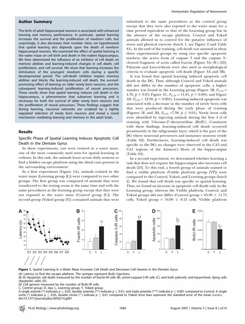
In these experiments, rats were trained in a water maze,one of the most commonly used tests for spatial learning inrodents. In this task, the animals learn across daily sessions tofind a hidden escape platform using the distal cues present inthe surrounding environment.
In a first experiment (Figure 1A), animals trained in thewater maze (Learning group [L]) were compared to two othergroups. The first group was composed of animals that weretransferred to the testing room at the same time and with thesame procedures as the learning group except that they werenot exposed to the water maze (Control group [C]). Thesecond group (Yoked group [Y]) contained animals that were
submitted to the same procedures as the control groupexcept that they were also exposed to the water maze for atime period equivalent to that of the Learning group but inthe absence of the escape platform. Control and Yokedanimals allowed us to control for the putative influence ofstress and physical exercise (batch 1, see Figure 2 and TableS1). At the end of the training, cell death was assessed in thesethree experimental groups by using two specific apoptoticmarkers, the active form of caspase 3 and the caspase 3–cleaved fragment of actin called fractin (Figure 3A–3C) [22].Pyknosis and karyorrhexis were also used as morphologicalcriteria to evaluate apoptotic cell death (Figure 3A and 3B).It was found that spatial learning induced apoptotic cell
death in the DG. Thus, although Control and Yoked animalsdid not differ in the number of apoptotic cells, a highernumber was found in the Learning group (Figure 1B, F2,16 ¼4.70, p , 0.05; Figure 1C, F2,16¼ 11.01, p , 0.001; and Figure1D, F2,16¼ 12.90, p , 0.001). Learning-induced apoptosis wasassociated with a decrease in the number of newly born cellsthat were produced during the early phase of training(Figures 1E and 3D, F2,16 ¼7.38, p � 0.01). Newly born cellswere identified by injecting animals during the first 4 d oftraining with 5-bromo-29-deoxyuridine (BrdU). Consistentwith these findings, learning-induced cell death occurredprominently in the subgranular layer, which is the part of theDG where neuronal precursors and immature neurons reside(Table S2). Furthermore, learning-induced cell death wasspecific to the DG; no changes were observed in the CA3 andCA1 regions of the Ammon’s Horn of the hippocampus(Table S3).In a second experiment, we determined whether learning a
task that does not require the hippocampus also increases celldeath [23]. To this end, a fourth group of animals trained tofind a visible platform (Visible platform group [VP]) werecompared to the Control, Yoked, and Learning groups (batch2). We found that cell death was specific to spatial learning.Thus, we found an increase in apoptotic cell death only in theLearning group, whereas the Visible platform, Control, andYoked groups did not differ (Control group ¼ 65.00 6 11.73cells, Yoked group ¼ 54.00 6 8.12 cells, Visible platform
Figure 1. Spatial Learning in a Water Maze Increases Cell Death and Decreases Cell Genesis in the Dentate Gyrus
(A) Latency to find the escape platform. The syringes represent BrdU injections.(B–D) Apoptotic cell death measured by the number of fractin-IR cells (B), active-caspase-3-IR cells (C), and both pyknotic and karyorrhexic dying cells(Apoptotic cells) (D).(E) Cell genesis measured by the number of BrdU-IR cells.C, Control group; D, day; L, Learning group; Y, Yoked group.A single asterisk (*) indicates p � 0.05, double asterisks (**) indicate p � 0.01, and triple asterisks (***) indicate p � 0.001 compared to Control. A singlecircle (8) indicates p � 0.05, double circles (88) indicate p � 0.01 compared to Yoked. Error bars represent the standard error of the mean (s.e.m.).doi:10.1371/journal.pbio.0050214.g001
PLoS Biology | www.plosbiology.org August 2007 | Volume 5 | Issue 8 | e2141684
Homeostatic Regulation of Neoneurons
Author Summary
The birth of adult hippocampal neurons is associated with enhancedlearning and memory performance. In particular, spatial learningincreases the survival and the proliferation of newborn cells, butsurprisingly, it also decreases their number. Here, we hypothesizedthat spatial learning also depends upon the death of newbornhippocampal neurons. We examined the effect of spatial learning inthe water maze on cell birth and death in the rodent hippocampus.We then determined the influence of an inhibitor of cell death onmemory abilities and learning-induced changes in cell death, cellproliferation, and cell survival. We show that learning increases theelimination of the youngest newborn cells during a specificdevelopmental period. The cell-death inhibitor impairs memoryabilities and blocks the learning-induced cell death, the survival-promoting effect of learning on older newly born neurons, and thesubsequent learning-induced proliferation of neural precursors.These results show that spatial learning induces cell death in thehippocampus, a phenomenon that subserves learning and isnecessary for both the survival of older newly born neurons andthe proliferation of neural precursors. These findings suggest thatduring learning, neuronal networks are sculpted by a tightlyregulated selection of newly born neurons and reveal a novelmechanism mediating learning and memory in the adult brain.
group ¼ 50.00 6 5.92 cells, Learning group ¼ 97.00 6 9.03cells, F3,16¼5.67, p , 0.001; Control group ¼ Yoked group ¼Visible platform group , Learning group, all comparisons atleast p � 0.01).
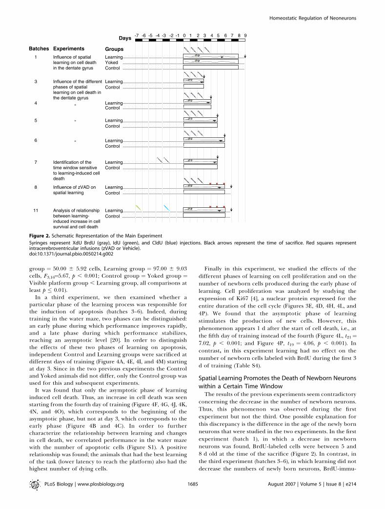
In a third experiment, we then examined whether aparticular phase of the learning process was responsible forthe induction of apoptosis (batches 3–6). Indeed, duringtraining in the water maze, two phases can be distinguished:an early phase during which performance improves rapidly,and a late phase during which performance stabilizes,reaching an asymptotic level [20]. In order to distinguishthe effects of these two phases of learning on apoptosis,independent Control and Learning groups were sacrificed atdifferent days of training (Figure 4A, 4E, 4I, and 4M) startingat day 3. Since in the two previous experiments the Controland Yoked animals did not differ, only the Control group wasused for this and subsequent experiments.
It was found that only the asymptotic phase of learninginduced cell death. Thus, an increase in cell death was seenstarting from the fourth day of training (Figure 4F, 4G, 4J, 4K,4N, and 4O), which corresponds to the beginning of theasymptotic phase, but not at day 3, which corresponds to theearly phase (Figure 4B and 4C). In order to furthercharacterize the relationship between learning and changesin cell death, we correlated performance in the water mazewith the number of apoptotic cells (Figure S1). A positiverelationship was found; the animals that had the best learningof the task (lower latency to reach the platform) also had thehighest number of dying cells.
Finally in this experiment, we studied the effects of thedifferent phases of learning on cell proliferation and on thenumber of newborn cells produced during the early phase oflearning. Cell proliferation was analyzed by studying theexpression of Ki67 [4], a nuclear protein expressed for theentire duration of the cell cycle (Figures 3E, 4D, 4H, 4L, and4P). We found that the asymptotic phase of learningstimulates the production of new cells. However, thisphenomenon appears 1 d after the start of cell death, i.e., atthe fifth day of training instead of the fourth (Figure 4L, t21¼7.02, p , 0.001; and Figure 4P, t19 ¼ 4.06, p , 0.001). Incontrast, in this experiment learning had no effect on thenumber of newborn cells labeled with BrdU during the first 3d of training (Table S4).
Spatial Learning Promotes the Death of Newborn Neurons
within a Certain Time WindowThe results of the previous experiments seem contradictory
concerning the decrease in the number of newborn neurons.Thus, this phenomenon was observed during the firstexperiment but not the third. One possible explanation forthis discrepancy is the difference in the age of the newly bornneurons that were studied in the two experiments. In the firstexperiment (batch 1), in which a decrease in newbornneurons was found, BrdU-labeled cells were between 5 and8 d old at the time of the sacrifice (Figure 2). In contrast, inthe third experiment (batches 3–6), in which learning did notdecrease the numbers of newly born neurons, BrdU-immu-
Figure 2. Schematic Representation of the Main Experiment
Syringes represent XdU BrdU (gray), IdU (green), and CldU (blue) injections. Black arrows represent the time of sacrifice. Red squares representintracerebroventricular infusions (zVAD or Vehicle).doi:10.1371/journal.pbio.0050214.g002
PLoS Biology | www.plosbiology.org August 2007 | Volume 5 | Issue 8 | e2141685
Homeostatic Regulation of Neoneurons

noreactive (IR) cells at the time of the sacrifice were less than5 d old (Figure 2).
These data suggest that learning promotes the death ofcells that have reached a certain level of maturation and areolder than 5 d. To test this hypothesis, additional groups ofanimals were trained in the water maze under conditionssimilar to those of experiment 3, but were injected with BrdUeither 3 or 4 d before the start of the behavioral training(batches 7a and 7b, Figure 5A). In this way, BrdU-labeled cellswould be either 7 or 8 d old at the end of the 5 d of training(Figure 2). We found that for both ages of BrdU-labeled cells,learning induced a decrease in the number of newly borncells (Figure 5B, group3 age interaction F1,20¼ 0.01, p¼ 0.90;group effect: F1,20 ¼ 12.57, p , 0.01) and, as expected, anincrease in cell death (Figure 5C, group3age interaction F1,20¼ 2.82, p ¼ 0.11; group effect: F1,20 ¼ 27.34, p , 0.001; andFigure 5D, group3age interaction F1,20¼1.83, p¼0.19; groupeffect: F1,20 ¼ 35.78, p , 0.001).
To confirm that this decrease in the number of BrdU-labeled cells was due to learning-induced death of newly bornneurons, we first evaluated the number of BrdU-labeled cellsthat expressed morphological manifestations of apoptosis. Incomparison to healthy BrdU-IR cells with large nuclei (’10lm, Figure 3H), apoptotic newly born BrdU-labeled cellsexhibited a small round shape and condensed, shrunken
nuclei (’3–4 lm, Figure 3I–3K). It was found that learningincreased the number of BrdU-IR pyknotic cells (Figure 5E,t12 ¼ �3.83, p � 0.01). We then studied the percentage ofBrdU-labeled cells expressing the immature neuronal markerdoublecortin (Dcx). It was found that this population of cellswas also decreased, most probably because the dying BrdU-labeled cells lost the Dcx labeling (Figures 3G and 5F, t8 ¼�2.30, p � 0.05). In addition, a quantitative evaluation basedon the extrapolated total number of BrdU-labeled neuronsshowed that learning induces a decrease of 34% in thispopulation of cells, which corresponds to a loss of approx-imately 1,600 newborn neurons (Figure 5G, t8 ¼�3.31, p �0.01).
Learning-Induced Apoptosis Is Critical for Spatial MemoryWe then examined whether learning-induced cell death
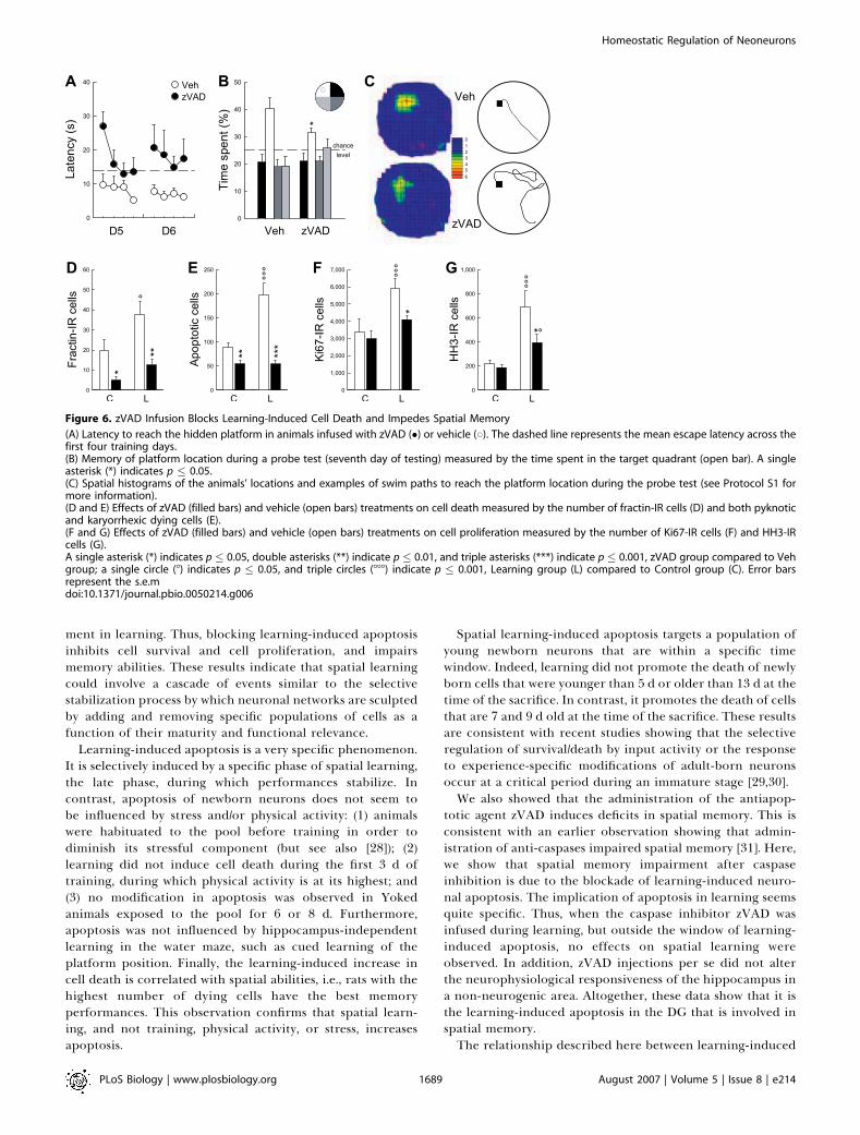
plays a role in the stabilization of performances in the watermaze. To address this issue, at the end of the fourth trainingsession, i.e., when learning-induced cell death becomesapparent, animals were infused in the lateral ventricles witheither the pan-caspase inhibitor z-Val-Ala-Asp-fluoromethyl-ketone (zVAD) or with vehicle [24,25] (batch 8). Thesetreatments were repeated for two subsequent training days.Before the beginning of the treatment, the vehicle- andzVAD-infused animals did not differ in their latency to findthe hidden platform (unpublished data; F9,198¼1.16, p¼0.32).However, after the infusions, zVAD-infused animals weresignificantly impaired, whereas vehicle-infused rats continuedto stabilize their performances (Figure 6A, F1,22 ¼ 15.64, p ,
0.001). This was particularly obvious in the first trial on day 5,during which zVAD-infused animals exhibited the largestimpairment, suggesting a deficit in retrieving what waslearned on the preceding day. In contrast, zVAD-treatedanimals were able to learn, within the session, the position ofthe hidden platform. These data strongly suggest thatinhibition of apoptosis disrupts the memory trace and notlearning per se.To further evaluate the strength of the memory trace after
zVAD treatment, a probe test was performed on the seventhday of training. The probe test consisted of exposing animalsto the water maze in the absence of the escape platform andrecording the time spent in the quadrant of the water mazethat contained the platform during training (target quad-rant). zVAD-infused animals spent less time than vehicle ratsin the target quadrant (Figure 6B and 6C, t22¼ 2.01, p � 0.05).In addition, several indices used to measure the efficiency ofthe swim paths to reach the goal location were also impairedin zVAD-infused animals (Table S5). These results confirmthat the inhibition of apoptosis when animals begin to masterthe task leads to an impairment of the memory for theplatform location.We found that zVAD treatment efficiently prevented
apoptosis (Figure 6D, F3,39 ¼ 9.61, p , 0.001; and Figure 6E,F3,39 ¼ 21.23, p , 0.001). However, we also found that zVADtreatment greatly reduced the increase in cell proliferationinduced by the late phase of learning. In this experiment, cellproliferation was measured by Ki67 staining (Figure 6F, F3,39¼ 6.72, p , 0.001), and also by a second marker of cell genesis,the phosphorylated histone H3 [4] (HH3, Figures 3F and 6G,F3,39¼6.89, p , 0.001). These results indicate that the increasein the production of new cells observed during theasymptotic phase of learning probably constitutes a compen-
Figure 3. Representative Examples of Apoptotic, Newborn, and
Proliferative Cells
(A) Fractin-IR pyknotic cell.(B) Fractin-IR karyorrhexic cell.(C) Active-caspase-3-IR cell.(D) BrdU-IR cells.(E) Ki67-IR cells.(F) HH3-IR cell in anaphase.(G) BrdU-labeled cells (red) are stained with Dcx (green), a typical markerfor immature newborn neurons.(H) Healthy BrdU-DAB-IR cell.(I) Pyknotic BrdU-DAB-IR cell.(J and K) Small BrdU-IR cell (red) exhibiting nuclear shrinkage usingFluoro Nissl Green counterstaining.Bar scales: (A–C), (F), and (H–K) indicate 5 lm; (D), (E), and (G) indicate 10lm.doi:10.1371/journal.pbio.0050214.g003
PLoS Biology | www.plosbiology.org August 2007 | Volume 5 | Issue 8 | e2141686
Homeostatic Regulation of Neoneurons

satory phenomenon triggered by learning-induced apoptosis.Thus, as shown in experiment 3 (Figure 4), learning-inducedcell proliferation follows learning-induced cell death and, asshown in the present experiment, this phenomenon isdecreased by blocking cell death.
In order to test for the specificity of the effects of zVAD, weperformed several complementary measures and experi-ments. Thus, after the probe test, animals were tested fortheir ability to find a visible platform (cued test), whichallowed for a measure of visuomotor processes (Table S5). Inthis case, zVAD treatment had no measurable effects onbehavioral performance. We also evaluated whether zVADwould modify the survival of newly born cells younger than 5
d, because these neurons are normally untouched by learning.This population of cells was labeled by injecting BrdU on day1 to day 3 of training (Figure 2). Again, zVAD treatment hadno measurable effects (t22 ¼ 0.87, p ¼ 0.39). Then, in asubsequent experiment (batch 9), we infused zVAD duringthe first 3 d of training, a period during which learning has noinfluence on cell death (Figure 4B and 4C). Consequently, ifthe effects of zVAD are mediated by a blockade of learning-induced cell death, this schedule of treatment should have nobehavioral effects. Indeed, we found that under theseconditions, zVAD infusions did not impair spatial memory(Figure S2). Finally, we performed physiological recordings inthe Ammon’s horn, a part of the hippocampus in which
Figure 4. The Stabilization of Spatial Performances Increases Cell Death and Cell Proliferation in the Dentate Gyrus
(A, E, I, and M) Latency to find the escape platform. The syringes represent BrdU injections. D, day.(B, F, J, and N) Apoptosis measured by the number of fractin-IR cells.(C, G, K, and O) Apoptosis measured by the number of both pyknotic and karyorrhexic dying cells.(D, H, L, and P) Cell proliferation measured by the number of Ki67-IR cells.A single asterisk (*) indicates p �0.05, double asterisks (**) indicate p � 0.01, and triple asterisks (***) indicate p � 0.001 compared to Control groups.Error bars represent the s.e.m.doi:10.1371/journal.pbio.0050214.g004
PLoS Biology | www.plosbiology.org August 2007 | Volume 5 | Issue 8 | e2141687
Homeostatic Regulation of Neoneurons

learning does not induce cell death (Table S3) and whichconsequently should not respond to zVAD treatment (batch10). Field recordings confirmed that zVAD did not alterexcitatory synaptic transmission within this area (Figure S3).
In conclusion, taken together, all these control experimentsconfirm that the disruption of spatial learning was due to aspecific inhibition of apoptosis by zVAD.
Learning-Induced Increases in Apoptosis, CellProliferation, and Survival of Newborn Neurons AreInterrelated Processes
It has previously been shown that training in a water mazeincreases the survival of newborn neurons that wereproduced 1 wk before the start of the training [19,26]. Itseems then that learning can induce increases in both thesurvival and death of newborn neurons. For this reason, in afinal experiment, we studied the relationships between thesetwo phenomena (batch 11). This experiment was performedby injecting the same animals with two thymidine analogs, 5-iodo-29-deoxyuridine (IdU) and 5-chloro-29-deoxyuridine(CldU) [27] (Figure S4). IdU and CldU were injected atdifferent times in order to analyze in the same subject the fateof newborn cells of different ages (Figure 2). IdU was injected7 d before training in order to label the newborn cells forwhich survival should be increased by learning. CldU wasinjected 3 d before the start of the training in order to labelnewly born cells that should die as a consequence of learning.Animals in these experiments were also infused either withvehicle or zVAD at the end of the fourth through sixth days oftraining. As found in the previous experiment, vehicle- and
zVAD-infused animals did not differ in their latency to findthe hidden platform during the first 4 d of training (F9,135 ¼0.52, p . 0.05). Similarly, an impairment was observed inzVAD-infused animals during the last two training days(Figure 7A, F1,15 ¼ 20.43, p , 0.001).In vehicle-treated rats, learning promoted the survival of
IdU-labeled cells generated 1 wk before exposure to the task,and this effect was blocked by zVAD infusion (Figure 7B, F3,24¼ 6.65, p , 0.01). This prosurvival effect of learning on IdU-labeled cells was associated with a decrease in the number ofIdU-IR pyknotic cells (Figure 7C, F3,24 ¼ 5.58, p , 0.01).Furthermore, as previously found here (Figure 5B), learningdecreased the survival of CldU-labeled cells that were born 3d before the start of the training, an effect suppressed byzVAD infusion (Figure 7D, F3,24¼ 4.50, p � 0.01). As expected,learning increased the number of CldU-IR pyknotic cells, andthis effect was blocked by zVAD infusion (Figure 7E, F3,24 ¼40.55, p � 0.001). These data show that learning-inducedincreases in survival and apoptosis of newborn cells areinterrelated processes. Thus, blocking learning-inducedapoptosis also blocks the increased survival of older neurons.
Discussion
The results of the experiments reported here show thatspatial learning promotes the survival of adult-born neuronsthat are relatively more mature, induces the death of cellsthat are more immature, and finally, stimulates proliferationof precursors. Blocking learning-induced cell death hasshown an interdependency of these events and their involve-
Figure 5. Spatial Learning Promotes the Death of Newborn Neurons Generated a Few Days before Training
(A) Latency to find the escape platform. Learning groups were injected with BrdU either 4 or 3 d before initiation of training as indicated by the syringes.D, day.(B) Newly born cells measured by the number of BrdU-IR cells.(C and D) Apoptotic cells measured by the number of fractin-IR cells (C) and both pyknotic and karyorrhexic dying cells (D).(E) Number of BrdU-IR cells exhibiting characteristics of dying cells.(F) Percentage of BrdU-IR cells expressing the immature neuronal marker Dcx.(G) Extrapolated number of newborn neurons.A single asterisk (*) indicates p � 0.05, double asterisks (**) indicate p � 0.01, and triple asterisks (***) indicate p � 0.001 compared to Control groups.Error bars represent the s.e.m.doi:10.1371/journal.pbio.0050214.g005
PLoS Biology | www.plosbiology.org August 2007 | Volume 5 | Issue 8 | e2141688
Homeostatic Regulation of Neoneurons
ment in learning. Thus, blocking learning-induced apoptosisinhibits cell survival and cell proliferation, and impairsmemory abilities. These results indicate that spatial learningcould involve a cascade of events similar to the selectivestabilization process by which neuronal networks are sculptedby adding and removing specific populations of cells as afunction of their maturity and functional relevance.
Learning-induced apoptosis is a very specific phenomenon.It is selectively induced by a specific phase of spatial learning,the late phase, during which performances stabilize. Incontrast, apoptosis of newborn neurons does not seem tobe influenced by stress and/or physical activity: (1) animalswere habituated to the pool before training in order todiminish its stressful component (but see also [28]); (2)learning did not induce cell death during the first 3 d oftraining, during which physical activity is at its highest; and(3) no modification in apoptosis was observed in Yokedanimals exposed to the pool for 6 or 8 d. Furthermore,apoptosis was not influenced by hippocampus-independentlearning in the water maze, such as cued learning of theplatform position. Finally, the learning-induced increase incell death is correlated with spatial abilities, i.e., rats with thehighest number of dying cells have the best memoryperformances. This observation confirms that spatial learn-ing, and not training, physical activity, or stress, increasesapoptosis.
Spatial learning-induced apoptosis targets a population ofyoung newborn neurons that are within a specific timewindow. Indeed, learning did not promote the death of newlyborn cells that were younger than 5 d or older than 13 d at thetime of the sacrifice. In contrast, it promotes the death of cellsthat are 7 and 9 d old at the time of the sacrifice. These resultsare consistent with recent studies showing that the selectiveregulation of survival/death by input activity or the responseto experience-specific modifications of adult-born neuronsoccur at a critical period during an immature stage [29,30].We also showed that the administration of the antiapop-
totic agent zVAD induces deficits in spatial memory. This isconsistent with an earlier observation showing that admin-istration of anti-caspases impaired spatial memory [31]. Here,we show that spatial memory impairment after caspaseinhibition is due to the blockade of learning-induced neuro-nal apoptosis. The implication of apoptosis in learning seemsquite specific. Thus, when the caspase inhibitor zVAD wasinfused during learning, but outside the window of learning-induced apoptosis, no effects on spatial learning wereobserved. In addition, zVAD injections per se did not alterthe neurophysiological responsiveness of the hippocampus ina non-neurogenic area. Altogether, these data show that it isthe learning-induced apoptosis in the DG that is involved inspatial memory.The relationship described here between learning-induced
Figure 6. zVAD Infusion Blocks Learning-Induced Cell Death and Impedes Spatial Memory
(A) Latency to reach the hidden platform in animals infused with zVAD (�) or vehicle (*). The dashed line represents the mean escape latency across thefirst four training days.(B) Memory of platform location during a probe test (seventh day of testing) measured by the time spent in the target quadrant (open bar). A singleasterisk (*) indicates p � 0.05.(C) Spatial histograms of the animals’ locations and examples of swim paths to reach the platform location during the probe test (see Protocol S1 formore information).(D and E) Effects of zVAD (filled bars) and vehicle (open bars) treatments on cell death measured by the number of fractin-IR cells (D) and both pyknoticand karyorrhexic dying cells (E).(F and G) Effects of zVAD (filled bars) and vehicle (open bars) treatments on cell proliferation measured by the number of Ki67-IR cells (F) and HH3-IRcells (G).A single asterisk (*) indicates p � 0.05, double asterisks (**) indicate p � 0.01, and triple asterisks (***) indicate p � 0.001, zVAD group compared to Vehgroup; a single circle (8) indicates p � 0.05, and triple circles (888) indicate p � 0.001, Learning group (L) compared to Control group (C). Error barsrepresent the s.e.mdoi:10.1371/journal.pbio.0050214.g006
PLoS Biology | www.plosbiology.org August 2007 | Volume 5 | Issue 8 | e2141689
Homeostatic Regulation of Neoneurons

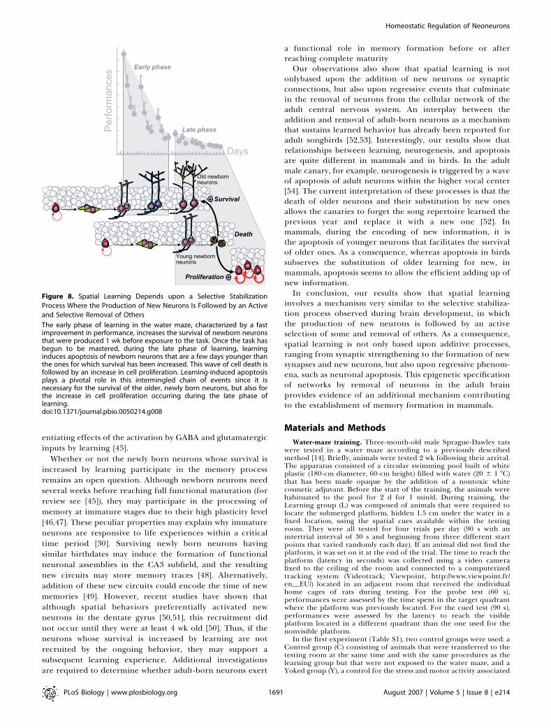
increases in survival, apoptosis, and proliferation of newborncells provides a three-step picture of the relationship betweenneurogenesis and spatial learning (Figure 8). First, acquisitionof the task induces an increase in the survival of newbornneurons generated 1 wk before the task and that conse-quently have reached an intermediate level of maturity.Second, once the task starts to be mastered, learning inducesapoptosis of newborn neurons that are a few days youngerthan those for which survival has been increased. Third,learning-induced apoptosis is followed by an increase in cellproliferation that provides the hippocampus with a new poolof young neurons [16,20].
This homeostatic regulation of neurogenesis by learning isconsistent with the selective stabilization theory according towhich regressive events will stabilize a particular set ofcontacts among many others, thereby sculpting the precisecircuits that are crucial for a given function [32]. It has beenestimated that during development, after an initial prolifer-ating phase during which a large number of newborn neuronsare produced, at least half of the initial neuronal populationis eliminated by apoptosis [33]. This neuronal eliminationserves several functions, among which is the regulation oftarget innervation. Indeed, neural function depends upon aprecise quantitative relationship between neurons: each axoninnervates an appropriate number of target cells and eachtarget cell is innervated by an appropriate number of axons.The decision for survival or death during development isgoverned by afferences and/or efferences [34,35].
In the case of hippocampal adult-born neurons, it might behypothesized that those cells that are successfully connected,both in terms of efferent output and afferent input, are theones that can be rescued by the stimuli generated in thecourse of learning. In favor of this hypothesis, it has beenshown that enhanced synaptic activity enhances cell survival[36]. In contrast, apoptosis could constitute a trimmingmechanism that suppresses more-immature neurons thathave not been selected by learning. Their suppression couldfavor the integration of older cells that have been stabilizedby activity-dependent stimuli generated in the course oflearning. These regressive events could also, by clearing the
network of nonspecific noise due to superfluous new neurons,enhance the signal-to-noise ratio. Supporting this idea, animprovement in the signal-to-noise ratio of motor cortexcells during motor skill learning has been linked to a practice-related improvement in behavioral performance [37].The precise mechanisms by which learning promotes the
survival or apoptosis of immature newborn neurons arecurrently unknown. However, analysis of the developmentalpattern of newborn neurons provides a certain number ofputative explanations. Newborn neurons follow a precisematuration of neuronal connectivity and function thatrequires about 1 mo. They extend their dendritic tree atvariable times after mitosis, and by 3 wk, their dendriticarborization resembles that of mature neurons [12,38]. Inaddition, as soon as 10 d after birth, newly born cells extendaxons into the CA3 subfield of the hippocampus [9,12]. Afterthe first week of maturation, they also receive depolarizingGABA inputs [39–44]. Toward the end of the second week,GABA inputs become progressively hyperpolarizing, and theadult-born neurons begin receiving functional glutamatergicdepolarizing afferents [11,29,40,41], a process that occurs inparallel with the formation of dendritic spines.On the basis of this developmental pattern, it seems likely
that newborn neurons that are younger than 5 d are notinfluenced by learning because they lack afferent inputs andhave not yet reached projection territories. Neurons that arein the window during which learning induces apoptosisshould have received functional depolarizing GABA inputs,although their dendritic tree would still be poorly developed,and these newborn cells should not have reached their targetarea. Thus, in response to learning-driven depolarization, thisimbalance between input and output activity may impede thesurvival of these cells and lead to their death. Older neuronsthat survive as a consequence of learning have a moredeveloped dendritic tree that receives depolarizing GABAinputs and starts to have some glutamatergic ones. Further-more, these newborn neurons have also reached the CA3subfield. It is then likely that these newborn neurons thathave reached a higher stage of maturation, with balancedinput/output connections, can benefit from the pro-differ-
Figure 7. Learning Increases the Number of New Neurons, and This Effect Is Blocked by zVAD Infusion
(A) Latency to reach the hidden platform in animals infused with zVAD (�) or vehicle (*). The dashed line represents the mean escape latency across thefirst four training days. The syringes represent IdU and CldU injections. D, day.(B) Effects of zVAD (filled bars) and vehicle (open bars) treatments on the survival of IdU-IR cells generated 7 d before exposure to the task.(C) Effects of zVAD (filled bars) and vehicle (open bars) treatments on the number of IdU-IR cells exhibiting characteristics of pyknotic cells.(D) Effects of zVAD (filled bars) and vehicle (open bars) treatments on the survival of CldU-IR cells generated 3 d before exposure to the task.(E) Effects of zVAD (filled bars) and vehicle (open bars) treatments on the number of CldU-IR cells exhibiting characteristics of pyknotic cells.A single asterisk (*) indicates p � 0.05, double asterisks (**) indicate p � 0.01, and triple asterisks (***) indicate p � 0.001, zVAD group compared to Vehgroup; a single circle (8) indicates p � 0.05, double circles (88) indicate p � 0.01, and triple circles (888) indicate p � 0.001, Learning group (L) compared toControl group (C) .doi:10.1371/journal.pbio.0050214.g007
PLoS Biology | www.plosbiology.org August 2007 | Volume 5 | Issue 8 | e2141690
Homeostatic Regulation of Neoneurons
entiating effects of the activation by GABA and glutamatergicinputs by learning [45].
Whether or not the newly born neurons whose survival isincreased by learning participate in the memory processremains an open question. Although newborn neurons needseveral weeks before reaching full functional maturation (forreview see [45]), they may participate in the processing ofmemory at immature stages due to their high plasticity level[46,47]. These peculiar properties may explain why immatureneurons are responsive to life experiences within a criticaltime period [30]. Surviving newly born neurons havingsimilar birthdates may induce the formation of functionalneuronal assemblies in the CA3 subfield, and the resultingnew circuits may store memory traces [48]. Alternatively,addition of these new circuits could encode the time of newmemories [49]. However, recent studies have shown thatalthough spatial behaviors preferentially activated newneurons in the dentate gyrus [50,51], this recruitment didnot occur until they were at least 4 wk old [50]. Thus, if theneurons whose survival is increased by learning are notrecruited by the ongoing behavior, they may support asubsequent learning experience. Additional investigationsare required to determine whether adult-born neurons exert
a functional role in memory formation before or afterreaching complete maturityOur observations also show that spatial learning is not
onlybased upon the addition of new neurons or synapticconnections, but also upon regressive events that culminatein the removal of neurons from the cellular network of theadult central nervous system. An interplay between theaddition and removal of adult-born neurons as a mechanismthat sustains learned behavior has already been reported foradult songbirds [52,53]. Interestingly, our results show thatrelationships between learning, neurogenesis, and apoptosisare quite different in mammals and in birds. In the adultmale canary, for example, neurogenesis is triggered by a waveof apoptosis of adult neurons within the higher vocal center[54]. The current interpretation of these processes is that thedeath of older neurons and their substitution by new onesallows the canaries to forget the song repertoire learned theprevious year and replace it with a new one [52]. Inmammals, during the encoding of new information, it isthe apoptosis of younger neurons that facilitates the survivalof older ones. As a consequence, whereas apoptosis in birdssubserves the substitution of older learning for new, inmammals, apoptosis seems to allow the efficient adding up ofnew information.In conclusion, our results show that spatial learning
involves a mechanism very similar to the selective stabiliza-tion process observed during brain development, in whichthe production of new neurons is followed by an activeselection of some and removal of others. As a consequence,spatial learning is not only based upon additive processes,ranging from synaptic strengthening to the formation of newsynapses and new neurons, but also upon regressive phenom-ena, such as neuronal apoptosis. This epigenetic specificationof networks by removal of neurons in the adult brainprovides evidence of an additional mechanism contributingto the establishment of memory formation in mammals.
Materials and Methods
Water-maze training. Three-month-old male Sprague-Dawley ratswere tested in a water maze according to a previously describedmethod [14]. Briefly, animals were tested 2 wk following their arrival.The apparatus consisted of a circular swimming pool built of whiteplastic (180-cm diameter, 60-cm height) filled with water (20 6 1 8C)that has been made opaque by the addition of a nontoxic whitecosmetic adjuvant. Before the start of the training, the animals werehabituated to the pool for 2 d for 1 min/d. During training, theLearning group (L) was composed of animals that were required tolocate the submerged platform, hidden 1.5 cm under the water in afixed location, using the spatial cues available within the testingroom. They were all tested for four trials per day (90 s with anintertrial interval of 30 s and beginning from three different startpoints that varied randomly each day). If an animal did not find theplatform, it was set on it at the end of the trial. The time to reach theplatform (latency in seconds) was collected using a video camerafixed to the ceiling of the room and connected to a computerizedtracking system (Videotrack; Viewpoint, http://www.viewpoint.fr/en_EU/) located in an adjacent room that received the individualhome cages of rats during testing. For the probe test (60 s),performances were assessed by the time spent in the target quadrantwhere the platform was previously located. For the cued test (90 s),performances were assessed by the latency to reach the visibleplatform located in a different quadrant than the one used for thenonvisible platform.
In the first experiment (Table S1), two control groups were used: aControl group (C) consisting of animals that were transferred to thetesting room at the same time and with the same procedures as thelearning group but that were not exposed to the water maze, and aYoked group (Y), a control for the stress and motor activity associated
Figure 8. Spatial Learning Depends upon a Selective Stabilization
Process Where the Production of New Neurons Is Followed by an Active
and Selective Removal of Others
The early phase of learning in the water maze, characterized by a fastimprovement in performance, increases the survival of newborn neuronsthat were produced 1 wk before exposure to the task. Once the task hasbegun to be mastered, during the late phase of learning, learninginduces apoptosis of newborn neurons that are a few days younger thanthe ones for which survival has been increased. This wave of cell death isfollowed by an increase in cell proliferation. Learning-induced apoptosisplays a pivotal role in this intermingled chain of events since it isnecessary for the survival of the older, newly born neurons, but also forthe increase in cell proliferation occurring during the late phase oflearning.doi:10.1371/journal.pbio.0050214.g008
PLoS Biology | www.plosbiology.org August 2007 | Volume 5 | Issue 8 | e2141691
Homeostatic Regulation of Neoneurons
with the water-maze training, composed of rats that were placed intothe pool without the platform and were paired for the duration of thetrial with the Learning animals. In the second experiment, threecontrol groups were used: a Control group, a Yoked group, and anadditional group of rats that were trained to find a visible platform(VP) in a fixed location. Animals in this group were all tested for fourtrials per day (90 s with an intertrial interval of 30 s and beginningfrom three different start points that varied randomly each day).Because in the first experiment the Yoked and Control groups didnot differ for cell genesis or cell death, and because in the secondexperiment the Visual Platform, Yoked, and Control groups also didnot differ, only the Control group was used for subsequent experi-ments. All experiments were performed in accordance with theEuropean Union (86/609/EEC) and the French National Committee(87/848) recommendations. Detailed analysis of swim paths made toreach the platform location during the probe test was performedusing the Wintrack software (see Protocol S1).
Intracerebroventricular surgery. Guide cannulae were implanted,according to a previously described method [55], above the rostralventricle in order not to cause lesions in the hippocampus. Twoweeks later, 6 ll (per infusion site) of vehicle (Ringer’s solution with1% DMSO) or of zVAD-fmk (at a concentration of 1 lg of zVAD/ll ofvehicle; Calbiochem, http://www.emdbiosciences.com/html/CBC/home.html) [25] solutions were infused at a constant rate (3 ll/min)in naive or in trained animals immediately after the last trial of the4th–6th days (batches 8 and 11) or of the 1st–3rd days (batch 9) oftraining.
Thymidine analog injections. Newly born cells were labeled by theincorporation of synthetic thymidine analog (XdU [where Xrepresents Br, Cl, or I]; Table S1). Rats (batches 1, 3–8, and 11) wereinjected with BrdU (intraperitoneal). The Learning groups receivedone daily BrdU injection 30 min before the first trials or a singleBrdU injection 3 or 4 d before the onset of training. Rats of the 11thexperiment received a single injection of IdU and of CldU [27],respectively, 7 and 3 d before the onset of training, both at equimolardoses of 50 mg BrdU/kg. The control groups were injected with XdUwithin the same period.
Immunohistochemistry. Animals were sacrificed 1 d (batches 1, 2,and 11) or 3 h after daily training session (batches 3–7), or after theprobe test (batches 8 and 9). Free-floating sections (50 lm) wereprocessed in a standard immunohistochemical procedure in order tovisualized BrdU (1/200; Dako, http://www.dako.com), IdU (1/1,000, BDPharMingen #347580; BD Biosciences, http://www.bdbiosciences.com),CldU (1/1,000; Accurate Chemical and Scientific Corporation, http://www.accuratechemical.com), Ki67 (1/200, Novocastra; Vision BioSys-tems, http://www.vision-bio.com), fractin (1/5,000, BD PharMingen#551527; BD Biosciences), HH3 (1/2,000, Cell Signaling #06–570;Upstate Biotechnology, http://www.upstate.com), and activated-cas-pase-3 (1/10,000, BD PharMingen #551150; BD Biosciences)[14,22,56].Sections were counterstained with thionine in order to visualizepyknotic cells, characterized by a condensed nucleus of smaller size,and karyorrhexic cells displaying chromatin clumps. The number ofimmunoreactive (IR) cells throughout the entire granule andsubgranular layers of the DG were estimated using the opticalfractionator method [14].
To examine the phenotype of BrdU-IR cells, one in ten sectionswere incubated with BrdU antibodies (1/500; Accurate), which wererevealed using a CY3–anti-rat antibody (1/1,000; Jackson Immunor-esearch, http://www.jacksonimmuno.com). Sections were then incu-bated with anti-DCX antibodies (1/1,000; Santa Cruz Biotechnology,http://www.scbt.com), which were visualized with an Alexa-488 anti-goat IgG (1/1,000; Jackson). The percentage of BrdU-labeled cellsexpressing Dcx was determined throughout the DG using a confocalmicroscope with helium–neon and argon lasers (DMR TCSSP2AOBS;Leica, http://www.leica-microsystems.com). To estimate the totalnumber of BrdU-labeled neurons, the percent of BrdU-IR cells co-labeled with Dcx was multiplied by the total number of BrdU-labeledcells.
Electrophysiology. Hippocampal slices (500 lm) were prepared asdescribed previously [57] from vehicle- and zVAD -infused rats by aninvestigator blind to the treatments. Slices were submerged in anoxygenated artificial cerebrospinal fluid (ACSF) comprising (in mM):NaCl 123, KCl 2.5, Na2HPO4 1, NaHCO3 26.2, Cacl2 2.4, MgCl2 1.2,glucose 10, bicuculline 0.02 (pH 7.4; 295 mosmol.kg�1; room temper-ature). A concentric bipolar steel electrode was placed in the stratumradiatum to evoke (0.01 Hz) field excitatory postsynaptic potentials(fEPSPs) recorded with a glass electrode filled with ACSF. Data werecollected with a multiclamp 700A (Axon Instruments, http://www.axon.com), filtered at 3 kHz, sampled at 10 kHz and analyzed offlineusing pClamp 9 (Axon Instruments). The initial slopes of the fEPSPs
were measured from approximately 10%–40% of the rising phase.Paired-pulse ratio corresponds to the slope ratio of the second fEPSPto the first fEPSP.
Statistical analysis. All data (mean 6 standard error of the mean)were analyzed by a Student t-test (two tailed) or by analysis ofvariance followed by Newman-Keuls test when necessary. Correlationanalysis was performed using the Spearman test.
Supporting Information
Figure S1. Correlations between the Performance in the Water Mazeand Apoptotic Cell Death
In order to characterize the relationship between learning andchanges in cell death, we correlated the performance in the watermaze (mean latency to find the platform after 3 to 6 d of training)with the number of fractin-IR cells or of pyknotic and karyorrhexiccells (Apoptotic cells). A significant correlation was found betweenthe behavioral performances and cell death ([A] fractin-IR cells: R ¼�0.47, p , 0.001; [B] Apoptotic cells: R¼�0.62, p , 0.001). In addition,the number of fractin-IR cells also correlated to the number ofapoptotic cells ([C] R¼ 0.65, p , 0.001).
Found at doi:10.1371/journal.pbio.0050214.sg001 (22 KB PDF).
Figure S2. Influence of zVAD Infusion during the Early Phase ofLearning on Spatial Learning
Vehicle or zVAD was infused during the first 3 d of water-mazetraining (batch 9). Results show vehicle- and zVAD-infused animalsdid not differ in their latency to find the hidden platform duringtraining ([A] F1,16¼0.07, p . 0.05) nor for the time spent in the targetquadrant during the probe test on the seventh day ([B] t16¼1.20, p .0.05).
Found at doi:10.1371/journal.pbio.0050214.sg002 (19 KB PDF).
Figure S3. Effects of the Blockade of Caspases-Mediated Cell Deathon Hippocampal Neurophysiological Response
Field recordings performed in hippocampal slices (batch 10) revealedthat zVAD did not alter the input–output relationship (F6,30¼ 0.34, p. 0.05) nor short-term facilitation at CA3-CA1 synapses, indicatingthat excitatory synaptic transmission (F6,30 ¼ 1.46, p . 0.05) wasunaffected by zVAD.(A) Example of CA1 field excitatory postsynaptic potentials (fEPSPs)obtained at different stimulus intensities.(B) Input–output relationships in animals infused with zVAD (�) orvehicle (*).(C) Example of short-term facilitation occurring when pairs of stimuliwere applied 50 ms apart at different stimulus intensities.(D) Facilitation (paired-pulse ratio) plotted as a function of stimulusstrength.
Found at doi:10.1371/journal.pbio.0050214.sg003 (28 KB PDF).
Figure S4. Example of IdU- and CldU-Labeled Cells
Cells having incorporated IdU or CldU were specifically revealed withBD #347580 (1/1,000e) or Accurate (1/1,000e) antibodies, respectively.There was no cross reactivity between antibodies. Scale bar indicates10 lm.GCL, granule cell layer.
Found at doi:10.1371/journal.pbio.0050214.sg004 (100 KB PDF).
Protocol S1. Detailed Analysis of Swim Paths during the Probe Test
Found at doi:10.1371/journal.pbio.0050214.sd001 (30 KB DOC).
Table S1. Summary of the Schedule of Pharmacological Treatmentsand Water-Maze Training
Found at doi:10.1371/journal.pbio.0050214.st001 (16 KB PDF).
Table S2. Repartition of Cell Death within the Dentate Gyrus
Learning-induced cell death occurred prominently in the subgranu-lar layer where neuronal precursors reside. Cell death was much lessin the granule cell layer composed mainly of mature neurons (F3,28¼65.78, p , 0.001).
Found at doi:10.1371/journal.pbio.0050214.st002 (12 KB PDF).
Table S3. Effect of Learning on Cell Death in the Different Regions ofthe Hippocampus
Learning did not modify the number of fractin-IR cells and of
PLoS Biology | www.plosbiology.org August 2007 | Volume 5 | Issue 8 | e2141692
Homeostatic Regulation of Neoneurons
pyknotic and karyorrhexic cells (Apoptotic cells) in the CA3 and CA1subfield of the Ammon’s Horn.
Found at doi:10.1371/journal.pbio.0050214.st003 (12 KB PDF).
Table S4. Time Course of the Influence of Learning on the Numberof BrdU-IR Cells
Spatial learning had no effect on the number of newborn cells labeledwith BrdU during the first 3 d of training (D1–D3, see Table S1). D,day.
Found at doi:10.1371/journal.pbio.0050214.st004 (11 KB PDF).
Table S5. Influence of zVAD Infusion during the Late Phase ofLearning on the Probe Test and the Cued Test
During the probe test, zVAD treatment impaired all the indices usedto measure the efficiency of the swim paths to reach the goal location(time to goal, Wishaw’s index, and cumulative search error and pathefficiency index). In contrast, during the cued test, zVAD treatmentdid not alter either visuomotor processes (average speed, and latencyto reach the visible platform).
Found at doi:10.1371/journal.pbio.0050214.st005 (11 KB PDF).
Acknowledgments
We greatly acknowledge Dr. F. Ichas, Dr. F. de Giorgi, Dr. M.Montcouquiol, Dr. N. Sans for their helpful comments and C. Dupuy,S. Aguerre, C. Aurousseau, and M. Kharouby for their technical help.
Author contributions. DD is the lead author. He contributed to theconcept and design of the experiments, and performed the experi-ments, analyzed the data, and revised the manuscript. AF performedzVAD stereotaxic injections, behavior experiments, and cell counting(batches 9 and 11). MDD contributed to the first experiment (batch 1).JJR and SL participated in immunohistochemistry. VL performed abehavioral experiment (batch 2). AP and SHRO performed electro-physiology recordings (batch 10). PVP contributed to the concept,designed some of the experiments, interpreted some of the data, andwrote the manuscript. DNA contributed to the concept, designed theexperiments, interpreted the data, wrote the manuscript, andprovided financial support.
Funding. This work was supported by the French National Instituteof Health and Medical Research (INSERM) and University ofBordeaux 2.
Competing interests. The authors have declared that no competinginterests exist.
References1. Ramon y Cajal S (1991) Cajal’s degeneration and regeneration of the
nervous system. May RM, translator; DeFelipe J, Jones EG, editors. NewYork: Oxford University Press. 766 p.
2. Altman J (1962) Are new neurones formed in the brains of adult mammals?Science 135: 1127–1128.
3. Gross CG (2000) Neurogenesis in the adult brain: Death of a dogma. NatRev Neurosci 1: 67–73.
4. Abrous DN, Koehl M, Le Moal M (2005) Adult neurogenesis: Fromprecursors to network and physiology. Physiol Rev 85: 523–569.
5. Cameron HA, McKay RD (2001) Adult neurogenesis produces a large poolof new granule cells in the dentate gyrus. J Comp Neurol 435: 406–417.
6. Rao MS, Shetty AK (2004) Efficacy of doublecortin as a marker to analysethe absolute number and dendritic growth of newly generated neurons inthe adult dentate gyrus. Eur J Neurosci 19: 234–246.
7. Dayer AG, Ford AA, Cleaver KM, Yassaee M, Cameron HA (2003) Short-term and long-term survival of new neurons in the rat dentate gyrus. JComp Neurol 460: 563–572.
8. Kempermann G, Gast D, Kronenberg G, Yamaguchi M, Gage FH (2003)Early determination and long-term persistence of adult-generated newneurons in the hippocampus of mice. Development 130: 391–399.
9. Hastings NB, Gould E (1999) Rapid extension of axons into the CA3 regionby adult-generated granule cells. J Comp Neurol 413: 146–154.
10. Stanfield BB, Trice JE (1988) Evidence that granule cells generated in thedentate gyrus of adult rats extend axonal projections. Exp Brain Res 72:399–406.
11. van Praag H, Schinder AF, Christie BR, Toni N, Palmer TD, et al. (2002)Functional neurogenesis in the adult hippocampus. Nature 415: 1030–1034.
12. Zhao C, Teng EM, Summers RG Jr, Ming GL, Gage FH (2006) Distinctmorphological stages of dentate granule neuron maturation in the adultmouse hippocampus. J Neurosci 26: 3–11.
13. O’Keefe J, Nadel L (1978) The hippocampus as a cognitive map. Oxford:Clarendon Press. 570 p.
14. Drapeau E, Mayo W, Aurousseau C, Le Moal M, Piazza PV, et al. (2003)Spatial memory performances of aged rats in the water maze predict levelsof hipppocampal neurogenesis. Proc Natl Acad Sci U S A 100: 14385–14390.
15. Kempermann G, Kuhn HG, Gage FH (1997) More hippocampal neurons inadult mice living in an enriched environment. Nature 386: 493–495.
16. Lemaire V, Koehl M, Le Moal M, Abrous DN (2000) Prenatal stress produceslearning deficits associated with an inhibition of neurogenesis in thehippocampus. Proc Natl Acad Sci U S A 97: 11032–11037.
17. Shors TJ, Miesegaes G, Beylin A, Zhao M, Rydel T, et al. (2001) Neurogenesisin the adult is involved in the formation of trace memories. Nature 410:372–376.
18. van Praag H, Kempermann G, Gage FH (1999) Running increases cellproliferation and neurogenesis in the adult mouse dentate gyrus. NatNeurosci 2: 266–270.
19. Gould E, Beylin A, Tanapat P, Reeves A, Shors TJ (1999) Learning enhancesadult neurogenesis in the hippocampal formation. Nat Neurosci 2: 260–265.
20. Dobrossy MDE, Aurousseau C, Le Moal M, Piazza PV, Abrous DN (2003)Differential effects of learning on neurogenesis: Learning increases ordecreases the number of newly born cells depending on their birth date.Mol Psychiatry 8: 974–982.
21. Ambrogini P, Orsini L, Mancini C, Ferri P, Ciaroni S, et al. (2004) Learningmay reduce neurogenesis in adult rat dentate gyrus. Neurosci Lett 359: 13–16.
22. Rossiter JP, Anderson LL, Yang F, Cole GM (2000) Caspase-cleaved actin(fractin) immunolabelling of Hirano bodies. Neuropathol Appl Neurobiol26: 342–346.
23. Morris RG, Garrud P, Rawlins JN, O’Keefe J (1982) Place navigation isimpaired in rats with hippocampal lesions. Nature 297: 681–683.
24. Cryns V, Yuan J (1998) Proteases to die for. Genes Dev 12: 1551–1570.25. Ekdahl CT, Mohapel P, Weber E, Bahr B, Blomgren K, et al. (2002) Caspase-
mediated death of newly formed neurons in the adult rat dentate gyrusfollowing status epilepticus. Eur J Neurosci 16: 1463–1471.
26. Hairston IS, Little MT, Scanlon MD, Barakat MT, Palmer TD, et al. (2005)Sleep restriction suppresses neurogenesis induced by hippocampus-dependent learning. J Neurophysiol 94: 4224–4233.
27. Bauer S, Patterson PH (2005) The cell cycle-apoptosis connection revisitedin the adult brain. J Cell Biol 171: 641–650.
28. Ehninger D, Kempermann G (2006) Paradoxical effects of learning theMorris water maze on adult hippocampal neurogenesis in mice may beexplained by a combination of stress and physical activity. Genes BrainBehav 5: 29–39.
29. Tashiro A, Sandler VM, Toni N, Zhao C, Gage FH (2006) NMDA-receptor-mediated, cell-specific integration of new neurons in adult dentate gyrus.Nature 442: 929–933.
30. Tashiro A, Makino H, Gage FH (2007) Experience-specific functionalmodification of the dentate gyrus through adult neurogenesis: A criticalperiod during an immature stage. J Neurosci 27: 3252–3259.
31. Dash PK, Blum S, Moore AN (2000) Caspase activity plays an essential rolein long-term memory. Neuroreport 11: 2811–2814.
32. Changeux JP, Danchin A (1976) Selective stabilisation of developingsynapses as a mechanism for the specification of neuronal networks.Nature 264: 705–712.
33. Oppenheim RW (1991) Cell death during development of the nervoussystem. Annu Rev Neurosci 14: 453–501.
34. Linden R (1994) The survival of developing neurons: A review of afferentcontrol. Neuroscience 58: 671–682.
35. Purves D (1986) The trophic theory of neural connections. Trends Neurosci9: 486–489.
36. Bruel-Jungerman E, Davis S, Rampon C, Laroche S (2006) Long-termpotentiation enhances neurogenesis in the adult dentate gyrus. J Neurosci26: 5888–5893.
37. Kargo WJ, Nitz DA (2004) Improvements in the signal-to-noise ratio ofmotor cortex cells distinguish early versus late phases of motor skilllearning. J Neurosci 24: 5560–5569.
38. Plumpe T, Ehninger D, Steiner B, Klempin F, Jessberger S, et al. (2006)Variability of doublecortin-associated dendrite maturation in adult hippo-campal neurogenesis is independent of the regulation of precursor cellproliferation. BMC Neurosci 7: 77.
39. Ambrogini P, Lattanzi D, Ciuffoli S, Agostini D, Bertini L, et al. (2004)Morpho-functional characterization of neuronal cells at different stages ofmaturation in granule cell layer of adult rat dentate gyrus. Brain Res 1017:21–31.
40. Esposito MS, Piatti VC, Laplagne DA, Morgenstern NA, Ferrari CC, et al.(2005) Neuronal differentiation in the adult hippocampus recapitulatesembryonic development. J Neurosci 25: 10074–10086.
41. Ge S, Goh EL, Sailor KA, Kitabatake Y, Ming GL, et al. (2006) GABAregulates synaptic integration of newly generated neurons in the adultbrain. Nature 439: 589–593.
42. Tozuka Y, Fukuda S, Namba T, Seki T, Hisatsune T (2005) GABAergicexcitation promotes neuronal differentiation in adult hippocampalprogenitor cells. Neuron 47: 803–815.
43. Wang LP, Kempermann G, Kettenmann H (2005) A subpopulation of
PLoS Biology | www.plosbiology.org August 2007 | Volume 5 | Issue 8 | e2141693
Homeostatic Regulation of Neoneurons

precursor cells in the mouse dentate gyrus receives synaptic GABAergicinput. Mol Cell Neurosci 29: 181–189.
44. Ben-Ari Y (2002) Excitatory actions of GABA during development: thenature of the nurture. Nat Rev Neurosci 3: 728–739.
45. Piatti VC, Esposito MS, Schinder AF (2006) The timing of neuronaldevelopment in adult hippocampal neurogenesis. Neuroscientist 12: 463–468.
46. Schmidt-Hieber C, Jonas P, Bischofberger J (2004) Enhanced synapticplasticity in newly generated granule cells of the adult hippocampus.Nature 429: 184–187.
47. Wang S, Scott BW, Wojtowicz JM (2000) Heterogenous properties ofdentate granule neurons in the adult rat. J Neurobiol 42: 248–257.
48. Becker S, Wojtowicz JM (2007) A model of hippocampal neurogenesis inmemory and mood disorders. Trends Cogn Sci 11: 70–76.
49. Aimone JB, Wiles J, Gage FH (2006) Potential role for adult neurogenesis inthe encoding of time in new memories. Nat Neurosci 9: 723–727.
50. Kee N, Teixeira CM, Wang AH, Frankland PW (2007) Preferentialincorporation of adult-generated granule cells into spatial memorynetworks in the dentate gyrus. Nat Neurosci 10: 355–362.
51. Ramirez-Amaya V, Marrone DF, Gage FH, Worley PF, Barnes CA (2006)Integration of new neurons into functional neural networks. J Neurosci 26:12237–12241.
52. Nottebohm F (2002) Why are some neurons replaced in adult brain? JNeurosci 22: 624–628.
53. Tramontin AD, Brenowitz EA (2000) Seasonal plasticity in the adult brain.Trends Neurosci 23: 251–258.
54. Scharff C, Kirn JR, Grossman M, Macklis JD, Nottebohm F (2000) Targetedneuronal death affects neuronal replacement and vocal behavior in adultsongbirds. Neuron 25: 481–492.
55. Mayo W, Lemaire V, Malaterre J, Rodriguez JJ, Cayre M, et al. (2005)Pregnenolone sulfate enhances neurogenesis and PSA-NCAM in young andaged hippocampus. Neurobiol Aging 26: 103–114.
56. Montaron MF, Drapeau E, Dupret D, Kitchener P, Aurousseau C, et al.(2006) Lifelong corticosterone level determines age-related decline inneurogenesis and memory. Neurobiol Aging 27: 645–654.
57. Castillo PE, Weisskopf MG, Nicoll RA (1994) The role of Ca2þ channels inhippocampal mossy fiber synaptic transmission and long-term potentia-tion. Neuron 12: 261–269.
PLoS Biology | www.plosbiology.org August 2007 | Volume 5 | Issue 8 | e2141694
Homeostatic Regulation of Neoneurons
Related Documents