Copyright © 2014 Korean Neurological Association 373 Print ISSN 1738-6586 / On-line ISSN 2005-5013 http://dx.doi.org/10.3988/jcn.2014.10.4.373 LETTER TO THE EDITOR J Clin Neurol 2014;10(4):373-374 We describe herein the case of a 37-year-old woman who pre- sented with slowly progressive lower-limb stiffness and gait ataxia. She complained of running difficulties since the age of 10 years, gait impairment since the age of 30 years, and uri- nary urgency during the past 2 years. Her family history was unremarkable. At the last examination she exhibited spastic gait, pyramidal signs, severe impairment of the vibration sense in the lower limbs, and the Romberg sign. She achieved a Spastic Paraplegia Rating Scale score of 9/52. Blood tests were normal except for elevated serum levels of cholesterol (223 mg/dL, normal range 110–200 mg/dL) and thyroid-stimulating hormone (4.67 μUI/mL, normal range 0.45–3.50 μUI/mL). The patient was treated with levo- thyroxine for previous autoimmune thyroiditis. Infectious and autoimmune diseases, vitamin deficiencies, and genetic mutations associated with the most frequently occurring in- herited ataxias and spastic paraplegias (SPGs) were excluded (i.e., Friedreich’s ataxia, spinocerebellar ataxia types 1, 2, 3, and 6, and SPG4 and 7). Brain MRI findings were normal, but spinal MRI revealed severe thinning of the dorsal medulla. Visual and brainstem auditory evoked potentials were normal, but motor evoked po- tentials exhibited prolonged central motor conduction times (CMCTs) from the abductor hallucis (26.5/26.3 ms for left/ right) and normal CMCTs from the thenar eminence muscles. Somatosensory evoked potentials demonstrated elongated central conduction times on stimulation of the median and tibial nerves: N13–N20 interval, 15.5/14.6 ms for left/right; and N22–P37 interval, 25.8/24.6 ms for left/right. The pa- tient’s cortical response amplitudes were mildly reduced for stimulation at the upper limbs (1.4/1.0 μV for left/right), and severely reduced for stimulation at the lower limbs (0.9/0.5 μV for left/right). Her peripheral motor-sensory conduction times were preserved. Magnetoencephalography (MEG) was conducted using a 306-channel helmet-shaped magnetoencephalograph (Neu- romag Triux, Elekta Neuromag TRIUX, Elekta OY, Helsinki, Finland) 1 with 1-Hz stimulation of the median, ulnar, and tibial nerves, to obtain somatosensory evoked fields (SEFs). For up- per-limb stimulation, generators of the first cortical peak of the SEF (corresponding to N20) were identified and local- ized in the sensorimotor cortical areas, although the latency was increased (Fig. 1). No magnetic components were ob- tained for lower-limb stimulation. The association between sensory ataxia and spastic para- plegia led us to suspect that this patient had SPG5: mutational screening for the gene encoding oxysterol-7-alpha hydroxy- lase (CYP7B1) 2 was performed, and the serum level of 27-hy- droxycholesterol was measured. SPG5 patients generally present with increased serum levels of 27-hydroxycholesterol due to a deficiency of the P450 family enzyme oxysterol-7-al- pha-hydroxylase. 3 Our patient carried two CYP7B1 missense mutations, both of which have been described previously: c.260G>T (p.G87V) and c.1250G>A (p.R417H). 2,4 The unaf- fected father carried the c.260G>T mutation, and the mother carried the c.1250G>A mutation. The diagnosis of SPG5 was confirmed by the finding of ele- vated serum 27-hydroxycholesterol (754.3 μg/L; assayed us- ing isotope-dilution mass spectroscopy). 3 The two parents of the patient had serum 27-hydroxycholesterol levels of 177.2 and 147.5 μg/L (range in 27 controls, 84.1–210.8 μg/L). Somatosensory Conduction Pathway in Spastic Paraplegia Type 5 Alessandra Vanotti, a Lorenzo Nanetti, a Davide Rossi Sebastiano, b Elisa Visani, b Dunja Duran, b Daniela Di Bella, a Elisa Sarto, a Claudio Caccia, a Valerio Leoni, a Franco Taroni, a Caterina Mariotti a a Genetics of Neurodegenerative and Metabolic Diseases Unit and b Neurophysiopathology and Epilepsy Centre, IRCCS Fondazione Istituto Neurologico Carlo Besta, Milan, Italy Open Access Received May 20, 2014 Revised July 26, 2014 Accepted July 28, 2014 Correspondence Lorenzo Nanetti, MD, Unit of Genetics of Neurodegenerative and Met- abolic Diseases, IRCCS Fondazione Istituto Neurologico Carlo Besta, Via Celoria 11, 20133 Milan, Italy Tel +39-02-23942519, Fax +39-02-23942140 E-mail [email protected] cc This is an Open Access article distributed under the terms of the Cre- ative Commons Attribution Non-Commercial License (http://creative- commons.org/licenses/by-nc/3.0) which permits unrestricted non-com- mercial use, distribution, and reproduction in any medium, provided the ori- ginal work is properly cited.

Welcome message from author
This document is posted to help you gain knowledge. Please leave a comment to let me know what you think about it! Share it to your friends and learn new things together.
Transcript
Copyright © 2014 Korean Neurological Association 373
Print ISSN 1738-6586 / On-line ISSN 2005-5013http://dx.doi.org/10.3988/jcn.2014.10.4.373
LETTER TO THE EDITORJ Clin Neurol 2014;10(4):373-374
We describe herein the case of a 37-year-old woman who pre-sented with slowly progressive lower-limb stiffness and gait ataxia. She complained of running difficulties since the age of 10 years, gait impairment since the age of 30 years, and uri-nary urgency during the past 2 years. Her family history was unremarkable. At the last examination she exhibited spastic gait, pyramidal signs, severe impairment of the vibration sense in the lower limbs, and the Romberg sign. She achieved a Spastic Paraplegia Rating Scale score of 9/52.
Blood tests were normal except for elevated serum levels of cholesterol (223 mg/dL, normal range 110–200 mg/dL) and thyroid-stimulating hormone (4.67 μUI/mL, normal range 0.45–3.50 μUI/mL). The patient was treated with levo-thyroxine for previous autoimmune thyroiditis. Infectious and autoimmune diseases, vitamin deficiencies, and genetic mutations associated with the most frequently occurring in-herited ataxias and spastic paraplegias (SPGs) were excluded (i.e., Friedreich’s ataxia, spinocerebellar ataxia types 1, 2, 3, and 6, and SPG4 and 7).
Brain MRI findings were normal, but spinal MRI revealed severe thinning of the dorsal medulla. Visual and brainstem auditory evoked potentials were normal, but motor evoked po-tentials exhibited prolonged central motor conduction times (CMCTs) from the abductor hallucis (26.5/26.3 ms for left/
right) and normal CMCTs from the thenar eminence muscles. Somatosensory evoked potentials demonstrated elongated central conduction times on stimulation of the median and tibial nerves: N13–N20 interval, 15.5/14.6 ms for left/right; and N22–P37 interval, 25.8/24.6 ms for left/right. The pa-tient’s cortical response amplitudes were mildly reduced for stimulation at the upper limbs (1.4/1.0 μV for left/right), and severely reduced for stimulation at the lower limbs (0.9/0.5 μV for left/right). Her peripheral motor-sensory conduction times were preserved.
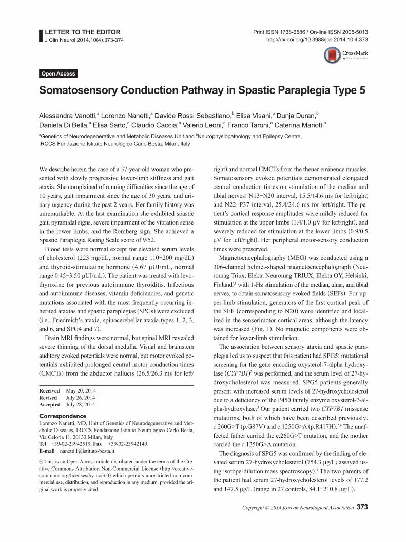
Magnetoencephalography (MEG) was conducted using a 306-channel helmet-shaped magnetoencephalograph (Neu-romag Triux, Elekta Neuromag TRIUX, Elekta OY, Helsinki, Finland)1 with 1-Hz stimulation of the median, ulnar, and tibial nerves, to obtain somatosensory evoked fields (SEFs). For up-per-limb stimulation, generators of the first cortical peak of the SEF (corresponding to N20) were identified and local-ized in the sensorimotor cortical areas, although the latency was increased (Fig. 1). No magnetic components were ob-tained for lower-limb stimulation.
The association between sensory ataxia and spastic para-plegia led us to suspect that this patient had SPG5: mutational screening for the gene encoding oxysterol-7-alpha hydroxy-lase (CYP7B1)2 was performed, and the serum level of 27-hy-droxycholesterol was measured. SPG5 patients generally present with increased serum levels of 27-hydroxycholesterol due to a deficiency of the P450 family enzyme oxysterol-7-al-pha-hydroxylase.3 Our patient carried two CYP7B1 missense mutations, both of which have been described previously: c.260G>T (p.G87V) and c.1250G>A (p.R417H).2,4 The unaf-fected father carried the c.260G>T mutation, and the mother carried the c.1250G>A mutation.
The diagnosis of SPG5 was confirmed by the finding of ele-vated serum 27-hydroxycholesterol (754.3 μg/L; assayed us-ing isotope-dilution mass spectroscopy).3 The two parents of the patient had serum 27-hydroxycholesterol levels of 177.2 and 147.5 μg/L (range in 27 controls, 84.1–210.8 μg/L).
Somatosensory Conduction Pathway in Spastic Paraplegia Type 5
Alessandra Vanotti,a Lorenzo Nanetti,a Davide Rossi Sebastiano,b Elisa Visani,b Dunja Duran,b Daniela Di Bella,a Elisa Sarto,a Claudio Caccia,a Valerio Leoni,a Franco Taroni,a Caterina MariottiaaGenetics of Neurodegenerative and Metabolic Diseases Unit and bNeurophysiopathology and Epilepsy Centre, IRCCS Fondazione Istituto Neurologico Carlo Besta, Milan, Italy
Open Access
Received May 20, 2014 Revised July 26, 2014 Accepted July 28, 2014
CorrespondenceLorenzo Nanetti, MD, Unit of Genetics of Neurodegenerative and Met-abolic Diseases, IRCCS Fondazione Istituto Neurologico Carlo Besta, Via Celoria 11, 20133 Milan, ItalyTel +39-02-23942519, Fax +39-02-23942140E-mail [email protected]
cc This is an Open Access article distributed under the terms of the Cre-ative Commons Attribution Non-Commercial License (http://creative-commons.org/licenses/by-nc/3.0) which permits unrestricted non-com-mercial use, distribution, and reproduction in any medium, provided the ori-ginal work is properly cited.
Neurophysiologic Characterization of SPG5 with MEG
374 J Clin Neurol 2014;10(4):373-374
Spastic paraplegia type 5 has been clinically described with either a “pure” or “complex” phenotype. Mild sensory abnor-malities have been described in pure hereditary SPGs, among which SPG4 is the most frequently occurring. Furthermore, 47–58% of pure hereditary SPG patients present with mild vibration-sense abnormalities without elongation of central somatosensory conduction times.5 Although several SPGs may present with vibration-sense abnormalities, this feature has only been consistently described in SPG5 patients (94–100%)2,4 in association with elongated somatosensory central conduction times.6
The data obtained in this study confirmed the presence of a severe clinical and neurophysiologic somatosensory impair-ment in SPG5 that is due to selective dorsal-column degen-eration. Our MEG study revealed that the magnetic cortical representation is conserved for upper-limb stimulation and absent for lower-limb stimulation, in agreement with the oc-currence of a “dying-back” dorsal-column degeneration.6 This finding should be confirmed in a larger cohort of SPG5 patients since the absence of evoked MEG signals has been observed even in normal healthy subjects. The neurophysio-logic somatosensory abnormalities in our patient were more pronounced and diffuse than corticospinal abnormalities.
These results contribute to the definition of the SPG5 phe-notype. Despite the rarity of SPG5, the refinement of clinical, biochemical, and neurophysiologic descriptions could im-
prove early diagnoses of this condition. Moreover, serum lev-els of 27-hydroxycholesterol represent a possible biomarker for therapeutic trials.
Conflicts of InterestThe authors have no financial conflicts of interest.
REFERENCES1. Burgess RC, Funke ME, Bowyer SM, Lewine JD, Kirsch HE, Bagić
AI, et al. American Clinical Magnetoencephalography Society Clini-cal Practice Guideline 2: presurgical functional brain mapping using magnetic evoked fields. J Clin Neurophysiol 2011;28:355-361.
2. Tsaousidou MK, Ouahchi K, Warner TT, Yang Y, Simpson MA, La-ing NG, et al. Sequence alterations within CYP7B1 implicate defec-tive cholesterol homeostasis in motor-neuron degeneration. Am J Hum Genet 2008;82:510-515.
3. Schüle R, Siddique T, Deng HX, Yang Y, Donkervoort S, Hansson M, et al. Marked accumulation of 27-hydroxycholesterol in SPG5 pa-tients with hereditary spastic paresis. J Lipid Res 2010;51:819-823.
4. Arnoldi A, Crimella C, Tenderini E, Martinuzzi A, D’Angelo MG, Musumeci O, et al. Clinical phenotype variability in patients with he-reditary spastic paraplegia type 5 associated with CYP7B1 mutations. Clin Genet 2012;81:150-157.
5. Sartucci F, Tovani S, Murri L, Sagliocco L. Motor and somatosensory evoked potentials in Autosomal Dominant Hereditary Spastic Parapa-resis (ADHSP) linked to chromosome 2p, SPG4. Brain Res Bull 2007; 74:243-249.
6. Manganelli F, Pisciotta C, Dubbioso R, Iodice R, Criscuolo C, Rug-giero L, et al. Electrophysiological characterisation in hereditary spas-tic paraplegia type 5. Clin Neurophysiol 2011;122:819-822.
Fig. 1. Cortical representation of the first peak of the somatosensory evoked fields obtained by stimulation of the right median nerve (red), right ulnar nerve (yellow), left median nerve (green), and left ulnar nerve (blue). Source local-ization was performed using standard-ized weighted low-resolution electromag-netic tomography on individual MRI.
Related Documents