Small supernumerary marker chromosomes (sSMC) - why do they break, where they break and how to distinguish harmful from harmless sSMC? Dissertation in partial fulfilment of the requirements for the academic degree of doctor rerum naturalium (Dr. rer. nat.) submitted to the Faculty Council of the School of Medicine at Friedrich Schiller University of Jena by M.Sc. Ahmed Basheer Hamid born on 30. June 1973 in Thi-Qar (Iraq)

Welcome message from author
This document is posted to help you gain knowledge. Please leave a comment to let me know what you think about it! Share it to your friends and learn new things together.
Transcript
Small supernumerary marker chromosomes (sSMC) - why do they break, where they break and how to distinguish harmful
from harmless sSMC?
Dissertation
in partial fulfilment of the requirements for the academic degree of
doctor rerum naturalium (Dr. rer. nat.)
submitted to the Faculty Council of the School of Medicine
at Friedrich Schiller University of Jena
by M.Sc. Ahmed Basheer Hamid
born on 30. June 1973 in Thi-Qar (Iraq)
Reviewers 1. PD Dr. rer. nat. / med. habil. Thomas Liehr
Institute of Human Genetics, Friedrich Schiller University of Jena 2. Prof. Dr. rer. nat. Wim Damen
Faculty of Biology and Pharmacy, Friedrich Schiller University of Jena 3. PD Mag. Dr. rer. nat. Irmgard Verdorfer
Department of Pathology, Medical University of Innsbruck
Date of the public disputation: 03.02.2015

I
This work is dedicated…
to my parents
to my dear wife Shaymaa
to my lovely kids Mustafa and Mayar
to the spirit of my dearest friend Dr. Zaid A. AL-Hilli
(10.09.1974 - 26.03.2011)
Abbreviations II
Abbreviations
aCGH Array-based comparative genomic hybridization AS Angelman syndrome BAC Bacterial Artificial Chromosome CCK Color changing karyotyping cenM-FISH Centromere specific multicolor FISH CES Cat eye syndrome CGH Comparative genomic hybridization CNV Copy number variant(s) COBRA Combined binary ratio labeling C-UBCA Centromere-near - unbalanced chromosomal abnormalities DAPI 4',-6-Diamidino-2-phenylindol-dihydrochlorid der Derivative chromosome dn de novo DNA Deoxyribonucleic Acid ESAC Extra structurally abnormal chromosome FISH Fluorescence in situ hybridization HCM-FISH Heterochromatin-directed M-FISH i Isochromosome idic Isodicentric chromosome inv dup Inverted duplicated chromosomes ISCN International System for Human Cytogenetic Nomenclature Kb Kilobase pairs mar Marker chromosome Mb Megabase pairs SNP Single nucleotide polymorphism MCB Multicolor banding mFISH multicolor FISH M-FISH Multiplex FISH min Centric minute chromosomes NCBI National Center for Biotechnology Information NORs Nucleolus-Organizing Regions OMIM Online Mendelian Inheritance in Man p Short arm of chromosome PAC Phage artificial chromosome PCL-FISH Pericentric-ladder-FISH PCR Polymerase chain reaction PKS Pallister-Killian syndrome PWS Prader-Willi syndrome q Long arm of chromosome
Abbreviations III
r Ring chromosomes rDNA Ribosomal Deoxyribonucleic Acid RNA Ribonucleic Acid rRNA Ribosomal ribonucleic acid SKY Spectral karyotyping SMC Supernumerary marker chromosome SNRPN Small nuclear ribonucleoprotein-associated protein N SRC Supernumerary ring chromosome sSMC Small supernumerary marker chromosomes subcenM-FISH Subcentromere specific multicolor FISH t Translocation UBCA Unbalanced chromosomal abnormalities UCSC University of California, Santa Cruz UPD Uniparental disomy WCP Whole-chromosome painting YAC Yeast artificial chromosome
Contents IV
Contents
Dedication…………………………………………………………………………………... I
Abbreviations………………………………………………………………………………. II-III
Contents…………………………………………………………………………………….. IV-V
Summary…………………………………………………………………………………… 1
Zusammenfassung………………………………………………………………………… 2
1. Introduction…………………………………………………………………………….. 3
1.1. Cytogenetics – how to characterize chromosomes…………………………………. 3
1.1.1. Classical and banding cytogenetics…………………………………………….. 3
1.1.2. Molecular cytogenetics………………………………………………………… 4
1.1.3. Comparative Genomic Hybridization (CGH) and aCGH……………………… 6
1.1.3.1. Microdissection and aCGH…………………………………………………… 7
1.2. Small supernumerary marker chromosomes (sSMC)………………………………. 8
1.2.1. Definition and nomenclature…………………………………………………… 8
1.2.2. Characterization………………………………………………………………… 9
1.2.3. Formation of sSMC…………………………………………………………….. 11
1.2.3.1. Mixtures of Different sSMC Shapes………………………………………. 13
1.2.4. Epidemiology of sSMC in genetics disorders………………………………….. 14
1.2.4.1. Clinical consequences of sSMC…………………………………………... 14
1.3. Aim of study/ Questions worked on………………………………………………… 15
2. Results………………………………………………………………………………….. 16
2.1. Basic papers of thesis…………………………...…………………………………… 16
2.2. Article-1……………………………………………………………………………... 18
2.3. Article-2……………………………………………………………………………... 23
2.4. Article-3……………………………………………………………………………... 28
2.5. Article-4……………………………………………………………………………... 34
2.6. Article-5……………………………………………………………………………... 45
2.7. Article-6……………………………………………………………………………... 53
2.8. Article-7……………………………………………………………………………... 55
2.9. Article-8……………………………………………………………………………... 62
2.10. Article-9……………………………………………………………………………. 69
2.11. Article-10…………………………………………………………………………... 75
2.12. Article-11…………………………………………………………………………... 79
Contents V
3. Discussion……………………………………………………………………………….. 84
3.1. Development of probe sets for detection of euchromatic presence in sSMC……….. 84
3.2. sSMC and localization of chromosomal breakpoints……………………………….. 87
3.3. Mosaicism in association with sSMC……………………………………………….. 88
3.4. sSMC and genotype-phenotype correlation…………………………………………. 90
4. Conclusions and Outlook…………………………………………….………………... 93
5. Bibliography ……………………………………………………………………………. 94
6. Appendix ………………………………………………………………………………... 105
6.1. List of own publications ……………………………………………………………. 105
6.2. Curriculum Vitae……………………………………………………………………. 108
6.3. Acknowledgements…………………………………………………………………. 111
6.4. Ehrenwörtliche Erklärung…………………………………………………………… 112
Summary 1
Summary:
Small supernumerary marker chromosomes (sSMC) are defined as additional centric
chromosome fragments too small to be identified or characterized unambiguously by banding
cytogenetics alone. Even though certain sSMC were associated with specific clinical pictures
and syndromes, for most of the sSMC only first steps towards genotype-phenotype
correlations were achieved. Therefore sSMC are still a problem in clinical cytogenetics and
can be harmful due to different mechanisms like induction of genomic imbalances and/or
uniparental disomy of the sSMC’s sister chromosomes. This study had the aim to provide new
insights into the questions (i) if and why sSMC include specific breakpoints and (ii) how to
distinguish harmful from harmless sSMC. Thus, several approaches for better sSMC
characterization (HCM-FISH) and/or, characterization of sSMC breakpoints were developed
(PCL-FISH; 1MB sets spanning the transitions of dosage-sensitive and dosage-insensitive
pericentric regions) and established. sSMC breakpoints were characterized in detail using
these new approaches, but also by microdissection based array-comparative genomic
hybridization. First hints were obtained that breakpoints involved in sSMC formation are
situated preferentially in gene- poor regions of the pericentric regions. Concerning genotype-
phenotype correlation of sSMC the present study further identified one new “complex sSMC”
associated syndrome: the der(13 or 21)t(13 or 21;18) syndrome, which is associated with a
mild clinical phenotype irrespective of partial trisomy 18p. Finally, influence of mosaicism on
sSMC-related phenotypes was studied in detail.
In conclusion, the present study provided important new data for genotype-phenotype
correlation and biological understanding of sSMC.

Summary 2
Zusammenfassung:
Kleine überzählige Marker-Chromosomen (sSMC) sind definiert als zusätzliche zentrische
Chromosomenfragmente, die zu klein sind, als allein mittels Zytogenetik identifiziert oder
eindeutig charakterisiert werden zu können. Auch wenn bestimmte sSMC schon mit
spezifischen Krankheitsbilder und Syndromen assoziiert werden konnten, wurden für die
meisten der sSMC bisher nur erste Schritte bezüglich Genotyp-Phänotyp-Korrelationen
erreicht. Daher sind sSMC immer noch ein Problem in der klinischen Zytogenetik und können
sich ungünstig auf den Phänotyp auswirken durch verschiedene Mechanismen, wie die
Induktion von genomischen Imbalancen und/oder einer uniparentalen Disomie der
Schwesterchromosomen des sSMC. Die hier vorliegende Studie hatte das Ziel, neue
Einsichten in die Fragen (i) ob und warum sSMC spezifische Bruchpunkte haben, und (ii) wie
schädliche von harmlosen sSMC zu unterscheiden sind. Daher wurden mehrere Ansätze für
eine bessere sSMC-Charakterisierung (HCM –FISH) und/oder die Charakterisierung von
sSMC Bruchpunkten entwickelt (PCL–FISH; 1MB Sondensets welche die Übergänge der
dosisempfindlichen und der dosisunempfindlichen perizentrischen Regionen unterscheiden).
sSMC Bruchpunkte wurden mit diesen neuen Ansätzen, aber auch im Detail durch
mikrodissektion-basierende-array-komparative genomische Hybridisierung charakterisiert.
Erste Hinweise wurden erhalten, dass die an der Bildung der sSMC-Bruchpunkte beteiligten
Regionen bevorzugt in genarmen Bereichen des Perizentromers liegen. Bezüglich der
Genotyp-Phänotyp-Korrelation von sSMC wurde in der vorliegenden Studie ein neues mit
einem "komplexen sSMC" verbundenes Syndrom definiert: das der(13 oder 21)t(13 oder
21;18)-Syndrom, welches mit einem milden klinischen Phänotyp aber einer partiellen
Trisomie 18p verbunden ist. Schließlich wurde der Einfluss von zellulären Mosaiken in
sSMC-Syndromen auf den Phänotyp im Detail untersucht.
Insgesamt liefert die vorliegende Studie wichtige neue Daten für Genotyp-Phänotyp-
Korrelation und das biologische Verständnis von sSMC.
1. Introduction 3
1. Introduction
1.1. Cytogenetics – how to characterize chromosomes
Chromosomes are the factors that distinguish one species from another and that enable the
transmission of genetic information from one generation to the next; the study of
chromosomes and cell division are referred to as Cytogenetics (Turnpenny and Ellard 2007).
Cytogenetic studies allow analyzing the chromosomal behavior in the organization and
transmission of genetic information, variability mechanisms and evolutionary pathways,
besides contributing to the genetic improvement of domestic species (Lacadena 1996) and its
essential role in clinical genetics. Therefore, cytogenetics is mainly focused on structure and
chemical/genetic organization of chromosomes, linking two formerly unrelated sciences,
cytology and genetics (Griffiths et al. 1996).
1.1.1. Classical and banding cytogenetics
The essential step for the emergence of modern human cytogenetics approach was when Tjio
and Levan (1956) correctly concluded that the normal human somatic cell contains 46
chromosomes (Tjio and Levan 1956) which was confirmed by examining meiotic
chromosomes (Ford and Hamerton 1956). In 1959, Lejeune and colleagues found the trisomy
for chromosome 21 as the underlying cause of the Langdon Down syndrome (Lejeune et al.
1959), and after that Nowell and Hungerford, in 1960 identified a minute chromosome in the
peripheral blood of patient with chronic granulocytic leukaemia which was called later
Philadelphia 1 chromosome (Nowell and Hungerford 1960). In 1961 Ilberry and coworkers
provided the first description of a small supernumerary marker chromosome (sSMC; see also
1.2) when reporting a boy with epicanthic fold and protuberant tongue and a karyotype
47,XY,+mar/46,XY (Ilberry et al. 1961). Later, Ellis and colleagues (1962) reported an
aberrant small acrocentric chromosome (Ellis et al. 1962), and Froland and colleagues (1963),
described a boy with several congenital defects with a karyotype 47,XY,+mar (Froland et al.
1963). This altogether opened broad prospects to clinical cytogenetic studies and the
literatures showed the relation between numerical and morphological chromosomal
aberrations and disease in man (Luthardt and Keitges 2001).
Classical cytogenetic staining approaches can provide information regarding the structure of
an sSMC (Rooney and Czepulkowski 1986). The size and shape is often more clearly
observed in solid-stained preparations, since chromosome banding approaches like G-banding
(Claussen et al. 2002) may suggest a particular chromosomal origin (Seabright 1971), such as
in case of tetrasomy 12p (Graf and Schwartz 2002). So-called chromosomal satellites
including the nucleolus-organizing regions (NORs) may be present at one or both ends of an

1. Introduction 4
sSMC and can be visualized either by silver staining or observation of satellite association
between the marker and other acrocentric chromosomes (Thangavelu et al. 1994).
Centromeric heterochromatin can be highlighted by C-banding (Gardner et al. 2012). If a
marker chromosome has two centromeres, one may be inactived, either in all or in a
proportion of cells, as recent studies of our group showed (Ewers et al. 2010).
DistamycinA/DAPI staining identifies the heterochromatin of chromosomes 1, 9, 15, and 16
and of the Y chromosome. In case an sSMC has chromosomal satellites and, in addition, a
distamycinA/DAPI-staining region, then an origin from chromosome 15 is likely, even
though this could not always be substantiated (Wisniewski et al. 1979, Callen 1991).
In 1971/1972 a system of nomenclature was proposed for banded human chromosomes and
chromosome abnormalities and was based on the patterns observed in different cells stained
with either of the chromosome banding techniques (Mitelman 1995). This international
system for human cytogenetic nomenclature (ISCN) is still in place and actualized regularly
(Shaffer et al. 2013). By standard banding techniques karyotypes a pattern of ~500 bands can
be achieved. G-bands made it possible for a detailed analysis of each chromosome to be
carried out, which led to improved definitions of different chromosomal aberrations and the
discovery of new cytogenetic syndromes in clinical pathology. Thus, nowadays, it is still the
starting point and gold standard of all cytogenetic techniques (Garcia-Sagredo 2008).
1.1.2. Molecular cytogenetics
The staining patterns produced on chromosomes by banding procedures are sometimes
ambiguous, and the resolution is limited by the optical characteristics of microscopes and the
complex manner in which DNA is packaged into chromosomes (Li and Pinkel 2006). But
further characterization of particular rearrangements requires additional techniques. Among
them, fluorescence in situ hybridization (FISH) becomes increasingly important in the
characterization of both constitutional and acquired chromosomal abnormalities (Gerdes et al.
1997). In 1969, Gall and Pardue described the hybridization of radioactively labeled rRNA to
tissue squashes allowing the in situ visualization of the complementary sequences, the rDNA,
within cells (Gall and Pardue 1969) and then, in 1986, Pinkel and coworkers (1986) and
Cremer and coworkers (1986) reported FISH using non-radioactively labeled probes. Since
then, FISH has been further developed and widely used for the detection of DNA or RNA
sequences (Pinkel et al.1986, Cremer et al. 1986). In situ hybridization is based on the specific
base pairing of two complementary nucleic acid sequences, the probe and the target
sequences. Hybridized probes are detected via fluorochromes using epifluorescence
1. Introduction 5
microscopy, via colorimetric enzyme assays by transmission light microscopy, or via metallic
compounds in the electron microscope (Joos et al. 1994).
The aim of the FISH technique is to characterize either imbalances, i.e gains or losses of
chromosome material, or specific breakpoints with or without imbalance (Kjeldsen and
Kølvraa 2002). A number of different types of probes for FISH can be distinguished on the
basis of the complexities of probe or target sequences: the alphoid and satellite probes
detecting repeat-targets; individual probes such as plasmid-, cosmid (35-55 kb)-, bacterial
artificial chromosomes (BACs)-, yeast artificial chromosomes (YACs) or P1 filamentous
phage artificial chromosomes (PACs) - clones detecting single copy sequences – nowadays
mainly used BACs; or composite probes are generated using PCR with sequence-specific
primers, allowing a specific painting of individual chromosomes or chromosomal regions
(Pinkel et al. 1988, Speicher 2005). Following the sequencing of the human genome, large-
insert clones that have been mapped and sequenced, and can be used as probes, are now
readily available for almost any genomic region. Probes can be selected easily using internet-
browsers such as Ensembl Cytoview, NCBI Map-Viewer or the UCSC genome browser
(Speicher and Carter 2005). This relatively new field of molecular cytogenetics, which makes
use of a variety of nucleic acid sequences as probes to cellular DNA targets has helped to
bridge the gap between molecular genetics and classical cytogenetic analyses (Teixeira 2002).
Nederlof and coworkers reported in 1989 the first multicolor FISH (three-color FISH)
experiments (Nederlof et al. 1989). For multicolor karyotyping with painting probes several
approaches were developed, including multiplex FISH (M-FISH), spectral karyotyping
(SKY), color changing karyotyping (CCK), and combined binary ratio labeling (COBRA)
(Liehr and Claussen 2002a,b). Molecular cytogenetics has provided new tools to characterize
aberrant karyotypes more precisely (Haddad et al. 1998) and became important component of
molecular diagnostics, particularly for diagnosing congenital syndromes in which the
underlying genetic defect is unknown (Speicher and Carter 2005).
1. Introduction 6
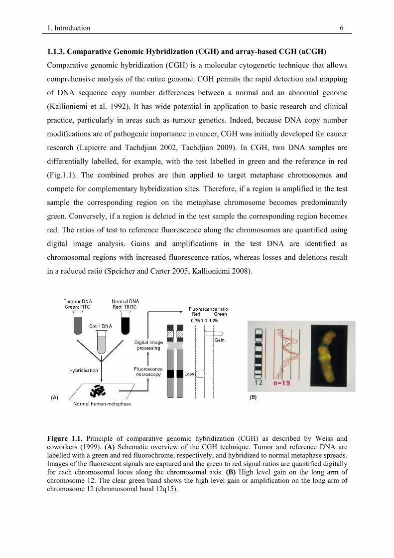
1.1.3. Comparative Genomic Hybridization (CGH) and array-based CGH (aCGH)
Comparative genomic hybridization (CGH) is a molecular cytogenetic technique that allows
comprehensive analysis of the entire genome. CGH permits the rapid detection and mapping
of DNA sequence copy number differences between a normal and an abnormal genome
(Kallioniemi et al. 1992). It has wide potential in application to basic research and clinical
practice, particularly in areas such as tumour genetics. Indeed, because DNA copy number
modifications are of pathogenic importance in cancer, CGH was initially developed for cancer
research (Lapierre and Tachdjian 2002, Tachdjian 2009). In CGH, two DNA samples are
differentially labelled, for example, with the test labelled in green and the reference in red
(Fig.1.1). The combined probes are then applied to target metaphase chromosomes and
compete for complementary hybridization sites. Therefore, if a region is amplified in the test
sample the corresponding region on the metaphase chromosome becomes predominantly
green. Conversely, if a region is deleted in the test sample the corresponding region becomes
red. The ratios of test to reference fluorescence along the chromosomes are quantified using
digital image analysis. Gains and amplifications in the test DNA are identified as
chromosomal regions with increased fluorescence ratios, whereas losses and deletions result
in a reduced ratio (Speicher and Carter 2005, Kallioniemi 2008).
Figure 1.1. Principle of comparative genomic hybridization (CGH) as described by Weiss and coworkers (1999). (A) Schematic overview of the CGH technique. Tumor and reference DNA are labelled with a green and red fluorochrome, respectively, and hybridized to normal metaphase spreads. Images of the fluorescent signals are captured and the green to red signal ratios are quantified digitally for each chromosomal locus along the chromosomal axis. (B) High level gain on the long arm of chromosome 12. The clear green band shows the high level gain or amplification on the long arm of chromosome 12 (chromosomal band 12q15).
1. Introduction 7
In analyzing the results of CGH, several limitations must also be taken into account. CGH can
spot sequence copy number changes only if more than 50 % of the cells analyzed contain a
chromosomal gain or loss. CGH is also impaired in its ability to identify balanced
chromosomal abnormalities for which there are no copy number changes, such as those found
in balanced translocations, inversions and intragenetic rearrangements (Tachdjian et al. 2008).
Genetic changes are detected and mapped on chromosomes when the size of the chromosomal
region affected is at least 10–12 Mb (Bentz et al. 1998).
Subsequently array-based CGH (aCGH) was established, an approach where arrays of
genomic sequences replaced the metaphase chromosomes as hybridization targets by large
numbers of mapped clones that are spotted onto a standard glass slide greatly increasing the
resolution of screening for genomic copy number gains and losses (Solinas-Toldo et al. 1997,
Pinkel et al. 1998). This solved many of the technical difficulties and problems caused by
working with cytogenetic chromosome preparations. The main advantage of aCGH is the
ability to perform copy number analyses with much higher resolution than was ever possible
using chromosomal CGH (Davies et al. 2005, Pinkel and Albertson 2005, Lockwood et al.
2006).
Two major groups of microarray-based platforms are currently used in clinical cytogenetics:
microarray-based comparative genomic hybridization (aCGH), and single nucleotide
polymorphism (SNP) genotyping-based arrays (Li and Andersson 2009). In aCGH, the most
apparent besides those already present in CGH includes the challenge of interpreting copy
number variants (CNVs) of unknown significance and distinguishing disease-causing CNVs
from normal CNV polymorphisms (Li and Andersson 2009, Bishop 2010).
1.1.3.1. Microdissection and aCGH
In 1981 Scalenghe and colleagues were the first to develop the chromosome microdissection
and microcloning technique (Scalenghe et al. 1981). Then, it was extended to human
chromosomes (Bates et al. 1986, Lüdecke et al. 1989, Senger et al. 1990). Microdissection
can be used to isolate derivative chromosomes form a balanced translocation or marker
chromosomes present in low mosaic. This DNA can be amplified and used in aCGH, thus
overcoming parts of its above mentioned limitations (1.1.3). For sSMC this approach was
applied by others (Shaw et al. 2004) and our group in single case studies (Liehr et al. 2006a,
Backx et al. 2007).
1. Introduction 8
1.2. Small supernumerary marker chromosomes (sSMC)
1.2.1. Definition and nomenclature
Small supernumerary marker chromosomes (sSMC) were first described in 1961 by Ilberry
and coworkers and, today, it is known that sSMCs are present in approximately 3.0 million
carriers worldwide in a population of 7 billion human beings (Liehr et al. 2004a, Liehr
2014a). sSMC can be defined as “small structurally abnormal chromosomes that occur in
addition to the normal 46 chromosomes” (Crolla et al. 1997), and according to the definition
of the ISCN 2013, a marker chromosome (mar) is a structurally abnormal chromosome that
cannot be unambiguously identified or characterized by conventional banding cytogenetics
(Shaffer et al. 2013). Numerous terms have been used in the literature to described sSMC in
the last few decades. The three best known are supernumerary marker chromosome (SMC)
which does not distinguish between larger and smaller SMCs, extra structurally abnormal
chromosome (ESAC), and supernumerary ring chromosome (SRC). In addition, other
designations summarized elsewhere were used (Liehr 2012, Liehr et al. 2004a). The
chromosomal origin of some sSMCs has been identified and associated with known
syndromes, such as isochromosome 12p [i(12p), OMIM #601803] Pallister-Killian syndrome
(PKS), isochromosome 18p [i(18pS), OMIM #614290] syndrome, Emanuel syndrome (ES) or
supernumerary-derivative chromosome 22 [der(22)t(11;22)(q23;q11.2), OMIM #609029]
syndrome, and inverted duplication 22 [inv dup(22q), OMIM #115470] cat eye syndrome
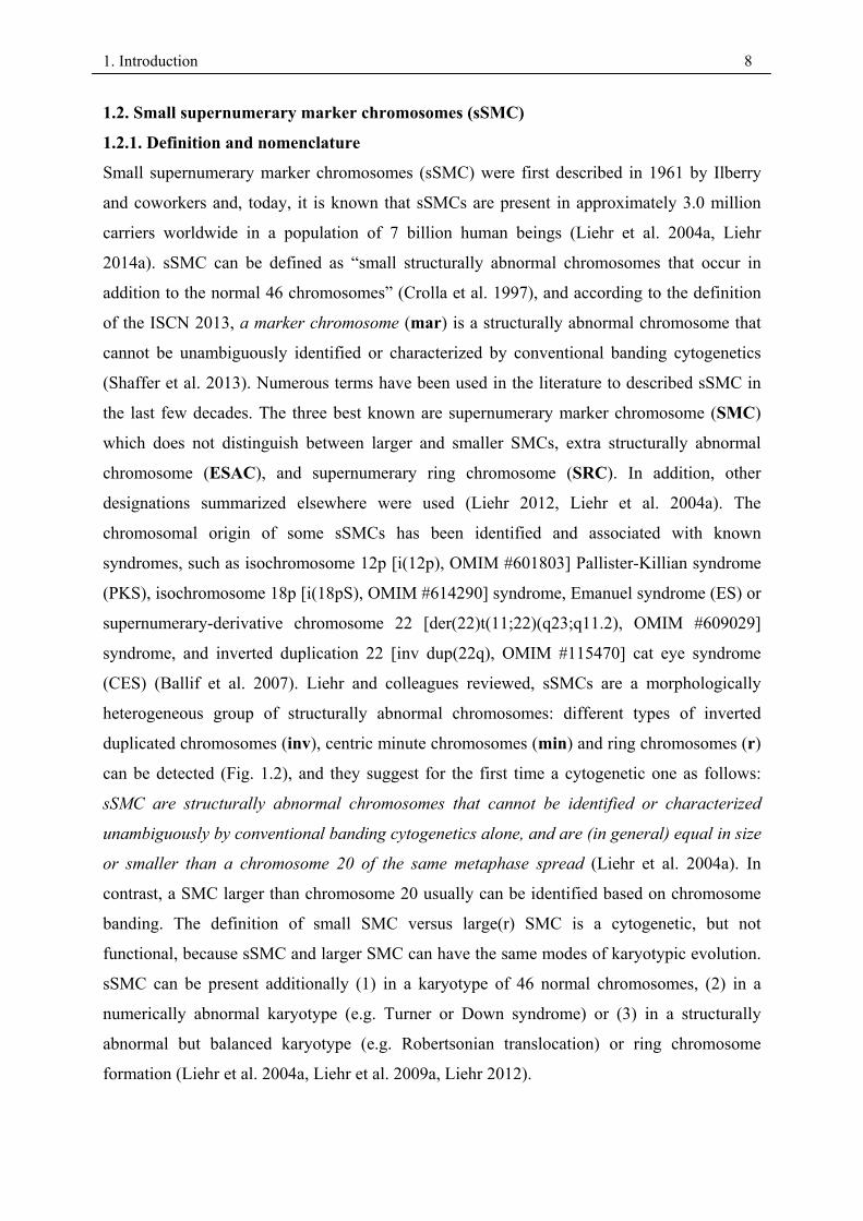
(CES) (Ballif et al. 2007). Liehr and colleagues reviewed, sSMCs are a morphologically
heterogeneous group of structurally abnormal chromosomes: different types of inverted
duplicated chromosomes (inv), centric minute chromosomes (min) and ring chromosomes (r)
can be detected (Fig. 1.2), and they suggest for the first time a cytogenetic one as follows:
sSMC are structurally abnormal chromosomes that cannot be identified or characterized
unambiguously by conventional banding cytogenetics alone, and are (in general) equal in size
or smaller than a chromosome 20 of the same metaphase spread (Liehr et al. 2004a). In
contrast, a SMC larger than chromosome 20 usually can be identified based on chromosome
banding. The definition of small SMC versus large(r) SMC is a cytogenetic, but not
functional, because sSMC and larger SMC can have the same modes of karyotypic evolution.
sSMC can be present additionally (1) in a karyotype of 46 normal chromosomes, (2) in a
numerically abnormal karyotype (e.g. Turner or Down syndrome) or (3) in a structurally
abnormal but balanced karyotype (e.g. Robertsonian translocation) or ring chromosome
formation (Liehr et al. 2004a, Liehr et al. 2009a, Liehr 2012).
1. Introduction 9
Figure.1.2. Different shapes of Small supernumerary marker chromosomes (sSMC). sSMCs can form
three basic types of shapes: ring-structure (r), inverted duplication (inv dup), and centric minute (min)
(Liehr et al. 2004a).
1.2.2. Characterization
Detection of an sSMC is nearly always unexpected by the clinician and more or less an
accidental result in cytogenetics. The origin of sSMC is almost impossible to establish by
routine cytogenetics alone, whereas fluorescence in situ hybridisation (FISH) methods are
highly suited for this (Starke et al. 2003a). A variety of molecular cytogenetic techniques that
provide more comprehensive analysis in a single or a few experiments have been described
for sSMC characterization. M-FISH, multicolor banding (MCB), whole-chromosome painting
(WCP), locus-specific FISH, centromere specific multicolor FISH (cenM-FISH),
subcentromere specific multicolor FISH (subcenM-FISH), microdissection coupled with
reverse painting and FISH approaches may all provide identification of the chromosome of
origin of SMCs (Nietzel et al. 2001, Brecevic et al. 2006, Pietrzak et al. 2007). Even if M-
FISH is readily available, this technique can result in ambiguous classification or
misclassification of sSMCs, particularly if they are small. In addition, these multicolor FISH
techniques cannot precisely determine the chromosome regions or breakpoints involved
(Tsuchiya et al. 2008). Usually, sSMC larger than chromosome 20 can be identified based on
chromosome-banding. Additionally, C-banding, silver staining of NOR or Q-banding were
used for sSMC characterization (Gersen and Keagle 2005). WCP-FISH approaches are well-
suited for the determination of the chromosomal origin of marker or derivative chromosomes
providing that they are larger than 17p, whereas if they are smaller, WCP-FISH is, in general,
non-informative (Haddad et al. 1998, Starke et al. 1999). Also, it is possible characterization
sSMC with a euchromatic content of approximately half of the short arm of chromosome 17p
1. Introduction 10
or more by the MCB technique is possible (Weise et al. 2002, Starke et al. 2003a). sSMCs
have also been successfully characterized by glass needle-based chromosome microdissection
and reverse chromosome painting (Starke et al. 2001). This approach is suited for all types of
sSMCs, including neocentromeric ones (Liehr et al. 2007). Although a comprehensive
characterization seems to be available by microdissection and reverse painting, it is restricted
as it provides on information regarding the orientation of eventual present chromosomal
fragments or the copy number of specific subregions, and no reliable information on the
presence or absence of centromere-near euchromatic content (Liehr et al. 2009a). aCGH is an
efficient and sensitive technique for detecting genome-wide copy number alterations at high
resolution (Shaffer et al. 2007). aCGH can now provide accurate characterization of SMCs in
terms of chromosomal origin, gene content, and other concomitant imbalances elsewhere in
the genome (Reddy et al. 2013). However, most successfully characterized sSMCs were
larger than 17p. Furthermore, centromeric and/or heterochromatic regions are problematic.
CGH has the advantage that it provides informative results on the euchromatic region(s)
involved in a sSMC, these regions must be larger than approximately 5-10 Mb to be visible in
CGH and can be overcome that, in principle, by application of aCGH, where much higher
resolutions can be achieved (Tsuchiya et al. 2008, Liehr et al. 2009a). Although aCGH using
chips that provide comprehensive genome coverage may become the technology of choice for
initial characterization of SMCs, G-banded and FISH analyses are still indispensable for
determining the structure and level of mosaicism of these chromosomes. G-banded analysis
may also be useful for detecting low level mosaic SMCs that could potentially be missed by
array CGH (Tsuchiya et al. 2008). sSMC can be best characterized for their chromosomal
origin by using centromeric probes. Nietzel and colleagues proposed the centromere-specific
multicolor FISH (cenM-FISH), as fast and easy method for sSMC characterization (Nietzel et
al. 2001). This approach overcomes the limitations of all the previously mentioned methods
concerning the informational value of the centromeric regions (Liehr et al. 2009a).
Several probe sets were suggested as approaches to detect the presence of euchromatic on an
sSMC. Besides FISH banding, approaches such as multicolor banding (Liehr et al. 2006b),
and subcentromeric multicolor-FISH (subcenM-FISH), a probe set comprising of 43 bacterial
or yeast artificial chromosome clones located in proximal regions of each human chromosome
(Starke et al. 2003a) were suggested. Still the approaches available at the beginning of the
present work were not ideal yet for sSMC-characterization.
As reported previously by (Chudoba et al. 1999, von Eggeling et al. 2002, Liehr et al. 2009a)
and others (reviewed in: Kotzot 2002a), they recommended that, after identification of the
1. Introduction 11
origin of the sSMC, its normal sister-chromosomes should be tested for their parental origin
to exclude a possible uniparental disomy (UPD). However, most sSMCs have yet to be
accurately characterized (Liehr et al. 2004b) because of variations in euchromatic DNA
content, different degrees of mosaicism, UPD of the chromosomes homologous to the sSMC,
and technical limitations of fluorescence in situ hybridization (FISH) and G-banding that do
not allow for accurate detection of sSMCs at high resolution (Starke et al. 2003a). This has
resulted in a lack of genotype/phenotype correlation for most sSMCs.
1.2.3. Formation of sSMC
Different mechanisms of sSMC formation including trisomic rescue, monosomic rescue, post
fertilization errors and gamete complementation have been proposed in the literature (Bartels
et al. 2003, Liehr et al. 2004a). Later, a new mechanism was proposed, that could provides a
possible explanation for the formation of multiple sSMC of different origin, in which sSMC
originated from transfection of chromosomes into the zygote derived from one or more
superfluous haploid pronuclei that would normally be degraded by deoxyribonucleases or
other means (Daniel and Malafiej 2003). Modes of sSMC-formation, which were not topic of
this work, are summarized in Fig. 1.3.
1. Introduction 12
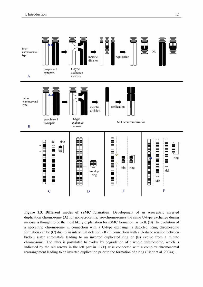
Figure 1.3. Different modes of sSMC formation: Development of an acrocentric inverted duplication chromosome (A) for non-acrocentric iso-chromosomes the same U-type exchange during meiosis is thought to be the most likely explanation for sSMC formation, as well. (B) The evolution of a neocentric chromosome in connection with a U-type exchange is depicted. Ring chromosome formation can be (C) due to an interstitial deletion, (D) in connection with a U-shape reunion between broken sister chromatids leading to an inverted duplicated ring or (E) evolve from a minute chromosome. The latter is postulated to evolve by degradation of a whole chromosome, which is indicated by the red arrows in the left part in E (F) arise connected with a complex chromosomal rearrangement leading to an inverted duplication prior to the formation of a ring (Liehr et al. 2004a).
1. Introduction 13
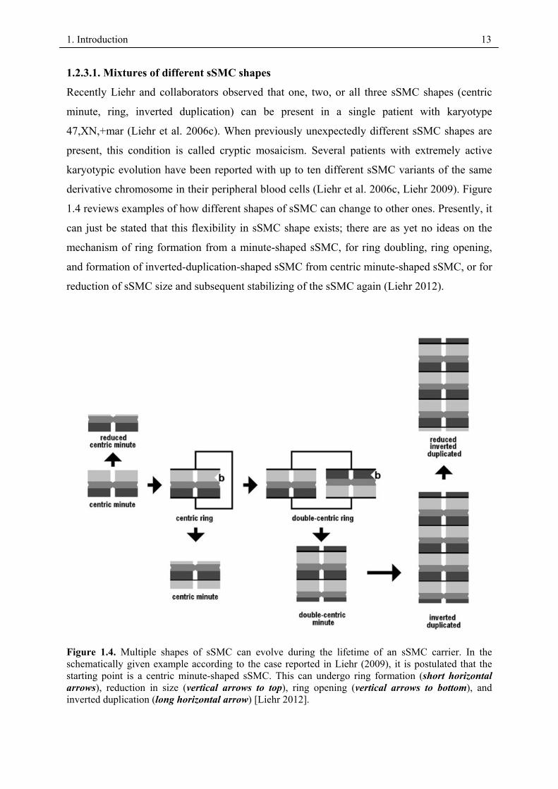
1.2.3.1. Mixtures of different sSMC shapes
Recently Liehr and collaborators observed that one, two, or all three sSMC shapes (centric
minute, ring, inverted duplication) can be present in a single patient with karyotype
47,XN,+mar (Liehr et al. 2006c). When previously unexpectedly different sSMC shapes are
present, this condition is called cryptic mosaicism. Several patients with extremely active
karyotypic evolution have been reported with up to ten different sSMC variants of the same
derivative chromosome in their peripheral blood cells (Liehr et al. 2006c, Liehr 2009). Figure
1.4 reviews examples of how different shapes of sSMC can change to other ones. Presently, it
can just be stated that this flexibility in sSMC shape exists; there are as yet no ideas on the
mechanism of ring formation from a minute-shaped sSMC, for ring doubling, ring opening,
and formation of inverted-duplication-shaped sSMC from centric minute-shaped sSMC, or for
reduction of sSMC size and subsequent stabilizing of the sSMC again (Liehr 2012).
Figure 1.4. Multiple shapes of sSMC can evolve during the lifetime of an sSMC carrier. In the schematically given example according to the case reported in Liehr (2009), it is postulated that the starting point is a centric minute-shaped sSMC. This can undergo ring formation (short horizontal arrows), reduction in size (vertical arrows to top), ring opening (vertical arrows to bottom), and inverted duplication (long horizontal arrow) [Liehr 2012].
1. Introduction 14
1.2.4. Epidemiology of sSMC in genetics disorders:
For several reasons sSMC are still a problem in clinical cytogenetics: (i) they are too small to
be characterized for their chromosomal origin by traditional banding techniques and require
molecular (cytogenetic) techniques for their identification (Liehr and Weise 2007); (ii) most
of the sSMCs have not been correlated with clinical syndromes, even though progress was
achieved, recently (Liehr et al. 2006c, Liehr 2014a); (iii) sSMC can be harmful due to
different mechanisms like induction of genomic imbalance and/or UPD of the sSMC’s sister
chromosomes (Liehr et al. 2004b); (iv) also sSMC can be found just by chance and cannot be
correlated with the clinical problems of a patient (Liehr 2010); finally (v) the percentage in
which an sSMC is present (mosaicism) can, but must not have an influence on the clinical
outcome (Liehr et al. 2004b, 2006c, Liehr 2014a). Thus, to understand the epidemiology of
sSMC comprehensive studies of sSMC need to be done.
1.2.4.1. Clinical consequences of sSMC
In approximately 30% of SMC carriers an abnormal phenotype is observed. The clinical
outcome of an sSMC is difficult to predict as they can have different phenotypic
consequences because of (1) differences in euchromatic DNA-content, (2) different degrees of
mosaicism, and/or (3) UPD of the chromosomes homologous to the SMC (Starke et al.
2003a). Also the risk for phenotypic abnormalities associated with a marker chromosome
depends on several factors, including inheritance, mode of ascertainment, chromosomal
origin, and the morphology, content, and structure of the marker (Graf et al. 2006). Thus, the
main problem is de novo sSMC detected prenatally, which are not characterized in detail. It
has been shown that most couples decide in such cases against the child, even though there is
a 2:1 chance that the developing child would be normal.
Certain marker chromosomes are consistently identifiable by G-banding and have a well-
established phenotype. Examples include i(12p), associated with PKS and i(18p), which cause
both mild–moderate mental retardation and a characteristic facial appearance (Callen et al.
1990), and for chromosome 15-derived marker chromosomes, often seen as isodicentric 15q.
FISH analysis allows discrimination between large markers that contain the SNRPN locus and
thus are tetrasomic for the Prader–Willi syndrome (PWS) or Angelman syndrome (AS)
critical region and those small markers that do not contain SNRPN (Crolla et al. 1995, Huang
et al. 1997, Eggermann et al. 2002, Baldwin 2008).

1. Introduction 15
1.3. Aims of the present study/ Questions worked on
The long term objective of the present project is get new insights in the regulation of gene-
expression within the pericentric region of the human genome. There was some evidence at
the begin of this study that there are dosage sensitive and insensitive regions around the
human centromeres. This evidence was supported by own studies and thus the main focus of
my studies were the following questions.
1) How to characterize sSMC quickly and comprehensively?
2) How to distinguish sSMC straight forward between benign and harmful?
3) Where are the borders of dosage-sensitive pericentric regions?
Or as summarized in the title of this thesis here should be studied why sSMC break, where
they break and how to distinguish harmful from harmless sSMC.
To answer these questions FISH-probe sets were established, ~400 new sSMC cases were
studied during the present work and microdissection based aCGH was systematically applied
in 80 sSMC cases.
162. Results
2.1. Basic papers of thesis
1. Liehr T, Weise A, Hamid AB, Fan X, Klein E, Aust N, Othman MAK, Mrasek K, Kosyakova
N. Multicolor FISH methods in current clinical diagnostics. Expert Rev Mol Diagn, 2013;13(3):251–255.
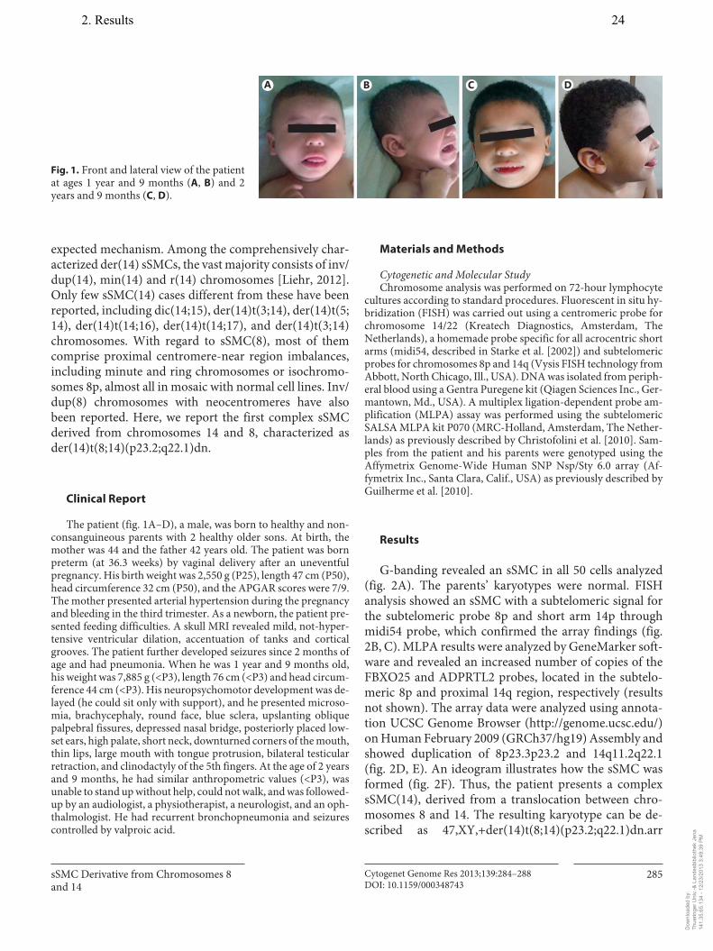
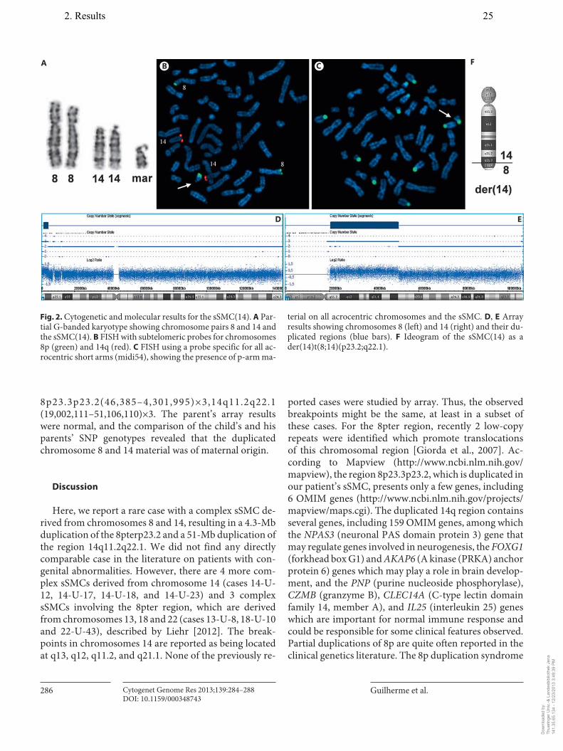
2. Guilherme RS, Dutra ARN, Perez ABA, Takeno SS, Oliveira MM, Kulikowski LD, Klein E, Hamid AB, Liehr T, Melaragno MI. First report of a small supernumerary der(8;14) marker chromosome. Cytogenet Genome Res, 2013; 139:284-288.
3. Liehr T, Cirkovic S, Lalic T, Guc-Scekic M, de Almeida C, Weimer J, Iourov I, Melaragno M I, Guilherme RS, Stefanou E-GG, Aktas D, Kreskowski K, Klein E, Ziegler M, Kosyakova N, Volleth M, Hamid AB. Complex small supernumerary marker chromosomes – an update. Molecular Cytogenetics, 2013; 6:46
4. Hamid AB, Kreskowski K, Weise A, Kosayakova N, Mrasek K, Voigt M, Guilherme RS, Wagner R, Hardekopf D, Pekova S, Karamysheva T, Liehr T, Klein E. How to narrow down chromosomal breakpoints in small and large derivative chromosomes – a new probe set. J Appl Genet , 2012;53(3):259-269.
5. Liehr T, Karamysheva T, Merkas M, Brecevic L, Hamid AB, Ewers E, Mrasek K, Kosyakova N, Weise A. Somatic mosaicism in cases with small supernumerary marker chromosomes. Curr Genomics, 2010;11:432-439.
6. Hamid AB, Liehr T. Pericentromeric BAC-probe set - thoughts about considering genedosage insensitive regions. Mol Cytogenet 2013; 6:45/comments.
7. Hamid AB, Weise A, Voigt M, Bucksch M, Kosyakova N, Liehr T, Klein E. Clinical impact of proximal autosomal imbalances. Balk J Med Genet, 2012; 15(2):15-21.
8. Bucksch M, Ziegler M, Kosayakova N, Mulhatino MV, Llerena Jr. JC, Morlot S, Fischer W, Polityko AD, Kulpanovich AI, Petersen MB, Belitz B, Trifonov V, Weise A, Liehr T, Hamid AB. A new multicolor fluorescence in situ hybridization probe set directed against human heterochromatin: HCM-FISH. J Histochem Cytochem, 2012;60(7):530-536.
9. Liehr T, Klein E, Mrasek K, Kosyakova N, Guilherme RS, Aust N, Venner C, Weise A, Hamid AB. Clinical impact of somatic mosaicism in cases with small supernumerary marker chromosomes. Cytogenet Genome Res, 2013;139(3):158–163.
10. Fernández-Toral J, Rodríguez L, Plasencia A, Martínez-Frías ML, Ewers E, Hamid AB, Ziegler M, Liehr T. Four small supernumerary marker chromosomes derived from chromosomes 6, 8, 11 and 12 in a patient with minimal clinical abnormalities: a case report. J Med Case Reports, 2010;4:239.
11. Papoulidis I, Manolakos E, Hamid AB, Klein E, Kosyakova N, Kordaß U, Kunz J, Siomou E, Kontodiou M, Tzimina M, Nicolaides P, Liehr T, Petersen MB. Tetrasomy 9p mosaicism associated with a normal phenotype in two cases. Cytogenet Genome Res, 2012;136:237–241.
172. Results
The main points of the present study - as highlighted in discussion - part are:
1) better sSMC characterization approaches,
2) characterization of chromosomal breakpoints involved in sSMC formation,
3) on mosaicism in sSMC, and overall,
4) on a refinement of the genotype-phenotype correlation in sSMC.
Those four points are also covered in the papers mentioned above as listed in the Table below:
Table.2.1. Articles of the present Ph.D. work is based on.
Article No. probe sets involved breakpoints mosaicism genotype/
phenotype 1 + - - - 2 + + - + 3 + + + + 4 + + + + 5 + - + - 6 + + - + 7 + + - + 8 + - - + 9 - - + - 10 - - - + 11 - - + +
2. Results
2.2. Article .1
Liehr T, Weise A, Hamid AB, Fan X, Klein E, Aust N, Othman MAK, Mrasek K, Kosyakova N. Multicolor FISH methods in current clinical diagnostics. Expert Rev Mol Diagn, 2013; 13(3): 251–255.
10.1586/ERM.12.146 251ISSN 1473-7159© 2013 Expert Reviews Ltdwww.expert-reviews.com
Review
Human genetics is a discipline, which includes pre- and post-natal counseling of patients and families. A genetic basis can be considered in individuals suffering from infertility and/or repeated abortions, or any kind of acquired or inherited syndrome [1]. At present, genetic coun-selors have a multitude of technical possibili-ties, some highly sophisticated, for the genetic analysis of an individual. Approaches such as next-generation sequencing of a whole genome has gained importance and has been helpful on many occasions [1,2].
Besides genetic counseling, another key element of human genetics is still the well-established approach of cytogenetics, includ-ing molecular cytogenetics [1]. In many western countries (e.g., Germany), insurance companies request, where appropriate, banding cytogenet-ics as the starting test for a genetic analysis. Thus, up to 40% of individuals in search of advice are still studied cytogenetically, and a subset of them are further analyzed by molecular cytogenetics [Schreyer I, Pers. Comm.]. Additionally, in most countries (except for North America and western Europe) cytogenetics is still the
gold standard for any genetic analysis, with molecular cytogenetics becoming available over the last decade.
After the introduction of array-comparative genomic hybridization (aCGH), cytogenetics/molecular cytogenetics were considered to be outdated by some researchers [3,4]. However, it is common knowledge that aCGH results can only be correctly interpreted if cytogenetics is performed in parallel; in addition, abnormal aCGH results need to be confirmed by a second method, such as molecular cytogenetics [5,6].
Molecular cytogeneticsIn banding cytogenetics – today often incor-rectly called ‘classical cytogenetics’ (classical cytogenetics is Giemsa or Orcein staining with-out any banding) – only chromosome morphol-ogy combined with a black and white banding pattern is evaluated. Thus, only changes within the normal banding pattern, size variations in a chromosomal band or the chromosome itself, and changes to the centromere index, can be detected [7]. To overcome these limitations, FISH approaches were introduced in the 1980s,
Thomas Liehr*, Anja Weise, Ahmed B Hamid, Xiaobo Fan, Elisabeth Klein, Nadine Aust, Moneeb AK Othman, Kristin Mrasek and Nadezda KosyakovaJena University Hospital, Friedrich Schiller University, Institute of Human Genetics, Kollegiengasse 10, Jena D-07743, Germany*Author for correspondence: Tel.: +49 364 193 5533 Fax: +49 364 193 5582 [email protected]
Multicolor FISH (mFISH) assays are currently indispensable for a precise description of derivative chromosomes. Routine application of such techniques on human chromosomes started in 1996 with the simultaneous use of all 24 human whole-chromosome painting probes in multiplex-FISH and spectral karyotyping. Since then, multiple approaches for chromosomal differentiation based on multicolor-FISH (MFISH) assays have been developed. Predominantly, they are applied to characterize marker or derivative chromosomes identified in conventional banding analysis. Since the introduction of array-based comparative genomic hybridization (aCGH), mFISH is also applied to verify and further delineate aCGH-detected aberrations. For the latter, it is important to consider the fact that aCGH cannot detect or characterize balanced rearrangements, which are important to be resolved in detail in infertility diagnostics. In addition, mFISH is necessary to distinguish different imbalanced situations detectable in aCGH; small supernumerary marker chromosomes have to be differentiated from insertions or unbalanced translocations. This review presents an overview on the available mFISH methods and their applications in pre- and post-natal clinical genetics.
Multicolor FISH methods in current clinical diagnostics
Expert Rev. Mol. Diagn. 13(3), 251–255 (2013)
Expert Review of Molecular Diagnostics
© 2013 Expert Reviews Ltd
10.1586/ERM.12.146
1473-7159
1744-8352
Review
Keywords: derivative chromosomes • marker chromosomes • multicolor fluorescence in situ hybridization • postnatal diagnostics • prenatal diagnostics
THeMed ArTICLe y Genetic & Genomics Applications
For reprint orders, please contact [email protected]
2. Results 18
ahamid
Textfeld
Expert Rev. Mol. Diagn. 13(3), (2013)252
Review
and the new field of ‘molecular cytogenetics’ was launched. For more information on FISH, readers are directed to [8], as such a discussion will not be covered in this review.
One-, two- and three-color FISH experiments are standard in every laboratory around the world performing molecular cytoge-netics. Multicolor FISH (mFISH) is defined as the simultaneous use of at least three different ligands or fluorochromes for the spe-cific labeling of DNA – excluding the counterstain [9]. Due to this definition, the first successful mFISH experiments were performed in 1989 [10]. The first mFISH probe sets were put together 7 years later in 1996 [11,12]. In the following review, the available mFISH probe sets for humans are summarized and their applications in pre- and post-natal diagnostics are highlighted.
mFISH probe setsWhole-chromosome painting-based mFISH probe setsBetween 1996 and 2000, simultaneous staining of each of the 24 human chromosomes in different colors using whole-chromosome painting (WCP) probes was described repeatedly as multiplex-FISH (M-FISH) [11], spectral karyotyping (SKY) [12], mFISH, combined binary ratio labeling–FISH or 24-color FISH (reviewed in [9]). Between four and seven fluorescence dyes were used either for combinatorial labeling and/or ratio labeling (combinatorial labeling: three up to seven fluorochromes are combined; each flourochrome combination is only used once. Ratio labeling: a maximum of three different fluorochromes are combined and mixed for each chromosome in different ratios). It was also shown that additional probes can be added to the basic 24-color FISH probe set (summarized in [9]). Today, the WCP-based mFISH probe sets are the most frequently applied probe sets in diagnostics; they are mostly designated as M-FISH or SKY probe sets [101].
mFISH probe sets for FISH-bandingThe definition of FISH-banding probe sets is “…any kind of FISH technique which provides the possibility to simultane-ously characterize several chromosomal subregions smaller than a chromosome arm – excluding the short arms of the acrocentric chromosomes; FISH-banding methods fitting that definition may have quite different characteristics, but share the ability to produce a DNA-specific chromosomal banding…” [13].
The most often applied and also commercially available mFISH probe set for FISH banding is the high-resolution multi color-banding (MCB) or m-banding technique [101]. It is based on overlapping microdissection libraries (partial chromosome paints [PCPs]) producing fluorescence profiles along the human chro-mosomes, which was first described using the example of chromo-some 5 in 1999 [14]. MCB/m-banding allows for differentiation of chromosome region-specific areas at the band and sub-band level at a resolution of 550 bands per haploid karyotype. In addition, the simultaneous use of all MCB PCPs in one hybridization step for the characterization of complex karyotypes is possible [15]. For the MCB probe set, a molecular definition of all underlying microdissection libraries was performed, which converted MCB into a DNA sequence-anchored probe set [16].
Besides these, there were many other mFISH-banding probe sets, which either were never finished for all human chromosomes or are no longer (commercially) available, such as cross-species color banding (Rx-FISH) or the Harlequin-FISH probe set [17]; spectral color banding [18] ; or interspersed PCR-based M-FISH [19]. There are also many probe sets leading to chromosome bar codes with different resolutions and applications (for a more detailed review, see [9]).
Centromeric probe-based mFISH probe setsSome mFISH probe sets are based on repetitive centromeric satel-lite probes. Such mFISH probe sets are extremely important in daily diagnostics, as combinations of different probes can princi-pally be chosen freely according to the individual case and ques-tion [20]. There is also an mFISH probe set that allows the simul-taneous characterization of all human centromeres in one step, the centromere-specific mFISH [21]. This probe set is especially useful for the characterization of the chromosomal origin of small supernumerary marker chromosomes (sSMC) [22,102].
Locus-specific probe-based mFISH probe setsmFISH probe sets based on locus-specific probes can be cre-ated by every laboratory and many are commercially available [103–108]. Some of the abovementioned chromosome bar codes were based on such locus-specific probes [9]. At present, mainly bacterial artificial chromosome (BAC) probes are used, as the necessary BACs can easily be tracked in genome browsers [109–111] and are offered commercially [103]. One of the most imaginative mFISH probe sets developed during the last few years is the one that enables a type of single cell-directed microsatellite analysis; the so-called parental origin determination FISH (pod-FISH) approach, detecting copy number variant regions in the human genome on a single-cell level [23].
mFISH-probe sets based on combinations of a variety of probesFinally, it is also possible to combine WCP, PCP, BAC or centro-meric probes in one probe set. Recent examples are: the subcen-tromere-specific mFISH [24], which can specifically characterize the centromere near euchromatic material; the hetero chromatin-M-FISH [25], which is specific for all larger heterochromatic regions in the human karyotype; or the 9het-mix [26], which ena-bles subdifferentiation of chromosome 9 heteromorphisms in the human population.
Diagnostic applications of mFISH probe setsmFISH probe sets are applied in pre- and post-natal clinical genet-ics (see below), tumor cytogenetics [9,13,101] and various research fields [9,13,101]. Here, the authors focus on their use in clinical genetics; that is, molecular cytogenetics performed on amnion, chorion, blood and, rarely, fibroblast cells. In all these tissues, it is possible to not only analyze the gain or loss of chromosomes or chromosomal segments in metaphase, but also in interphase. Structural rearrangements are normally studied on metaphase chromosomes in clinical genetics.
Liehr, Weise, Hamid et al.
2. Results 19
ahamid
Textfeld
253www.expert-reviews.com
Review
mFISH-probe sets applied in the interphaseInterphase mFISH diagnostics normally use the abovementioned combination of centromeric and/or locus-specific probes; PCPs and WCPs can be applied in interphase research, but are not suited for routine applications. The most commonly used diag-nostic probe set is the AneuVision® (Vysis Inc., IL, USA) probe set [106] or comparable ones [104,106], suited to detect the most frequent numerical chromosomal aberrations of the human fetus in the second trimester [27]. Preimplantation diagnostics are per-formed with the aim of detecting up to 70% of the most frequent numerical chromosome aberrations responsible for spontaneous abortions [9].
All of the following mentioned applications are performed on metaphase chromosome preparations. Molecular cytogenetics is normally performed as a secondary diagnostic test; thus below, the primary test is listed as an entry criteria for mFISH.
mFISH used after a cytogenetic normal resultBanding cytogenetics in mentally retarded patients or prenatal cases with specific sonographic signs quite often give normal results. Still, the clinical signs may be indicative for some syn-dromes to be excluded by FISH, namely microdeletion or micro-duplication syndromes [28]. Therefore, the clinician needs to provide a suspected diagnosis and then the FISH probes for the corresponding microdeletion, or microduplication syndrome may be applied. In most cases, these probes are applied in two-color FISH experiments [104–106]; however, there was a suggestion for a simultaneous screening for Prader–Willi/Angelman (15q11.13), Williams–Beuren (7q11.23), Smith–Magenis (17p11.2) and DiGeorge/velocardiofacial (22q11.2) syndromes in one so-called ‘multiFISH’ assay [29]. In addition, there were mFISH probe sets with locus-specific probes for the subtelo meric regions, which were successfully applied to detect genetic imbalances in up to 6% of patients with iodiopathic mental retardation [9]. However, in many instances such probe sets are now successfully replaced by real-time PCR, multiplex ligation-dependent probe amplification or aCGH settings [30].
mFISH used after an abnormal cytogenetic resultA cytogenetically abnormal result, which needs further mFISH testing, can include mosaics [31], larger derivative chromo-somes (balanced and unbalanced) [32] and/or the presence of an sSMC [22,102].
M-FISH or SKY probe sets will only be used in cases of complex chromosomal rearrangements [32] or if a derivative chromosome contains additional material of completely unknown origin [33]. As soon as the origin of the involved chromosomes is known, the chromosomal breakpoints are of interest and can be deter-mined by mFISH-banding and/or locus-specific probes [9,13,101]. However, WCP-based mFISH-probe sets and mFISH-banding probe sets are not helpful for the characterization of sSMC or of heterochromatic variants.
Cytogenetically visible heterochromatic variants can be best characterized by the recently reported heterochromatin M-FISH probe set [25] or subsets of them [26,33].
sSMC, excluding neocentric ones [102], can be best character-ized for their origin by centromere-specific M-FISH [21,22,24,102]. Subcentromere-specific M-FISH [24] is a straightforward approach for defining their euchromatic content, which might further be delineated by the pericentric ladder FISH probe set [34]. The latter enables a breakpoint analysis on a 10-Mb resolution.
mFISH used after an abnormal aCGH resultSince aCGH is applied for the characterization of subchromo-somal imbalanced rearrangements [3–5], this is another starting point for the application of molecular cytogenetics. Here, indi-vidual combinations of locus-specific (BAC) probes are used to prove or contradict a gain or loss suggested after aCGH [5,35].
aCGH is not necessarily fully informative with regards to the number of copies gained in the patient; for example, a threefold gain of 18p detected in aCGH may be a hint of an intrachromo-somal duplication or a derivative chromosome t(autosome;18)(?;p10) of the corresponding region in all cells of the patient. However, it can also be a hint on a mosaic kary otype 47,XN,+i(18)(p10)(50%)/46,XN(50%). FISH and mFISH applications follow-ing detection of an abnormal aCGH result were already repeatedly published [34–36] and are routine in clinical genetic diagnostics.
ConclusionAt present, mFISH methods are well established in clinical diagnostics. Apart from their longstanding role in refining and confirming cytogenetic results, mFISH approaches have gained additional importance in the verification of aCGH results. This underlines the truth that every approach has advantages and dis-advantages: the conventional approach-banding cytogenetics has a lower resolution but provides a highly informative ‘in situ’ view on the human genome; aCGH, however, results in a higher resolu-tion but gives a result more distant from the in vivo situation and can only detect imbalanced rearrangements. Both approaches are connected by molecular cytogenetics, and a comprehensive view on a pre- or post-natal clinical case is most often only possible after applying several of the currently available approaches, including mFISH, in a majority of them.
Expert commentaryMolecular cytogenetics, especially mFISH, is still a progressive field. New mFISH probe sets are being developed up to the pre-sent date [25,34]. Otherwise, mFISH is necessary to confirm and refine diagnostic findings of cytogenetics and aCGH. Therefore, the method is the connecting approach for banding cytogenetics and molecular genetics.
Five-year viewThe field of molecular cytogenetics/mFISH is an important tool to define and visualize chromosomal changes detectable in pre- and postnatal diagnostics. According to the fact that mFISH gained importance during the last years rather than lose it, in 5 years from now, it will be at least as significant as diagnostics are now. It can be expected that even findings seen in next-generation sequencing are necessary to be confirmed by mFISH in future [37].
Multicolor FISH methods in current clinical diagnostics
2. Results 20
ahamid
Textfeld
Expert Rev. Mol. Diagn. 13(3), (2013)254
Review
Financial and competing interest disclosureThis article was supported in parts by BMBF/DLR (ARM 08/001, BLR 08/004, RUS 09/006 and BLR 10/006) and Else Kröner-Fresenius-Stiftung (2011_A42). The authors have no other relevant affiliations or financial
involvement with any organization or entity with a financial interest in or financial conflict with the subject matter or materials discussed in the manuscript apart from those disclosed.
No writing assistance was utilized in the production of this manuscript.
Key issues
• Human genetics is a discipline that includes pre- and post-natal counseling of patients and families, (molecular) cytogenetics and molecular genetics.
• In molecular cytogenetics, multiple multicolor FISH (mFISH) approaches are now available.
• mFISH is performed based on whole or partial chromosome painting, centromeric or locus-specific DNA probes.
• Since 1996, new mFISH probe sets have been established every year, and this development is still ongoing.
• mFISH can be applied during interphase and metaphase.
• mFISH assays are indispensable for a precise description of derivative chromosomes identified in banding cytogenetics.
• Small supernumerary marker chromosomes can still be best analyzed by mFISH.
• In the last few years, mFISH has become an important instrument for array-comparitive genomic hybridization confirmation.
ReferencesPapers of special note have been highlighted as:
• of interest•• of considerable interest
1 Lewis R. Human Genetics. Mcgraw-Hill Higher Education, NY, USA (2011).
2 Desai AN, Jere A. Next-generation sequencing: ready for the clinics? Clin. Genet. 81(6), 503–510 (2012).
3 Ahn JW, Mann K, Walsh S et al. Validation and implementation of array comparative genomic hybridisation as a first line test in place of postnatal karyotyping for genome imbalance. Mol. Cytogenet. 3, 9 (2010).
4 Gao J, Liu C, Yao F et al. Array-based comparative genomic hybridization is more informative than conventional karyotyping and fluorescence in situ hybridization in the analysis of first-trimester spontaneous abortion. Mol. Cytogenet. 5(1), 33 (2012).
5 Kumar RA, Sudi J, Babatz TD et al. A de novo 1p34.2 microdeletion identifies the synaptic vesicle gene RIMS3 as a novel candidate for autism. J. Med. Genet. 47(2), 81–90 (2010).
6 Chen CP, Huang HK, Su YN et al. Trisomy 7 mosaicism at amniocentesis: interphase FISH, QF-PCR, and aCGH analyses on uncultured amniocytes for rapid distinguishing of true mosaicism from pseudomosaicism. Taiwan J. Obstet. Gynecol. 51(1), 77–82 (2012).
7 Claussen U, Michel S, Mühlig P et al. Demystifying chromosome preparation and the implications for the concept of chromosome condensation during mitosis. Cytogenet. Genome Res. 98(2–3), 136–146 (2002).
•• Explains chromosome preparations indetail and the biology behind the process.
8 Chang SS, Mark HF. Emerging molecular cytogenetic technologies. Cytobios 90(360), 7–22 (1997).
•• Describes the basics of molecularcytogenetics.
9 Liehr T, Starke H, Weise A, Lehrer H, Claussen U. Multicolor FISH probe sets and their applications. Histol. Histopathol. 19(1), 229–237 (2004).
• Reviews multicolor FISH sets.
10 Nederlof PM, Robinson D, Abuknesha R et al. Three-color fluorescence in situ hybridization for the simultaneous detection of multiple nucleic acid sequences. Cytometry 10(1), 20–27 (1989).
11 Speicher MR, Gwyn Ballard S, Ward DC. Karyotyping human chromosomes by combinatorial multi-fluor FISH. Nat. Genet. 12(4), 368–375 (1996).
12 Schröck E, du Manoir S, Veldman T et al. Multicolor spectral karyotyping of human chromosomes. Science 273(5274), 494–497 (1996).
13 Liehr T, Heller A, Starke H, Claussen U. FISH banding methods: applications in research and diagnostics. Expert Rev. Mol. Diagn. 2(3), 217–225 (2002).
• Reviews FISH banding methods and theirapplications.
14 Chudoba I, Plesch A, Lörch T, Lemke J, Claussen U, Senger G. High resolution multicolor-banding: a new technique for refined FISH analysis of human chromosomes. Cytogenet. Cell Genet. 84(3–4), 156–160 (1999).
15 Weise A, Heller A, Starke H et al. Multitude multicolor chromosome banding (mMCB) – a comprehensive one-step
multicolor FISH banding method. Cytogenet. Genome Res. 103(1–2), 34–39 (2003).
16 Weise A, Mrasek K, Fickelscher I et al. Molecular definition of high-resolution multicolor banding probes: first within the human DNA sequence anchored FISH banding probe set. J. Histochem. Cytochem. 56(5), 487–493 (2008).
17 Müller S, Wienberg J. Advances in the development of chromosome bar codes: integration of M-FISH and Rx-FISH technology. Medgen. 12(4), 474–477 (2000).
18 Kakazu N, Abe T. Multicolor banding technique, spectral color banding (SCAN): new development and applications. Cytogenet. Genome Res. 114(3–4), 250–256 (2006).
19 Aurich-Costa J, Vannier A, Grégoire E, Nowak F, Cherif D. IPM-FISH, a new M-FISH approach using IRS-PCR painting probes: application to the analysis of seven human prostate cell lines. Genes. Chromosomes Cancer 30(2), 143–160 (2001).
20 Starke H, Schreyer I, Kähler C et al. Molecular cytogenetic characterization of a prenatally detected supernumerary minute marker chromosome 8. Prenat. Diagn. 19(12), 1169–1174 (1999).
21 Nietzel A, Rocchi M, Starke H et al. A new multicolor-FISH approach for the characterization of marker chromosomes: centromere-specific multicolor-FISH (cenM-FISH). Hum. Genet. 108(3), 199–204 (2001).
22 Liehr T, Ewers E, Kosyakova N et al. Handling small supernumerary marker chromosomes in prenatal diagnostics.
Liehr, Weise, Hamid et al.
2. Results 21
ahamid
Textfeld
255www.expert-reviews.com
Review
Expert Rev. Mol. Diagn. 9(4), 317–324 (2009).
23 Weise A, Gross M, Hinreiner S, Witthuhn V, Mkrtchyan H, Liehr T. POD-FISH: a new technique for parental origin determination based on copy number variation polymorphism. Methods Mol. Biol. 659, 291–298 (2010).
24 Liehr T, Starke H, Heller A et al. Multi-color fluorescence in situ hybridization (FISH) applied to FISH-banding. Cytogenet. Genome Res. 114(3-4), 240–244 (2006).
• Reviews small supernumerary markerchromosomes.
25 Bucksch M, Ziegler M, Kosayakova N et al. A new multicolor fluorescence in situ hybridization probe set directed against human heterochromatin: HCM-FISH. J. Histochem. Cytochem. 60(7), 530–536 (2012).
26 Starke H, Seidel J, Henn W et al. Homologous sequences at human chromosome 9 bands p12 and q13-21.1 are involved in different patterns of pericentric rearrangements. Eur. J. Hum. Genet. 10(12), 790–800 (2002).
27 Weise A, Liehr T. Fluorescence in situ hybridization for prenatal screening of chromosomal aneuploidies. Expert Rev. Mol. Diagn. 8(4), 355–357 (2008).
28 Weise A, Mrasek K, Klein E et al. Microdeletion and microduplication syndromes. J. Histochem. Cytochem. 55(3), 185–190 (2012).
• Describes microdeletion andmicroduplication syndromes.
29 Ligon AH, Beaudet AL, Shaffer LG. Simultaneous, multilocus FISH analysis for detection of microdeletions in the diagnostic evaluation of developmental delay and mental retardation. Am. J. Hum. Genet. 61(1), 51–59 (1997).
30 Sauter SM, Böhm D, Bartels I et al. Partial trisomy of distal 19q detected by quantita-
tive real-time PCR and FISH in a girl with mild facial dysmorphism, hypotonia and developmental delay. Am. J. Med. Genet. 143A(10), 1091–1099 (2007).
31 Liehr T, Karamysheva T, Merkas M et al. Somatic mosaicism in cases with small supernumerary marker chromosomes. Curr. Genomics 11(6), 432–439 (2010).
32 Pellestor F, Anahory T, Lefort G et al. Complex chromosomal rearrangements: origin and meiotic behavior. Hum. Reprod. Update 17(4), 476–494 (2011).
33 Trifonov V, Seidel J, Starke H et al. Enlarged chromosome 13 p-arm hiding a cryptic partial trisomy 6p22.2-pter. Prenat. Diagn. 23(5), 427–430 (2003).
34 Hamid AB, Kreskowski K, Weise A et al. How to narrow down chromosomal breakpoints in small and large derivative chromosomes – a new probe set. J. Appl. Genet. 53(3), 259–269 (2012).
35 Liehr T, Starke H, Senger G, Melotte C, Weise A, Vermeesch JR. Overrepresenta-tion of small supernumerary marker chromosomes (sSMC) from chromosome 6 origin in cases with multiple sSMC. Am. J. Med. Genet. A 140(1), 46–51 (2006).
36 Carreira IM, Melo JB, Rodrigues C et al. Molecular cytogenetic characterisation of a mosaic add(12)(p13.3) with an inv dup(3)(q26.31 → qter) detected in an autistic boy. Mol. Cytogenet. 2, 16 (2009).
37 Kloosterman WP, Tavakoli-Yaraki M, van Roosmalen MJ et al. Constitutional chromothripsis rearrangements involve clustered double-stranded DNA breaks and nonhomologous repair mechanisms. Cell Rep. 1(6), 648–655 (2012).
Websites
101 Liehr T. Basics and literature on multicolor fluorescence in situ hybridization applica-tion (2012). www.fish.uniklinikum-jena.de/mFISH.html (Accessed 10 October 2012)
•• Collection of all relevant mFISHliterature.
102 Liehr T. Small supernumerary marker chromosomes (2012). www.fish.uniklinikum-jena.de/sSMC.html (Accessed 10 October 2012)
•• Collection of all relevant mFISHliterature. Most comprehensive resourcefor small supernumerary markerchromosomes.
103 BACPAC Resources Center (BPRC). http://bacpac.chori.org (Accessed 10 October 2012)
104 MetaSystems GmbH. www.metasystems-international.com (Accessed 10 October 2012)
105 Kreatech diagnostic. www.kreatech.com (Accessed 10 October 2012)
106 Abbott/Vysis. www.abbottmolecular.com/us/home.html (Accessed 10 October 2012)
107 Cytocell. www.cytocell.com (Accessed 10 October 2012)
108 DAKO. www.dako.com (Accessed 10 October 2012)
109 UCSC Human Genome Browser – hg19 assembly. http://genome.ucsc.edu/cgi-bin/hgGateway?hgsid=95241316&clade=vertebrate&org=Human&db=hg19 (Accessed 10 October 2012)
110 NCBI Map Viewer Homo sapiens (human) Build 37.3. www.ncbi.nlm.nih.gov/mapview/maps.cgi?ORG=hum&MAPS=ideogr,est,loc&LINKS=ON&VERBOSE=ON&CHR=5 (Accessed 10 October 2012)
111 Ensembl Genome Browser. www.ensembl.org (Accessed 10 October 2012)
Multicolor FISH methods in current clinical diagnostics
2. Results 22
ahamid
Textfeld
ahamid
Textfeld
2. Results
2.3. Article .2
Guilherme RS, Dutra ARN, Perez ABA, Takeno SS, Oliveira MM, Kulikowski LD, Klein E, Hamid AB, Liehr T, Melaragno MI. First report of a small supernumerary der(8;14) marker chromosome. Cytogenet Genome Res, 2013; 139:284-288.


2. Results
2.4. Article .3
Liehr T, Cirkovic S, Lalic T, Guc-Scekic M, de Almeida C, Weimer J, Iourov I, Melaragno MI, Guilherme RS, Stefanou E-GG, Aktas D, Kreskowski K, Klein E, Ziegler M, Kosyakova N, Volleth M, Hamid AB. Complex small supernumerary marker chromosomes – an update. Molecular Cytogenetics, 2013; 6:46
RESEARCH Open Access
Complex small supernumerary markerchromosomes – an updateThomas Liehr1,12*, Sanja Cirkovic2, Tanja Lalic2, Marija Guc-Scekic2,3, Cynthia de Almeida4, Jörg Weimer5,
Ivan Iourov6,7, Maria Isabel Melaragno8, Roberta S Guilherme8, Eunice-Georgia G Stefanou9, Dilek Aktas10,
Katharina Kreskowski1, Elisabeth Klein1, Monika Ziegler1, Nadezda Kosyakova1, Marianne Volleth11
and Ahmed B Hamid1
Abstract
Background: Complex small supernumerary marker chromosomes (sSMC) constitute one of the smallest subgroups
of sSMC in general. Complex sSMC consist of chromosomal material derived from more than one chromosome; the
best known representative of this group is the derivative chromosome 22 {der(22)t(11;22)} or Emanuel syndrome. In
2008 we speculated that complex sSMC could be part of an underestimated entity.
Results: Here, the overall yet reported 412 complex sSMC are summarized. They constitute 8.4% of all yet in detail
characterized sSMC cases. The majority of the complex sSMC is contributed by patients suffering from Emanuel
syndrome (82%). Besides there are a der(22)t(8;22)(q24.1;q11.1) and a der(13)t(13;18)(q11;p11.21) or der(21)t(18;21)
(p11.21;q11.1) = der(13 or 21)t(13 or 21;18) syndrome. The latter two represent another 2.6% and 2.2% of the
complex sSMC-cases, respectively. The large majority of complex sSMC has a centric minute shape and derives from
an acrocentric chromosome. Nonetheless, complex sSMC can involve material from each chromosomal origin. Most
complex sSMC are inherited form a balanced translocation in one parent and are non-mosaic. Interestingly, there
are hot spots for the chromosomal breakpoints involved.
Conclusions: Complex sSMC need to be considered in diagnostics, especially in non-mosaic, centric minute shaped
sSMC. As yet three complex-sSMC-associated syndromes are identified. As recurrent breakpoints in the complex
sSMC were characterized, it is to be expected that more syndromes are identified in this subgroup of sSMC. Overall,
complex sSMC emphasize once more the importance of detailed cytogenetic analyses, especially in patients with
idiopathic mental retardation.
Keywords: Complex small supernumerary marker chromosomes (sSMC), Genotype-phenotype correlation,
Mosaicism, SSMC shape, Emanuel syndrome
BackgroundSmall supernumerary marker chromosomes (sSMC) are
structurally abnormal chromosomes that cannot be identi-
fied or characterized in detail by banding cytogenetics, are
generally about the size of or smaller than a chromosome
20, and molecular cytogenetic techniques are necessary for
their comprehensive characterization [1]. It is estimated
that there are ~3 million of sSMC carriers in the human
population of 7 billion individuals. Fortunately, only in 1/3
of the cases the sSMC is associated with clinical abnor-
malities [2]. Besides some specific syndromes, i.e.
Pallister-Killian {= i(12p), OMIM #601803}, isochromo-
some 18p {i(18p), OMIM #614290}, cat-eye {i(22p ~ q),
OMIM #115470}, idic(15) {no OMIM number} and
Emanuel or derivative chromosome 22 {der(22)t(11;22),
OMIM #609029} syndromes [2], for the remaining sSMC-
cases only first steps towards genotype-phenotype correla-
tions were achieved [2,3].
sSMC can present with different shapes (ring-, centric
minute- and inverted duplication-shape), and consist in
the majority of the cases of pericentric chromosomal ma-
terial. Besides, sSMC can be derived from any part of the
* Correspondence: [email protected] University Hospital, Friedrich Schiller University, Institute of Human
Genetics, Kollegiengasse 10, Jena D-07743, Germany12Institut für Humangenetik, Postfach, Jena D-07740, Germany
Full list of author information is available at the end of the article
© 2013 Liehr et al.; licensee BioMed Central Ltd. This is an Open Access article distributed under the terms of the CreativeCommons Attribution License (http://creativecommons.org/licenses/by/2.0), which permits unrestricted use, distribution, andreproduction in any medium, provided the original work is properly cited. The Creative Commons Public Domain Dedicationwaiver (http://creativecommons.org/publicdomain/zero/1.0/) applies to the data made available in this article, unless otherwisestated.
Liehr et al. Molecular Cytogenetics 2013, 6:46
http://www.molecularcytogenetics.org/content/6/1/46
2. Results 28
human chromosomes and form neocentrics [2,4]. If they
derived from the chromosomal ends, in most cases they
lead to partial tetrasomies [2]; for one of those conditions
also an OMIM entry was introduced recently (#614846 -
tetrasomy 15q26 syndrome).
One of the smallest subgroup of sSMC is constituted by
the so-called complex marker chromosomes [5]. ‘Complex’
are such sSMC which consist of chromosomal material de-
rived from more than one chromosome [1]. Thus, besides
the aforementioned large group of Emanuel or derivative
chromosome 22 {der(22)t(11;22), OMIM #609029} syn-
drome cases, there was identified a second recurrent
complex sSMC in 2010, designated as supernumerary
der(22)t(8;22) syndrome {OMIM #613700} [6].
In 2008 we speculated that the then described 22 com-
plex sSMC cases, excluding the der(22)t(11;22) cases,
could be part of an underestimated entity [5]. Here the yet
reported 412 complex sSMC cases are summarized based
on the sSMC database (http://www.fish.uniklinikum-jena.
de/sSMC.html, [3]) and analyzed for their chromosomal
constitution, breakpoints and special features.
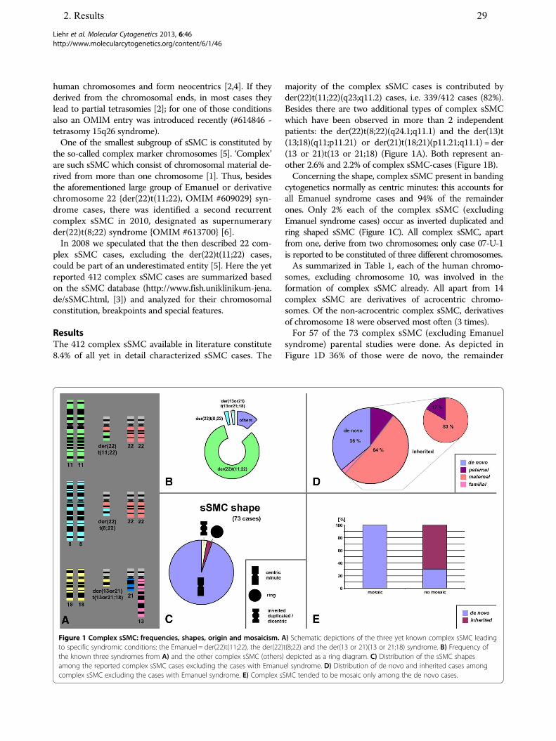
Results
The 412 complex sSMC available in literature constitute
8.4% of all yet in detail characterized sSMC cases. The
majority of the complex sSMC cases is contributed by
der(22)t(11;22)(q23;q11.2) cases, i.e. 339/412 cases (82%).
Besides there are two additional types of complex sSMC
which have been observed in more than 2 independent
patients: the der(22)t(8;22)(q24.1;q11.1) and the der(13)t
(13;18)(q11;p11.21) or der(21)t(18;21)(p11.21;q11.1) = der
(13 or 21)t(13 or 21;18) (Figure 1A). Both represent an-
other 2.6% and 2.2% of complex sSMC-cases (Figure 1B).
Concerning the shape, complex sSMC present in banding
cytogenetics normally as centric minutes: this accounts for
all Emanuel syndrome cases and 94% of the remainder
ones. Only 2% each of the complex sSMC (excluding
Emanuel syndrome cases) occur as inverted duplicated and
ring shaped sSMC (Figure 1C). All complex sSMC, apart
from one, derive from two chromosomes; only case 07-U-1
is reported to be constituted of three different chromosomes.
As summarized in Table 1, each of the human chromo-
somes, excluding chromosome 10, was involved in the
formation of complex sSMC already. All apart from 14
complex sSMC are derivatives of acrocentric chromo-
somes. Of the non-acrocentric complex sSMC, derivatives
of chromosome 18 were observed most often (3 times).
For 57 of the 73 complex sSMC (excluding Emanuel
syndrome) parental studies were done. As depicted in
Figure 1D 36% of those were de novo, the remainder
Figure 1 Complex sSMC: frequencies, shapes, origin and mosaicism. A) Schematic depictions of the three yet known complex sSMC leading
to specific syndromic conditions: the Emanuel = der(22)t(11;22), the der(22)t(8;22) and the der(13 or 21)(13 or 21;18) syndrome. B) Frequency of
the known three syndromes from A) and the other complex sSMC (others) depicted as a ring diagram. C) Distribution of the sSMC shapes
among the reported complex sSMC cases excluding the cases with Emanuel syndrome. D) Distribution of de novo and inherited cases among
complex sSMC excluding the cases with Emanuel syndrome. E) Complex sSMC tended to be mosaic only among the de novo cases.
Liehr et al. Molecular Cytogenetics 2013, 6:46 Page 2 of 6
http://www.molecularcytogenetics.org/content/6/1/46
2. Results 29
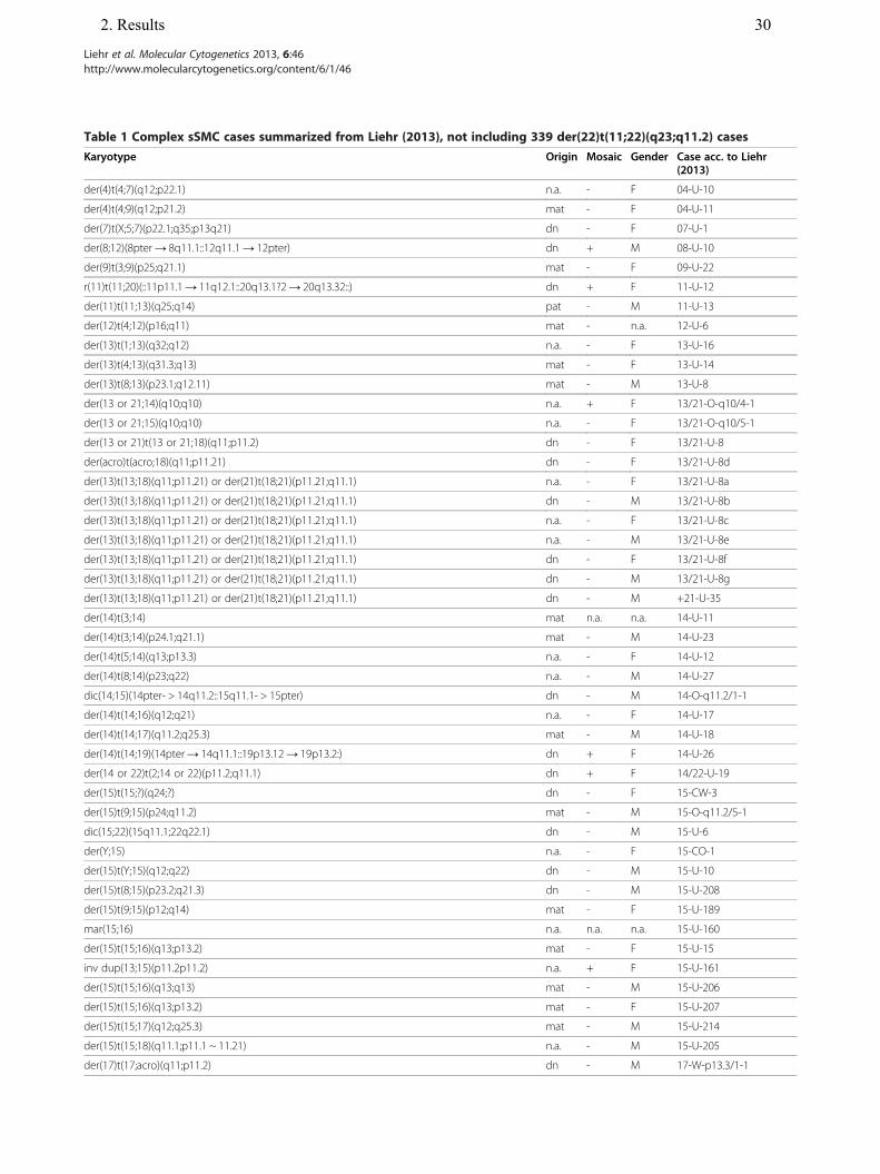
Table 1 Complex sSMC cases summarized from Liehr (2013), not including 339 der(22)t(11;22)(q23;q11.2) cases
Karyotype Origin Mosaic Gender Case acc. to Liehr(2013)
der(4)t(4;7)(q12;p22.1) n.a. - F 04-U-10
der(4)t(4;9)(q12;p21.2) mat - F 04-U-11
der(7)t(X;5;7)(p22.1;q35;p13q21) dn - F 07-U-1
der(8;12)(8pter→ 8q11.1::12q11.1→ 12pter) dn + M 08-U-10
der(9)t(3;9)(p25;q21.1) mat - F 09-U-22
r(11)t(11;20)(::11p11.1→ 11q12.1::20q13.1?2→ 20q13.32::) dn + F 11-U-12
der(11)t(11;13)(q25;q14) pat - M 11-U-13
der(12)t(4;12)(p16;q11) mat - n.a. 12-U-6
der(13)t(1;13)(q32;q12) n.a. - F 13-U-16
der(13)t(4;13)(q31.3;q13) mat - F 13-U-14
der(13)t(8;13)(p23.1;q12.11) mat - M 13-U-8
der(13 or 21;14)(q10;q10) n.a. + F 13/21-O-q10/4-1
der(13 or 21;15)(q10;q10) n.a. - F 13/21-O-q10/5-1
der(13 or 21)t(13 or 21;18)(q11;p11.2) dn - F 13/21-U-8
der(acro)t(acro;18)(q11;p11.21) dn - F 13/21-U-8d
der(13)t(13;18)(q11;p11.21) or der(21)t(18;21)(p11.21;q11.1) n.a. - F 13/21-U-8a
der(13)t(13;18)(q11;p11.21) or der(21)t(18;21)(p11.21;q11.1) dn - M 13/21-U-8b
der(13)t(13;18)(q11;p11.21) or der(21)t(18;21)(p11.21;q11.1) n.a. - F 13/21-U-8c
der(13)t(13;18)(q11;p11.21) or der(21)t(18;21)(p11.21;q11.1) n.a. - M 13/21-U-8e
der(13)t(13;18)(q11;p11.21) or der(21)t(18;21)(p11.21;q11.1) dn - F 13/21-U-8f
der(13)t(13;18)(q11;p11.21) or der(21)t(18;21)(p11.21;q11.1) dn - M 13/21-U-8g
der(13)t(13;18)(q11;p11.21) or der(21)t(18;21)(p11.21;q11.1) dn - M +21-U-35
der(14)t(3;14) mat n.a. n.a. 14-U-11
der(14)t(3;14)(p24.1;q21.1) mat - M 14-U-23
der(14)t(5;14)(q13;p13.3) n.a. - F 14-U-12
der(14)t(8;14)(p23;q22) n.a. - M 14-U-27
dic(14;15)(14pter- > 14q11.2::15q11.1- > 15pter) dn - M 14-O-q11.2/1-1
der(14)t(14;16)(q12;q21) n.a. - F 14-U-17
der(14)t(14;17)(q11.2;q25.3) mat - M 14-U-18
der(14)t(14;19)(14pter→ 14q11.1::19p13.12→ 19p13.2:) dn + F 14-U-26
der(14 or 22)t(2;14 or 22)(p11.2;q11.1) dn + F 14/22-U-19
der(15)t(15;?)(q24;?) dn - F 15-CW-3
der(15)t(9;15)(p24;q11.2) mat - M 15-O-q11.2/5-1
dic(15;22)(15q11.1;22q22.1) dn - M 15-U-6
der(Y;15) n.a. - F 15-CO-1
der(15)t(Y;15)(q12;q22) dn - M 15-U-10
der(15)t(8;15)(p23.2;q21.3) dn - M 15-U-208
der(15)t(9;15)(p12;q14) mat - F 15-U-189
mar(15;16) n.a. n.a. n.a. 15-U-160
der(15)t(15;16)(q13;p13.2) mat - F 15-U-15
inv dup(13;15)(p11.2p11.2) n.a. + F 15-U-161
der(15)t(15;16)(q13;q13) mat - M 15-U-206
der(15)t(15;16)(q13;p13.2) mat - F 15-U-207
der(15)t(15;17)(q12;q25.3) mat - M 15-U-214
der(15)t(15;18)(q11.1;p11.1 ~ 11.21) n.a. - M 15-U-205
der(17)t(17;acro)(q11;p11.2) dn - M 17-W-p13.3/1-1
Liehr et al. Molecular Cytogenetics 2013, 6:46 Page 3 of 6
http://www.molecularcytogenetics.org/content/6/1/46
2. Results 30

ones were inherited form a balanced translocation in
one parent. The majority of the latter group (83%) was
maternally derived. Interestingly, mosaic cases with karyo-
types 47,XN,+mar/46,XN were only seen in de novo com-
plex sSMC (Figure 1E). However, no balanced translocation
t(13;18)(q11;p11.21) or t(18;21)(p11.21;q11.1) was seen yet
in any of the corresponding nine cases.
In the 73 complex sSMC only 67 breakpoints were in-
volved. 44/67 breakpoints were unique, the remainder
observed two to 14 times (Table 2).
Finally, only seven of the 73 (~10%) complex sSMC-cases
not leading to Emanuel syndrome (case numbers 13/21-O-
q10/4-1, 13/21-O-q10/5-1, 14-O-q11.2/1-1, 15-O-q11.2/5-
1, 15-CO-1, 22-O-q11/3-1, 22-O-q11.21/3-1) were not
associated with clinical signs (Table 1). However, clinically
affected carriers of a der(13 or 21)t(13 or 21;18) inherited
the sSMC in parts by their mothers, which were considered
to be clinically normal [3].
DiscussionIn 2008 complex sSMC seamed to be something rather
unusual, apart from the cases with Emanuel syndrome [5].
Since then ~4 times more complex sSMC were character-
ized and reported, thus enabling more detailed follow up
analyses of our previous studies.
~40% (408 of 1,040) of all centric minute shaped sSMC
are complex sSMC, including der(22)t(11;22) cases [3]; the
latter needs to be kept in mind, if a minute shaped sSMC
is detected in diagnostics. Moreover, if a centric minute
shaped sSMC turns out to be NOR-positive at one end,
thus being acrocentric derived, this means that there is a
70% chance that it is a complex sSMC: of the yet known
567 centric minute shaped sSMC 408 are complex [3].
Also, if a centric minute shaped sSMC is present in all
cells of the carrier, this might be another hint for a com-
plex sSMC. Centric minute shaped non-complex sSMC
are mosaic in ~70% of the cases [3], while complex sSMC
Table 1 Complex sSMC cases summarized from Liehr (2013), not including 339 der(22)t(11;22)(q23;q11.2) cases
(Continued)
der(18)t(2;18)(p23.1;q11.1) dn + F 18-U-24
der(18)t(8;18)(p23.2 ~ 23.1;q11.1) n.a. - M 18-U-10
der(19)t(18;19) n.a. n.a. F 19-U-15
der(18)t(18;21 or 22) fam n.a. n.a. 18-CW-2
der(21)t(4;21)(q32.1;q21.2) mat - F 21-U-15
der(21)t(7;21)(p21;q21.3) mat - M 21-U-7
der(13/21;22)(13/21pter→ 13/21q11::22q11.1 ~ 11.2→ 22q11.21 ~ 11.22: :22q11.21 ~ 11.22→22pter)
dn - F 22-Wces-5-101
der(22)t(6;22)(p22.1;q11.21) ?pat - F 22-U-53
der(22)t(8;22)(q24.1;q11.2) pat - M 22-U-11
der(22)t(8;22)(q24.1;q11.1) mat - M/F 22-U-11a1/a2
der(22)t(8;22)(q24.1;q11.1) pat - M 22-U-11b
der(22)t(8;22)(q24.1;q11.1) mat - M 22-U-11c
der(22)t(8;22)(q24.1;q11.1) mat - M 22-U-11d
der(22)t(8;22)(q24.1;q11.1) mat - M 22-U-11e
der(22)t(8;22)(q24.1;q11.1) mat - M 22-U-11f
der(22)t(8;22)(q24.13;q11.21) n.a. - M 22-U-11g
der(22)t(8;22)(q24.13;q11.21) pat - F 22-U-11h
der(22)t(8;22)(q24.1;q11.2) mat - M 22-U-11i
der(22)t(8;22)(q24.1;q11.2) n.a. - M 22-U-11j
der(22)t(8;22)(p22;q11.21) mat - M 22-U-43
der(22)t(9;22)(p13.1;q11) mat - M 22-U-57
der(22)t(12;22)(p12;q11.2-12) dn - M 22-U-18
der(22)t(12;22)(p13.3;q12) mat - M 22-U-18a
der(22)t(14;22)(q31;q11) mat - F 22-O-q11/3-1
der(22)t(17;22)(17pter→ 17p10::22q10→ 22pter) mat - M 22-U-6
der(22)t(17;22)(p13.3;q11.21) pat - M 22-O-q11.21/3-1
der(22)t(19;22)(q13.42;q11.1) n.a. - M 22-U-50
r(15)ins(15;5)(?;q35.5q35.3)der(18)(:p11.21→ q11.1:)der(18)(:p11.1→ q11.1:) dn + M mult 3-9
Liehr et al. Molecular Cytogenetics 2013, 6:46 Page 4 of 6
http://www.molecularcytogenetics.org/content/6/1/46
2. Results 31
are mosaic in only ~10% of the cases. This indicates the
importance of cytogenetic analyses, as only this kind of
study enables to characterize the sSMC-shape and mosai-
cism reliably, and gives first hints on the possible complex
nature of an sSMC.
In 2010 the der(22)t(8;22)(q24.1;q11.1) syndrome was
reported. It was suggested that, like in Emanuel syndrome,
a 3:1 meiotic non-disjunction is causative for the occur-
rence of the corresponding sSMC in the offspring of t
(8;22)(q24.1;q11.1) carriers [6]. Besides in the present
study it became obvious that there is at least one more
syndrome present among the patients with complex
sSMC – nine patients with a der(13 or 21)t(13 or 21;18)
were reported yet. It is not known yet if it is always de
novo or can also be due to a balanced t(13;18)(q11;
p11.21) or t(18;21)(p11.21;q11.1) in one of the parents.
However, in contrast to most other complex-sSMC as-
sociated syndromes symptoms are very variable, even
though a complete trisomy 18p is induced [3].
64% of complex sSMC are due to parental balanced
translocations, 36% are de novo. This is a much lower rate
that seen in sSMC in general, with a de novo rate of 70%
[2; 3]. Still, like in other sSMC the majority of them is ma-
ternally derived [2].
At present it seems, complex sSMC fall into two major
groups: such with unique and such with (more) common
breakpoints. The later group comprises at present 23
different breakpoints involved 2 to 14 times in one of
the 73 complex sSMC. As reason for this preference sev-
eral mechanisms are discussed, including palindrome
mediated recurrent translocations [6], homologous re-
combination between olfactory receptor gene clusters
[7] or an involvement of fragile sites in the formation of
constitutional breakpoints [8].
While the formation of complex sSMC due to a paren-
tal balanced translocation is comprehensible, it is un-
clear how such sSMC are formed de novo. Mosaicism in
the germ-cells of one parent may be a possible explan-
ation. Also, as only de novo cases have been seen in mo-
saic yet (Figure 1E), postzygotic origin of de novo cases
has also to be considered.
As complex sSMC comprise in most cases besides
centromeric material also chromosomal parts from
gene-rich subtelomeric regions, it is not surprising that
in the majority of the cases the clinical consequences
are adverse. The seven cases with complex sSMC and no
clinical signs only comprised genomic regions without
dosage-dependant genes or even only heterochromatin.
Conclusions
In conclusion, complex sSMC are with 8.4% (including
Emanuel syndrome cases) or ~1.5% (excluding der(22)t
(11;22) cases) an essential part of the reported sSMC
cases. Their frequency was really underestimated in 2008.
Especially in cases of clinical abnormal patients with a
centric minute shaped sSMC present in 100% of the cells
a complex sSMC should be considered.
Methods
Data was acquired from the freely available sSMC database
(http://www.fish.uniklinikum-jena.de/sSMC.html, [3]). 412
sSMC cases were identified as being complex among the
4,913 sSMC cases summarized there. The 339 der(22)t
(11;22)(q23;q11.2) cases were not further analyzed; in Table 1
only the details on chromosomal constitution, parental ori-
gin, mosaicism and gender for the remainder 73 complex
sSMC cases are summarized. Data from Table 1 together
with previous knowledge on non-complex sSMC are bases
for the here reported and discussed results.
Competing interests
The authors declare that they have no competing interests.
Authors’ contributions
SC, TaL, MG-S, CdA, JW, II, IMM, RSG, E-GGS, DA provided the case and/or did
primary cytogenetic and parts of FISH-tests; KK, EK, MZ, NK, ABH and TL did
detailed FISH studies. TL drafted the paper and all authors read and approved
the final manuscript.
Table 2 Breakpoints present between two and fourteen
times in 73 complex sSMC
Present X times Breakpoint
4q12
4q31.3 ~ q32.1
12q11
13q11 ~ q11.2
13q13 ~ q14
14/22q10 ~ q11.1
15q11.2 ~ 12
15q21.3 ~ q22
16p13.2
17p10 ~ q11
17q25.3
2 21q21.2 ~ 21.3
5q35
14q11.1 ~ 11.2
15q13
3 22q11.21 ~ q12
4 15q10 ~ q11.1
5 8p22 ~ p23
9 22q11.1 ~ 11.21
10 22q10 ~ 22q11.1
11 8q24.1
13/21q11
14 18p11.1 ~ 11.21
Liehr et al. Molecular Cytogenetics 2013, 6:46 Page 5 of 6
http://www.molecularcytogenetics.org/content/6/1/46
2. Results 32
Acknowledgments
Supported in parts by the DAAD (PhD fellowship to ABH), DAAD (project
54387576), DLR/BMBF (project RUS 11/002) and the Else-Kröner-Fresenius Stif-
tung (2011_A42).
Author details1Jena University Hospital, Friedrich Schiller University, Institute of Human
Genetics, Kollegiengasse 10, Jena D-07743, Germany. 2Laboratory for Medical
Genetics, Mother and Child Health Care Institute of Serbia "Dr Vukan Cupic",
Radoje Dakic str. 6-8, Belgrade 11070, Serbia. 3University of Belgrade, Faculty
of Biology, Belgrade, Serbia. 4Military Hospital associated with "Universidad
de la República (UDELAR)", Montevideo, Uruguay. 5Department of
Gynecology and Obstetrics, UKSH Campus Kiel, Arnold-Heller-Str. 3; House
24, Kiel 24105, Germany. 6Research Center for Mental Health, RAMS, Moscow,
Russia. 7Institute of Pediatrics and Children Surgery, RF Ministry of Health,
Moscow, Russia. 8Department of Morphology and Genetics, Universidade
Federal de São Paulo, Rua Botucatu 740, São Paulo SP, 04023-900, Brazil.9Department of Pediatrics, Laboratory of Medical Genetics, University General
Hospital of Patras, Rion, Patras 26504, Greece. 10Hacettepe University School
of Medicine, Dept of Medical Genetics, 06100 Sihhiye, Ankara, Turkey.11Institut für Humangenetik, Universitätsklinikum, Leipziger Str. 44,
Magdeburg 39120, Germany. 12Institut für Humangenetik, Postfach, Jena
D-07740, Germany.
Received: 23 September 2013 Accepted: 26 September 2013
Published: 31 October 2013
References
1. Liehr T, Claussen U, Starke H: Small supernumerary marker chromosomes
(sSMC) in humans. Cytogenet Genome Res 2004, 107:55–67.
2. Liehr T: Small Supernumerary Marker Chromosomes (sSMC) A Guide for
Human Geneticists and Clinicians; With contributions by UNIQUE (The Rare
Chromosome Disorder Support Group). Heidelberg, Dordrecht, London,
New York: Springer; 2012.
3. Liehr T: Small supernumerary marker chromosomes. http://www.fish.
uniklinikum-jena.de/sSMC.html. [accessed 23/09/2013.
4. Liehr T, Utine GE, Trautmann U, Rauch A, Kuechler A, Pietrzak J, Bocian E,
Kosyakova N, Mrasek K, Boduroglu K, Weise A, Aktas D: Neocentric small
supernumerary marker chromosomes (sSMC)–three more cases and
review of the literature. Cytogenet Genome Res 2007, 118:31–37.
5. Trifonov V, Fluri S, Binkert F, Nandini A, Anderson J, Rodriguez L, Gross M,
Kosyakova N, Mkrtchyan H, Ewers E, Reich D, Weise A, Liehr T: Complex
rearranged small supernumerary marker chromosomes (sSMC), three new
cases; evidence for an underestimated entity? Mol Cytogenet 2008, 1:6.
6. Sheridan MB, Kato T, Haldeman-Englert C, Jalali GR, Milunsky JM, Zou Y,
Klaes R, Gimelli G, Gimelli S, Gemmill RM, Drabkin HA, Hacker AM, Brown J,
Tomkins D, Shaikh TH, Kurahashi H, Zackai EH, Emanuel BS: A palindrome-
mediated recurrent translocation with 3:1 meiotic nondisjunction: the t
(8;22)(q24.13;q11.21). Am J Hum Genet 2010, 87:209–218.
7. Maas NM, Van Vooren S, Hannes F, Van Buggenhout G, Mysliwiec M,
Moreau Y, Fagan K, Midro A, Engiz O, Balci S, Parker MJ, Sznajer Y, Devriendt
K, Fryns JP, Vermeesch JR: The t(4;8) is mediated by homologous
recombination between olfactory receptor gene clusters, but other 4p16
translocations occur at random. Genet Couns 2007, 18:357–365.
8. Liehr T, Kosayakova N, Schröder J, Ziegler M, Kreskowski K, Pohle B, Bhatt S,
Theuss L, Wilhelm K, Weise A, Mrasek K: Evidence for correlation of fragile
sites and chromosomal breakpoints in carriers of constitutional balanced
chromosomal rearrangements. Balk J Med Genet 2011, 14:13–16.
doi:10.1186/1755-8166-6-46Cite this article as: Liehr et al.: Complex small supernumerary markerchromosomes – an update. Molecular Cytogenetics 2013 6:46.
Submit your next manuscript to BioMed Centraland take full advantage of:
• Convenient online submission
• Thorough peer review
• No space constraints or color figure charges
• Immediate publication on acceptance
• Inclusion in PubMed, CAS, Scopus and Google Scholar
• Research which is freely available for redistribution
Submit your manuscript at www.biomedcentral.com/submit
Liehr et al. Molecular Cytogenetics 2013, 6:46 Page 6 of 6
http://www.molecularcytogenetics.org/content/6/1/46
2. Results 33
2. Results
2.5. Article .4
Hamid AB, Kreskowski K, Weise A, Kosayakova N, Mrasek K, Voigt M, Guilherme RS, Wagner R, Hardekopf D, Pekova S, Karamysheva T, Liehr T, Klein E. How to narrow down chromosomal breakpoints in small and large derivative chromosomes – a new probe set. J Appl Genet, 2012; 53(3):259-269.
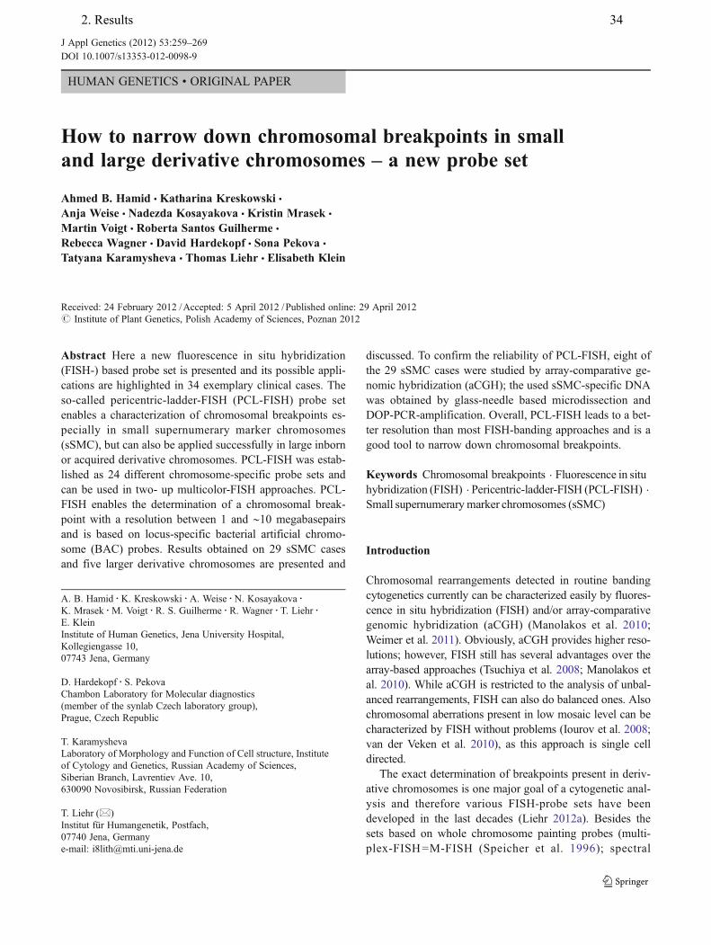
HUMAN GENETICS • ORIGINAL PAPER
How to narrow down chromosomal breakpoints in smalland large derivative chromosomes – a new probe set
Ahmed B. Hamid & Katharina Kreskowski &Anja Weise & Nadezda Kosayakova & Kristin Mrasek &
Martin Voigt & Roberta Santos Guilherme &
Rebecca Wagner & David Hardekopf & Sona Pekova &
Tatyana Karamysheva & Thomas Liehr & Elisabeth Klein
Received: 24 February 2012 /Accepted: 5 April 2012 /Published online: 29 April 2012# Institute of Plant Genetics, Polish Academy of Sciences, Poznan 2012
Abstract Here a new fluorescence in situ hybridization(FISH-) based probe set is presented and its possible appli-cations are highlighted in 34 exemplary clinical cases. Theso-called pericentric-ladder-FISH (PCL-FISH) probe setenables a characterization of chromosomal breakpoints es-pecially in small supernumerary marker chromosomes(sSMC), but can also be applied successfully in large inbornor acquired derivative chromosomes. PCL-FISH was estab-lished as 24 different chromosome-specific probe sets andcan be used in two- up multicolor-FISH approaches. PCL-FISH enables the determination of a chromosomal break-point with a resolution between 1 and ∼10 megabasepairsand is based on locus-specific bacterial artificial chromo-some (BAC) probes. Results obtained on 29 sSMC casesand five larger derivative chromosomes are presented and
discussed. To confirm the reliability of PCL-FISH, eight ofthe 29 sSMC cases were studied by array-comparative ge-nomic hybridization (aCGH); the used sSMC-specific DNAwas obtained by glass-needle based microdissection andDOP-PCR-amplification. Overall, PCL-FISH leads to a bet-ter resolution than most FISH-banding approaches and is agood tool to narrow down chromosomal breakpoints.
Keywords Chromosomal breakpoints . Fluorescence in situhybridization (FISH) . Pericentric-ladder-FISH (PCL-FISH) .
Small supernumerary marker chromosomes (sSMC)
Introduction
Chromosomal rearrangements detected in routine bandingcytogenetics currently can be characterized easily by fluores-cence in situ hybridization (FISH) and/or array-comparativegenomic hybridization (aCGH) (Manolakos et al. 2010;Weimer et al. 2011). Obviously, aCGH provides higher reso-lutions; however, FISH still has several advantages over thearray-based approaches (Tsuchiya et al. 2008; Manolakos etal. 2010). While aCGH is restricted to the analysis of unbal-anced rearrangements, FISH can also do balanced ones. Alsochromosomal aberrations present in low mosaic level can becharacterized by FISH without problems (Iourov et al. 2008;van der Veken et al. 2010), as this approach is single celldirected.
The exact determination of breakpoints present in deriv-ative chromosomes is one major goal of a cytogenetic anal-ysis and therefore various FISH-probe sets have beendeveloped in the last decades (Liehr 2012a). Besides thesets based on whole chromosome painting probes (multi-plex-FISH0M-FISH (Speicher et al. 1996); spectral
A. B. Hamid :K. Kreskowski :A. Weise :N. Kosayakova :K. Mrasek :M. Voigt : R. S. Guilherme : R. Wagner : T. Liehr :E. KleinInstitute of Human Genetics, Jena University Hospital,Kollegiengasse 10,07743 Jena, Germany
D. Hardekopf : S. PekovaChambon Laboratory for Molecular diagnostics(member of the synlab Czech laboratory group),Prague, Czech Republic
T. KaramyshevaLaboratory of Morphology and Function of Cell structure, Instituteof Cytology and Genetics, Russian Academy of Sciences,Siberian Branch, Lavrentiev Ave. 10,630090 Novosibirsk, Russian Federation
T. Liehr (*)Institut für Humangenetik, Postfach,07740 Jena, Germanye-mail: [email protected]
J Appl Genetics (2012) 53:259–269DOI 10.1007/s13353-012-0098-9
2. Results 34

karyotyping0SKY (Schröck et al. 1996)) or all centro-meric probes (centromere-specific M-FISH0cenM-FISH(Nietzel et al. 2001)), also various FISH-bandingapproaches (Liehr et al. 2002a) were introduced. Oneearly idea on how to implement FISH-banding waschromosome-bar coding, using well mapped locus-specific probes (Lengauer et al. 1992). However, no suchprobe set was ever finished for routine use in humanchromosomes (Liehr et al. 2006a).
Here we present a new FISH probe set based on 174bacterial artificial chromosome (BAC) probes calledpericentric-ladder-FISH (PCL-FISH). It enables achromosome-specific characterization of breakpoints with aresolution between 1 and ∼10 megabasepairs (Mb) in smalland large inborn or acquired derivative chromosomes; it isdirected mainly toward the pericentric regions, as it is primar-ily intended for characterization of small supernumerarymarker chromosomes (sSMC) (Liehr et al. 2004 and 2006b).PCL-FISH was successfully applied in 29 cases with sSMCand in five patients with larger derivative chromosomes.
Material and methods
PCL-FISH probe set
The PCL-FISH probe set (Fig. 1) is based on 174 BACprobes (Table 1). As centromere-near “starting points”
for each of the chromosome-arms served probes used inthe previously published so-called subcentromere-specificmulticolor- (subcenM-) FISH probes set (Liehr et al. 2006b).These probes are denominated Np1 or Nq1 in Table 1, where-by “N” stands for the corresponding chromosome number.Distal from the probes Np1 or Nq1 follow between two andsix other BAC probes with an average distance of 10 Mb toeach other. Those were selected from the published humanDNA-sequence. The BAC-probes were either kindly providedfrom the Sanger Center, Cambridge, UK, or purchased viaBAC/PAC Chori, Oakland, CA, USA.
Fifty to 100 ng of DNA per BAC-probe was in vitroamplified and labeled by degenerated oligonucleotideprimed polymerase chain reaction (DOP-PCR) (Teleniuset al. 1992). Amplification procedure followed a pub-lished scheme (Fig. 2A in Liehr et al. 2002b). Here weused only the fluorochromes SpectrumOrange (SO) anddiethylaminocoumarin (DEAC) for labeling of the BAC-probes and combined them with the corresponding com-mercially available centromeric probe labeled with agreen fluorochrome (Kreatech, Amsterdam, The Nether-lands; see Fig. 1). Still, it would be no problem to alsolabel the BACs with more and/ or combined fluoro-chromes to achieve additional colors and an individualidentification of the probes. As in the present studyPCL-FISH was exclusively used to narrow down chro-mosomal breakpoints in derivative chromosomes withknown structures, a three-color FISH as shown in
Fig. 1 Karyogram combined oftwo homologous for eachchromosome-position labeledwith the PCL-FISH probe set.The chromosome-pairs are tak-en from 24 different experi-ments and one metaphase, each,except for the Y-chromosomes,which are from two differentmetaphases. A cytogeneticallynormal female and a male werehybridized, each. Locus-specific probes are labeled inred and blue according to thescheme shown in Table 1;corresponding centromericprobes labeled in green wereadditionally applied
260 J Appl Genetics (2012) 53:259–269
2. Results 35
ahamid
Textfeld
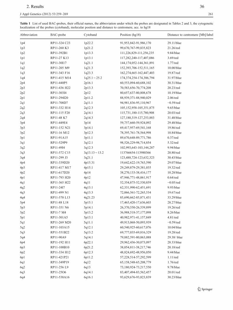
Table 1 List of used BAC-probes, their official names, the abbreviation under which the probes are designated in Tables 2 and 3, the cytogeneticlocalization of the probes (cytoband), molecular position and distance to centromere, acc. to hg18
Abbreviation BAC-probe Cytoband Position (hg18) Distance to centromere [Mb]/label
1p4 RP11-324 C23 1p22.2 91,953,842-91,986,170 29.11/blue
1p3 RP11-260 K3 1p21.2 99,670,767-99,835,823 21.26/red
1p2 RP11-392B1 1p13.3 111,226,829-111,256,235 9.84/blue
1p1 RP11-27 K13 1p13.1 117,282,240-117,407,086 3.69/red
1q1 RP11-30I17 1q21.1 144,174,052-144,361,891 1.77/red
1q2 RP11-205 M9 1q21.3 152,393,706-152,511,165 10.00/blue
1q3 RP11-343 F16 1q23.3 162,274,665-162,447,403 19.87/red
1q4 RP11-415 M14 1q25.1õ25.2 174,374,254-174,386,794 31.97/blue
2p4 RP11-440P5 2p16.1 60,553,094-60,688,102 30.31/blue
2p3 RP11-436 H22 2p13.3 70,583,656-70,774,208 20.23/red
2p2 RP11-303I4 2p12 80,657,637-80,808,678 10.19/blue
2p1 RP11-294I20 2p11.2 88,939,371-88,940,029 2.06/red
2q1 RP11-708D7 2q11.1 94,981,836-95,110,967 −0.59/red
2q2 RP11-332 H14 2q12.3 105,152,958-105,351,875 9.65/blue
2q3 RP11-115 F20 2q14.1 115,731,180-115,780,908 20.03/red
2q4 RP11-48 K7 2q14.3 127,180,319-127,253,883 31.48/blue
3p4 RP11-449E4 3p14 59,757,660-59,924,092 29.48/blue
3p3 RP11-152 N21 3p14.1 69,417,957-69,541,168 19.86/red
3p2 RP11-16 M12 3p12.3 78,395,761-78,564,998 10.84/blue
3p1 RP11-91A15 3p11.1 89,670,648-89,771,786 0.37/red
3q1 RP11-529P9 3q12.1 98,526,229-98,714,854 5.32/red
3q2 RP11-49I4 3q12.3 102,993,643-103,144,287 9.94/blue
3q3 RP11-572 C15 3q13.13õ13.2 113766654-113980366 20.80/red
3q4 RP11-299 J3 3q21.1 123,488,724-123,632,323 30.43/blue
4p4 RP11-339D20 4p15.31 19,642,622-19,763,590 29.07/blue
4p3 RP11-417 M17 4p15.1 29,249,879-29,381,035 19.32/red
4p2 RP11-617D20 4p14 38,270,133-38,416,177 10.28/blue
4p1 RP11-793 H20 4p12 47,946,771-48,061,917 0.64/red
4q1 RP11-365 H22 4q11 52,354,875-52,530,859 −0.05/red
4q2 RP11-24I7 4q13.1 62,331,990-62,451,691 9.93/blue
4q3 RP11-499 N1 4q13.3 72,066,561-72,265,534 19.67/red
4q4 RP11-570 L13 4q21.23 85,690,662-85,871,431 33.29/blue
5p4 RP11-88 L18 5p15.1 17,465,420-17,636,603 28.27/blue
5p3 RP11-351 N6 5p14.1 26,370,550-26,539,899 19.26/red
5p2 RP11-7 M4 5p13.2 36,988,518-37,177,098 8.26/blue
5p1 RP11-301A5 5p13.1 40,982,971-41,157,849 4.81/red
5q1 RP11-269 M20 5q11.1 49,913,068-50,093,939 −0.59/red
5q2 RP11-103A15 5q12.1 60,540,925-60,617,076 10.04/blue
5q3 RP11-551B22 5q13.2 69,777,035-69,816,329 19.28/red
5q4 RP11-90A9 5q14.1 79,882,591-80,063,088 29.38/ blue
6p4 RP11-192 H11 6p22.1 29,962,436-30,073,097 28.33/blue
6p3 RP11-100B10 6p21.2 38,054,811-38,217,746 20.18/red
6p2 RP11-334 H12 6p12.3 48,824,692-48,956,050 9.44/blue
6p1 RP11-421P21 6p11.2 57,228,514-57,292,599 1.11/red
6q1 RP11-349P19 6q12 65,158,548-65,208,779 1.76/red
6q2 RP11-256 L9 6q13 73,180,924-73,217,550 9.78/blue
6q3 RP11-25O6 6q14.1 83,407,494-83,562,457 20.01/red
6q4 RP11-538A16 6q16.1 93,629,676-93,823,839 30.23/blue
J Appl Genetics (2012) 53:259–269 261
2. Results 36
ahamid
Textfeld

Table 1 (continued)
Abbreviation BAC-probe Cytoband Position (hg18) Distance to centromere [Mb]/label
7p4 RP4-781A18 7p15.2 27,976,171-28,166,812 29.23/blue
7p3 RP11-302 L6 7p14.2õ14.1 37,482,196-37,597,749 19.80/red
7p2 RP11-651 K8 7p12.3 46,734,018-46,843,890 10.56/blue
7p1 RP11-10 F11 7p11.2 55,222,879-55,347,931 2.05/red
7q1 RP11-144 H20 7q11.21 61,606,122-61,791,403 0.51/red
7q2 RP11-53 M13 7q11.22 71,272,716-71,279,228 10.18/blue
7q3 RP11-448A3 7q21.11 81,263,387-81,349,902 20.16/red
7q4 RP11-313 N23 7q21.2 91,801,440-91,966,446 30.70/blue
8p4 RP11-366 J3 8p22 14,724,716-14,893,465 27.31/blue
8p3 RP11-115 K10 8p21.2 24,116,701-24,305,391 17.89/red
8p2 RP11-11 N9 8p12 32,909,252-33,028,630 9.71/blue
8p1 RP11-503E24 8p11.21 42,503,724-42,674,302 −0.47/red
8q1 RP11-197I11 8q11.23 53,135,835-53,175,019 5.04/red
8q2 RP11-430 H18 8q12.1 58,249,439-58,400,298 10.15/blue
8q3 RP11-409 C19 8q13.2 68,012,453-68,120,862 19.91/red
8q4 RP11-652 L3 8q21.11 78,197,078-78,348,048 30.10/blue
9p4 RP11-492A12 9p22.2 17,361,476-17,489,085 29.21/blue
9p3 RP11-438B23 9p21.2 27,866,315-28,042,166 18.66/red
9p2 RP11-61I3 9p13.2 37,607,753-37,776,264 8.92/blue
9p1 RP11-113O24 9p13.1 38,263,089-38,427,295 8.27/red
9q1 RP11-109D9 9q21.11 71,449,567-71,636,843 1.45/red
9q2 RP11-574 G7 9q21.2 79,668,774-79,884,072 9.67/blue
9q3 RP11-249 H20 9q21.33 89,375,412-89,446,315 19.38/red
9q4 RP11-535 C21 9q22.33 99,744,616-99,824,931 29.74/blue
10p4 RP11-575 N15 10p14 8,728,048-8,906,592 29.89/blue
10p3 RP11-51E20 10p12.31 20,784,567-20,938,614 17.86/red
10p2 RP11-350D11 10p11.23 30,675,157-30,865,135 7.93/blue
10p1 RP11-365P10 10p11.21 36,945,343-36,974,907 1.85/red
10q1 RP11-92P6 10q11.21 43,174,613-43,219,888 1.07/red
10q2 RP11-532 F4 10q11.23 52,151,487-52,328,351 10.05/blue
10q3 RP11-166B18 10q21.2 61,904,197-62,069,022 19.80/red
10q4 RP11-367 H5 10q22.1 71,504,053-71,672,097 29.40/blue
11p4 RP11-701I24 11p15 20,531,828.-20,709,763 30.69/blue
11p3 RP11-297A4 11p13 31,195,990-31,386,312 20.01/red
11p2 RP11-1 G18 11p12 41,077,222-41,230,282 10.17/blue
11p1 RP11-397 M16 11p11.2 48,260,247-48,436,072 2.96/red
11q1 RP11-644A8 11q12.1 56,558,447-56,613,460 0.16/red
11q2 RP11-157 K17 11q13.1 66,670,500-66,841,190 10.44/blue
11q3 RP11-263 C24 11q13.5 75,265,230-75,385,345 18.87/red
11q4 RP11-665E10 11q14.2 87,158,646-87,317,430 30.92/blue
12p4 RP11-298 J22 12p13.33 2,247,231-2,406,919 30.79/blue
12p3 RP11-161A14 12p13.1 13,255,212-13,430,711 19.77/red
12p2 RP11-12D15 12p12.1 22,250,749-22,369,579 10.83/blue
12p1 RP11-310I24 12p11.22 29,551,976-29,607,135 3.59/red
12q1 RP11-498B21 12q12 39,833,150-39,900,092 3.33/red
12q2 RP11-89 H19 12q13.11 46,571,184-46,627,459 10.07/blue
12q3 RP11-181 L23 12q13.3 56,118,000-56,288,135 19.62/red
12q4 RP11-542B15 12q15 66,138,644-66,250,773 29.64/blue
13q1 RP11-523 H24 13q12.11 19,137,338-19,306,540 0.74/red
262 J Appl Genetics (2012) 53:259–269
2. Results 37
ahamid
Textfeld
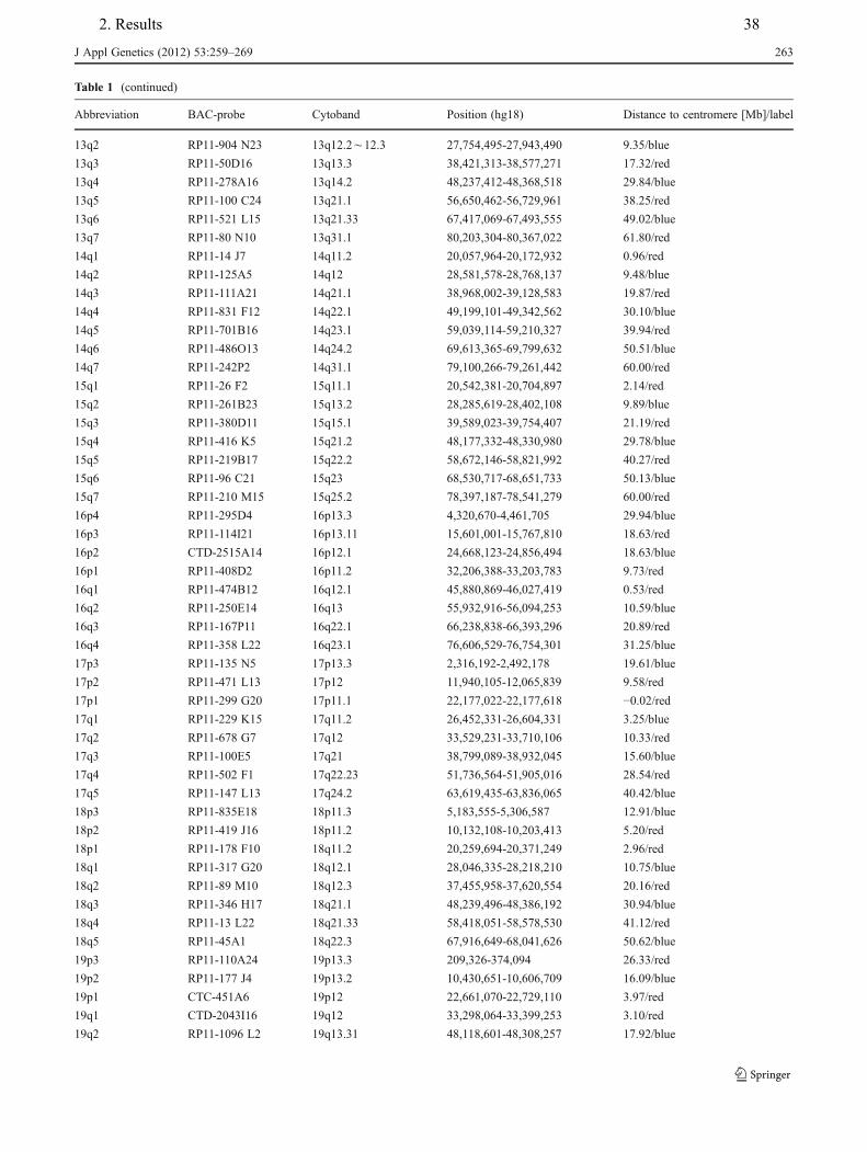
Table 1 (continued)
Abbreviation BAC-probe Cytoband Position (hg18) Distance to centromere [Mb]/label
13q2 RP11-904 N23 13q12.2õ12.3 27,754,495-27,943,490 9.35/blue
13q3 RP11-50D16 13q13.3 38,421,313-38,577,271 17.32/red
13q4 RP11-278A16 13q14.2 48,237,412-48,368,518 29.84/blue
13q5 RP11-100 C24 13q21.1 56,650,462-56,729,961 38.25/red
13q6 RP11-521 L15 13q21.33 67,417,069-67,493,555 49.02/blue
13q7 RP11-80 N10 13q31.1 80,203,304-80,367,022 61.80/red
14q1 RP11-14 J7 14q11.2 20,057,964-20,172,932 0.96/red
14q2 RP11-125A5 14q12 28,581,578-28,768,137 9.48/blue
14q3 RP11-111A21 14q21.1 38,968,002-39,128,583 19.87/red
14q4 RP11-831 F12 14q22.1 49,199,101-49,342,562 30.10/blue
14q5 RP11-701B16 14q23.1 59,039,114-59,210,327 39.94/red
14q6 RP11-486O13 14q24.2 69,613,365-69,799,632 50.51/blue
14q7 RP11-242P2 14q31.1 79,100,266-79,261,442 60.00/red
15q1 RP11-26 F2 15q11.1 20,542,381-20,704,897 2.14/red
15q2 RP11-261B23 15q13.2 28,285,619-28,402,108 9.89/blue
15q3 RP11-380D11 15q15.1 39,589,023-39,754,407 21.19/red
15q4 RP11-416 K5 15q21.2 48,177,332-48,330,980 29.78/blue
15q5 RP11-219B17 15q22.2 58,672,146-58,821,992 40.27/red
15q6 RP11-96 C21 15q23 68,530,717-68,651,733 50.13/blue
15q7 RP11-210 M15 15q25.2 78,397,187-78,541,279 60.00/red
16p4 RP11-295D4 16p13.3 4,320,670-4,461,705 29.94/blue
16p3 RP11-114I21 16p13.11 15,601,001-15,767,810 18.63/red
16p2 CTD-2515A14 16p12.1 24,668,123-24,856,494 18.63/blue
16p1 RP11-408D2 16p11.2 32,206,388-33,203,783 9.73/red
16q1 RP11-474B12 16q12.1 45,880,869-46,027,419 0.53/red
16q2 RP11-250E14 16q13 55,932,916-56,094,253 10.59/blue
16q3 RP11-167P11 16q22.1 66,238,838-66,393,296 20.89/red
16q4 RP11-358 L22 16q23.1 76,606,529-76,754,301 31.25/blue
17p3 RP11-135 N5 17p13.3 2,316,192-2,492,178 19.61/blue
17p2 RP11-471 L13 17p12 11,940,105-12,065,839 9.58/red
17p1 RP11-299 G20 17p11.1 22,177,022-22,177,618 −0.02/red
17q1 RP11-229 K15 17q11.2 26,452,331-26,604,331 3.25/blue
17q2 RP11-678 G7 17q12 33,529,231-33,710,106 10.33/red
17q3 RP11-100E5 17q21 38,799,089-38,932,045 15.60/blue
17q4 RP11-502 F1 17q22.23 51,736,564-51,905,016 28.54/red
17q5 RP11-147 L13 17q24.2 63,619,435-63,836,065 40.42/blue
18p3 RP11-835E18 18p11.3 5,183,555-5,306,587 12.91/blue
18p2 RP11-419 J16 18p11.2 10,132,108-10,203,413 5.20/red
18p1 RP11-178 F10 18q11.2 20,259,694-20,371,249 2.96/red
18q1 RP11-317 G20 18q12.1 28,046,335-28,218,210 10.75/blue
18q2 RP11-89 M10 18q12.3 37,455,958-37,620,554 20.16/red
18q3 RP11-346 H17 18q21.1 48,239,496-48,386,192 30.94/blue
18q4 RP11-13 L22 18q21.33 58,418,051-58,578,530 41.12/red
18q5 RP11-45A1 18q22.3 67,916,649-68,041,626 50.62/blue
19p3 RP11-110A24 19p13.3 209,326-374,094 26.33/red
19p2 RP11-177 J4 19p13.2 10,430,651-10,606,709 16.09/blue
19p1 CTC-451A6 19p12 22,661,070-22,729,110 3.97/red
19q1 CTD-2043I16 19q12 33,298,064-33,399,253 3.10/red
19q2 RP11-1096 L2 19q13.31 48,118,601-48,308,257 17.92/blue
J Appl Genetics (2012) 53:259–269 263
2. Results 38
ahamid
Textfeld
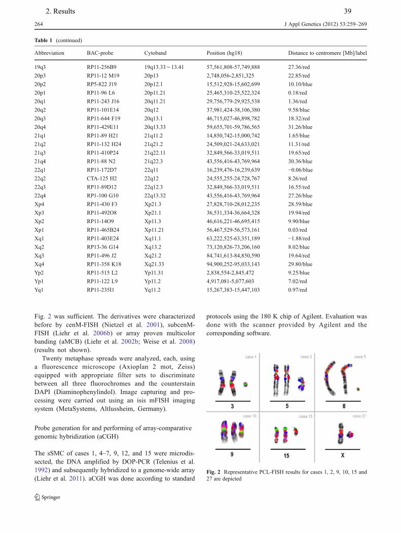
Fig. 2 was sufficient. The derivatives were characterizedbefore by cenM-FISH (Nietzel et al. 2001), subcenM-FISH (Liehr et al. 2006b) or array proven multicolorbanding (aMCB) (Liehr et al. 2002b; Weise et al. 2008)(results not shown).
Twenty metaphase spreads were analyzed, each, usinga fluorescence microscope (Axioplan 2 mot, Zeiss)equipped with appropriate filter sets to discriminatebetween all three fluorochromes and the counterstainDAPI (Diaminophenylindol). Image capturing and pro-cessing were carried out using an isis mFISH imagingsystem (MetaSystems, Altlussheim, Germany).
Probe generation for and performing of array-comparativegenomic hybridization (aCGH)
The sSMC of cases 1, 4–7, 9, 12, and 15 were microdis-sected, the DNA amplified by DOP-PCR (Telenius et al.1992) and subsequently hybridized to a genome-wide array(Liehr et al. 2011). aCGH was done according to standard
protocols using the 180 K chip of Agilent. Evaluation wasdone with the scanner provided by Agilent and thecorresponding software.
Table 1 (continued)
Abbreviation BAC-probe Cytoband Position (hg18) Distance to centromere [Mb]/label
19q3 RP11-256B9 19q13.33õ13.41 57,561,808-57,749,888 27.36/red
20p3 RP11-12 M19 20p13 2,748,056-2,851,325 22.85/red
20p2 RP5-822 J19 20p12.1 15,512,928-15,602,699 10.10/blue
20p1 RP11-96 L6 20p11.21 25,465,310-25,522,324 0.18/red
20q1 RP11-243 J16 20q11.21 29,756,779-29,925,538 1.36/red
20q2 RP11-101E14 20q12 37,981,424-38,106,380 9.58/blue
20q3 RP11-644 F19 20q13.1 46,715,027-46,898,782 18.32/red
20q4 RP11-429E11 20q13.33 59,655,701-59,786,565 31.26/blue
21q1 RP11-89 H21 21q11.2 14,850,742-15,000,742 1.65/blue
21q2 RP11-132 H24 21q21.2 24,509,021-24,633,021 11.31/red
21q3 RP11-410P24 21q22.11 32,849,566-33,019,511 19.65/red
21q4 RP11-88 N2 21q22.3 43,556,416-43,769,964 30.36/blue
22q1 RP11-172D7 22q11 16,239,476-16,239,639 −0.06/blue
22q2 CTA-125 H2 22q12 24,555,255-24,728,767 8.26/red
22q3 RP11-89D12 22q12.3 32,849,566-33,019,511 16.55/red
22q4 RP1-100 G10 22q13.32 43,556,416-43,769,964 27.26/blue
Xp4 RP11-430 F3 Xp21.3 27,828,710-28,012,235 28.59/blue
Xp3 RP11-492O8 Xp21.1 36,531,334-36,664,328 19.94/red
Xp2 RP11-14O9 Xp11.3 46,616,221-46,695,415 9.90/blue
Xp1 RP11-465B24 Xp11.21 56,467,529-56,573,161 0.03/red
Xq1 RP11-403E24 Xq11.1 63,222,525-63,351,189 −1.88/red
Xq2 RP13-36 G14 Xq13.2 73,120,826-73,206,160 8.02/blue
Xq3 RP11-496 J2 Xq21.2 84,741,613-84,850,590 19.64/red
Xq4 RP11-358 K18 Xq21.33 94,900,252-95,033,143 29.80/blue
Yp2 RP11-515 L2 Yp11.31 2,838,554-2,845,472 9.25/blue
Yp1 RP11-122 L9 Yp11.2 4,917,081-5,077,603 7.02/red
Yq1 RP11-235I1 Yq11.2 15,267,383-15,447,103 0.97/red
Fig. 2 Representative PCL-FISH results for cases 1, 2, 9, 10, 15 and27 are depicted
264 J Appl Genetics (2012) 53:259–269
2. Results 39
ahamid
Textfeld
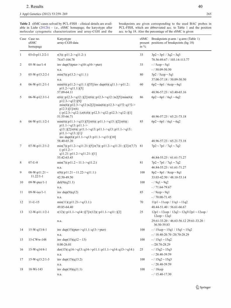
Table 2 sSMC-cases solved by PCL-FISH – clinical details are avail-able in Liehr (2012b) – i.e., sSMC homepage; the karyotype aftermolecular cytogenetic characterization and array-CGH and the
breakpoints are given corresponding to the used BAC probes inPCL-FISH, which are abbreviated acc. to Table 1 and the positionacc. to hg 18. Also the percentage of the sSMC is given
Case Case no.sSMChomepage
Karyotype sSMCpresentin %
Breakpoints p-arm / q-arm (Table 1)array-CGH-data positions of breakpoints (hg 18)
1 03-O-p11.2/2-1 r(3)(::p11.2->q11.2::) 33 3p2õ3p1 / 3q2õ3q3
74.67-104.78 78.56-89.67 / 103.14-113.77
2 05-W-iso/1-4 inv dup(5)(pter->q10::q10->pter) 33 - / 5cepõ5q1
n.a. - / 50.09-50.50
3 05-W-p13.2/2-1 min(5)(:p13.2->q11.1:) 80 5p2 / 5cepõ5q1
n.a. 37.00-37.18 / 50.09-50.50
4 06-W-p11.2/1-1 min(6)(:p11.2->q11.1:)[55]/inv dup(6)(:q11.1->p11.2::p11.2->q11.1:)[5]
60 6p2õ6p1 / 6cepõ6q1
57.09-64.11 48.96-57.23 / 63.40-65.16
5 06-W-p12.3/1-1 r(6)(::p12.3->q12::)[2]/r(6)(::p12.3->q12::)x2[5]/min(6)(:p12.3->q12:)[9]/min(6)(:p12.3->q12:)x2[2]/min(6)(:p12.3->q1?2::q1?2->p12.3:)[1]/r(6)(::p12.3->q12::),r(6;6)(::p12.3->q12::p12.3->q12::)[1]
86 6p2õ6p1 / 6q1õ6q2
51.55-66.71 48.96-57.23 / 65.21-73.18
6 06-W-p11.1/2-1 min(6)(:p11.1->q13:)[5]/r(6)(::p11.1->q13::)[2]/r(6)(::p11.1->q13::p11.1->q13::)[2]/r(6)(::p11.1->q13::p11.1->q13::p11.1->q13::p11.1->q13::)[1]/inv dup(6)(:p11.1->q13::p11.1->q13:)[10]
85 6p2õ6p1 / 6q1õ6q2
58.40-65.20 48.96-57.23 / 65.21-73.18
7 07-W-p11.2/1-2 min(7)(:p11.2->q11.21:)[5]/r(7)(::p11.2->q11.21::)[2]/r(7;7)(::p11.2->q11.21::p11.2->q11.21::)[1]
81 7p2õ7p1 / 7q1õ7q2
55.42-63.45 46.84-55.23 / 61.61-71.27
8 07-U-8 min(7)(:p11.2õ11.1->q11.2:) 81 7p2õ7p1 / 7q1õ7q2
n.a. 46.84-55.23 / 61.61-71.27
9 08-W-p11.21õ11.22/1-1
r(8)(:p11.21õ11.22->q11.1:) 100 8p2õ8p1 / 8cepõ8q1
42.50-49.50 33.03-42.50 / 48.10-53.14
10 09-W-pter/1-1 del(9)(q21.1) 100 - / 9q1õ9q2
n.a. - / 71.64-79.67
11 09-W-iso/1-1 inv dup(9)(q12) 85 - / 9cepõ9q1
n.a. - / 70.00-71.45
12 11-U-15 min(11)(:p11.21->q13.1:) 70 11p1õ11cep / 11q1õ11q2
49.85-64.40 48.44-51.40 / 56.61-66.67
13 12-W-p11.1/2-1 r(12)(::p11.1->q14::)[7]/r(12)(::p11.1->q11::)[2] 25 12p1õ12cep / 12q2õ12q312p1õ12cep /12cepõ12q1
n.a. 29.61-33.20 / 46.63-56.12 29.61-33.20 /36.50-39.83
14 15-W-q13/4-1 inv dup(15)(pter->q11.1::q13->pter) 100 - / 15cepõ15q1 / 15q1õ15q2
n.a. - / 18.40-20.70 /20.70-28.29
15 15-CWw-148 inv dup(15)(q12õ13) 100 - / 15q1õ15q2
0.00-26.01 - /20.70-28.29
16 15-W-q14/4-1 der(15)(:q14->q13::q14->p11.1::p11.1->q14::q13->q14:) 25 - / 15q2õ15q3
n.a. - / 28.40-39.59
17 15-W-q13.2/1-3 inv dup(15)(q13.2) 100 - / 15q2õ15q3
n.a. - / 28.40-39.59
18 18-Wi-143 inv dup(18)(q11.1) 100 - / 18cep
n.a. - / 15.40-17.30
J Appl Genetics (2012) 53:259–269 265
2. Results 40
ahamid
Textfeld
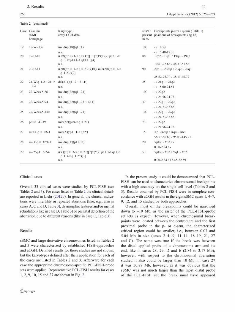
Clinical cases
Overall, 33 clinical cases were studied by PCL-FISH (seeTables 2 and 3). For cases listed in Table 2 the clinical detailsare reported in Liehr (2012b). In general, the clinical indica-tions were infertility or repeated abortions (like, e.g., also incases A, C and D, Table 3), dysmorphic features and/or mentalretardation (like in case B, Table 3) or prenatal detection of theaberration due to different reasons (like in case E, Table 3).
Results
sSMC and large derivative chromosomes listed in Tables 2and 3 were characterized by established FISH-approachesand aCGH. Detailed results for these studies are not shown,but the karyotypes defined after their application for each ofthe cases are listed in Tables 2 and 3. Afterward for eachcase the appropriate chromosome-specific PCL-FISH-probesets were applied. Representative PCL-FISH results for cases1, 2, 9, 10, 15 and 27 are shown in Fig. 2.
In the present study it could be demonstrated that PCL-FISH can be used to characterize chromosomal breakpointswith a high accuracy on the single cell level (Tables 2 and3). Results obtained by PCL-FISH were in complete con-cordance with aCGH results in the eight sSMC cases 1, 4–7,9, 12, and 15 studied by both approaches.
Overall, most of the breakpoints could be narroweddown to ∼10 Mb, as the raster of the PCL-FISH-probeset lets us expect. However, when chromosomal break-points were located between the centromere and the firstproximal probe in the p- or q-arm, the characterizedcritical region could be smaller, i.e., between 0.03 and5.04 Mb in size (cases 2–4, 9, 11–14, 18–19, 21, 27and C). The same was true if the break was betweenthe distal applied probe of a chromosome arm and itsend, like in cases 28, 29, D and E (2.84 to 3.17 Mb);however, with respect to the chromosomal aberrationstudied it also could be larger than 10 Mb: in case 27it was 50.88 Mb, however, as it was obvious that thesSMC was not much larger than the most distal probeof the PCL-FISH set the break must have appeared
Table 2 (continued)
Case Case no.sSMChomepage
Karyotype sSMCpresentin %
Breakpoints p-arm / q-arm (Table 1)array-CGH-data positions of breakpoints (hg 18)
19 18-Wi-132 inv dup(18)(q11.1) 100 - / 18cep
n.a. - / 15.40-17.3020 19-U-10 r(19)(::p13.1->q13.1::)[17]/r(19;19)(::p13.1->
q13.1::p13.1->q13.1::)[4]88 19p2õ19p1 / 19q2õ19q3
n.a. 10.61-22.66 / 48.31-57.56
21 20-U-11 r(20)(::p11.1->q11.23::)[10]/ min(20)(:p11.1->q11.23:)[2]
90 20p1õ20cep / 20q2õ20q3
n.a. 25.52-25.70 / 38.11-46.72
22 21-W-q11.2õ21.1/1-2
del(21)(q11.2õ21.1:) 25 - / 21q1õ21q2
n.a. - / 15.00-24.51
23 22-Wces-5-86 inv dup(22)(q11.21) 100 - / 22q2
n.a. - / 24.56-24.73
24 22-Wces-5-94 inv dup(22)(q11.23õ12.1) 37 - / 22q1õ22q2
n.a. - / 24.73-32.85
25 22-Wces-5-130 inv dup(22)(q11.21) 100 - / 22q1õ22q2
n.a. - / 24.73-32.85
26 plus21-U-39 min(22)(pter->q11.21) 73 - / 22q2
n.a. - / 24.56-24.73
27 minX-p11.1/6-1 min(X)(:p11.1->q22:) 15 Xp1-Xcep / Xq4õXtel
n.a. 56.57-56.60 / 95.03-145.91
28 m-iY-p11.32/1-3 inv dup(Y)(p11.32) 20 YpterõYp2 / -
n.a. 0.00-2.84 / -
29 m-rY-p11.3/2-4 r(Y)(::p11.3->q11.2::)[7]/r(Y)(::p11.3->q11.2::p11.3->q11.2::)[3]
53 YpterõYp2 / Yq1õYq2
n.a. 0.00-2.84 / 15.45-22.59
266 J Appl Genetics (2012) 53:259–269
2. Results 41
ahamid
Textfeld
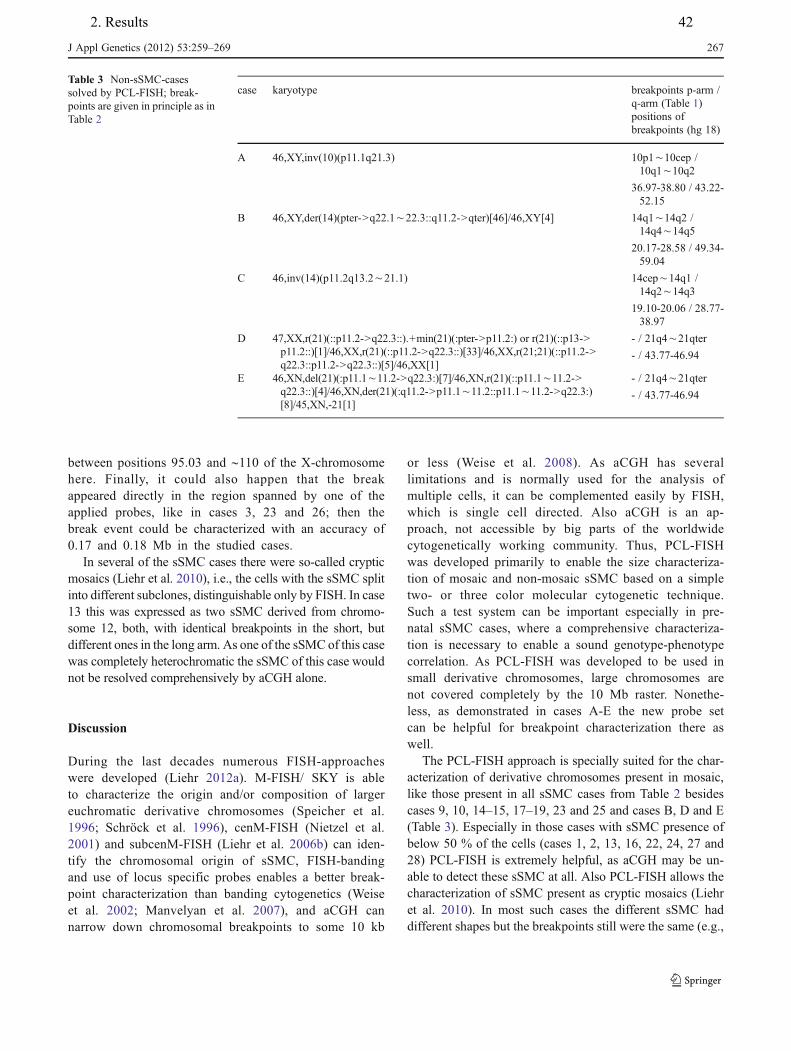
between positions 95.03 and ∼110 of the X-chromosomehere. Finally, it could also happen that the breakappeared directly in the region spanned by one of theapplied probes, like in cases 3, 23 and 26; then thebreak event could be characterized with an accuracy of0.17 and 0.18 Mb in the studied cases.
In several of the sSMC cases there were so-called crypticmosaics (Liehr et al. 2010), i.e., the cells with the sSMC splitinto different subclones, distinguishable only by FISH. In case13 this was expressed as two sSMC derived from chromo-some 12, both, with identical breakpoints in the short, butdifferent ones in the long arm. As one of the sSMC of this casewas completely heterochromatic the sSMC of this case wouldnot be resolved comprehensively by aCGH alone.
Discussion
During the last decades numerous FISH-approacheswere developed (Liehr 2012a). M-FISH/ SKY is ableto characterize the origin and/or composition of largereuchromatic derivative chromosomes (Speicher et al.1996; Schröck et al. 1996), cenM-FISH (Nietzel et al.2001) and subcenM-FISH (Liehr et al. 2006b) can iden-tify the chromosomal origin of sSMC, FISH-bandingand use of locus specific probes enables a better break-point characterization than banding cytogenetics (Weiseet al. 2002; Manvelyan et al. 2007), and aCGH cannarrow down chromosomal breakpoints to some 10 kb
or less (Weise et al. 2008). As aCGH has severallimitations and is normally used for the analysis ofmultiple cells, it can be complemented easily by FISH,which is single cell directed. Also aCGH is an ap-proach, not accessible by big parts of the worldwidecytogenetically working community. Thus, PCL-FISHwas developed primarily to enable the size characteriza-tion of mosaic and non-mosaic sSMC based on a simpletwo- or three color molecular cytogenetic technique.Such a test system can be important especially in pre-natal sSMC cases, where a comprehensive characteriza-tion is necessary to enable a sound genotype-phenotypecorrelation. As PCL-FISH was developed to be used insmall derivative chromosomes, large chromosomes arenot covered completely by the 10 Mb raster. Nonethe-less, as demonstrated in cases A-E the new probe setcan be helpful for breakpoint characterization there aswell.
The PCL-FISH approach is specially suited for the char-acterization of derivative chromosomes present in mosaic,like those present in all sSMC cases from Table 2 besidescases 9, 10, 14–15, 17–19, 23 and 25 and cases B, D and E(Table 3). Especially in those cases with sSMC presence ofbelow 50 % of the cells (cases 1, 2, 13, 16, 22, 24, 27 and28) PCL-FISH is extremely helpful, as aCGH may be un-able to detect these sSMC at all. Also PCL-FISH allows thecharacterization of sSMC present as cryptic mosaics (Liehret al. 2010). In most such cases the different sSMC haddifferent shapes but the breakpoints still were the same (e.g.,
Table 3 Non-sSMC-casessolved by PCL-FISH; break-points are given in principle as inTable 2
case karyotype breakpoints p-arm /q-arm (Table 1)positions ofbreakpoints (hg 18)
A 46,XY,inv(10)(p11.1q21.3) 10p1õ10cep /10q1õ10q2
36.97-38.80 / 43.22-52.15
B 46,XY,der(14)(pter->q22.1õ22.3::q11.2->qter)[46]/46,XY[4] 14q1õ14q2 /14q4õ14q5
20.17-28.58 / 49.34-59.04
C 46,inv(14)(p11.2q13.2õ21.1) 14cepõ14q1 /14q2õ14q3
19.10-20.06 / 28.77-38.97
D 47,XX,r(21)(::p11.2->q22.3::).+min(21)(:pter->p11.2:) or r(21)(::p13->p11.2::)[1]/46,XX,r(21)(::p11.2->q22.3::)[33]/46,XX,r(21;21)(::p11.2->q22.3::p11.2->q22.3::)[5]/46,XX[1]
- / 21q4õ21qter
- / 43.77-46.94
E 46,XN,del(21)(:p11.1õ11.2->q22.3:)[7]/46,XN,r(21)(::p11.1õ11.2->q22.3::)[4]/46,XN,der(21)(:q11.2->p11.1õ11.2::p11.1õ11.2->q22.3:)[8]/45,XN,-21[1]
- / 21q4õ21qter
- / 43.77-46.94
J Appl Genetics (2012) 53:259–269 267
2. Results 42
ahamid
Textfeld
cases 4–7, 20 and 29). However, in some instances thebreakpoints can differ, like in case 13; here PCL-FISH couldeasily define all three involved breakpoints.
In general, PCL-FISH is a pericentromeric region direct-ed bar-code FISH approach. However, to the best of ourknowledge none of the previously published ones werecreated to constitute a 10 Mb raster along chromosomalregions. Also none of the locus-specific-probe-based bar-code FISH approaches were ever established for the wholehuman genome (Liehr et al. 2006a). PCL-FISH can be usedimmediately after identification of the chromosomal originof a derivative; however, this was not done in the presentstudy. Here all derivatives were previously characterized bywell established molecular cytogenetic approaches and/oraCGH; those results are listed in Tables 2 and 3 and servedto control the accuracy of the new established probe set.
Overall, it could be demonstrated that PCL-FISH charac-terizes chromosomal breakpoints reliably (Tables 2 and 3),as they were in concordance with previous FISH and aCGHresults. In those breakpoints not characterized by aCGH,PCL-FISH not only confirmed the previous FISH-resultsbut concretized them with an accuracy of 0.03 to ∼10 Mb,as outlined in Results.
In conclusion we present a new FISH probe set easily andeffectively applicable in clinical cytogenetic routine diag-nostics. It could be enlarged by additional probes, e.g.,BACs in 10 Mb distance covering all human genome andnot only the pericentric regions. Also, applications of PCL-FISH in tumor cytogenetics, as well as in evolution researchstudies are principally possible.
Acknowledgments The clinical cases were kindly provided by thefollowing colleagues: Australia: J Anderson, Brisbane; Belgium: Dr. J.Vermeesch, Leuven; France: Dr. C. Yardin, Montpellier; Germany: Dr.I. Bartels, Göttingen; Dr. B. Belitz, Berlin; Dr. U. Beudt, Frankfurt; Dr.H.-M. Burow, Oberkirch; Dr. A. Dufke, Tübingen; Dr. G. Gillessen-Kasebach, Lübeck; Dr. D. Huhle, Leipzig; Dr. A. Kuechler, Essen; Dr.T. Martin, Homburg; Dr. A. Meiner, Halle; Dr. D. Mitter, Leipzig; Dr.S. Morlot, Hannover; Dr. A. Ovens-Raeder, München, Dr. G. Schwan,Dortmund; Dr. S. Singer, Tübingen; Dr. S. Spranger, Bremen; Portu-gal: Dr. J. Melo, Coimbra; Serbia: Dr. G. Josik, Vinca; Turkey: Dr. B.Seher, Ankara; UK: Dr. K. Ren Ong, Birmingham.
Supported in parts by Deutsche Forschungsgemeinschaft (DFG LI820/22-1), Else Kröner-Fresenius-Stiftung (2011_A42), the DeutscherAkademischer Austauschdienst (DAAD), the Monika-Kutzner-Stiftung and the Stefan-Morsch-Stiftung.
References
Iourov IY, Vorsanova SG, Yurov YB (2008) Chromosomal mosaicismgoes global. Mol Cytogenet 1:26
Lengauer C, Green ED, Cremer T (1992) Fluorescence in situ hybrid-ization of YAC clones after Alu-PCR amplification. Genomics13:826–828
Liehr T (2012a) Basics and literature on multicolor fluorescence in situhybridization application. http://www.fish.uniklinikum-jena.de/mFISH.html. [accessed 09/02/2012]
Liehr T (2012b) Small supernumerary marker chromosomes. http://www.fish.uniklinikum-jena.de/sSMC.html. [accessed 09/02/2012]
Liehr T, Heller A, Starke H, Claussen U (2002a) FISH bandingmethods: applications in research and diagnostics. Exp Rev MolDiagn 2:217–225
Liehr T, Heller A, Starke H, Rubtsov N, Trifonov V, Mrasek K, WeiseA, Kuechler A, Claussen U (2002b) Microdissection based highresolution multicolor banding for all 24 human chromosomes. IntJ Mol Med 9:335–339
Liehr T, Claussen U, Starke H (2004) Small supernumerary markerchromosomes (sSMC) in humans. Cytogenet Genome Res107:55–67
Liehr T, Starke H, Heller A, Kosyakova N, Mrasek K, Gross M, KarstC, Steinhaeuser U, Hunstig F, Fickelscher I, Kuechler A, TrifonovV, Romanenko SA, Weise A (2006a) Multicolor fluorescence insitu hybridization (FISH) applied to FISH-banding. CytogenetGenome Res 114:240–244
Liehr T,Mrasek K,Weise A, DufkeA, Rodríguez L,Martínez Guardia N,Sanchís A, Vermeesch JR, Ramel C, Polityko A, Haas OA,Anderson J, Claussen U, von Eggeling F, Starke H (2006b) Smallsupernumerary marker chromosomes–progress towards a genotype-phenotype correlation. Cytogenet Genome Res 112:23–34
Liehr T, Karamysheva T, Merkas M, Brecevic L, Hamid AB, Ewers E,Mrasek K, Kosyakova N, Weise A (2010) Somatic mosaicism incases with small supernumerary marker chromosomes. CurrGenomics 11:432–439
Liehr T, Bartels I, Zoll B, Ewers E, Mrasek K, Kosyakova N, MerkasM, Hamid AB, von Eggeling F, Posorski N, Weise A (2011) Isthere a yet unreported unbalanced chromosomal abnormalitywithout phenotypic consequences in proximal 4p? CytogenetGenome Res 132:121–123
Manolakos E, Vetro A, Kefalas K, Rapti S-M, Louizou E, Garas A,Kitsos G, Vasileiadis L, Tsoplou P, Eleftheriades M, Peitsidis P,Orru S, Liehr T, Petersen MB, Thomaidis L (2010) The use ofarray-CGH in a cohort of Greek children with developmentaldelay. Mol Cytogenet 3:22
Manvelyan M, Schreyer I, Höls-Herpertz I, Köhler S, Niemann R, HehrU, Belitz B, Bartels I, Götz J, Huhle D, Kossakiewicz M, TittelbachH, Neubauer S, Polityko A, Mazauric ML, Wegner R, Stumm M,Küpferling P, Süss F, Kunze H, Weise A, Liehr T, Mrasek K (2007)Forty-eight new cases with infertility due to balanced chromosomalrearrangements: detailed molecular cytogenetic analysis of the 90involved breakpoints. Int J Mol Med 19:855–864
Nietzel A, Rocchi M, Starke H, Heller A, Fiedler W, Wlodarska I,Loncarevic IF, Beensen V, Claussen U, Liehr T (2001) A newmulticolor-FISH approach for the characterization of marker chro-mosomes: centromere-specific multicolor-FISH (cenM-FISH).Hum Genet 108:199–204
Schröck E, du Manoir S, Veldman T, Schoell B, Wienberg J, Ferguson-Smith MA, Ning Y, Ledbetter DH, Bar-Am I, Soenksen D, GariniY, Ried T (1996) Multicolor spectral karyotyping of human chro-mosomes. Science 273:494–497
Speicher MR, Gwyn Ballard S, Ward DC (1996) Karyotyping humanchromosomes by combinatorial multi-fluor FISH. Nat Genet12:368–375
Telenius H, Carter NP, Bebb CE, Nordenskjöld M, Ponder BA,Tunnacliffe A (1992) Degenerate oligonucleotide-primedPCR: general amplification of target DNA by a single de-generate primer. Genomics 13:718–725
Tsuchiya KD, Opheim KE, Hannibal MC, Hing AV, Glass IA, Raff ML,Norwood T, Torchia BA (2008) Unexpected structural complexityof supernumerarymarker chromosomes characterized bymicroarraycomparative genomic hybridization. Mol Cytogenet 1:7
268 J Appl Genetics (2012) 53:259–269
2. Results 43
ahamid
Textfeld

van der Veken LT, Dieleman MMJ, Douben H, van de Brug JC, van deGraaf R, Hoogeboom AJM, Poddighe PJ, de Klein A (2010) Lowgrade mosaic for a complex supernumerary ring chromosome 18in an adult patient with multiple congenital anomalies. Mol Cyto-genet 3:13
Weimer J, Heidemann S, von Kaisenberg CS, Grote W, Arnold N,Bens S, Caliebe A (2011) Isolated trisomy 7q21.2-31.31 resultingfrom a complex familial rearrangement involving chromosomes7, 9 and 10. Mol Cytogenet 4:28
Weise A, Starke H, Heller A, Tönnies H, VollethM, StummM, Senger G,Nietzel A, Claussen U, Liehr T (2002) Chromosome 2 aberrations inclinical cases characterised by high resolution multicolour bandingand region specific FISH probes. J Med Genet 39:434–439
Weise A, Mrasek K, Fickelscher I, Claussen U, Cheung SW, Cai WW,Liehr T, Kosyakova N (2008) Molecular definition of high-resolution multicolor banding probes: first within the humanDNA sequence anchored FISH banding probe set. J HistochemCytochem 56:487–493
J Appl Genetics (2012) 53:259–269 269
2. Results 44
ahamid
Textfeld
2. Results
2.6. Article .5
Liehr T, Karamysheva T, Merkas M, Brecevic L, Hamid AB, Ewers E, Mrasek K, Kosyakova N, Weise A. Somatic mosaicism in cases with small supernumerary marker chromosomes. Curr Genomics, 2010; 11:432-439.
432 Current Genomics, 2010, 11, 432-439
1389-2029/10 $55.00+.00 ©2010 Bentham Science Publishers Ltd.
Somatic Mosaicism in Cases with Small Supernumerary Marker Chromo-somes
Thomas Liehr*,1, Tatyana Karamysheva2, Martina Merkas1,3, Lukrecija Brecevic3, Ahmed B. Hamid1, Elisabeth Ewers1, Kristin Mrasek1, Nadezda Kosyakova1 and Anja Weise1
1Jena University Hospital, Institute of Human Genetics and Anthropology, Jena, Germany 2Institute for Cytology and Genetics, Nowosibirsk, Russian Federation 3School of Medicine Zagreb University, Croatian Institute for Brain Research, Zagreb, Croatia
Abstract: Somatic mosaicism is something that is observed in everyday lives of cytogeneticists. Chromosome instability is one of the leading causes of large-scale genome variation analyzable since the correct human chromosome number was established in 1956. Somatic mosaicism is also a well-known fact to be present in cases with small supernumerary marker chromosomes (sSMC), i.e. karyotypes of 47,+mar/46. In this study, the data available in the literature were collected con-cerning the frequency mosaicism in different subgroups of patients with sSMC. Of 3124 cases with sSMC 1626 (52%) present with somatic mosaicism. Some groups like patients with Emanuel-, cat-eye- or i(18p)- syndrome only tend rarely to develop mosaicism, while in Pallister-Killian syndrome every patient is mosaic. In general, acrocentric and non-acrocentric derived sSMCs are differently susceptible to mosaicism; non-acrocentric derived ones are hereby the less sta-ble ones. Even though, in the overwhelming majority of the cases, somatic mosaicism does not have any detectable clini-cal effects, there are rare cases with altered clinical outcomes due to mosaicism. This is extremely important for prenatal genetic counseling. Overall, as mosaicism is something to be considered in at least every second sSMC case, array-CGH studies cannot be offered as a screening test to reliably detect this kind of chromosomal aberration, as low level mosaic cases and cryptic mosaics are missed by that.
Received on: April 20, 2010 - Revised on: May 30, 2010 - Accepted on: June 01, 2010
Keywords: Mosaic, small supernumerary marker chromosomes (sSMC), genotype-phenotype correlation.
SMALL SUPERNUMERARY MARKER CHROMO-SOMES (sSMC)
In 1956, the exact chromosomal number in humans was established [1]. Since then it was possible to delineate nu-merical chromosomal aberrations in any body tissue where chromosomes could be prepared from, including clinical [2] and tumor cases [3]. After the introduction of molecular cy-togenetics [4-7], it became even possible to analyze numeri-cal chromosomal aberrations in non-dividing cells [8]. By that also low-level chromosomal aberrations could be de-tected in tumor [9-13], various clinical [14-18] and neuronal diseases [19-27], embryonic tissues [28-32] and different tissue types [9, 13, 15, 33-35]. Overall it can be stated that chromosome instability is one of the main causes of large-scale genome variation [36-39]. For review of cytogenetic and molecular cytogenetics see Refs. [4-5, 40]. Small supernumerary marker chromosomes (sSMC) are reported in 0.043% of newborn infants, 0.077% of prenatal cases, 0.433% of mentally retarded patients and 0.171% of subfertile people [41]. They are defined as structurally ab-normal chromosomes that cannot be identified or character-ized unambiguously by conventional banding cytogenetics
*Address correspondence to this author at the Institute of Human Genetics and Anthropology, Kollegiengasse 10, D-07743 Jena, Germany; Tel: 0049-3641-935533; Fax: 0049-3641-935582; E-mail: [email protected]
alone, and are generally equal in size or smaller than a chro- mosome 20 of the same metaphase spread; sSMC can either be present additionally in (1) an otherwise normal karyotype, (2) a numerically abnormal karyotype (like Turner- or Down-syndrome) or (3) a structurally abnormal but balanced karyotype with or without ring chromosome formation [42]. sSMCs are normally detected by banding cytogenetics in mentally retarded patients, in subfertile persons or during prenatal diagnosis and particularly prenatally ascertained ones, are not easy to correlate with a clinical outcome. It is known that ~30% of sSMCs are derived from chromosome 15; ~11% are i(12p) = Pallister-Killian, ~10% are der(22)- Emanuel-,~7% are inv dup (22)-cat-eye- and ~6% are i(18p)- syndrome associated sSMC [42]. sSMC are for several reasons still a problem in clinical cytogenetics: (i) they are too small to be characterized for their chromosomal origin by traditional banding techniques and require molecular (cytogenetic) techniques for their identification [41]; (ii) apart from the correlation of about one-third of the sSMC cases with a specific clinical picture, as mentioned above, most of the sSMCs have not been corre-lated with clinical syndromes, even though progress was achieved, recently [43, 44]; (iii) sSMC can be harmful due to different mechanisms like induction of genomic imbalance and/or uniparental disomy [42]; (iv) also sSMC can be found just by chance and cannot be correlated with the clinical problems of a patient [44]; finally (v) the percentage in
2. Results 45
ahamid
Textfeld
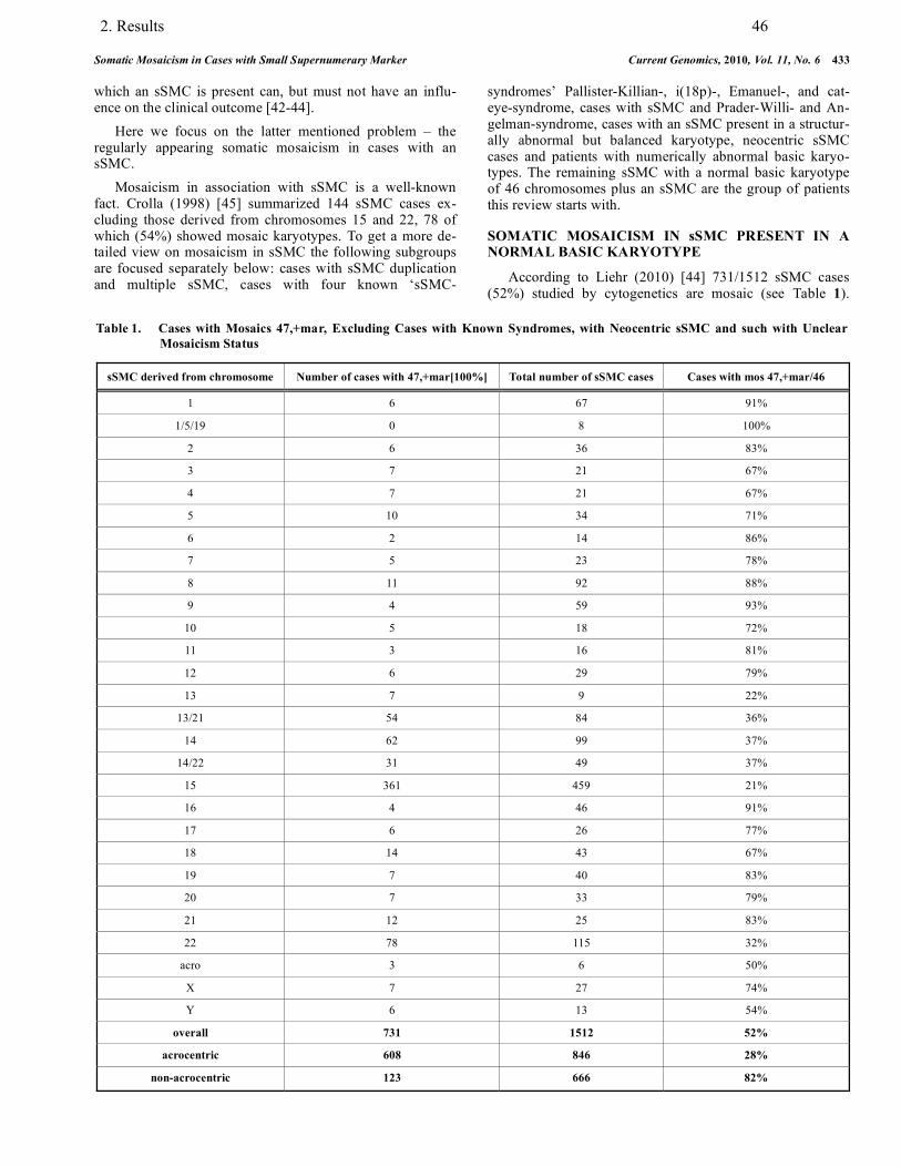
Somatic Mosaicism in Cases with Small Supernumerary Marker Current Genomics, 2010, Vol. 11, No. 6 433
which an sSMC is present can, but must not have an influ-ence on the clinical outcome [42-44]. Here we focus on the latter mentioned problem – the regularly appearing somatic mosaicism in cases with an sSMC. Mosaicism in association with sSMC is a well-known fact. Crolla (1998) [45] summarized 144 sSMC cases ex-cluding those derived from chromosomes 15 and 22, 78 of which (54%) showed mosaic karyotypes. To get a more de-tailed view on mosaicism in sSMC the following subgroups are focused separately below: cases with sSMC duplication and multiple sSMC, cases with four known ‘sSMC-
syndromes’ Pallister-Killian-, i(18p)-, Emanuel-, and cat-eye-syndrome, cases with sSMC and Prader-Willi- and An-gelman-syndrome, cases with an sSMC present in a structur-ally abnormal but balanced karyotype, neocentric sSMC cases and patients with numerically abnormal basic karyo-types. The remaining sSMC with a normal basic karyotype of 46 chromosomes plus an sSMC are the group of patients this review starts with.
SOMATIC MOSAICISM IN sSMC PRESENT IN A NORMAL BASIC KARYOTYPE
According to Liehr (2010) [44] 731/1512 sSMC cases (52%) studied by cytogenetics are mosaic (see Table 1).
Table 1. Cases with Mosaics 47,+mar, Excluding Cases with Known Syndromes, with Neocentric sSMC and such with Unclear Mosaicism Status
sSMC derived from chromosome Number of cases with 47,+mar[100%] Total number of sSMC cases Cases with mos 47,+mar/46
1 6 67 91%
1/5/19 0 8 100%
2 6 36 83%
3 7 21 67%
4 7 21 67%
5 10 34 71%
6 2 14 86%
7 5 23 78%
8 11 92 88%
9 4 59 93%
10 5 18 72%
11 3 16 81%
12 6 29 79%
13 7 9 22%
13/21 54 84 36%
14 62 99 37%
14/22 31 49 37%
15 361 459 21%
16 4 46 91%
17 6 26 77%
18 14 43 67%
19 7 40 83%
20 7 33 79%
21 12 25 83%
22 78 115 32%
acro 3 6 50%
X 7 27 74%
Y 6 13 54%
overall 731 1512 52%
acrocentric 608 846 28%
non-acrocentric 123 666 82%
2. Results 46
ahamid
Textfeld

434 Current Genomics, 2010, Vol. 11, No. 6 Liehr et al.
However, there is a strong difference between acrocentric and non-acrocentric derived sSMC: while 72% of acrocentric derived sSMC present no mosaic, 82% of non-acrocentric derived sSMCs are mosaic. The real grade and complexity of mosaicism seems to be even slightly higher as recently repeatedly cryptic mosaicism was detected in sSMC cases by molecular cytogenetics. There were either cases showing an sSMC in all studied metaphase spreads, however, interphase-FISH in uncultured cells showed a mosaic situation like in case 16-CW-2 [44]. More often it is found that more than one variant of an sSMC is present in different studied cells of a patient. As summa-rized in Table 2, at least 5% of sSMC cases have, after a
detailed molecular cytogenetic analysis, a more complex mosaicism than suggested after simple cytogenetic diagnos-tics. In 20% of these cases, unexpected complex somatic mosaicism was detectable where cytogenetics did not sug-gest any mosaicism, i.e. in cases 04-U-7, 08-W-p11.2/1-2, 11-O-p11.1/2-1, 11-U-9, 13-U-13, 15-W-q11.1+q11.2/1-1, 21-O-q11.21/1-1, 21-U-5, 22-O-q11.1/5-1, 22-O-q11.1/5-2, 0X-W-p11.?3/1-1, 0X-W-p11.21/1-1 [44]. Interestingly, acrocentric derived sSMC are by far more stable than non-acrocentric derived ones (2% versus 9%, Table 2). Cryptic mosaicism appears as some sSMC tend to rear-range and/or be reduced in size during karyotypic evolution. This can lead to double ring formation or inverted duplica-
Table 2. Cases with Cryptic Mosaics 47,+mar, Excluding Cases with Known Syndromes, with Neocentric sSMC and such with Unclear Mosaicism Status
sSMC derived from chromosome Number of cases with cryptic mosaicism Number of cases with cryptic mosaicism
1 0/67 0%
1/5/19 0/8 0%
2 2/36 5%
3 5/21 24%
4 1/21 5%
5 5/34 15%
6 4/14 29%
7 4/23 17%
8 8/92 9%
9 6/59 10%
10 0/18 0%
11 4/16 25%
12 2/29 7%
13 2/9 15%
13/21 0/84 0%
14 2/99 2%
14/22 0/49 0%
15 4/459 1%
16 3/46 19%
17 0/26 0%
18 2/43 5%
19 6/40 15%
20 4/33 12%
21 2/25 8%
22 5/115 4%
acro 0/6 0%
X 2/27 7%
Y 0/13 0%
overall 73/1512 5%
acrocentric 15/846 2%
non-acrocentric 58/666 9%
2. Results 47
ahamid
Textfeld
ahamid
Textfeld
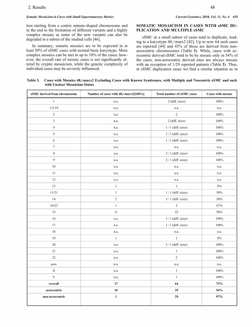
Somatic Mosaicism in Cases with Small Supernumerary Marker Current Genomics, 2010, Vol. 11, No. 6 435
tion starting from a centric minute-shaped chromosome and in the end to the formation of different variants and a highly complex mosaic as some of the new variants can also be degraded in a subset of the studied cells [46]. In summary, somatic mosaics are to be expected in at least 50% of sSMC cases with normal basic karyotype. More complex mosaics can be met in up to 10% of the cases; how-ever, the overall rate of mosaic cases is not significantly al-tered by cryptic mosaicism, while the genetic complexity of individual cases may be severely influenced.
SOMATIC MOSAICISM IN CASES WITH sSMC DU-PLICATION AND MULTIPLE sSMC
sSMC in a small subset of cases tend to duplicate, lead-ing to a karyotype 48,+marx2 [42]. Up to now 64 such cases are reported [44] and 45% of those are derived from non-acrocentric chromosomes (Table 3). While, cases with ac-rocentric derived sSMC tend to be by mosaic only in 54% of the cases, non-acrocentric derived ones are always mosaic with an exception of 1/29 reported patients (Table 3). Thus, in sSMC duplication cases we find a similar situation as in
Table 3. Cases with Mosaics 48,+marx2 Excluding Cases with Known Syndromes, with Multiple and Neocentric sSMC and such with Unclear Mosaicism Status
sSMC derived from chromosome Number of cases with 48,+marx2[100%] Total number of sSMC cases Cases with mosaic
1 n.a. 2 (diff. sizes) 100%
1/5/19 n.a. n.a. n.a.
2 n.a. 2 100%
3 n.a. 2 (diff. sizes) 100%
4 n.a. 1 / 1 (diff. sizes) 100%
5 n.a. 1 / 1 (diff. sizes) 100%
6 n.a. 1 / 1 (diff. sizes) 100%
7 n.a. n.a. n.a.
8 n.a. 2 / 1 (diff. sizes) 100%
9 n.a. 2 / 1 (diff. sizes) 100%
10 n.a. n.a. n.a.
11 n.a. n.a. n.a.
12 n.a. n.a. n.a.
13 1 1 0%
13/21 1 1 / 1 (diff. sizes) 50%
14 2 3 / 1 (diff. sizes) 50%
14/22 1 3 67%
15 11 22 50%
16 n.a. 1 / 1 (diff. sizes) 100%
17 n.a. 1 / 1 (diff. sizes) 100%
18 n.a. n.a. n.a.
19 1 1 0%
20 n.a. 3 / 1 (diff. sizes) 100%
21 n.a. 1 100%
22 n.a. 2 100%
acro n.a. n.a. n.a.
X n.a. 1 100%
Y n.a. 1 100%
overall 17 64 73%
acrocentric 16 35 54%
non-acrocentric 1 29 97%
2. Results 48
ahamid
Textfeld
436 Current Genomics, 2010, Vol. 11, No. 6 Liehr et al.
cases with one single sSMC and a karyotype 47, +mar con-cerning mosaicism. Multiple sSMC cases [42] differ from sSMC duplication ones by the fact that the observed sSMC are not derived from the identical chromosome. Only 48 such cases are known by now [44], having between 2 and 7 sSMC of dif-ferent origin, each; and all reported cases with multiple sSMC are mosaic. Formation of this rare cytogenetic condi-tion is unclear, even though polysomic rescue or triploid rescue maybe suggested. As in most cases markedly chro-mosomal imbalances are induced by multiple sSMC pres-ence, ~90-95% of them are correlated with clinical symp-toms, irrespective of mosaicism status detectable in periph-eral blood.
SOMATIC MOSAICISM PRESENT IN THE FOUR KNOWN ‘sSMC-SYNDROMES’: PALLISTER-KILLIAN-, I(18P)-, EMANUEL-, AND CAT-EYE-SYNDROME
Somatic mosaicism is reported to different extents in four sSMC-related syndromes. Patients suffering from Pallister-Killian-syndrome (PKS) due to the presence of an additional isochromosome 12p are known to have somatic mosaicism in practically every case. In peripheral blood the +(12p) tends to be lost either already during pregnancy or shortly after birth in practically all cells. In the alternatively studied skin fibroblasts, the sSMC de-rived from chromosome 12 is normally easily to detect in >70% to 100% of the cells [47]. However, besides a mosaic of cells with 46 and 47 chromosomes exceptional cases also with two different shapes of sSMC (12-Wpks-4, 12-Wpks-159, [44]) or two isochromosomes 12p (12-Wpks-174 [44]) are also reported. In isochromosome 18p syndrome mosaicism is rather rare. But also here exceptional cases are known having the full clinical phenotype but normal karyotype in some of the body cells (18-Wi-42, 18-Wi-153, 18-Wi-154, 18-Wi-157 [44]). In case 18-Wi-41 [44] the i(18p) was derived from the clinically normal mother; the latter had the i(18p) in only 4% of her peripheral blood cells. Also, an interesting case of somatic mosaicism is 18-Wi-158 [44] showing prenatally an i(18p) in 35% of the amnion cells but postnatal only normal cells in peripheral blood, being a normal child. To the best of our knowledge no mosaic cases are known by now for Emanuel-syndrome (ES) [44]. Also in cat-eye-syndrome (CES) mosaicism is rather rare. sSMC derived from chromosome 22 having two different shapes were seen in CES (22-Wces-2 [44]) or minimal mosaicism with a nor-mal cell line (22-Wces-3-03, 22-Wces-5, 22-Wces-5-119 [44]). Overall, somatic mosaicism is, compared to other sSMC derived from the corresponding chromosomes, over-represented in PKS (100% vs. 79%) and under-represented in i(18p) syndrome (4% vs. 67%), ES (0% vs. 32%) and CES (3% vs. 32%).
SOMATIC MOSAICISM IN PRADER-WILLI- AND ANGELMAN-SYNDROME WITH sSMC
26 sSMC cases with Prader-Willi-syndrome (PWS) and 7 with Angelman-syndrome (AS) can be found in the literature
[44]. 15 of these are PWS (58%) while only 1 of these AS cases (14%) is mosaic with respect to sSMC presence [44]. As the corresponding syndromes were caused either by uni-parental disomy or microdeletion the sSMC presence has no direct influence on the clinical outcome; neither has mo-saicism.
SOMATIC MOSAICISM IN sSMC PRESENT IN STRUCTURALLY ABNORMAL BUT BALANCED KARYOTYPE
Another rare cytogenetic variant of sSMC presence is that of a structurally abnormal but balanced karyotype (McClintock mechanism) [48]. Such cases can either be connected with a neocentromere formation (see section be-low) or both the derivatives share the available centromeric alpha-satellite sequences. If in such case mosaicism appears, i.e. loss of the sSMC, relevant genetic material is lost and this leads normally to clinical problems as described for the following cases: 03-W-p11/1-1, 04-W-p15.3/1-1, 04-W-p12/1-1 [44]. If no or only very low grade mosaicism is pre-sent the carrier of such a karyotype can be completely nor-mal (e.g. 02-O-p12/1-1, 06-O-p22.3/1-1, 06-O-p22.3/1-1, 08-O-p11.1/2-1, 12-U-4, 17-O-p11.2/2-1, mother of 19-W-10/2-1, mother of 22-W-q11.2/2-1 [44]).
SOMATIC MOSAICISM IN NEOCENTRIC sSMC
For mosaicism in neocentric sSMC formed by McClin-tock mechanism, [48] the same holds true, like for the aforementioned centric sSMC present in structurally abnor-mal but balanced karyotype. If balanced and no or only minimal mosaicism is present, the carriers of such a chromo-somal condition are clinically normal. If the neocentric sSMC is lost in a higher percentage of the body cells this has an adverse prognosis.
In general, in at least around 50% of the cases with a neocentric sSMC somatic mosaicism is observable (Table 4). Strikingly, as in centric sSMC, mosaicism is more frequent in non-acrocentric derived compared to acrocentric derived ones (58% vs. 24%).
SOMATIC MOSAICISM IN sSMC PRESENT IN NU-MERICALLY ABNORMAL BASIC KARYOTYPES
As above mentioned, sSMC can appear in a numerically normal basic karyotype of 46 chromosomes, but also in numerically abnormal basic karyotypes [42]. Up to now, sSMC are reported additionally to a basic karyotype 45, X (= Turner syndrome), 47, XXY (= Klinefelter syndrome), 47, XXX (triple-X syndrome) and 47, +21 (Down syndrome) [44, 49-51].
542 cases are available in the literature with a basic karyotype typical for Turner syndrome and an additional sSMC, i.e. 46,X,+mar [44, 49]. Only 73 of these are reported without mosaicism; thus, 76% of these Turner syndrome cases are mosaic [44].
Only three cases, each of them are known by now with Klinefelter- or triple-X syndrome and an additional sSMC. Concerning the Klinefelter-syndrome two cases of those are mosaic (07-U-6, 0X-U3) and one not (09-U5) [44]. For tri-
2. Results 49
ahamid
Textfeld
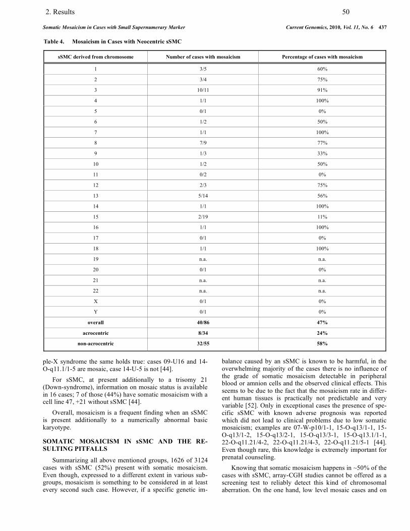
Somatic Mosaicism in Cases with Small Supernumerary Marker Current Genomics, 2010, Vol. 11, No. 6 437
ple-X syndrome the same holds true: cases 09-U16 and 14-O-q11.1/1-5 are mosaic, case 14-U-5 is not [44]. For sSMC, at present additionally to a trisomy 21 (Down-syndrome), information on mosaic status is available in 16 cases; 7 of those (44%) have somatic mosaicism with a cell line 47, +21 without sSMC [44]. Overall, mosaicism is a frequent finding when an sSMC is present additionally to a numerically abnormal basic karyotype.
SOMATIC MOSAICISM IN sSMC AND THE RE-SULTING PITFALLS
Summarizing all above mentioned groups, 1626 of 3124 cases with sSMC (52%) present with somatic mosaicism. Even though, expressed to a different extent in various sub-groups, mosaicism is something to be considered in at least every second such case. However, if a specific genetic im-
balance caused by an sSMC is known to be harmful, in the overwhelming majority of the cases there is no influence of the grade of somatic mosaicism detectable in peripheral blood or amnion cells and the observed clinical effects. This seems to be due to the fact that the mosaicism rate in differ-ent human tissues is practically not predictable and very variable [52]. Only in exceptional cases the presence of spe-cific sSMC with known adverse prognosis was reported which did not lead to clinical problems due to low somatic mosaicism; examples are 07-W-p10/1-1, 15-O-q13/1-1, 15-O-q13/1-2, 15-O-q13/2-1, 15-O-q13/3-1, 15-O-q13.1/1-1, 22-O-q11.21/4-2, 22-O-q11.21/4-3, 22-O-q11.21/5-1 [44]. Even though rare, this knowledge is extremely important for prenatal counseling. Knowing that somatic mosaicism happens in ~50% of the cases with sSMC, array-CGH studies cannot be offered as a screening test to reliably detect this kind of chromosomal aberration. On the one hand, low level mosaic cases and on
Table 4. Mosaicism in Cases with Neocentric sSMC
sSMC derived from chromosome Number of cases with mosaicism Percentage of cases with mosaicism
1 3/5 60%
2 3/4 75%
3 10/11 91%
4 1/1 100%
5 0/1 0%
6 1/2 50%
7 1/1 100%
8 7/9 77%
9 1/3 33%
10 1/2 50%
11 0/2 0%
12 2/3 75%
13 5/14 56%
14 1/1 100%
15 2/19 11%
16 1/1 100%
17 0/1 0%
18 1/1 100%
19 n.a. n.a.
20 0/1 0%
21 n.a. n.a.
22 n.a. n.a.
X 0/1 0%
Y 0/1 0%
overall 40/86 47%
acrocentric 8/34 24%
non-acrocentric 32/55 58%
2. Results 50
ahamid
Textfeld
438 Current Genomics, 2010, Vol. 11, No. 6 Liehr et al.
the other hand, cryptic mosaics are missed. Thus, cytogenet-ics is still the gold-standard to detect any kind of chromoso-mal aberration, which then, in further steps can be character-ized by molecular (cyto-) genetic approaches. Interestingly, acrocentric and non-acrocentric derived sSMC are differently susceptible to mosaicism; acrocentric derived ones are hereby the more stable ones. This holds true for centric and neocentric sSMC, and an explanation is there-fore at present not available.
CONCLUSION
Somatic mosaicism is a feature of the human body, which has to be considered much more than up to now in future. It is known as a fact, but not understood why man with age (in peripheral blood) develops something like a ‘Turner-syndrome-mosaic’ 46,XY/45,X. Similarly, in cases with sSMC it is known since years, that PKS patients lose the i(12p) in peripheral blood or that some inv dup(15) sSMC are stable and cytogenetically identical ones in an-other carrier are not. For all these facts to the best of our knowledge, no studies were undertaken to come closer to an understanding of these phenomena. Here we present, some kind of starting point for such studies, for the first time a detailed ‘mosaicism map’ for the different subtypes of sSMC.
ACKNOWLEDGEMENTS
This work was supported in parts by DAAD (D07/00070), BMBF/DLR (BLR 08/004 and ARM 08/001), Prochance 2008 and 2009, and DFG (LI 820/22-1).
REFERENCES [1] Tjio, J.-H.; Levan, A. The chromosome number of man. Hereditas,
1956, 42, 1-6. [2] Yunis, J.J.; Chandler, M.E. High-resolution chromosome analysis
in clinical medicine. Prog. Clin. Pathol., 1978, 7, 267-288. [3] Mitelman, F. Cytogenetics of experimental neoplasms and non-
random chromosome correlations in man. Clin. Haematol., 1980, 9, 195-219.
[4] Liehr, T.; Claussen, U. Current developments in human molecular cytogenetic techniques. Curr. Mol. Med., 2002, 2, 283-297.
[5] Liehr, T.; Claussen, U. Multicolor-FISH approaches for the charac-terization of human chromosomes in clinical genetics and tumor cytogenetics. Curr. Genomics, 2002, 3, 213-235.
[6] Liehr, T.; Starke, H.; Weise, A.; Lehrer, H.; Claussen, U. Multi-color FISH probe sets and their applications. Histol. Histopathol., 2004, 19, 229-237.
[7] Liehr, T.; Starke, H.; Heller, A.; Kosyakova, N.; Mrasek, K.; Gross, M.; Karst, C.; Steinhaeuser, U.; Hunstig, F.; Fickelscher, I.; Kuechler, A.; Trifonov, V.; Romanenko, S.A.; Weise A. Multicolor fluorescence in situ hybridization (FISH) applied for FISH-banding. Cytogenet. Genome Res., 2006, 114, 240-244.
[8] Vorsanova, S.G.; Yurov, Y.B.; Iourov, I.Y. Human interphase chromosomes: a review of available molecular cytogenetic tech-nologies. Mol. Cytogenet., 2010, 3, 1.
[9] Gebhart, E.; Liehr, T.; Harrer, P.; Reichardt, S.; Schmitt, G.; Thoma, K.; Gramatzki, M.; Trautmann, U. Determination by inter-phase FISH of the clonality of aberrant karyotypes in human hema-topoietic neoplasias. Leuk. Lymphoma, 1995, 17, 295-302.
[10] Gebhart, E.; Liehr, T. Clonality determined by fluorescence in situ hybridization of single-cell aberrations in hematopoietic neoplasias. Cancer Genet. Cytogenet., 1999, 113, 193-194.
[11] Karst, C.; Gross, M.; Haase, D.; Wedding, U.; Höffken, K.; Liehr, T.; Mkrtchyan, H. Novel cryptic chromosomal rearrangements de-tected in acute lymphoblastic leukemia (ALL) detected by applica-
tion of new multicolor fluorescent in situ hybridization approaches. Int. J. Oncol., 2006, 28, 891-897.
[12] Gross, M.; Mkrtchyan, H.; Glaser, M.; Fricke, H.J.; Höffken, K.; Heller, A.; Weise, A.; Liehr T. Delineation of yet unknown cryptic subtelomere aberrations in 50% of acute myeloid leukemia with normal GTG-banding karyotype. Int. J. Oncol., 2009, 34, 417-423.
[13] Dimmler, A.; Kiesewetter, F.; Liehr, T.; Neubauer, S.; Schell, H.; Gebhart, E. Interphase-FISH examinations in paraffin sections from benign, precancerous, and cancerous lesions of the skin and oral mucosa. Int. J. Oncol., 1997, 10, 83-88.
[14] Iourov, I.Y.; Vorsanova, S.G.; Liehr, T.; Monakhov, V.V.; So-loviev, I.V.; Yurov, Y.B. Dynamic mosaicism manifesting as loss; gain and rearrangement of an isodicentric Y chromosome in a male child with growth retardation and abnormal external genitalia. Cy-togenet. Genome Res., 2008, 121, 302-306.
[15] Liehr, T.; Ziegler, M. Rapid prenatal diagnostics in the interphase nucleus – procedure and cut-off rates. J. Histochem. Cytochem., 2005, 53, 289-291.
[16] Liehr, T.; Rautenstrauss, B.; Grehl, H.; Bathke, K.D.; Ekici, A.; Rauch, A.; Rott, H.D. Mosaicism for the Charcot-Marie-Tooth dis-ease type 1A duplication suggests somatic reversion. Hum. Genet., 1996, 98, 22-28.
[17] Koç, A.; Kan, D.; Karaer, K.; Ergün, M.A.; Karao uz, M.Y.; Gücüyener, K.; Hinreiner, S.; Liehr, T.; Perçin, E.F. An unexpected finding in a child with neurological problems: mosaic ring chromo-some 18. Eur. J. Pediatr., 2008, 167, 655-659.
[18] Soysal, Y.; Balci, S.; Hekimler, K.; Liehr, T.; Ewers, E.; Schou-mans, J.; Bui, T.H.; Içduygu, F.M.; Kosyakova, N.; Imirzalio lu, N. Characterization of double ring chromosome 4 mosaicism asso-ciated with bilateral hip dislocation, cortical dysgenesis, and epi-lepsy. Am. J. Med. Genet. A, 2009, 149A, 2782-2787.
[19] Yurov, Y.B.; Vostrikov, V.M.; Vorsanova, S.G.; Monakhov, V.V.; Iourov, I.Y. Multicolor fluorescent in situ hybridization on post mortem brain in schizophrenia as an approach for identification of low-level chromosomal aneuploidy in neuropsychiatric diseases. Brain Dev., 2001, 23(Suppl 1), 186-190.
[20] Yurov, Y.B.; Iourov, I.Y.; Monakhov, V.V.; Soloviev, I.V.; Vostri-kov, V.M.; Vorsanova, S.G. The variation of aneuploidy frequency in the developing and adult human brain revealed by an interphase FISH study. J. Histochem. Cytochem., 2005, 53, 385-390.
[21] Iourov, I.Y.; Liehr, T.; Vorsanova, S.G.; Kolotii, A.D.; Yurov, Y.B. Visualization of interphase chromosomes in postmitotic cells of the human brain by multicolour banding (MCB). Chromosome Res., 2006, 14, 223-229.
[22] Yurov, Y.B.; Iourov, I.Y.; Vorsanova, S.G.; Liehr, T.; Kolotii, A.D.; Kutsev, S.I.; Pellestor, F.; Beresheva, A.K.; Demidova, I.A.; Kravets, V.S.; Monakhov, V.V.; Soloviev, I.V. Aneuploidy and confined chromosomal mosaicism in the developing human brain. PLoS ONE, 2007, 2, e558.
[23] Yurov, Y.B.; Vorsanova, S.G.; Iourov, I.Y.; Demidova, I.A.; Ber-esheva, A.K.; Kravetz, V.S.; Monakhov, V.V.; Kolotii, A.D.; Voi-nova-Ulas, V.Y.; Gorbachevskaya, N.L. Unexplained autism is fre-quently associated with low-level mosaic aneuploidy. J. Med. Genet., 2007, 44, 521-535.
[24] Yurov, Y.B.; Iourov, I.Y.; Vorsanova, S.G.; Demidova. I.A.; Kravetz, V.S.; Beresheva, A.K.; Kolotii, A.D.; Monakchov, V.V.; Uranova, N.A.; Vostrikov, V.M.; Soloviev, I.V.; Liehr, T. The schizophrenia brain exhibits low-level aneuploidy involving chro-mosome 1. Schizophr. Res., 2008, 98, 137-147.
[25] Iourov, I.Y.; Vorsanova, S.G.; Yurov, Y.B. Molecular cytogenetics and cytogenomics of brain diseases. Curr. Genomics, 2008, 9, 452-465.
[26] Iourov, I.Y.; Vorsanova, S.G.; Liehr, T.; Yurov, Y.B. Aneuploidy in the normal; Alzheimer's disease and ataxia-telangiectasia brain: differential expression and pathological meaning. Neurobiol. Dis., 2009, 34, 212-220.
[27] Iourov, I.Y.; Vorsanova, S.G.; Liehr, T.; Kolotii, A.D.; Yurov, Y.B. Increased chromosome instability dramatically disrupts neural genome integrity and mediates cerebellar degeneration in the ataxia-telangiectasia brain. Hum. Mol. Genet., 2009, 18, 2656-2669.
[28] Vanneste, E.; Voet, T.; Le Caignec, C.; Ampe, M.; Königs, P.; Mellote, C.; Debrock, S.; Amyere, M.; Vikkula, M.; Schuit, F.; Fryns, J.P.; Verbeke, G., D'Hooghe, T.; Moreau, Y.; Vermeesch, J.R. Chromosome instability is common in human cleavage-stage embryos. Nat. Med., 2009, 15, 577-583.
2. Results 51
ahamid
Textfeld

Somatic Mosaicism in Cases with Small Supernumerary Marker Current Genomics, 2010, Vol. 11, No. 6 439
[29] Vorsanova, S.G.; Kolotii, A.D.; Iourov, I.Y.; Monakhov, V.V.; Kirillova, E.A.; Soloviev, I.V.; Yurov, Y.B. Evidence for high fre-quency of chromosomal mosaicism in spontaneous abortions re-vealed by interphase FISH analysis. J. Histochem. Cytochem., 2005, 53, 375-380.
[30] Vorsanova, S.G.; Iourov, I.Y.; Demidova, I.A.; Kirillova, E.A.; Soloviev, I.V.; Yurov, Y.B. Chimerism and multiple numerical chromosome imbalances in a spontaneously aborted fetus. Tsitol. Genet., 2006, 40, 28-30.
[31] Vorsanova, S.G.; Yurov, Y.B.; Deryagin, G.V.; Soloviev, I.V.; Bytenskaya, G.A. Diagnosis of aneuploidy by in situ hybridization: analysis of interphase nuclei. Bull. Exp. Biol. Med., 1991, 112, 413-415.
[32] Mkrtchyan, H.; Gross, M.; Hinreiner, S.; Polytiko, A.; Manvelyan, M.; Mrasek, K.; Kosyakova, N.; Ewers, E.; Nelle, H.; Liehr, T.; Volleth, M.; Weise, A. Early embryonic chromosome instability re-sults in stable mosaic pattern in human tissues. PLoS One, 2010, 5, e9591.
[33] Felka, T.; Lemke, J.; Lemke, C.; Michel, S.; Liehr, T.; Claussen, U. DNA degradation during maturation of erythrocytes-molecular cy-togenetic characterization of Howell–Jolly bodies. Cytogenet. Ge-nome Res., 2007, 119, 2-8.
[34] Kinne, R.W.; Liehr, T.; Beensen, V.; Kunisch, E.; Zimmermann, T.; Holland, H.; Pfeiffer, R.; Stahl, H.-D.; Lungershausen, W.; Hein, G.; Roth, A.; Emmrich, F.; Claussen, U.; Froster, U.G. Mo-saic chromosomal aberrations in synovial fibroblasts of patients with rheumatoid arthritis, osteoarthritis, and other inflammatory joint diseases. Arthritis. Res., 2001, 3, 319-330.
[35] Kinne, R.W.; Kunisch, W.; Beensen, V.; Zimmermann, T.; Emmrich, F.; Petrow, P.; Lungershausen, W.; Hein, G.; Braun, R.K.; Foerster, M.; Kroegel, C.; Winter, R.; Liesaus, E.; Fuhrmann, R.A.; Roth, A.; Claussen, U.; Liehr, T. Synovial fibroblasts and synovial macrophages from patients with rheumatoid arthritis and other inflammatory joint diseases show chromosomal aberrations. Genes Chromosomes Cancer, 2003, 38, 53-67.
[36] Iourov, I.Y.; Vorsanova, S.G.; Soloviev, I.V.; Yurov, Y.B. Inter-phase FISH: detection of intercellular genomic variations and so-matic chromosomal mosaicism. In Fluorescence in situ hybridiza-tion (FISH) - Application guide. Liehr T. Ed. Berlin; Heidelberg: Springer Verlag; 2009, pp. 301-311.
[37] Iourov, I.Y.; Vorsanova, S.G.; Yurov, Y.B. Chromosomal varia-tions in mammalian neuronal cells: known facts and attractive hy-potheses. Int. Rev. Cytol., 2006, 249, 143-191.
[38] Iourov, I.Y.; Vorsanova, S.G.; Yurov, Y.B. Intercellular genomic (chromosomal) variations resulting in somatic mosaicism: mecha-nisms and consequences. Curr. Genomics, 2006, 7, 435-446.
[39] Iourov, I.Y.; Vorsanova, S.G.; Yurov, Y.B. Chromosomal mosia-cism goes global. Mol. Cytogenet., 2008, 1, 26.
[40] Pathak, S. Cytogenetic research techniques in humans and labora-tory animals that can be applied most profitably to livestock. J. Dairy Sci., 1979, 62, 836-843.
[41] Liehr, T.; Weise, A. Frequency of small supernumerary marker chromosomes in prenatal, newborn, developmentally retarded and infertility diagnostics. Int. J. Mol. Med., 2007, 19, 719-731.
[42] Liehr, T.; Claussen, U.; Starke, H. Small supernumerary marker chromosomes (sSMC) in humans. Cytogenet. Genome Res., 2004, 107, 55-67.
[43] Liehr, T.; Mrasek, K.; Weise, A.; Dufke, A.; Rodríguez, L.; Martínez Guardia, N.; Sanchís, A.; Vermeesch, JR.; Ramel, C.; Polityko, A.; Haas, O.A.; Anderson, J.; Claussen, U.; von Eggeling, F.; Starke, H. Small supernumerary marker chromosomes--progress towards a genotype-phenotype correlation. Cytogenet. Genome Res., 2006, 112, 23-34.
[44] Liehr, T. Homepage on small supernumerary marker chromosomes (sSMC). http://www.med.uni-jena.de/fish/sSMC/00START.htm (Accessed January 11, 2010).
[45] Crolla, J.A. FISH and molecular studies of autosomal supernumer-ary marker chromosomes excluding those derived from chromo-some 15: II. Review of the literature. Am. J. Med. Genet., 1998, 75, 367-381.
[46] Liehr, T. Small supernumerary marker chromosomes (sSMCs): a spotlight on some nomenclature problems. J. Histochem. Cyto-chem., 2009, 57, 991-993.
[47] Liehr, T.; Wegner, R.-D.; Stumm, M.; Joksi, G.; Polityko, A.; Kosyakova, N.; Ewers, E.; Reich, D.; Wagner, R.; Weise, A. Pallis-ter-Killian syndrome. Rare phenotypic features and variable karyo-types. Balkan J. Med. Genet. 2008, 12, 65-67
[48] Baldwin, E.L.; May, L.F.; Justice, A.N.; Martin, C.L.; Ledbetter, D.H. Mechanisms and consequences of small supernumerary marker chromosomes: from Barbara McClintock to modern ge-netic-counseling issues. Am. J. Hum. Genet., 2008, 82, 398-410.
[49] Liehr, T.; Mrasek, K.; Hinreiner, S.; Reich, D.; Ewers, E.; Bartels, I.; Seidel, J.; Manolakis, E.; Petersen, M.; Polityko, A.; Dufke, A.; Iourov, I.; Trifonov, V.; Vermeesch, J.; Weise, A. Small supernu-merary marker chromosomes (sSMC) in patients with a karyotype 45.;X/46.;X.;+mar – 17 new cases and a review of the literature. Sex. Dev., 2007, 1, 353-362.
[50] Starke, H.; Mitulla, B.; Nietzel, A.; Heller, A.; Beensen, V.; Grosswendt, G.; Claussen, U.; von Eggeling, F.; Liehr, T. First pa-tient with trisomy 21 accompanied by an additional der(4)(:p11 Ā q11:) plus partial uniparental disomy 4p15-16. Am. J. Med. Genet., 2003, 116A, 26-30.
[51] Liehr, T.; Mrasek, K.; Starke, H.; Claussen, U.; Schreiber, G. Un-usual small supernumerary marker chromosome (sSMC) 9 in a Klinefelter patient. Cytogenet. Genome Res., 2005, 111, 179-181.
[52] Fickelscher, I.; Starke, H.; Schulze, E.; Ernst, G.; Kosyakova, N.; Mkrtchyan, H.; Macdermont, K.; Sebire, N.; Liehr, T. A further case with a small supernumerary marker chromosome (sSMC) de-rived from chromosome 1-evidence for high variability in mo-saicism in different tissues of sSMC carriers. Prenat. Diagn., 2007, 27, 783-785.
2. Results 52
ahamid
Textfeld
2. Results
2.7. Article .6
Hamid AB, Liehr T. Pericentromeric BAC-probe set - thoughts about considering genedosage insensitive regions. Mol Cytogenet, 2013; 6:45/comments.
•
•
•
2. Results 53
•
2. Results 54
2. Results
2.8. Article .7
Hamid AB, Weise A, Voigt M, Bucksch M, Kosyakova N, Liehr T, Klein E. Clinical impact of proximal autosomal imbalances. Balk J Med Genet, 2012; 15(2): 15-21.
ahamid
Textfeld
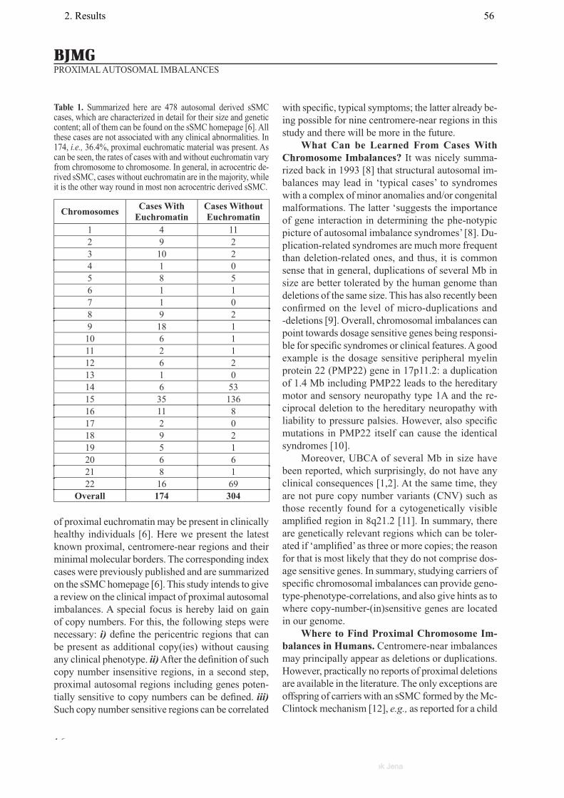
ahamid
Textfeld

ahamid
Textfeld

ahamid
Textfeld
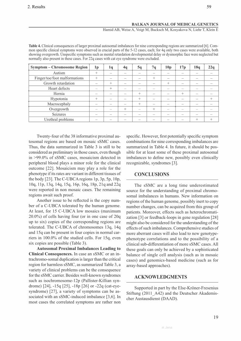
ahamid
Textfeld
ahamid
Textfeld
ahamid
Textfeld
2. Results
2.9. Article .8
Bucksch M, Ziegler M, Kosayakova N, Mulhatino MV, Llerena Jr. JC, Morlot S, Fischer W, Polityko AD, Kulpanovich AI, Petersen MB, Belitz B, Trifonov V, Weise A, Liehr T, Hamid AB. A new multicolor fluorescence in situ hybridization probe set directed against human heterochromatin: HCM-FISH. J Histochem Cytochem, 2012; 60(7):530-536.
Journal of Histochemistry & Cytochemistry 60(7) 530 –536© The Author(s) 2012Reprints and permission: sagepub.com/journalsPermissions.navDOI: 10.1369/0022155412441708http://jhc.sagepub.com
A detailed characterization of chromosomal rearrangements detected in routine banding cytogenetics can nowadays be done easily by fluorescence in situ hybridization (FISH) and/or array-comparative genomic hybridization (aCGH) (Manolakos et al. 2010; Weimer et al. 2011). While in aCGH, a higher resolution may be achieved, FISH still has several advantages over the array-based approaches (Mano-lakos et al. 2010). FISH allows, for example, the analysis of balanced rearrangements, of chromosomal aberrations pres-ent only in low mosaic levels, and of the large heterochro-matic regions of the human genome. The acrocentric short arms; the centric and the large pericentric regions of chro-mosomes 1, 9, and 16; as well as the band Yq12 cannot be analyzed by aCGH.
A multitude of multicolor FISH (mFISH) probe sets have been developed in the last decades (Liehr 2012a). They were implemented for use in one experiment: 1) all 24 human whole chromosome painting probes (multiplex FISH = M-FISH [Speicher et al. 1996]; spectral karyotyp-ing = SKY [Schröck et al. 1996]) or 2) all centromeric probes (centromere-specific M-FISH = cenM-FISH [Nietzel et al. 2001]). Also, 3) various FISH banding
441708 JHCXXX10.1369/0022155412441708Bucksch et al.Heterochromatin-M-FISH (HCM-FISH)2012© The Author(s) 2010
Reprints and permission:sagepub.com/journalsPermissions.nav
Received for publication January 10, 2012; accepted February 13, 2012.
Corresponding Author:Thomas Liehr, Institute of Human Genetics, Jena University Hospital, Kollegiengasse 10, D-07743 Jena, Germany. Email: [email protected]
A New Multicolor Fluorescence In Situ Hybridization Probe Set Directed Against Human Heterochromatin: HCM-FISH
Maria Bucksch, Monika Ziegler, Nadezda Kosayakova, Milene V. Mulhatino, Juan C. Llerena Jr., Susanne Morlot, Wolfgang Fischer, Anna D. Polityko, Anna I. Kulpanovich, Michael B. Petersen, Britta Belitz, Vladimir Trifonov, Anja Weise, Thomas Liehr, and Ahmed B. HamidInstitute of Human Genetics, Jena University Hospital, Jena, Germany (MB,MZ,NK,AW,TL,ABH); Medical Genetics Department, Fernandes Figueira Institute, FIOCRUZ, Rio de Janeiro, Brazil (MVM,JCL); MVZ Wagnerstibbe, Hannover, Germany (SM,WF); National Medical Center, Minsk, Belarus (ADP); Belarus State Medical University, Minsk, Belarus (AIK); Molecular Biology–Genetics–Biotechnology, EuroGenetica, Athens, Greece (MBP); Praxis für Humangenetik, Berlin, Germany (BB); and Molecular and Cellular Biology Department, Institute of Chemical Biology and Fundamental Medicine, SВ RAS, Novosibirsk, Russia (VT).
Summary
A new multicolor fluorescence in situ hybridization (mFISH) probe set is presented, and its possible applications are highlighted in 25 clinical cases. The so-called heterochromatin-M-FISH (HCM-FISH) probe set enables a one-step characterization of the large heterochromatic regions within the human genome. HCM-FISH closes a gap in the now available mFISH probe sets, as those do not normally cover the acrocentric short arms; the large pericentric regions of chromosomes 1, 9, and 16; as well as the band Yq12. Still, these regions can be involved in different kinds of chromosomal rearrangements such as translocations, insertions, inversions, amplifications, and marker chromosome formations. Here, examples are given for all these kinds of chromosomal aberrations, detected as constitutional rearrangements in clinical cases. Application perspectives of the probe set in tumors as well as in evolutionary cytogenetic studies are given. (J Histochem Cytochem 60:530–536, 2012)
Keywords
multicolor fluorescence in situ hybridization (mFISH), heterochromatin-M-FISH (HCM-FISH) probe set, heteromorphism, small supernumerary marker chromosome (sSMC), insertion, translocation
Article
2. Results 62
Heterochromatin-M-FISH (HCM-FISH) 531
approaches (Liehr et al. 2002a) were introduced as well as 4) combinations of centromeric with locus-specific and/or partial chromosome painting probes (e.g., subcentromere-specific M-FISH = subcenM-FISH [Liehr et al. 2006]). These probe sets are highly suited to characterize simple and complex chromosomal aberrations (approaches 1 and 3) or small supernumerary marker chromosomes (sSMC) (Liehr et al. 2004, 2006) (approaches 2 and 4). Recently, even a probe set was introduced to substantiate indirectly epigenetic changes (parental origin determination FISH = POD-FISH [Weise et al. 2008]).
Here, we present a new mFISH probe set specifically directed against the large heterochromatic regions within the human genome. This so-called heterochromatin-M-FISH (HCM-FISH) set was successfully established and applied already in 30 cases, where its application saved sample material and time. We present 25 representative cases studied by HCM-FISH and discuss the possible appli-cations of this new probe set.
Materials and MethodsHCM-FISH Probe Set
The HCM-FISH probe set (Fig. 1) is based on eight glass-needle microdissection (midi)–derived and one P1 artificial chromosome (PAC) probe (RP5–1174A5 = dj1174A5); the latter was kindly provided by Dr. M. Rocchi (Bari, Italy). The latter probe is specific for the nucleolus organizer region (NOR), which contains several tandem copies of ribosomal RNA genes and in humans is clustered on the short arms of chromosomes 13, 14, 15, 21, and 22; that is, the acrocentric chromosomes (Trifonov et al. 2003). Midi was done as previously reported (Liehr et al. 2002b). Midi probes for the regions 1q12, 9q12, 15p12~11.2 (i.e., a β-satellite–specific probe), 16q11.2, 19p12~19q12, and Yq12 were established for this probe set, while those probes for 9p12/9q13 (midi 36) (Starke et al. 2002) and for all acrocentric short arms (midi 54) were as previously reported (Mrasek et al. 2003).
The DNA of the nine probes was amplified in vitro and labeled by degenerated oligonucleotide primer polymerase chain reaction (DOP-PCR) according to standard proce-dures (Telenius et al. 1992). The amplification procedure followed a published scheme (Fig. 2A in Liehr et al. 2002b). The used fluorochromes Spectrum Green (SG), Spectrum Orange (SO), Texas Red (TR), cyanine 5 (CY5), and dieth-ylaminocoumarin (DEAC) were applied for the nine DNA probes as depicted in Figure 1A. Thus, each DNA probe obtained its unique fluorochrome combination, which could be transformed into pseudocolors (Fig. 1) using the soft-ware mentioned below.
Twenty metaphase spreads were analyzed, each using a fluorescence microscope (Axioplan 2 MOT; Carl Zeiss,
Oberkochen, Germany) equipped with appropriate filter sets to discriminate between all five fluorochromes and the counterstain 4’,6-diamidino-2-phenylindole (DAPI). Image capturing and processing were carried out using an Isis mFISH imaging system (MetaSystems; Altlussheim, Germany).
Clinical CasesOverall, 30 clinical cases were studied already by HCM-FISH (Table 1). The clinical indications were infertility, repeated abortions, dysmorphic features and/or mental retardation, or a prenatal cytogenetic study due to advanced maternal age (Table 1). In all studied cases, apart from cases 1 and 1a to 1d, which were normal controls, banding cytogenetics revealed an aberrant karyotype. M-FISH was not informative in cases 2, 4, 6, 7, 8, 10, 11, and 14 (results not shown). In cases 3, 5, 12, 13, and 15, heteromorphisms were suggested after Giemsa stained chromsomes banding. In the additional redundant 11 cases (Table 1), similar observations were made. In case 9, HCM-FISH was applied directly, as an sSMC derived from an acrocentric chromosome was suggested.
ResultsIn the present study, it could be demonstrated that HCM-FISH can be used to characterize within one single step chromosomal rearrangements with gross involvement of heterochromatic material. The HCM-FISH probe set was established first in five control cases (result shown for case 1 in Fig. 1A and for case 1b in Fig. 1B). The probe mix appeared to work reliably and stably and stained the fore-seen chromosomal regions as expected. Afterwards, it was applied in the five groups of patients listed below (Table 1). All chromosomal aberrations in cases 2 to 14 were initially detected by GTG banding.
1. Heterochromatic material attached to the tip of a nonacrocentric chromosomal arm: In cases 2, 4, 4a, and 4b, the short arm of a acrocentric chro-mosome unable to be further characterized was attached to the short arm of a chromosome 1 (Fig. 1C) or the long arm of a Y chromosome (Fig. 1E).
2. Heterochromatic material attached to the end of an acrocentric chromosomal arm: In cases 5-8, 12, 13, and 15, the short arms of different acrocentric chro-mosomes were enlarged. Chromosome 15p–specific β-satellite DNA was amplified in one chromosome 15 of case 5 (Fig. 1F); additionally, double satel-lites (dss) were present on the second chromosome 15 and one chromosome 22 in case 5. Furthermore, chromosome 22 of case 5 with dss had a so-called increase in the length of the stalk of the short arm (pstk+) (Fig. 1F). Similar heteromorphisms were
2. Results 63
ahamid
Textfeld
532 Bucksch et al.
the reason for the enlargements of acrocentric p-arms in cases 12 (including cases 12a and 12b), 13 (including cases 13a and 13b), and 15: dss, pstk+, or double stalks (pstkpstk) were character-ized (Table 1). In cases 6, 7, and 8 (including cases
6a and 8a–8d), heterochromatic material derived from Yq12 was added to the short arms of a chro-mosome 13, 14 (Fig. 1G), or 15.
3. Heterochromatic material inserted in an autosome: In cases 10 and 11, undefined additional material
Figure 1. (A) Label scheme used for the heterochromatin multicolor fluorescence in situ hybridization (HCM-FISH) probe set. The pseudocolors used for the corresponding region-specific DNA probes in C through J are used to indicate the fluorochromes applied to generate the HCM-FISH probe set. Also, the nine labeled chromosome pairs and the Y chromosome are shown in pseudocolor depiction below the scheme. Eight microdissection (midi)–derived and one cosmid probe (dj1174A5) were labeled by Spectrum Green (SG), Spectrum Orange (SO), Texas Red (TR), cyanine 5 (CY5), and diethylaminocoumarin (DEAC) as depicted. acro-p-arms, short arms of all acrocentric human chromosomes; NOR, nucleolus organizer region. (B) Real color depiction of a female metaphase after HCM-FISH. All labeled chromosomes are highlighted by the chromosome numbers. Chromosomes are counterstained in dark blue by 4’,6-diamidino-2-phenylindole (DAPI), CY5 and SG are depicted in greenish colors, TR and SO are in reddish ones, and DEAC is in light blue. (C-J) Typical FISH results after application of the HCM-FISH probe set on a metaphase of a normal control (see Table 1). (C) HCM-FISH revealed in one hybridization step the nature of the derivative chromosome 1 (arrowhead) in case 2 (Table 1), that is, der(1)t(1;acro)(p36.33;p11.2). (D) In case 3 (Table 1), a suggested 16qh+ (arrowhead) could be confirmed. (E) der(Y)t(Y;acro)(q11.2;p12) was characterized in case 4 (Table 1). (F) In case 5 (Table 1), the short arms of both chromosomes 15 and one chromosome 22 looked abnormal. By HCM-FISH, the following could be defined: one chromosome 15 has an enlarged β-satellite–positive region (left chromosome 15), the second chromosome had double satellites (right chromosome 15), and the chromosome 22 in question had an enlarged midi-positive region plus double satellites (arrowhead). (G) In case 7 (Table 1), the extremely enlarged short arm of one chromosome 14 (arrowhead) derived from Yq12 and a final karyotype of 46,XX,der(14)t(Y;14)(q12;p13) was characterized. (H) A small supernumerary marker chromosome (sSMC) was present in case 9 (Table 1). It was initially suggested to be derived from an acrocentric chromosome; however, HCM-FISH characterized the sSMC as a derivative of the short arm of chromosome 9 (arrowhead): del(9)(q11.1~12). (I) The unknown material inserted in 4q34.2 of case 10 (Table 1) was characterized by HCM-FISH as derived from the short arm of an acrocentric chromosome (arrowhead). (J) In case 11 (Table 1), short arm material derived from an acrocentric chromosome was inserted in a derivative chromosome 16 in p11.2 (arrowhead).
2. Results 64
ahamid
Textfeld
Heterochromatin-M-FISH (HCM-FISH) 533
was inserted into a chromosome 4 and 16, respec-tively. By HCM-FISH, this material was defined to be derived from an acrocentric short arm (Fig. 1I and 1J).
4. Enlargement of heterochromatic blocks in auto-somes: In cases 3 and 14, the heterochromatic blocks of one chromosome 16 and 9, respectively, were enlarged. In case 3, it was an enlargement of 16q11.2, describable as 16qh+ (Fig. 1D). In case 14, the enlargement resulted from an additional band derived from DNA homologous to midi 36 (specific for 9p12/9q13).
5. Potentially heterochromatic sSMC: Case 9 was studied by the HCM-FISH probe set, as a hetero-chromatic; it was most likely that acrocentric chro-mosome–derived sSMC was expected according to GTG banding. Surprisingly, this sSMC turned out to be del(9)(q11.1~12), also describable as der(9)(pter->q11.1~12:) (Fig. 1H).
Discussion
During the last decades, numerous mFISH approaches have been developed (Liehr 2012a): M-FISH/SKY is able
to characterize the origin and/or composition of larger euchromatic-derivative chromosomes (Speicher et al. 1996; Schröck et al. 1996); cenM-FISH can identify the chromosomal origin of sSMC (Nietzel et al. 2001); FISH banding and the use of locus-specific probes enable a bet-ter breakpoint characterization than banding cytogenetics (Weise et al. 2002; Manvelyan et al. 2007); and POD-FISH is able to determine the parental origin of derivative chro-mosomes on a single cell level (Polityko et al. 2009). Even though there were already probe sets specific for some of the large heterochromatic human chromosomal regions, like pericentromere of chromosome 9 (Starke et al. 2002), or short arms of all acrocentric chromosomes (Trifonov et al. 2003), no probe set was available up to now that was directed against all of them. The HCM-FISH probe set closes this gap in mFISH approaches; within one single step, chromosomal rearrangements with gross involvement of heterochromatic material can be characterized, as shown for cases 2 to 15.
Here, HCM-FISH was applied for the characterization of five different kinds of chromosomal rearrangements and proved to be a helpful tool in clinical cytogenetic diagnostics. However, the HCM-FISH probe set could also be used to answer questions in other fields, such as tumor cytogenetics
Table 1. Cases Solved by HCM-FISH
Case No. Clinical Indication Final Cytogenetic Result
1 None: normal control 46,XY 2 Infertility 46,XX,der(1)t(1;acro)(p36.33;p11.2) 3 Prenatally detected; advanced maternal age 46,XY,16qh+ 4 Infertility 46,X,der(Y)t(Y;acro)(q11.2;p12) 5 Dysmorphic features 46,XY,15βsat+,15pss,22pstk+pss 6 Infertility 46,XY,der(13)t(Y;13)(q11.2;p12) 7 Prenatally detected; advanced maternal age 46,XX,der(14)t(Y;14)(q12;p13) 8 Infertility 46,XY,der(15)t(Y;15)(q12;p13) 9 Dysmorphic features, mentally retarded 47,XX,+del(9)(q11.1~12)10 Dysmorphic features, mentally retarded 46,XX,der(4)ins(4;acro)(q34.2;p11.2p12)11 Dysmorphic features, mentally retarded 46,XX,inv(2)(q31q37.3),ins(16;acro)(p11.2;p11.2p12)12 Repeated abortions 46,XX,14pstkpstk,21pstk+13 Repeated abortions 46,XX,22pstk+pss14 Repeated abortions 46,XX,inv(9)(var5)15 Infertility 46,XY,21pstkpstk15 Additional Redundant Cases
1a None: normal control 46,XY1b–1d None: normal control 46,XX4a–4b Infertility 46,X,der(Y)t(Y;acro)(q11.2;p12)6a Infertility 46,XX,der(13)t(Y;13)(q11.2;p12)8a–8b Infertility 46,XY,der(15)t(Y;15)(q12;p13)8c–8d Infertility 46,XX,der(15)t(Y;15)(q12;p13)12a Repeated abortions 46,XY,14pstkpstk12b Repeated abortions 46,XY,21pstk+13a–13b Repeated abortions 46,XX,22pstk+pss
2. Results 65
ahamid
Textfeld
ahamid
Textfeld
534 Bucksch et al.
or evolutionary studies. Examples would be interstitial het-erochromatin in tumor-associated derivative chromosomes (Doneda et al. 1989) or studies on evolutionarily conserved heterochromatin (Mrasek et al. 2003).
If heterochromatic material is attached to the tip of a nona-crocentric chromosomal arm, the carrier can be clinically normal and only detected due to infertility or clinically affected due to essential loss of subtelomeric material in the “receiving” chromosome. There are cases reported with attached heterochromatin derived from an acrocentric short arm, similar to the present cases 2 and 4 (Weise et al. 2002), or derived from Yq12 (de Ravel et al. 2004; Hiraki et al. 2006). Yet, there are no other terminal additions of hetero-chromatic material reported as inborn rearrangements. However, in tumor cytogenetics, terminal translocations with breakpoints in 16q11.2 (Tsuda et al. 1999) or the pericentric region of chromosome 19 (Nagel et al. 2009) are reported.
Heterochromatic material attached to the end of an acro-centric chromosomal arm can have various sources. In gen-eral, such derivative acrocentric chromosomes are considered to be heteromorphic variations without any clinical mean-ing. They can be found in infertility patients and in those with clinical problems. In rare cases, the clinical phenotype of a patient is due to euchromatin translocated to an acrocen-tric short arm (Trifonov et al. 2003). The cases included in this study had only heterochromatic variants, considered to have no clinical meaning. However, their influence on fertil-ity is still a matter of discussion (Codina-Pascual et al. 2006). In cases 5, 12, 13, and 15, the enlargement of one or more acrocentric short arms was due to double satellite formation (dss), increase in the length of the stalk of the short arm (pstk+), or double stalks (pstkpstk). These are well-known length variations in heterochromatic segments described in the corresponding standard literature (Shaffer et al. 2009). In most of them, the NOR is involved; however, systematic studies aligning results from NOR silver staining (Goodpasture et al. 1976) and FISH studies using a NOR-specific or an rDNA probe are still lacking. In case 5 also, 15p-specific β-satellite DNA was amplified on one chromo-some 15, a variant less frequently observed (Acar et al. 1999) and not yet included in Shaffer et al. (2009). Finally, the short arm of an acrocentric chromosome can be enlarged due to an unbalanced translocation of Yq12 material (cases 6–8). Most frequently observed are der(15)t(Y;15)(q12;p13) (Chen et al. 2007), while corresponding derivatives of chro-mosomes 13 (Morris et al. 1987), 14 (Buys et al. 1979), 21 (Ng et al. 2006), or 22 are rarely or have not been seen up to now.
Insertion of heterochromatic material into a chromo-some arm of an autosome was present in cases 10 and 11 of this study. HCM-FISH showed in one step that this material was derived from an acrocentric short arm, once with and once without the NOR region. Similar reports are scarcely available in the literature (Watt et al. 1984; Reddy and
Sulcova 1998; Guttenbach et al. 1998; Chen et al. 2004). However, even such an insertion in an X chromosome was seen once (Tamagaki et al. 2000). Also, heterochromatic material from the pericentric region of chromosome 9 may be inserted into euchromatic (own unpublished observa-tion) of heterochromatic material of other chromosomes (Doneda et al. 1998). Furthermore, Yq12 (Ashton-Prolla et al. 1997) and 16q11.2 material (McKeever et al. 1996) were observed to be inserted in another chromosome. Moreover, heterochromatic insertions such as Yq12 have been observed in tumor cytogenetics (Sala et al. 2007).
Enlargement of heterochromatic blocks in autosomes, specifically in chromosomes 1, 9, and 16, is well known and described in Shaffer et al. (2009) and elsewhere (Starke et al. 2002). Variants such as qh+, ph+, and qh– can be eas-ily characterized by HCM-FISH. Also, the variants of chro-mosome 9 reported in Starke et al. (2002) can be visualized, similar to here in case 14.
Finally, HCM-FISH is suited to be used for the one-step characterization of potentially heterochromatic sSMC cases. In case 9, an acrocentric chromosome–derived sSMC was expected but turned out to be del(9)(q11.1~12). Thus, sSMC, being largely C banding-positive, are good candi-dates to be tested by HCM-FISH. sSMC derived from chro-mosomes 1, 9, 16, 19, or any acrocentric chromosome, can be determined or at least narrowed for their origin using this probe set, including such sSMC being Yq12 positive. Thus, over 55% of sSMC can be characterized with this simple probe set (Liehr 2012b).
In conclusion, we present a new mFISH probe set easily and effectively applicable in clinical cytogenetic routine diagnostics. It could be enlarged by additional probes, for example, an rDNA probe (Muravenko et al. 2001), midi probes of obviously heterochromatic sSMC of unclear ori-gin within the human genome (Mackie Ogilvie et al. 2001), or regions of cytogenetically visible copy number variants (Manvelyan et al. 2011). The application of HCM-FISH will be helpful in tumor cytogenetics as well as in evolu-tion research studies; for the latter, the addition of species-specific heterochromatic DNA probes would also be recommended.
Declaration of Conflicting Interests
The authors declared no potential conflicts of interest with respect to the research, authorship, and/or publication of this article.
Funding
The authors disclosed receipt of the following financial support for the research, authorship, and/or publication of this article: This work was supported in part by Bundesministerium für Bildung und Forschung/Deutsche Luft-und Raumfahrtbehörder (BMBF/DLR) (BLR 08/004 and BRA 09/020), Deutsche Forschungsgemeinschaft (DFG) (LI 820/19–1, LI 820/32–1), Else Kröner-Fresenius-Stiftung (2011_A42), and Dr. Robert Pfleger Stiftung.
2. Results 66
ahamid
Textfeld

Heterochromatin-M-FISH (HCM-FISH) 535
References
Acar H, Cora T, Erkul I. 1999. Coexistence of inverted Y, chromo-some 15p+ and abnormal phenotype. Genet Couns. 10:163–170.
Ashton-Prolla P, Gershin IF, Babu A, Neu RL, Zinberg RE, Will-ner JP, Desnick RJ, Cotter PD. 1997. Prenatal diagnosis of a familial interchromosomal insertion of Y chromosome hetero-chromatin. Am J Med Genet. 73:470–473.
Buys CH, Anders GJ, Borkent-Ypma JM, Blenkers-Platter JA, van der Hoek-van der Veen AY. 1979. Familial transmission of a translocation Y/14. Hum Genet. 53:125–127.
Chen CP, Chern SR, Lee CC, Chen WL, Wang W. 2004. Prenatal diagnosis of interstitially satellited 6p. Prenat Diagn. 24: 430–433.
Chen Y, Chen G, Lian Y, Gao X, Huang J, Qiao J. 2007. A normal birth following preimplantation genetic diagnosis by FISH determination in the carriers of der(15)t(Y;15)(Yq12;15p11) translocations: two case reports. J Assist Reprod Genet. 24:483–488.
Codina-Pascual M, Navarro J, Oliver-Bonet M, Kraus J, Speicher MR, Arango O, Egozcue J, Benet J. 2006. Behaviour of human heterochromatic regions during the synapsis of homologous chromosomes. Hum Reprod. 21:1490–1497.
de Ravel TJ, Fryns JP, Van Driessche J, Vermeesch JR. 2004. Complex chromosome re-arrangement 45,X,t(Y;9) in a girl with sex reversal and mental retardation. Am J Med Genet A. 124A:259–262.
Doneda L, Gandolfi P, Nocera G, Larizza L. 1998. A rare chromo-some 5 heterochromatic variant derived from insertion of 9qh satellite 3 sequences. Chromosome Res. 6:411–414.
Doneda L, Ginelli E, Agresti A, Larizza L. 1989. In situ hybridiza-tion analysis of interstitial C-heterochromatin in marker chro-mosomes of two human melanomas. Cancer Res. 49:433–438.
Goodpasture C, Bloom SE, Hsu TC, Arrighi FE. 1976. Human nucleolus organizers: the satellites or the stalks? Am J Hum Genet 28:559–566.
Guttenbach M, Nassar N, Feichtinger W, Steinlein C, Nanda I, Wanner G, Kerem B, Schmid M. 1998. An interstitial nucleo-lus organizer region in the long arm of human chromosome 7: cytogenetic characterization and familial segregation. Cyto-genet Cell Genet. 80:104–112.
Hiraki Y, Fujita H, Yamamori S, Ohashi H, Eguchi M, Harada N, Mizuguchi T, Matsumoto N. 2006. Mild craniosynostosis with 1p36.3 trisomy and 1p36.3 deletion syndrome caused by famil-ial translocation t(Y;1). Am J Med Genet A. 140:1773–1777.
Liehr T. 2012a. Basics and literature on multicolor fluorescence in situ hybridization application. http://www.fish.uniklinikum-jena.de/mFISH.html. Accessed September 12, 2012.
Liehr T. 2012b. Small supernumerary marker chromosomes. http://www.fish.uniklinikum-jena.de/sSMC.html. Accessed September 12, 2012.
Liehr T, Claussen U, Starke H. 2004. Small supernumerary marker chromosomes (sSMC) in humans. Cytogenet Genome Res. 107:55–67.
Liehr T, Heller A, Starke H, Claussen U. 2002a. FISH banding methods: applications in research and diagnostics. Expert Rev Mol Diagn. 2:217–225.
Liehr T, Heller A, Starke H, Rubtsov N, Trifonov V, Mrasek K, Weise A, Kuechler A, Claussen U. 2002b. Microdissection based high resolution multicolor banding for all 24 human chromosomes. Int J Mol Med. 9:335–339.
Liehr T, Mrasek K, Weise A, Dufke A, Rodríguez L, Martínez Guardia N, Sanchís A, Vermeesch JR, Ramel C, Polityko A, et al. 2006. Small supernumerary marker chromosomes: prog-ress towards a genotype-phenotype correlation. Cytogenet Genome Res. 112:23–34.
Mackie Ogilvie C, Harrison RH, Horsley SW, Hodgson SV, Kearney L. 2001. A mitotically stable marker chromosome negative for whole chromosome libraries, centromere probes and chromosome specific telomere regions: a novel class of supernumerary marker chromosome? Cytogenet Cell Genet. 92:69–73.
Manolakos E, Vetro A, Kefalas K, Rapti S-M, Louizou E, Garas A, Kitsos G, Vasileiadis L, Tsoplou P, Eleftheriades M, et al. 2010. The use of array-CGH in a cohort of Greek children with developmental delay. Mol Cytogenet. 3:22.
Manvelyan M, Cremer FW, Lancé J, Kläs R, Kelbova C, Ramel C, Reichenbach H, Schmidt C, Ewers E, Kreskowski K, et al. 2011. New cytogenetically visible copy number variant in region 8q21.2. Mol Cytogenet. 4:1.
Manvelyan M, Schreyer I, Höls-Herpertz I, Köhler S, Niemann R, Hehr U, Belitz B, Bartels I, Götz J, Huhle D, et al. 2007. Forty-eight new cases with infertility due to balanced chromosomal rearrangements: detailed molecular cytogenetic analysis of the 90 involved breakpoints. Int J Mol Med. 19:855–864.
McKeever PE, Dennis TR, Burgess AC, Meltzer PS, Marchuk DA, Trent JM. 1996. Chromosome breakpoint at 17q11.2 and insertion of DNA from three different chromosomes in a glioblastoma with exceptional glial fibrillary acidic protein expression. Cancer Genet Cytogenet. 87:41–47.
Morris MI, Hanson FW, Tennant FR. 1987. A novel Y/13 familial translocation. Am J Obstet Gynecol. 157:857–858.
Mrasek K, Heller A, Rubtsov N, Trifonov V, Starke H, Claussen U, Liehr T. 2003. Detailed Hylobates lar karyotype defined by 25-color FISH and multicolor banding. Int J Mol Med. 12:139–146.
Muravenko OV, Badaeva ED, Amosova AV, Shostak NG, Popov KV, Zelenin AV. 2001. [Localization of DNA probes for human ribosomal genes on barley chromosomes]. Genetika. 37:1721–1724.
Nagel I, Akasaka T, Klapper W, Gesk S, Böttcher S, Ritgen M, Harder L, Kneba M, Dyer MJ, Siebert R. 2009. Identification of the gene encoding cyclin E1 (CCNE1) as a novel IGH trans-location partner in t(14;19)(q32;q12) in diffuse large B-cell lymphoma. Haematologica. 94:1020–1023.
Ng LK, Kwok YK, Tang LY, Ng PP, Ghosh A, Lau ET, Tang MH. 2006. Prenatal detection of a de novo Yqh-acrocentric translo-cation. Clin Biochem. 39:219–223.
2. Results 67
ahamid
Textfeld
536 Bucksch et al.
Nietzel A, Rocchi M, Starke H, Heller A, Fiedler W, Wlodarska I, Loncarevic IF, Beensen V, Claussen U, Liehr T. 2001. A new multicolor-FISH approach for the characterization of marker chromosomes: centromere-specific multicolor-FISH (cenM-FISH). Hum Genet. 108:199–204.
Polityko AD, Khurs OM, Kulpanovich AI, Mosse KA, Solnt-sava AV, Rumyantseva NV, Naumchik IV, Liehr T, Weise A, Mkrtchyan H. 2009. Paternally derived der(7)t(Y;7)(p11.1~11.2;p22.3)dn in a mosaic case with Turner syndrome. Eur J Med Genet. 52:207–210.
Reddy KS, Sulcova V. 1998. The mobile nature of acrocentric ele-ments illustrated by three unusual chromosome variants. Hum Genet. 102:653–662.
Sala E, Villa N, Crosti F, Miozzo M, Perego P, Cappellini A, Bon-azzi C, Barisani D, Dalprà L. 2007. Endometrioid-like yolk sac and Sertoli-Leydig cell tumors in a carrier of a Y hetero-chromatin insertion into 1qh region: a causal association? Cancer Genet Cytogenet. 173:164–169.
Schröck E, du Manoir S, Veldman T, Schoell B, Wienberg J, Ferguson-Smith MA, Ning Y, Ledbetter DH, Bar-Am I, Soenksen D, et al. 1996. Multicolor spectral karyotyping of human chromosomes. Science. 273:494–497.
Shaffer LG, Slovak ML, Campbell LJ, eds. 2009. ISCN 2009: An International System for Human Cytogenetic Nomenclature. Basel: S. Karger.
Speicher MR, Gwyn Ballard S, Ward DC. 1996. Karyotyping human chromosomes by combinatorial multi-fluor FISH. Nat Genet.12:368–375.
Starke H, Seidel J, Henn W, Reichardt S, Volleth M, Stumm M, Behrend C, Sandig KR, Kelbova C, Senger G, et al. 2002. Homologous sequences at human chromosome 9 bands p12 and q13–21.1 are involved in different patterns of pericentric rearrangements. Eur J Hum Genet. 10:790–800.
Tamagaki A, Shima M, Tomita R, Okumura M, Shibata M, Morichika S, Kurahashi H, Giddings JC, Yoshioka A, Yokobayashi Y. 2000. Segregation of a pure form of spastic paraplegia and NOR insertion into Xq11.2. Am J Med Genet. 94:5–8.
Telenius H, Carter NP, Bebb CE, Nordenskjöld M, Ponder BA, Tunnacliffe A. 1992. Degenerate oligonucleotide-primed PCR: general amplification of target DNA by a single degen-erate primer. Genomics. 13:718–725.
Trifonov V, Seidel J, Starke H, Martina P, Beensen V, Ziegler M, Hartmann I, Heller A, Nietzel A, Claussen U, Liehr T. 2003. Enlarged chromosome 13 p-arm hiding a cryptic partial tri-somy 6p22.2-pter. Prenat Diagn. 23:427–430.
Tsuda H, Takarabe T, Fukutomi T, Hirohashi S. 1999. der(16)t(1;16)/der(1;16) in breast cancer detected by fluorescence in situ hybridization is an indicator of better patient prognosis. Genes Chromosomes Cancer. 24:72–77.
Watt JL, Couzin DA, Lloyd DJ, Stephen GS, McKay E. 1984. A familial insertion involving an active nucleolar organiser within chromosome 12. J Med Genet. 21:379–384.
Weimer J, Heidemann S, von Kaisenberg CS, Grote W, Arnold N, Bens S, Caliebe A. 2011. Isolated trisomy 7q21.2–31.31 resulting from a complex familial rearrangement involving chromosomes 7, 9 and 10. Mol Cytogenet. 4:28.
Weise A, Gross M, Mrasek K, Mkrtchyan H, Horsthemke B, Jon-srud C, Von Eggeling F, Hinreiner S, Witthuhn V, Claussen U, Liehr T. 2008. Parental-origin-determination fluorescence in situ hybridization distinguishes homologous human chromo-somes on a single-cell level. Int J Mol Med. 21:189–200.
Weise A, Starke H, Heller A, Tönnies H, Volleth M, Stumm M, Gabriele S, Nietzel A, Claussen U, Liehr T. 2002. Chromo-some 2 aberrations in clinical cases characterised by high reso-lution multicolour banding and region specific FISH probes. J Med Genet. 39:434–439.
2. Results 68
ahamid
Textfeld

2. Results
2.10. Article .9
Liehr T, Klein E, Mrasek K, Kosyakova N, Guilherme RS, Aust N, Venner C, Weise A, Hamid AB. Clinical impact of somatic mosaicism in cases with small supernumerary marker chromosomes. Cytogenet Genome Res, 2013; 139(3): 158–163.
E-Mail [email protected]/cgr
Cytogenet Genome Res 2013;139:158–163 DOI: 10.1159/000346026
Clinical Impact of Somatic Mosaicism in Cases with Small Supernumerary Marker Chromosomes
T. Liehr E. Klein K. Mrasek N. Kosyakova R.S. Guilherme N. Aust C. Venner
A. Weise A.B. Hamid
Institute of Human Genetics, Jena University Hospital, Jena , Germany
Small Supernumerary Marker Chromosomes
Small supernumerary marker chromosomes (sSMC) are defined as structurally abnormal chromosomes that cannot be identified or characterized unambiguously by conventional banding cytogenetics alone; they are gener-ally equal in size or smaller than a chromosome 20 of the same metaphase spread. sSMC can either be present ad-ditionally in (1) an otherwise normal karyotype, (2) a nu-merically abnormal karyotype (like Turner- or Down-syndrome) or (3) a structurally abnormal but balanced karyotype with or without ring chromosome formation. Overall, sSMC are too small to be considered for their chromosomal origin by traditional routine banding tech-niques, and molecular cytogenetic approaches are need-ed for their exact characterization [Liehr et al., 2004]. The general risk for an abnormal phenotype in prenatally as-certained de novo cases with sSMC is given as 26–30% [Liehr and Weise, 2007].
Different factors have to be considered to establish a potential clinical impact of a prenatally ascertained de novo sSMC case. The sheer size of the extra chromosome is less important; rather, the question if the sSMC con-sists of hetero- or euchromatin has to be answered first, together with the characterization of its chromosomal origin. As shown first in 2006 [Liehr et al., 2006], a ge-notype-phenotype correlation can be based on the re-
Key Words
Molecular cytogenetics � Mosaicism � Small supernumerary marker chromosome
Abstract
Somatic mosaicism is present in slightly more than 50% of small supernumerary marker chromosome (sSMC) carriers. Interestingly, non-acrocentric derived sSMC show mosa-icism much more frequently than acrocentric ones. sSMC can be present in different mosaic rates, which may go below 5% of the studied cells. Also cryptic mosaicism can be present and mosaics may be differently expressed in different tissues of the body. Even though in the overwhelming majority of the cases somatic sSMC mosaicism has no direct clinical ef-fect, there are also cases with altered clinical outcomes due to mosaicism. Also clinically important is the fact that a de novo sSMC, even present in mosaic, may be a hint of unipa-rental disomy (UPD). As it is under discussion to possibly re-place standard karyotyping by methods like array-CGH, the impracticality of the latter to detect low-level sSMC mosaics and/or UPD has to be considered as well. Overall, sSMC mo-saicism has to be studied carefully in each individual case, as it can be extremely informative and of importance, especial-ly for prenatal genetic counseling.
Copyright © 2012 S. Karger AG, Basel
Published online: December 29, 2012
Dr. Thomas Liehr Institute of Human Genetics, Jena University Hospital Kollegiengasse 10 DE–07743 Jena (Germany) E-Mail i8lith @ mti.uni-jena.de
© 2012 S. Karger AG, Basel1424–8581/13/1393–0158$38.00/0
2. Results 69

Somatic Mosaicism in sSMC Cytogenet Genome Res 2013;139:158–163 DOI: 10.1159/000346026
159
gions and sizes of chromosomal imbalances induced by the sSMC [Liehr, 2012]. However, even if an sSMC is, ac-cording to that, considered to be harmless, it still is im-portant to test for a uniparental disomy (UPD) of the corresponding sister chromosomes [Liehr et al., 2011], as around 5% of de novo sSMC are correlated with a UPD [Liehr et al., 2004]. Besides, in � 14% of the cases, an sSMC is present additionally to a numerical chromo-somal aberration like trisomy 13, 18 or 21, monosomy X, or any other numerical aberration of the gonosomes. For these latter cases, it is common sense that the sSMC is more or less negligible for clinical outcome, as the effects of a whole chromosome gain or loss are much stronger than that of an sSMC.
One of the most puzzling problems in sSMC cases is mosaicism, as in general, the percentage in which an sSMC is present can, but must not, have an influence on the clinical outcome [Liehr et al., 2004, 2006]. Thus, this review is focusing on this special question and tries to give an answer on the clinical impact of somatic mosa-icism in cases with sSMC.
Somatic Mosaicism in sSMC Cases
Somatic mosaicism is not only observed in the every-day-life of cytogeneticists [Gebhart and Liehr, 1999; Yurov et al., 2007; Iourov et al., 2008], but also in recent research projects [Yurov et al., 2009; Mkrtchyan et al., 2010]. However, of the almost 400 different human body tissues, normally only one tissue, i.e. blood, amnion or chorion cells, or fibroblasts are studied cytogenetically. Thus, nobody can know the real rate of somatic mosa-icism in any studied individual. Though, few postmor-tem studies in carriers of sSMC indicate up to now that frequency of sSMC-carrying cells is highly variable from tissue to tissue [Fickelscher et al., 2007]. This has to be considered for the following thoughts, especially, as mo-saicism ranges from very low, i.e. less than 0.5% of studied cells with an sSMC, to very high, i.e. (practically) all cells of the studied tissue with sSMC [Liehr et al., 2011].
Frequency and Clinical Impact of Somatic Mosaicism
in sSMC Cases
Basic Karyotype 47,XN,+mar In sSMC, carriers having karyotypes of 47,+mar a mo-
saic of 47,+mar/46 is present in 52% of the cases, excluding the well-defined sSMC-related syndromes mentioned in
the following paragraph. However, there is a difference be-tween acrocentric and non-acrocentric derived sSMC: 28% of acrocentric derived sSMC, compared to 82% of non-ac-rocentric derived sSMC are mosaic [Liehr et al., 2010].
Somatic mosaicism is reported also in the known sSMC-related syndromes isochromosome-5p-syndrome (92%), isochromosome-8p-syndrome (95%), isochromo-some-9p-syndrome ( � 90%), isochromosome-12p-syn-drome = Pallister-Killian-syndrome (100%), isochromo-some-15q-syndrome (15%), isochromosome-18p-syn-drome (4%), Emanuel-syndrome (0%), and cat-eye-syndrome (3%) [Liehr et al., 2010; Liehr 2012].
If a specific genetic imbalance caused by an sSMC is known to be harmful [Liehr et al., 2006; Liehr 2012], in the overwhelming majority of the cases there is no influence of the grade of somatic mosaicism detectable in peripheral blood or amnion cells and the observed clinical effects. This is suggested to be due to the fact that the mosaicism rate in different human tissues is, as aforementioned, not predictable and very variable [Fickelscher et al., 2007].
However, in exceptional cases the presence of an sSMC with known adverse prognosis was reported, which did, surprisingly, not lead to clinical problems. Most likely ex-planation for this finding is somatic mosaicism; examples are listed in table 1 . Especially noteworthy is that by now 2/23 isochromosome-5p-, 5/51 isochromosome-9p-, 1/271 isochromosome 12p-, 4/400 isochromosome-15q-, 1/229 isochromosome-18p, and 4/192 cat-eye-chromosome-car-riers showed no clinical symptoms due to low-grade-mo-saicism. Besides, there are other cases reported for sSMC derived from chromosomes 5, 7, 8, and 20 which should lead to clinical symptoms, but did not, as a large cell line with normal karyotype was predominant. In case of sSMC (7), the father had the extra chromosome in only 35% of his blood lymphocytes, while the phenotypically impaired son was carrier in 100% of his blood cells. A similar ex-ample with good outcome is case 15-O-q13.1/1-1, in which a r(15)(p11.2q13.1) was present in mother and daughter in mosaic and did not harm any of them. Also possible, as among the cases listed in table 1 , the sSMC may not be the reason for clinical signs and symptoms [Nelle et al., 2010]. Overall, even though rare, considering mosaicism as a possibility with clinical impact in comparable sSMC cases is extremely important for prenatal counseling.
Basic Karyotype 46,X,+mar Basically, a karyotype 45,X may be connected with a
Turner syndrome [Liehr et al., 2007]. In patients with a karyotype 45,X,+mar, mosaics like 45,X/45,X,+mar/46,XN are found in 87% of the cases. If the corresponding
2. Results 70
ahamid
Textfeld
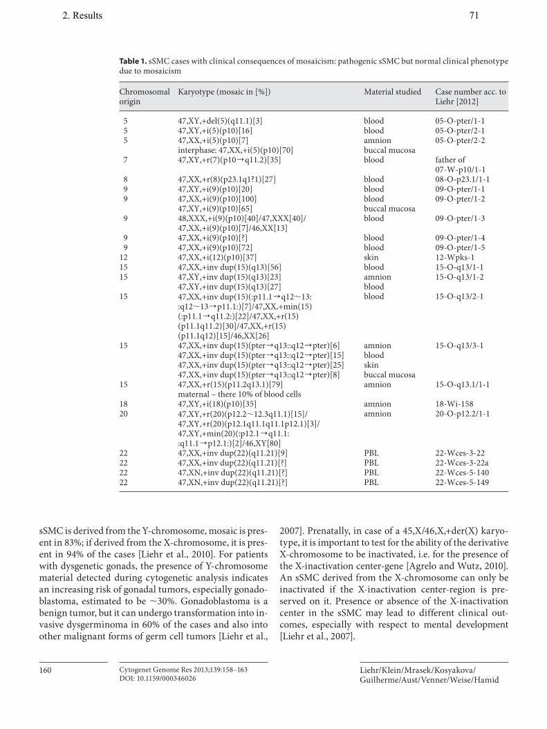
Liehr/Klein/Mrasek/Kosyakova/Guilherme/Aust/Venner/Weise/Hamid
Cytogenet Genome Res 2013;139:158–163 DOI: 10.1159/000346026
160
sSMC is derived from the Y-chromosome, mosaic is pres-ent in 83%; if derived from the X-chromosome, it is pres-ent in 94% of the cases [Liehr et al., 2010]. For patients with dysgenetic gonads, the presence of Y-chromosome material detected during cytogenetic analysis indicates an increasing risk of gonadal tumors, especially gonado-blastoma, estimated to be � 30%. Gonadoblastoma is a benign tumor, but it can undergo transformation into in-vasive dysgerminoma in 60% of the cases and also into other malignant forms of germ cell tumors [Liehr et al.,
2007]. Prenatally, in case of a 45,X/46,X,+der(X) karyo-type, it is important to test for the ability of the derivative X-chromosome to be inactivated, i.e. for the presence of the X-inactivation center-gene [Agrelo and Wutz, 2010]. An sSMC derived from the X-chromosome can only be inactivated if the X-inactivation center-region is pre-served on it. Presence or absence of the X-inactivation center in the sSMC may lead to different clinical out-comes, especially with respect to mental development [Liehr et al., 2007].
Table 1. s SMC cases with clinical consequences of mosaicism: pathogenic sSMC but normal clinical phenotype due to mosaicism
Chromosomal origin
Karyotype (mosaic in [%]) Material studied Case number acc. to Liehr [2012]
5 47,XY,+del(5)(q11.1)[3] blood 05-O-pter/1-15 47,XY,+i(5)(p10)[16] blood 05-O-pter/2-15 47,XX,+i(5)(p10)[7]
interphase: 47,XX,+i(5)(p10)[70]amnionbuccal mucosa
05-O-pter/2-2
7 47,XY,+r(7)(p10]q11.2)[35] blood father of 07-W-p10/1-1
8 47,XX,+r(8)(p23.1q1?1)[27] blood 08-O-p23.1/1-19 47,XY,+i(9)(p10)[20] blood 09-O-pter/1-19 47,XX,+i(9)(p10)[100]
47,XY,+i(9)(p10)[65]bloodbuccal mucosa
09-O-pter/1-2
9 48,XXX,+i(9)(p10)[40]/47,XXX[40]/47,XX,+i(9)(p10)[7]/46,XX[13]
blood 09-O-pter/1-3
9 47,XX,+i(9)(p10)[?] blood 09-O-pter/1-49 47,XX,+i(9)(p10)[72] blood 09-O-pter/1-5
12 47,XX,+i(12)(p10)[37] skin 12-Wpks-115 47,XX,+inv dup(15)(q13)[56] blood 15-O-q13/1-115 47,XY,+inv dup(15)(q13)[23]
47,XY,+inv dup(15)(q13)[27]amnionblood
15-O-q13/1-2
15 47,XX,+inv dup(15)(:p11.1]q12�13::q12�13]p11.1:)[7]/47,XX,+min(15)(:p11.1]q11.2:)[22]/47,XX,+r(15)(p11.1q11.2)[30]/47,XX,+r(15)(p11.1q12)[15]/46,XX[26]
blood 15-O-q13/2-1
15 47,XX,+inv dup(15)(pter]q13::q12]pter)[6]47,XX,+inv dup(15)(pter]q13::q12]pter)[15]47,XX,+inv dup(15)(pter]q13::q12]pter)[25]47,XX,+inv dup(15)(pter]q13::q12]pter)[8]
amnionbloodskinbuccal mucosa
15-O-q13/3-1
15 47,XX,+r(15)(p11.2q13.1)[79]maternal – there 10% of blood cells
amnion 15-O-q13.1/1-1
18 47,XY,+i(18)(p10)[35] amnion 18-Wi-15820 47,XY,+r(20)(p12.2�12.3q11.1)[15]/
47,XY,+r(20)(p12.1q11.1q11.1p12.1)[3]/47,XY,+min(20)(:p12.1]q11.1::q11.1]p12.1:)[2]/46,XY[80]
amnion 20-O-p12.2/1-1
22 47,XX,+inv dup(22)(q11.21)[9] PBL 22-Wces-3-2222 47,XX,+inv dup(22)(q11.21)[?] PBL 22-Wces-3-22a22 47,XN,+inv dup(22)(q11.21)[?] PBL 22-Wces-5-140 22 47,XN,+inv dup(22)(q11.21)[?] PBL 22-Wces-5-149
2. Results 71
ahamid
Textfeld
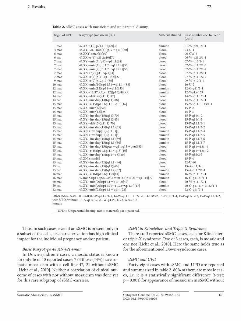
Somatic Mosaicism in sSMC Cytogenet Genome Res 2013;139:158–163 DOI: 10.1159/000346026
161
Thus, in such cases, even if an sSMC is present only in a subset of the cells, its characterization has high clinical impact for the individual pregnancy and/or patient.
Basic Karyotype 48,XN,+21,+mar In Down-syndrome cases, a mosaic status is known
for only 16 of 40 reported cases; 7 of those (44%) have so-matic mosaicism with a cell line 47,+21 without sSMC [Liehr et al., 2010]. Neither a correlation of clinical out-come of cases with nor without mosaicism was done yet for this rare subgroup of sSMC-carriers.
sSMC in Klinefelter- and Triple-X-Syndrome There are 3 reported sSMC-cases, each for Klinefelter-
or triple-X syndrome. Two of 3 cases, each, is mosaic and one not [Liehr et al., 2010]. Here the same holds true as for the aforementioned Down-syndrome cases.
sSMC and UPD Forty-eight cases with sSMC and UPD are reported
and summarized in table 2 . 80% of them are mosaic cas-es, i.e. it is a statistically significant difference (t-test:p = 0.001) for appearance of mosaicism in sSMC without
Table 2. s SMC cases with mosaicism and uniparental disomy
Origin of UPD Karyotype (mosaic in [%]) Material studied Case number acc. to Liehr[2012]
1 mat 47,XX,r(1)(::p21.1]q12)[3] amnion 01-W-p21.1/1-14 mat 48,XY,+21,+min(4)(:p12]q11:)[80] blood 04-U-16 mat 48,XXY,+mar(6)[60] blood 06-CW-36 pat 47,XX,+r(6)(p21.2q10)[74] blood 06-W-p21.2/1-17 mat 47,XY,+min(7)(p12]p11.1:)[8] blood 07-W-p12/1-17 mat 47,XY,+min(7)(:p11.2]q11.21:)[36] blood 07-W-p11.2/1-37 mat 47,XY,+min(7)(:p11.2]q11.21:)[56] blood 07-W-p11.2/1-47 mat 47,XN,+r(7)(p11.2q21)[4] blood 07-W-p11.2/2-17 mat 47,XX,+r(7)(p11.1q11.2?2)[27] blood 07-W-p11.1/2-29 mat 47,XX,+r(9)(p12q10)[36] blood 09-W-p12/1-1
10 mat 47,XX,+min(10)(:p12.31]q11.1:)[88] blood 10-U-212 mat 47,XX,+min(12)(:p11]q11:)[53] amnion 12-O-p11/1-112 mat 47,XX,+12/47,XX,+i(12)(p10)/46,XX amnion 12-Wpks-15914 mat 47,XY,+del(14)(q11.1)[87] blood 14-W-q11.1/3-114 pat 47,XX,+inv dup(14)(q11)[88] blood 14-W-q11.1/2-115 mat 47,XY,+r(15)(p11.1q11.1�q13)[16] blood 15-W-q11.1�13/1-115 mat 47,XX,+mar(X)[50] blood 15-P-215 mat 47,XX,+mar(15)[25] blood 15-P-315 mat 47,XY,+inv dup(15)(q11)[70] blood 15-P-q11/1-215 mat 47,XY,+inv dup(15)(q11)[45] blood 15-P-q11/1-515 mat 47,XY,+del(15)(q11.1)[70] blood 15-P-q11.1/1-115 mat 47,XX,+inv dup(15)(q11.1)[55] blood 15-P-q11.1/2-215 mat 47,XN,+inv dup(15)(q11.1)[?] amnion 15-P-q11.1/2-4 15 mat 47,XN,+inv dup(15)(q11.1)[?] amnion 15-P-q11.1/2-515 mat 47,XY,+inv dup(15)(q11.1)[39] amnion 15-P-q11.1/2-615 mat 47,XX,+inv dup(15)(q11.1)[50] amnion 15-P-q11.1/2-715 mat 47,XY,+inv dup(15)(pter]q11::q13]pter)[85] blood 15-P-q11�13/1-115 mat 47,XY,+r(15)(p11.1q11.1�q13)[16] blood 15-P-q11�13/1-215 mat 47,XX,+inv dup(15)(q12�13)[20] blood 15-P-q12/2-315 mat 47,XN,+mar[8] blood 15-P-415 mat 47,XY,+inv dup(22)(q11.1)[46] blood 22-U-4015 pat 47,XY,+inv dup(15)(q11)[60] blood 15-A-q11/1-115 pat 47,XY,+inv dup(15)(q11.2)[32] amnion 15-A-q11.2/1-116 mat 47,XY,+r(16)(p11.1q11.2)[84] amnion 16-W-p11.1/3-116 mat 47,inv(X)(p11.4p22.3)Y,+min(16)(:p11.21]q11.1:)[72] amnion 16-O-p11.21/1-120 mat 47,XY,+min(20)(:p11.1]q11.1:)[42] blood 20-W-p11.1/2-120 pat 47,XY,+min(20)(:p11.21�11.22]q11.1:)[17] amnion 20-O-p11.21�11.22/1-122 mat 47,XX,+min(22)(:p11.1]q11:)[22] blood 22-O-q11/2-1
Other sSMC cases with UPD, without mosaic
03-U-8; 07-W-p11.2/1-1; 14-W-q11.1�11.2/1-1; 14-CW-2; 15-P-q11/1-4; 15-P-q11/1-13; 15-P-q11.1/1-2, 15-A-q11/1-2; 20-W-p13/3-1; 22-Wces-5-81
U PD = Uniparental disomy; mat = maternal; pat = paternal.
2. Results 72
ahamid
Textfeld
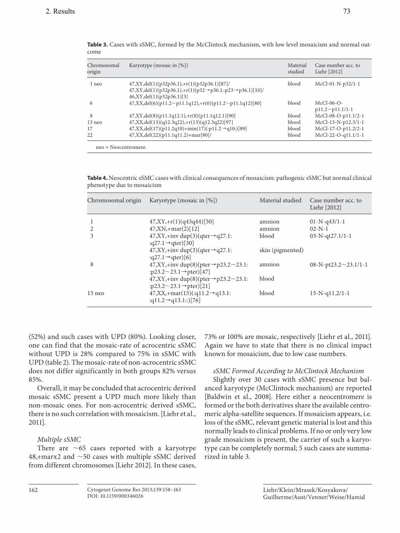
Liehr/Klein/Mrasek/Kosyakova/Guilherme/Aust/Venner/Weise/Hamid
Cytogenet Genome Res 2013;139:158–163 DOI: 10.1159/000346026
162
(52%) and such cases with UPD (80%). Looking closer, one can find that the mosaic-rate of acrocentric sSMC without UPD is 28% compared to 75% in sSMC with UPD ( table 2 ). The mosaic-rate of non-acrocentric sSMC does not differ significantly in both groups 82% versus 85%.
Overall, it may be concluded that acrocentric derived mosaic sSMC present a UPD much more likely than non-mosaic ones. For non-acrocentric derived sSMC, there is no such correlation with mosaicism. [Liehr et al., 2011].
Multiple sSMC There are � 65 cases reported with a karyotype
48,+marx2 and � 50 cases with multiple sSMC derived from different chromosomes [Liehr 2012]. In these cases,
73% or 100% are mosaic, respectively [Liehr et al., 2011]. Again we have to state that there is no clinical impact known for mosaicism, due to low case numbers.
sSMC Formed According to McClintock Mechanism Slightly over 30 cases with sSMC presence but bal-
anced karyotype (McClintock mechanism) are reported [Baldwin et al., 2008]. Here either a neocentromere is formed or the both derivatives share the available centro-meric alpha-satellite sequences. If mosaicism appears, i.e. loss of the sSMC, relevant genetic material is lost and this normally leads to clinical problems. If no or only very low grade mosaicism is present, the carrier of such a karyo-type can be completely normal; 5 such cases are summa-rized in table 3 .
Table 3. C ases with sSMC, formed by the McClintock mechanism, with low level mosaicism and normal out-come
Chromosomal origin
Karyotype (mosaic in [%]) Material studied
Case number acc. to Liehr [2012]
1 neo 47,XY,del(1)(p32p36.1),+r(1)(p32p36.1)[87]/47,XY,del(1)(p32p36.1),+r(1)(p32]p36.1::p23]p36.1)[10]/46,XY,del(1)(p32p36.1)[3]
blood McCl-01-N-p32/1-1
6 47,XX,del(6)(p11.2�p11.1q12),+r(6)(p11.2�p11.1q12)[80] blood McCl-06-O-p11.2�p11.1/1-1
8 47,XY,del(8)(p11.1q12.1),+r(8)(p11.1q12.1)[90] blood McCl-08-O-p11.1/2-113 neo 47,XX,del(13)(q12.3q22),+r(13)(q12.3q22)[97] blood McCl-13-N-p12.3/1-117 47,XX,del(17)(p11.2q10)+min(17)(:p11.2]q10:)[89] blood McCl-17-O-p11.2/2-122 47,XX,del(22)(p11.1q11.2)+mar[80]/ blood McCl-22-O-q11.1/1-1
n eo = Neocentromere.
Table 4. N eocentric sSMC cases with clinical consequences of mosaicism: pathogenic sSMC but normal clinical phenotype due to mosaicism
Chromosomal origin Karyotype (mosaic in [%]) Material studied Case number acc. to Liehr [2012]
1 47,XY,+r(1)(q43q44)[50] amnion 01-N-q43/1-12 47,XN,+mar(2)[12] amnion 02-N-13 47,XY,+inv dup(3)(qter]q27.1:
:q27.1]qter)[30]47,XY,+inv dup(3)(qter]q27.1::q27.1]qter)[6]
blood
skin (pigmented)
03-N-qt27.1/1-1
8 47,XY,+inv dup(8)(pter]p23.2�23.1::p23.2�23.1]pter)[47]47,XY,+inv dup(8)(pter]p23.2�23.1::p23.2�23.1]pter)[21]
amnion
blood
08-N-pt23.2�23.1/1-1
15 neo 47,XX,+mar(15)(:q11.2]q13.1::q11.2]q13.1::)[76]
blood 15-N-q11.2/1-1
2. Results 73
ahamid
Textfeld
Somatic Mosaicism in sSMC Cytogenet Genome Res 2013;139:158–163 DOI: 10.1159/000346026
163
Neocentric sSMC In at least 50% of the known � 100 neocentric sSMC
cases, somatic mosaicism is present. Strikingly, as in cen-tric sSMC, mosaicism is more frequent in non-acrocentric derived compared to acrocentric derived ones (58% vs. 24%) [Liehr et al., 2010]. In table 4 , the 5 known neocentric sSMC cases present in mosaic and normal clinical out-come are collected (i.e. in � 8% of the mosaic neocentric sSMC carriers, a normal clinical outcome is reported).
sSMC Carriers with Cryptic Mosaicism The real grade and complexity of mosaicism may be
even higher in � 5% of the sSMC cases, considering that recently cryptic mosaicism was repeatedly detected, which means sSMC cases can have more complex rearranged sSMC in mosaic than expected after cytogenetic analysis. Acrocentric derived sSMC are by far more stable than non-acrocentric derived ones (2 vs. 9%) [Liehr et al., 2010]. With this knowledge, clinical consequences are to be ex-pected because cryptic mosaics may lead e.g. to partial tet-ra- instead of trisomies. As it is known that trisomy 18p is tolerated hardly without any clinical signs, tetrasomy 18p, i.e. isochromosome-18p-syndrome, is associated with se-vere mental and physical problems. However, the finding of cryptic mosaics maybe to new, and thus, no correlation with this fact is possible in sSMC by now.
Summary and Conclusion
Somatic mosaicism is present in � 50% of the cases with sSMC. Acrocentric and non-acrocentric derived sSMC are differently susceptible to mosaicism. Acrocen-tric derived are the more stable ones, and surprisingly, this holds true for centric and neocentric sSMC. Also, there is an enhanced susceptibility for UPD formation in mosaic acrocentric- than in non-acrocentric-derived sSMC.
It has to be stressed that the only reliable approach to detect sSMC present in (low-level) mosaic is banding cy-togenetics. Array-CGH studies cannot be offered as a screening test to reliably detect this kind of chromosom-al aberration. Thus, especially when considering somatic mosaicism, cytogenetics is still the gold-standard to de-tect any kind of chromosomal aberration, which after-wards may be characterized in detail by molecular (cyto) genetic approaches.
Acknowledgements
This work was supported in parts by Else Kröner-Fresenius-Stiftung (2011_A42), ProChance 2008 and 2009, and Deutsche Forschungsgemeinschaft (DFG) (LI 820/22-1).
References
Agrelo R, Wutz A: ConteXt of change–X inacti-vation and disease. EMBO Mol Med 2: 6–15 (2010).
Baldwin EL, May LF, Justice AN, Martin CL, Ledbetter DH: Mechanisms and conse-quences of small supernumerary marker chromosomes: from Barbara McClintock to modern genetic-counseling issues. Am J Hum Genet 82: 398–410 (2008).
Fickelscher I, Starke H, Schulze E, Ernst G,Kosyakova N, et al: A further case with a small supernumerary marker chromosome (sSMC) derived from chromosome 1 – evi-dence for high variability in mosaicism in different tissues of sSMC carriers. Prenat Di-agn 27: 783–785 (2007).
Gebhart E, Liehr T: Clonality determined by flu-orescence in situ hybridization of single-cell aberrations in hematopoietic neoplasias. Cancer Genet Cytogenet 113: 193–194 (1999).
Iourov IY, Vorsanova SG, Yurov YB: Chromo-somal mosiacism goes global. Mol Cytogenet 1: 26 (2008).
Liehr T: Small supernumerary marker chromo-somes (sSMC). http://www.med.uni-jena.de/fish/sSMC/00START.htm (2012).
Liehr T, Weise A: Frequency of small supernu-merary marker chromosomes in prenatal, newborn, developmentally retarded and in-fertility diagnostics. Int J Mol Med 19: 719–731 (2007).
Liehr T, Claussen U, Starke H: Small supernu-merary marker chromosomes (sSMC) in hu-mans. Cytogenet Genome Res 107: 55–67 (2004).
Liehr T, Mrasek K, Weise A, Dufke A, Rodríguez L, et al: Small supernumerary marker chro-mosomes–progress towards a genotype-phe-notype correlation. Cytogenet Genome Res 112: 23–34 (2006).
Liehr T, Mrasek K, Hinreiner S, Reich D, Ewers E, et al: Small supernumerary marker chro-mosomes (sSMC) in patients with a 45,X/46,X,+mar karyotype – 17 new cases and a review of the literature. Sex Dev 1: 353–362 (2007).
Liehr T, Karamysheva T, Merkas M, Brecevic L, Hamid AB, et al: Somatic mosaicism in cases with small supernumerary marker chromo-somes. Curr Genomics 11: 432–439 (2010).
Liehr T, Ewers E, Hamid AB, Kosyakova N, Voigt M, et al: Small supernumerary marker chro-mosomes and uniparental disomy have a sto-ry to tell. J Histochem Cytochem 59: 842–848 (2011).
Mkrtchyan H, Gross M, Hinreiner S, Polytiko A, Manvelyan M, et al: Early embryonic chro-mosome instability results in stable mosaic pattern in human tissues. PLoS One 5:e9591 (2010).
Nelle H, Schreyer I, Ewers E, Mrasek K, Kosya-kova N, et al: Presence of harmless small su-pernumerary marker chromosomes ham-pers molecular genetic diagnosis: a case re-port. Mol Med Report 3: 571–574 (2010).
Yurov YB, Vorsanova SG, Iourov IY, Demidova IA, Beresheva AK, et al: Unexplained autism is frequently associated with low-level mo-saic aneuploidy. J Med Genet 44: 521–525 (2007).
Yurov YB, Vorsanova SG, Iourov IY: GIN’n’CIN hypothesis of brain aging: deciphering the role of somatic genetic instabilities and neu-ral aneuploidy during ontogeny. Mol Cyto-genet 2: 23 (2009).
2. Results 74
ahamid
Textfeld
2. Results
2.11. Article.10
Fernández-Toral J, Rodríguez L, Plasencia A, Martínez-Frías ML, Ewers E, Hamid AB, Ziegler M, Liehr T. Four small supernumerary marker chromosomes derived from chromosomes 6, 8, 11 and 12 in a patient with minimal clinical abnormalities: a case report. J Med Case Reports, 2010; 4:239.
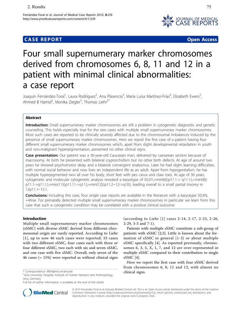
CASE REPORT Open Access
Four small supernumerary marker chromosomesderived from chromosomes 6, 8, 11 and 12 in apatient with minimal clinical abnormalities:a case reportJoaquín Fernández-Toral1, Laura Rodríguez2, Ana Plasencia3, María Luisa Martínez-Frías4, Elisabeth Ewers5,Ahmed B Hamid5, Monika Ziegler5, Thomas Liehr5*
Abstract
Introduction: Small supernumerary marker chromosomes are still a problem in cytogenetic diagnostic and geneticcounseling. This holds especially true for the rare cases with multiple small supernumerary marker chromosomes.Most such cases are reported to be clinically severely affected due to the chromosomal imbalances induced by thepresence of small supernumerary marker chromosomes. Here we report the first case of a patient having fourdifferent small supernumerary marker chromosomes which, apart from slight developmental retardation in youthand non-malignant hyperpigmentation, presented no other clinical signs.
Case presentation: Our patient was a 30-year-old Caucasian man, delivered by caesarean section because ofmacrosomy. At birth he presented with bilateral cryptorchidism but no other birth defects. At age of around twoyears he showed psychomotor delay and a bilateral convergent strabismus. Later he had slight learning difficulties,with normal social behavior and now lives an independent life as an adult. Apart from hypogenitalism, he hasmultiple hyperpigmented nevi all over his body, short feet with pes cavus and claw toes. At age of 30 years,cytogenetic and molecular cytogenetic analysis revealed a karyotype of 50,XY,+min(6)(:p11.1-> q11.1:),+min(8)(:p11.1->q11.1:),+min(11)(:p11.11->q11:),+min(12)(:p11.2~12->q10:), leading overall to a small partial trisomy in12p11.1~12.1.
Conclusions: Including this case, four single case reports are available in the literature with a karyotype 50,XN,+4mar. For prenatally detected multiple small supernumerary marker chromosomes in particular we learn from thiscase that such a cytogenetic condition may be correlated with a positive clinical outcome.
IntroductionMultiple small supernumerary marker chromosomes(sSMC) with diverse sSMC derived from different chro-mosomal origin are rarely reported. According to Liehr[1], up to now 46 such cases were reported: 33 caseswith two different sSMC, four cases each with three orfour different sSMC, two each with six and seven sSMC,and one case with five sSMC. Overall, only seven of the46 cases (= 15%) were reported as without clinical signs
(according to Liehr [1] cases 2-14, 2-17, 2-23, 2-26,2-29, 3-3 and 7-1).Patients with multiple sSMC constitute a sub-group of
patients with sSMC [2,3]. Little is known about the for-mation of sSMC in general [1-3] or about multiplesSMC specifically [4]. As reported previously, chromo-somes 6, 3, 5, X, 1, 7, and 12 are over-represented inmultiple sSMC compared to their contribution to singlesSMC [4].Here we report the first case with four sSMC derived
from chromosomes 6, 8, 11 and 12, with almost noclinical signs.* Correspondence: [email protected]
5Jena University Hospital, Institute of Human Genetics and Anthropology,Jena, GermanyFull list of author information is available at the end of the article
Fernández-Toral et al. Journal of Medical Case Reports 2010, 4:239http://www.jmedicalcasereports.com/content/4/1/239 JOURNAL OF MEDICAL
CASE REPORTS
© 2010 Fernández-Toral et al; licensee BioMed Central Ltd. This is an Open Access article distributed under the terms of the CreativeCommons Attribution License (http://creativecommons.org/licenses/by/2.0), which permits unrestricted use, distribution, andreproduction in any medium, provided the original work is properly cited.
2. Results 75
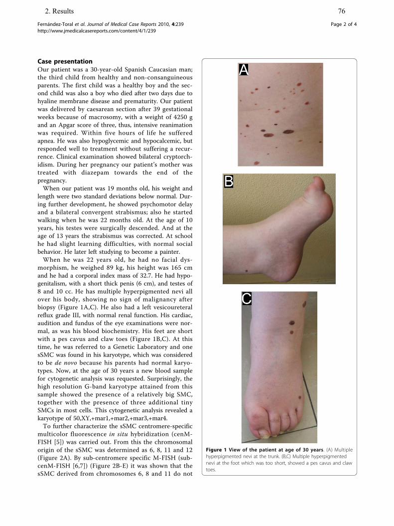
Case presentationOur patient was a 30-year-old Spanish Caucasian man;the third child from healthy and non-consanguineousparents. The first child was a healthy boy and the sec-ond child was also a boy who died after two days due tohyaline membrane disease and prematurity. Our patientwas delivered by caesarean section after 39 gestationalweeks because of macrosomy, with a weight of 4250 gand an Apgar score of three, thus, intensive reanimationwas required. Within five hours of life he sufferedapnea. He was also hypoglycemic and hypocalcemic, butresponded well to treatment without suffering a recur-rence. Clinical examination showed bilateral cryptorch-idism. During her pregnancy our patient’s mother wastreated with diazepam towards the end of thepregnancy.When our patient was 19 months old, his weight and
length were two standard deviations below normal. Dur-ing further development, he showed psychomotor delayand a bilateral convergent strabismus; also he startedwalking when he was 22 months old. At the age of 10years, his testes were surgically descended. And at theage of 13 years the strabismus was corrected. At schoolhe had slight learning difficulties, with normal socialbehavior. He later left studying to become a painter.When he was 22 years old, he had no facial dys-
morphism, he weighed 89 kg, his height was 165 cmand he had a corporal index mass of 32.7. He had hypo-genitalism, with a short thick penis (6 cm), and testes of8 and 10 cc. He has multiple hyperpigmented nevi allover his body, showing no sign of malignancy afterbiopsy (Figure 1A,C). He also had a left vesicoureteralreflux grade III, with normal renal function. His cardiac,audition and fundus of the eye examinations were nor-mal, as was his blood biochemistry. His feet are shortwith a pes cavus and claw toes (Figure 1B,C). At thistime, he was referred to a Genetic Laboratory and onesSMC was found in his karyotype, which was consideredto be de novo because his parents had normal karyo-types. Now, at the age of 30 years a new blood samplefor cytogenetic analysis was requested. Surprisingly, thehigh resolution G-band karyotype attained from thissample showed the presence of a relatively big SMC,together with the presence of three additional tinySMCs in most cells. This cytogenetic analysis revealed akaryotype of 50,XY,+mar1,+mar2,+mar3,+mar4.To further characterize the sSMC centromere-specific
multicolor fluorescence in situ hybridization (cenM-FISH [5]) was carried out. From this the chromosomalorigin of the sSMC was determined as 6, 8, 11 and 12(Figure 2A). By sub-centromere specific M-FISH (sub-cenM-FISH [6,7]) (Figure 2B-E) it was shown that thesSMC derived from chromosomes 6, 8 and 11 do not
Figure 1 View of the patient at age of 30 years. (A) Multiplehyperpigmented nevi at the trunk. (B,C) Multiple hyperpigmentednevi at the foot which was too short, showed a pes cavus and clawtoes.
Fernández-Toral et al. Journal of Medical Case Reports 2010, 4:239http://www.jmedicalcasereports.com/content/4/1/239
Page 2 of 4
2. Results 76
ahamid
Textfeld

contain any detectable euchromatic material. Only forthe derivative of chromosome 12 centromere-near mate-rial in 12p12.1 could be detected. The final karyotypewas 50,XY,+min(6)(:p11.1->q11.1:),+min(8)(:p11.1->q11.1:),+min(11)(:p11.11->q11:),+min(12)(:p11.2~12->q10:).
DiscussionHere we report the fourth unusual case with four differ-ent sSMC and the 34th case with multiple sSMC. It isthe eighth case with no or only minor clinical signs dueto the sSMC presence. The only detectable sSMC-related chromosomal imbalance is a small partial tris-omy 12p11.2~12.1. According to Liehr [8] there are sev-eral cases with a partial trisomy 12p12 due to an sSMCwhich were all clinically normal. Thus, this region seemsto be a potentially transmittable unbalanced chromoso-mal abnormality (UBCA) without causing clinical pro-blems (see case 12-O-p11.1/1-1 [8]). Similar UBCA wererecently reported for a multitude of chromosomalregions [9] and especially for the centromere nearregions [3]. Thus, it is not clear if the sSMC have apositive correlation with the observed clinical symptoms.Moreover, it is interesting that the multiple sSMC
derive in the present case from chromosomes 6, 8, 11and 12. Chromosomes 6 and 12 are over-represented inmultiple sSMC cases reported to date compared to theircontribution to single sSMC [4]. This might pointtowards a specific way of formation of multiple sSMCduring meiosis [10].
ConclusionsThe present case confirms that multiple sSMC may becorrelated with an almost normal clinical outcome. This
is especially important for the correct genetic counselingof similar pre-natal cases. Furthermore, a small partialtrisomy12p11.2~12.1 seems to correlate largely to no clinical
effects. Finally, involvement of chromosome 6 in sSMCformation seems to be correlated with the tendency ofmultiple sSMC formation.
ConsentWritten informed consent was obtained from the patientfor publication of this case report and any accompany-ing images. A copy of the written consent is availablefor review by the Editor-in-Chief of this journal.
AcknowledgementsSupported in parts by the DFG (LI 820/22-1) and DAAD (D07/00070).
Author details1Pediatría y jefe de sección de genética pediatrica del HUCA, Oviedo, Spain.2AbaCid-Genética Hospital de Madrid Norte Sanchinarro, Madrid, Spain.3Servicio de genética del HUCA. Oviedo, Spain. 4Estudio ColaborativoEspañol de Malformaciones Congénitas (ECEMC) del Centro de Investigaciónsobre Anomalías Congénitas (CIAC), Instituto de Salud Carlos III, Ministerio deSanidad y Consumo, Madrid, Spain. 5Jena University Hospital, Institute ofHuman Genetics and Anthropology, Jena, Germany.
Authors’ contributionsLR performed the cytogenetic studies in the present case. JFT and APcollected the data relative to this case report and provided geneticcounseling to the parents. MLMF supervised the cytogenetic analysis asDirector of the ECEMC. EE, ABH, MZ and TL did the molecular cytogeneticanalysis and interpretation. TL drafted the paper and all authors contributedto the finalizing of the manuscript.
Competing interestsThe authors declare that they have no competing interests.
Received: 29 October 2009 Accepted: 3 August 2010Published: 3 August 2010
References1. Liehr T: Small supernumerary marker chromosome (sSMC) homepage.
[http://www.med.uni-jena.de/fish/sSMC/00START.htm], Accessed on 7.October 2009..
2. Liehr T, Claussen U, Starke H: Small supernumerary marker chromosomes(sSMC) in humans. Cytogenet Genome Res 2004, 107:55-67.
3. Liehr T, Mrasek K, Weise A, Dufke A, Rodríguez L, Martínez Guardia N,Sanchís A, Vermeesch JR, Ramel C, Polityko A, Haas OA, Anderson J,Claussen U, von Eggeling F, Starke H: Small supernumerary markerchromosomes–progress towards a genotype-phenotype correlation.Cytogenet Genome Res 2006, 112:23-34.
4. Liehr T, Starke H, Senger G, Melotte C, Weise A, Vermeesch JR:Overrepresentation of small supernumerary marker chromosomes(sSMC) from chromosome 6 origin in cases with multiple sSMC. Am JMed Genet A 2006, 140:46-51.
5. Nietzel A, Rocchi M, Starke H, Heller A, Fiedler W, Wlodarska I, Loncarevic IF,Beensen V, Claussen U, Liehr T: A new multicolor-FISH approach for thecharacterization of marker chromosomes: centromere-specificmulticolor-FISH (cenM-FISH). Hum Genet 2001, 108:199-204.
6. Starke H, Nietzel A, Weise A, Heller A, Mrasek K, Belitz B, Kelbova C,Volleth M, Albrecht B, Mitulla B, Trappe R, Bartels I, Adolph S, Dufke A,Singer S, Stumm M, Wegner RD, Seidel J, Schmidt A, Kuechler A, Schreyer I,Claussen U, von Eggeling F, Liehr T: Small supernumerary markerchromosomes (SMCs): genotype-phenotype correlation andclassification. Hum Genet 2003, 114:51-67.
Figure 2 FISH results obtained on the chromosomes of thereported patient. (A) cenM-FISH revealed that the four sSMC werederivatives of chromosomes 6, 8, 11, and 12. (B-E) subcenM-FISHrevealed absence of euchromatic material in sSMC derived fromchromosomes 6, 8 and 11 and presence of centromere nearmaterial on the sSMC(12).
Fernández-Toral et al. Journal of Medical Case Reports 2010, 4:239http://www.jmedicalcasereports.com/content/4/1/239
Page 3 of 4
2. Results 77
ahamid
Textfeld
7. Mrasek K, Heller A, Rubtsov N, Trifonov V, Starke H, Claussen U, Liehr T:Detailed Hylobates lar karyotype defined by 25-color FISH andmulticolor banding. Int J Mol Med 2003, 12:139-146.
8. Liehr T: Small supernumerary marker chromosome (sSMC) homepage -subpage for sSMC derived from chromosome 12. [http://www.med.uni-jena.de/fish/sSMC/12.htm], Accessed on 7. October 2009..
9. Barber JC: UBCA anomaly register. [https://www.som.soton.ac.uk/research/Geneticsdiv/anomaly%20register/default.htm], Accessed on 7. October2009..
10. Mackie-Ogilvie C, Waddle K, Mandeville J, Seller MJ, Docherty Z: Rapididentification of multiple supernumerary ring chromosomes with a newFISH technique. J Med Genet 1997, 34:912-916.
doi:10.1186/1752-1947-4-239Cite this article as: Fernández-Toral et al.: Four small supernumerarymarker chromosomes derived from chromosomes 6, 8, 11 and 12 in apatient with minimal clinical abnormalities: a case report. Journal ofMedical Case Reports 2010 4:239.
Submit your next manuscript to BioMed Centraland take full advantage of:
• Convenient online submission
• Thorough peer review
• No space constraints or color figure charges
• Immediate publication on acceptance
• Inclusion in PubMed, CAS, Scopus and Google Scholar
• Research which is freely available for redistribution
Submit your manuscript at www.biomedcentral.com/submit
Fernández-Toral et al. Journal of Medical Case Reports 2010, 4:239http://www.jmedicalcasereports.com/content/4/1/239
Page 4 of 4
2. Results 78
ahamid
Textfeld
2. Results
2.12. Article.11
Papoulidis I, Manolakos E, Hamid AB, Klein E, Kosyakova N, Kordaß U, Kunz J, Siomou E, Kontodiou M, Tzimina M, Nicolaides P, Liehr T, Petersen MB. Tetrasomy 9p mosaicism associated with a normal phenotype in two cases. Cytogenet Genome Res, 2012; 136:237–241.

Fax +41 61 306 12 34E-Mail [email protected]
Original Article
Cytogenet Genome Res 2012;136:237–241 DOI: 10.1159/000337520
Tetrasomy 9p Mosaicism Associated with a Normal Phenotype in Two Cases
I. Papoulidis a M. Kontodiou a M. Tzimina a I. Saitis b A.B. Hamid c E. Klein c
N. Kosyakova c U. Kordaß d J. Kunz e E. Siomou a P. Nicolaides f S. Orru g
L. Thomaidis h T. Liehr c M.B. Petersen a E. Manolakos a, g
a Eurogenetica S.A., Thessaloniki and Athens , b Genemed, Heraklion , Greece; c Institute of Human Genetics,Jena University Hospital, Jena , d Institute of Human Genetics, Greifswald University Hospital, Greifswald , e Institute of Human Genetics, Charité, Berlin , Germany; f Emvrioiatriki S.A., Athens , Greece; g Cattedra diGenetica Medica, Ospedale Binaghi, Universita di Cagliari, Cagliari, Italy; h Developmental Assessment Unit,2nd Department of Pediatrics, P. & A. Kyriakou Children’s Hospital, National and Kapodistrian University ofAthens School of Medicine, Athens , Greece
Small supernumerary marker chromosomes (sSMCs) are reported in 0.044% of newborn infants and in 0.125% of subfertile individuals [Liehr and Weise, 2007]. sSMCs are structurally abnormal chromosomes that cannot be identified or characterized unambiguously by conven-tional cytogenetics alone, and they are generally equal in size or smaller than chromosome 20 of the same meta-phase spread [Liehr et al., 2004]. To date, only one report is available for a triple-X syndrome patient with an addi-tional sSMC [Lee-Jones et al., 2004], and only 3 cases have been reported so far with mosaic tetrasomy 9p that pres-ent no clinical symptoms [Sait and Wetzler, 2003; McAu-liffe et al., 2005; Baronchelli et al., 2011].
In the present study, we report 2 patients with tetra-somy 9p mosaicism and an apparently normal pheno-type. The first individual was cytogenetically studied be-cause of a de novo inversion in a chromosome 7, observed in a previous pregnancy. The second proband was re-ferred for cytogenetic studies as part of in vitro fertiliza-tion (IVF) pre-testing due to her husband’s azoospermia. The results of the molecular, clinical, and cytogenetic findings are presented and compared to reports previ-ously published.
Key Words
Mosaicism � Normal phenotype � Tetrasomy 9p
Abstract
Tetrasomy 9p is a rare chromosomal syndrome and about 30% of known cases exhibit mosaicism. Approximately 50 of the reported cases with tetrasomy 9p mosaicism show a characteristic facial appearance, growth failure, and devel-opmental delay. However, 3 patients with mosaicism for iso-chromosome 9p and a normal phenotype have also been reported. We report 2 additional cases of clinically normal young females with tetrasomy 9p mosaicism, one of whom also exhibited X chromosome aneuploidy mosaicism lead-ing to an overall of 6 different cell lines. STR analysis per-formed on this complex mosaic case indicated that the extra isochromosome was of maternal origin while the X chromo-some aneuploidy was of paternal origin, indicating a postzy-gotic event. Copyright © 2012 S. Karger AG, Basel
Accepted: February 13, 2012 by M. Schmid Published online: April 5, 2012
E. Manolakos Eurogenetica S.A. Alexandroupoleos 23, Ampelokipi GR–115 27 Athens (Greece) Tel. +30 21 0747 4904, E-Mail emanolakosgr @ yahoo.gr
© 2012 S. Karger AG, Basel1424–8581/12/1364–0237$38.00/0
Accessible online at:www.karger.com/cgr
2. Results 79
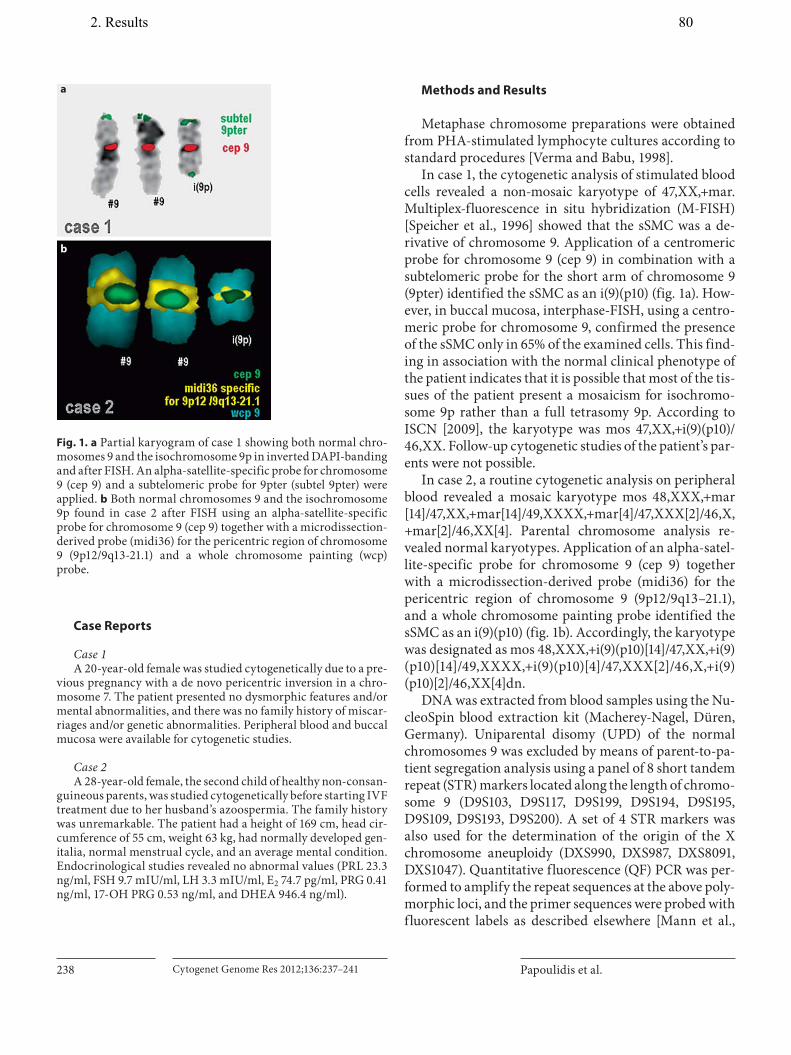
Papoulidis et al. Cytogenet Genome Res 2012;136:237–241238
Case Reports
Case 1 A 20-year-old female was studied cytogenetically due to a pre-
vious pregnancy with a de novo pericentric inversion in a chro-mosome 7. The patient presented no dysmorphic features and/or mental abnormalities, and there was no family history of miscar-riages and/or genetic abnormalities. Peripheral blood and buccal mucosa were available for cytogenetic studies.
Case 2 A 28-year-old female, the second child of healthy non-consan-
guineous parents, was studied cytogenetically before starting IVF treatment due to her husband’s azoospermia. The family history was unremarkable. The patient had a height of 169 cm, head cir-cumference of 55 cm, weight 63 kg, had normally developed gen-italia, normal menstrual cycle, and an average mental condition. Endocrinological studies revealed no abnormal values (PRL 23.3 ng/ml, FSH 9.7 mIU/ml, LH 3.3 mIU/ml, E 2 74.7 pg/ml, PRG 0.41 ng/ml, 17-OH PRG 0.53 ng/ml, and DHEA 946.4 ng/ml).
Methods and Results
Metaphase chromosome preparations were obtained from PHA-stimulated lymphocyte cultures according to standard procedures [Verma and Babu, 1998].
In case 1, the cytogenetic analysis of stimulated blood cells revealed a non-mosaic karyotype of 47,XX,+mar. Multiplex-fluorescence in situ hybridization (M-FISH) [Speicher et al., 1996] showed that the sSMC was a de-rivative of chromosome 9. Application of a centromeric probe for chromosome 9 (cep 9) in combination with a subtelomeric probe for the short arm of chromosome 9 (9pter) identified the sSMC as an i(9)(p10) ( fig. 1 a). How-ever, in buccal mucosa, interphase-FISH, using a centro-meric probe for chromosome 9, confirmed the presence of the sSMC only in 65% of the examined cells. This find-ing in association with the normal clinical phenotype of the patient indicates that it is possible that most of the tis-sues of the patient present a mosaicism for isochromo-some 9p rather than a full tetrasomy 9p. According to ISCN [2009], the karyotype was mos 47,XX,+i(9)(p10)/46,XX. Follow-up cytogenetic studies of the patient’s par-ents were not possible.
In case 2, a routine cytogenetic analysis on peripheral blood revealed a mosaic karyotype mos 48,XXX,+mar[14]/47,XX,+mar[14]/49,XXXX,+mar[4]/47,XXX[2]/46,X,+mar[2]/46,XX[4]. Parental chromosome analysis re-vealed normal karyotypes. Application of an alpha-satel-lite-specific probe for chromosome 9 (cep 9) together with a microdissection-derived probe (midi36) for the pericentric region of chromosome 9 (9p12/9q13–21.1), and a whole chromosome painting probe identified the sSMC as an i(9)(p10) ( fig. 1 b). Accordingly, the karyotype was designated as mos 48,XXX,+i(9)(p10)[14]/47,XX,+i(9)(p10)[14]/49,XXXX,+i(9)(p10)[4]/47,XXX[2]/46,X,+i(9)(p10)[2]/46,XX[4]dn.
DNA was extracted from blood samples using the Nu-cleoSpin blood extraction kit (Macherey-Nagel, Düren, Germany). Uniparental disomy (UPD) of the normal chromosomes 9 was excluded by means of parent-to-pa-tient segregation analysis using a panel of 8 short tandem repeat (STR) markers located along the length of chromo-some 9 (D9S103, D9S117, D9S199, D9S194, D9S195, D9S109, D9S193, D9S200). A set of 4 STR markers was also used for the determination of the origin of the X chromosome aneuploidy (DXS990, DXS987, DXS8091, DXS1047). Quantitative fluorescence (QF) PCR was per-formed to amplify the repeat sequences at the above poly-morphic loci, and the primer sequences were probed with fluorescent labels as described elsewhere [Mann et al.,
a
b
Fig. 1. a Partial karyogram of case 1 showing both normal chro-mosomes 9 and the isochromosome 9p in inverted DAPI-banding and after FISH. An alpha-satellite-specific probe for chromosome 9 (cep 9) and a subtelomeric probe for 9pter (subtel 9pter) were applied. b Both normal chromosomes 9 and the isochromosome 9p found in case 2 after FISH using an alpha-satellite-specific probe for chromosome 9 (cep 9) together with a microdissection-derived probe (midi36) for the pericentric region of chromosome 9 (9p12/9q13-21.1) and a whole chromosome painting (wcp) probe.
2. Results 80
ahamid
Textfeld
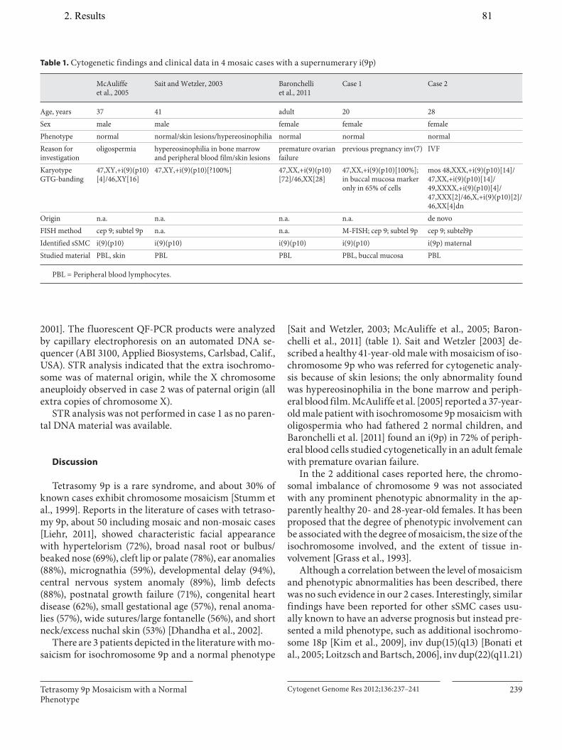
Tetrasomy 9p Mosaicism with a Normal Phenotype
Cytogenet Genome Res 2012;136:237–241 239
2001]. The fluorescent QF-PCR products were analyzed by capillary electrophoresis on an automated DNA se-quencer (ABI 3100, Applied Biosystems, Carlsbad, Calif., USA). STR analysis indicated that the extra isochromo-some was of maternal origin, while the X chromosome aneuploidy observed in case 2 was of paternal origin (all extra copies of chromosome X).
STR analysis was not performed in case 1 as no paren-tal DNA material was available.
Discussion
Tetrasomy 9p is a rare syndrome, and about 30% of known cases exhibit chromosome mosaicism [Stumm et al., 1999]. Reports in the literature of cases with tetraso-my 9p, about 50 including mosaic and non-mosaic cases [Liehr, 2011], showed characteristic facial appearance with hypertelorism (72%), broad nasal root or bulbus/beaked nose (69%), cleft lip or palate (78%), ear anomalies (88%), micrognathia (59%), developmental delay (94%), central nervous system anomaly (89%), limb defects (88%), postnatal growth failure (71%), congenital heart disease (62%), small gestational age (57%), renal anoma-lies (57%), wide sutures/large fontanelle (56%), and short neck/excess nuchal skin (53%) [Dhandha et al., 2002].
There are 3 patients depicted in the literature with mo-saicism for isochromosome 9p and a normal phenotype
[Sait and Wetzler, 2003; McAuliffe et al., 2005; Baron-chelli et al., 2011] ( table 1 ). Sait and Wetzler [2003] de-scribed a healthy 41-year-old male with mosaicism of iso-chromosome 9p who was referred for cytogenetic analy-sis because of skin lesions; the only abnormality found was hypereosinophilia in the bone marrow and periph-eral blood film. McAuliffe et al. [2005] reported a 37-year-old male patient with isochromosome 9p mosaicism with oligospermia who had fathered 2 normal children, and Baronchelli et al. [2011] found an i(9p) in 72% of periph-eral blood cells studied cytogenetically in an adult female with premature ovarian failure.
In the 2 additional cases reported here, the chromo-somal imbalance of chromosome 9 was not associated with any prominent phenotypic abnormality in the ap-parently healthy 20- and 28-year-old females. It has been proposed that the degree of phenotypic involvement can be associated with the degree of mosaicism, the size of the isochromosome involved, and the extent of tissue in-volvement [Grass et al., 1993].
Although a correlation between the level of mosaicism and phenotypic abnormalities has been described, there was no such evidence in our 2 cases. Interestingly, similar findings have been reported for other sSMC cases usu-ally known to have an adverse prognosis but instead pre-sented a mild phenotype, such as additional isochromo-some 18p [Kim et al., 2009], inv dup(15)(q13) [Bonati et al., 2005; Loitzsch and Bartsch, 2006], inv dup(22)(q11.21)
Table 1. Cytogenetic findings and clinical data in 4 mosaic cases with a supernumerary i(9p)
McAuliffeet al., 2005
Sait and Wetzler, 2003 Baronchelliet al., 2011
Case 1 Case 2
Age, years 37 41 adult 20 28Sex male male female female femalePhenotype normal normal/skin lesions/hypereosinophilia normal normal normalReason for investigation
oligospermia hypereosinophilia in bone marrowand peripheral blood film/skin lesions
premature ovarian failure
previous pregnancy inv(7) IVF
KaryotypeGTG-banding
47,XY,+i(9)(p10)[4]/46,XY[16]
47,XY,+i(9)(p10)[?100%] 47,XX,+i(9)(p10)[72]/46,XX[28]
47,XX,+i(9)(p10)[100%];in buccal mucosa markeronly in 65% of cells
mos 48,XXX,+i(9)(p10)[14]/47,XX,+i(9)(p10)[14]/49,XXXX,+i(9)(p10)[4]/47,XXX[2]/46,X,+i(9)(p10)[2]/46,XX[4]dn
Origin n.a. n.a. n.a. n.a. de novoFISH method cep 9; subtel 9p n.a. n.a. M-FISH; cep 9; subtel 9p cep 9; subtel9pIdentified sSMC i(9)(p10) i(9)(p10) i(9)(p10) i(9)(p10) i(9p) maternalStudied material PBL, skin PBL PBL PBL, buccal mucosa PBL
P BL = Peripheral blood lymphocytes.
2. Results 81
ahamid
Textfeld
Papoulidis et al. Cytogenet Genome Res 2012;136:237–241240
leading generally to cat eye syndrome [Lin et al., 2006], or even isochromosome 12p leading to Pallister-Killian syn-drome [Genevieve et al., 2003]. In our case 1, no mosa-icism was evident after studying blood lymphocytes; it became obvious only after interphase-FISH of the buccal mucosa. Still, only a few tissues were studied in both of our cases.
The supernumerary isochromosome 9p in case 2 was a de novo finding as in all of the so far described cases [Dutly et al., 1998; Eggerman et al., 1998; Wyandt et al., 2000] and of maternal origin. It seems that errors in ma-ternal meiosis may be responsible for the origin of the isochromosome and that non-disjunction during meiosis II could be followed by rearrangements leading to dupli-cation of the short arm and loss of the long arm in the majority of cases [Dutly et al., 1998].
For sSMCs in general, the predominant mechanism of origin has been shown to be ring chromosome formation by centromere misdivision, the so-called McClintock mechanism [Baldwin et al., 2008].
Trisomy X occurs from a non-disjunction event in which the X chromosomes fail to properly separate dur-ing cell division, either during gametogenesis or after conception [May et al., 1990]. Studies made to determine the parental origin of the additional X chromosome dem-onstrated that in 58–63% of cases the extra X chromo-some derived from maternal meiosis I errors, in 16–17% from maternal meiosis II errors, and in 18–20% from post-zygotic non-disjunction [Hall et al., 2006; Hassold et al., 2007]. One study [Wallerstein et al., 2004] with mo-
saic trisomy X (such as 45,X/47,XXX) suggested that cas-es of mosaicism may result from a post-zygotic non-dis-junction event as could be the cause in our case 2. This case presented with a normal stature, while women with a mosaic karyotype of 45,X/47,XXX generally develop a short stature [Syber and McCauley, 2004]. The severity of the short stature has been correlated with the distribution of cell lines in 47,XXX/45,X/46,XX mosaicism [Partsch et al., 1994].
Mosaicism for tetrasomy 9p is a challenging issue in terms of prenatal diagnosis and genetic counseling as the abnormality may not be detectable in the amniotic fluid and fetal ultrasound assessment can be normal through-out pregnancy. In one reported case, amniocentesis due to advanced maternal age showed a normal fetal karyo-type. However, further cytogenetic analysis due to post-natal developmental delay revealed mosaic tetrasomy 9p in blood and skin cells [Eggermann et al., 1998].
Our 2 cases of healthy females can be regarded as rep-resenting the one end of the spectrum of karyotype-phe-notype correlation in chromosomal aneuploidies [Avra-mopoulos et al., 1997]. In most such cases, however, tis-sue-specific mosaicism has not been fully investigated.
Acknowledgments
This article was supported in parts by the Else-Kröner-Frese-nius Stiftung and the DAAD.
References
Avramopoulos D, Kennerknecht I, Barbi G, Eck-ert D, Delabar JM, et al: A case of apparent trisomy 21 without the Down’s syndrome phenotype. J Med Genet 34: 597–600 (1997).
Baldwin EL, May LF, Justice AN, Martin CL, Ledbetter DH: Mechanisms and conse-quences of small supernumerary marker chromosomes: from Barbara McClintock to modern genetic-counselling issues. Am J Hum Genet 82: 398–410 (2008).
Baronchelli S, Conconi D, Panzeri E, Bentivegna A, Redaelli S, et al: Cytogenetics of prema-ture ovarian failure: an investigation on 269 affected women. J Biomed Biotechnol 2011: 370195 (2011).
Bonati MT, Finelli P, Giardino D, Gottardi G, Roberts W, Larizza L: Trisomy 15q25.2-qter in an autistic child: genotype-phenotype correlations. Am J Med Genet A 133: 184–188 (2005).
Dhandha S, Hogge WA, Surti U, McPherson E: Three cases of tetrasomy 9p. Am J Med Gen-et 113: 375–380 (2002).
Dutly F, Balmer D, Baumer A, Binkert F, Schinzel A: Isochromosomes 12p and 9p: parental or-igin and possible mechanisms of formation. Eur J Hum Genet 6: 140–144 (1998).
Eggermann T, Rossier E, Theurer-Mainka U, Backsch C, Klein-Vogler U, et al: New case of mosaic tetrasomy 9p with additional neuro-metabolic findings. Am J Med Genet 75: 530–533 (1998).
Genevieve D, Cormier-Daire V, Sanlaville D, Faivre L, Gosset P, et al: Mild phenotype in a 15-year-old boy with Pallister-Killian syn-drome. Am J Med Genet 116A:90–93 (2003).
Grass FS, Parke JC Jr, Kirkman HN, Christensen V, Roddey OF, et al: Tetrasomy 9p: tissue-limited idic(9p) in a child with mild manifes-tations and a normal CVS result. Report and review. Am J Med Genet 47: 812–816 (1993).
Hall H, Hunt P, Hassold T: Meiosis and sex chro-mosome aneuploidy: how meiotic errors cause aneuploidy; how aneuploidy causes meiotic errors. Curr Opin Genet Dev 16: 323–329 (2006).
Hassold T, Hall H, Hunt P: The origin of human aneuploidy: where we have been, where we are going. Hum Mol Genet 16 Spec No. 2:R203–208 (2007).
ISCN 2009: An International System for Human Cytogenetic Nomenclature (200p), Shaffer LG, Slovak ML, Campbel LJ (eds) (S. Karger, Basel 2009).
Kim M, Park C, Park S, Kim M, Lee B, et al: Pre-natal diagnosis of a de novo mosaic isochro-mosome 18p: karyotype discordance be-tween amniocytes and fetal/neonatal blood. ASHG 2009; abstract only online, informa-tion from poster.
2. Results 82
ahamid
Textfeld

Tetrasomy 9p Mosaicism with a Normal Phenotype
Cytogenet Genome Res 2012;136:237–241 241
Lee-Jones L, Williams T, Little E, Sampson J: Tri-somy 14pter ] q21: a case with associated ovarian germ cell tumor and review of the literature. Am J Med Genet A 128A:78–84 (2004).
Liehr T: Small supernumerary marker chromo-somes. http://www.fish.uniklinikum-jena.de/sSMC.html. [accessed 20/02/2012].
Liehr T, Weise A: Frequency of small supernu-merary marker chromosomes in prenatal, newborn, developmentally retarded and in-fertility diagnostics. Int J Mol Med 19: 719–731 (2007).
Liehr T, Claussen U, Starke H: Small supernumer-ary marker chromosomes (sSMC) in humans. Cytogenet Genome Res 107: 55–67 (2004).
Lin CC, Hsieh YY, Wang CH, Li YC, Hsieh LJ, et al: Prenatal detection and characterization of a small supernumerary marker chromo-some (sSMC) derived from chromosome 22 with apparently normal phenotype. Prenat Diagn 26: 898–902 (2006).
Loitzsch A, Bartsch O: Healthy 12-year-old boy with mosaic inv dup(15)(q13). Am J Med Genet A 140: 640–643 (2006).
Mann K, Fox SP, Abbs SJ, Yau SC, Scriven PN, et al: Development and implementation of a new rapid aneuploidy diagnostic service within the UK National Health Service and implications for the future of prenatal diag-nosis. Lancet 358: 1057–1061 (2001).
May KM, Jacobs PA, Lee M, Ratcliffe S, Robinson A, et al: The parental origin of the extra X chromosome in 47,XXX females. Am J Hum Genet 46: 754–761 (1990).
McAuliffe F, Winsor EJ, Chitayat D: Tetrasomy 9p mosaicism associated with a normal phe-notype. Fetal Diagn Ther 20: 219–222 (2005).
Partsch CJ, Pankau R, Sippell WG, Tolksdorf M: Normal growth and normalization of hyper-gonadotropic hypogonadism in atypical Turner syndrome (45,X/46,XX/47,XXX). Correlation of body height with distribution of cell lines. Eur J Pediatr 153: 451–455 (1994).
Sait SNJ, Wetzler M: Tetrasomy 9p with no ap-parent phenotype characteristics (abstract), 53th ASHG Annual Meeting: Nr 678 (2003).
Speicher MR, Gwyn Ballard S, Ward DC: Karyo-typing human chromosomes by combinato-rial multi-fluor FISH. Nat Genet 12: 368–375 (1996).
Stumm M, Tönnies H, Mandon U, Götze A, Krebs P, Wieacker PF: Mosaic tetrasomy 9p in a girl with multiple congenital anomalies: cytogenetic and molecular-cytogenetic studies. Eur J Pediatr 158: 571–575 (1999).
Syber VR, McCauley E: Turner’s syndrome. N Engl J Med 351: 1227–1238 (2004).
Verma RS, Babu A: Human Chromosomes – Manual of Basis Technologies, ed 4, pp 6–71 (Pergamon Press, New York 1998).
Wallerstein R, Musen E, McCarrier J, Aisenberg J, Chartoff A, et al: Turner syndrome pheno-type with 47,XXX karyotype: further inves-tigation warranted? Am J Med Genet A 125A:106–107 (2004).
Wyandt HE, Lebo RV, Fenerci EY, Sadhu DN, Milunsky JM: Tandem duplication/deletion in a maternally derived chromosome 9 su-pernumerary derivative resulting in 9p tri-somy and partial 9q tetrasomy. Am J Med Genet 93: 305–312 (2000).
Notes Added in Proof
A more intense literature search re-vealed four (4) additional i(9p) cases with normal phenotype. For more details visit: http://www.fish.uniklinikum-jena.de/sSMC/sSMC+by+chromosome/sSMC+9.html#i9p).
2. Results 83
ahamid
Textfeld
3. Discussion 84
3. Discussion
As mentioned in Introduction part: Detection of an sSMC is nearly always unexpected by the
clinician and more or less an accidental result in cytogenetics; the origin of an sSMC is almost
impossible to establish by routine cytogenetics alone, whereas FISH methods are highly
suited for this (Starke et al. 2003a). The majority of sSMC comprise exclusively of material
derived from one chromosome. Of those, only a very small subset does not consist of
consecutive chromosomal material, but has complex intrachromosomal rearrangements (Liehr
et al. 2004a). Approximately 30% of the marker chromosome cases are familial and they
impose a low risk of abnormality (Crolla et al. 1998, Hastings et al. 1999), whereas ~30% de
novo cases present statistically an increased risk of inborn defects due to sSMC (Warburton
1984, Liehr 2014a). In prenatal clinical cytogenetics, sSMC are still a problem: the question
is, has it been correlated with clinical syndromes and is it harmful? Thus, in this study I
worked on
1) better sSMC characterization approaches,
2) characterization of chromosomal breakpoints involved in sSMC formation,
3) on mosaicism in sSMC, and overall,
4) on a refinement of the genotype-phenotype correlation in sSMC.
3.1. Development of probe sets for detection of euchromatic presence in sSMC
To obtain additional information regarding genotype-phenotype correlations, sSMC need to
characterized as precisely as possible. Several FISH-based techniques have been developed
during the last decades to achieve this end (Liehr 2014b). Specific probe sets were suggested
to detect the presence of euchromatic on an sSMC after identification of its chromosomal
origin (e.g. by cenM-FISH (Nietzel et al. 2001)). Strikingly euchromatin can be present on an
sSMC and must not cause any harm in the carrier; it depends which exact genetic imbalance
was induced. As above mentioned, a detailed sSMC characterization is especially necessary in
prenatal cases (Liehr 2014a).
In (article 1) we reviewed the effectiveness of multicolor FISH (mFISH) methods in current
clinical diagnostics. mFISH is defined as the simultaneous use of at least three different
ligands or fluorochromes for the specific labeling of DNA – excluding the counterstain (Liehr
et al. 2004c). Due to this definition, the first successful mFISH experiments were performed
in 1989 (Nederlof et al. 1989). aCGH is an efficient and sensitive technique for detecting
genome-wide copy number alterations at high resolution (Shaffer et al. 2007), and can narrow
down chromosomal breakpoints to some 10 kb or less (Weise et al. 2008). Also aCGH now
3. Discussion 85
provides accurate characterization of sSMCs in terms of chromosomal origin, gene content,
and other concomitant imbalances elsewhere in the genome (Reddy et al. 2013). Moreover
sSMCs have to be differentiated from insertions or unbalanced translocations, and individual
combinations of locus-specific (BAC) probes are used to prove or contradict a gain or loss
suggested after aCGH (Liehr et al. 2006a, Kumar et al. 2010). All available probe sets like
whole chromosome painting mFISH probe sets (Liehr and Claussen 2002a,b, Liehr et al.
2004c), cenM-FISH (Nietzel et al. 2001), subcenM-FISH (Liehr et al. 2006c) or FISH-
banding (Weise et al. 2002, Starke et al. 2003a) and use of locus specific probes enabled a
better sSMC characterization than banding cytogenetics (Weise et al. 2002, Manvelyan et al.
2007). Also sSMCs have been successfully characterized by glass needle-based chromosome
microdissection and reverse chromosome painting (Starke et al. 2001) or use of the sSMC-
derived DNA in aCGH (article 6).
For the present work combinations of these approaches were very successfully applied for the
characterization of a complex sSMC derived from chromosomes 14 and 8, as der(14)t(8;
14)(p23.2;q22.1)dn in (article 2). And also they were applied for our comprehensive study
(article 3) in 412 reported complex sSMC. Also major parts of the 5,200 sSMC cases
collected in our sSMC database (Liehr 2014a) were characterized by the standard approaches
mentioned in the previous paragraph.
Still aCGH and mentioned mFISH-approaches and probes-sets were not suited for the
comprehensive characterization of sSMC in each case as each technique has its limitations.
M-FISH include the inability to detect most intra-chromosomal abnormalities such as
inversions and inter-chromosomal anomalies especially if they are 3 Mb or less (Fan 2002).
And although aCGH is a more sensitive technique, which can significantly, narrow down
sSMC breakpoints, available ‘chips’ often do not completely cover the pericentromeric
regions and furthermore cannot detect low-level and/or cryptic mosaic sSMCs (Ballif et al.
2006, Ballif et al. 2007, Baldwin et al. 2008, Li and Andersson 2009, Sheth et al. 2011, Vetro
et al. 2012, Reddy et al. 2013). Thus, part of the present work was to establish the pericentric-
ladder-FISH (PCL-FISH) probe set (article 4), which is especially suited to narrow down
chromosomal breakpoints in derivative chromosomes of known origin, including sSMC. This
probe set has been used in dual-color/multicolor-FISH approaches, and it is a specific tool for
the pericentromeric regions, which enables sSMC breakpoint characterization with a
resolution between 1 and ~10 Mb. Pericentromeric regions of human chromosomes are
transitional territories between centromeric heterochromatin and euchromatic regions. They
represent complex mosaic structures, including coding sequences interspersed with non-
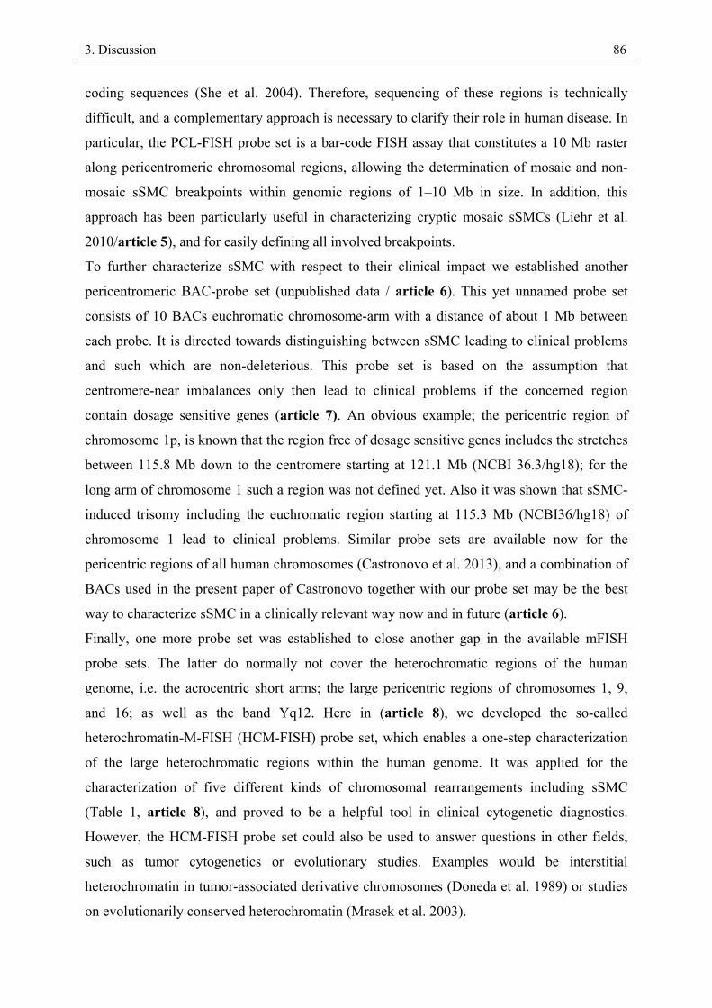
3. Discussion 86
coding sequences (She et al. 2004). Therefore, sequencing of these regions is technically
difficult, and a complementary approach is necessary to clarify their role in human disease. In
particular, the PCL-FISH probe set is a bar-code FISH assay that constitutes a 10 Mb raster
along pericentromeric chromosomal regions, allowing the determination of mosaic and non-
mosaic sSMC breakpoints within genomic regions of 1–10 Mb in size. In addition, this
approach has been particularly useful in characterizing cryptic mosaic sSMCs (Liehr et al.
2010/article 5), and for easily defining all involved breakpoints.
To further characterize sSMC with respect to their clinical impact we established another
pericentromeric BAC-probe set (unpublished data / article 6). This yet unnamed probe set
consists of 10 BACs euchromatic chromosome-arm with a distance of about 1 Mb between
each probe. It is directed towards distinguishing between sSMC leading to clinical problems
and such which are non-deleterious. This probe set is based on the assumption that
centromere-near imbalances only then lead to clinical problems if the concerned region
contain dosage sensitive genes (article 7). An obvious example; the pericentric region of
chromosome 1p, is known that the region free of dosage sensitive genes includes the stretches
between 115.8 Mb down to the centromere starting at 121.1 Mb (NCBI 36.3/hg18); for the
long arm of chromosome 1 such a region was not defined yet. Also it was shown that sSMC-
induced trisomy including the euchromatic region starting at 115.3 Mb (NCBI36/hg18) of
chromosome 1 lead to clinical problems. Similar probe sets are available now for the
pericentric regions of all human chromosomes (Castronovo et al. 2013), and a combination of
BACs used in the present paper of Castronovo together with our probe set may be the best
way to characterize sSMC in a clinically relevant way now and in future (article 6).
Finally, one more probe set was established to close another gap in the available mFISH
probe sets. The latter do normally not cover the heterochromatic regions of the human
genome, i.e. the acrocentric short arms; the large pericentric regions of chromosomes 1, 9,
and 16; as well as the band Yq12. Here in (article 8), we developed the so-called
heterochromatin-M-FISH (HCM-FISH) probe set, which enables a one-step characterization
of the large heterochromatic regions within the human genome. It was applied for the
characterization of five different kinds of chromosomal rearrangements including sSMC
(Table 1, article 8), and proved to be a helpful tool in clinical cytogenetic diagnostics.
However, the HCM-FISH probe set could also be used to answer questions in other fields,
such as tumor cytogenetics or evolutionary studies. Examples would be interstitial
heterochromatin in tumor-associated derivative chromosomes (Doneda et al. 1989) or studies
on evolutionarily conserved heterochromatin (Mrasek et al. 2003).
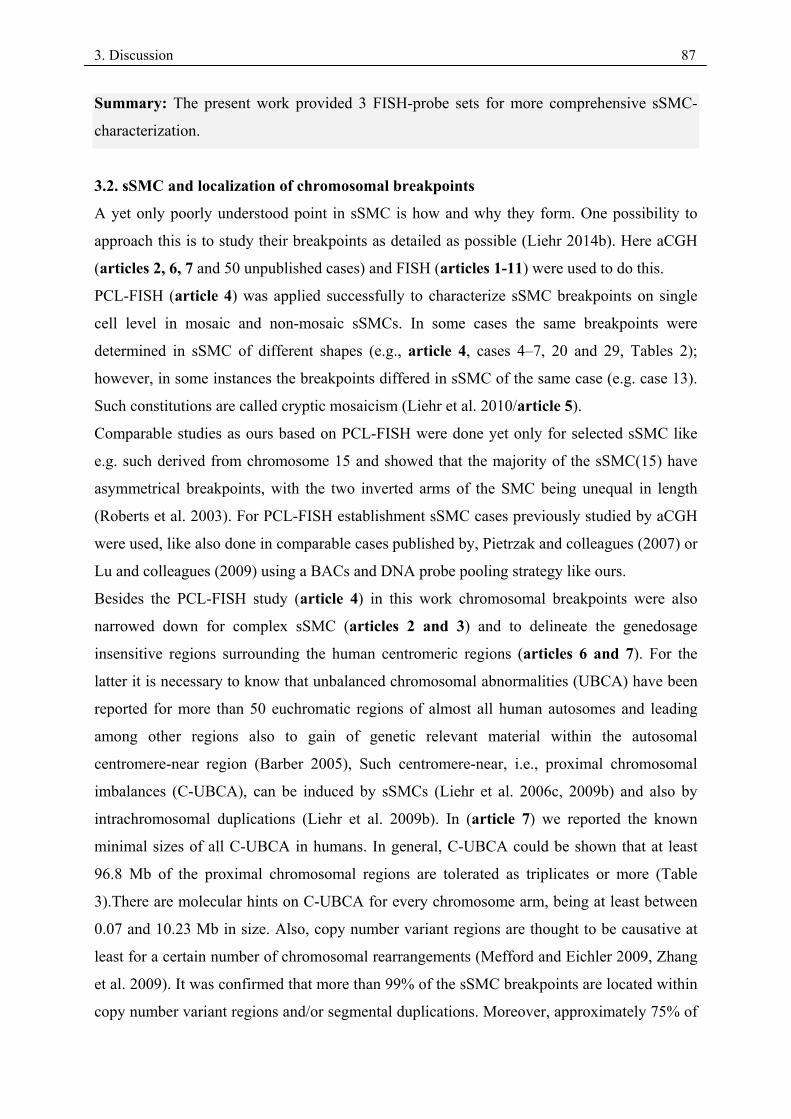
3. Discussion 87
Summary: The present work provided 3 FISH-probe sets for more comprehensive sSMC-
characterization.
3.2. sSMC and localization of chromosomal breakpoints
A yet only poorly understood point in sSMC is how and why they form. One possibility to
approach this is to study their breakpoints as detailed as possible (Liehr 2014b). Here aCGH
(articles 2, 6, 7 and 50 unpublished cases) and FISH (articles 1-11) were used to do this.
PCL-FISH (article 4) was applied successfully to characterize sSMC breakpoints on single
cell level in mosaic and non-mosaic sSMCs. In some cases the same breakpoints were
determined in sSMC of different shapes (e.g., article 4, cases 4–7, 20 and 29, Tables 2);
however, in some instances the breakpoints differed in sSMC of the same case (e.g. case 13).
Such constitutions are called cryptic mosaicism (Liehr et al. 2010/article 5).
Comparable studies as ours based on PCL-FISH were done yet only for selected sSMC like
e.g. such derived from chromosome 15 and showed that the majority of the sSMC(15) have
asymmetrical breakpoints, with the two inverted arms of the SMC being unequal in length
(Roberts et al. 2003). For PCL-FISH establishment sSMC cases previously studied by aCGH
were used, like also done in comparable cases published by, Pietrzak and colleagues (2007) or
Lu and colleagues (2009) using a BACs and DNA probe pooling strategy like ours.
Besides the PCL-FISH study (article 4) in this work chromosomal breakpoints were also
narrowed down for complex sSMC (articles 2 and 3) and to delineate the genedosage
insensitive regions surrounding the human centromeric regions (articles 6 and 7). For the
latter it is necessary to know that unbalanced chromosomal abnormalities (UBCA) have been
reported for more than 50 euchromatic regions of almost all human autosomes and leading
among other regions also to gain of genetic relevant material within the autosomal
centromere-near region (Barber 2005), Such centromere-near, i.e., proximal chromosomal
imbalances (C-UBCA), can be induced by sSMCs (Liehr et al. 2006c, 2009b) and also by
intrachromosomal duplications (Liehr et al. 2009b). In (article 7) we reported the known
minimal sizes of all C-UBCA in humans. In general, C-UBCA could be shown that at least
96.8 Mb of the proximal chromosomal regions are tolerated as triplicates or more (Table
3).There are molecular hints on C-UBCA for every chromosome arm, being at least between
0.07 and 10.23 Mb in size. Also, copy number variant regions are thought to be causative at
least for a certain number of chromosomal rearrangements (Mefford and Eichler 2009, Zhang
et al. 2009). It was confirmed that more than 99% of the sSMC breakpoints are located within
copy number variant regions and/or segmental duplications. Moreover, approximately 75% of
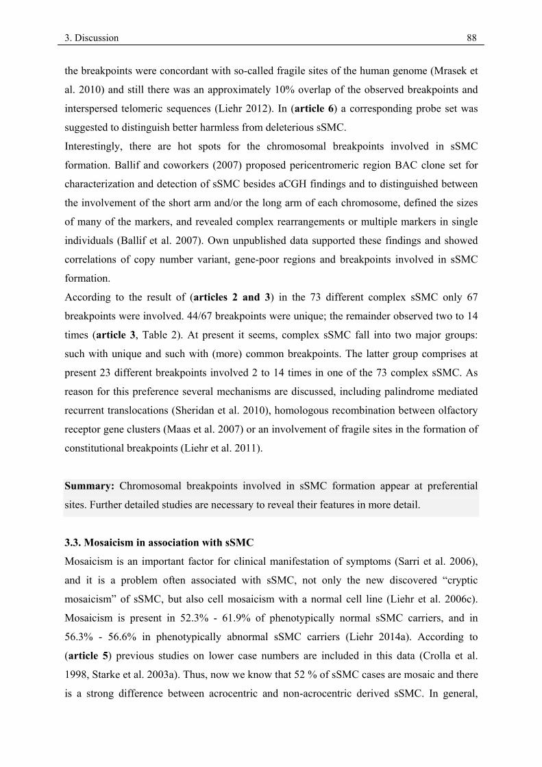
3. Discussion 88
the breakpoints were concordant with so-called fragile sites of the human genome (Mrasek et
al. 2010) and still there was an approximately 10% overlap of the observed breakpoints and
interspersed telomeric sequences (Liehr 2012). In (article 6) a corresponding probe set was
suggested to distinguish better harmless from deleterious sSMC.
Interestingly, there are hot spots for the chromosomal breakpoints involved in sSMC
formation. Ballif and coworkers (2007) proposed pericentromeric region BAC clone set for
characterization and detection of sSMC besides aCGH findings and to distinguished between
the involvement of the short arm and/or the long arm of each chromosome, defined the sizes
of many of the markers, and revealed complex rearrangements or multiple markers in single
individuals (Ballif et al. 2007). Own unpublished data supported these findings and showed
correlations of copy number variant, gene-poor regions and breakpoints involved in sSMC
formation.
According to the result of (articles 2 and 3) in the 73 different complex sSMC only 67
breakpoints were involved. 44/67 breakpoints were unique; the remainder observed two to 14
times (article 3, Table 2). At present it seems, complex sSMC fall into two major groups:
such with unique and such with (more) common breakpoints. The latter group comprises at
present 23 different breakpoints involved 2 to 14 times in one of the 73 complex sSMC. As
reason for this preference several mechanisms are discussed, including palindrome mediated
recurrent translocations (Sheridan et al. 2010), homologous recombination between olfactory
receptor gene clusters (Maas et al. 2007) or an involvement of fragile sites in the formation of
constitutional breakpoints (Liehr et al. 2011).
Summary: Chromosomal breakpoints involved in sSMC formation appear at preferential
sites. Further detailed studies are necessary to reveal their features in more detail.
3.3. Mosaicism in association with sSMC
Mosaicism is an important factor for clinical manifestation of symptoms (Sarri et al. 2006),
and it is a problem often associated with sSMC, not only the new discovered “cryptic
mosaicism” of sSMC, but also cell mosaicism with a normal cell line (Liehr et al. 2006c).
Mosaicism is present in 52.3% - 61.9% of phenotypically normal sSMC carriers, and in
56.3% - 56.6% in phenotypically abnormal sSMC carriers (Liehr 2014a). According to
(article 5) previous studies on lower case numbers are included in this data (Crolla et al.
1998, Starke et al. 2003a). Thus, now we know that 52 % of sSMC cases are mosaic and there
is a strong difference between acrocentric and non-acrocentric derived sSMC. In general,
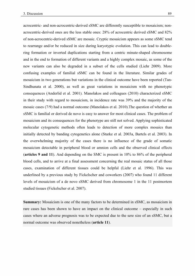
3. Discussion 89
acrocentric- and non-acrocentric-derived sSMC are differently susceptible to mosaicism; non-
acrocentric-derived ones are the less stable ones: 28% of acrocentric derived sSMC and 82%
of non-acrocentric-derived sSMC are mosaic. Cryptic mosaicism appears as some sSMC tend
to rearrange and/or be reduced in size during karyotypic evolution. This can lead to double-
ring formation or inverted duplications starting from a centric minute-shaped chromosome
and in the end to formation of different variants and a highly complex mosaic, as some of the
new variants can also be degraded in a subset of the cells studied (Liehr 2009). More
confusing examples of familial sSMC can be found in the literature. Similar grades of
mosaicism in two generations but variations in the clinical outcome have been reported (Tan-
Sindhunata et al. 2000), as well as great variations in mosaicism with no phenotypic
consequences (Anderlid et al. 2001). Manolakos and colleagues (2010) characterized sSMC
in their study with regard to mosaicism, in incidence rate was 39% and the majority of the
mosaic cases (7/9) had a normal outcome (Manolakos et al. 2010).The question of whether an
sSMC is familial or derived de novo is easy to answer for most clinical cases. The problem of
mosaicism and its consequences for the phenotype are still not solved. Applying sophisticated
molecular cytogenetic methods often leads to detection of more complex mosaics than
initially detected by banding cytogenetics alone (Starke et al. 2003a, Bartels et al. 2003). In
the overwhelming majority of the cases there is no influence of the grade of somatic
mosaicism detectable in peripheral blood or amnion cells and the observed clinical effects
(articles 9 and 11). And depending on the SMC is present in 10% to 86% of the peripheral
blood cells, and to arrive at a final assessment concerning the real mosaic status of all those
cases, examination of different tissues could be helpful (Liehr et al. 1996). This was
underlined by a previous study by Fickelscher and coworkers (2007) who found 11 different
levels of mosaicism of a de novo sSMC derived from chromosome 1 in the 11 postmortem
studied tissues (Fickelscher et al. 2007).
Summary: Mosaicism is one of the many factors to be determined in sSMC, as mosaicism in
rare cases has been shown to have an impact on the clinical outcome – especially in such
cases where an adverse prognosis was to be expected due to the sere size of an sSMC, but a
normal outcome was observed nonetheless (article 11).
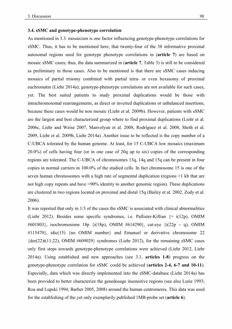
3. Discussion 90
3.4. sSMC and genotype-phenotype correlation
As mentioned in 3.3. mosaicism is one factor influencing genotype-phenotype correlations for
sSMC. Thus, it has to be mentioned here, that twenty-four of the 38 informative proximal
autosomal regions used for genotype phenotype correlations in (article 7) are based on
mosaic sSMC cases; thus, the data summarized in (article 7, Table 3) is still to be considered
as preliminary in those cases. Also to be mentioned is that there are sSMC cases inducing
mosaics of partial trisomy combined with partial tetra- or even hexasomy of proximal
euchromatin (Liehr 2014a); genotype-phenotype correlations are not available for such cases,
yet. The best suited patients to study proximal duplications would be those with
intrachromosomal rearrangements, as direct or inverted duplications or unbalanced insertions,
because these cases would be non mosaic (Liehr et al. 2009b). However, patients with sSMC
are the largest and best characterized group where to find proximal duplications (Liehr et al.
2006c, Liehr and Weise 2007, Manvelyan et al. 2008, Rodríguez et al. 2008, Sheth et al.
2009, Liehr et al. 2009b, Liehr 2014a). Another issue to be reflected is the copy number of a
C-UBCA tolerated by the human genome. At least, for 15 C-UBCA low mosaics (maximum
20.0%) of cells having four (or in one case of 20q up to six) copies of the corresponding
regions are tolerated. The C-UBCA of chromosomes 13q, 14q and 15q can be present in four
copies in normal carriers in 100.0% of the studied cells. In fact chromosome 15 is one of the
seven human chromosomes with a high rate of segmental duplication (regions >1 kb that are
not high copy repeats and have >90% identity to another genomic region). These duplications
are clustered in two regions located on proximal and distal 15q (Bailey et al. 2002, Zody et al.
2006).
It was reported that only in 1/3 of the cases the sSMC is associated with clinical abnormalities
(Liehr 2012). Besides some specific syndromes, i.e. Pallister-Killian {= i(12p), OMIM
#601803}, isochromosome 18p {i(18p), OMIM #614290}, cat-eye {i(22p ~ q), OMIM
#115470}, idic(15) {no OMIM number} and Emanuel or derivative chromosome 22
{der(22)t(11;22), OMIM #609029} syndromes (Liehr 2012), for the remaining sSMC cases
only first steps towards genotype-phenotype correlations were achieved (Liehr 2012, Liehr
2014a). Using established and new approaches (see 3.1, articles 1-8) progress on the
genotype-phenotype correlation for sSMC could be achieved (articles 2-4, 6-7 and 10-11).
Especially, data which was directly implemented into the sSMC-database (Liehr 2014a) has
been provided to better characterize the genedosage insensitive regions (see also Lurie 1993;
Roa and Lupski 1994, Barber 2005, 2008) around the human centromeres. This data was used
for the establishing of the yet only exemplarily published 1MB-probe set (article 6).
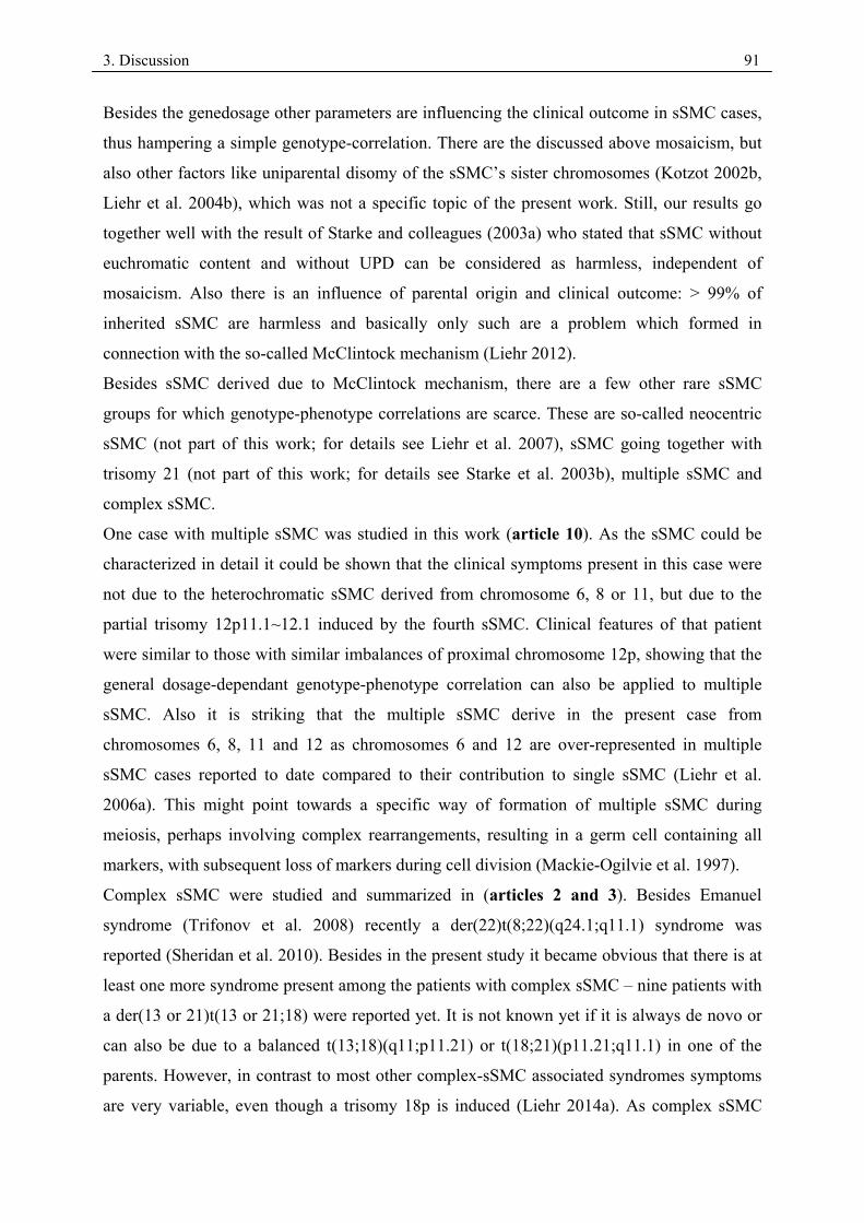
3. Discussion 91
Besides the genedosage other parameters are influencing the clinical outcome in sSMC cases,
thus hampering a simple genotype-correlation. There are the discussed above mosaicism, but
also other factors like uniparental disomy of the sSMC’s sister chromosomes (Kotzot 2002b,
Liehr et al. 2004b), which was not a specific topic of the present work. Still, our results go
together well with the result of Starke and colleagues (2003a) who stated that sSMC without
euchromatic content and without UPD can be considered as harmless, independent of
mosaicism. Also there is an influence of parental origin and clinical outcome: > 99% of
inherited sSMC are harmless and basically only such are a problem which formed in
connection with the so-called McClintock mechanism (Liehr 2012).
Besides sSMC derived due to McClintock mechanism, there are a few other rare sSMC
groups for which genotype-phenotype correlations are scarce. These are so-called neocentric
sSMC (not part of this work; for details see Liehr et al. 2007), sSMC going together with
trisomy 21 (not part of this work; for details see Starke et al. 2003b), multiple sSMC and
complex sSMC.
One case with multiple sSMC was studied in this work (article 10). As the sSMC could be
characterized in detail it could be shown that the clinical symptoms present in this case were
not due to the heterochromatic sSMC derived from chromosome 6, 8 or 11, but due to the
partial trisomy 12p11.1~12.1 induced by the fourth sSMC. Clinical features of that patient
were similar to those with similar imbalances of proximal chromosome 12p, showing that the
general dosage-dependant genotype-phenotype correlation can also be applied to multiple
sSMC. Also it is striking that the multiple sSMC derive in the present case from
chromosomes 6, 8, 11 and 12 as chromosomes 6 and 12 are over-represented in multiple
sSMC cases reported to date compared to their contribution to single sSMC (Liehr et al.
2006a). This might point towards a specific way of formation of multiple sSMC during
meiosis, perhaps involving complex rearrangements, resulting in a germ cell containing all
markers, with subsequent loss of markers during cell division (Mackie-Ogilvie et al. 1997).
Complex sSMC were studied and summarized in (articles 2 and 3). Besides Emanuel
syndrome (Trifonov et al. 2008) recently a der(22)t(8;22)(q24.1;q11.1) syndrome was
reported (Sheridan et al. 2010). Besides in the present study it became obvious that there is at
least one more syndrome present among the patients with complex sSMC – nine patients with
a der(13 or 21)t(13 or 21;18) were reported yet. It is not known yet if it is always de novo or
can also be due to a balanced t(13;18)(q11;p11.21) or t(18;21)(p11.21;q11.1) in one of the
parents. However, in contrast to most other complex-sSMC associated syndromes symptoms
are very variable, even though a trisomy 18p is induced (Liehr 2014a). As complex sSMC
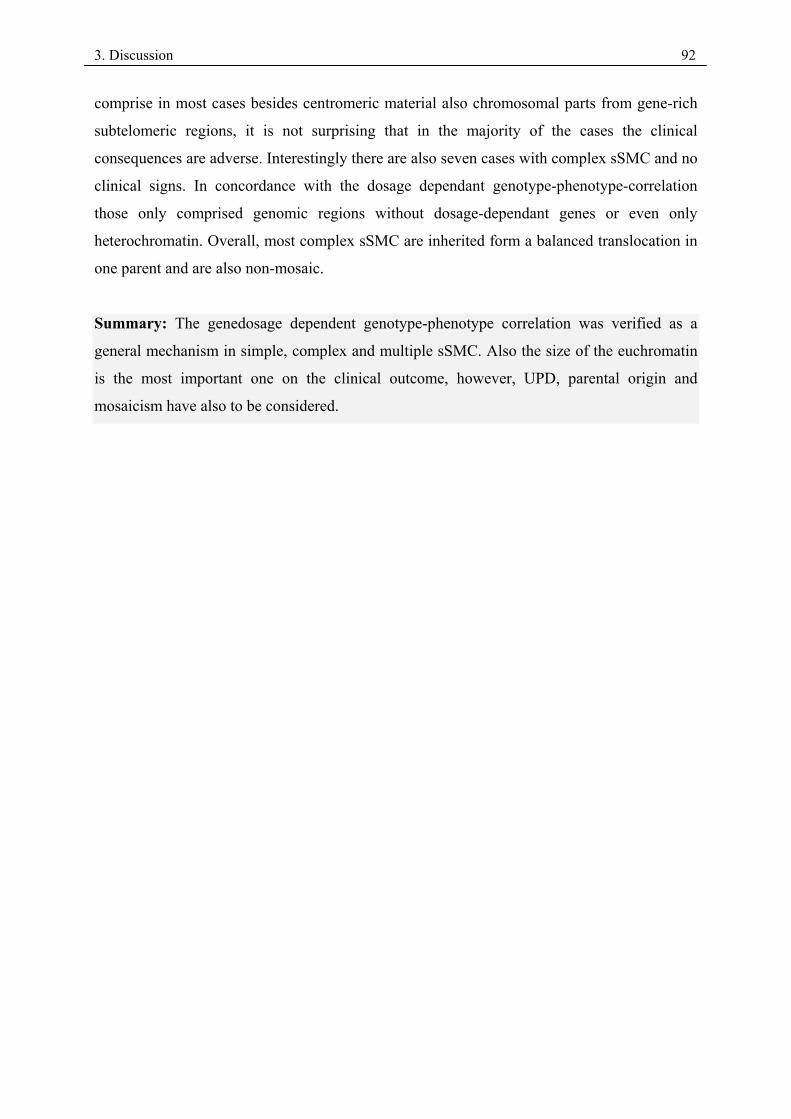
3. Discussion 92
comprise in most cases besides centromeric material also chromosomal parts from gene-rich
subtelomeric regions, it is not surprising that in the majority of the cases the clinical
consequences are adverse. Interestingly there are also seven cases with complex sSMC and no
clinical signs. In concordance with the dosage dependant genotype-phenotype-correlation
those only comprised genomic regions without dosage-dependant genes or even only
heterochromatin. Overall, most complex sSMC are inherited form a balanced translocation in
one parent and are also non-mosaic.
Summary: The genedosage dependent genotype-phenotype correlation was verified as a
general mechanism in simple, complex and multiple sSMC. Also the size of the euchromatin
is the most important one on the clinical outcome, however, UPD, parental origin and
mosaicism have also to be considered.
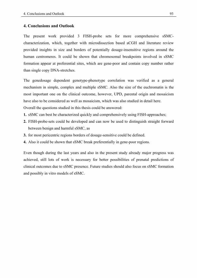
4. Conclusions and Outlook 93
4. Conclusions and Outlook The present work provided 3 FISH-probe sets for more comprehensive sSMC-
characterization, which, together with microdissection based aCGH and literature review
provided insights in size and borders of potentially dosage-insensitive regions around the
human centromeres. It could be shown that chromosomal breakpoints involved in sSMC
formation appear at preferential sites, which are gene-poor and contain copy number rather
than single copy DNA-stretches.
The genedosage dependent genotype-phenotype correlation was verified as a general
mechanism in simple, complex and multiple sSMC. Also the size of the euchromatin is the
most important one on the clinical outcome, however, UPD, parental origin and mosaicism
have also to be considered as well as mosaicism, which was also studied in detail here.
Overall the questions studied in this thesis could be answered:
1. sSMC can best be characterized quickly and comprehensively using FISH-approaches;
2. FISH-probe-sets could be developed and can now be used to distinguish straight forward
between benign and harmful sSMC, as
3. for most pericentric regions borders of dosage-sensitive could be defined.
4. Also it could be shown that sSMC break preferentially in gene-poor regions.
Even though during the last years and also in the present study already major progress was
achieved, still lots of work is necessary for better possibilities of prenatal predictions of
clinical outcomes due to sSMC presence. Future studies should also focus on sSMC formation
and possibly in vitro models of sSMC.
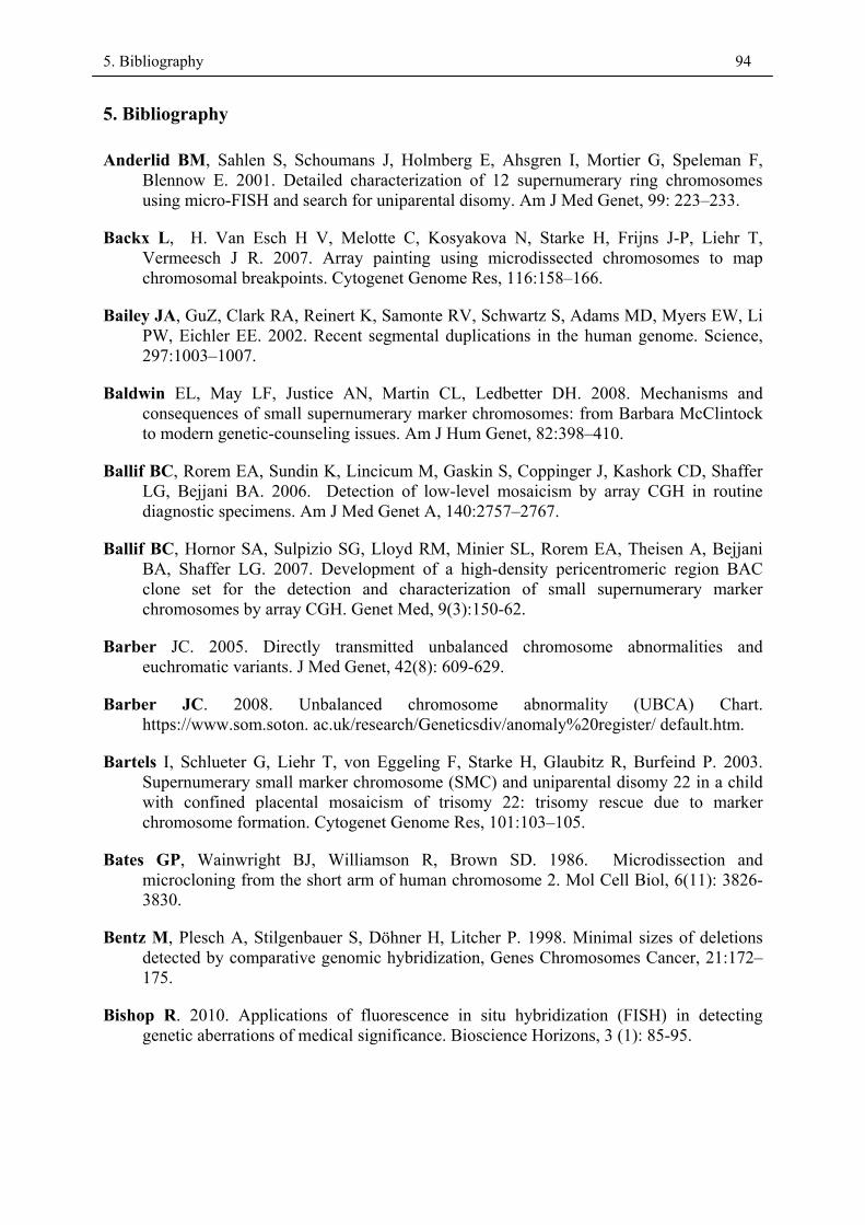
5. Bibliography 94
5. Bibliography
Anderlid BM, Sahlen S, Schoumans J, Holmberg E, Ahsgren I, Mortier G, Speleman F, Blennow E. 2001. Detailed characterization of 12 supernumerary ring chromosomes using micro-FISH and search for uniparental disomy. Am J Med Genet, 99: 223–233.
Backx L, H. Van Esch H V, Melotte C, Kosyakova N, Starke H, Frijns J-P, Liehr T, Vermeesch J R. 2007. Array painting using microdissected chromosomes to map chromosomal breakpoints. Cytogenet Genome Res, 116:158–166.
Bailey JA, GuZ, Clark RA, Reinert K, Samonte RV, Schwartz S, Adams MD, Myers EW, Li PW, Eichler EE. 2002. Recent segmental duplications in the human genome. Science, 297:1003–1007.
Baldwin EL, May LF, Justice AN, Martin CL, Ledbetter DH. 2008. Mechanisms and consequences of small supernumerary marker chromosomes: from Barbara McClintock to modern genetic-counseling issues. Am J Hum Genet, 82:398–410.
Ballif BC, Rorem EA, Sundin K, Lincicum M, Gaskin S, Coppinger J, Kashork CD, Shaffer LG, Bejjani BA. 2006. Detection of low-level mosaicism by array CGH in routine diagnostic specimens. Am J Med Genet A, 140:2757–2767.
Ballif BC, Hornor SA, Sulpizio SG, Lloyd RM, Minier SL, Rorem EA, Theisen A, Bejjani BA, Shaffer LG. 2007. Development of a high-density pericentromeric region BAC clone set for the detection and characterization of small supernumerary marker chromosomes by array CGH. Genet Med, 9(3):150-62.
Barber JC. 2005. Directly transmitted unbalanced chromosome abnormalities and euchromatic variants. J Med Genet, 42(8): 609-629.
Barber JC. 2008. Unbalanced chromosome abnormality (UBCA) Chart. https://www.som.soton. ac.uk/research/Geneticsdiv/anomaly%20register/ default.htm.
Bartels I, Schlueter G, Liehr T, von Eggeling F, Starke H, Glaubitz R, Burfeind P. 2003. Supernumerary small marker chromosome (SMC) and uniparental disomy 22 in a child with confined placental mosaicism of trisomy 22: trisomy rescue due to marker chromosome formation. Cytogenet Genome Res, 101:103–105.
Bates GP, Wainwright BJ, Williamson R, Brown SD. 1986. Microdissection and microcloning from the short arm of human chromosome 2. Mol Cell Biol, 6(11): 3826-3830.
Bentz M, Plesch A, Stilgenbauer S, Döhner H, Litcher P. 1998. Minimal sizes of deletions detected by comparative genomic hybridization, Genes Chromosomes Cancer, 21:172–175.
Bishop R. 2010. Applications of fluorescence in situ hybridization (FISH) in detecting genetic aberrations of medical significance. Bioscience Horizons, 3 (1): 85-95.
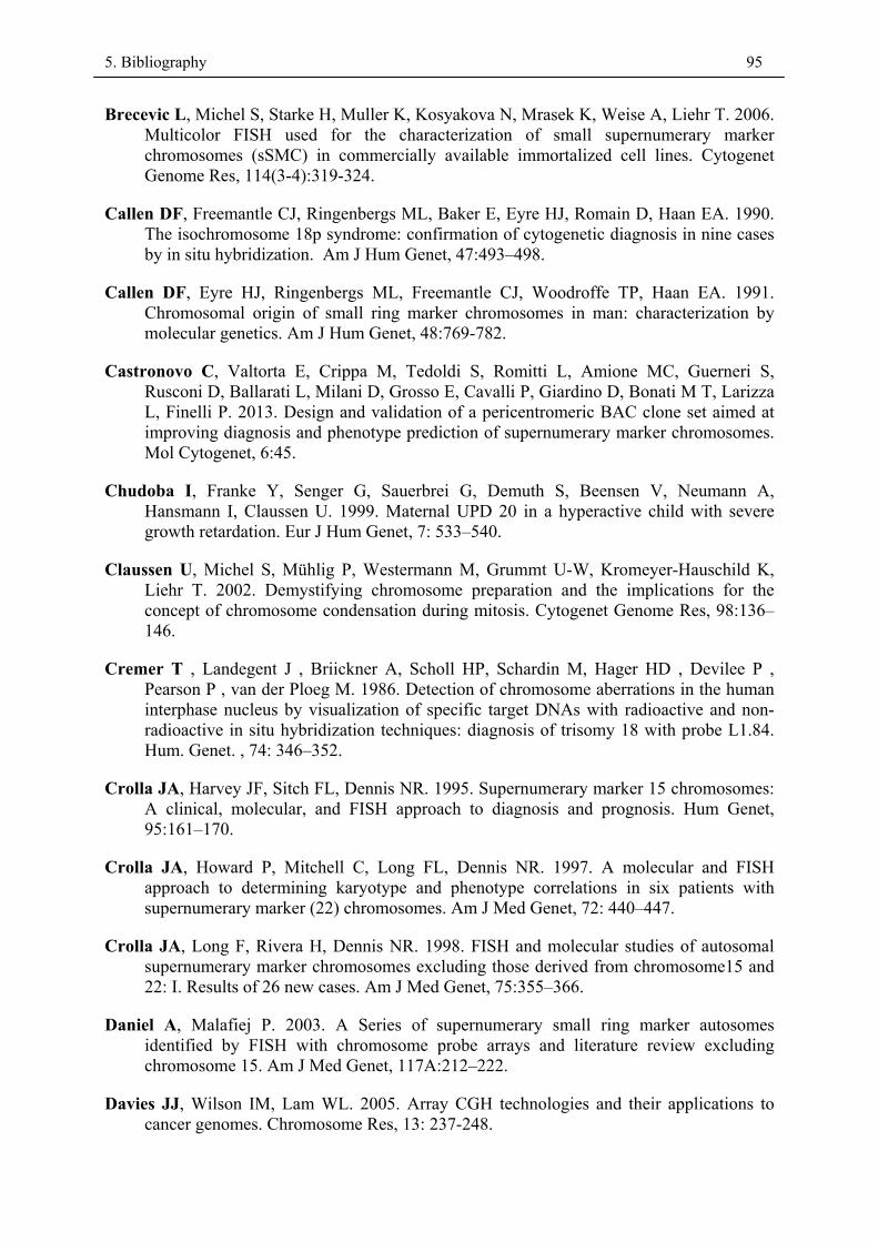
5. Bibliography 95
Brecevic L, Michel S, Starke H, Muller K, Kosyakova N, Mrasek K, Weise A, Liehr T. 2006. Multicolor FISH used for the characterization of small supernumerary marker chromosomes (sSMC) in commercially available immortalized cell lines. Cytogenet Genome Res, 114(3-4):319-324.
Callen DF, Freemantle CJ, Ringenbergs ML, Baker E, Eyre HJ, Romain D, Haan EA. 1990. The isochromosome 18p syndrome: confirmation of cytogenetic diagnosis in nine cases by in situ hybridization. Am J Hum Genet, 47:493–498.
Callen DF, Eyre HJ, Ringenbergs ML, Freemantle CJ, Woodroffe TP, Haan EA. 1991. Chromosomal origin of small ring marker chromosomes in man: characterization by molecular genetics. Am J Hum Genet, 48:769-782.
Castronovo C, Valtorta E, Crippa M, Tedoldi S, Romitti L, Amione MC, Guerneri S, Rusconi D, Ballarati L, Milani D, Grosso E, Cavalli P, Giardino D, Bonati M T, Larizza L, Finelli P. 2013. Design and validation of a pericentromeric BAC clone set aimed at improving diagnosis and phenotype prediction of supernumerary marker chromosomes. Mol Cytogenet, 6:45.
Chudoba I, Franke Y, Senger G, Sauerbrei G, Demuth S, Beensen V, Neumann A, Hansmann I, Claussen U. 1999. Maternal UPD 20 in a hyperactive child with severe growth retardation. Eur J Hum Genet, 7: 533–540.
Claussen U, Michel S, Mühlig P, Westermann M, Grummt U-W, Kromeyer-Hauschild K, Liehr T. 2002. Demystifying chromosome preparation and the implications for the concept of chromosome condensation during mitosis. Cytogenet Genome Res, 98:136–146.
Cremer T , Landegent J , Briickner A, Scholl HP, Schardin M, Hager HD , Devilee P , Pearson P , van der Ploeg M. 1986. Detection of chromosome aberrations in the human interphase nucleus by visualization of specific target DNAs with radioactive and non-radioactive in situ hybridization techniques: diagnosis of trisomy 18 with probe L1.84. Hum. Genet. , 74: 346–352.
Crolla JA, Harvey JF, Sitch FL, Dennis NR. 1995. Supernumerary marker 15 chromosomes: A clinical, molecular, and FISH approach to diagnosis and prognosis. Hum Genet, 95:161–170.
Crolla JA, Howard P, Mitchell C, Long FL, Dennis NR. 1997. A molecular and FISH approach to determining karyotype and phenotype correlations in six patients with supernumerary marker (22) chromosomes. Am J Med Genet, 72: 440–447.
Crolla JA, Long F, Rivera H, Dennis NR. 1998. FISH and molecular studies of autosomal supernumerary marker chromosomes excluding those derived from chromosome15 and 22: I. Results of 26 new cases. Am J Med Genet, 75:355–366.
Daniel A, Malafiej P. 2003. A Series of supernumerary small ring marker autosomes identified by FISH with chromosome probe arrays and literature review excluding chromosome 15. Am J Med Genet, 117A:212–222.
Davies JJ, Wilson IM, Lam WL. 2005. Array CGH technologies and their applications to cancer genomes. Chromosome Res, 13: 237-248.
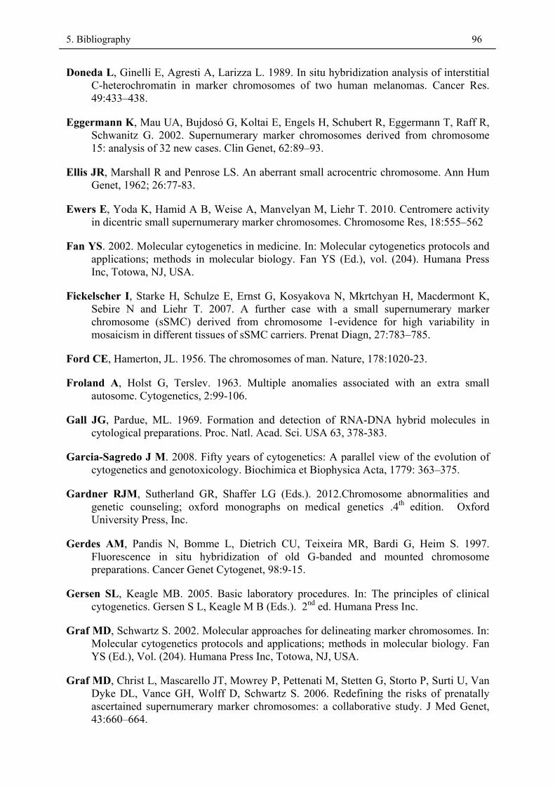
5. Bibliography 96
Doneda L, Ginelli E, Agresti A, Larizza L. 1989. In situ hybridization analysis of interstitial C-heterochromatin in marker chromosomes of two human melanomas. Cancer Res. 49:433–438.
Eggermann K, Mau UA, Bujdosó G, Koltai E, Engels H, Schubert R, Eggermann T, Raff R, Schwanitz G. 2002. Supernumerary marker chromosomes derived from chromosome 15: analysis of 32 new cases. Clin Genet, 62:89–93.
Ellis JR, Marshall R and Penrose LS. An aberrant small acrocentric chromosome. Ann Hum Genet, 1962; 26:77-83.
Ewers E, Yoda K, Hamid A B, Weise A, Manvelyan M, Liehr T. 2010. Centromere activity in dicentric small supernumerary marker chromosomes. Chromosome Res, 18:555–562
Fan YS. 2002. Molecular cytogenetics in medicine. In: Molecular cytogenetics protocols and applications; methods in molecular biology. Fan YS (Ed.), vol. (204). Humana Press Inc, Totowa, NJ, USA.
Fickelscher I, Starke H, Schulze E, Ernst G, Kosyakova N, Mkrtchyan H, Macdermont K, Sebire N and Liehr T. 2007. A further case with a small supernumerary marker chromosome (sSMC) derived from chromosome 1-evidence for high variability in mosaicism in different tissues of sSMC carriers. Prenat Diagn, 27:783–785.
Ford CE, Hamerton, JL. 1956. The chromosomes of man. Nature, 178:1020-23.
Froland A, Holst G, Terslev. 1963. Multiple anomalies associated with an extra small autosome. Cytogenetics, 2:99-106.
Gall JG, Pardue, ML. 1969. Formation and detection of RNA-DNA hybrid molecules in cytological preparations. Proc. Natl. Acad. Sci. USA 63, 378-383.
Garcia-Sagredo J M. 2008. Fifty years of cytogenetics: A parallel view of the evolution of cytogenetics and genotoxicology. Biochimica et Biophysica Acta, 1779: 363–375.
Gardner RJM, Sutherland GR, Shaffer LG (Eds.). 2012.Chromosome abnormalities and genetic counseling; oxford monographs on medical genetics .4th edition. Oxford University Press, Inc.
Gerdes AM, Pandis N, Bomme L, Dietrich CU, Teixeira MR, Bardi G, Heim S. 1997. Fluorescence in situ hybridization of old G-banded and mounted chromosome preparations. Cancer Genet Cytogenet, 98:9-15.
Gersen SL, Keagle MB. 2005. Basic laboratory procedures. In: The principles of clinical cytogenetics. Gersen S L, Keagle M B (Eds.). 2nd ed. Humana Press Inc.
Graf MD, Schwartz S. 2002. Molecular approaches for delineating marker chromosomes. In: Molecular cytogenetics protocols and applications; methods in molecular biology. Fan YS (Ed.), Vol. (204). Humana Press Inc, Totowa, NJ, USA.
Graf MD, Christ L, Mascarello JT, Mowrey P, Pettenati M, Stetten G, Storto P, Surti U, Van Dyke DL, Vance GH, Wolff D, Schwartz S. 2006. Redefining the risks of prenatally ascertained supernumerary marker chromosomes: a collaborative study. J Med Genet, 43:660–664.

5. Bibliography 97
Griffiths AJF, Miller JH, Suzuki DT (Eds.).1996. An introduction to genetic analysis, edited by, R. C. Lewontin and W. M. Gelbart .W. H. Freeman and Company, New York, USA, 915p.
Haddad BR, Schröck E, Meck J, Cowan J, Young H, Ferguson-Smith MA, Manoir S du, Ried T. 1998. Identification of de novo chromosomal markers and derivatives by spectral karyotyping. Hum Genet, 103:619–625.
Hastings RJ, Nisbet DL, Waters K, Spencer T, Chitty LS. 1999. Prenatal detection of extra structurally abnormal chromosomes (ESACs): New cases and a review of the literature. Prenat Diagn, 19:436–445.
Huang B, Crolla JA, Christian SL, Wolf-Ledbetter ME, Macha ME, Papenhausen PN , Ledbetter DH. 1997. Refined molecular characterization of the breakpoints in small inv dup(15) chromosomes. Hum Genet, 99:11–17.
Ilberry PLT, Lee CWG, Winn SM. 1961. Incomplete trisomy in a mongoloid child exhibiting minimal stigmata. Med J Austr, 48:182–184.
Joos S, Fink TM, Rätsch A, Lichter P. 1994. Mapping and chromosome analysis: the potential of fluorescence in situ hybridization. J Biotechnol, 35:135-153.
Kallioniemi A. 2008. CGH microarrays and cancer. Current Opinion in Biotechnology, 19:36–40.
Kallioniemi A, Kallioniemi OP, Sudar D, Rutovitz D, Gray JW, Waldman F, Pinkel D. 1992. Comparative genomic hybridization for molecular cytogenetic analysis of solid tumours, Science, 258: 818–821.
Kjeldsen E, Kølvraa S. 2002. FISH techniques, FISH probe and their applications in medicine and biology – an overview. In: Rautenstrauß BW, Liehr T (Eds.). FISH technology. Springer-Verlag Berlin Heidelberg.
Kotzot D. 2002a. Review and meta-analysis of systematic searches for uniparental disomy (UPD) other than UPD 15. Am J Med Genet, 111:366–375.
Kotzot D. 2002b. Supernumerary marker chromosomes (SMC) and uniparental disomy (UPD): coincidence or consequence? J Med Genet, 39: 775–778.
Kumar RA, Sudi J, Babatz TD, Brune CW, Oswald D, Yen M, Nowak NJ, Cook EH, Christian SL, Dobyns WB. 2010. A de novo 1p34.2 microdeletion identifies the synaptic vesicle gene RIMS3 as a novel candidate for autism. J Med Genet, 47(2):81-90.
Lacadena LR (Ed.). 1996. Citogenética, Editorial Complutense, Madrid. Spain.
Lapierre JM, Tachdjian G. 2002. Comparative genomic hybridization. In: Encyclopedia of Life Sciences (ELS). John Wiley &Sons, Ltd. DOI: 10.1038/npg.els.0002651.
Lejeune J, Gautier M, Turpin R. 1959. Etude des chromosomes somatiques de neuf enfants mongoliens. C. R. Acad.sci., Paris, 248: 1721.
Li M, Pinkel D. 2006. Clinical cytogenetics and molecular cytogenetics. J Zhejiang Uni Science B, 7(2): 162-163.
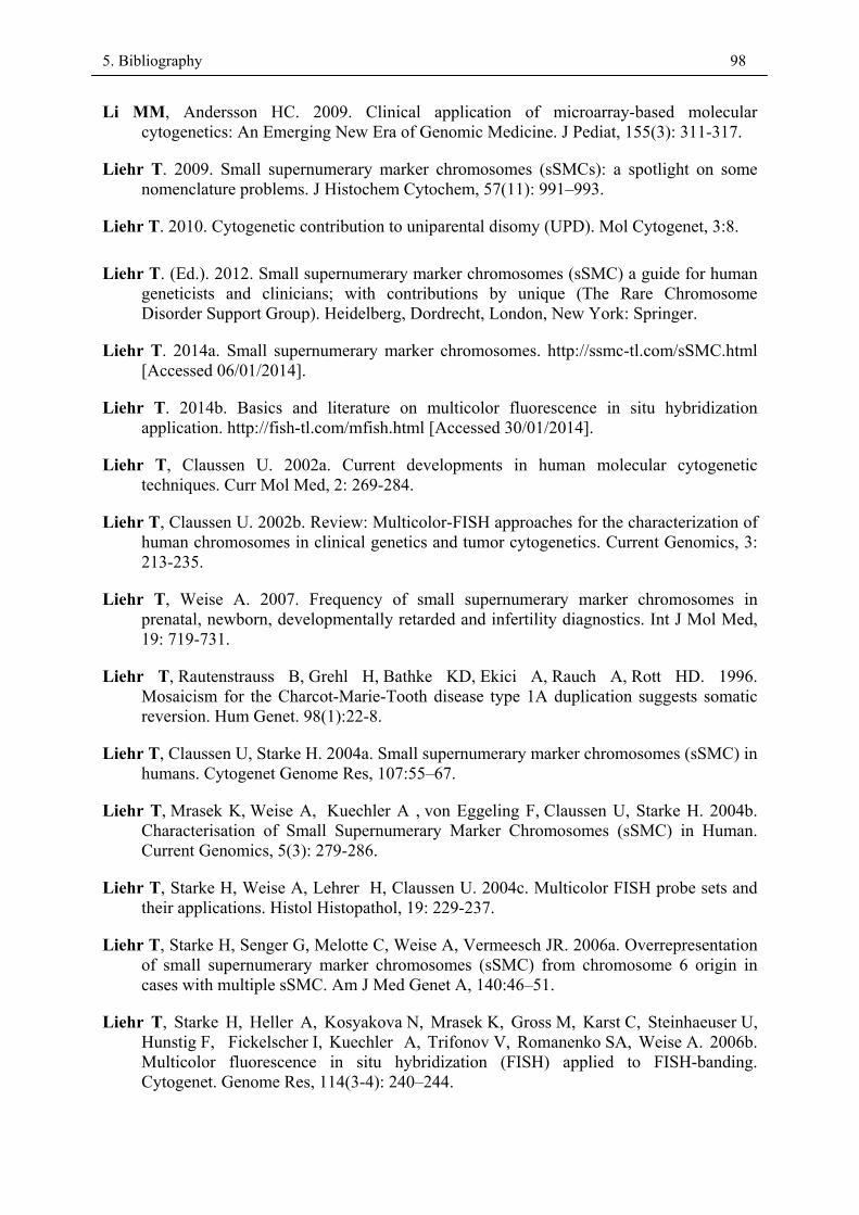
5. Bibliography 98
Li MM, Andersson HC. 2009. Clinical application of microarray-based molecular cytogenetics: An Emerging New Era of Genomic Medicine. J Pediat, 155(3): 311-317.
Liehr T. 2009. Small supernumerary marker chromosomes (sSMCs): a spotlight on some nomenclature problems. J Histochem Cytochem, 57(11): 991–993.
Liehr T. 2010. Cytogenetic contribution to uniparental disomy (UPD). Mol Cytogenet, 3:8.
Liehr T. (Ed.). 2012. Small supernumerary marker chromosomes (sSMC) a guide for human geneticists and clinicians; with contributions by unique (The Rare Chromosome Disorder Support Group). Heidelberg, Dordrecht, London, New York: Springer.
Liehr T. 2014a. Small supernumerary marker chromosomes. http://ssmc-tl.com/sSMC.html [Accessed 06/01/2014].
Liehr T. 2014b. Basics and literature on multicolor fluorescence in situ hybridization application. http://fish-tl.com/mfish.html [Accessed 30/01/2014].
Liehr T, Claussen U. 2002a. Current developments in human molecular cytogenetic techniques. Curr Mol Med, 2: 269-284.
Liehr T, Claussen U. 2002b. Review: Multicolor-FISH approaches for the characterization of human chromosomes in clinical genetics and tumor cytogenetics. Current Genomics, 3: 213-235.
Liehr T, Weise A. 2007. Frequency of small supernumerary marker chromosomes in prenatal, newborn, developmentally retarded and infertility diagnostics. Int J Mol Med, 19: 719-731.
Liehr T, Rautenstrauss B, Grehl H, Bathke KD, Ekici A, Rauch A, Rott HD. 1996. Mosaicism for the Charcot-Marie-Tooth disease type 1A duplication suggests somatic reversion. Hum Genet. 98(1):22-8.
Liehr T, Claussen U, Starke H. 2004a. Small supernumerary marker chromosomes (sSMC) in humans. Cytogenet Genome Res, 107:55–67.
Liehr T, Mrasek K, Weise A, Kuechler A , von Eggeling F, Claussen U, Starke H. 2004b. Characterisation of Small Supernumerary Marker Chromosomes (sSMC) in Human. Current Genomics, 5(3): 279-286.
Liehr T, Starke H, Weise A, Lehrer H, Claussen U. 2004c. Multicolor FISH probe sets and their applications. Histol Histopathol, 19: 229-237.
Liehr T, Starke H, Senger G, Melotte C, Weise A, Vermeesch JR. 2006a. Overrepresentation of small supernumerary marker chromosomes (sSMC) from chromosome 6 origin in cases with multiple sSMC. Am J Med Genet A, 140:46–51.
Liehr T, Starke H, Heller A, Kosyakova N, Mrasek K, Gross M, Karst C, Steinhaeuser U, Hunstig F, Fickelscher I, Kuechler A, Trifonov V, Romanenko SA, Weise A. 2006b. Multicolor fluorescence in situ hybridization (FISH) applied to FISH-banding. Cytogenet. Genome Res, 114(3-4): 240–244.
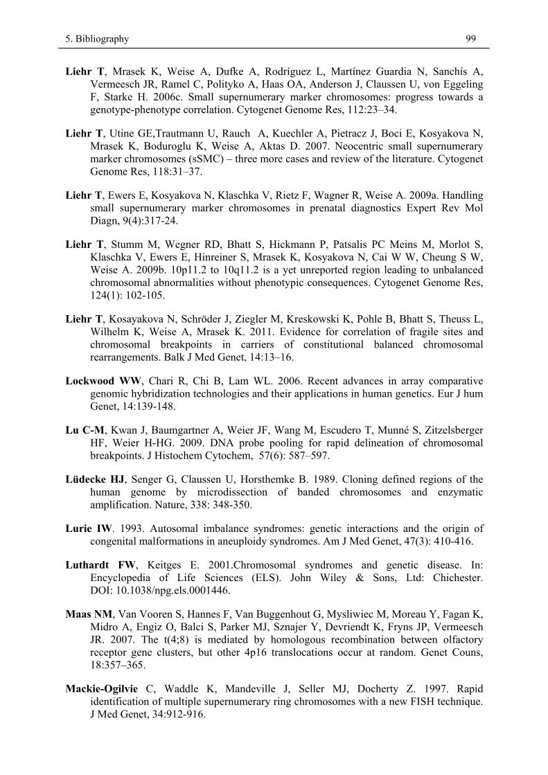
5. Bibliography 99
Liehr T, Mrasek K, Weise A, Dufke A, Rodríguez L, Martínez Guardia N, Sanchís A, Vermeesch JR, Ramel C, Polityko A, Haas OA, Anderson J, Claussen U, von Eggeling F, Starke H. 2006c. Small supernumerary marker chromosomes: progress towards a genotype-phenotype correlation. Cytogenet Genome Res, 112:23–34.
Liehr T, Utine GE,Trautmann U, Rauch A, Kuechler A, Pietracz J, Boci E, Kosyakova N, Mrasek K, Boduroglu K, Weise A, Aktas D. 2007. Neocentric small supernumerary marker chromosomes (sSMC) – three more cases and review of the literature. Cytogenet Genome Res, 118:31–37.
Liehr T, Ewers E, Kosyakova N, Klaschka V, Rietz F, Wagner R, Weise A. 2009a. Handling small supernumerary marker chromosomes in prenatal diagnostics Expert Rev Mol Diagn, 9(4):317-24.
Liehr T, Stumm M, Wegner RD, Bhatt S, Hickmann P, Patsalis PC Meins M, Morlot S, Klaschka V, Ewers E, Hinreiner S, Mrasek K, Kosyakova N, Cai W W, Cheung S W, Weise A. 2009b. 10p11.2 to 10q11.2 is a yet unreported region leading to unbalanced chromosomal abnormalities without phenotypic consequences. Cytogenet Genome Res, 124(1): 102-105.
Liehr T, Kosayakova N, Schröder J, Ziegler M, Kreskowski K, Pohle B, Bhatt S, Theuss L, Wilhelm K, Weise A, Mrasek K. 2011. Evidence for correlation of fragile sites and chromosomal breakpoints in carriers of constitutional balanced chromosomal rearrangements. Balk J Med Genet, 14:13–16.
Lockwood WW, Chari R, Chi B, Lam WL. 2006. Recent advances in array comparative genomic hybridization technologies and their applications in human genetics. Eur J hum Genet, 14:139-148.
Lu C-M, Kwan J, Baumgartner A, Weier JF, Wang M, Escudero T, Munné S, Zitzelsberger HF, Weier H-HG. 2009. DNA probe pooling for rapid delineation of chromosomal breakpoints. J Histochem Cytochem, 57(6): 587–597.
Lüdecke HJ, Senger G, Claussen U, Horsthemke B. 1989. Cloning defined regions of the human genome by microdissection of banded chromosomes and enzymatic amplification. Nature, 338: 348-350.
Lurie IW. 1993. Autosomal imbalance syndromes: genetic interactions and the origin of congenital malformations in aneuploidy syndromes. Am J Med Genet, 47(3): 410-416.
Luthardt FW, Keitges E. 2001.Chromosomal syndromes and genetic disease. In: Encyclopedia of Life Sciences (ELS). John Wiley & Sons, Ltd: Chichester. DOI: 10.1038/npg.els.0001446.
Maas NM, Van Vooren S, Hannes F, Van Buggenhout G, Mysliwiec M, Moreau Y, Fagan K, Midro A, Engiz O, Balci S, Parker MJ, Sznajer Y, Devriendt K, Fryns JP, Vermeesch JR. 2007. The t(4;8) is mediated by homologous recombination between olfactory receptor gene clusters, but other 4p16 translocations occur at random. Genet Couns, 18:357–365.
Mackie-Ogilvie C, Waddle K, Mandeville J, Seller MJ, Docherty Z. 1997. Rapid identification of multiple supernumerary ring chromosomes with a new FISH technique. J Med Genet, 34:912-916.
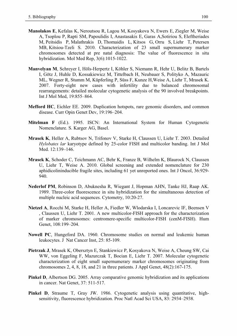
5. Bibliography 100
Manolakos E, Kefalas K, Neroutsou R, Lagou M, Kosyakova N, Ewers E, Ziegler M, Weise A, Tsoplou P, Rapti SM, Papoulidis I, Anastasakis E, Garas A,Sotiriou S, Eleftheriades M, Peitsidis P, Malathrakis D, Thomaidis L, Kitsos G, Orru S, Liehr T, Petersen MB, Kitsiou-Tzeli S. 2010. Characterization of 23 small supernumerary marker chromosomes detected at pre natal diagnosis: The value of fluorescence in situ hybridization. Mol Med Rep, 3(6):1015-1022.
Manvelyan M, Schreyer I, Höls-Herpertz I, Köhler S, Niemann R, Hehr U, Belitz B, Bartels I, Götz J, Huhle D, Kossakiewicz M, Tittelbach H, Neubauer S, Polityko A, Mazauric ML, Wegner R, Stumm M, Küpferling P, Süss F, Kunze H,Weise A, Liehr T, Mrasek K. 2007. Forty-eight new cases with infertility due to balanced chromosomal rearrangements: detailed molecular cytogenetic analysis of the 90 involved breakpoints. Int J Mol Med, 19:855–864.
Mefford HC, Eichler EE. 2009. Duplication hotspots, rare genomic disorders, and common disease. Curr Opin Genet Dev, 19:196–204.
Mitelman F (Ed.). 1995. ISCN: An International System for Human Cytogenetic Nomenclature. S. Karger AG, Basel.
Mrasek K, Heller A, Rubtsov N, Trifonov V, Starke H, Claussen U, Liehr T. 2003. Detailed Hylobates lar karyotype defined by 25-color FISH and multicolor banding. Int J Mol Med. 12:139–146.
Mrasek K, Schoder C, Teichmann AC, Behr K, Franze B, Wilhelm K, Blaurock N, Claussen U, Liehr T, Weise A. 2010. Global screening and extended nomenclature for 230 aphidicolininducible fragile sites, including 61 yet unreported ones. Int J Oncol, 36:929-940.
Nederlof PM, Robinson D, Abuknesha R, Wiegant J, Hopman AHN, Tanke HJ, Raap AK. 1989. Three-color fluorescence in situ hybridization for the simultaneous detection of multiple nucleic acid sequences. Cytometry, 10:20-27.
Nietzel A, Rocchi M, Starke H, Heller A, Fiedler W, Wlodarska I, Loncarevic IF, Beensen V , Claussen U, Liehr T. 2001. A new multicolor-FISH approach for the characterization of marker chromosomes: centromere-specific multicolor-FISH (cenM-FISH). Hum Genet, 108:199–204.
Nowell PC, Hungeford DA. 1960. Chromosome studies on normal and leukemic human leukocytes. J Nat Cancer Inst, 25: 85-109.
Pietrzak J, Mrasek K, Obersztyn E, Stankiewicz P, Kosyakova N, Weise A, Cheung SW, Cai WW, von Eggeling F, Mazurczak T, Bocian E, Liehr T. 2007. Molecular cytogenetic characterization of eight small supernumerary marker chromosomes originating from chromosomes 2, 4, 8, 18, and 21 in three patients. J Appl Genet, 48(2):167-175.
Pinkel D, Albertson DG. 2005. Array comparative genomic hybridization and its applications in cancer. Nat Genet, 37: 511-517.
Pinkel D, Straume T, Gray JW. 1986. Cytogenetic analysis using quantitative, high-sensitivity, fluorescence hybridization. Proc Natl Acad Sci USA, 83: 2934–2938.
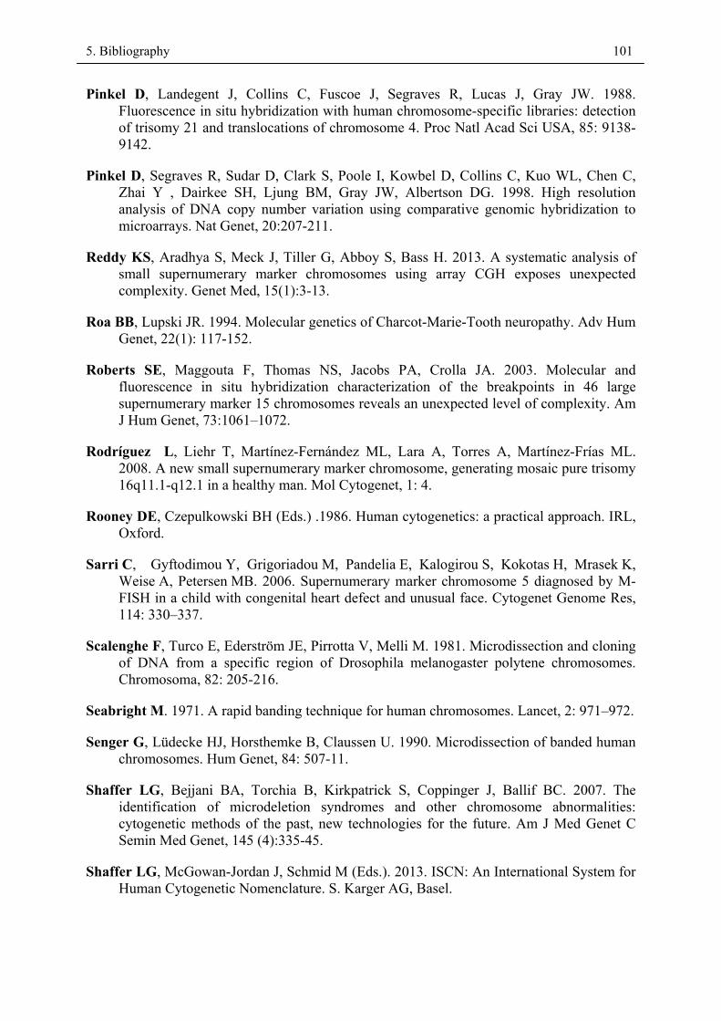
5. Bibliography 101
Pinkel D, Landegent J, Collins C, Fuscoe J, Segraves R, Lucas J, Gray JW. 1988. Fluorescence in situ hybridization with human chromosome-specific libraries: detection of trisomy 21 and translocations of chromosome 4. Proc Natl Acad Sci USA, 85: 9138-9142.
Pinkel D, Segraves R, Sudar D, Clark S, Poole I, Kowbel D, Collins C, Kuo WL, Chen C, Zhai Y , Dairkee SH, Ljung BM, Gray JW, Albertson DG. 1998. High resolution analysis of DNA copy number variation using comparative genomic hybridization to microarrays. Nat Genet, 20:207-211.
Reddy KS, Aradhya S, Meck J, Tiller G, Abboy S, Bass H. 2013. A systematic analysis of small supernumerary marker chromosomes using array CGH exposes unexpected complexity. Genet Med, 15(1):3-13.
Roa BB, Lupski JR. 1994. Molecular genetics of Charcot-Marie-Tooth neuropathy. Adv Hum Genet, 22(1): 117-152.
Roberts SE, Maggouta F, Thomas NS, Jacobs PA, Crolla JA. 2003. Molecular and fluorescence in situ hybridization characterization of the breakpoints in 46 large supernumerary marker 15 chromosomes reveals an unexpected level of complexity. Am J Hum Genet, 73:1061–1072.
Rodríguez L, Liehr T, Martínez-Fernández ML, Lara A, Torres A, Martínez-Frías ML. 2008. A new small supernumerary marker chromosome, generating mosaic pure trisomy 16q11.1-q12.1 in a healthy man. Mol Cytogenet, 1: 4.
Rooney DE, Czepulkowski BH (Eds.) .1986. Human cytogenetics: a practical approach. IRL, Oxford.
Sarri C, Gyftodimou Y, Grigoriadou M, Pandelia E, Kalogirou S, Kokotas H, Mrasek K, Weise A, Petersen MB. 2006. Supernumerary marker chromosome 5 diagnosed by M-FISH in a child with congenital heart defect and unusual face. Cytogenet Genome Res, 114: 330–337.
Scalenghe F, Turco E, Ederström JE, Pirrotta V, Melli M. 1981. Microdissection and cloning of DNA from a specific region of Drosophila melanogaster polytene chromosomes. Chromosoma, 82: 205-216.
Seabright M. 1971. A rapid banding technique for human chromosomes. Lancet, 2: 971–972.
Senger G, Lüdecke HJ, Horsthemke B, Claussen U. 1990. Microdissection of banded human chromosomes. Hum Genet, 84: 507-11.
Shaffer LG, Bejjani BA, Torchia B, Kirkpatrick S, Coppinger J, Ballif BC. 2007. The identification of microdeletion syndromes and other chromosome abnormalities: cytogenetic methods of the past, new technologies for the future. Am J Med Genet C Semin Med Genet, 145 (4):335-45.
Shaffer LG, McGowan-Jordan J, Schmid M (Eds.). 2013. ISCN: An International System for Human Cytogenetic Nomenclature. S. Karger AG, Basel.
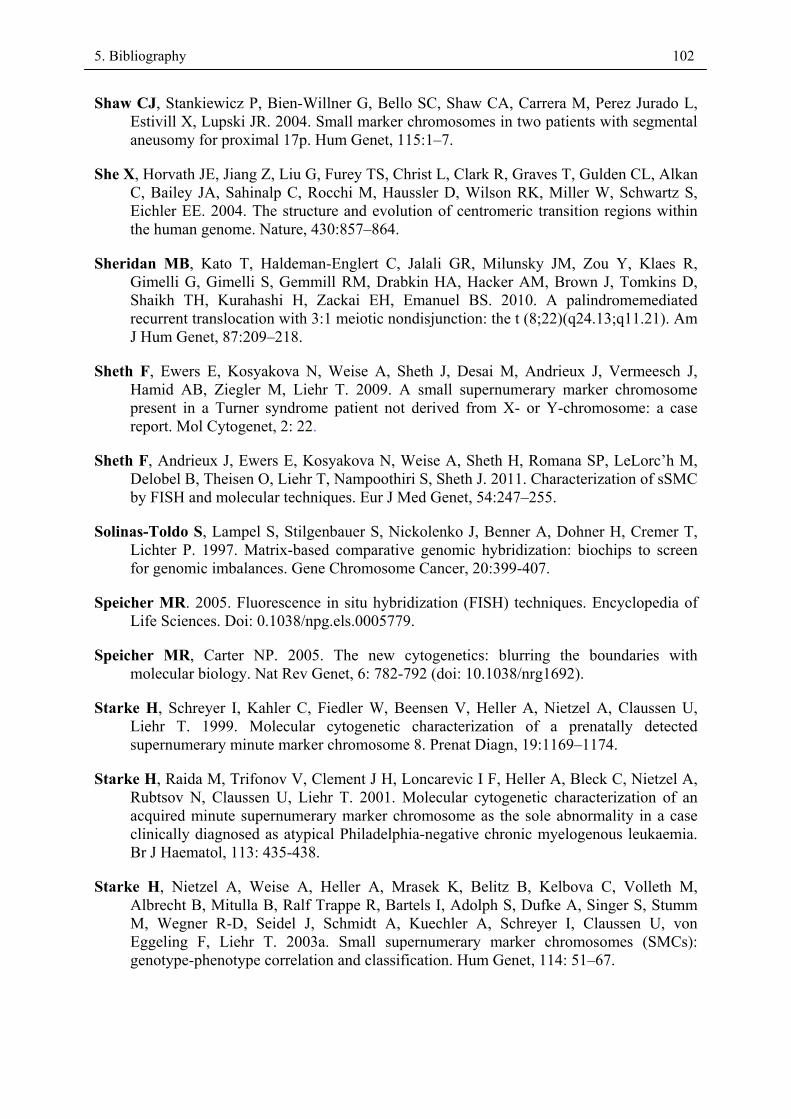
5. Bibliography 102
Shaw CJ, Stankiewicz P, Bien-Willner G, Bello SC, Shaw CA, Carrera M, Perez Jurado L, Estivill X, Lupski JR. 2004. Small marker chromosomes in two patients with segmental aneusomy for proximal 17p. Hum Genet, 115:1–7.
She X, Horvath JE, Jiang Z, Liu G, Furey TS, Christ L, Clark R, Graves T, Gulden CL, Alkan C, Bailey JA, Sahinalp C, Rocchi M, Haussler D, Wilson RK, Miller W, Schwartz S, Eichler EE. 2004. The structure and evolution of centromeric transition regions within the human genome. Nature, 430:857–864.
Sheridan MB, Kato T, Haldeman-Englert C, Jalali GR, Milunsky JM, Zou Y, Klaes R, Gimelli G, Gimelli S, Gemmill RM, Drabkin HA, Hacker AM, Brown J, Tomkins D, Shaikh TH, Kurahashi H, Zackai EH, Emanuel BS. 2010. A palindromemediated recurrent translocation with 3:1 meiotic nondisjunction: the t (8;22)(q24.13;q11.21). Am J Hum Genet, 87:209–218.
Sheth F, Ewers E, Kosyakova N, Weise A, Sheth J, Desai M, Andrieux J, Vermeesch J, Hamid AB, Ziegler M, Liehr T. 2009. A small supernumerary marker chromosome present in a Turner syndrome patient not derived from X- or Y-chromosome: a case report. Mol Cytogenet, 2: 22.
Sheth F, Andrieux J, Ewers E, Kosyakova N, Weise A, Sheth H, Romana SP, LeLorc’h M, Delobel B, Theisen O, Liehr T, Nampoothiri S, Sheth J. 2011. Characterization of sSMC by FISH and molecular techniques. Eur J Med Genet, 54:247–255.
Solinas-Toldo S, Lampel S, Stilgenbauer S, Nickolenko J, Benner A, Dohner H, Cremer T, Lichter P. 1997. Matrix-based comparative genomic hybridization: biochips to screen for genomic imbalances. Gene Chromosome Cancer, 20:399-407.
Speicher MR. 2005. Fluorescence in situ hybridization (FISH) techniques. Encyclopedia of Life Sciences. Doi: 0.1038/npg.els.0005779.
Speicher MR, Carter NP. 2005. The new cytogenetics: blurring the boundaries with molecular biology. Nat Rev Genet, 6: 782-792 (doi: 10.1038/nrg1692).
Starke H, Schreyer I, Kahler C, Fiedler W, Beensen V, Heller A, Nietzel A, Claussen U, Liehr T. 1999. Molecular cytogenetic characterization of a prenatally detected supernumerary minute marker chromosome 8. Prenat Diagn, 19:1169–1174.
Starke H, Raida M, Trifonov V, Clement J H, Loncarevic I F, Heller A, Bleck C, Nietzel A, Rubtsov N, Claussen U, Liehr T. 2001. Molecular cytogenetic characterization of an acquired minute supernumerary marker chromosome as the sole abnormality in a case clinically diagnosed as atypical Philadelphia-negative chronic myelogenous leukaemia. Br J Haematol, 113: 435-438.
Starke H, Nietzel A, Weise A, Heller A, Mrasek K, Belitz B, Kelbova C, Volleth M, Albrecht B, Mitulla B, Ralf Trappe R, Bartels I, Adolph S, Dufke A, Singer S, Stumm M, Wegner R-D, Seidel J, Schmidt A, Kuechler A, Schreyer I, Claussen U, von Eggeling F, Liehr T. 2003a. Small supernumerary marker chromosomes (SMCs): genotype-phenotype correlation and classification. Hum Genet, 114: 51–67.
5. Bibliography 103
Starke H, Mitulla B, Nietzel A, Heller A, Beensen V, Grosswendt G, Claussen U,von Eggeling F, Liehr T. 2003b. First patient with trisomy 21 accompanied by an additional der(4)(:p11→ q11:) plus partial uniparental disomy 4p15-16. Am J Med Genet A, 116A(1):26-30.
Tachdjian G. 2009. Comparative genomic hybridization. In: Encyclopedia of Life Sciences (ELS). John Wiley &Sons, Ltd: Chichester. DOI: 10.1002 / 9780470015902.a0002651.pub2.
Tachdjian G, Aboura A, Portnoï MF, Pasquier M, Bourcigaux N, Simon T, Rousseau G, Finkel L, Benkhalifa M, Christin-Maitre S. 2008. Cryptic Xp duplication including the SHOX gene in a woman with 46,X,del(X)(q21.31) and premature ovarian failure. Human Reproduction, 23: 222–226.
Tan-Sindhunata G, Castedo S, Leegte B, Mulder I, vd Veen AY, vd Hout AH, Wiersma TJ, van Essen AJ. 2000. Molecular cytogenetic characterization of a small, familial supernumerary ring chromosome 7 associated with mental retardation and an abnormal phenotype. Am J Med Genet, 92:147–152.
Teixeira MR. 2002. Combined classical and molecular cytogenetic analysis of cancer. Eur. J. Cancer, 38: 1580–1584.
Thangavelu M, Pergament E, Espinosa R, Stefan K, Bohlander SK. 1994. Characterization of marker chromosomes by microdissection and fluorescence in situ hybridization. Prenat Diagn, 14: 583-588.
Tjio J H, Levan A. 1956. The chromosome number of man. Hereditas, 42:1-6.
Trifonov V, Fluri S, Binkert F, Nandini A, Anderson J, Rodriguez L, Gross M, Kosyakova N, Mkrtchyan H, Ewers E, Reich D, Weise A, Liehr T. 2008. Complex rearranged small supernumerary marker chromosomes (sSMC), three new cases; evidence for an underestimated entity? Mol Cytogenet, 1:6.
Tsuchiya KD, Opheim KE, Hannibal MC, Hing AV, Glass IA, Raff ML, Norwood T, Torchia BA. 2008. Unexpected structural complexity of supernumerary marker chromosomes characterized by microarray comparative genomic hybridization. Mol Cytogenet, 1: 7.
Turnpenny PD, Ellard S (Eds.). 2007. EMERY’S elements of medical genetics.13th Edition, Churchill Livingstone Elsevire.
Vetro A, Manolakos E, Petersen MB, Thomaidis L, Liehr T, Croci G, Franchi F, Marinelli M, Meneghelli E, Dal Bello B, Cesari S, Iasci A, Arrigo G, Zuffardi O. 2012. Unexpected results in the constitution of small supernumerary marker chromosomes. Eur J Med Genet, 55:185–190.
Von Eggeling F, Hoppe C, Bartz U, Starke H, Houge G, Claussen U, Ernst G, Kotzot D, Liehr T. 2002. Maternal uniparental disomy 12 in a healthy girl with a 47,XX, +der (12) (:p11→q11:)/46,XX karyotype. J Med Genet, 39:519–521.
Warburton D. 1984. Outcome of case of de novo structural rearrangements diagnosed at amniocentesis. Prenat Diagn, 4:69–80.
5. Bibliography 104
Weise A, Starke H, Heller A, Tönnies H, Volleth M, Stumm M, Senger G, Nietzel A, Claussen U, Liehr T. 2002. Chromosome 2 aberrations in clinical cases characterised by high resolution multicolour banding and region specific FISH probes. J Med Genet, 39:434–439.
Weise A, Mrasek K, Fickelscher I, Claussen U, Cheung SW, Cai WW, Liehr T, Kosyakova N. 2008. Molecular definition of high-resolution multicolor banding probes: first within the human DNA sequence anchored FISH banding probe set. J Histochem Cytochem, 56:487–493.
Weiss MM, Hermsen MAJA, Meijer GA, van Grieken NCT, Baak JPA, Kuipers EJ, van Diest PJ. 1999. Comparative genomic hybridization. J Clin Pathol: Mol Pathol, 52:243–251.
Wisniewski K, Hassold T, Heffelfinger J, Higgins JV. 1979. Cytogenetic and clinical studies in five cases of inv dup (15). Hum Genet 50: 259-270.
Zhang F, Gu W, Hurles ME, Lupski JR. 2009. Copy number variation in human health, disease, and evolution. Annu Rev Genomics Hum Genet, 10:451–481.
Zody MC, Garber M, Sharpe T, Young SK, Rowen L, O’Neill K, Whittaker CA, Kamal M,Chang JL,CuomoCA. 2006. Analysis of theDNAsequence and duplication history of human chromosome 15. Nature, 440:671–675.

6. Appendix 105
6. Appendix 6.1. List of own publications
Hamid AB, Al-Mashhdani AA, Al-Taee FS, Alwan AH, Fadle RS, Jaffar TH, Jaber TF. Association of Karyotypic abnormalities, Hormones and Cholesterol levels with Infertility. Iraqi J Genet, 2008; 1(1):11-16.
Hamid AB, Al-Mashhdani AA, Ismaéel NH, Fadle RS. Detection of Y chromosome loss in
infertile men (Azoospermia and Oligozoospermia). Iraqi J Genet, 2008; 1(1):37–42. Hamid AB, Yaseen NY, Dalloul RA-H . Y Chromosome loss in elderlies and some cancer
patients. Iraqi J Genet, 2008; 1(1):49–58. Hamid AB, Yaseen NY, Al – Hilli ZA-M, Ali AM, Salah Al-Deen M, Al-Mukhtar AA.
Chromosomal Analysis in Fetuses by using Prenatal Diagnosis Technique. Iraqi J Genet, 2008; 1(2): 8-15.
Al-Mukhtar AA, Hamid AB, Ismaéel NH, Alwan AH. Micronuclei in cultured lymphocytes from
workers of sewage water treatment stations. Iraqi J Genet, 2008; 1(2): 39-42. Sheth F, Ewers E, Kosyakova N, Weise A, Sheth J, Desai M, Andrieux J, Vermeesch J, Hamid
AB, Ziegler M, Liehr T. A small supernumerary marker chromosome present in a Turner syndrome patient not derived from X- or Y-chromosome. Mol Cytogenet, 2009; 2:22.
Liehr T, Wegner R-D, Stumm M, Martin T, Gillessen-Kaesbach G, Kosyakova N, Ewers E, Hamid AB, von Eggeling F, Hentschel J, Ziegler M, Weise A. Three new cases with small supernumerary marker chromosomes 1 and normal phenotype. J Chin Med Assoc, 2010; 73: 205-207.
Nelle H, Schreyer I, Ewers E, Mrasek K, Kosyakova N, Merkas M, Hamid AB, Weise A, Liehr T. Harmless familial small supernumerary marker chromosome 22 hampers diagnosis of fragile X-syndrome. Molecular Medicine Reports, 2010; 3:571-574.
Ewers E, Yoda K, Hamid AB, Weise A, Manvelyan M, Liehr T. Centromere activity in dicentric small supernumerary marker chromosomes. Chromosome Res, 2010; 18:555-562.
Liehr T, Karamysheva T, Merkas M, Brecevic L, Hamid AB, Ewers E, Mrasek K, Kosyakova N, Weise A. Somatic mosaicism in cases with small supernumerary marker chromosomes. Curr Genomics, 2010; 11:432-439.
Fernández-Toral J, Rodríguez L, Plasencia A, Martínez-Frías ML, Ewers E, Hamid AB, Ziegler M, Liehr T. Four small supernumerary marker chromosomes derived from chromosomes 6, 8, 11 and 12 in a patient with minimal clinical abnormalities – a case report. J Med Case Reports, 2010; 4:239.
Liehr T, Bartels I, Zoll B, Ewers E, Mrasek K, Kosyakova N, Merkas M, Hamid AB, von Eggeling F, Posorski N, Weise A. Is there a yet unreported unbalanced chromosomal abnormalities without phenotypic consequences in proximal 4p? Cytogenet Genome Res, 2011; 132: 121-123.
6. Appendix 106
Liehr T, Ewers E, Hamid AB, Kosyakova N, Voigt M, Weise A, Manvelyan M. Small supernumerary marker chromosomes and uniparental disomy have a story to tell. J Histochem Cytochem, 2011; 59: 842-848.
Papoulidis I, Manolakos E, Hamid AB, Klein E, Kosyakova N, Kordaß U, Kunz J, Siomou E, Kontodiou M, Tzimina M, Nicolaides P, Liehr T, Petersen MB. Tetrasomy 9p mosaicism associated with a normal phenotype in two cases. Cytogenet Genome Res, 2012; 136: 237–241.
Klein E, Manvelyan M, Simonyan I, Hamid AB, Guilherme RS, Liehr T, Karamysheva T. Centromeric association of small supernumerary marker chromosomes with their sister chromosomes detected by three dimensional molecular cytogenetics. Molecular Cytogenetics, 2012; 5:15.
Bucksch M, Ziegler M, Kosayakova N, Mulhatino MV, Llerena Jr. JC, Morlot S, Fischer W, Polityko AD, Kulpanovich AI, Petersen MB, Belitz B, Trifonov V, Weise A, Liehr T, Hamid AB. A new multicolor-fluorescence in situ hybridization probe set directed against human heterochromatin: HCM-FISH. J Histochem Cytochem.2012; 60(7): 530-536.
Guilherme RS, Klein E, Venner C, Hamid AB, Bhatt S, Melaragno MI, Volleth M, Polityko A, Kulpanovich A, Kosyakova N, Liehr T. Human ring chromosomes and small supernumerary marker chromosomes – do they have telomeres? Chromosome Res, 2012; 20:825–835.
Hamid AB, Weise A, Voigt M, Bucksch M, Kosyakova N, Liehr T, Klein E. Clinical impact of proximal autosomal imbalances. Balk J Med Genet, 2012; 15(2): 15-21.
Hamid AB, Kreskowski K, Weise A, Kosayakova N, Mrasek K, Voigt M, Guilherme RS, Wagner R, Hardekopf D, Pekova S, Karamysheva T, Liehr T, Klein E. How to narrow down chromosomal breakpoints in small and large derivative chromosomes – a new probe set. J Appl Genet , 2012; 53(3): 259-269.
Guilherme RS, Dutra ARN, Perez ABA, Takeno SS, Oliveira MM, Kulikowski LD, Klein E, Hamid AB, Liehr T, Melaragno MI. First Report of a Small Supernumerary der(8;14) Marker Chromosome. Cytogenet Genome Res, 2013; 139:284-288.
Ou J, Wang W, Liehr T, Klein E, Hamid AB, Wang F, Duan C, Li H. Characterization of three small supernumerary marker chromosomes (sSMC) in humans. J Matern Fetal Neonatal Med,2013; 26(1):106–108.
Liehr T, Klein E, Mrasek K, Kosyakova N, Guilherme RS, Aust N, Venner C, Weise A, Hamid AB. Clinical impact of somatic mosaicism in cases with small supernumerary marker chromosomes. Cytogenet Genome Res, 2013;139(3):158–163.
Kosyakova N, Hamid AB, Chaveerach A, Pinthong K, Siripiyasing P, Supiwong W, Romanenko S, Trifonov V, Fan X. Generation of multicolor banding probes for chromosomes of different species. Molecular Cytogenetics, 2013; 6:6.
Liehr T, Weise A, Hamid AB, Fan X, Klein E, Aust N, Othman MAK, Mrasek K, Kosyakova N. Multicolor fluorescence in situ hybridization methods in current clinical diagnostics. Exp Rev Mol Diag, 2013; 13(3): 251–255.
6. Appendix 107
Guilherme R S, Klein E, Hamid AB, Bhatt S, Volleth M, Polityko A, Kulpanovich A, Dufke A, Albrecht B, Morlot S, Brecevic L, Petersen M B, Manolakos E, Kosyakova N, Liehr T. Human ring chromosomes-new insights for their clinical significance. Balkan J Med Genet, 2013; 16(1):13-19.
Abo-Zeid MAM, Liehr T, El-Daly SM, Gamal-Eldeen AM, Glei M, Shabakae A, Bhatt S, Hamid AB. Molecular cytogenetic evaluation of the efficacy of photodynamic therapy by indocyanine green in breast adenocarcinoma MCF-7 cells. Photodiagnosis and Photodynamic Therapy, 2013; 10: 194-202.
Liehr T, Cirkovic S, Lalic T, Guc-Scekic M, de Almeida C, Weimer J, Iourov I, Melaragno M I, Guilherme R S, Stefanou E-G G, Aktas D, Kreskowski1 K, Klein1 E, Ziegler1 M, Kosyakova1 N, Volleth M, Hamid AB. Complex small supernumerary marker chromosomes – an update. Molecular Cytogenetics, 2013; 6:46.
Hamid AB, Liehr T. Pericentromeric BAC-probe set - thoughts about considering genedosage insensitive regions. Mol Cytogenet 2013; 6:45/comments.
Spittel H, Kubek F, Kreskowski K, Ziegler M, Klein E, Hamid AB, Kosyakova N, Radhakrishnan G, Junge A, Kozlowski P, Schulze B, Martin T, Huhle D, Mehnert K, Rodríguez L, Ergun M A, Sarri C, Militaru M, Stipoljev F, Tittelbach H, Vasheghani F, Cioffi MB, Hussein SS, Fan X, Volleth M, Liehr T. Mitotic stability of small supernumerary marker chromosomes : A study based on 93 immortalized cell lines. Cytogenet Genome Res, 2014; 142: 151-160.
Klein E, Hamid AB, Volleth M, Liehr T. Human dicentric chromosomes and their centromere activity. MedGen 2014, 26:168 (Abstractnr. P-CytoG-177).
Jena, 28. 05. 2014
6. Appendix 108
6.2. Curriculum Vitae
- Personal information
- Full Name: Ahmed Basheer Hamid
- Address : Saalstr. 10, 07743 Jena, Germany
• Tel.: (+49) 0176 31563353
• Email: [email protected]
- Birth date: 30 June 1973
- Education
- 1979 – 1985 Al-Rifaie Primary school, Thi-Qar, Iraq.
- 1985 – 1991 Al-Rifaie Secondary school, Thi-Qar, Iraq.
- 1991 – 1995 B.Sc. in Biology from College of Education, Baghdad University, Iraq
- 2000 – 2003 M.Sc. degree in Zoology / Cytogenetics from College of Science, Al-
Mustansiriya University, Iraq
- Award
DAAD fellowship for Ph.D. study in Friedrich Schiller University of Jena (2009-2013)
-Work experience
- Cell Culture and Chromosomal analysis with GTG Banding.
- Molecular Cytogenetics; Fluorescent in-situ hybridization (FISH) techniques.
- Array- based Comparative Genomic Hybridization (a-CGH) technique.
- Glass Needle Base Microdissection technique.
- Degenerate Oligonucleotide-Primed (DOP-PCR) technique.
- DNA Reamplification and Labelling technique.
- Plasmid DNA Purification.
- Record of Employment
- 1998 – 1999 Asst. researcher, Department of Cancer Research, Iraqi Centre for Cancer
& Medical Genetics Research (ICCMGR).
- 1999 – 2000 Chief of laboratories / ICCMGR.
6. Appendix 109
- 2000 – 2003 M.Sc. Student in College of Science/Al-Mustansiriya University / Iraq, in
Cytogenetics field and the thesis title “A Study of The Frequency of Y Chromosome
Loss in Some Cancers and Elderlies”
- 2003 – 2004 Researcher, Department of Medical Genetics, ICCMGR.
- 2004 Vice-Chairman and Researcher, Department of Medical Genetics, ICCMGR.
- 2004 – 2007 Chairman and Researcher, Department of Medical Genetics, ICCMGR
- 2007 – 2008 Lecturer in Cytology, Genetic and Ecology laboratories / Biology
Department / College of science / Thi-Qar University.
- 2008 – 2009 Researcher, Department of Medical Genetics, ICCMGR.
- October 2009 till now Ph.D. student in group of PD Dr. rer. nat./ med.habil. Thomas
Liehr / Molecular Cytogenetics Department / Institute for Human Genetics / Jena
University Hospital / Friedrich Schiller University of Jena.
- October 2013 till now scientist co-worker in group of PD Dr. rer. nat./ med.habil.
Thomas Liehr / Molecular Cytogenetics Department / Institute for Human Genetics /
Jena University Hospital / Friedrich Schiller University of Jena.
- Contributions in the international conferences and workshops
- 21st Annual Meeting of the German Society of Human Genetics – Hamburg 2-4
March 2010.
- Annual Conference of the German Genetic Society (GfG) Evolution of Primates,
16-18 September 2010, Jena, Germany.
- 12th Day of Microscopy Jena 9th November 2011 (Zeizz Workshop).
- 3rd B-Chromosome Conference, Leibniz Institute of Plant Genetics and Crop Plant
Research (IPK)- Gatersleben, Germany, 7 – 9 April 2014.
- National Research Center of Mental Health, RAS, Moscow, Russia, 27 – 31 August
2012 (Workshop).
- Personal skills and competences
- Training Courses
1. Academic Teaching Course from 13 to 29 October 2003 / Teaching Methods Unit /
College of Education / Al-Mustansiriya University.
2. First International Course on Small Supernumerary Marker Chromosomes (sSMC) -
Institute for Human Genetics / Jena University Hospital, 2009.
6. Appendix 110
3. International Course on Fluorescence in Situ Hybridization (FISH, a-CGH,
Microdissection) of marker chromosomes - Institute for Human Genetics / Jena
University Hospital, 2009.
- Languages
- Mother tongue: Arabic
- Other languages: English
German (Basic user / A2)
Language certificates:
1. English Language Test Certificate at University of Baghdad in 2000.
2. TOEFL. ITP- English Language Test Certificate 2009.
3. DUO Deutsch – Uni Online Certificate (13 March 2009 – 30 June 2009)
4. Intensive German language course – interDaf e.V. am Herder – Institute of Leipzig
University (02 June2009 – 30 September 2009).
Computer certificates:
1. Proficiency Certificate in using the computer from 1 to 30 August 2000, Consulting
Bureau / College of Science / Al-Mustansiriya University.
2. Proficiency Certificate in using the computer from 3 to 20 July 2005 / Consultation
Bureau of Information Systems and Computers / Baghdad University.
Jena, 28. 05. 2014
Ahmed Basheer Hamid
6. Appendix 111
6.3. Acknowledgements
First and foremost I would like to extend my sincerest thanks and gratitude and appreciation
to my supervisor PD Dr. rer. nat./ med. habil. Thomas Liehr, for providing me the
opportunity to accomplish my doctoral thesis in his lab and throughout supporting me with his
patience and excellent scientific supervision. I am very grateful for your encouragement and
effort and your suggestions for corrections which improved the quality of this thesis
enormously. Without you this thesis would not have been completed.
I also extend my thanks and gratitude to German Academic Exchange Service (DAAD) to
give me the opportunity to complete my study at doctorate through support me financially and
morally. My heartfelt thanks to all the people working there, especially those whom
responsible for the Iraq department.
Many thanks to The institute directors Prof. Aria Baniahmad and Prof. Christian Hübner
for enabling my PhD at the Institute of Human Genetics at the University Hospital Jena.
Without Dr. med. Nadezda Kosyakova, I would not have been able to accomplish my work.
Your support in the practical training made it possible to carry out microdissection at all. I am
thankful for you.
Thank you very much to Monika Ziegler and Katharina Kreskowski for your cooperation
with me during the practical application in my study and provide me what I needs to
laboratory work.
At last, many thanks to Dr. Samarth Bhatt for helpful hints and tricks and for entertaining
hours in the lab. I would like to thank all my colleagues at work to support me morally.
I am extremely grateful to my parents and family for their everlasting care and support in
many different ways which strengthens my resolve to work ahead.
Finally, my great special thanks go to my wife, who is always beside me to give me the
necessary support and my lovely children who give hope and motivation to continue in my
work.
6. Appendix 112
6.4. Ehrenwörtliche Erklärung
Hiermit erkläre ich, dass mir die Promotionsordnung der Medizinischen Fakultät der
Friedrich-Schiller-Universität bekannt ist,
ich die Dissertation selbst angefertigt habe und alle von mir benutzten Hilfsmittel,
persönlichen Mitteilungen und Quellen in meiner Arbeit angegeben sind,
mich folgende Personen bei der Auswahl und Auswertung des Materials sowie bei der
Herstellung des Manuskripts unterstützt haben: PD Dr. rer. nat./ med. habil. Thomas Liehr.
die Hilfe eines Promotionsberaters nicht in Anspruch genommen wurde und dass Dritte weder
unmittelbar noch mittelbar geldwerte Leistungen von mir für Arbeiten erhalten haben, die im
Zusammenhang mit dem Inhalt der vorgelegten Dissertation stehen,
dass ich die Dissertation noch nicht als Prüfungsarbeit für eine staatliche oder andere
wissenschaftliche Prüfung eingereicht habe und
dass ich die gleiche, eine in wesentlichen Teilen ähnliche oder eine andere Abhandlung nicht
bei einer anderen Hochschule als Dissertation eingereicht habe.
Jena, 28. 05. 2014
Ahmed Basheer Hamid
Related Documents


![Brief CV English[1]«Supernumerary marker chromosomes (SMC’s) in Turner syndrome are mostly derived from Y chromosome». Clinical Genetics, 51: 184 - 190, 1997 I.F: 3.276 9. …](https://static.cupdf.com/doc/110x72/5f03cf897e708231d40ae2a1/brief-cv-english1-supernumerary-marker-chromosomes-smcas-in-turner-syndrome.jpg)