The Genome of Nectria haematococca: Contribution of Supernumerary Chromosomes to Gene Expansion Jeffrey J. Coleman 1,2. , Steve D. Rounsley 3. , Marianela Rodriguez-Carres 1,4. , Alan Kuo 5 , Catherine C. Wasmann 1 , Jane Grimwood 6,7 , Jeremy Schmutz 6,7 , Masatoki Taga 8 , Gerard J. White 1 , Shiguo Zhou 9 , David C. Schwartz 9 , Michael Freitag 10 , Li-jun Ma 11 , Etienne G. J. Danchin 12,13 , Bernard Henrissat 12 , Pedro M. Coutinho 12 , David R. Nelson 14 , Dave Straney 15 , Carolyn A. Napoli 1 , Bridget M. Barker 1 , Michael Gribskov 16 , Martijn Rep 17 , Scott Kroken 1 , Istva ´ n Molna ´r 18 , Christopher Rensing 19 , John C. Kennell 20 , Jorge Zamora 1 , Mark L. Farman 21 , Eric U. Selker 22 , Asaf Salamov 5 , Harris Shapiro 5 , Jasmyn Pangilinan 5 , Erika Lindquist 5 , Casey Lamers 9 , Igor V. Grigoriev 5 , David M. Geiser 23 , Sarah F. Covert 24 , Esteban Temporini 1,25 , Hans D. VanEtten 1 * 1 Department of Plant Sciences, University of Arizona, Tucson, Arizona, United States of America, 2 Massachusetts General Hospital, Boston, Massachusetts, United States of America, 3 BIO5 Institute and Department of Plant Sciences, University of Arizona, Tucson, Arizona, United States of America, 4 Department of Biology, Duke University, Durham, North Carolina, United States of America, 5 United States Department of Energy Joint Genome Institute, Walnut Creek, California, United States of America, 6 Joint Genome Institute—Stanford Human Genome Center, Palo Alto, California, United States of America, 7 Hudson Alpha Genome Sequencing Center, Hudson Alpha Institute for Biotechnology, Huntsville, Alabama, United States of America, 8 Department of Biology, Okayama University, Okayama, Japan, 9 Laboratory for Molecule and Computational Genomics, University of Wisconsin, Madison, Wisconsin, United States of America, 10 Department of Biochemistry and Biophysics and Center for Genome Research and Biocomputing, Oregon State University, Corvallis, Oregon, United States of America, 11 The Broad Institute, Cambridge, Massachusetts, United States of America, 12 Architecture et Fonction des Macromole ´cules Biologiques, CNRS, Universite ´s Aix-Marseille I & II, Marseille, France, 13 Institut National de la Recherche Agronomique, Centre de recherche de Sophia-Antipolis, Sophia-Antipolis, France, 14 Department of Molecular Sciences, University of Tennessee, Memphis, Tennessee, United States of America, 15 Department of Cell Biology and Molecular Genetics, University of Maryland, College Park, Maryland, United States of America, 16 Department of Biological Sciences, Purdue University, West Lafayette, Indiana, United States of America, 17 Plant Pathology, University of Amsterdam, Amsterdam, The Netherlands, 18 South West Center for Natural Products Research and Commercialization, Office of Arid Lands Studies, University of Arizona, Tucson, Arizona, United States of America, 19 Department of Soil, Water, and Environmental Science, University of Arizona, Tucson, Arizona, United States of America, 20 Department of Biology, Saint Louis University, St. Louis, Missouri, United States of America, 21 Department of Plant Pathology, University of Kentucky, Lexington, Kentucky, United States of America, 22 Institute of Molecular Biology, University of Oregon, Eugene, Oregon, United States of America, 23 Fusarium Research Center, Department of Plant Pathology, The Pennsylvania State University, University Park, Pennsylvania, United States of America, 24 Warnell School of Forestry and Natural Resources, University of Georgia, Athens, Georgia, United States of America, 25 Vilmorin Inc., Tucson, Arizona, United States of America Abstract The ascomycetous fungus Nectria haematococca, (asexual name Fusarium solani), is a member of a group of .50 species known as the ‘‘Fusarium solani species complex’’. Members of this complex have diverse biological properties including the ability to cause disease on .100 genera of plants and opportunistic infections in humans. The current research analyzed the most extensively studied member of this complex, N. haematococca mating population VI (MPVI). Several genes controlling the ability of individual isolates of this species to colonize specific habitats are located on supernumerary chromosomes. Optical mapping revealed that the sequenced isolate has 17 chromosomes ranging from 530 kb to 6.52 Mb and that the physical size of the genome, 54.43 Mb, and the number of predicted genes, 15,707, are among the largest reported for ascomycetes. Two classes of genes have contributed to gene expansion: specific genes that are not found in other fungi including its closest sequenced relative, Fusarium graminearum; and genes that commonly occur as single copies in other fungi but are present as multiple copies in N. haematococca MPVI. Some of these additional genes appear to have resulted from gene duplication events, while others may have been acquired through horizontal gene transfer. The supernumerary nature of three chromosomes, 14, 15, and 17, was confirmed by their absence in pulsed field gel electrophoresis experiments of some isolates and by demonstrating that these isolates lacked chromosome-specific sequences found on the ends of these chromosomes. These supernumerary chromosomes contain more repeat sequences, are enriched in unique and duplicated genes, and have a lower G+C content in comparison to the other chromosomes. Although the origin(s) of the extra genes and the supernumerary chromosomes is not known, the gene expansion and its large genome size are consistent with this species’ diverse range of habitats. Furthermore, the presence of unique genes on supernumerary chromosomes might account for individual isolates having different environmental niches. PLoS Genetics | www.plosgenetics.org 1 August 2009 | Volume 5 | Issue 8 | e1000618

Welcome message from author
This document is posted to help you gain knowledge. Please leave a comment to let me know what you think about it! Share it to your friends and learn new things together.
Transcript
The Genome of Nectria haematococca: Contribution ofSupernumerary Chromosomes to Gene ExpansionJeffrey J. Coleman1,2., Steve D. Rounsley3., Marianela Rodriguez-Carres1,4., Alan Kuo5, Catherine C.
Wasmann1, Jane Grimwood6,7, Jeremy Schmutz6,7, Masatoki Taga8, Gerard J. White1, Shiguo Zhou9,
David C. Schwartz9, Michael Freitag10, Li-jun Ma11, Etienne G. J. Danchin12,13, Bernard Henrissat12,
Pedro M. Coutinho12, David R. Nelson14, Dave Straney15, Carolyn A. Napoli1, Bridget M. Barker1, Michael
Gribskov16, Martijn Rep17, Scott Kroken1, Istvan Molnar18, Christopher Rensing19, John C. Kennell20,
Jorge Zamora1, Mark L. Farman21, Eric U. Selker22, Asaf Salamov5, Harris Shapiro5, Jasmyn Pangilinan5,
Erika Lindquist5, Casey Lamers9, Igor V. Grigoriev5, David M. Geiser23, Sarah F. Covert24, Esteban
Temporini1,25 , Hans D. VanEtten1*
1 Department of Plant Sciences, University of Arizona, Tucson, Arizona, United States of America, 2 Massachusetts General Hospital, Boston, Massachusetts, United States
of America, 3 BIO5 Institute and Department of Plant Sciences, University of Arizona, Tucson, Arizona, United States of America, 4 Department of Biology, Duke University,
Durham, North Carolina, United States of America, 5 United States Department of Energy Joint Genome Institute, Walnut Creek, California, United States of America,
6 Joint Genome Institute—Stanford Human Genome Center, Palo Alto, California, United States of America, 7 Hudson Alpha Genome Sequencing Center, Hudson Alpha
Institute for Biotechnology, Huntsville, Alabama, United States of America, 8 Department of Biology, Okayama University, Okayama, Japan, 9 Laboratory for Molecule and
Computational Genomics, University of Wisconsin, Madison, Wisconsin, United States of America, 10 Department of Biochemistry and Biophysics and Center for Genome
Research and Biocomputing, Oregon State University, Corvallis, Oregon, United States of America, 11 The Broad Institute, Cambridge, Massachusetts, United States of
America, 12 Architecture et Fonction des Macromolecules Biologiques, CNRS, Universites Aix-Marseille I & II, Marseille, France, 13 Institut National de la Recherche
Agronomique, Centre de recherche de Sophia-Antipolis, Sophia-Antipolis, France, 14 Department of Molecular Sciences, University of Tennessee, Memphis, Tennessee,
United States of America, 15 Department of Cell Biology and Molecular Genetics, University of Maryland, College Park, Maryland, United States of America, 16 Department
of Biological Sciences, Purdue University, West Lafayette, Indiana, United States of America, 17 Plant Pathology, University of Amsterdam, Amsterdam, The Netherlands,
18 South West Center for Natural Products Research and Commercialization, Office of Arid Lands Studies, University of Arizona, Tucson, Arizona, United States of America,
19 Department of Soil, Water, and Environmental Science, University of Arizona, Tucson, Arizona, United States of America, 20 Department of Biology, Saint Louis
University, St. Louis, Missouri, United States of America, 21 Department of Plant Pathology, University of Kentucky, Lexington, Kentucky, United States of America,
22 Institute of Molecular Biology, University of Oregon, Eugene, Oregon, United States of America, 23 Fusarium Research Center, Department of Plant Pathology, The
Pennsylvania State University, University Park, Pennsylvania, United States of America, 24 Warnell School of Forestry and Natural Resources, University of Georgia, Athens,
Georgia, United States of America, 25 Vilmorin Inc., Tucson, Arizona, United States of America
Abstract
The ascomycetous fungus Nectria haematococca, (asexual name Fusarium solani), is a member of a group of .50 speciesknown as the ‘‘Fusarium solani species complex’’. Members of this complex have diverse biological properties including theability to cause disease on .100 genera of plants and opportunistic infections in humans. The current research analyzed themost extensively studied member of this complex, N. haematococca mating population VI (MPVI). Several genes controllingthe ability of individual isolates of this species to colonize specific habitats are located on supernumerary chromosomes.Optical mapping revealed that the sequenced isolate has 17 chromosomes ranging from 530 kb to 6.52 Mb and that thephysical size of the genome, 54.43 Mb, and the number of predicted genes, 15,707, are among the largest reported forascomycetes. Two classes of genes have contributed to gene expansion: specific genes that are not found in other fungiincluding its closest sequenced relative, Fusarium graminearum; and genes that commonly occur as single copies in otherfungi but are present as multiple copies in N. haematococca MPVI. Some of these additional genes appear to have resultedfrom gene duplication events, while others may have been acquired through horizontal gene transfer. The supernumerarynature of three chromosomes, 14, 15, and 17, was confirmed by their absence in pulsed field gel electrophoresisexperiments of some isolates and by demonstrating that these isolates lacked chromosome-specific sequences found onthe ends of these chromosomes. These supernumerary chromosomes contain more repeat sequences, are enriched inunique and duplicated genes, and have a lower G+C content in comparison to the other chromosomes. Although theorigin(s) of the extra genes and the supernumerary chromosomes is not known, the gene expansion and its large genomesize are consistent with this species’ diverse range of habitats. Furthermore, the presence of unique genes onsupernumerary chromosomes might account for individual isolates having different environmental niches.
PLoS Genetics | www.plosgenetics.org 1 August 2009 | Volume 5 | Issue 8 | e1000618
Citation: Coleman JJ, Rounsley SD, Rodriguez-Carres M, Kuo A, Wasmann CC, et al. (2009) The Genome of Nectria haematococca: Contribution of SupernumeraryChromosomes to Gene Expansion. PLoS Genet 5(8): e1000618. doi:10.1371/journal.pgen.1000618
Editor: Hiten D. Madhani, University of California San Francisco, United States of America
Received April 20, 2009; Accepted July 27, 2009; Published August 28, 2009
This is an open-access article distributed under the terms of the Creative Commons Public Domain declaration which stipulates that, once placed in the publicdomain, this work may be freely reproduced, distributed, transmitted, modified, built upon, or otherwise used by anyone for any lawful purpose.
Funding: This work was performed under the auspices of the US Department of Energy’s Office of Science, Biological and Environmental Research Program, andby the University of California, Lawrence Berkeley National Laboratory under Contract DE-AC02-05CH11231, Lawrence Livermore National Laboratory underContract DE-AC52-07NA27344, and Los Alamos National Laboratory under Contract DE-AC02-06NA25396. Personnel at these laboratories were involved in allaspects of this research. The sequence of Nectria haematococca is available at http://www.jgi.doe.gov/nectria. Partial support for personnel was also obtainedfrom NRA/USDA grant 2008-00645.
Competing Interests: The authors have declared that no competing interests exist.
* E-mail: [email protected]
. These authors contributed equally to the research.
Introduction
The fungus Nectria haematococca, commonly referred to by its
asexual name Fusarium solani, is a member of a monophyletic clade
that includes over 50 phylogenetic species known as the ‘‘Fusarium
solani species complex’’ [1,2]. Members of the F. solani species
complex are able to colonize an impressive variety of environ-
ments. As saprobes, they are present in agricultural and non-
cultivated habitats, such as forests, scrub communities, savannahs,
prairies, swamps, littoral zones, coastal zones, and deserts [3]. As
pathogens, members of this complex are responsible for disease on
,100 genera of plants [4], and they represent one of the most
important group of pathogens associated with opportunistic fungal
infections and keratitis in humans [2,5–8]. Because of their diverse
host range, some members have been proposed for the biological
control of weeds and other pathogens [9–11]. Extreme environ-
ments are not beyond the reach of these fungi. F. solani is among
the fungal species recovered from the highly radioactive inner
parts of the damaged nuclear reactor at Chernobyl [12]. These
fungi are capable of growing in anaerobic conditions in the soil
[13] and are tolerant to many compounds shown to be toxic to
other fungi [14,15]. F. solani also has been found growing in the
caves at Lascaux, France where it is damaging the 15,000 year-old
paintings [16]. The ability of these fungi to adapt to so many
different environments reflects their genetic plasticity and
metabolic diversity. Individual members of this species complex
can degrade hydrocarbons, organofluorine compounds, lignin,
metal cyanides, and pesticides in the soil [17–29].
The most extensively studied species of the F. solani complex is
‘‘mating population’’ (MP) VI of N. haematococca (also called
Haematonectria haematococca [30]). The term ‘‘mating population’’
defines a group of isolates that are sexually fertile with one
another, indicating that they are a biological species. Like other F.
solani species, N. haematococcaa MPVI isolates can live in many
habitats [31] and classical and molecular genetic analyses have
demonstrated that the genes controlling the ability of individual
isolates to colonize specific habitats are located on conditionally-
dispensable supernumerary chromosomes (‘‘CD chromosomes’’),
which were first described in fungi in 1991 using N. haematococca
MPVI [32]. CD chromosomes are defined as supernumerary
chromosomes that are not required for growth under all conditions
but confer an adaptive advantage in certain habitats [33].
Subsequent research has demonstrated that in N. haematococca
MPVI, genes on these chromosomes are involved in resistance to
plant antimicrobials, utilization of specific carbon and nitrogen
sources, and in host-specific pathogenicity [33–35]. In addition,
the properties of the chromosomes and the properties of the genes
on these chromosomes, suggest that some of these genes and
perhaps even the entire chromosome(s) might have been acquired
through horizontal gene transfer (HGT) and have properties
similar to the genomic islands of bacteria [36]. The genomic
sequence of N. haematococca MPVI has not only the potential to
reveal a multitude of metabolic pathways involved in inhabiting
many different types of environments, but also to expand on our
understanding of the impact of gene flow on fungal evolution.
Results
General genome featuresOptical mapping revealed that N. haematococca MPVI isolate 77-
13-4 has 17 chromosomes ranging from 530 kb to 6.52 Mb and
that the physical size of the genome, 54.43 Mb, is larger than that
of any other published ascomycete (Table S1). This is 15% greater
than that of the most closely related sequenced fungus, Fusarium
graminearum (sexual name: Gibberella zeae), which is known to have
undergone significant gene expansion itself [37]. The average gene
length (1.67 kb), number of exons (3.08), intron size (84 nt), and
the size of the encoded protein (480 aa) (Table S2) are similar to
other sequenced ascomycetes [38,39]. Of the gene families in N.
haematococca MPVI with at least ten genes, 77% (226 of 293) have
more genes than their counterpart in F. graminearum; 18% have
more than twice as many genes (Figure S1). As might be expected
for a metabolically diverse fungus that can live in so many habitats,
among the gene families with the greatest numerical increases are
carbohydrate-active enzymes, oxidoreductases, and various mono-
oxygenases and dioxygenases (Table S3).
Chromosomal location of genes similar to other fungiTo determine why N. haematococca MPVI might have more genes
than F. graminearum and to see if these ‘‘extra’’ genes are similar to
genes from other fungi, the proteome of N. haematococca MPVI was
compared to the genomes of eight other sequenced fungi (F.
graminearum, Aspergillus oryzae, A. nidulans, Coccidioides immitis, Chaeto-
mium globosum, Magnaporthe oryzae, Neurospora crassa, and Saccharomyces
cerevisiae). The predicted genes of N. haematococca MPVI were
classified into three groups: those most similar to F. graminearum,
those most similar to genes from the other fungi used in the
comparison, or those with no similarity to genes from any of the
included genomes. 61.5% were most similar to F. graminearum genes,
28.5% were more similar to the genes from other fungi, and 6.4%
had no good match with any of the other genomes. Among the
genes with the highest similarity outside F. graminearum, the highest
similarity was to Aspergillus species (1786 genes), particularly to A.
oryzae (811 genes).
The percentage of the genes in each category also was
determined for each chromosome. With the exception of
chromosome 7, the majority (.60%) of the genes on the large
Nectria haematococca and Supernumerary Chromosomes
PLoS Genetics | www.plosgenetics.org 2 August 2009 | Volume 5 | Issue 8 | e1000618
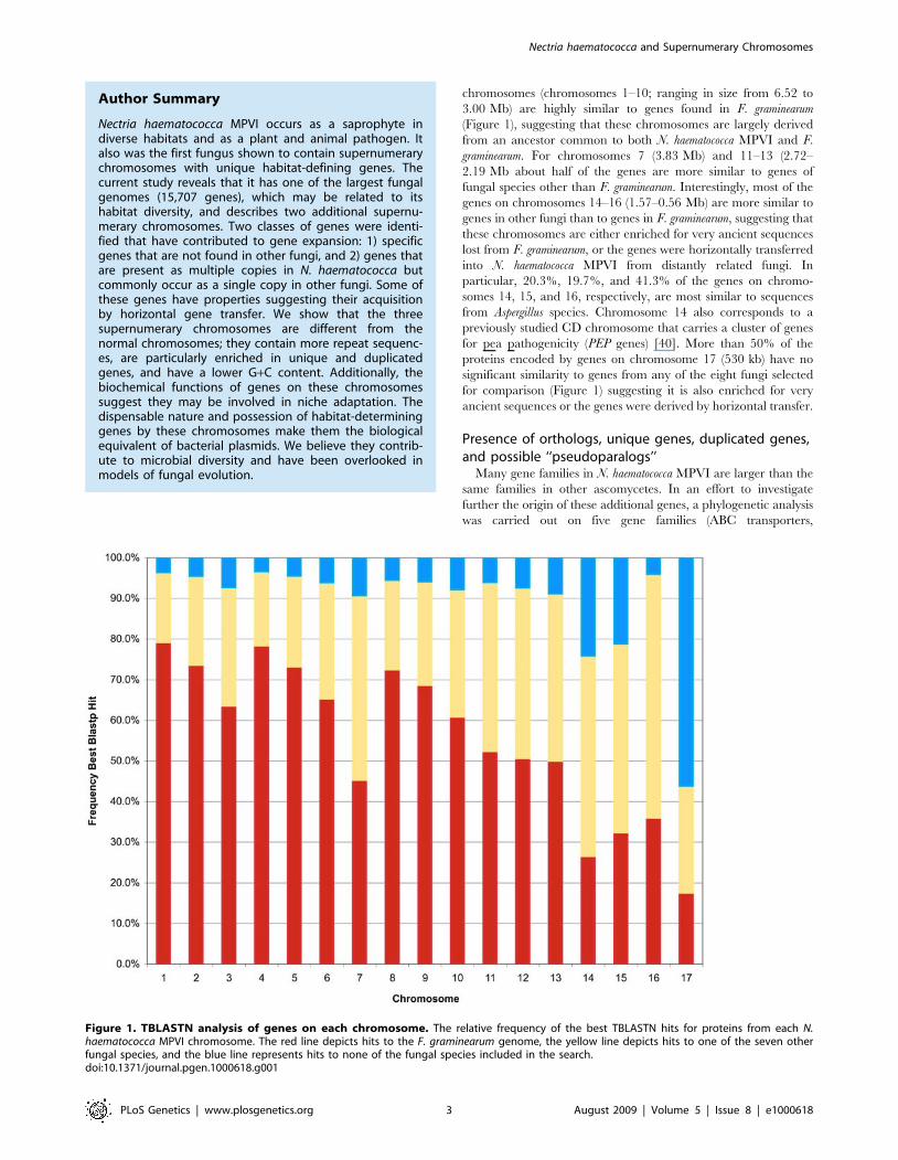
chromosomes (chromosomes 1–10; ranging in size from 6.52 to
3.00 Mb) are highly similar to genes found in F. graminearum
(Figure 1), suggesting that these chromosomes are largely derived
from an ancestor common to both N. haematococca MPVI and F.
graminearum. For chromosomes 7 (3.83 Mb) and 11–13 (2.72–
2.19 Mb about half of the genes are more similar to genes of
fungal species other than F. graminearum. Interestingly, most of the
genes on chromosomes 14–16 (1.57–0.56 Mb) are more similar to
genes in other fungi than to genes in F. graminearum, suggesting that
these chromosomes are either enriched for very ancient sequences
lost from F. graminearum, or the genes were horizontally transferred
into N. haematococca MPVI from distantly related fungi. In
particular, 20.3%, 19.7%, and 41.3% of the genes on chromo-
somes 14, 15, and 16, respectively, are most similar to sequences
from Aspergillus species. Chromosome 14 also corresponds to a
previously studied CD chromosome that carries a cluster of genes
for pea pathogenicity (PEP genes) [40]. More than 50% of the
proteins encoded by genes on chromosome 17 (530 kb) have no
significant similarity to genes from any of the eight fungi selected
for comparison (Figure 1) suggesting it is also enriched for very
ancient sequences or the genes were derived by horizontal transfer.
Presence of orthologs, unique genes, duplicated genes,and possible ‘‘pseudoparalogs’’
Many gene families in N. haematococca MPVI are larger than the
same families in other ascomycetes. In an effort to investigate
further the origin of these additional genes, a phylogenetic analysis
was carried out on five gene families (ABC transporters,
Figure 1. TBLASTN analysis of genes on each chromosome. The relative frequency of the best TBLASTN hits for proteins from each N.haematococca MPVI chromosome. The red line depicts hits to the F. graminearum genome, the yellow line depicts hits to one of the seven otherfungal species, and the blue line represents hits to none of the fungal species included in the search.doi:10.1371/journal.pgen.1000618.g001
Author Summary
Nectria haematococca MPVI occurs as a saprophyte indiverse habitats and as a plant and animal pathogen. Italso was the first fungus shown to contain supernumerarychromosomes with unique habitat-defining genes. Thecurrent study reveals that it has one of the largest fungalgenomes (15,707 genes), which may be related to itshabitat diversity, and describes two additional supernu-merary chromosomes. Two classes of genes were identi-fied that have contributed to gene expansion: 1) specificgenes that are not found in other fungi, and 2) genes thatare present as multiple copies in N. haematococca butcommonly occur as a single copy in other fungi. Some ofthese genes have properties suggesting their acquisitionby horizontal gene transfer. We show that the threesupernumerary chromosomes are different from thenormal chromosomes; they contain more repeat sequenc-es, are particularly enriched in unique and duplicatedgenes, and have a lower G+C content. Additionally, thebiochemical functions of genes on these chromosomessuggest they may be involved in niche adaptation. Thedispensable nature and possession of habitat-determininggenes by these chromosomes make them the biologicalequivalent of bacterial plasmids. We believe they contrib-ute to microbial diversity and have been overlooked inmodels of fungal evolution.
Nectria haematococca and Supernumerary Chromosomes
PLoS Genetics | www.plosgenetics.org 3 August 2009 | Volume 5 | Issue 8 | e1000618
carbohydrate-active enzymes, P450 monooxygenases, binuclear
zinc transcription factors, and chromatin genes; Tables S4, S5, S6,
S7, S8). This analysis divided the extra genes into two groups: 1)
genes specific to N. haematococca MPVI that are not found in F.
graminearum and other fungi, and 2) genes that are present as
multiple copies in N. haematococca MPVI but are commonly
represented by a single copy in other fungi. In some cases the
multiple copies (i.e., paralogs) appear to result from lineage-
specific gene duplication (Figure 2). However, in other cases the
paralogs are more closely related to a gene from distantly related
fungal species (often an Aspergillus species); thus, the gene
phylogeny does not reflect the species phylogeny (Figure 2). A
specific example of this phenomenon is shown in Figure 3 using
the phylogenetically conserved ABC transporter gene YOR1. The
YOR1 homologs in 27 fungi were identified by a protein similarity
search, and their phylogenetic relationship determined (Figure 3).
N. haematococca MPVI has two copies of YOR1 (Nh63546 and
Nh73313). Nh63546 appears to be an ortholog of YOR1 in F.
graminearum (FGSG_07325), which is the closest relative of N.
haematococca MPVI included in this analysis. In contrast, Nh73313
does not demonstrate the expected phylogenetic placement
(Figure 3). While grouped with other YOR1 homologs, Nh73313
appears distantly related to the F. graminearum YOR1 and its N.
haematococca MPVI ortholog, Nh63546. Genes that demonstrate an
incongruent phylogenetic topology, as illustrated in Figure 2 and
as specifically shown for Nh73313 in Figure 3, have been called
‘pseudoparalogs’ [41]. A pseudoparalog is a copy of a gene that
Figure 2. Phylogenetic placement of paralogs in N. haemato-cocca MPVI. N. haematococca 1 is the ortholog. (A) Placement of agene at this position implies a recent gene duplication. (B) Placement ofa gene at this position indicates the gene may be a pseudoparalog.doi:10.1371/journal.pgen.1000618.g002
Figure 3. The phylogenetic relationship of the ABC transporter YOR1 from selected fungal genomes. Maximum parsimony analysis wasused to establish the phylogenetic relationship between the ortholog (Nh63546, red box) and the pseudoparalog (Nh73313, blue box) of N.haematococca MPVI.doi:10.1371/journal.pgen.1000618.g003
Nectria haematococca and Supernumerary Chromosomes
PLoS Genetics | www.plosgenetics.org 4 August 2009 | Volume 5 | Issue 8 | e1000618
appears paralogous in a single genome analysis, but when
sequences from another genome are included, it appears as if
the gene were transferred laterally into the genome. However, it
has been pointed out recently that the same topology can occur if
there is gene duplication, diversification, and differential gene loss
(DDL) [42,43]. Specific and duplicated genes were observed
within all five gene families and apparent pseudoparalogous genes
were found in all families except the chromatin genes. An example
of a pseudoparalog for each gene family is given in the footnotes of
Tables S4, S5, S6, S7.
Since it has been proposed that HGT could account for some of
the genes on the 1.6-Mb CD chromosome of N. haematococca MPVI
[34,36], and four of the five expanded gene families included
pseudoparalogs, a global analysis of the genome was undertaken to
identify possible pseudoparalogs and genes unique to N.
haematococca MPVI. Reciprocal BLASTp searches between the F.
graminearum and N. haematococca MPVI proteomes resulted in the
identification of 8,922 possible orthologs representing 56.8% of the
genes in N. haematococca MPVI. The remaining 6,785 genes in N.
haematococca MPVI were identified as ‘unique’ genes. It is within
these unique genes that pseudoparalogs are found. To identify
possible pseudoparalogs, the unique genes from N. haematococca
were compared to the F. graminearum proteome and the orthologs
of N. haematococca MPVI with a reciprocal BLASTp approach. A
liberal arbitrary cut off of 40% identity over a 40-amino acid
length was used to limit the results. A non-stringent cut off for
orthologs was used as it created a more comprehensive search for
possible pseudoparalogs. Those unique genes that had mutual best
hits to both genes of a F. graminearum-N. haematococca MPVI
ortholog-pair were classified as possible pseudoparalogs. For
example, two CAX (calcium exchange) transporter genes were
found in this set; one (Nh65123) is orthologous to F. graminearum
FGSG_01606 and the phylogenetic placement of the second,
Nh101770, suggests it is a pseudoparalog (Figure S2). Using this
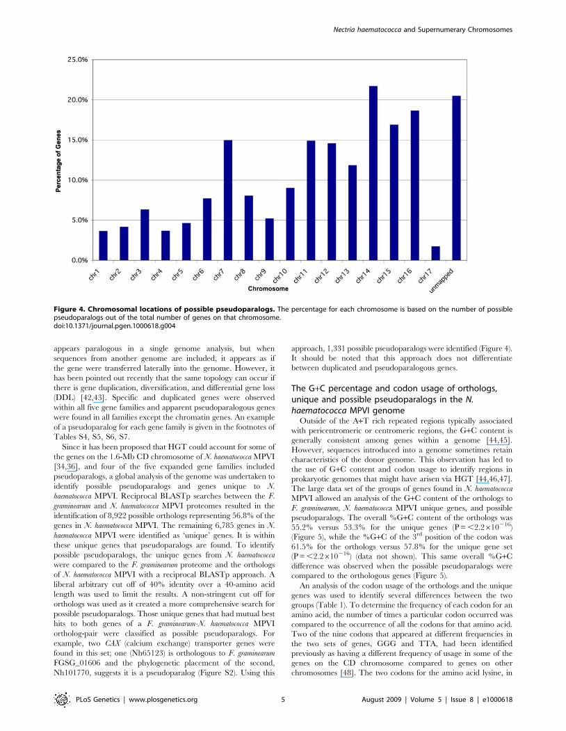
approach, 1,331 possible pseudoparalogs were identified (Figure 4).
It should be noted that this approach does not differentiate
between duplicated and pseudoparalogous genes.
The G+C percentage and codon usage of orthologs,unique and possible pseudoparalogs in the N.haematococca MPVI genome
Outside of the A+T rich repeated regions typically associated
with pericentromeric or centromeric regions, the G+C content is
generally consistent among genes within a genome [44,45].
However, sequences introduced into a genome sometimes retain
characteristics of the donor genome. This observation has led to
the use of G+C content and codon usage to identify regions in
prokaryotic genomes that might have arisen via HGT [44,46,47].
The large data set of the groups of genes found in N. haematococca
MPVI allowed an analysis of the G+C content of the orthologs to
F. graminearum, N. haematococca MPVI unique genes, and possible
pseudoparalogs. The overall %G+C content of the orthologs was
55.2% versus 53.3% for the unique genes (P = ,2.2610216)
(Figure 5), while the %G+C of the 3rd position of the codon was
61.5% for the orthologs versus 57.8% for the unique gene set
(P = ,2.2610216) (data not shown). This same overall %G+C
difference was observed when the possible pseudoparalogs were
compared to the orthologous genes (Figure 5).
An analysis of the codon usage of the orthologs and the unique
genes was used to identify several differences between the two
groups (Table 1). To determine the frequency of each codon for an
amino acid, the number of times a particular codon occurred was
compared to the occurrence of all the codons for that amino acid.
Two of the nine codons that appeared at different frequencies in
the two sets of genes, GGG and TTA, had been identified
previously as having a different frequency of usage in some of the
genes on the CD chromosome compared to genes on other
chromosomes [48]. The two codons for the amino acid lysine, in
Figure 4. Chromosomal locations of possible pseudoparalogs. The percentage for each chromosome is based on the number of possiblepseudoparalogs out of the total number of genes on that chromosome.doi:10.1371/journal.pgen.1000618.g004
Nectria haematococca and Supernumerary Chromosomes
PLoS Genetics | www.plosgenetics.org 5 August 2009 | Volume 5 | Issue 8 | e1000618
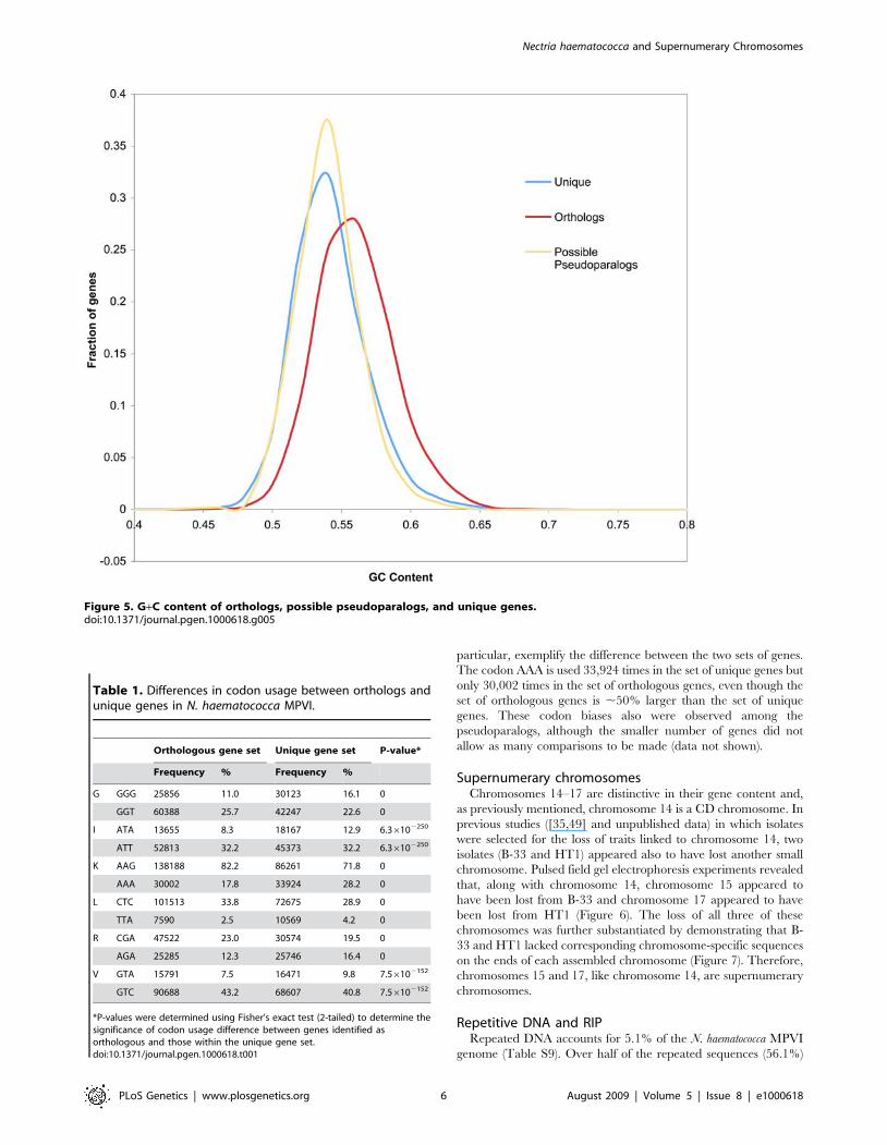
particular, exemplify the difference between the two sets of genes.
The codon AAA is used 33,924 times in the set of unique genes but
only 30,002 times in the set of orthologous genes, even though the
set of orthologous genes is ,50% larger than the set of unique
genes. These codon biases also were observed among the
pseudoparalogs, although the smaller number of genes did not
allow as many comparisons to be made (data not shown).
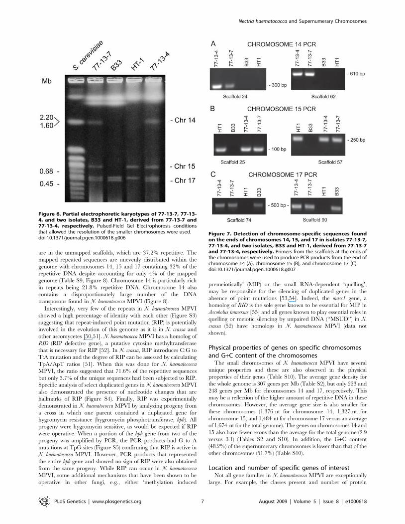
Supernumerary chromosomesChromosomes 14–17 are distinctive in their gene content and,
as previously mentioned, chromosome 14 is a CD chromosome. In
previous studies ([35,49] and unpublished data) in which isolates
were selected for the loss of traits linked to chromosome 14, two
isolates (B-33 and HT1) appeared also to have lost another small
chromosome. Pulsed field gel electrophoresis experiments revealed
that, along with chromosome 14, chromosome 15 appeared to
have been lost from B-33 and chromosome 17 appeared to have
been lost from HT1 (Figure 6). The loss of all three of these
chromosomes was further substantiated by demonstrating that B-
33 and HT1 lacked corresponding chromosome-specific sequences
on the ends of each assembled chromosome (Figure 7). Therefore,
chromosomes 15 and 17, like chromosome 14, are supernumerary
chromosomes.
Repetitive DNA and RIPRepeated DNA accounts for 5.1% of the N. haematococca MPVI
genome (Table S9). Over half of the repeated sequences (56.1%)
Table 1. Differences in codon usage between orthologs andunique genes in N. haematococca MPVI.
Orthologous gene set Unique gene set P-value*
Frequency % Frequency %
G GGG 25856 11.0 30123 16.1 0
GGT 60388 25.7 42247 22.6 0
I ATA 13655 8.3 18167 12.9 6.36102250
ATT 52813 32.2 45373 32.2 6.36102250
K AAG 138188 82.2 86261 71.8 0
AAA 30002 17.8 33924 28.2 0
L CTC 101513 33.8 72675 28.9 0
TTA 7590 2.5 10569 4.2 0
R CGA 47522 23.0 30574 19.5 0
AGA 25285 12.3 25746 16.4 0
V GTA 15791 7.5 16471 9.8 7.56102152
GTC 90688 43.2 68607 40.8 7.56102152
*P-values were determined using Fisher’s exact test (2-tailed) to determine thesignificance of codon usage difference between genes identified asorthologous and those within the unique gene set.doi:10.1371/journal.pgen.1000618.t001
Figure 5. G+C content of orthologs, possible pseudoparalogs, and unique genes.doi:10.1371/journal.pgen.1000618.g005
Nectria haematococca and Supernumerary Chromosomes
PLoS Genetics | www.plosgenetics.org 6 August 2009 | Volume 5 | Issue 8 | e1000618
are in the unmapped scaffolds, which are 37.2% repetitive. The
mapped repeated sequences are unevenly distributed within the
genome with chromosomes 14, 15 and 17 containing 32% of the
repetitive DNA despite accounting for only 4% of the mapped
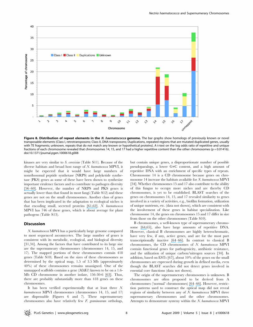
genome (Table S9, Figure 8). Chromosome 14 is particularly rich
in repeats being 21.8% repetitive DNA. Chromosome 14 also
contains a disproportionately large number of the DNA
transposons found in N. haematococca MPVI (Figure 8).
Interestingly, very few of the repeats in N. haematococca MPVI
showed a high percentage of identity with each other (Figure S3)
suggesting that repeat-induced point mutation (RIP) is potentially
involved in the evolution of this genome as it is in N. crassa and
other ascomycetes [50,51]. N. haematococca MPVI has a homolog of
RID (RIP defective gene), a putative cytosine methyltransferase
that is necessary for RIP [52]. In N. crassa, RIP introduces C:G to
T:A mutation and the degree of RIP can be assessed by calculating
TpA/ApT ratios [51]. When this was done for N. haematococca
MPVI, the ratio suggested that 71.6% of the repetitive sequences
but only 3.7% of the unique sequences had been subjected to RIP.
Specific analysis of select duplicated genes in N. haematococca MPVI
also demonstrated the presence of nucleotide changes that are
hallmarks of RIP (Figure S4). Finally, RIP was experimentally
demonstrated in N. haematococca MPVI by analyzing progeny from
a cross in which one parent contained a duplicated gene for
hygromycin resistance (hygromycin phosphotransferase, hph). All
progeny were hygromycin sensitive, as would be expected if RIP
were operative. When a portion of the hph gene from two of the
progeny was amplified by PCR, the PCR products had G to A
mutations at TpG sites (Figure S5) confirming that RIP is active in
N. haematococca MPVI. However, PCR products that represented
the entire hph gene and showed no sign of RIP were also obtained
from the same progeny. While RIP can occur in N. haematococca
MPVI, some additional mechanisms that have been shown to be
operative in other fungi, e.g., either ‘methylation induced
premeiotically’ (MIP) or the small RNA-dependent ‘quelling’,
may be responsible for the silencing of duplicated genes in the
absence of point mutations [53,54]. Indeed, the masc1 gene, a
homolog of RID is the sole gene known to be essential for MIP in
Ascobolus immersus [55] and all genes known to play essential roles in
quelling or meiotic silencing by unpaired DNA (‘‘MSUD’’) in N.
crassa (52) have homologs in N. haematococca MPVI (data not
shown).
Physical properties of genes on specific chromosomesand G+C content of the chromosomes
The small chromosomes of N. haematococca MPVI have several
unique properties and these are also observed in the physical
properties of their genes (Table S10). The average gene density for
the whole genome is 307 genes per Mb (Table S2), but only 223 and
248 genes per Mb for chromosomes 14 and 17, respectively. This
may be a reflection of the higher amount of repetitive DNA in these
chromosomes. However, the average gene size is also smaller for
these chromosomes (1,376 nt for chromosome 14, 1,327 nt for
chromosome 15, and 1,484 nt for chromosome 17 versus an average
of 1,674 nt for the total genome). The genes on chromosomes 14 and
15 also have fewer exons than the average for the total genome (2.9
versus 3.1) (Tables S2 and S10). In addition, the G+C content
(48.2%) of the supernumerary chromosomes is lower than that of the
other chromosomes (51.7%) (Table S10).
Location and number of specific genes of interestNot all gene families in N. haematococca MPVI are exceptionally
large. For example, the classes present and number of protein
Figure 6. Partial electrophoretic karyotypes of 77-13-7, 77-13-4, and two isolates, B33 and HT-1, derived from 77-13-7 and77-13-4, respectively. Pulsed-Field Gel Electrophoresis conditionsthat allowed the resolution of the smaller chromosomes were used.doi:10.1371/journal.pgen.1000618.g006
Figure 7. Detection of chromosome-specific sequences foundon the ends of chromosomes 14, 15, and 17 in isolates 77-13-7,77-13-4, and two isolates, B33 and HT-1, derived from 77-13-7and 77-13-4, respectively. Primers from the scaffolds at the ends ofthe chromosomes were used to produce PCR products from the end ofchromosome 14 (A), chromosome 15 (B), and chromosome 17 (C).doi:10.1371/journal.pgen.1000618.g007
Nectria haematococca and Supernumerary Chromosomes
PLoS Genetics | www.plosgenetics.org 7 August 2009 | Volume 5 | Issue 8 | e1000618
kinases are very similar to S. cerevisiae (Table S11). Because of the
diverse habitats and broad host range of N. haematococca MPVI, it
might be expected that it would have large numbers of
nonribosomal peptide synthetase (NRPS) and polyketide synthe-
tase (PKS) genes as some of these have been shown to synthesize
important virulence factors and to contribute to pathogen diversity
[56–60]. However, the number of NRPS and PKS genes is
actually lower than that found in most fungi (Table S12) and these
genes are not on the small chromosomes. Another class of genes
that has been implicated in the adaptation to ecological niches is
that encoding small, secreted proteins [61,62]. N. haematococca
MPVI has 746 of these genes, which is about average for plant
pathogens (Table S13).
Discussion
N. haematococca MPVI has a particularly large genome compared
to most sequenced ascomycetes. The large number of genes is
consistent with its metabolic, ecological, and biological diversity
[31,34]. Among the factors that have contributed to its large size
are the supernumeary chromosomes (chromosomes 14, 15, and
17). The mapped portions of these chromosomes contain 418
genes (Table S10). Based on the sizes of these chromosomes as
determined by the optical map, 1.5 of 3.5 Mb (approximately
40%) of these chromosomes remains unassigned. One of the
unmapped scaffolds contains a gene (MAK1) known to be on a 1.6-
Mb CD chromosome in another isolate, 156-30-6 [63]. Thus,
there are probably substantially more than 418 genes on these
chromosomes.
It has been verified experimentally that at least three N.
haematococca MPVI chromosomes (chromosomes 14, 15, and 17)
are dispensable (Figures 6 and 7). These supernumerary
chromosomes also have relatively few F. graminearum orthologs,
but contain unique genes, a disproportionate number of possible
pseudoparalogs, a lower G+C content, and a high amount of
repetitive DNA with an enrichment of specific types of repeats.
Chromosome 14 is a CD chromosome because genes on chro-
mosome 14 increase the habitats available for N. haematococca MPVI
[34]. Whether chromosomes 15 and 17 also contribute to the ability
of this fungus to occupy more niches and are thereby CD
chromosomes, is yet to be established. BLAST searches of the
genes on chromosomes 14, 15, and 17 revealed similarity to genes
involved in a variety of activities, e.g., biofilm formation, utilization
of unique nutrients, etc. (data not shown), which are consistent with
the involvement of these genes in habitat specialization. Like
chromosome 14, the genes on chromosomes 15 and 17 differ in size
from those on the other chromosomes (Table S10).
B chromosomes, a well-known type of supernumerary chromo-
some [64,65], also have large amounts of repetitive DNA.
However, classical B chromosomes are highly heterochromatic,
have very few, if any, active genes, and are for the most part
transcriptionally inactive [64–66]. In contrast to classical B
chromosomes, the CD chromosomes of N. haematococca MPVI
contain functional genes for pathogenicity, antibiotic resistance,
and the utilization of unique carbon/nitrogen sources [34]. In
addition, based on ESTs [67], about 10% of the genes on the small
chromosomes are expressed during growth in defined media, even
though the BLAST searches did not detect genes involved in
essential core functions (data not shown).
The origin of the supernumerary chromosomes is unknown. B
chromosomes are often proposed to be derived from A
chromosomes (‘normal’ chromosomes) [64–66]. However, restric-
tion patterns used to construct the optical map did not reveal
regions of similarity between any of N. haematococca MPVI three
supernumerary chromosomes and the other chromosomes.
Attempts to demonstrate synteny within the N. haematococca MPVI
Figure 8. Distribution of repeat elements in the N. haematococca genome. The bar graphs show homologs of previously known or noveltransposable elements (Class I, retrotransposons; Class II, DNA transposons; Duplications, repeated regions that are mutated duplicated genes, usuallywith TE fragments; unknown, repeats that do not match any known or hypothetical proteins). A t-test on the log odds ratio of repetitive and uniquefractions of each chromosome revealed that chromosomes 14, 15, and 17 had a higher repetitive content than the other chromosomes (p = 0.01416).doi:10.1371/journal.pgen.1000618.g008
Nectria haematococca and Supernumerary Chromosomes
PLoS Genetics | www.plosgenetics.org 8 August 2009 | Volume 5 | Issue 8 | e1000618
genome by identifying three-gene pairs in the same order and
orientation failed to demonstrate any large-scale segmental
duplications. However, six paired regions of up to 50 kb were
found. Although three of these regions are on chromosome 14, two
of the regions are similar to DNA on unmapped scaffolds and one
is on chromosome 6, which appears to be part of the main genome
complement. The RIP system of N. haematococca MPVI acting on
ancient genome duplications in combination with gene loss might
conceal any large-scale duplications, if they occurred [68]. It also
has been proposed that B chromosomes could arise from A
chromosomes following interspecific hybridization in sexual
crosses [66]. In this situation, it is possible that the B chromosome
would be the only remnants of an original A chromosome.
Extending the mechanism of hybridization to include the well-
known parasexual cycle that occurs in some fungi provides another
explanation of the origin of supernumerary chromosomes in N.
haematococca MPVI. As with sexual hybridization there are
numerous barriers between vegetative fusion of different fungal
species with the major one being vegetative incompatibility [69],
which acts at the intraspecies level. However, as recently pointed
out by Sanders [70], it would seem unlikely that the barriers to
vegetative fusion are so efficient as to prevent entirely the fusion of
fungal cells and the subsequent exchange of DNA. Such an event
of DNA exchange need not be common; in principle it need
happen only once, particularly if the transferred genetic material
provided a selective advantage. The exchange of supernumerary
chromosomes has been demonstrated in the laboratory, but thus
far only between members of the same species [71,72].
Supernumerary chromosomes do not explain entirely the large
size of the N. haematococca MPVI genome. Unique genes on non-
supernumerary chromosomes with atypical G+C content and
codon preference are also potential pseudoparalogs obtained by
HGT [41,44,46,47]. Although HGT has not been considered to
be a common mechanism of gene acquisition in fungi [73], there is
an increasing number of examples of apparent HGT [74–76].
However, the data are often inconclusive and subject to different
interpretations [77]. For example, genes may be classified as
pseudoparalogs because of lineage-specific gene loss, poor
phylogenetic taxon sampling, artifacts due to long branch
attraction, and other variables that obfuscate gene origins via
DDL or HGT [42,43]. Many of the properties of the
supernumerary chromosomes are similar to genomic islands of
A. fumigatus [42]. These species-specific regions of DNA can be as
large as 400 kb, and contain A. fumigatus-specific genes and large
amounts of repetitive DNA. Genomic islands have smaller genes
with fewer exons than the core genes found in Aspergillus species.
The genes on these genomic islands, like those on the
supernumerary chromosomes, encode metabolic processes that
appear to be involved in the adaptation to different ecological
niches. The origin of the genes on these islands was originally
attributed to HGT, but after comparisons to genomes of
additional Aspergillus species, Fedorova et al. [42] proposed that
the genes on the genomic islands of Aspergillus arose by DDL.
Nevertheless, the ability of N. haematococca MPVI to acquire genes
by HGT or a similar mechanism would bypass the evolutionary
restraint that an active RIP system places on a fungus by hindering
the utility of gene duplications as a means to create new genes
functions [50].
The genomic islands of A. fumigatus tend to be located in the sub-
telomeric regions of chromosomes. In other fungi, genes involved in
specific habitat associations, including pathogenicity, also are often
located in the sub-telomeric regions [78]. In N. haematococca MPVI,
gene placements differ among the chromosomes. Chromosomes 1,
4, 5, and 9 have most of their unique genes in the sub-telomeric
regions (Figure 9). On the other large chromosomes, the unique
genes are distributed in clusters throughout the chromosome.
Chromosome 7 is unusual in that it is a large chromosome (3.0 Mb)
that has some properties of the smaller chromosomes. For example,
the majority of the genes on chromosome 7 are unique to N.
haematococca MPVI (Figure 9). Chromosome 7 also has the largest
number of possible pseudoparalogs (159) of all the chromosomes
(Figure 4).
Only one isolate of N. haematococca MPVI (77-13-4) was sequenced
in the current study and it is a third generation progeny from a
laboratory cross between two field isolates. In N. crassa, where RIP
was first defined, both RIP and the translocation of chromosomal
fragments occur during the sexual cycle [79]. Thus, the crossing of
the original isolates of N. haematococca MPVI might have increased
the degree of RIP and/or affected the locations of genes in 77-13-4.
An examination of field isolates of this fungus has revealed a high
degree of chromosomal polymorphism and differences in the
locations of studied genes [80]. Thus, it would be of great interest to
determine the sequence of field isolates of N. haematococca MPVI and
other species within the F. solani species complex and to compare the
sizes and organization of their genomes to F. graminearum and to
other fusaria in the Gibberella clade.
Materials and Methods
IsolatesN. haematococca MPVI isolates 77-13-4 (FGSC 9596, Fungal
Genetics Stock Center) and 77-13-7 are ascospore isolates from a
third generation cross between two field isolates: one (T2) obtained
from a infected pea plant in NY and the other (T219) obtained
from soil in a potato field in PA [81,82]. Isolates HT1 and B-33
were derived from isolates 77-13-4 and 77-13-7, respectively, after
treatment with benomyl and selection for the loss of chromosome
14 [35,49].
DNA isolation and sequencingHigh molecular weight genomic DNA was isolated from
protoplasts of 77-13-4 prepared by treatment of mycelia with a
combination of lytic enzymes as described previously [83]. Whole-
genome shotgun libraries (3-kb, 8-kb, and 40-kb DNA inserts) of
77-13-4 were constructed and sequenced as previously described
[84].
RNA isolation and sequencingTwo cDNA libraries were constructed and sequenced to facilitate
the automated gene calling programs. The RNA for the cDNA
libraries was obtained from mycelia treated in two different ways:
both were grown in a rich medium, potato dextrose broth, (PDB)
(Difco, Sparks, MD) and one of these was treated with pisatin. Spores
of 77-13-4 (105 ml21) from V8 agar medium [85] were added to
100 ml of PDB in a 250 ml Erlenmeyer flask and grown at room
temperature on a rotary shaker (180 rpm) for 24 h. The mycelium
was collected by vacuum filtration and stored at 280uC. Mycelium
for the pisatin-treated library was collected after overnight growth in
PDB, washed in 10 ml of 0.7 M NaCl, and added to 50 ml of
phosphate buffer (50 mM potassium phosphate buffer, pH 6.5).
After 2 hours, pisatin in DMSO was added to a final concentration
of 30 mg of pisatin/ml and 1% DMSO and after another 4.5 hours
the mycelium was collected and frozen at 280uC.
For RNA isolation the mycelia were lyophilized and ground
under liquid nitrogen using a mortar and pestle. RNA was isolated
as described in Mandel et al. [86] with the exception that ground
mycelia (200 mg) were placed in an RNase-free 2-ml tube, to
which 0.7 ml of ice-cold breaking buffer was added, the mycelia
Nectria haematococca and Supernumerary Chromosomes
PLoS Genetics | www.plosgenetics.org 9 August 2009 | Volume 5 | Issue 8 | e1000618
were suspended, and 0.6 ml of acid phenol added. cDNA libraries
were constructed and sequenced as described previously with
minor differences that include: the size ranges of cDNA (0.6 k–
2 kb and .2 kb), the cloning vector (pMCL200cDNA), and the
sequencing primers (Fw: 59-AGGAAACAGCTATGACCA-39,
Rv: 59-GTTTTCCCAGTCACGACGTTGTA-39) [87]. 24,793
ESTs were obtained from mycelium grown in the PDB medium
and 8,327 from the mycelium treated with pisatin.
Genome finishing methodsInitial read layouts from the whole genome shotgun assembly
were converted into a Phred/Phrap/Consed pipeline [88] and,
following manual inspection of the assembled sequences, finishing
was performed by resequencing plasmid subclones and by walking
on plasmid subclones or fosmids using custom primers. All
finishing reactions were performed with 4:1 BigDye to dGTP
BigDye terminator chemistry (Applied Biosystems). Repeats in the
sequence were resolved by transposon-hopping 8-kb plasmid
clones. Fosmid clones were shotgun sequenced and finished to fill
large gaps, resolve large repeats, and to extend into chromosome
telomere regions where possible. After finishing, the genome
remained in 209 scaffolds as a result of many regions of the
genome being apparently unclonable in the shotgun libraries
constructed for this project.
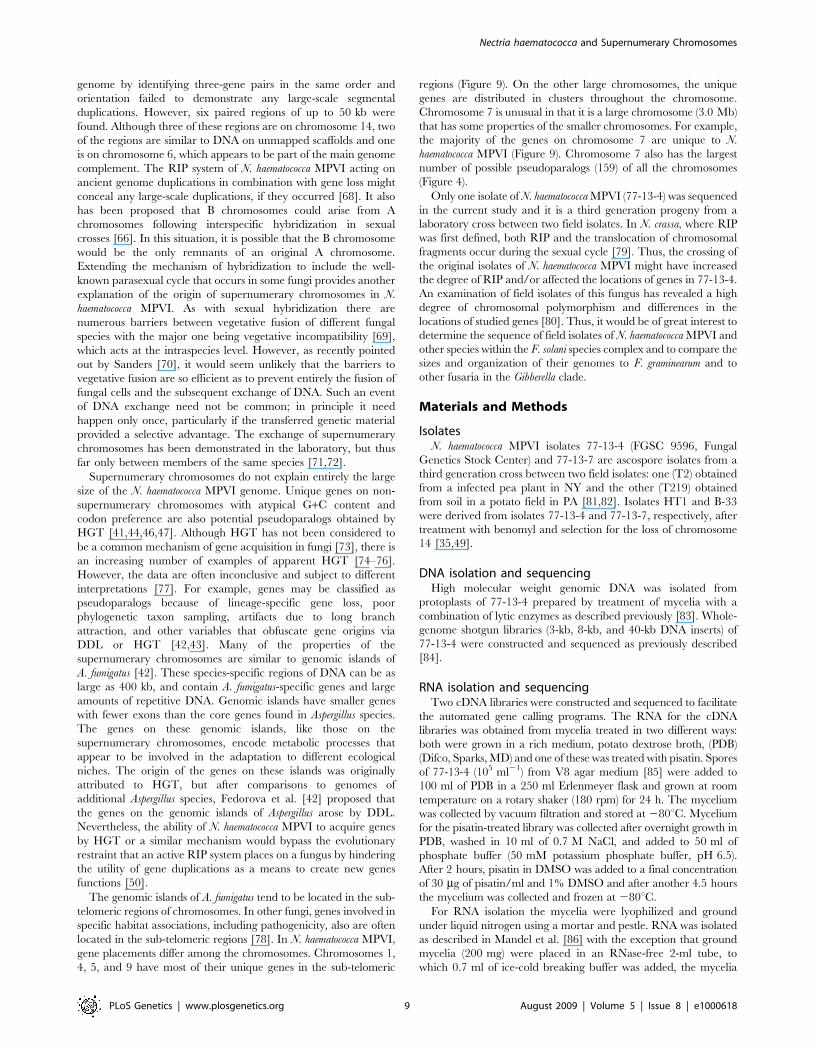
Figure 9. Distribution of orthologs, possible pseudoparalogs, and unique genes on each of the chromosomes of N. haematococcaMPVI. Compositional statistical analysis using an additive log-ratio transformation [102] reveals that the distribution of genes within the three classesis statistically different on chromosomes 14, 15, and 17 than on the other chromosomes (p = 1.05e-5).doi:10.1371/journal.pgen.1000618.g009
Nectria haematococca and Supernumerary Chromosomes
PLoS Genetics | www.plosgenetics.org 10 August 2009 | Volume 5 | Issue 8 | e1000618
The resulting assembly was joined and validated by alignments
to a N. haematococca optical map (generated by digestion with NheI),
with 92.26% of the sequence (72 scaffolds or 47,191,137 bp) being
placed onto 17 chromosome optical maps. 3,958,438 bp of the
137 smaller scaffolds remain unplaced because of the lack of
sufficient restriction sites. The genome consists of 51,149,575 base
pairs of finished sequence with an estimated error rate of less than
1 error in 100,000 bp.
Optical mappingProtoplasts of 77-13-4, made as described above [83], were directly
lysed with a protoplast lysing solution (10 mM EDTA, 5 mM EGTA,
1 mg/ml proteinase K, pH 8.0) by heating to 65uC for 30 min to 1 hr
and then incubating overnight at 37uC. The protoplast concentration
was adjusted to ,700 protoplasts per microliter. Optical mapping
operations followed previously published techniques [89]; briefly,
randomly sheared high molecular weight DNA was loaded onto
optical mapping chips for restriction digestion by NheI (New England
Biolabs). DNA was stained with YOYO-1 fluorochrome (Invitrogen)
and the chips were scanned on an automated fluorescence microscope
system for image capture, analysis, and map construction [90].
Resulting single-molecule restriction maps were assembled into
genome-wide contigs [91,92] that served as map scaffolds for sequence
joining and validation efforts.
Gene prediction and automated annotationGene models (15,707) were predicted and automatically
annotated using the Joint Genome Institute (JGI) Annotation
Pipeline. Several gene predictors were used on repeat masked
assembly: ab initio FGENESH and homology-based FGENESH+[93] and Genewise [94]. The predicted gene models were verified,
corrected, and extended using 33,142 N. haematococca MPVI ESTs.
All predicted gene models were functionally annotated by homology
to proteins from the NCBI non-redundant set and classified
according to Gene Ontology [95], eukaryotic orthologous groups
(KOGs [96]), and KEGG metabolic pathways [97]. Of the 15,707
models, 93% were complete models, 25% were supported with EST
aligment, 94% with NR alignment, 73% with Swissprot alignment
and 52% with Pfam alignments. For every locus the ‘best’repre-
sentative model was selected based on EST and homology support,
to produce a non-redundant representative set, subject to manual
curation and the analysis described here.
Best fit analysis of N. haematococca to other fungiEach predicted protein from the N. haematococca MPVI genome was
used as a query in a TBLASTN search of a database consisting of the
genomes of A. oryzae, A. nidulans, C. globosum, C. immitis, F. graminearum,
M. oryzae, N. crassa, and S. cerevisiae. For each protein query, the
genome with the best hit below a threshold of 1e-5 was identified.
Phylogenetic analysisThe predicted amino acid sequences for hypothetical proteins
were aligned with ClustalW 1.81 [98]. The resulting alignment
files were imported into MacClade 4.08 [99] for manual editing
and exclusion of all ambiguously aligned regions. Heuristic
searches for maximum parsimony (MP) were conducted in PAUP*
[100], and neighbor-joining distance trees were generated in
MacVector 10.0.2 (Symantec Corporation). Statistical support was
calculated from 1,000 bootstrap replicates.
RIP indexTo identify repetitive regions of the genome in the absence of
a curated repeat library, the genome was divided into 1 kb
non-overlapping windows and BLASTN was used to align each
against the complete genome. If the only match greater than
500 bp was a self-match, then the window was labeled as
unique, otherwise it was labeled as repetitive. For each 1 kb
window, the ratio of TpA/ApT frequency was calculated.
Experimental demonstration of RIPA transformant (Tr78.2) of N. haematococca that contained several
copies of the hph gene from the transformation vector pCWHyg1
[101], was crossed (cross 370) with 77-15-7 as previously described
[82]. The hph gene is linked to the homoserine utilization
phenotype (HUT) in Tr78.2 and 370 progeny were screened for
hygromycin sensitivity and HUT. All forty progeny from cross 370
were sensitive to hygromycin, and half were HUT+. DNA was
isolated from two hygromycin sensitive/HUT+ isolates (370-4 and
370-8) as previously described [32]. Sequences of hph were
amplified using PCR and the primers Hyg-F (59-CGGA-
GATTCGTCGTTCTGAAGAG-39) and Hyg-R (59-TTCTACA-
CAGCCATCGGTCCAG-39) following the manufacturer’s pro-
tocol (Invitrogen) and the following set of conditions (94uC for
45 sec, 59uC for 30 sec, 72uC for 1.5 min, for 35 cycles). The
resulting 1,242 bp products containing hph were cloned into the
pGEM T-EZ vector (Promega Corporation, Madison, WI) and hph
was sequenced using the Hyg2-F (59-ACGCGACAACTGAGT-
GACTG-39) primer adjacent to hph. Sequencing was done by the
Genomic Analysis and Technology Core (GATC) facility at the
University of Arizona.
Pulsed-field gel electrophoresis (PFGE) analyses ofchromosome-sized DNA
77-13-7 and its derivative B-33 have been described [49], as
have 77-13-4 and its derivative HT-1 [35]. The preparation of
chromosome-sized DNAs and the PFGE were performed
essentially as described previously [35,72]. For making protoplasts
of 77-13-7 and B-33, the enzyme mixture of Garmaroodi and
Taga [72] was used, while the enzyme mixture of Rodriguez-
Carres et al. [35] was used for 77-13-4 and HT-1. Protoplasts (ca.
36108 protoplasts/ml) were embedded in low melting tempera-
ture agarose (Bio-Rad Laboratories Inc., Hercules, CA) and
chromosome-sized DNAs were separated in 0.56TBE buffer on a
1% (w/v) pulsed field certified agarose (Bio-Rad Laboratories) gel
with a contour-clamped homogeneous electric field apparatus
(CHEF-DR II, Bio-Rad Laboratories). Running conditions were
5.4 V/cm and pulse time of 120 s for 13 h followed by 180 s for
13 h. Chromosomal DNAs of S. cerevisiae (Bio-Rad Laboratories)
were used as the size markers.
Attempts to detect chromosome-specific sequencesfound on the ends of supernumerary chromosomes
Genomic DNA of N. haematococca MPVI was isolated as
previously described [83]. PCR was used to test for the presence
of sequences found on the ends of the assembled sequences
representing chromosomes 14, 15, and 17 in isolates 77-13-7, 77-
13-4, B-33, and HT-1. Primers were designed to amplify regions of
scaffolds located on the ends of the respective chromosomes:
scaffolds 24 and 62 for chromosome 14, scaffolds 25 and 57 for
chromosome 15, and scaffolds 74 and 90 for chromosome 17.
These sequences were blasted against the N. haematococca MPVI
genome sequence to confirm that they were not found on
chromosomes other than those of interest.
Sequences of the primers (Invitrogen) used for PCR were as
follows: for chromosome 14, scaffold 24: forward primer, 59-
GCCAGGAGATCGAGATATGA-39 and reverse primer, 59-
Nectria haematococca and Supernumerary Chromosomes
PLoS Genetics | www.plosgenetics.org 11 August 2009 | Volume 5 | Issue 8 | e1000618
GTGGATGAGATCGGTGTTTC-39; for chromosome 14, scaf-
fold 62: forward primer, 59-CTCCATCTTCTCGGCAATGT-39
and reverse primer, 59-CTTGGTTCACTCGCATACTTG-39;
for chromosome 15, scaffold 25: forward primer, 59-GACCGT-
CAAGGGAGCTACAG-39 and reverse primer, 59-ATCAGGG-
GTCATGTGAAGC-39; for chromosome 15, scaffold 57: forward
primer, 59-GGCCTTTGTACTCGCATTTA-39 and reverse primer,
59-GACCCTCTGCCTTCTTCTTC-39; for chromosome 17, scaf-
fold 74: forward primer, 59- CGCCCACTTCTTTGTCTCTA-39
and reverse primer, 59-AGCGAATTCATTTGAAGCAG -39; and
for chromosome 17, scaffold 90: forward primer, 59-GGAGACGTT-
GATGAGATTGG -39 and reverse primer, 59-CATCTGTTGA-
ACCCACACAA -39. Each reaction had a total volume of 50 ml
containing 300 nM forward primer, 300 nM reverse primer, 1 ml
(,50 ng) DNA template, 5 mM MgCl2, 5 ml 106 PCR buffer,
200 mM (each) of dATP, dTTP, dCTP, and dGTP, and 1 unit Taq
DNA Polymerase. The PCR protocol consisted of an initial
denaturation step of 95uC for 3 min., 35 cycles at 95uC for 30 sec,
55uC for 30 sec, and 72uC for 30 sec, and a final elongation step at
72uC for 30 sec. PCR products were run on a 0.8% agarose gel
containing ethidium bromide and visualized under UV light. Genomic
DNA of N. haematococca isolates 77-13-4 and 77-13-7 was used as
positive controls.
Search for segmental duplicationsBlastP searches of the protein set against itself with a threshold
value of 1e-20 identified 2,259 gene pairs as best-bidirectional hits.
Segmental duplicated regions were defined as genomic regions
that share at least three genes in the same order and orientation,
while the distance between neighboring gene pairs is less than
50 kb.
Supporting Information
Figure S1 Ratio of the number of genes in gene families in N.
haematococca MPVI versus F. graminearum. Only gene families that
had $10 members in N. haematococca MPVI were used in the
analysis. The number of genes per family was derived from
Interpro calls made by the JGI for N. haematococca MPVI, and by
the Munich Institute for Protein Sequences (MIPS) for F.
graminearum.
Found at: doi:10.1371/journal.pgen.1000618.s001 (1.27 MB TIF)
Figure S2 The CAX (calcium exchanger) transporter clade from
select fungal genomes. Maximum parsimony analysis was used to
establish the phylogenetic relationship between the ortholog
(Nh65123, red box) and the pseudoparalog (Nh101770, blue
box) of N. haematococca MPVI.
Found at: doi:10.1371/journal.pgen.1000618.s002 (8.06 MB TIF)
Figure S3 Distibution of repeat identity. NC7 is N. crassa, MG5
is M. oryzae, AN1 is A. nidulans, ‘‘FG3 Repeats’’ is F. graminearum
and ‘‘FS Repeats’’ is N. haematococca MPVI.
Found at: doi:10.1371/journal.pgen.1000618.s003 (4.09 MB TIF)
Figure S4 Effect of RIP on a family of telomere-associated
helicases (TAH) in N. haematococca MPVI. Partial alignment of the
12 predicted TAH genes (tah), spanning only the first three
conserved motifs. The top row shows the predicted translation of
the fourth tah gene on scaffold 45 (TAH_45-4). While many
mutations occur in the wobble position, note the presence of
nonsense codons (*). Nucleotides in red (G to A change) and
orange (C to T change) can be explained by a single RIP-type
mutation, while nucleotides in pink denoted non-RIP-type
transversions. Conversion of RIP-type C:G to T:A mutations
back to the likely original sequence (‘‘de-RIP’’, blue), results in a
consensus sequence (TAH_ORI) that closely resembles that of the
Metarhizium anisopliae TAH1 sequence (MaTAH1; note absence of
nonsense codons in the derived consensus sequence, residues in
red indicate changes compared to the TAH_45-4 sequence). De-
RIP of the complete coding region results in a single large ORF
without predicted introns or nonsense codons, similar to the M.
anisopliae TAH1 gene (Inglis PW, Rigden DJ, Mello LV, Louis EJ,
Valadares-Inglis MC 2005 Monomorphic subtelomeric DNA in
the filamentous fungus, Metarhizium anisopliae, contains a RecQ
helicase-like gene. Mol Genet Genomics 274: 79–90).
Found at: doi:10.1371/journal.pgen.1000618.s004 (7.54 MB TIF)
Figure S5 Repeat-induced point mutation (RIP) in N. haemato-
cocca MPVI. The hygromycin resistance (hph) gene is mutated from
G to A at multiple TpG positions (indicated in red) in isolates 370-
4 and 370-8.
Found at: doi:10.1371/journal.pgen.1000618.s005 (1.36 MB TIF)
Table S1 Comparison of genome statistics of several filamentous
ascomycete fungi.
Found at: doi:10.1371/journal.pgen.1000618.s006 (0.06 MB
DOC)
Table S2 Properties of the genes of N. haematococca MPVI.
Found at: doi:10.1371/journal.pgen.1000618.s007 (0.03 MB
DOC)
Table S3 Gene families that are at least two-fold larger in Nectria
haematococca MPVI than in Fusarium graminearum.
Found at: doi:10.1371/journal.pgen.1000618.s008 (0.09 MB
DOC)
Table S4 The number of ABC transporters in Nectria haematococca
MPVI compared to other fungi.
Found at: doi:10.1371/journal.pgen.1000618.s009 (0.05 MB
DOC)
Table S5 Carbohydrate-active enzymes in N. haematococca MPVI
compared to other fungi.
Found at: doi:10.1371/journal.pgen.1000618.s010 (0.07 MB
DOC)
Table S6 The number of cytochrome P450 genes in Nectria
haematococca MPVI compared to other fungi.
Found at: doi:10.1371/journal.pgen.1000618.s011 (0.06 MB
DOC)
Table S7 Number of predicted genes in Nectria haematococca
MPVI that contain transcription factor motifs compared to other
fungi.
Found at: doi:10.1371/journal.pgen.1000618.s012 (0.10 MB
DOC)
Table S8 The number of chromatin genes in N. haematococca
MPVI compared to other fungi.
Found at: doi:10.1371/journal.pgen.1000618.s013 (0.05 MB
DOC)
Table S9 Distribution of repeat elements in the genome of
Nectria haematococca MPVI.
Found at: doi:10.1371/journal.pgen.1000618.s014 (0.08 MB
DOC)
Table S10 Properties of the chromosomes and genes on each
chromosome in N. haematococca MPVI.
Found at: doi:10.1371/journal.pgen.1000618.s015 (0.09 MB
DOC)
Table S11 The protein kinases of N. haematococca MPVI
compared to S. cerevisiae.
Nectria haematococca and Supernumerary Chromosomes
PLoS Genetics | www.plosgenetics.org 12 August 2009 | Volume 5 | Issue 8 | e1000618
Found at: doi:10.1371/journal.pgen.1000618.s016 (0.03 MB
DOC)
Table S12 The number of polyketide synthases (PKS) and
nonribosomal peptide synthetases (NRPS) of Nectria haemato-
cocca MPVI compared to other fungi.
Found at: doi:10.1371/journal.pgen.1000618.s017 (0.06 MB
DOC)
Table S13 Distribution of Small Secreted Proteins (SSP) among
filamentous Ascomycetes as identified by SignalP.
Found at: doi:10.1371/journal.pgen.1000618.s018 (0.05 MB
DOC)
Author Contributions
Conceived and designed the experiments: CCW DMG SFC ET HDV.
Performed the experiments: JJC SDR MRC JS MT GJW SZ MF LjM
EGJD DRN DS BMB MR JZ MLF AS HS JP EL CL. Analyzed the data:
JJC SDR MRC AK CCW JG MT GJW SZ DCS MF LjM EGJD BH
PMC DRN DS CAN BMB MG MR SK IM CR JCK JZ MLF EUS EL
IVG SFC. Contributed reagents/materials/analysis tools: PMC. Wrote the
paper: JJC MRC CCW GJW HDV.
References
1. O’Donnell K (2000) Molecular phylogeny of the Nectria haematococca-Fusarium
solani species complex. Mycologia 92: 919–938.
2. Zhang N, O’Donnell K, Sutton DA, Nalim FA, Summerbell RC, et al. (2006)
Members of the Fusarium solani species complex that cause infections in both
humans and plants are common in the environment. Journal of ClinicalMicrobiology 44: 2186–2190.
3. Mandeel QA (1996) Survey of Fusarium species in an arid environment of
Bahrain .4. Prevalence of Fusarium species in various soil groups using severalisolation techniques. Cryptogamie Mycologie 17: 149–163.
4. Farr DF, Bills GF, Chamuris GP, Rossman AY (1989) Fungi on plants and
plant products in the United States. St. Paul (Minnesota): American
Phytopatholgy Society Press. 1252 p.
5. Boutati EI, Anaissie EJ (1997) Fusarium, a significant emerging pathogen inpatients with hematologic malignancy: Ten years’ experience at a cancer center
and implications for management. Blood 90: 999–1008.
6. Groll AH, Walsh TJ (2001) Uncommon opportunistic fungi: new nosocomialthreats. Clinical Microbiology and Infection 7: 8–24.
7. Guarro J, Gene J (1995) Opportunistic fusarial infections in humans. European
Journal of Clinical Microbiology & Infectious Diseases 14: 741–754.
8. Chang DC, Grant GB, O’Donnell K, Wannemuehler KA, Noble-Wang J, et
al. (2006) Multistate outbreak of Fusarium keratitis associated with use of acontact lens solution. JAMA-Journal of the American Medical Association 296:
953–963.
9. Abbas HK, Egley GH (1996) Influence of unrefined corn oil and surface-activeagents on the germination and infectivity of Alternaria helianthi. Biocontrol
Science and Technology 6: 531–538.
10. Chen SY, Dickson DW, Mitchell DJ (1996) Pathogenicity of fungi to eggs of
Heterodera glycines. Journal of Nematology 28: 148–158.
11. Bernard EC, Self LH, Tyler DD (1997) Fungal parasitism of soybean cystnematode, Heterodera glycines (Nemata: Heteroderidae), in differing cropping-
tillage regimes. Applied Soil Ecology 5: 57–70.
12. Zhdanova NN, Zakharchenko VA, Vember VV, Nakonechnaya LT (2000)Fungi from Chernobyl: mycobiota of the inner regions of the containment
structures of the damaged nuclear reactor. Mycological Research 104:
1421–1426.
13. Wainwright M, Ali TA, Killham K (1994) Anaerobic growth of fungalmycelium from soil particles onto nutrient-free silica-gel. Mycological Research
98: 761–762.
14. Pujol I, Guarro J, Gene J, Sala J (1997) In-vitro antifungal susceptibility ofclinical and environmental Fusarium spp. strains. Journal of Antimicrobial
Chemotherapy 39: 163–167.
15. Espinel-Ingroff A (1998) Comparison of in vitro activities of the new triazole
SCH56592 and the echinocandins MK-0991 (L-743,872) and LY303366against opportunistic filamentous and dimorphic fungi and yeasts. Journal of
Clinical Microbiology 36: 2950–2956.
16. Dupont J, Jacquet C, Dennetiere B, Lacoste S, Bousta F, et al. (2007) Invasionof the French Paleolithic painted cave of Lascaux by members of the Fusarium
solani species complex. Mycologia 99: 526–533.
17. Barclay M, Hart A, Knowles CJ, Meeussen JCL, Tett VA (1998)
Biodegradation of metal cyanides by mixed and pure cultures of fungi.Enzyme and Microbial Technology 22: 223–231.
18. Chakraborty SK, Bhattacharyya A (1991) Degradation of butachlor by 2 soil
fungi. Chemosphere 23: 99–105.
19. Colombo JC, Cabello M, Arambarri AM (1996) Biodegradation of aliphaticand aromatic hydrocarbons by natural soil microflora and pure cultures of
imperfect and lignolitic fungi. Environmental Pollution 94: 355–362.
20. Falcon MA, Rodriguez A, Carnicero A, Regalado V, Perestelo F, et al. (1995)
Isolation of microorganisms with lignin transformation potential from soil ofTenerife Island. Soil Biology & Biochemistry 27: 121–126.
21. Hemida SK, Bagy MMK, Khallil AM (1993) Utilization of hydrocarbons by
fungi. Cryptogamie Mycologie 14: 207–213.
22. Hsu JC, Camper ND (1979) Degradation of ioxynil by a soil fungus, Fusarium
solani. Soil Biology & Biochemistry 11: 19–22.
23. Katayama T, Sogo M (1989) An optically-active compound formed by the
reduction of an alpha-ketonic lignin substructure model-compound by Fusarium
solani m-13-1. Mokuzai Gakkaishi 35: 1116–1124.
24. Mitra J, Mukherjee PK, Kale SP, Murthy NBK (2001) Bioremediation of DDTin soil by genetically improved strains of soil fungus Fusarium solani.
Biodegradation 12: 235–245.
25. Rafin C, Potin O, Veignie E, Sahraoui ALH, Sancholle M (2000) Degradationof benzo[a]pyrene as sole carbon source by a non white rot fungus, Fusarium
solani. Polycyclic Aromatic Compounds 21: 311–329.
26. Rodriguez A, Perestelo F, Carnicero A, Regalado V, Perez R, et al. (1996)Degradation of natural lignins and lignocellulosic substrates by soil-inhabiting
fungi imperfecti. Fems Microbiology Ecology 21: 213–219.
27. Romero MC, Salvioli ML, Cazau MC, Arambarri AM (2002) Pyrenedegradation by yeasts and filamentous fungi. Environmental Pollution 117:
159–163.
28. Saparrat MCN, Martinez MJ, Tournier HA, Cabello MN, Arambarri AM(2000) Production of ligninolytic enzymes by Fusarium solani strains isolated from
different substrata. World Journal of Microbiology & Biotechnology 16:799–803.
29. Walker JRL, Lien BC (1981) Metabolism of fluoroacetate by a soil Pseudomonas
sp and Fusarium solani. Soil Biology & Biochemistry 13: 231–235.30. Rossman AY, Samuels GJ, Rogerson CT, Lowen R (1999) Genera of
Bionectriaceae, Hypocreaceae and Nectriaceae (Hypocreales, Ascomycetes).
Studies in Mycology 42: 1–248.31. VanEtten HD, Kistler HC (1988) Nectria haematococca mating populations I
and VI. In: Sidhu GS, ed. Advances in Plant Pathology. New York: Academic
Press. pp 189–206.32. Miao VP, Covert SF, Vanetten HD (1991) A fungal gene for antibiotic-
resistance on a dispensable (B) chromosome. Science 254: 1773–1776.
33. Covert SF (1998) Supernumerary chromosomes in filamentous fungi. CurrentGenetics 33: 311–319.
34. VanEtten HD, Straney D, Covert S, Kistler C (2001) Update on selected topics
of the genetics of Nectria haematococca mating population VI with specialemphasis on its conditionally dispensable (CD) chromosomes: a source of
habitat specific genes. In: Summerell JFL BA, Backhouse D, Bryden WL,Burgess LW, eds. Fusarium. Saint Paul (Minnesota): APS Press. pp 97–112.
35. Rodriguez-Carres M, White G, Tsuchiya D, Taga M, VanEtten HD (2008)
The supernumerary chromosome of Nectria haematococca that carries pea-pathogenicity-related genes also carries a trait for pea rhizosphere compet-
itiveness. Applied and Environmental Microbiology 74: 3849–3856.
36. Temporini ED, VanEtten HD (2004) An analysis of the phylogeneticdistribution of the pea pathogenicity genes of Nectria haematococca MPVI
supports the hypothesis of their origin by horizontal transfer and uncovers apotentially new pathogen of garden pea: Neocosmospora boniensis. Current
Genetics 46: 29–36.
37. Cuomo CA, Gueldener U, Xu JR, Trail F, Turgeon BG, et al. (2007) TheFusarium graminearum genome reveals a link between localized polymorphism
and pathogen specialization. Science 317: 1400–1402.
38. Galagan JE, Henn MR, Ma LJ, Cuomo CA, Birren B (2005) Genomics of thefungal kingdom: Insights into eukaryotic biology. Genome Research 15:
1620–1631.
39. Stajich JE, Dietrich FS, Roy SW (2007) Comparative genomic analysis offungal genomes reveals intron-rich ancestors. Genome Biology 8: R223.
40. Han YN, Liu XG, Benny U, Kistler HC, VanEtten HD (2001) Genes
determining pathogenicity to pea are clustered on a supernumerarychromosome in the fungal plant pathogen Nectria haematococca. Plant Journal
25: 305–314.
41. Koonin EV (2005) Orthologs, paralogs, and evolutionary genomics. AnnualReview of Genetics 39: 309–338.
42. Fedorova ND, Khaldi N, Joardar VS, Maiti R, Amedeo P, et al. (2008)Genomic islands in the pathogenic filamentous fungus Aspergillus fumigatus. Plos
Genetics 4: e1000046. doi:10.1371/journal.pgen.1000046.
43. Khaldi N, Wolfe KH (2008) Elusive origins of the extra genes in Aspergillus
oryzae. PLoS ONE 3: e3036. doi:10.1371/journal.pone.0003036.
44. Muto A, Osawa S (1987) The guanine and cytosine content of genomic DNA
and bacterial evolution. Proc Natl Acad Sci U S A 84: 166–169.
45. Karlin S, Campbell AM, Mrazek J (1998) Comparative DNA analysis acrossdiverse genomes. Annual Review of Genetics 32: 185–225.
46. Lawrence JG, Ochman H (1998) Molecular archaeology of the Escherichia coli
genome. Proc Natl Acad Sci U S A 95: 9413–9417.
Nectria haematococca and Supernumerary Chromosomes
PLoS Genetics | www.plosgenetics.org 13 August 2009 | Volume 5 | Issue 8 | e1000618
47. Ochman H, Lawrence JG, Groisman EA (2000) Lateral gene transfer and the
nature of bacterial innovation. Nature 405: 299–304.
48. Liu XG, Inlow M, VanEtten HD (2003) Expression profiles of pea
pathogenicity (PEP) genes in vivo and in vitro, characterization of the flanking
regions of the PEP cluster and evidence that the PEP cluster region resulted
from horizontal gene transfer in the fungal pathogen Nectria haematococca.
Current Genetics 44: 95–103.
49. VanEtten H, Jorgensen S, Enkerli J, Covert SF (1998) Inducing the loss of
conditionally dispensable chromosomes in Nectria haematococca during vegetative
growth. Current Genetics 33: 299–303.
50. Galagan JE, Selker EU (2004) RIP: the evolutionary cost of genome defense.
Trends in Genetics 20: 417–423.
51. Galagan JE, Calvo SE, Borkovich KA, Selker EU, Read ND, et al. (2003) The
genome sequence of the filamentous fungus Neurospora crassa. Nature 422:
859–868.
52. Freitag M, Williams RL, Kothe GO, Selker EU (2002) A cytosine
methyltransferase homologue is essential for repeat-induced point mutation
in Neurospora crassa. Proc Natl Acad Sci U S A 99: 8802–8807.
53. Rossignol JL, Faugeron G (1995) MIP - An epigenetic gene silencing process in
Ascobolus immersus. Gene Silencing in Higher Plants and Related Phenomena in
Other Eukaryotes. pp 179–191.
54. Catalanotto C, Nolan T, Cogoni C (2006) Homology effects in Neurospora crassa.
Fems Microbiology Letters 254: 182–189.
55. Malagnac F, Wendel B, Goyon C, Faugeron G, Zickler D, et al. (1997) A gene
essential for de novo methylation and development in ascobolus reveals a novel
type of eukaryotic DNA methyltransferase structure. Cell 91: 281–290.
56. Donadio S, Monciardini P, Sosio M (2007) Polyketide synthases and
nonribosomal peptide synthetases: the emerging view from bacterial genomics.
Natural Product Reports 24: 1073–1109.
57. Jegorov A, Haiduch M, Sulc M, Havlicek V (2006) Nonribosomal cyclic
peptides: specific markers of fungal infections. Journal of Mass Spectrometry
41: 563–576.
58. Johnson R, Voisey C, Johnson L, Pratt J, Fleetwood D, et al. (2007)
Distribution of NRPS gene families within the Neotyphodium/Epichloe complex.
Fungal Genetics and Biology 44: 1180–1190.
59. Lu SW, Kroken S, Lee BN, Robbertse B, Churchill ACL, et al. (2003) A novel
class of gene controlling virulence in plant pathogenic ascomycete fungi. Proc
Natl Acad Sci U S A 100: 5980–5985.
60. Xu YQ, Orozco R, Wijeratne EMK, Gunatilaka AAL, Stock SP, et al. (2008)
Biosynthesis of the Cyclooligomer Depsipeptide Beauvericin, a Virulence
Factor of the Entomopathogenic Fungus Beauveria bassiana. Chemistry &
Biology 15: 898–907.
61. Rep M (2005) Small proteins of plant-pathogenic fungi secreted during host
colonization. Fems Microbiology Letters 253: 19–27.
62. van der Does HC, Rep M (2007) Virulence genes and the evolution of host
specificity in plant-pathogenic fungi. Molecular Plant-Microbe Interactions 20:
1175–1182.
63. Covert SF, Enkerli J, Miao VPW, VanEtten HD (1996) A gene for maackiain
detoxification from a dispensable chromosome of Nectria haematococca. Molecular
and General Genetics 251: 397–406.
64. Camacho JPM, Sharbel TF, Beukeboom LW (2000) B-chromosome evolution.
Phil Trans Roy Soc Lond B-Biological Sciences 355: 163–178.
65. Jones RN, Gonzalez-Sanchez M, Gonzalez-Garcia M, Vega JM, Puertas MJ
(2008) Chromosomes with a life of their own. Cytogenetic and Genome
Research 120: 265–280.
66. Jones N, Houben A (2003) B chromosomes in plants: escapees from the A
chromosome genome? Trends in Plant Science 8: 417–423.
67. Joint Genome Institute (2008) N. haematococca v2.0. http://genome.jgi-psf.org/
Necha2/Necha2.home.html.
68. Semon M, Wolfe KH (2007) Consequences of genome duplication. Current
Opinion in Genetics & Development 17: 505–512.
69. Glass NL, Kaneko I (2003) Fatal attraction: Nonself recognition and
heterokaryon incompatibility in filamentous fungi. Eukaryotic Cell 2: 1–8.
70. Sanders IR (2006) Rapid disease emergence through horizontal gene transfer
between eukaryotes. Trends in Ecology & Evolution 21: 656–658.
71. He CZ, Rusu AG, Poplawski AM, Irwin JAG, Manners JM (1998) Transfer of
a supernumerary chromosome between vegetatively incompatible biotypes of
the fungus Colletotrichum gloeosporioides. Genetics 150: 1459–1466.
72. Garmaroodi HS, Taga M (2007) Duplication of a conditionally dispensable
chromosome carrying pea pathogenicity (PEP) gene clusters in Nectria
haematococca. Molecular Plant-Microbe Interactions 20: 1495–1504.
73. Andersson JO (2005) Lateral gene transfer in eukaryotes. Cellular and
Molecular Life Sciences 62: 1182–1197.
74. Khaldi N, Collemare J, Lebrun MH, Wolfe KH (2008) Evidence for horizontal
transfer of a secondary metabolite gene cluster between fungi. Genome Biology9: R18.
75. Friesen TL, Stukenbrock EH, Liu ZH, Meinhardt S, Ling H, et al. (2006)
Emergence of a new disease as a result of interspecific virulence gene transfer.Nature Genetics 38: 953–956.
76. Garcia-Vallve S, Romeu A, Palau J (2000) Horizontal gene transfer of glycosylhydrolases of the rumen fungi. Molecular Biology and Evolution 17: 352–361.
77. Rosewich UL, Kistler HC (2000) Role of horizontal gene transfer in the
evolution of fungi. Annual Review of Phytopathology 38: 325–363.78. Farman ML (2007) Telomeres in the rice blast fungus Magnaporthe oryzae: the
world of the end as we know it. Fems Microbiology Letters 273: 125–132.79. Selker EU (1990) Premeiotic instability of repeated sequences in Neurospora
crassa. Annual Review of Genetics 24: 579–613.80. Miao VPW, Matthews DE, Vanetten HD (1991) Identification and
chromosomal locations of a family of cytochrome-P-450 genes for pisatin
detoxification in the fungus Nectria haematococca. Molecular & General Genetics226: 214–223.
81. Kistler HC, VanEtten HD (1984) 3 non-allelic genes for pisatin demethylationin the fungus Nectria haematococca. Journal of General Microbiology 130:
2595–2603.
82. Kistler HC, VanEtten HD (1984) Regulation of pisatin demethylation in Nectria
haematococca and its influence on pisatin tolerance and virulence. Journal of
General Microbiology 130: 2605–2613.83. Temporini ED, VanEtten HD (2002) Distribution of the pea pathogenicity
(PEP) genes in the fungus Nectria haematococca mating population VI. CurrentGenetics 41: 107–114.
84. Chain P, Lamerdin J, Larimer F, Regala W, Lao V, et al. (2003) Complete
genome sequence of the ammonia-oxidizing bacterium and obligate chemo-lithoautotroph Nitrosomonas europaea. Journal of Bacteriology 185: 2759–2773.
85. Stevens RB (1974) Mycology Guidebook. Seattle: University of WashingtonPress.
86. Mandel MA, Galgiani JN, Kroken S, Orbach MJ (2006) Coccidioides posadasii
contains single chitin synthase genes corresponding to classes I to VII. FungalGenetics and Biology 43: 775–788.
87. Jeffries TW, Grigoriev IV, Grimwood J, Laplaza JM, Aerts A, et al. (2007)Genome sequence of the lignocellulose-bioconverting and xylose-fermenting
yeast Pichia stipitis. Nature Biotechnology 25: 319–326.88. Gordon D, Abajian C, Green P (1998) Consed: A graphical tool for sequence
finishing. Genome Research 8: 195–202.
89. Zhou S, Bechner MC, Place M, Churas CP, Pape L, et al. (2007) Validation ofrice genome sequence by optical mapping. BMC Genomics 8: 278.
90. Dimalanta ET, Lim A, Runnheim R, Lamers C, Churas C, et al. (2004) Amicrofluidic system for large DNA molecule arrays. Analytical Chemistry 76:
5293–5301.
91. Valouev A, Schwartz DC, Zhou SG, Waterman MS (2006) An algorithm forassembly of ordered restriction maps from single DNA molecules. Proc Natl
Acad Sci U S A 103: 15770–15775.92. Zhou S, Herschleb J, Schwartz DC (2007) A Single Molecule System for Whole
Genome Analysis. In: Mitchelson KR, ed. New Methods for DNA Sequencing.Amsterdam: Elsevier.
93. Salamov AA, Solovyev VV (2000) Ab initio gene finding in Drosophila genomic
DNA. Genome Research 10: 516–522.94. Birney E, Durbin R (2000) Using GeneWise in the Drosophila annotation
experiment. Genome Research 10: 547–548.95. Ashburner M, Ball CA, Blake JA, Botstein D, Butler H, et al. (2000) Gene
Ontology: tool for the unification of biology. Nature Genetics 25: 25–29.
96. Koonin EV, Fedorova ND, Jackson JD, Jacobs AR, Krylov DM, et al. (2004) Acomprehensive evolutionary classification of proteins encoded in complete
eukaryotic genomes. Genome Biology 5: R7.97. Kanehisa M, Goto S, Kawashima S, Okuno Y, Hattori M (2004) The KEGG
resource for deciphering the genome. Nucleic Acids Research 32: D277–D280.
98. Thompson JD, Higgins DG, Gibson TJ (1994) CLUSTAL-W - Improving thesensitivity of progressive multiple sequence alignment through sequence
weighting, position-specific gap penalties and weight matrix choice. NucleicAcids Research 22: 4673–4680.
99. Maddison WP, Maddison DR (1997) MacClade. Analysis of phylogeny andcharacter evolution. 3.07 v. Sunderland (Massachusetts): Sinauer Associates.
100. Swofford DL PAUP*. Phylogenetic analysis using parsimony (*and other
methods). 4th ed. Sunderland (Massachusetts): Sinauer Associates, Inc).101. Wasmann CC, VanEtten HD (1996) Transformation-mediated chromosome
loss and disruption of a gene for pisatin demethylase decrease the virulence ofNectria haematococca on pea. Molecular Plant-Microbe Interactions 9: 793–803.
102. Aitchison J (1986) Statistical Analysis of Compositional Data. New York:
Chapman and Hall.
Nectria haematococca and Supernumerary Chromosomes
PLoS Genetics | www.plosgenetics.org 14 August 2009 | Volume 5 | Issue 8 | e1000618
Related Documents