Loyola University Chicago Loyola University Chicago Loyola eCommons Loyola eCommons Master's Theses Theses and Dissertations 1969 Skeletal Maturation in Orthodontic Patients Skeletal Maturation in Orthodontic Patients James Spiros Rozes Loyola University Chicago Follow this and additional works at: https://ecommons.luc.edu/luc_theses Part of the Dentistry Commons Recommended Citation Recommended Citation Rozes, James Spiros, "Skeletal Maturation in Orthodontic Patients" (1969). Master's Theses. 2343. https://ecommons.luc.edu/luc_theses/2343 This Thesis is brought to you for free and open access by the Theses and Dissertations at Loyola eCommons. It has been accepted for inclusion in Master's Theses by an authorized administrator of Loyola eCommons. For more information, please contact [email protected]. This work is licensed under a Creative Commons Attribution-Noncommercial-No Derivative Works 3.0 License. Copyright © 1969 James Spiros Rozes

Welcome message from author
This document is posted to help you gain knowledge. Please leave a comment to let me know what you think about it! Share it to your friends and learn new things together.
Transcript
Loyola University Chicago Loyola University Chicago
Loyola eCommons Loyola eCommons
Master's Theses Theses and Dissertations
1969
Skeletal Maturation in Orthodontic Patients Skeletal Maturation in Orthodontic Patients
James Spiros Rozes Loyola University Chicago
Follow this and additional works at: https://ecommons.luc.edu/luc_theses
Part of the Dentistry Commons
Recommended Citation Recommended Citation Rozes, James Spiros, "Skeletal Maturation in Orthodontic Patients" (1969). Master's Theses. 2343. https://ecommons.luc.edu/luc_theses/2343
This Thesis is brought to you for free and open access by the Theses and Dissertations at Loyola eCommons. It has been accepted for inclusion in Master's Theses by an authorized administrator of Loyola eCommons. For more information, please contact [email protected].
This work is licensed under a Creative Commons Attribution-Noncommercial-No Derivative Works 3.0 License. Copyright © 1969 James Spiros Rozes

i •
----cc-c=c:-c_c±l==_-=-=--=-=-=-=-====--=-=-----
SKELEI'AL MATURATION IN
ORTHODONTIC PATIENTS
by.
JAMES SPIROS ROZES
A THESIS SUBMITTED TO THE FACULTY OF THE GRADUATE SCHOOL OF LOYOLA UNIVERSITY IN PARTIAL FULFILLMENT OF
THE REQUIREMENTS FOR THE DEGREE OF MASTER OF SCIENCE
JUNE
--Ceccccc_ ==#==-==--==--=---=--=-==========-=--==--=-==---=========c=--=--=--=-==tt===

I'
ii l
CHAPTER I.
CHAPTER II.
CHAPTER III.
CHAPTER IV.
CHAPTER v.
TABLE OF CONTENTS
Introduction and Review of the Literature •••
Methods anq Materials.
Findings. • • • • • •
Discussion • • • • • •
Conclusion. • • • • • •
LIST OF REFERENCES. • • • • • • • •
• • • • 1
• • • • 18
• • • • 23
• • • • 38
• • • • .50
• • • • .51
=#========-==- --==-~---------=-================================================'*=====~
11

. I
LIST OF TABLES
Table Page 1. STUDY 1. Malocclusion in Boys. • • • • • 23
2. STUDY 1. Malocclusion in GirlE: • • • • • 24
J. STUDY 1. Mean Chronologic Ages • • • • • 25
4. STUDY 2. Class I and Class II Malocclusions • • • • • • • • • 26-27
5. STUDY J. Class I Malocclusions • • • • • 28-29
6. STUDY 4. Class II Malocclusions. • • • • 30
7. STUDY 4. Skeletal Malocclusions. • • • • Jl
8. STUDY 4. Moderate Arch Length Discrepancy • • • • • • • • • • 32
i11
II I• .1 •I I• ii j I !
GrEt.ph 1.
2.
3.
4.
5.
STUDY 1.
STUDY 1.
STUDY 2.
STUDY 3.
STUDY 4.
LIST OF GRAPHS
Malocclusion in Boys. • •
Malocclusion 1n Girls •
Class I and Class II Malocclusions • • • • ..
Class I Malocclusions •
Class II Malocclusions.
==fF====o=__=-~--------------_-_ _:_-::=_==-------=---------=--=--=c----c.c _____ _
iv
Pa.ge • • • • • 33
• • • • • 34
• • • • • 35
• • • • • )6
.. • .. • • 37

II 11
11
CHAP'!'ER I
INTRODUCTION AND REVIEW
OF THE LITERATURE
It is ess~ntial that the orthodontist be fully
aware if his patients are developmentally advanced,
retarded or normal in relation to their chronological
age. The simple alignment of teeth in a growing child
to certain arbitrary standards with no concern to the
child's future growth and dev~lopment will certainly not
insure successful results. Knowledge of a child's develoP-
mental status, both physiological and dental, is of
particular importance, timing being especially important
in orthodontic diagnosis and treatment planning. The
purpose of this paper is to determine the particular
relationship between malocclusion and physiologic develoP
ment, as reflected by skeletal age. Orthodontic patients
have a condition deviating from the norm in the dento-
facial area. Do these same patients also differ from
the normal population in the progress of their general
osseous development? Malocclusion is caused by a variety
of factors: local, environmental and genetic. Do
certain particular types of malocclusion have any
specific relation to the overall physiologic development
of the body? Are deviations in the growth of the
- 1 -

- 2 -
maxillo-facial complex accompanied by or reflected by
particular deviations in the overall growth of the
skeleton? Will a particular child with retarded or
delayed growth in his mandible resulting in a so-called
.. skeletal malocclusion'' also show a tendency to be
retarded in his overall skeletal development? Do those
malocclusions which are characterized by disharmonies
between the teeth and their respective dental bases show
any particular relation to overall general development?
Implicit here is the question of the pattern of growth.
~his paper will attempt to answer some of these questions.
There are many ways in which the precise develoP
mental status, or its progress toward maturity, can be
expresseds 1) dental age, 2) secondary sex characteristic
age, 3) morphological age based on height, weight, etc.
and 4) skeletal age. Of these, the most widely applied,
and th&t which will be utilized in this study, is that
of skeletal age.
Skeletal age is an indication of the maturation of
the skeleton as a whole. Essentially it 1s a measure of
the progress which the bones have made towards attaining
adult form. Each individual bone of the skeleton begins
as a center of ossification and subsequently passes
through stages of characteristic enlargement and reshaping
to reach its adult form. Standards obtained by means of
roentgenograms are employed to determine the order, rate,
=-====#=====================================================================tj'======j

- 3 -
tiine of appearance and progress of ossification of various
centers of skeletal ossification. Certain bones and joints,
such as the carpals, the femur, the elbow joint ~nd the
skull are used for this purpose. ~hP- hann, the wrist and
the distal epiphyses of the rad~.us and ulnfl characteris
tically present a large number of centers of ossification
so~e of which are present at birth. Although other
centers of postnatal ep1physeal ossificetion may be
utilized, the hand and wrist method is that most widely
employed in the assessment of skeletal age.
Two of the most important current qualitative
procedures are the ttatlastt methbd of Todd (1937) and
Greulich a.nd Pyle (1959), and the .. maturity score•• or
Oxford method (Acheson, 1954, 19571 Tanner, 1962). Both
of these procedures formulate a sequence of standards,
and transform these standards into a scale of Measure
ment.
modd (1937) and Greulich and Pyle (1950, 1959)
classified the roentgeno~a~s of a homo~eneous group of
children from the Cleveland, Ohio eree. accord.inP: to age
and sex. The films of each age-sex series were arrayed
in order of their relative skeletal status, from the
least mature to the most mature. ~he film chosen as the
standard for each group was the 'r!ost representative of
the pattern of skeletal ~nturat1on, or, the anatomical
mode. Each stanclard. is then a.ss1gned an age, in years
=====-'tt=================================================================d!:=====::d

- 4 -
and months, depicting as accurately as possible the modal
degree of skeletal development attained by the children
of the same sex at a specific chronological age. 'T'he
interval scale resulting is simply chronological.
Contrasted to this Inspectional Technique is the
Oxford method, which does not categorize hand-wrist radio
graphs by age and sex. For each specific ossifying
center, without consideration of age, the films are
arrayed from the least to the most mature. A series of
standards representing a characteristic developmental
shape for each particular center, common to all children,
is arbitrarily chosen. A number is then assigned to each
osseous center denoting its rank in the sequence of
standards, the numbers (1, 2, 3, etc.} representing
clearly definable shapes of the specific osseous center
from onset of ossification to maturity. 'T'he interval
scale here ls an ordinal one.
Literature concerning not only the concept cf
skeletal age, but its very validity as a diagnostic
tool, its assessment of growth and development, ls both
extremely prolific and contradictory. This author will
attempt to elucidate the main points of controversy
surrounding the very concept of the tfcarpal index.••
The discussion will then deal with its relation to the
orthodontic pa.ti ent.
Ranke (1896) is considered to have been the first
====-=-=#===========================================================================#=====-~~

- 5 -
----~ --='c·=-=tt===================================l:!::===
to study skeletal developmente.l progress utilizinp wrist
roentgenograms. Pryor (1907) was the first worker in
this country to investigate the skeletal development of
the hRnd and wrist by means of X-rays and was the first
to call attention to the skeletal precocity of the female
as compared to the male.
The order, age range and probable time of appearance
of the carpa.l bone centers have been investigated by
severe.l researchers1 Stevenson {1924), Pryor {1925),
Sawtell (1929), Flory (193~), Stuart (1939), Flecker
( 1942), Pyle and SontAg ( 1943), Englemark ( 1948), No beck
(1954) and Pyle (1961}. Thou~h there exist some dis
crepancies within these investigations, it is generally
a~eed that one may assign certain time periods for the
appearance of certain ossifying centers whi.ch are
relatively accurate. As a rule, the appearance and
development of the carpal centers is postnatal. While
there may be var1a.tion in the appearance of naviculare,
multa.ngulum majus and minus, the average time of appearance,
as reported in the literature, of these bones is1
Bone Male Female
Capitatum 3 mos. 3 mos.
Hamatum 6 '!"lOSe 3 mos.
Triquetrum 2-1/2 yrs. 2 yrs.
Luna tum 3-1/2 yrs. '3 yrs.
!'~Ul tani:r,ulum Majus 6-1/2 yrs. 4 yrs.

- 6 -
=#=--============-=-=--========-----·---·-··-----------------·-------
Bone ",,.a.le Female
Naviculare i;-1/2 yrs. 4 yrs.
:·"ul tangulum Ytnus 6-1/2 yrs. 4-1/2 yrs.
fisiforme 11-J/l~ yrs. 9 yrs.
Pryor (1907) was the first to report cases 1n
which ossification occurred in an a.typical order. ·rhese
atypica.l cases were probably, in the light of more
recent developments, the result of childhood illnesses
or other unfavorable environmental factors. Reynolds
(1943), though, reported the occurrence of atypical .
sequences of ossification in the hand and wrist which
appeared to have a genetic basis, since they tend to
recur among children of the same families. Greulich and
Pyle {1959) feel that such cases are much too infrequent
to invalidate the typical pattern of hand and wrist
ossification. They point out that the predominant pa.ttern
of ossification among the children of Reynold's sample
was identical to that of their sample.
Early workers, Todd (1937) and Francis and Werle
(1939), felt that acute illnesses early in childhood
were more apt to produce delayed development of the car-
pal bones (the primary centers of ossification) than the
epiphyseal centers. Hewitt et al (1955) re-emphasized
the greater susceptibility of the maturation of the car
pals than of the other centers of the hand and wrist to
interference by 1ntercurrent illnesses. -----=(
II
- ~ I I
- 7 -
------···- ---------------------------- - - ---.. -------------·---·---------- --------·-------------------- - -· -----·---- - ·--· - . " -------------------------~----------- ---- ----------- -- ------------------- --~---------------------------- --- ---- ---- ----------- -----'
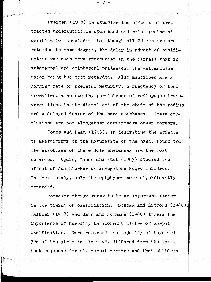
Dre1zen (1958) in study1n~ the effects of pro-
tracted undernutr1t1on upon hand and wrist postnatA.1
ossif icat1on concluded thet though all 28 centers are
retarded to some degree, the delay in advent of oss1f1-
cation was much more pronounced in the carpals than in
metacarpal and epiphyseal phalanges, the multangulum
major being the most retarded. Also mentioned are a
lagginp: rate of skeletal maturity, a. frequency of bone
anomalies, a noteworthy persistence of radiopaque trans-
verse lines in the distal end of the shaft of the radius
and a delayed fusion of the hand epiphyses. 'T'hese con-
clusions are not altogether conf1rmed'ty other workers.
Jones and Dean (1956), in describing the effects
of Kwashiorkor on the maturation of the hand, found that
the epiphyses of the middle phalanges are the most
retarded. Age.in, Masse and Hunt (1963) studied the
affect of Kwashiorkor on Senegalese Negro children.
In their study, only the epiphyses were significantly
retarded.
Heredity though seems to be an important factor
in the timing of ossification. Sontag and Lipford (1960),
Falkner (1958) and Garn and Rohme.nn (1960) stress the
imports.nee of heredity in aberrant timing of carpal
ossification. Gt=!rn reported the majority of boys end
39% of the girls in U.s study differed from the text-
book sequence for six carpal centers e.nd that children
-- ·---=ti==============--====· ==================!:I=====

- 8 -
-===-==lf==================================================================ti=======
with deviant ossification centers were not characterized
by more episodes of illness during the first 8even years
of life. He concludes that the variability is th& result
of heredity, not illness.
~he validity of the Inspectional ~echnique of
Greulich and Pyle, which does presume a rather fixed
pattern of development of the carpal bones and a sub
jecti ve averaging of all centers, has received much
criticism.
Robinow (1942) applied a fe.ctor analysis to a
selection of osseous centers in the limbs. He founrl two
rather distinct groups of developing centers in the hand
and wrist: One consisting of the long (epiphyseal) bones,
the other comprising the round (carpal) bones. Each
eroup is independent of the other centers being advanced
or retarded within their own group with no correlation
to the state of development of the centers of the other
group. Pyle and Sontag (1943) found thet the time of
onset of ossification is definitely more variable in the
carpals than in the epiphyses. Baer and Durkatz (1957)
reported that in both sexes the carpal bones, taken as
a group, show a considerably higher average percentage
of asymmetry than the average for the ep1physes. Any
bilateral asym,"Iletry which does occur in a population of
normal children is a function of the variability in the
initiation time of the ossification process.

- 9 -
Mainland (1953, 1954) evaluated systematic and
variable errors in the assessment of carpal roentgenograms.
He found no significant variable error associated with the
Greulich-Pyle Atlas, but felt that a range of ±11.1 months
should be assigned to each film for 95% probability if
the standard deviation of a particular sampling is 4
months. Furthermore, the systematic error, or bias,
varied significantly between independent reading of the
same film. Though these studies are ref erred to con
tinually in the literature, Greulich and Pyle (1959)
feel that Mainland's findings are invalid because of
the use of a relatively untrained observer in the film
assessment. Their studies show a remarkably high
correlation between trained observers in film assessment.
A sophisticated study of the reliability of
assessing skeletal maturity by the Inspectional Technique
was conducted by Acheson et al (1963). He reported that
the range of mean values of six observers' readings is
only just over four skeletal months, an extremely close
relationship. Acheson further suggests that the most
practical and logical method of assessing a radiograph
which shows a disparity in the pattern of skeletal
maturation, an event which is not altogether uncommon,
is to ignore the carpus. Acheson gives three reasons•
1) The carpus is the region of the hand and wrist whose
maturation is the most susceptible to interference because
9f===================================================*===~"

I I I I I i f '
- 10 -
~=====c=#============-=-=--=-~-=--=---=======---=-~-=-=-================================#=====
of environmental vicissitudes. 2) The onset of ossifi-
cation in the carpus is more ve.riable than in other
"communalities" in the hand, but nevertheless, follows
familial patterns {Garn and Hohmann 1959, 1960, 1962).
J) In measuring skeletal maturity, one should be concerned
with predictions in the epiphyseal bones (long and short
bones) than the carpal bones (round bones), which by
definition develop differently.
Johnston (1965) in a similar study found the car-
pals to be a source of significant variable error in
girls in repeated readings b~ individuals. In addition,
they lagged behind epiphyseal centers, relative to the
standards. He also recommended that they (the carpals)
not be considered in assigning skeletal age.
Michelson (1946) was the first to suggest that
individual bones should be standardized as regards to
their degree of progressive maturation. He described
22 consecutive stages of epiphyseal ossification and
union.
Acheson (1964) reported that the lack of precision
in carpal rating of particular films indicates that
either the applicability of the described standards for
these bones should be reviewed or that the carpal con
tribution to the overall assessment of the skeletal
maturity should be excluded.
Upon first introduction of the ••point-count"
i_-Jr.
,'
"11
~r :I'.
======i!:=====================================================:===========!t======Y'·
- 11 -
method. many investigators believed it to offer greater
objectivity. precision and more correctly picture the
continuous nature of skeletal maturation. However,
Acheson (1966), in studying the reliability of assessing
skeletal maturity. reported that systematic error, is
greater utilizing the 0 point-count•• method than the
Atlas method, because, whereas the Greulich-Pyle techn1-
que involves one single judgment, the Tanner-Whitehouse
or .. point-count" technique requires a series of 20
separate judgm.entss the more mature the hand, the greater
number of stages any one bone may have achieved. Con-
sequently, the variability is greater for the more mature
stages. Small differences in judgment as regards to
shape are greatly exaggerated because of the mathematical
conversion to an ordinal system. Though the bone specific
approach offers a more finely calibrated scale and may
reduce the random error of a single reading, shape
recognition and judgments make it very susceptible to
individualistic interpretations or bias. As with the
Inspectional method, its reliability is ~reatly enhanced
when the carpus is ignored. Simply by breaking the
maturing hand down into its component parts and considering
it bone by bone, Acheson and Tanner seem not to have
offered any quick solution to the problem of precision
in readings.
The growth and development of the hand and wrist

- 12 -
bones as reflected in the carpal index have been correlated
to other aspects of growth and development in general,
not always with the same degree of success,
Milo Hellman (1928) in studying the ossification of
the epiphyseal cartilages in the hand reported that the
greatest increase in height follows by just one year the
greatest growth increment in the bones of the hand,
suggesting differential growth patterns.
Howard (1928) found that a study of the growth
characteristics of the carpus, long bones and epiphyses
did not definitely establish ~oncomitant growth in the
jaws, In general, individual growth patterns are parallel,
but the rates of speed may vary.
Elgenmark (1946) reported that correlation cal
culations prove that there are significant and positive
correlations of equal degree between the differentiation
of the ossifying centers and age, hei~ht and weight. In
infancy, height shows a stronger correlation with ossi
fying center development than does age. In later years,
he feels the correlation is equal.
Acheson (1957) related physique to the rate of
skeletal maturity in boys. He found that ectomorphs
varied s1gn1f1cantly as slow maturers, whereas there was
no significant difference between endomorphs and mesomorphs.
'rhe eotomorphs, when compared to the endomorphs, were
slower maturers yet significantly taller. He concludes

- 13 -
that the basic relationship between physique and skeletal
maturation is genetically controlled, this relationship
being masked and distorted by environment.
Rose (1960) measured and compared the growth and
sizes of the various facial areas with those of certain
otherl:ndy measurements. He concluded that measures of
a carpe.1 rank are an ineffectual guide to the growth and
development of the facial areas in the para.pubertal period.
The best indicator is stature and body weight.
Garn (1961) studied specific six-month increments
in height, weight and number"of postnatal hand-wrist
ossification centers in over 150 children. He reported
that the ir-crements for height and weight, and especially
ossification increments, were markedly skewed, indicating
the inapplicability of means and standard deviations. He
concluded that the increments were under control of
individual genetic patterns.
Bjork (1967) found close association between the
age at maximum growth in body height a:nd the age when
ossification of the metacarpal-phalangeal sesamo1d of
the thumb occurred, the sesamoid usually ossifying one
year before maximum pubertal skeletal growth.
The relation between dental maturation, as assessed
from the degree of root formation, and skeletal maturity,
as assessed from hand-wrist roentgenograms, has been
studied extensively. The findings in these reports are

not in full agreement.
Todd ( 1932) quoted Broa.dbent a~ finding "the.t e.
child which is retarded in skeletal growth is usually also
retarded in tooth development."
Shuttleworth (1939) compared the total number of
teeth erupted with early, medium and late maturers,
depending on the age of peak height velocity. He con-
eluded that early maturing boys and girls were also ad-
vanced in dental development.
Sutow, Teraski and Owaha {1954) in examining over
1,000 Japanese children found that in each age group •
those children having advanced skeletal development had
a greater number of erupted permanent teeth.
Demisch and Wartman {1957) found a high positive
correlation, with an approximately straight line trend,
between the degree of calcification of the mandibular
third molar and' skeletal and chronological ages. Their
findings support the theoretical contention that a
relationship exists between the maturation of various
tissue systems.
Lamons and Gray {1958) investigated the relation
ship to chronological age, finding that it is a slightly
better index of tooth development than is skeletal age.
They further noted that skeletal and dental age may vary
independently.
Hotz (1959) studied the relation of dental calcif1-

- 15 -
--- . --- ----- -------------- _'" _________ ------· "" ___ ------ ----- ---------~------·----------------
cation to chronological and skeletal age. He reported
that a very close relationship exists between dental,
chronological and skeletal age.
Lauterstein (1961} conducted a cross-sectional
study into dental development and skeletal a.ge. He
found that an intimate correlation exists between .. root
age and bone age. 11
Green (1961) reported that correlation coefficients
between dental a.ge, skeletal age and chronological age
were moderately high. The closest association was not
between dental and skeletal gges, but between dental
and chronological ages. Furthermore, skeletal age,
height and weight showed a slight tendency to be related,
suggesting that the factors which control skeletal
growth and development are also important in determining
height and weight. Individual patterns, however, were
markedly varied, emphasizing the hazard of applying
norms of development based on central tendencies to
individual children.
Steel (1965) emphasized the existence of a wide
range in both dental maturity status and skeletal
maturity in 12 year old boys and girls. He demonstrated
statistically that there is no direct interdependence
between dental and skeletal maturation. He concluded
that though maturation is a continuous process, it does
not necessarily progress at a constant rate. Maturation --- -------------------~------ ------··------------- ---·-=-=--·-=-=--=----- -------~

II - 16 -
occurs in uncoordinated waves within the various systems,
in this particular case, dental and skeletal systems.
There have been only two published papers dealing
with the inter-relationship of malocclusion and skeletal
maturity. Bambka and Van Natta (19.59), in a longitudinal
study of occlusion in relation to skeletal maturation,
investigated Krogman's statement that there exists a
higher rate and more severe cases of malocclusion in
maturational laggards. A sampling of malocclusions we.s
categorized according to the Angle classification and
compared to a sample of normal occlusions. In Angle
Cl. II malooclusion, the sample was further subdivided
according to the degree of distal positioning of the
mandibular first molar, the most distal position repre
senting the most severe malocclusion. After a statis
tical analysis of the data, the findings were as follows•
1. No evidence of higher rates of malocclusion
in maturational laggards.
2. No evidence of more severe malocclusion
in maturational laggards.
J. No evidence of association between time
of skeletal maturation and severity of
malocclusion.
Johnston (1965) studied a group of malocclusions
classified according to Angle which were further sub
divided, according to the Steiner cephalometric analysis,
--~-~--~-=-~-==-==--=-===-=======-==-=-=--=====================tt====~

- 17 -
into skeletal categories. He found the only significant
differences between skeletal age and chronolog1c age
exist in the group where the malocclusions were purely
skeletal, the mean skeletal age being retarded in this
category. He concludes that maturational retardation is
a factor in a Cl. II malocclusion, insofar as skeletal
factors are concerned. Where deviations from the average
maturation pattern exist, concomitant devia.tions from
the usual facial growth patterns will also exist in
those dimensions related to skeletal mgturation. "Phough
differential relationships exist between certain growth
measurements and skeletal maturation, in the mandible,
spurts and lags in maturation activity are accompanied
by similar spurts and lags in some aspects of growth.
The findings in these two reports are not altogethe
in full agreement. Further research is still required.
---~

CHAPTER II
METHODS AND MA'rERIALS
The sampling of malocclusions to be considered in
this study were chosen from patients undergoing ortho
dontic therapy at Loyola University's School of Dental
Medicine Department of Orthodontics. The sample con
sists of 51 males and 66 femaleso The children are
Caucasian; their ages ranging from 119 months to 211
months. The mean age of all children is 151.65 months.
The following records were obtained for each individual
patients 1) a previous medical and dental history,
2) study casts of maxillary and mandibular arches,
J) a lateral cephalogram and 4) a radiograph of the
left hand and wrist.
Patients with a pa.st dental history of perverted
oral habits (thumb-sucking, lip..biting and sucking and
tongue thrust) were excluded from the sample1 whereas,
patients with a past medical history of protracted
illness during childhood were included.
The arch length discrepancy in the maxillary and
mandibular arches was determined by a Ha.yes-Nance Analy
sis. Those patients with congentially absent teeth
were excluded from the sample. The molar relationship
was classified according to Angle. --~- ·---~--·---~--~-·--------------~-~-----
- 18 -

- 19 -
The Sassoun1 Archial Analysis (1954) was traced
for each lateral cephalogram. Also the following
measurements from the Steiner Analysis were also traceda
SNA, SNB, ANB, GoGnSN, SND and Po to NB.
A skeletal maturity status of each patient was
determined by the Inspectional ~echnique utilizing the
Greulich-Pyle Atlas. Two 1nd1v1dual assessments were
made for each rad1ograph. Those rad1ographs showing a
discrepancy in readings of more than three months were
reassessed, the mean value of three readings of more
than three months were reassessed, the mean value of
three readings being considered as the skeletal age of
the particular radiograph in question.
Study 1
The entire sample of malocclusions, regardless
of nature, will be compared statistically with the
means and standard deviations of skeletal age used in
establishing the standard of reference for the Greulich
Pyle Atlas. Boys and girls will be compared individually
for each specific age. The Student "t" will be employed
to determine significance.
Study 2
The sample of malocclusions will be divided into
two categories, Cl. I and Cl. II, according to the
Angle classification. The skeletal ages of the two
groups will be compared both as a whole and individually

J
I . - 20 -
c .. 1·-cccc===--==-..==.c.c=cc·.c.=..-=c=:====--_c=--=:c=--cc==-=cc:=-=c==.cc:.cccc.====-cc=.===.cccc-...====c.c=.co~==--==--===-=--1 according to age. The Student ,.t" will be employed to
determine significance.
Study 3
The sample of Angle Cl. I malocclusions will be
subdivided according to the degree of arch length dis-
crepancy into two categories.
The arch length discrepancy of the first group
ranges from -6mm to -18.8mm in both maxillary and mandi-
bular dental arches, with a mean of -7.2mm for the
mandibular arch and -8.Jmm for the maxillary dental
arch. This group will be considered Cl. I severe arch
length discrepancies.
The arch length discrepancy of the second group
ranges from -5.6mm to +12mm in both maxillary and
mandibular arches, with a mean of -J.4mm for the mandi
bular arch and -2.9mm for the maxillary arch. This
group will be considered Cl. I moderate arch length dis-
crepancy.
'rhe mean cephalometric values are practially
identical for both groups of malocclusionsa
SNA SNB ANB SND GoGnSN Po to NB
81.J degrees 77.6 degrees 3.7 degrees
72.9 degrees J8.5 degrees
2.0mm
The Sasounni Archial Analysis shows good antero-
posterior relationship between maxilla and mandible.

- 21 -
These two groups of skeletal ages will be compared
both as a whole and individually according to age. The
Student "t" will be employed to determine s1gn1f1cance.
Study 4
The sample of Cl. II malocclusions will be sub-
divided into two groups. ·rhe first group will be termed
"skeletal" malocclusions.
The mean cephalometr1c values for this group are
as follows:
SNA SNB ANB SND GoGnSN Po to NB
82.3 degrees 73.8 degrees
8 .• 5 degrees 68.9 degrees 43.1 degrees
Omm
The Sasounni Arch1al Analysis reveals that each
case is retrognathic, the reason being either mandibular
body insufficiency or a distal positioning of the corpus.
The mean arch length discrepancy for the mandible is
-0.5mm, for the maxilla +l.2mm. The molar relation in
each case is in full Angle Cl. II malocclusion, the
d1sto-buccal cusp of the maxillary first molar occluding
in the buccal groove of the mandibular first molar,
both left and right.
~he second group of Cl. II malocclusions will be
considered m.odera.te arch length discrepancies, with no
skeletal retrogr.athism.
The mean arch length discrepancy is -6.3mm in the
-----==--=~'ii' I
i1I
I
I
arch.
- 22 -
.. - - - - - .. - --------------··------ ·-~-- ------·----··-- ------ ··------·--------~-----
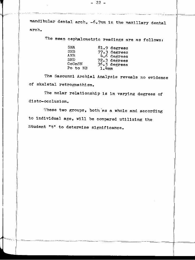
dental arch, -6.7mm in the maxillary dental
The mean cephalometric readings are as follows1
SNA SNB ANB SND GoGnSN Po to NB
81.9 degrees 77.3 degrees 4.6 degrees
72.3 degrees 36.5 degrees l.4mm
The Sasounni Archial Analysis reveals no evidence
of skeletal retrogna.thism.
The molar relationship is in varying degrees of
disto-occlusion.
'i'hese two groups, both "e.s a whole and according
to individual age, will be compared utilizing the
Student "t" to determine significance.
""=-'--tt---===~=-'-"-==="----'-"=--=---'-====c_c=cc=--e =-==-cc-=="-'====~-'-'-'--'--'-'"'''-==~===~-===-= ------------------------=--==--=-=-=tf=====-=-= 11 11
" 'I 11

------·---·-------------------~~~----------·---------··-------·-- -· ---------·-----·------------------·------------------- --------
CHAPTER III
FINDINGS
STUDY 1
BOYS
Skeletal Age (in months)
.5 Percent Chronologic Nwnber of Standard Student Level of
A e Hand-films ~ Deviation "t" Probabilit
*1 9 yrs. 160 113.90 9.00 *2 1 117.0
1 10 yrs. 177 125.68 9.79 2 .5 126.J
1 11 yrs. 1.54 137.32 10.09 1.20 1.96 2 8 139.8 12.0
1 12 yrs. 16.5 148.82 10.J8 1 • .56 1.96 2 11 151.4 8.74
1 13 yrs. 17.5 1.58.39 10.44 1.62 1.96 2 9 161.7 7.93
1 14 yrs. 163 170.02 10.72 1.78 1.96 2 9 175.7 6.37
1 15 yrs. 124 182.72 11.32 2 0 0 0
1 16 yrs. 99 19.5.32 12.86 2 1 19.5.0
1 17 yrs. 68 206.21 13.0.5 2 2 19.5.0
*l Data from Greulich and Pyle, Atlas 2f. Radiographic Develo ment of the Hand and Wrist, Stanford University Press, !9'59.----
*2 Sample of selected patients from Loyola University Depart-ment of Orthodontics, Chicago, Illinois.
I
---------~- ----1 - 23 - I

- 24 -
---- ---- - ··--- ------ --- - --- -- -- - - - ------ ------ -----~ ---- -------- ~- - - ------ --- --- -- ---- ----
STUDY 1 Continued)
GIRLS
Skeletal Age (in months)
5 Percent C hronologic Number of Standard Student Level of
A e Hand-films Mean Deviation "t" Probabilit -1 9 yrs. 195 113.86 10.74 2 3 121.0
1 10 yrs. 206 125.66 • 11.73 1.71 1.96 2 9 120.J 7.98
1 11 yrs. 203 1)7.87 11.94 1.43 1.96 2 11 135.6 10.60
1 12 yrs. 198 149.62 10.24 1.19 1.96 2 17 151.6 11.70
1 13 yrs. 179 162.28 10.67 l.OJ 1.96 2 18 161.1 12.)4
l 14 yrs. 170 174.25 11.JO 2 4 177.0
l 15 yrs. 117 18).62 9.23 2 1 189.0
1 16 yrs. 64 189.44 7.31 2 2 192.0
The Student's "t" in each case being less than the 5 percent probability level the coefficients are shown to be not significant.

- 25 -
- --~---------·----" - - ------ --· - -
STUDY 1 (continued)
Inspection of the mean chronologic ages of both
boys and girls revealed no significant difference. Since
the following studies are concerned only with relative
advancement or retardation of skeletal age, both boys
and girls are included in the following studies.
The mean chronologic ages are as follows1
Age Group (in years)
9
10
11
12
lJ
14
15
16
Mean Chronologic Age (in months)
Boys Girls
. 119. 0 115.3
126.3 123.9
137.9 lJS.5
148.7 149.9
161.8 160.5
173.2 17J.8
195.0 189.0
205.0 200.0
1
:11 --------------- --------- --,==========-=c=-====-=-==-==cc.====-=c===-=-= -==== -== ------=--= - --====== II~
!1
1

------ ------ -----· ' - 26 -
STUDY 2
CLASS I AND CLASS II MALOCCLUSIONS
Mean Chrono- Mean Skele- 5 Percent Number of logic Age Standard tal Age Standard Student Level of Hand-films (1n months~ Deviation ~in months) Deviation "t" Probab111tl Cl. I 49 151.6 17.3 151.4 18.1 .97 1.96 Cl. II 56 151.7 16.9 150.8 17.5
The Student's "t" being less than the 5 percent probability level the coefficients are shown to be not significant.
Number of Hand-films
Cl. I 2 Cl. II 2
Cl. I 5 Cl. II 9
Cl. I 11 Cl. II 8
Cl. I 10 Cl. II 17
Cl. I 12 Cl. II 15
Cl. I 6 Cl. II 6
Cl. I 1 Cl. II 0
Mean Chrono-logic Age ~1n months)
116.0 116.5
124.3 123.0
137.9 139.8
148.o 150.1
161.3 160.5
171.5 174.3
186.o 0
Mean Skele-tal Age
(in months~
120.0 120.0
126.o 118.0
133.l 129.0
150.6 150.9
160.0 162.0
171.0 179.0
189.0 0
Student "t"
2.21
2.01
5 Percent Level of Probability
2.2it;
2.23
------- - ---- -- ---- ---
- 27 -
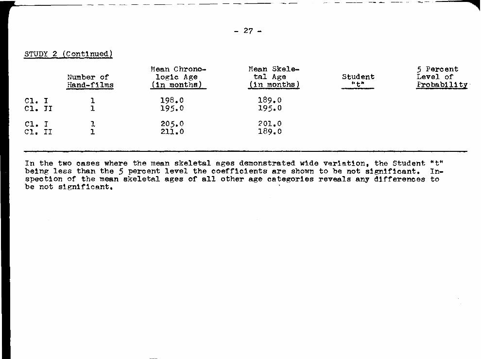
STUDY 2 (Continued)
Mean c hrono- Mean Skele- 5 Percent Number of logic Age tal Age Student Level of Hand-films (in months} (in months} ••ttt Proba.b111ty·
Cl. I 1 198.o 189.0 Cl. JI 1 195.0 195.0
Cl. I 1 205.0 201.0 Cl. II 1 211.0 189.0
In the two cases where the mean skeletal ages demonstrated wide v~r1ation, the Student "t" being less than the 5 percent level the coefficients are shown to be not significant. Inspection of the mean skeletal ages of all other age categories reveals any differences to be not s1gn1f1cant.

STUDY 3
Severe ALD Moderate ALD
---------- -- --.----------- ,
Mean Chrono-Number of logic Age Hand-films (1n months)
20 2.5
151.8 151.h
- 28 -
CLASS I MALOCCLUSIONS
Mean Ske!e-Standard tal Age Deviation (in months)
12.J 11.9
151.9 150.1
5 Percent. Standard Student Level of Deviation fft" Probability
17.J 17.8 .78 1.96
The Student "t" being less than the 5 percent level of probability the coefficients are shown to be not significant.
Number of Hand-films
Severe ALD 1 Moderate ALD 1
Severe ALD 1 Modera. te ALD 3
Severe AID J Moderate ALD 8
Severe ALD 5 Moderate ALD 5
Severe ALD 8 M odera. te ALD 4
---==--=..-~
M ea.n C hrono-logic Age
(in months}
119.0 llJ.O
121.0 125.J
1J8.0 1J7.9
149.0 147.0
161.J 161.3
:r-1ea.n Skele-tal Age
(in months)
12J.O 117.0
12J.O 127.0
lJJ.O 1J6.5
154.2 148.8
161.J 157.5
Student "t••
1.87
5 Percent Level of Probability
2.31

... ----~- - - -- - --
STUDY 3 (Continued)
Severe ALD Moderate ALD
Number of Hand-films
2 4
- ---- -- ·- ---- - --
r,Tean Chronologie Age ~in months)
17).0 170.8
- 29 -
Mean Skeletal Age
(1n months)
171.0 171.0
~ -- - --- --· ~
Student "t"
5 Percent Level of Probability
The Student "t" of age group showing the greatest difference between the mean skeletal ages being less than the 5 percent probability level the coefficient is shown to be not s1gn1f1cant.

STUDY 4
Skeletal Moderate ALD
- JO -
CLASS II MALOCCLUSIONS
Nean Chrono-Number of logic Age Sta~dard Hand-films {1n months) Deviation
16 24
145.1 152.2
11.32 10.4
5 Percent Student Level of
''t" Probab111 ty
The Student "t" being less than the 5 percent pro~ab111ty level the coeff1e1ent for the difference between the chronolog1c ages of the two samples is shown to be not significant.
Skeletal Moderate ALD
:·Iea.n Skele-Number of tal Age Standard Hand-films (1n months) Deviation
16 24
1)8.2 154.8
17.84 15.40
5 Percent Student Level of
"t" Probability
The Student "t" being greater than the 5 percent probability level, the mean difference of skeletal ages is shown to be s1gn1f1cRnt.

- 31 -
STUDY 4 (Continued)
SKEL~'TAL MALOCCLUSIONS
T·I ean c hrono- Mean Skele- 5 Percent Age Grouping Nu.'ll ber of logic Age tal Age Student Level of
(in ;!ea.rs) Hand-films (in months) (in months) ft t fl Probability -10 3 128.o 12.5.0 3.07 4.30
11 5 139.0 123.0 3.29 2.31
12 .5 148.6 149.4
13 3 161.3 159.0
The mean skeletal ages of only the 11 yea.r old malocclusions are shown to be significantly lower than the concomitant chronolog1c ages.
Inspection of the means reveals the difference between chronologic and skeletal ages of the 12 and 13 year old medocclusions to be not s1gn1ficar1t.
r

- 3?. -
§TUDY 4 (Continued)
MODEBP. TE ALD
!'4 ean C hrono- Mean Skele-Age Grouping Numbe:- of lo~ic Age tal Age (in ye9.rs) Hand-films {in months) (in months) 10 1 . 120. 0 117.0 11 3 1.38.J 139.0 12 9 151.0 153.0 13 11 159.9 161.2
Inspection of the means-reveals the difference between the skeletal and chronolog1o ages to be not s1gn1f 1cant.

- 33 -
--===~===================-=-=-==~-~'=================================*=====
STUDY 1
Skeletal Age 1n Years
17
16
15
14
13
12
11
10
9
MALOCCLUSION IN BOYS
/
/ /
--"' _,, ;,...
/ //
/'
/
I . i
/
j ,-/
/
"" /
,,.;
9 10 11 12 13 14 15 16 17 Chronolog1c Age 1n Years
a Sample used 1n determining standards --- as reported in Greulich-Pyle Atlas.
------• Sample of selected patients undergoing orthodontic treatment.

• I
l
- 34 -
STUDY 1 (Continued)
Skeletal Age in Years
17
16
15
14
lJ
12
11
10
9
9
MALOCCLUSION IN GIRLS
10 11 12 13 14 15 Chronologic Age in Years
16 17
~~~• Sample used in determining standards as reported in Greulich-Pyle Atlas.
------• Sample of selected patients undergoing orthodontic treatment.
~==:====#======-==-==-==-======================================-==· ---=- ====\l=====

STUDY 2
Skeletal Age 1n Years
17
16
15
14
13
12
11
10
9
- 35 -
CLASS I AND CLASS II MALOCCLUSIONS
9
.--
/
/
·-{
/
/ •. '""'~"···"
10 11 12 13 14 15 Chronolog1c Age in Years
------• Class I malocclusion
1 Class II malocclusion ---
16 17
~======#o=========================================~~~=-=='ff====

STUDY 3
Skeletal Age 1n Years
16
15
14
13
12
11
10
9
9
- 36 -
CLASS I MALOCCLUSIONS
/
{ /
/'J I
/
10 11 12 13 14 Chronologic Age 1n Years
15
~~~' Class I severe arch length discrepancy.
------1 Class I moderate arch length discrepancy.
16
~=·====!f==================================================M=====

- 37 -
--=±~-=--=-"'-=-===-=--=--=·=-=-===··-=--=-=--:-=.-==-=---=--=--- ----- ------·-------------
S7UDY 4
Skeletal Age in Years
14-
lJ·
11-
10·
CLASS II MALOCCLUSIONS
I
10
---
;
/ /
/
/
' 11
-/r I 1 ·
I
I
12
;/'
// ,_..::::. _...
13 14 Chronologic Age in Years
1 Class II skeletal malocclusion.
------• Class II moderate arch length discrepancy.

CHAPrER IV
DISCUSSION
The carpal index. as a guide to the overall skele
tal maturation of the individual patient, is subject to
great variation. One problem which is stressed repeatedly
in the literature is the need for population specific
norms• Koski (1961), Johnston (1963), Acheson (1966).
It has been a well-established fact that children
developing under different environmental circumstances
mature at different rates. The sample used in this
study was not large enough to allow adjustment of the
mean skeletal age values for each particular age category.
The highly selected population of well-to-do children of
suburban Cleveland used in establishing the norms of the
Greulich-Pyle Atlas are certainly not the same as the
children of metropolitan Chicago, of an obviously lower
income level, undergoing orthodontic therapy at a clinic.
Another problem is the averaging of all centers in
the hand and wrist. In the assessment of many films the
carpal bones and epiphyses showed clear discrepancies as
regards the maturational level of each group. These
films were assessed following the suggestion of Acheson
(1963) and Johnston (1965) in that the carpal bones were
not considered in the final assessment. This procedure
~~=====l:I=:============================================================~-=---=--=--==--=-=-=!!===
- 38 -

- 39 -
is contrary to that advocated in the Atlas and is Another
source of discrepancy of mean values between standard
and sample. In view of these considerations, it is this
author's opinion that differences of six months or less
between skeletal and chronologic should notl:J:3 considered
as outright evidence of skeletal advancement or retardation.
Since the very standard deviations of the skeletal age
values of the standards range from 6.3 months to 13.5
months, this consideration is not altogether unfair.
Study 1 revealed no difference between the sample
of orthodontic patients and ~hose children of the standard.
This comparison, however, is made with certain reservations.
All children of the selected sample were characterized by
a malocclusion requiring orthodontic therapy. These
malocclusions ranged in severity from simple crowding of
one tooth to severe antero-posterior discrepancies between
maxilla and mandible. The specific dental status of the
sample used in establishing the standards is unknown.
It would be unreasonable to assume that these children
have normal occlusions; certainly. a certain percentage
of these children are cha:racterized by the dento-fac1al
disharmony typical of the orthodontic sample. If, how
ever, one considers the standard truly representative of
the overall population, 1nclud1ng those children with
dento-facial disharmonies, it is clearly evident that
the sample of orthodontic patients do not differ stat1s-

- 40 -
tically one iota as regards their overall skeletal
maturity from the standards. The progress of general
osseous development of orthodontic is identical to the
standards. In this sense, one may infer that the broad
spectrum of malocclusions are not accompanied by dis
crepancies in overall skeletal development and that
children with malocclusions mature, as a whole, at a
rate identical with children representative of a normal
population.
In Study 2 the malocclusions were subdivided
according to the Angle classification into Cl. I and
Cl. II categories. The skeletal ages of both categories
related statistically not significant to the standards,
both mean skeletal ages being within one month of the
mean chronologic age. Furthermore, there was no statis
tical difference between the two categor1esr the Student
"t" was .97, a "t" value of 1.96 necessary for significance.
The Angle classification is based solely in the
positioning and relation of the maxillary and mandibular
first molars, the osseous bases usually relating as a
whole in a similar manner. The Angle system does not
itself take into account discrepancies in a vertical or
lateral plane. Although the antero-posterior relation
ship of the teeth may be the most important single con
sideration, this classification system does not lend
itself to the determination of the overall skeletal

- 41 -
--=====#=====================~-==--~~--~-=-=-===============================#======
pattern. ~he mere presence of a Cl. I molar relation
does not necessarily reflect perfect antero-posterior
skeletal relation of maxilla and mandible, indicative
of coordinated ideal growth patterns. Similarly, a Cl. II
molar relationship does not necessarily reflect a dis
harmony in growth between maxilla and mandible. For
example, a not uncommon cause of a Cl. II molar relation
is premature loss of maxillary deciduous second molars
with mesial drift of the maxillary first molars. The
resulting Cl. II malocclusion cannot possibly be related
to disharmonious growth maxilla and mandible. A myriad
of local factors can affect the molar relationship.
Bambka and Van Natta (without attempting to fur
ther categorize malocclusions according to facial and
skeletal patterns) used just this class~.f1cation to sub
divide their sample. Any index of skeletal mAturity is
useless when applied to tooth relationships. True skele
tal malocclusions must be determined and dealt with
separately.
Study J revealed that, regardless of degree of
arch length discrepancy, malocclusions relate to standards
of overall skeletal maturity within normal limits.
The following are quoted in the literature as
etiologic factors 1n malocclusions characterized by arch
length discrepancys

r _ 42 _
L=o==#====="--=======i!== 1 1. Supernumary teeth
2. Congenitally missing teeth
J. Anomalies of tooth size
4. Anomalies of tooth shape
5. Abnormal muscular attachments
6. Premature loss of deciduous teeth
7. Prolonged retention and abnormal
resorption of deciduous teeth
8. Delayed eruption of permanent teeth
9. Abnormal eruptive path
10. Ankylosis
11. Dental caries
12. Heredity
The sample utilized in this study were selected such
that factors as congenital defects, predisposing meta
bolic climate and disease, abnormal pressure habits and
anomalies in number of teet~ (the first and second factors
listed) were not et1olog1cally relevant to the malocclusion
Eruption, its sequence and rate as an etiologic
factor in arch length discrepancy, is relative to this
study. Do disharmonies of dental maturation, as assessed
from the degree of root formation or eruptive patterns,
resulting in a particular malocclusion type relate to
disharmonies in overall skeletal maturation. The litera
ture, on the whole, regards the correlation between
eruption and skeletal maturation as very low. The mal-
~=====llo===========----=====================================================1it========
- 43 -
occlusions represented here, where abnormal eruptive
patterns were indeed an etiologic factor, showed no con
comitant deviation from skeletal norms, either among
themselves when categorized by severity, or as a whole.
Brodie (1944) reported that temporary disharmonies
(crowding) are caused by a slow or fast rate of growth
in the bone compared to eruption of teeth. Are such
deviant growth rates in the jaws accompanied by a similar
pattern in overall skeletal maturation? Apparently not.
Assuming that Brodie's findings are correct, the precise
pattern of jaw growth, erratip increments of which may
result in dental crowding, 1s in no way coordinated with
overall skeletal maturation as a whole, as assessed from
the carpal roentgenogram.
Study 4 statistically compared two particular types
of malocclusions within the Cl. II category, skeletal
malocclusions and severe arch length discrepancies. ~he
skeletal malocclusions demonstrated no individual tooth
disharmonies, the problem being limited solely to the
antero-posterior relation of the osseous bases, as demon
strated cephalometrically. The mandible in each parti
cular case was determined to be lagging behind the
maxilla relative to the standardss the antero-posterior
relation of the maxilla was in good relation to other
cranio-facial landmarks. The soft tissue profile of each
patient was markedly retrognathic because of this posterior

~--
- 44 -
-----····-------·----·-·---·-- ---~------- - ---------·--- ---------------------
positioning of the mandible.
The sample of Cl. II moderate arch length discre
pancies demonstrated no similar skeletal retrogna.thism.
Individual tooth disharmonies were marked complicated by
problems of severe dental overbite and deep bite. Both
samples, though grouped in the Cl. II category according
to Angle, represent two distinct and separate malocclusion
types. One a skeletal imbalance, the other a dental im-
balance.
The sample of Cl. II severe arch length discre
pancies compared statistically not significant to the
standards, relative to degree of skeletal maturation.
As discussed previously, these findings are not alto
gether surprising, since the malocclusion was largely
of dental origin.
The sample of skeletal Ealocclus1ons, as a whole,
was slightly retarded skeletally relative to the stan-
dards. The age groupings, however, differed in mean
skeletal ages when considered individually. The children
of 11 years lagged considerably 1n skeletal maturation;
the children of 10, 12 and 13 years demonstrated skeletal
ages no greater than 3.0 months variance from the chrono-
logic age. Yet children of all ages possessed the same
skeleto-facial characteristics. Furthermore, the 11
year old skeletal malocclusions lagged s1gn1f1cantly
behind 11 year old arch length discrepancies, whereas
- lJ-5 -
. - -"- -- --- -- -----·-- ----· .. -- --·--- -----·----------·---- --- ---------------~---------- - - -- ----- --- - --- --- --· - -·- - . ------·- --------------- -------------------
in 12 and 13 year olds, no significa.nt difference in
skeletal maturation was evidencecl.
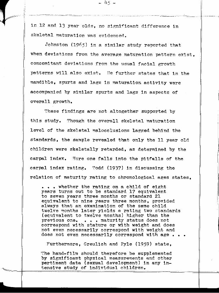
Johnston (19h5) in a similar study reported that
when deviat:lons from the average mature.ti on pa.ttern exist,
concomitant deviations from the usual facial growth
patterns will also exist. He further states that in the
mandible, spurts and lags in maturatiou activity were
accompanied by similar spurts and lags in aspects of
overall growth.
These findings a~e not altogether supported by
this study. Though the overall skeletal maturation
level of the ske1.etal malocclusions lagp~ed behind the
standards, the sample revealed that only the 11 year old
children were skeletally retarded, as determined by the
carpal index. Here one falls into the pitfalls of the
carpal index rating. ~odd (1937) in discussing the
relation of maturity rating to chronological ages states,
• - • whether the rating on a child of eight years turns out to be standard 17 equivalent to seven years three months or standard 21 equivalent to nine yea.rs three months, provided always that an examination of the same child twelve months later yields a rating two standards (equivalent to twelve months) higher than the previous one. • •• maturity status does not correspond with stature or with weight and does not even necessarily correspond with weight and does not even necessarily correspond with age •••
Furthermore, Greulich and Pyle (1959) state,
The hand-film should therefore be supplemented by significant physical measurements and other pertinent data (sexual development) in any in-
----· ----··--------... ---------...
~--=--==rt=======t=e=n==s=i=v=e==s=t=u=d=y==o=f===i=n=d=1=v=i=d=u=a=l==c=h==i=l=d=r=e=n=.=============-==-=--====l1F=======
- 46 -
=====- ... ---
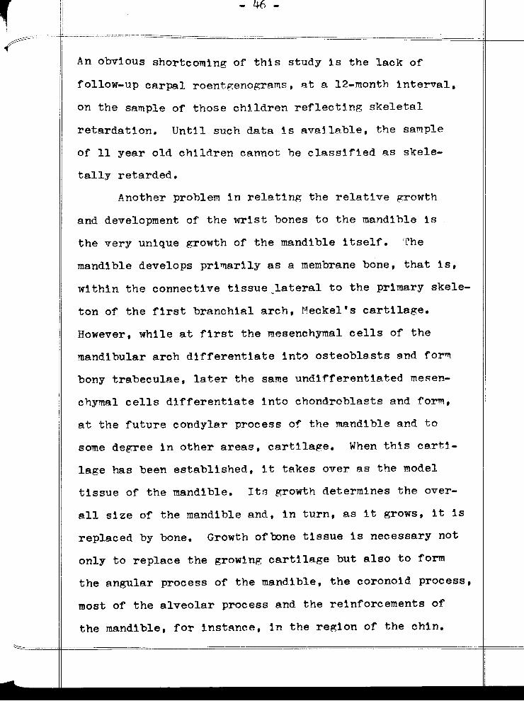
An obvious shortcoming of this study 1s the lack of
follow-up carpal roentgenograms, at a 12-rnonth interval,
on the sample of those children reflecting skeletal
retardation. Until such data is available, the sample
of 11 year old children cannot be classified as skele-
tally retarded.
Another problem in relating the relative growth
and development of the wrist bones to the mandible is
the very unique growth of the mandible itself. 'rhe
mandible develops pri'Il.B.r1ly as a membrane bone, that is,
within the connective tissue.lateral to the primary skele
ton of the first branchial arch, Meckel's cartilage.
However, while at first the mesenchymal cells of the
mandibular arch differentiate into osteoblasts end fornt
bony trabeculae, later the same undifferentiated mesen-
chymal cells differentiate into chondroblasts and form,
at the future condylar process of the mandible and to
some degree in other areas, cartilafte. When this ce.rt1.-
lage has been established, it takes over as the model
tissue of the mandible. Its growth determines the over-
all size of the mandible and, in turn, as it grows, it is
replaced by bone. Growth of bone tissue is necessary not
only to replace the growing cartilage but also to form
the a.ngular process of the mand.i ble, the coronoid process,
most of the alveolar process and the reinforcements of
the mandible, for instance, in the region of the chin.

~··
::::::.=:::_:;:----
- 1~7 -
Hodeling resorption at the neck of the mandible, at the
anterior border of the coronoid process and in other
areas is equally important.
Thus, it would seem that the mandible behaves very
much like any other long or tubular bone, at least from
the time cartilage has appeared in the condylar area.
But this is only partly true.
Sicher (1957) has stated that the epiphyseal cart1-
lages, the articular cartilages and the cartilage at the
cranial base grow by int~rstitial or expansive growth.
-rhat means that cells of this cartilage proliferate by
mitotic division, form new cartilaginous intercellular
substanca and thus spread the cartilage apart. Expansive
or interstitial growth, therefore, rests on the division
of already differentiated cells, the chondrocytes. On
the other hand, it is known that cartilage can and does
grow by what is aptly called appositional or additive
growth. For instance, a costal cartilage grows longer by
expansion, but thicker by apposition. 'T'ha.t means that in
the deepest layers of the perichondrium undifferenti~ted
mesench;rma.l cells gradually differentiate into chondro-
blasts and then into chondrocytes. ?herefore, new carti-
lage is added to whatever was there before. Differentiated
cartilage cells do not divide, but undifferentiated cells
differentiate into cartilage cells.
The growing cartilage at the mandibular condyle,

- 48 -
developing within the primary undifferentiated mesenchyme
of the embryo, during growth and beyond that time, is
covered by connective tissue that is but a highly differen
tiated perichondrium that later takes over as the articu
lating cushion of the condyle. That fact stamps the
cartilage in the mandibular condyle as something unique
in the mammalian or in the human skeleton. It is unqiue
because this cartilage grows mainly, or possibly entirely,
by apposition. The cartilage in the mandibular condyle
is added to by new differentiation of mesenchymal cells
into cartilage cells. ~here ~s no, or possibly just
occasional, mitotic division of differentiated cartilage
cells. It ls clear that in the light of these observations
the mandible behaves differently from all the other bones
of our skeleton, especially from the cranium itself.
Any attempt to correlate the skeletal progress of hand-
wrist roentgenograms with growth and development of the
mandible, as a factor in Cl. II skeletal malocclusions,
would then indeed be difficult.
It is this author's opinion that the evident skele
tal retardation in 11 year old Cl. II skeletal malocclusions
is inconclusive because of the lack of a longitudinal
study of their relative rates of skeletal maturation and
other pertinent diagnostic ma.terial. Another problem is
the number of the sample (5), much too small to conclude
specific trends of overall skeletal maturation. ~he
~---=-==#=================================================·--=~-=-=-===~-~-----=--=---==r===-==--=-=--
- 1-1-9 -
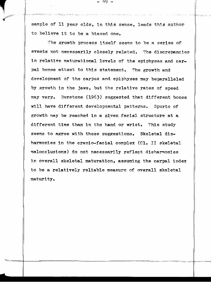
sample of 11 year olds, in this sense, leads this author
to believe it to be a biased one.
The growth process itself seems to be a series of
events not necessarily closely related. ~he discrepancies
in relative maturational levels of the epiphyses and car
pal bones attest to this statement. The growth and
development of the carpus and eplphyses may beparalleled
by growth in the jaws, but the relative rates of speed
may vary. Burstone (1963) suggested that different bones
will have different developmental patterns. Spurts of
growth ma.y be reached in a gi.ven facial structure at a
different time than in the hand or wrist. This study
seems to agree with these suggestions. Skeletal dis-
harmonies in the cranio-facial complex (Cl. II skeletal
malocclusions) do not necessarily reflect disharmonies
in overall skeletal maturation, assuming the carpal index
to be a relatively reliable measure of overall skeletal
maturity.
:::::::."'::.::·_===Ii========= ---... -.. __ -_-_"_.----=:_--:::-_____ =If==--=~

f
CHAP'TlER V
CONCLUSIONS
1) ~he broad spectrum of malocclusions 1s not
accompanied by d1sharmon,_es in overall skel~-
tal development; chtldi·en with malocclusions
mature, as a whole, at a rate identical with
children representative of the normal popu-
la ti on.
2) The skeletal ages o( malocclusions subdivided
into Cl. I and Cl. II categories according
to the Angle classif1cat1on relate statis-
t1cally not significant to standards of mean
skeletal age.
J) ~he severity of arch length discrepancy
(dental crowding} of Cl. I malocclusions
is statistically not related to skeletal
maturation, as assessed from carpal roent-
geno~rams.
4) Skeletal disharmonies of the cranio-facial
complex (1.e. Cl. II skeletal malocclusions)
do not necessarily reflect disharmonies in
overall skeletal maturation, as assessed
from carpal roentgeno~rams.
- 50 -

LIS.,, OP REF'EREtJCES
Acheson, R. M, 1954. A method of assessing skeletal maturity from radiographs. J. Ana. 881 498-509.
Acheson, R. M. 1957· The relationship between physique and rate of skeletal maturation in boys. Human Biol. 291 H57-193.
Acheson, R. Vi.; Fowler, G.r Fry, s. I.r Janes, n,, Koski, K.; Urbano, P.; and Van Der Werff Ten Bosch, J. J. 196J. Studies in the reliability of assessing skeletal maturity from x-rays. Human Biol. 351 317-349.
Baer, H. J. 1957. Bilateral asymrnetry in skeletal maturation of hand and wrist. Am. J. Phys. Anthropol. 151 181-196.
Bambha, J. L., and Van Natta, P. 19.59· A longitudinal study of occlusion and tooth eruption in relation to skeletal maturation. Am. J. Orthodont. 451 847-855.
Becks, IT.; Collins, D. A.1 and Freytag, R. M. 1946. Changes in oral structures of the dog persisting after chronic overdoses of vitamin D. Am. J. Orthodont. 321 463-471.
Bjork, A. 1967. Prediction of age of maximum pubertal growth. Ang. Orth. 53• 907-919.
Burstone, c. J. 1963. Processes of maturation and growth prediction. Am. J. Orthodont. 491 907-919.
Carter, T. M. 1926. Technique and devices used in radiographic study of the wr1st bones of children. Educ. Psychol. 171 237-247.
Dre1zen, s. r Snodere.sse, R. M.; Parker, G, s.; and Spies, C. C, 1954. !1aturation of bone centers in hand and wrist of children with chronic nutr1t1ve failure. Am. J. Dis. Child, 871 429-439.
~"'=====i=========================================================-=====---
- 51 -
- 52 -
==IF====================-~=====--~=-=-=-=====================-=-=-=-=-==-======#======= r==-'<i
~----
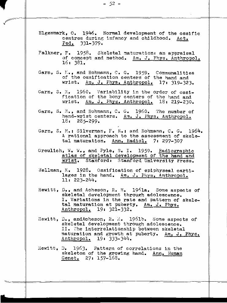
Elgenmark, o. 1946. Normal development of the oss1f1c centres during infancy and childhood. Act. Ped. 331-379 •
Falkner, F. 1958. Skeletal maturations an appraisal of concept and method. Am. J. Phys. Anthropol. 161 381.
Garn, s. E., and Rehmann, c. G. 1959. Communalities of the ossification centers of the hand and wrist. Am. J. Phys. Ant!:!ropol. 171 319-323.
Garn, s. x. 1960. Variability in the order of ossification of the bony centers of the hand and wrist. Am. J. Phys. Anthropol. 181 219-230.
Garn, s. Vi. , and Bohmann, C. hand-wrist centers. 18' 285-299.
G. 1960. The number of Arn. J. Phys. Anthr2.,Eol.
Garn, s. M.; Silverman, F. N.; and Rehmann, c. G. 1964. A rational approach to the assessment of skeletal maturation. Ann. Radiol. 7• 297-307
Greulich, w. w., and Pyle, s. I. 1959. Radiographic atlas of skeletal development of the hand and wrist. Stanford• Stanford University Press.
Hellman, K. 1928. Ossification of epiphysea.1 cartilages in the hand. Am. J. Phys. Anthropol. 11~ 223-244.
Hewitt, D., and Acheson, R. M. 196la.. Some aspects of skeletal development through adolescence. r. Variations in the rate and pattern of skeletal maturation at puberty. Am. J. Phys. Anthropol. 191 321-332.
Hewitt, D., andAcheson, R. E. 196lb. Some a.spec ts of skeletal development through adolescence. II. ~he interrelationship between skeletal maturation and growth at puberty. Am. J. Phys. Anthropol. 191 333-341.t.
Hewitt, D. 1963. Pattern of correlations in the skeleton of the growing hand. !.!:!.!:!• Human Genet. 271 157-168.

Bun.t,
- 53 -
E. E., and Gleiser, of age and sex of bones and teeth. 479-487.
I. 1955· The estimation preadolescent children from Am. J. Ph;ys. Anthropol. 131
Johnston, F. E. 1962. Skeletal age and its prediction in Philadelphia children. Human Biol. 341 192-202.
Johnston, F. E. 1963. Skeletal age and its prediction in Philadelphia children. quman Biol. 35• 192-202.
Johnston, F. E. 1964. The relationship of certain growth variables to chronological and skeletal age. Hu.man Biol!_ J6a 16-27.
Johnston, F. E. 1965a.. Contribution of the carpal bones to the assessment of skeletal age. Am. J. Phys. Anthrcpol. 231 349-354. -
Johnston, F. E. 1965b. Skeletal maturation and cephalofacial development. Ang. Orth. 35• 1-11.
Koski, K.; Haataja, J.; and Lappalainen, M. 1961. Skeletal development of hand and wrist in Finnish children. Am. J. Phys. Anthropol. 191 379-382.
Krogman, w. M. 1950. 'T'he concept of meturity from a morphological viewpoint. Child Dev. 21: 25-32.
Lamons, F. P., and Gray, s. w. 1958. A study of the relationship between tooth eruption age, skeletal development age, and chronological age in sixty-one Atlanta children. Am. J. Orthodont. 441 687-692.
nainland, D. 195J. ~'valuation of the skeletal age method of astimating children's development. I. Systematic errors in the assessment of roentgenograms. Pediatrics 121 114-129.
Me.inland, D. 1954. Evaluation of the skeletal age method of estimating children's development. II. Variable errors in the assessment of roentgenograms. J. Pediat. 131 165-173.
'=====~========================================================--=-=-~--=--===*=====

r'i; .... ..;··r -
r==c·=·-·==+====-=-=---=---=-====--==--=-=---==-.c=-=-·---·
~~asse, G. 1963. Skeletal maturation of the hand and wrist in ·,:est African c~ildren. 'iuman Biol. 35 a 3-25.
Michaelson, N. 1946. A method for assessing the d~velopment of the hana. sxeleton. Am. J. Phys. Anthropol. 4a 235-242.
Noback, c. ?.. 1954. The appearance of ossification and the fusion of bones, Am. J. Phys. Anthropol. 12 a 63-70.
Noba.ck, c. R.; :Moss, M. L.; and Leszeznska, F. 1960. Digital epiphyseal fusion of the hand in adolescences a longitudinal study. Am. J. Phys. Anthropol. 18: 13-17.
Robinow, ;; • 1942. t>.ppearance of ossification centers. Groupings obtained from factor analysis. ~ J. Diseases Child •• 641 229-236.
Bose, G. J. 1960. A cross-sectional study of the relationship of facial areas with several body di~ensions. Ang. Orth. JO: 6-lJ.
Sicher, H. 1957. Skeletal dis~armonies and malocclusion • Am. J. Orthodonh 1-1-3; 679-1)84.
Sontag, L. w., and Lipford, J. 1943. The effect of illness and other factors on the &ppearance pattern of skeletal epiphyses. J. Fediat. 23: 391-409.
Todd, T. w. 1930. 'l'he roentgenographic appraisement of skeletal differentiation. Child Devel. 11 298-JllS.
Todd, T. w. 1937. Atlas of skeletal maturation (hand). St. Louis: C. V. Mosby.
~--~·====*'==========================--=--=--==-==================-=-=-===-===----------_-

•
APPROVAL SHEET
The thesis submitted by Dr. James Spiros Rozas
has been read and approved by members of the Department
of Oral Biology.
The final copies have been examined by the Director
of the Thesis and the signature which appears below veri-
fies the fact that any necessary changes have been incor-
porated and that the thesis is now given final approval
with reference to content, form and mechanical accuracy.
The thesis is therefore accepted in partial fulfill-
ment of the requirements for the Degree of Master of
Science.
[;,,!le) I ·I , I I/' ' / :' l~
r I r· r '
! '.:-./? f )ff_ 1.,. L· .., f /
Signatu 'I
I I v
Advisor
Related Documents