Hindawi Publishing Corporation Case Reports in Dentistry Volume 2013, Article ID 397809, 6 pages http://dx.doi.org/10.1155/2013/397809 Case Report Surgical Orthodontic Treatment of Severe Skeletal Class II Fahad F. Alsulaimani, 1 Maisa O. Al-Sebaei, 2 and Ahmed R. Afify 1 1 Orthodontic Division, Department of Preventive Dental Sciences, Faculty of Dentistry, King Abdul-Aziz University, Jeddah, Saudi Arabia 2 Oral and Maxillofacial Surgery Department, Faculty of Dentistry, King Abdul-Aziz University, Jeddah, Saudi Arabia Correspondence should be addressed to Fahad F. Alsulaimani; fahad [email protected] Received 13 January 2013; Accepted 20 February 2013 Academic Editors: I. Anic and G. G´ omez-Moreno Copyright © 2013 Fahad F. Alsulaimani et al. is is an open access article distributed under the Creative Commons Attribution License, which permits unrestricted use, distribution, and reproduction in any medium, provided the original work is properly cited. is paper describes an adult Saudi male patient who presented with a severe skeletal class II deformity. e case was managed with a combination of presurgical orthodontic treatment followed by a double jaw orthognathic surgery and then another phase of orthodontic treatment for final occlusal detailing. Extraction of the four first premolars was done during the presurgical orthodontic phase of treatment to decompensate upper and lower incisors and to give room for surgical setback of the maxillary anterior segment. Double jaw surgery was performed: bilateral sagittal split ramus osteotomy for 8 mm mandibular advancement combined with three-piece Le Fort I maxillary osteotomy, 6 mm setback of the anterior segment, 8 mm impaction of the maxilla, and 5 mm advancement genioplasty. Although the anteroposterior discrepancy and the facial convexity were so severe, highly acceptable results were obtained, both esthetically as well as occlusally. 1. Introduction Orthognathic surgery is considered for the treatment of den- tofacial skeletal deformities for more than 100 years ago. Interestingly, the first jaw deformity correction was per- formed without anesthesia in the United States by Simon Hullihen, an American general surgeon, in the mid of the 19th century. Dentofacial skeletal deformities always cause severe func- tional and esthetic problems to the patient. In adult severe cases, the combined approach, orthodontic and orthognathic surgery, is always the treatment of choice, and the results obtained usually ensure a better esthetic, functional, and stable results [1–5]. Class II skeletal deformity is characterized by an exag- gerated sagittal distance between the maxilla and the man- dible, which could be the result of maxillary prognathism, mandibular retrognathism, or both. Presurgical orthodontic decompensation is essential to enable the surgeon to make a considerable amount of surgical correction, otherwise the esthetic and functional outcome of the entire procedure will not be that ideal [1–3]. 2. Case Report A 21-year-old Saudi male was referred to the orthodontic clinic, Faculty of Dentistry, King Abdul-Aziz University for the treatment of “Bothering anterior teeth.” At the first consultation visit, the patient expressed his great concerns about his anterior teeth in addition to his severely retruded chin. His medical history was nonrelevant except for a scar resulting from closure of an upper-leſt unilateral cleſt lip. e clinical examination of the patient revealed a severe skeletal class II pattern with a severe mandibular retrog- nathism. e frontal facial view showed a mesofacial pattern, slightly deviated nose to the right, an excessive lower face height, and an interlabial gap of 21 mm. e interpupillary line was parallel to the occlusal plan, and the lips were incompetent at rest with the lower lip resting behind the upper incisors. At rest, there is a 10 mm incisal show in addition to 4mm of the gum. e lips are incompetent at rest with a short upper lip, while the lower lip is resting behind the upper incisors. e upper midline is deviated to the right by 3 mm. Upon smiling, there was a severe gingival show around 12 mm. e lateral view of the face revealed an average nose, a normal nasolabial angle, a convex profile,

Welcome message from author
This document is posted to help you gain knowledge. Please leave a comment to let me know what you think about it! Share it to your friends and learn new things together.
Transcript
Hindawi Publishing CorporationCase Reports in DentistryVolume 2013, Article ID 397809, 6 pageshttp://dx.doi.org/10.1155/2013/397809
Case ReportSurgical Orthodontic Treatment of Severe Skeletal Class II
Fahad F. Alsulaimani,1 Maisa O. Al-Sebaei,2 and Ahmed R. Afify1
1 Orthodontic Division, Department of Preventive Dental Sciences, Faculty of Dentistry, King Abdul-Aziz University,Jeddah, Saudi Arabia
2Oral and Maxillofacial Surgery Department, Faculty of Dentistry, King Abdul-Aziz University, Jeddah, Saudi Arabia
Correspondence should be addressed to Fahad F. Alsulaimani; fahad [email protected]
Received 13 January 2013; Accepted 20 February 2013
Academic Editors: I. Anic and G. Gomez-Moreno
Copyright © 2013 Fahad F. Alsulaimani et al. This is an open access article distributed under the Creative Commons AttributionLicense, which permits unrestricted use, distribution, and reproduction in any medium, provided the original work is properlycited.
This paper describes an adult Saudi male patient who presented with a severe skeletal class II deformity. The case was managedwith a combination of presurgical orthodontic treatment followed by a double jaw orthognathic surgery and then another phase oforthodontic treatment for final occlusal detailing. Extraction of the four first premolars was done during the presurgical orthodonticphase of treatment to decompensate upper and lower incisors and to give room for surgical setback of the maxillary anteriorsegment. Double jaw surgery was performed: bilateral sagittal split ramus osteotomy for 8mmmandibular advancement combinedwith three-piece Le Fort I maxillary osteotomy, 6mm setback of the anterior segment, 8mm impaction of the maxilla, and 5mmadvancement genioplasty. Although the anteroposterior discrepancy and the facial convexity were so severe, highly acceptableresults were obtained, both esthetically as well as occlusally.
1. Introduction
Orthognathic surgery is considered for the treatment of den-tofacial skeletal deformities for more than 100 years ago.Interestingly, the first jaw deformity correction was per-formed without anesthesia in the United States by SimonHullihen, anAmerican general surgeon, in themid of the 19thcentury.
Dentofacial skeletal deformities always cause severe func-tional and esthetic problems to the patient. In adult severecases, the combined approach, orthodontic and orthognathicsurgery, is always the treatment of choice, and the resultsobtained usually ensure a better esthetic, functional, andstable results [1–5].
Class II skeletal deformity is characterized by an exag-gerated sagittal distance between the maxilla and the man-dible, which could be the result of maxillary prognathism,mandibular retrognathism, or both.
Presurgical orthodontic decompensation is essential toenable the surgeon tomake a considerable amount of surgicalcorrection, otherwise the esthetic and functional outcome ofthe entire procedure will not be that ideal [1–3].
2. Case Report
A 21-year-old Saudi male was referred to the orthodonticclinic, Faculty of Dentistry, King Abdul-Aziz University forthe treatment of “Bothering anterior teeth.” At the firstconsultation visit, the patient expressed his great concernsabout his anterior teeth in addition to his severely retrudedchin. His medical history was nonrelevant except for a scarresulting from closure of an upper-left unilateral cleft lip.
The clinical examination of the patient revealed a severeskeletal class II pattern with a severe mandibular retrog-nathism.The frontal facial view showed a mesofacial pattern,slightly deviated nose to the right, an excessive lower faceheight, and an interlabial gap of 21mm. The interpupillaryline was parallel to the occlusal plan, and the lips wereincompetent at rest with the lower lip resting behind theupper incisors. At rest, there is a 10mm incisal show inaddition to 4mm of the gum. The lips are incompetent atrest with a short upper lip, while the lower lip is restingbehind the upper incisors. The upper midline is deviated tothe right by 3mm. Upon smiling, there was a severe gingivalshow around 12mm. The lateral view of the face revealedan average nose, a normal nasolabial angle, a convex profile,
2 Case Reports in Dentistry
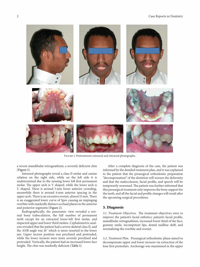
Figure 1: Pretreatment extraoral and intraoral photographs.
a severe mandibular retrognathism, a severely deficient chin(Figure 1).
Intraoral photographs reveal a class II molar and caninerelation on the right side, while on the left side it isundetermined due to the missing lower left first permanentmolar. The upper arch is V shaped, while the lower arch isU shaped. There is around 5mm lower anterior crowding,meanwhile there is around 6mm anterior spacing in theupper arch.There is an excessive overjet, almost 15mm.Thereis an exaggerated lower curve of Spee causing an impingingoverbite withmarkedly distinct occlusal planes in the anteriorand posterior segments (Figure 2).
Radiographically, the panoramic view revealed a nor-mal bony trabeculation, the full number of permanentteeth except for an extracted lower-left first molar, andimpacted upper and lower thirdmolars. Cephalometric anal-ysis revealed that the patient had a severe skeletal class II, andthe ANB angle was 16∘ which is more resorted to the lowerjaw. Upper incisor position was proclined and protruded,while the lower incisors were more severely proclined andprotruded. Vertically, the patient had an increased lower faceheight. The chin was markedly deficient (Table 1).
After a complete diagnosis of the case, the patient wasinformed by the detailed treatment plan, and it was explainedto the patient that the presurgical orthodontic preparation“decompensation” of the dentition will worsen the deformityand that the malocclusion, facial profile, and speech will betemporarily worsened.The patient was further informed thatthis presurgical treatment only improves the bony support forthe teeth, and all the facial and profile changes will result afterthe upcoming surgical procedures.
3. Diagnosis
3.1. Treatment Objectives. The treatment objectives were toimprove the patient’s facial esthetics: patient’s facial profile,mandibular retrognathism, increased lower third of the face,gummy smile, incompetent lips, dental midline shift, andnormalizing the overbite and overjet.
3.2. Treatment Plan. Presurgical orthodontic phase aimed todecompensate upper and lower incisors via extraction of thefour first premolars. Anchorage was maximized in the upper
Case Reports in Dentistry 3
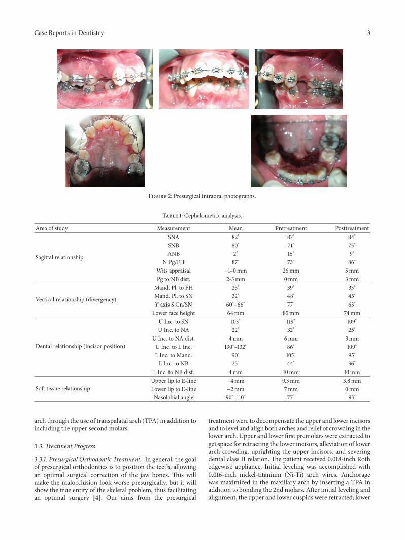
Figure 2: Presurgical intraoral photographs.
Table 1: Cephalometric analysis.
Area of study Measurement Mean Pretreatment Posttreatment
Sagittal relationship
SNA 82∘ 87∘ 84∘
SNB 80∘ 71∘ 75∘
ANB 2∘ 16∘ 9∘
N Pg/FH 87∘ 73∘ 86∘
Wits appraisal −1–0mm 26mm 5mmPg to NB dist. 2-3mm 0mm 3mm
Vertical relationship (divergency)
Mand. Pl. to FH 25∘ 39∘ 33∘
Mand. Pl. to SN 32∘ 48∘ 45∘
𝑌 axis S Gn/SN 60∘–66∘ 77∘ 63∘
Lower face height 64mm 85mm 74mm
Dental relationship (incisor position)
U Inc. to SN 103∘ 119∘ 109∘
U Inc. to NA 22∘ 32∘ 25∘
U Inc. to NA dist. 4mm 6mm 3mmU Inc. to L Inc. 130∘–132∘ 86∘ 109∘
L Inc. to Mand. 90∘ 105∘ 95∘
L Inc. to NB 25∘ 44∘ 36∘
L Inc. to NB dist. 4mm 10mm 10mm
Soft tissue relationshipUpper lip to E-line −4mm 9.3mm 3.8mmLower lip to E-line −2mm 7mm 0mmNasolabial angle 90∘–110∘ 77∘ 93∘
arch through the use of transpalatal arch (TPA) in addition toincluding the upper second molars.
3.3. Treatment Progress
3.3.1. Presurgical Orthodontic Treatment. In general, the goalof presurgical orthodontics is to position the teeth, allowingan optimal surgical correction of the jaw bones. This willmake the malocclusion look worse presurgically, but it willshow the true entity of the skeletal problem, thus facilitatingan optimal surgery [4]. Our aims from the presurgical
treatment were to decompensate the upper and lower incisorsand to level and align both arches and relief of crowding in thelower arch. Upper and lower first premolars were extracted toget space for retracting the lower incisors, alleviation of lowerarch crowding, uprighting the upper incisors, and severingdental class II relation. The patient received 0.018-inch Rothedgewise appliance. Initial leveling was accomplished with0.016-inch nickel-titanium (Ni-Ti) arch wires. Anchoragewas maximized in the maxillary arch by inserting a TPA inaddition to bonding the 2ndmolars. After initial leveling andalignment, the upper and lower cuspids were retracted; lower
4 Case Reports in Dentistry
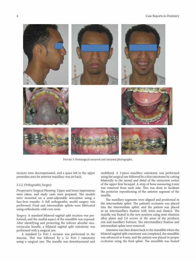
Figure 3: Postsurgical extraoral and intraoral photographs.
incisors were decompensated, and a space left in the upperpremolars area for anterior maxillary was set back.
3.3.2. Orthognathic Surgery
Preoperative Surgical Planning. Upper and lower impressionswere taken, and study casts were prepared. The modelswere mounted on a semi-adjustable articulator using aface-bow transfer. A full orthognathic model surgery wasperformed. Final and intermediate splints were fabricatedusing orthodontic cold-cure resin.
Surgery. A standard bilateral sagittal split incision was per-formed, and the medial aspect of the mandible was exposed.After identifying and protecting the inferior alveolar neu-rovascular bundle, a bilateral sagittal split osteotomy wasperformed with a surgical saw.
A standard Le Fort I incision was performed in themucosa. This was followed by a Le Fort I osteotomyusing a surgical saw. The maxilla was downfractured and
mobilized. A 3-piece maxillary osteotomy was performedusing the surgical saw followed by a thin osteotome by cuttingbilaterally in the mesial and distal of the extraction socketof the upper first bicuspid. A strip of bone measuring 6mmwas removed from each side. This was done to facilitatethe posterior repositioning of the anterior segment of themaxilla.
The maxillary segments were aligned and positioned inthe intermediate splint. The patient’s occlusion was placedinto the intermediate splint, and the patient was placedin an intermaxillary fixation with wires and elastics. Themaxilla was fixated in the new position using mini titaniumalloy plates and 2.0 screws in the areas of the pyriformrim and maxillary buttress. The intermaxillary fixation andintermediate splint were removed.
Attentionwas then drawn back to themandible where thebilateral sagittal split osteotomywas completed, themandiblewas advanced to 8mm, and the patient was placed in properocclusion using the final splint. The mandible was fixated
Case Reports in Dentistry 5
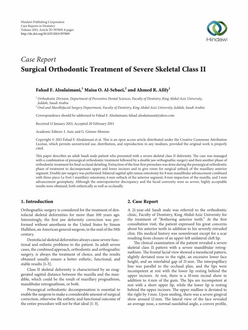
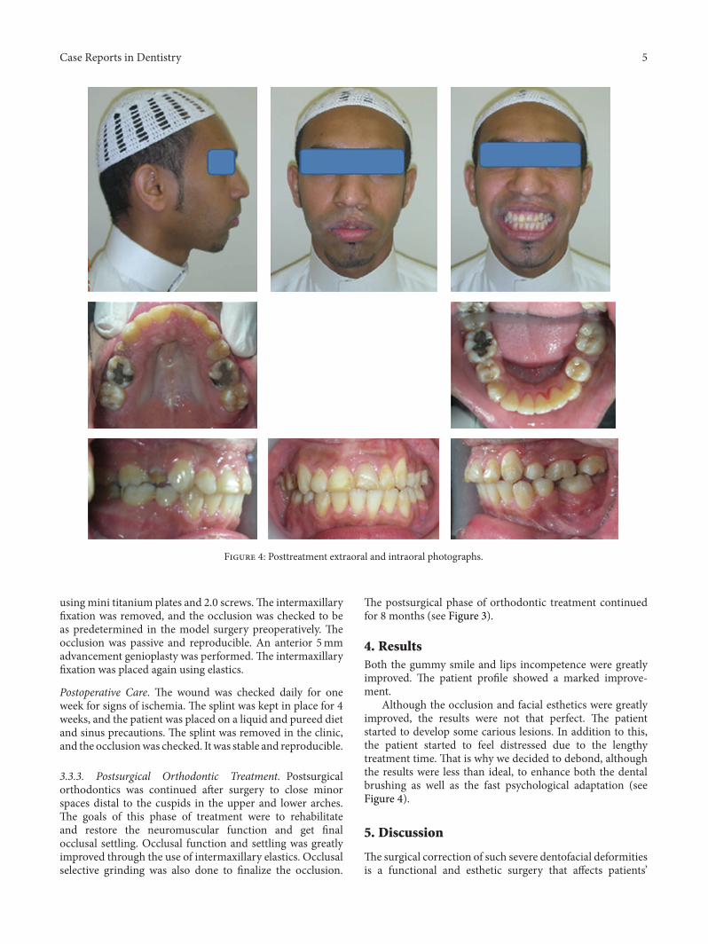
Figure 4: Posttreatment extraoral and intraoral photographs.
using mini titanium plates and 2.0 screws.The intermaxillaryfixation was removed, and the occlusion was checked to beas predetermined in the model surgery preoperatively. Theocclusion was passive and reproducible. An anterior 5mmadvancement genioplasty was performed.The intermaxillaryfixation was placed again using elastics.
Postoperative Care. The wound was checked daily for oneweek for signs of ischemia. The splint was kept in place for 4weeks, and the patient was placed on a liquid and pureed dietand sinus precautions. The splint was removed in the clinic,and the occlusionwas checked. It was stable and reproducible.
3.3.3. Postsurgical Orthodontic Treatment. Postsurgicalorthodontics was continued after surgery to close minorspaces distal to the cuspids in the upper and lower arches.The goals of this phase of treatment were to rehabilitateand restore the neuromuscular function and get finalocclusal settling. Occlusal function and settling was greatlyimproved through the use of intermaxillary elastics. Occlusalselective grinding was also done to finalize the occlusion.
The postsurgical phase of orthodontic treatment continuedfor 8 months (see Figure 3).
4. ResultsBoth the gummy smile and lips incompetence were greatlyimproved. The patient profile showed a marked improve-ment.
Although the occlusion and facial esthetics were greatlyimproved, the results were not that perfect. The patientstarted to develop some carious lesions. In addition to this,the patient started to feel distressed due to the lengthytreatment time. That is why we decided to debond, althoughthe results were less than ideal, to enhance both the dentalbrushing as well as the fast psychological adaptation (seeFigure 4).
5. Discussion
The surgical correction of such severe dentofacial deformitiesis a functional and esthetic surgery that affects patients’
6 Case Reports in Dentistry
(a) (b) (c)
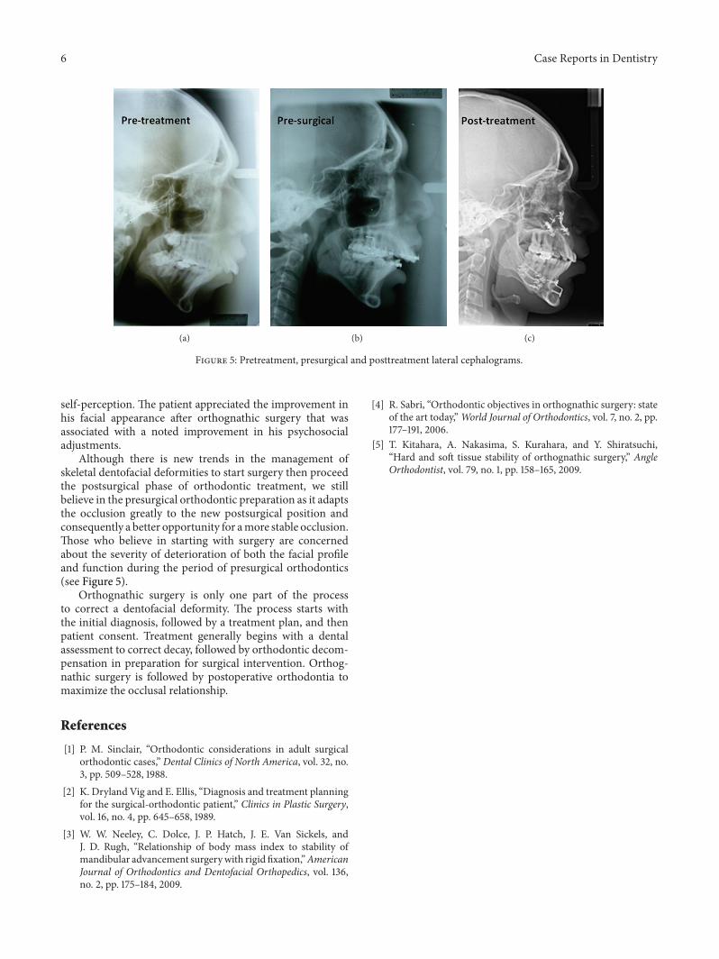
Figure 5: Pretreatment, presurgical and posttreatment lateral cephalograms.
self-perception. The patient appreciated the improvement inhis facial appearance after orthognathic surgery that wasassociated with a noted improvement in his psychosocialadjustments.
Although there is new trends in the management ofskeletal dentofacial deformities to start surgery then proceedthe postsurgical phase of orthodontic treatment, we stillbelieve in the presurgical orthodontic preparation as it adaptsthe occlusion greatly to the new postsurgical position andconsequently a better opportunity for amore stable occlusion.Those who believe in starting with surgery are concernedabout the severity of deterioration of both the facial profileand function during the period of presurgical orthodontics(see Figure 5).
Orthognathic surgery is only one part of the processto correct a dentofacial deformity. The process starts withthe initial diagnosis, followed by a treatment plan, and thenpatient consent. Treatment generally begins with a dentalassessment to correct decay, followed by orthodontic decom-pensation in preparation for surgical intervention. Orthog-nathic surgery is followed by postoperative orthodontia tomaximize the occlusal relationship.
References
[1] P. M. Sinclair, “Orthodontic considerations in adult surgicalorthodontic cases,” Dental Clinics of North America, vol. 32, no.3, pp. 509–528, 1988.
[2] K. Dryland Vig and E. Ellis, “Diagnosis and treatment planningfor the surgical-orthodontic patient,” Clinics in Plastic Surgery,vol. 16, no. 4, pp. 645–658, 1989.
[3] W. W. Neeley, C. Dolce, J. P. Hatch, J. E. Van Sickels, andJ. D. Rugh, “Relationship of body mass index to stability ofmandibular advancement surgerywith rigid fixation,”AmericanJournal of Orthodontics and Dentofacial Orthopedics, vol. 136,no. 2, pp. 175–184, 2009.
[4] R. Sabri, “Orthodontic objectives in orthognathic surgery: stateof the art today,”World Journal of Orthodontics, vol. 7, no. 2, pp.177–191, 2006.
[5] T. Kitahara, A. Nakasima, S. Kurahara, and Y. Shiratsuchi,“Hard and soft tissue stability of orthognathic surgery,” AngleOrthodontist, vol. 79, no. 1, pp. 158–165, 2009.

Submit your manuscripts athttp://www.hindawi.com
Hindawi Publishing Corporationhttp://www.hindawi.com Volume 2014
Oral OncologyJournal of
DentistryInternational Journal of
Hindawi Publishing Corporationhttp://www.hindawi.com Volume 2014
Hindawi Publishing Corporationhttp://www.hindawi.com Volume 2014
International Journal of
Biomaterials
Hindawi Publishing Corporationhttp://www.hindawi.com Volume 2014
BioMed Research International
Hindawi Publishing Corporationhttp://www.hindawi.com Volume 2014
Case Reports in Dentistry
Hindawi Publishing Corporationhttp://www.hindawi.com Volume 2014
Oral ImplantsJournal of
Hindawi Publishing Corporationhttp://www.hindawi.com Volume 2014
Anesthesiology Research and Practice
Hindawi Publishing Corporationhttp://www.hindawi.com Volume 2014
Radiology Research and Practice
Environmental and Public Health
Journal of
Hindawi Publishing Corporationhttp://www.hindawi.com Volume 2014
The Scientific World JournalHindawi Publishing Corporation http://www.hindawi.com Volume 2014
Hindawi Publishing Corporationhttp://www.hindawi.com Volume 2014
Dental SurgeryJournal of
Drug DeliveryJournal of
Hindawi Publishing Corporationhttp://www.hindawi.com Volume 2014
Hindawi Publishing Corporationhttp://www.hindawi.com Volume 2014
Oral DiseasesJournal of
Hindawi Publishing Corporationhttp://www.hindawi.com Volume 2014
Computational and Mathematical Methods in Medicine
ScientificaHindawi Publishing Corporationhttp://www.hindawi.com Volume 2014
PainResearch and TreatmentHindawi Publishing Corporationhttp://www.hindawi.com Volume 2014
Preventive MedicineAdvances in
Hindawi Publishing Corporationhttp://www.hindawi.com Volume 2014
EndocrinologyInternational Journal of
Hindawi Publishing Corporationhttp://www.hindawi.com Volume 2014
Hindawi Publishing Corporationhttp://www.hindawi.com Volume 2014
OrthopedicsAdvances in
Related Documents