SIMULATION OF BONE AGING AND VIRTUAL REALITY VISUALIZATION OF CANCELLOUS BONE STRUCTURE Ajay V. Sonar, Ph.D. Candidate; James J. Carroll * , Ph.D., Associate Professor Department of Electrical and Computer Engineering Wallace H. Coulter School of Engineering Clarkson University, Box 5720 Potsdam, NY 13699-5720 (315)268-7726 [voice] // (315)268-7600 [fax] Email: [email protected] * Corresponding author Lisa Sabini, M.S. Candidate; Kathleen A. Issen, Ph.D., P.E., Associate Professor Mechanical & Aeronautical Engineering Wallace H. Coulter School of Engineering Clarkson University, Box 5720 Potsdam, NY 13699- 5725 ABSTRACT This paper describes a bone modeling effort and virtual reality visualization aimed towards characterizing and predicting human vertebral compression fractures with emphasis on quantifying osteoporosis and aging. A 3D model of the trabecular bone structure is constructed from a stack of μCT images of a human vertebra. Parameters for quantifying the bone are calculated for this structure from the 3D model and validated by comparing the results with values calculated from standard stereological methods. The resulting structure is then aged using biologically based computational algorithms. These structures are then visualized in a virtual reality environment enabling detailed examination of the regional microstructures to assess the potential for compression fracture formation. 1. INTRODUCTION Image based reconstruction is a challenging problem. It has applications in various fields, such as military training, the entertainment industry, and medicine. Accurate 3D models of body organs/mechanisms can assist in understanding the mechanism in a more sophisticated and useful manner than the conventional 2D MRI or CT scan images, or by gross pathologic examination alone. 978-1-4244-1643-1/08/$25.00 ©2008 IEEE For example, the microstructure of the cancellous bone plays an important role in the overall strength and quality of the bone. It also determines the ability of bone to withstand external loading caused by everyday activity. Hence, the detailed study and quantification of these microstructures is of great importance. Bone, which is a dynamic porous material, is continuously remodeled; some of the factors affecting this process include hormonal changes and mechanical loading [8, 12]. This paper describes an ongoing effort to combine biologically-based factors in simulating the bone remodeling process. 2. MODELING CANCELLOUS BONE MICROSTRUCTURE FROM μCT SCANS A stack of μCT images of a human vertebra were used to create a model of the cancellous bone microstructure. The reconstruction process involves the following steps: (1) image adjustment, (2) identifying regions of interest, (3) segmentation and thresholding, and (4) voxel reconstruction and surface reconstruction [1, 2]. Once the above steps are completed, a simulated aging is applied to the resulting voxel-based bone model as detailed below. A full image of one of the slice of μCT scan is shown in Figure 1(a). This slice is taken perpendicular to the superior-inferior axis (perpendicular to the spinal column) Because of physical limitations on the size of the simulated bone specimen that can be physically manufactured and tested in a later phase of the project, the μCT scans are cored, i.e., segmented, to a diameter of 480 pixels, as shown in Figure 1(b). The aspect

Welcome message from author
This document is posted to help you gain knowledge. Please leave a comment to let me know what you think about it! Share it to your friends and learn new things together.
Transcript

SIMULATION OF BONE AGING AND VIRTUAL REALITY VISUALIZATION OF CANCELLOUS BONE STRUCTURE
Ajay V. Sonar, Ph.D. Candidate;
James J. Carroll*, Ph.D., Associate Professor Department of Electrical and Computer Engineering
Wallace H. Coulter School of Engineering Clarkson University, Box 5720
Potsdam, NY 13699-5720 (315)268-7726 [voice] // (315)268-7600 [fax]
Email: [email protected] *Corresponding author
Lisa Sabini, M.S. Candidate;
Kathleen A. Issen, Ph.D., P.E., Associate Professor Mechanical & Aeronautical Engineering
Wallace H. Coulter School of Engineering Clarkson University, Box 5720
Potsdam, NY 13699- 5725
ABSTRACT This paper describes a bone modeling effort and virtual reality visualization aimed towards characterizing and predicting human vertebral compression fractures with emphasis on quantifying osteoporosis and aging. A 3D model of the trabecular bone structure is constructed from a stack of µCT images of a human vertebra. Parameters for quantifying the bone are calculated for this structure from the 3D model and validated by comparing the results with values calculated from standard stereological methods. The resulting structure is then aged using biologically based computational algorithms. These structures are then visualized in a virtual reality environment enabling detailed examination of the regional microstructures to assess the potential for compression fracture formation.
1. INTRODUCTION
Image based reconstruction is a challenging problem. It has applications in various fields, such as military training, the entertainment industry, and medicine. Accurate 3D models of body organs/mechanisms can assist in understanding the mechanism in a more sophisticated and useful manner than the conventional 2D MRI or CT scan images, or by gross pathologic examination alone.
978-1-4244-1643-1/08/$25.00 ©2008 IEEE
For example, the microstructure of the cancellous bone plays an important role in the overall strength and quality of the bone. It also determines the ability of bone to withstand external loading caused by everyday activity. Hence, the detailed study and quantification of these microstructures is of great importance. Bone, which is a dynamic porous material, is continuously remodeled; some of the factors affecting this process include hormonal changes and mechanical loading [8, 12]. This paper describes an ongoing effort to combine biologically-based factors in simulating the bone remodeling process.
2. MODELING CANCELLOUS BONE
MICROSTRUCTURE FROM µCT SCANS A stack of µCT images of a human vertebra were used to create a model of the cancellous bone microstructure. The reconstruction process involves the following steps: (1) image adjustment, (2) identifying regions of interest, (3) segmentation and thresholding, and (4) voxel reconstruction and surface reconstruction [1, 2]. Once the above steps are completed, a simulated aging is applied to the resulting voxel-based bone model as detailed below. A full image of one of the slice of µCT scan is shown in Figure 1(a). This slice is taken perpendicular to the superior-inferior axis (perpendicular to the spinal column) Because of physical limitations on the size of the simulated bone specimen that can be physically manufactured and tested in a later phase of the project, the µCT scans are cored, i.e., segmented, to a diameter of 480 pixels, as shown in Figure 1(b). The aspect

ratio of a single vertebral body (diameter/height) is 1.33. To maintain this aspect ratio on the cored samples, 384 slices are selected from the original µCT scan images. Figure 2 (a) shows the grayscale representation of the segmented core from the full µCT scan. This segmented image is then thresholded to generate a binary image. Thresholding is done to distinguish the bone from the surrounding marrow. Thresholding was performed using the protocol established in [13]. Each image is thresholded individually with a value of 10.2% of its maximal gray value. The result of thresholding is shown in Figure 2(b). In this image, the white region indicates the trabecular bone and the black region is the background/bone marrow. The next stage is voxel reconstruction. A voxel is a three-dimensional pixel element, i.e., a cube with user specified length, width and height. For convenience, the dimensions are set to produce a unit voxel, i.e., a volume of dimension one. The resulting voxel reconstruction is performed by first
building a voxel space that is approximately the same size as the original specimen. The size of the unit voxel defines the resolution of the final reconstructed model, i.e., a smaller unit voxel generates a higher resolution model. Each layer of voxels from the voxel space is projected on to one slice of the µCT scan. The voxels that intersect the trabecular bone region, i.e., the black region in the pre-processed images of Figure 2 (b), are retained and the remaining voxels are discarded. Once this process is run on all scans within the core, the remaining voxels represent the volume of the specimen. Figure 3 (a) shows a voxel model of a small section of the core. The voxel model is an approximation of the actual volume. The actual surface might cut through some of the voxels. Thus, to generate an accurate surface model from the carved voxels, a Marching cube algorithm is used [2].
(a) (b)
Figure 1: (a) A full µCT and (b) the segmented core (in blue) used for reconstruction.
(a) Core (Grayscale) (b) Core (Binary)
Figure 2: Image preprocessing steps.

The resulting surface reconstruction of the core section is shown in Figure 3 (b). The surface model of trabecular bone generated from the entire segmented core of Figure 1 (b) is shown in Figure 4.
3. QUANTIFICATION OF BONE STRUCTURE AND
MODEL VERIFICATION Bone is a dynamic porous structure whose porosity
evolves as a result of a pathologic condition or due to normal adaptive response to a mechanical or physiological stimulus. Cancellous bone may become more compact, or compact bone may become more porous. Such changes strongly affect the mechanical properties of bone [9]. Bone is composed of three primary substances: water, mineral and organic matrix that is largely collagen which gives bone its
flexibility and tensile strength. The strength of a bone depends on the relative amounts of each substance and the architectural organization of the micro-architecture. Hence it is important to investigate the changes in the cancellous bone microstructure. Based on a detailed literature study [3, 4], five basic parameters were selected to characterize the resulting bone structure: bone volume faction ( VV ), surface density ( VS ),
trabecular thickness ( ThTb. ), trabecular separation ( SpTb. ) and trabecular number ( NTb. ). Stereology is the traditional method of calculating these parameters based on 2D µCT scans [15], and is considered as the gold standard for obtaining quantitative information on cancellous bone structure.
(a) (b)
Figure 3: Voxel model of the section of the core sample (a) and the surface model of the same (b).
Figure 4: 3D model of the core sample.
Parameters like volume fraction and surface density can be calculated directly from these images, but other important
parameters such as trabecular thickness, trabecular separation and the trabecular number are derived indirectly
assuming a fixed structural model [15]. Let VT be the total volume of the region of bone. This is divided in to two parts: the hard bony matrix, VB and the soft tissue in the
space between the hard bony matrix, VM . Let SB be the total surface area of the bone. Two of the basic parameters can be calculated directly as shown, /V V VV B T= (Volume Fraction),
/V S VS B T= (Surface Density).
The remaining three parameters are calculated indirectly as shown
( )2 2. V
V
VTbTh
S−
= ; .2VSTb N = ;
( )2. V
V
VTb Sp
S= .
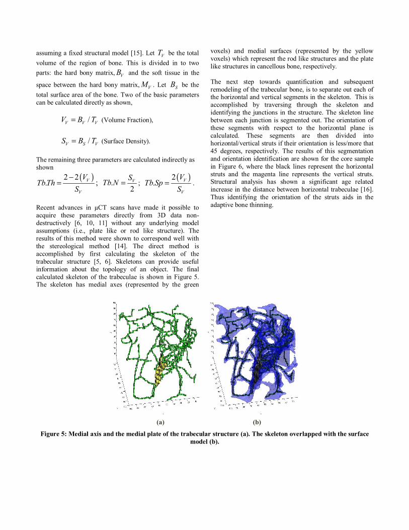
Recent advances in µCT scans have made it possible to acquire these parameters directly from 3D data non-destructively [6, 10, 11] without any underlying model assumptions (i.e., plate like or rod like structure). The results of this method were shown to correspond well with the stereological method [14]. The direct method is accomplished by first calculating the skeleton of the trabecular structure [5, 6]. Skeletons can provide useful information about the topology of an object. The final calculated skeleton of the trabeculae is shown in Figure 5. The skeleton has medial axes (represented by the green
voxels) and medial surfaces (represented by the yellow voxels) which represent the rod like structures and the plate like structures in cancellous bone, respectively. The next step towards quantification and subsequent remodeling of the trabecular bone, is to separate out each of the horizontal and vertical segments in the skeleton. This is accomplished by traversing through the skeleton and identifying the junctions in the structure. The skeleton line between each junction is segmented out. The orientation of these segments with respect to the horizontal plane is calculated. These segments are then divided into horizontal/vertical struts if their orientation is less/more that 45 degrees, respectively. The results of this segmentation and orientation identification are shown for the core sample in Figure 6, where the black lines represent the horizontal struts and the magenta line represents the vertical struts. Structural analysis has shown a significant age related increase in the distance between horizontal trabeculae [16]. Thus identifying the orientation of the struts aids in the adaptive bone thinning.
(a) (b)
Figure 5: Medial axis and the medial plate of the trabecular structure (a). The skeleton overlapped with the surface model (b).

Figure 6: Segmented struts and their orientations.
4. COMPUTATIONAL BONE AGING ALGORITHM
Human bones reach maturity between 30-35 years of
age. After this point, the bone starts to deteriorate, i.e., there is a decrease in the mechanical properties and the ability to remodel and produce new bone. In the case of osteoporosis, the deterioration occurs faster. There are several serious drawbacks in using human bone to understand the microstructural changes due to aging or osteoporosis. Samples are difficult to obtain from human donors, and require special handling and storage. Since each specimen consists of unique microstructure, tests/analysis on several specimens with similar bone volume fraction will exhibit variations that cannot be attributed directly to the volume fraction, i.e., since the microstructure will not have been the same before osteoporosis thinned it. Because of these natural microstructural variations between specimens, large numbers of specimens are required to characterize the relationship between the different parameters which quantify the microstructure. Even so, only general trends can be determined. Therefore, a method to simulate the effects of osteoporosis is required to produce a suite of specimens based on an initial same healthy specimen. This work aims toward creating such a suite of specimens.
Studies have shown that cancellous bone microstructure
tends to retain the vertical struts and loose the horizontal struts during ageing [16]. Additionally, the struts are modified such that they align along the principal stress axis. Another factor that affects the modification of bone is the activity of the osteoblast cells (responsible for bone formation) and osteoclast cells (responsible for bone destruction). The activity of these cells is closely related to the stress throughout the structure [8, 9]. When there is a balance between the activities of these cells, i.e. the rate of removal equals the rate of formation, the bone retain its strength. Micro finite element analysis (FEA) can be used to locate regions of high and low stress, which in turn can be used to trigger remodeling at particular locations.
Incorporating these biologically based factors into a bone aging algorithm may produce simulated bone ageing that is structurally closer to actual cancellous bone. The simplest method to age bone structures is to remove material from the bone surface uniformly, i.e., uniform thinning, although this is not representative of how actual bone is modified biologically in the human body. An example of uniform thinning is shown below in Figure 7.
(a) Original Structure.
(b) Thinned Structure.
Figure 7: Uniform thinning.
5. VIRTUAL REALITY VISUALIZATION
Visualization can greatly enhance the ability of
researchers to analyze complex data sets, like those associated with biomedical studies. In this particular application, the generated 3D surface models of both the original structure and the aged bone structure can be visualized using different stereoscopic techniques, e.g., passive or active displays which are more immersive than the conventional display/screen. An initial effort has been made to facilitate interactive navigation through the complex trabecular structure using custom human-computer interfaces, e.g. gyro-mouse and stereoscopic head-mounted
display with Intersense-based head tracking. The resulting visualization models facilitate more efficient study of the complex biological structures. For example, the results of an FEA can be texture mapped onto the 3D bone model to augment a researchers' ability to identify stress localization in bone structure. A screen shot of the trabecular bone in the virtual environment is shown in Figure 8.
Figure 8: Virtual reality visualization of trabecular bone
structure.
6. CONCLUSION AND FUTURE WORK
In this paper we discussed the various factors affecting
the microstructure of cancellous bone due to aging and osteoporosis. Different methods and parameters used to simulate the biologically based aging of bone were discussed. A complete implementation of the above methods has yet to be fully incorporated. The virtual reality visualization was achieved for a sample of the cancellous structure. A real time visualization of the aging process that shows meaningful changes in the microstructure is currently being developed.
11. REFERENCES [1] Adem Yasar Mulayim, Ulas Yilmaz and Volkan Atalay, “Silhouette based 3D Model Reconstruction from Multiple Images”, IEEE Transactions on Systems, Man and Cybernetics, Part B. [2] W. Lorensen, H. Cline. “Marching Cubes: A high resolution 3D surface construction algorithm”, ACM Computer Graphics, Vol. 21, No. 4, pages: 163-170, July 1987. [3] Ralph Muller, W. C. Hayes, “Biomechanical Competence of Microstructural Bone in the Progress of Adaptive Bone Remodeling”, SPIE, Vol. 3149, 1997. [4] H. S. Kim, S.T.S. Al-Hassani, “A morphological model of vertebral trabecular bone”, Journal of Biomechanics Vol. 35, 2002.
[5] Tao Ju, M. Baker, W. Chiu, “Computing a family of skeletons of volumetric models for shape description”, Computer Aided Design, 2007. [6] Martin Stauber, Ralph Muller, “Volumetric spatial decomposition of trabecular bone in to rods and plates – A new method for local bone morphometry”, Bone, Vol. 38, 2006 [7] R. Bruce Martin, David B. Burr, Neil A. Sharky, “Skeletal Tissue Mechanics”, 1998 Springer-Verlag New York. [8] M. Mullender, B. Van Rietbergen, P. Ruegsegger, R. Huiskes, “Effect of Mechanical set point of Bone Cells on Mechanical Control of Trabecular Bone Architecture”, Bone, Vol. 22, 1998. [9] R. Ruimerman, P. Hilbers, B. van Rietbergen, R. Huiskes, “A theoretical framework for strain related trabecular bone maintenance and adaptation”, Journal of Biomechanics, Vol. 38, 2005. [10] R. J. Fajardo, R. Muller, “Three dimensional analysis of nonhuman primate trabecular architecture using micro computed tomography”, American Journal of Physical Anthropology, Vol. 115, 2001. [11] T. Hildebrand, P. Ruegsegger, “A new method for the model independent assessment of thickness in three-dimensional images”, Journal of Microscopy, Vol. 185:76-75, 1997. [12] Vincent Lemaire, Frank L. Tobin, Larry D. Greller, Carolyn R. Cho, Larry J. Suva, “Modeling interactions between osteoblast and osteoclast activities in bone remodeling”, Journal of Theorotical Biology, Vol.229:293-309, 2004. [13] R. Muller, H. Van Campenhout, B. Van Damme, G Van Der Perre, J. Dequeker, T. Hildebrand, P. Ruegsegger, “Morphometric analysis of human bone biopsies: a quantitative structural comparison of histological sections and micro-computed tomography”, Bone Vol. 23:59-66, 1998. [14] J. S. Thomsen, A. Laib, B. Koller, S. Prohaska, LI. Mosekilde, W. Gowin, “Stereological measures of trabecular bone structure: comparison of 3D micro computed tomography with 2D histological sections in human proximal tibial bone biopsies”, Journal of Microscopy, Vol. 218:171-179, May 2005. [15] A. M. Parfitt, C. H. E. Mathews, A. R. Villanueva, M. Kleerekoper, B. Frame, D. S. Rao, “Relationships between surface, volume, and thickness of Ilica Trabecular Bone in aging and Osteoporosis. Implications for the microanatomic and cellular mechanisms of bone loss”, J. Clin. Invest. Vol. 72:1396-1409, 1983. [16] Li Mosekilde, “Sex differences in age related loss of vertebral trabecular bone mass and structure – biomechanical consequences”, Bone, Vol. 10:425-432, 1989.
Related Documents


![Bone Mineral Density and Fatty Degeneration of Thigh ... … · Progressive reduction of bone mass and muscle mass are inevitable processes which occur with aging.[1,2] Bone mineral](https://static.cupdf.com/doc/110x72/5eae336dd5e77a5b1a56c4a8/bone-mineral-density-and-fatty-degeneration-of-thigh-progressive-reduction.jpg)
![Nicotinamide phosphoribosyltransferase postpones rat bone ... · entering a senescent state during aging [1]. Accordingly, the aging of SCs is crucially implicated in individual aging](https://static.cupdf.com/doc/110x72/5f1daac5b2c2c1053d52d773/nicotinamide-phosphoribosyltransferase-postpones-rat-bone-entering-a-senescent.jpg)