Simulated urban carbon monoxide air pollution exacerbates rat heart ischemia-reperfusion injury G. Meyer, 1 L. André, 2 S. Tanguy, 1 J. Boissiere, 2 C. Farah, 1 F. Lopez-Lauri, 4 S. Gayrard, 1 S. Richard, 2 F. Boucher, 3 O. Cazorla, 2 P. Obert, 1 and C. Reboul 1 1 Research Laboratory: Equipe d’Accueil 4278, Physiology and Physiopathology of Cardiovascular Adaptations to Exercise, and 4 Research Laboratory: Equipe d’Accueil 4279, Faculty of Sciences, Avignon University, Avignon; 2 Research Laboratory: Institut National de la Santé et de la Recherche Médicale Unité 637, Cardiovascular Physiopathology, Montpellier University, Faculty of Medicine, Montpellier; and 3 Research Laboratory: Unite Mixte de Recherche 5525 Physiologie cardio- Respiratoire Expérimentale Théorique et Appliquée-Techniques de l’Ingénierie Médicale et de la Complexité, Grenoble University Joseph Fourier, Grenoble, France Submitted 15 December 2009; accepted in final form 28 February 2010 Meyer G, André L, Tanguy S, Boissiere J, Farah C, Lopez- Lauri F, Gayrard S, Richard S, Boucher F, Cazorla O, Obert P, Reboul C. Simulated urban carbon monoxide air pollution exacer- bates rat heart ischemia-reperfusion injury. Am J Physiol Heart Circ Physiol 298: H1445–H1453, 2010. First published March 5, 2010; doi:10.1152/ajpheart.01194.2009.—Myocardial damages due to ische- mia-reperfusion (I/R) are recognized to be the result of a complex interplay between genetic and environmental factors. Epidemiological studies suggested that, among environmental factors, carbon monox- ide (CO) urban pollution can be linked to cardiac diseases and mortality. The aim of this work was to evaluate the impact of exposure to CO pollution on cardiac sensitivity to I/R. Regional myocardial I/R was performed on isolated perfused hearts from rats exposed for 4 wk to air enriched with CO (30 –100 ppm). Functional variables, reper- fusion ventricular arrhythmias (VA) and cellular damages (infarct size, lactate dehydrogenase release) were assessed. Sarcomere length shortening and Ca 2 handling were evaluated in intact isolated car- diomyocytes during a cellular anoxia-reoxygenation protocol. The major results show that prolonged CO exposure worsens myocardial I/R injuries, resulting in increased severity of postischemic VA, impaired recovery of myocardial function, and increased infarct size (60 5 vs. 33 2% of ischemic zone). The aggravating effects of CO exposure on I/R could be explained by a reduced myocardial enzymatic antioxidant status (superoxide dismutase 45%; glutathi- one peroxidase 49%) associated with impaired intracellular Ca 2 handling. In conclusion, our results are consistent with the idea that chronic CO pollution dramatically increases the severity of myocar- dial I/R injuries. environmental pollution; myocardial infarction; antioxidant status MYOCARDIAL DAMAGES RESULTING from ischemia-reperfusion (I/R) are a major cause of morbidity and mortality in western nations. Those myocardial I/R injuries result in cardiac dys- function, arrhythmias, as well as irreversible myocyte damages (25, 28). The sensitivity of the myocardium to I/R-induced cellular injuries is recognized today to be the result of a complex interplay between genetic, pathological, and environ- mental factors. Moreover, it appears to be aggravated in several diseases, including hypertension, metabolic disorders, and complications from cigarette smoking and environmental pol- lution. Among the environmental factors that could influence the development of cardiovascular diseases, several epidemiolog- ical studies have recently suggested that urban atmospheric pollution may exert adverse effects on cardiovascular health (7, 11, 12, 15). Among the numerous pollutants, carbon monoxide (CO) has been described as one of the main pollutants respon- sible for the development of cardiovascular diseases (9, 35). In urban environments, CO concentration usually varies from 2 to 40 ppm, but during heavy traffic it may be as high as 170 ppm (10, 34, 40). At this level, CO exposure has been correlated with hospital admissions, mortality, and morbidity related to cardiovascular diseases (9, 13, 29). Today, although the patho- physiological mechanisms regarding acute CO poisoning are well known, mechanisms associated with chronic exposure to lower concentrations of CO, consistent with urban environ- mental pollution, remain unclear. We (2) and Bye et al. (14) have recently reported that prolonged CO exposure induces a pathological myocardial cellular phenotype characterized by a major remodeling of the excitation-contraction coupling. Such deleterious consequences may worsen the effect of cardiovas- cular diseases. The aim of this experimental work was to challenge the impact of a chronic exposure to simulated CO urban pollution on the sensitivity of the myocardium to I/R in a rat model. The major result shows that prolonged exposure to CO at a level found in the urban environment has a dramatic deleterious impact on the sensitivity of the myocardium to I/R. METHODS Experiments complied with the Guide for the Care and Use of Laboratory Animals published by the United States National Institutes of Health (National Institutes of Health Publications no. 85-23, revised 1996) and was approved by the French Ministry of Agriculture. Animals and CO Exposure Adult, male Wistar rats (n 64; 345 7 g; Charles River Laboratories) were randomly assigned to the following two groups: CO rats (exposed for 4 wk to simulated CO urban pollution, n 32) and control animals (Ctrl rats, exposed to standard filtered air, n 32). CO rats were housed in an airtight exposure container for 4 wk. Exposure was performed according to a 12:12-h CO in the air-ambient air cycle. To simulate CO urban pollution, exposure was performed as follows: during CO exposure, a CO concentration of 30 ppm was maintained in the airtight container and monitored with an aspirative CO analyzer (CHEMGARD Infrared Gas Monitor NEMA 4 Version; Address for reprint requests and other correspondence: C. Reboul, Research Laboratory: EA 4278, Physiology and Physiopathology of Cardiovascular Adaptations to Exercise, Faculty of Sciences, Avignon Univ., F-84000 Av- ignon, France (e-mail: [email protected]). Am J Physiol Heart Circ Physiol 298: H1445–H1453, 2010. First published March 5, 2010; doi:10.1152/ajpheart.01194.2009. 0363-6135/10 $8.00 Copyright © 2010 the American Physiological Society http://www.ajpheart.org H1445

Welcome message from author
This document is posted to help you gain knowledge. Please leave a comment to let me know what you think about it! Share it to your friends and learn new things together.
Transcript
Simulated urban carbon monoxide air pollution exacerbates rat heartischemia-reperfusion injury
G. Meyer,1 L. André,2 S. Tanguy,1 J. Boissiere,2 C. Farah,1 F. Lopez-Lauri,4 S. Gayrard,1 S. Richard,2
F. Boucher,3 O. Cazorla,2 P. Obert,1 and C. Reboul11Research Laboratory: Equipe d’Accueil 4278, Physiology and Physiopathology of Cardiovascular Adaptations to Exercise,and 4Research Laboratory: Equipe d’Accueil 4279, Faculty of Sciences, Avignon University, Avignon; 2Research Laboratory:Institut National de la Santé et de la Recherche Médicale Unité 637, Cardiovascular Physiopathology, MontpellierUniversity, Faculty of Medicine, Montpellier; and 3Research Laboratory: Unite Mixte de Recherche 5525 Physiologie cardio-Respiratoire Expérimentale Théorique et Appliquée-Techniques de l’Ingénierie Médicale et de la Complexité, GrenobleUniversity Joseph Fourier, Grenoble, France
Submitted 15 December 2009; accepted in final form 28 February 2010
Meyer G, André L, Tanguy S, Boissiere J, Farah C, Lopez-Lauri F, Gayrard S, Richard S, Boucher F, Cazorla O, Obert P,Reboul C. Simulated urban carbon monoxide air pollution exacer-bates rat heart ischemia-reperfusion injury. Am J Physiol Heart CircPhysiol 298: H1445–H1453, 2010. First published March 5, 2010;doi:10.1152/ajpheart.01194.2009.—Myocardial damages due to ische-mia-reperfusion (I/R) are recognized to be the result of a complexinterplay between genetic and environmental factors. Epidemiologicalstudies suggested that, among environmental factors, carbon monox-ide (CO) urban pollution can be linked to cardiac diseases andmortality. The aim of this work was to evaluate the impact of exposureto CO pollution on cardiac sensitivity to I/R. Regional myocardial I/Rwas performed on isolated perfused hearts from rats exposed for 4 wkto air enriched with CO (30–100 ppm). Functional variables, reper-fusion ventricular arrhythmias (VA) and cellular damages (infarctsize, lactate dehydrogenase release) were assessed. Sarcomere lengthshortening and Ca2� handling were evaluated in intact isolated car-diomyocytes during a cellular anoxia-reoxygenation protocol. Themajor results show that prolonged CO exposure worsens myocardialI/R injuries, resulting in increased severity of postischemic VA,impaired recovery of myocardial function, and increased infarct size(60 � 5 vs. 33 � 2% of ischemic zone). The aggravating effects ofCO exposure on I/R could be explained by a reduced myocardialenzymatic antioxidant status (superoxide dismutase �45%; glutathi-one peroxidase �49%) associated with impaired intracellular Ca2�
handling. In conclusion, our results are consistent with the idea thatchronic CO pollution dramatically increases the severity of myocar-dial I/R injuries.
environmental pollution; myocardial infarction; antioxidant status
MYOCARDIAL DAMAGES RESULTING from ischemia-reperfusion(I/R) are a major cause of morbidity and mortality in westernnations. Those myocardial I/R injuries result in cardiac dys-function, arrhythmias, as well as irreversible myocyte damages(25, 28). The sensitivity of the myocardium to I/R-inducedcellular injuries is recognized today to be the result of acomplex interplay between genetic, pathological, and environ-mental factors. Moreover, it appears to be aggravated in severaldiseases, including hypertension, metabolic disorders, andcomplications from cigarette smoking and environmental pol-lution.
Among the environmental factors that could influence thedevelopment of cardiovascular diseases, several epidemiolog-ical studies have recently suggested that urban atmosphericpollution may exert adverse effects on cardiovascular health (7,11, 12, 15). Among the numerous pollutants, carbon monoxide(CO) has been described as one of the main pollutants respon-sible for the development of cardiovascular diseases (9, 35). Inurban environments, CO concentration usually varies from 2 to40 ppm, but during heavy traffic it may be as high as 170 ppm(10, 34, 40). At this level, CO exposure has been correlatedwith hospital admissions, mortality, and morbidity related tocardiovascular diseases (9, 13, 29). Today, although the patho-physiological mechanisms regarding acute CO poisoning arewell known, mechanisms associated with chronic exposure tolower concentrations of CO, consistent with urban environ-mental pollution, remain unclear. We (2) and Bye et al. (14)have recently reported that prolonged CO exposure induces apathological myocardial cellular phenotype characterized by amajor remodeling of the excitation-contraction coupling. Suchdeleterious consequences may worsen the effect of cardiovas-cular diseases.
The aim of this experimental work was to challenge theimpact of a chronic exposure to simulated CO urban pollutionon the sensitivity of the myocardium to I/R in a rat model. Themajor result shows that prolonged exposure to CO at a levelfound in the urban environment has a dramatic deleteriousimpact on the sensitivity of the myocardium to I/R.
METHODS
Experiments complied with the Guide for the Care and Use ofLaboratory Animals published by the United States National Institutes ofHealth (National Institutes of Health Publications no. 85-23, revised1996) and was approved by the French Ministry of Agriculture.
Animals and CO Exposure
Adult, male Wistar rats (n � 64; 345 � 7 g; Charles RiverLaboratories) were randomly assigned to the following two groups:CO rats (exposed for 4 wk to simulated CO urban pollution, n � 32)and control animals (Ctrl rats, exposed to standard filtered air, n �32). CO rats were housed in an airtight exposure container for 4 wk.Exposure was performed according to a 12:12-h CO in the air-ambientair cycle. To simulate CO urban pollution, exposure was performed asfollows: during CO exposure, a CO concentration of 30 ppm wasmaintained in the airtight container and monitored with an aspirativeCO analyzer (CHEMGARD Infrared Gas Monitor NEMA 4 Version;
Address for reprint requests and other correspondence: C. Reboul, ResearchLaboratory: EA 4278, Physiology and Physiopathology of CardiovascularAdaptations to Exercise, Faculty of Sciences, Avignon Univ., F-84000 Av-ignon, France (e-mail: [email protected]).
Am J Physiol Heart Circ Physiol 298: H1445–H1453, 2010.First published March 5, 2010; doi:10.1152/ajpheart.01194.2009.
0363-6135/10 $8.00 Copyright © 2010 the American Physiological Societyhttp://www.ajpheart.org H1445
MSA). This initial concentration was supplemented with five 1-hpeaks at 100 ppm CO. During ambient air exposure, animals wereplaced in the laboratory animal house with a CO concentration of 0ppm. Ctrl rats were confined in the laboratory animal house for 4 wkand were manipulated daily. At the end of the 4-wk CO exposure, ratswere housed 24 h in standard filtered air to avoid the acute effects ofCO on the myocardium.
Regional Myocardial Ischemia and Reperfusion on IsolatedPerfused Rat Heart
Rats were anaesthetized with pentobarbital sodium (50 mg/kg ip).A thoracotomy was performed, and the heart was rapidly removed, bycutting the great vessels, and immersed in ice-cold Krebs solution.The heart was transferred to the perfusion apparatus, and the aorta wascannulated for perfusion with oxygenated (95% O2-5% CO2) Krebssolution (37°C) composed of (in mM) 118.3 NaCl, 25 NaHCO3, 4.7KCl, 1.2 MgSO4, 1.2 KH2PO4, 11.1 glucose, and 2.5 CaCl (pH �7.4). The hearts were perfused at a constant pressure (80 mmHg).
Evaluation of postischemic reperfusion injury. On a first set of rats(n � 8/group), the atrioventricular node was crushed using fineforceps, the right atrium was excised, and the hearts were paced at arate of 300 beats/min with an electrical stimulator (low voltagestimulator, BSL MP35 SS58L, 3V). An ultrathin, water-filled balloonwas inserted in the left ventricle via the mitral valve, and the balloonvolume was adjusted to achieve a left ventricular (LV) end diastolicpressure of 5 mmHg. Coronary blood flow was measured by collec-tion of the infiltration effluents. A suture on a round-bodied needlewas placed around the left anterior descending coronary artery (LAD),and the suture ends were placed around a small length of tubing toform a snare. The heart was allowed to stabilize for 30 min. Followingthe stabilization period, the LAD was occluded for 30 min. Subse-quently, the snare occluder was released to allow reperfusion of thepreviously ischemic vascular bed for 120 min. The LAD was thenreoccluded, and Evans blue dye solution (5 ml, 2%) was injected inthe left ventricle to allow perfused (stained blue) and nonperfused(unstained) areas of the heart to be distinguished. After removal of thehearts, it was divided into five slices perpendicular to the apex-baseaxis. Triphenyltetrazolium chloride staining (0.5 mg/ml for 20 min at37°C) was used to assess myocardial tissue viability and to determinemyocardial infarct size. Tissue slices were photographed, and area atrisk and infarcted area were then determined using a computer-basedsystem (ImageJ; NIH).
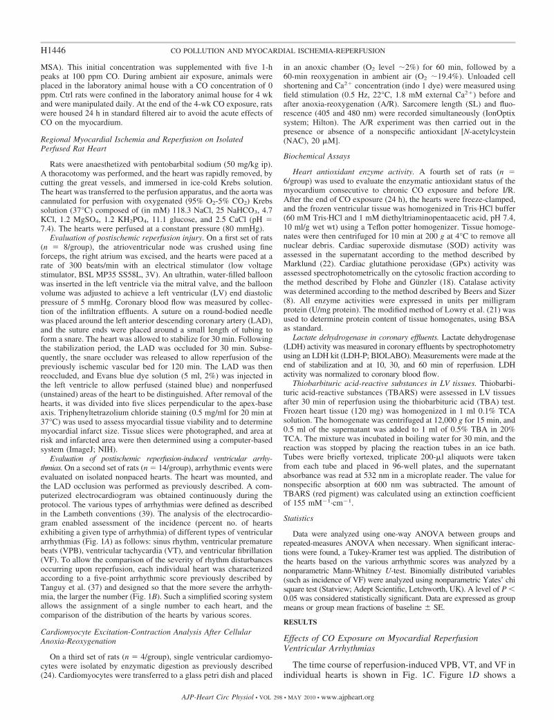
Evaluation of postischemic reperfusion-induced ventricular arrhy-thmias. On a second set of rats (n � 14/group), arrhythmic events wereevaluated on isolated nonpaced hearts. The heart was mounted, andthe LAD occlusion was performed as previously described. A com-puterized electrocardiogram was obtained continuously during theprotocol. The various types of arrhythmias were defined as describedin the Lambeth conventions (39). The analysis of the electrocardio-gram enabled assessment of the incidence (percent no. of heartsexhibiting a given type of arrhythmia) of different types of ventriculararrhythmias (Fig. 1A) as follows: sinus rhythm, ventricular prematurebeats (VPB), ventricular tachycardia (VT), and ventricular fibrillation(VF). To allow the comparison of the severity of rhythm disturbancesoccurring upon reperfusion, each individual heart was characterizedaccording to a five-point arrhythmic score previously described byTanguy et al. (37) and designed so that the more severe the arrhyth-mia, the larger the number (Fig. 1B). Such a simplified scoring systemallows the assignment of a single number to each heart, and thecomparison of the distribution of the hearts by various scores.
Cardiomyocyte Excitation-Contraction Analysis After CellularAnoxia-Reoxygenation
On a third set of rats (n � 4/group), single ventricular cardiomyo-cytes were isolated by enzymatic digestion as previously described(24). Cardiomyocytes were transferred to a glass petri dish and placed
in an anoxic chamber (O2 level �2%) for 60 min, followed by a60-min reoxygenation in ambient air (O2 �19.4%). Unloaded cellshortening and Ca2� concentration (indo 1 dye) were measured usingfield stimulation (0.5 Hz, 22°C, 1.8 mM external Ca2�) before andafter anoxia-reoxygenation (A/R). Sarcomere length (SL) and fluo-rescence (405 and 480 nm) were recorded simultaneously (IonOptixsystem; Hilton). The A/R experiment was then carried out in thepresence or absence of a nonspecific antioxidant [N-acetylcystein(NAC), 20 �M].
Biochemical Assays
Heart antioxidant enzyme activity. A fourth set of rats (n �6/group) was used to evaluate the enzymatic antioxidant status of themyocardium consecutive to chronic CO exposure and before I/R.After the end of CO exposure (24 h), the hearts were freeze-clamped,and the frozen ventricular tissue was homogenized in Tris·HCl buffer(60 mM Tris·HCl and 1 mM diethyltriaminopentaacetic acid, pH 7.4,10 ml/g wet wt) using a Teflon potter homogenizer. Tissue homoge-nates were then centrifuged for 10 min at 200 g at 4°C to remove allnuclear debris. Cardiac superoxide dismutase (SOD) activity wasassessed in the supernatant according to the method described byMarklund (22). Cardiac glutathione peroxidase (GPx) activity wasassessed spectrophotometrically on the cytosolic fraction according tothe method described by Flohe and Günzler (18). Catalase activitywas determined according to the method described by Beers and Sizer(8). All enzyme activities were expressed in units per milligramprotein (U/mg protein). The modified method of Lowry et al. (21) wasused to determine protein content of tissue homogenates, using BSAas standard.
Lactate dehydrogenase in coronary effluents. Lactate dehydrogenase(LDH) activity was measured in coronary effluents by spectrophotometryusing an LDH kit (LDH-P; BIOLABO). Measurements were made at theend of stabilization and at 10, 30, and 60 min of reperfusion. LDHactivity was normalized to coronary blood flow.
Thiobarbituric acid-reactive substances in LV tissues. Thiobarbi-turic acid-reactive substances (TBARS) were assessed in LV tissuesafter 30 min of reperfusion using the thiobarbituric acid (TBA) test.Frozen heart tissue (120 mg) was homogenized in 1 ml 0.1% TCAsolution. The homogenate was centrifuged at 12,000 g for 15 min, and0.5 ml of the supernatant was added to 1 ml of 0.5% TBA in 20%TCA. The mixture was incubated in boiling water for 30 min, and thereaction was stopped by placing the reaction tubes in an ice bath.Tubes were briefly vortexed, triplicate 200-�l aliquots were takenfrom each tube and placed in 96-well plates, and the supernatantabsorbance was read at 532 nm in a microplate reader. The value fornonspecific absorption at 600 nm was subtracted. The amount ofTBARS (red pigment) was calculated using an extinction coefficientof 155 mM�1·cm�1.
Statistics
Data were analyzed using one-way ANOVA between groups andrepeated-measures ANOVA when necessary. When significant interac-tions were found, a Tukey-Kramer test was applied. The distribution ofthe hearts based on the various arrhythmic scores was analyzed by anonparametric Mann-Whitney U-test. Binomially distributed variables(such as incidence of VF) were analyzed using nonparametric Yates’ chisquare test (Statview; Adept Scientific, Letchworth, UK). A level of P �0.05 was considered statistically significant. Data are expressed as groupmeans or group mean fractions of baseline � SE.
RESULTS
Effects of CO Exposure on Myocardial ReperfusionVentricular Arrhythmias
The time course of reperfusion-induced VPB, VT, and VF inindividual hearts is shown in Fig. 1C. Figure 1D shows a
H1446 CO POLLUTION AND MYOCARDIAL ISCHEMIA-REPERFUSION
AJP-Heart Circ Physiol • VOL 298 • MAY 2010 • www.ajpheart.org
significant difference in the distribution of the arrhythmicscores observed in both experimental groups. According to theincreased mean arrhythmic score observed in CO rats duringreperfusion, the arrhythmia severity was higher in CO ratscompared with Ctrl rats (Fig. 1E). Moreover, sustained VFwere triggered more frequently in the CO rats compared withCtrl rats (54 vs. 9%, P � 0.05 nonparametric Yates’ chi squaretest).
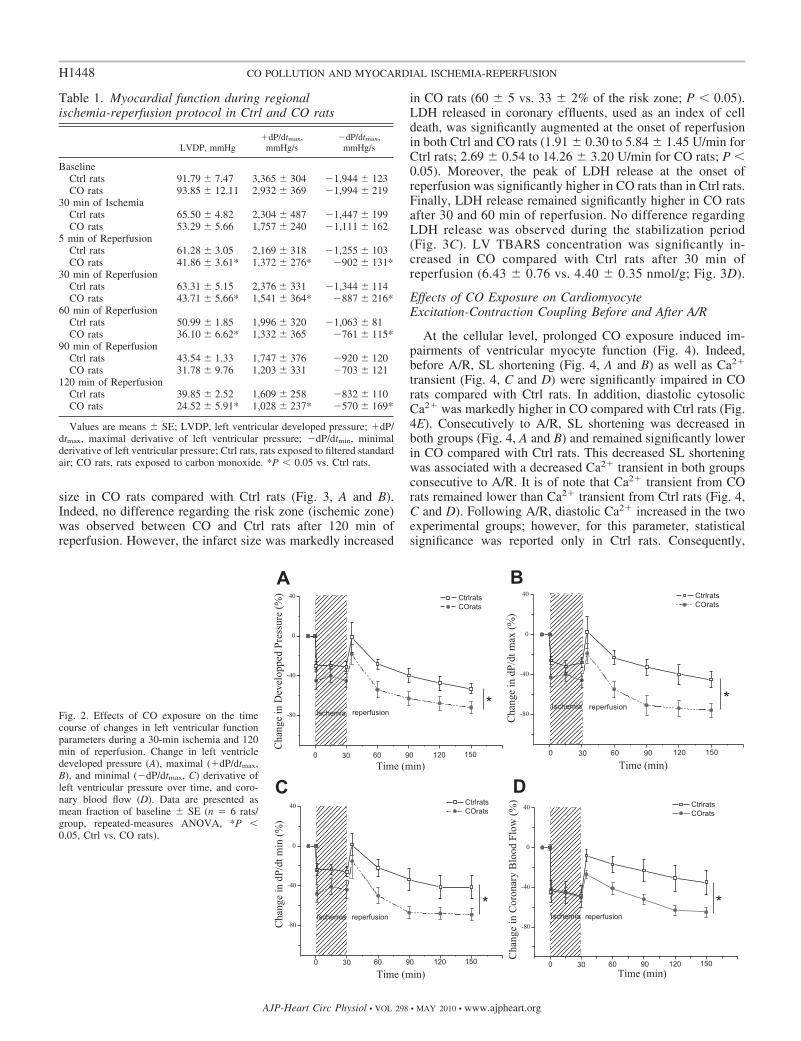
Effects of CO Exposure on Postischemic Recovery ofMyocardial Function
Any difference regarding cardiac function was reportedbetween CO and Ctrl rats before ischemia (Table 1). Postische-mic recovery of LV developed pressure and contractility,assessed during reperfusion, was significantly lower in CO ratscompared with Ctrl (Fig. 2). Indeed, the postischemic recovery
of LV developed pressure, �dP/dtmax, and �dP/dtmax weresignificantly altered in the CO rats compared with Ctrl rats(Fig. 2, A–C).
These functional results were paired with some deleteri-ous effects of CO exposure on postischemic myocardialcoronary blood flow recovery (Fig. 2D). Indeed, although nodifference in coronary blood flow was reported between COand Ctrl rats before ischemia (Ctrl rats: 11.5 � 0.9 ml/minvs. CO rats: 11.8 � 1.4 ml/min), coronary blood flow wassignificantly lower in CO rats compared with Ctrl ratsduring reperfusion.
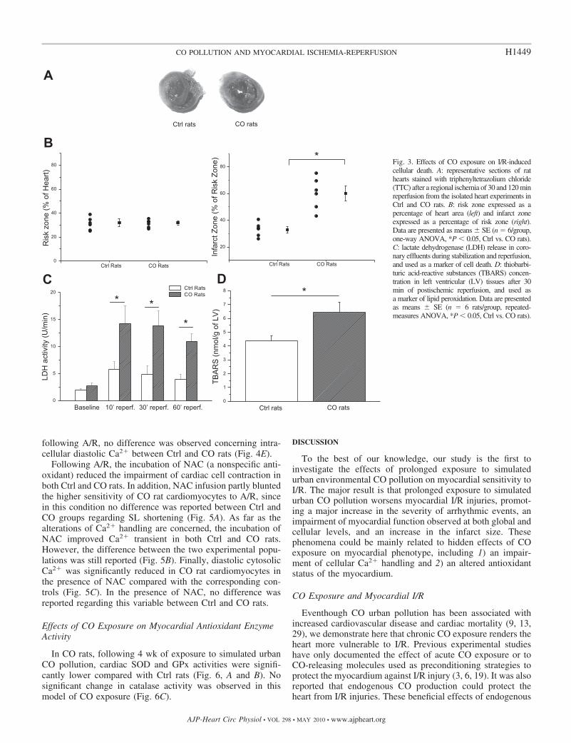
Effects of CO Exposure on Myocardial PostischemicReperfusion-Induced Cellular Death
The deleterious effects of CO exposure on I/R-inducedinjury were characterized by an increase in myocardial infarct
Fig. 1. Effects of carbon monoxide (CO) expo-sure on the severity of postischemic reperfu-sion-induced arrhythmic events. Electrocardio-graphic recordings (A) of normal sinus rhythmand of arrhythmias identified during postisch-emic reperfusion as described in the LambethConvention [sinus rhythm, ventricular prema-ture beat (VPB), ventricular tachycardia (VT),and ventricular fibrillation (VF)] and relatedarrhythmic scores (B). C: time course of reper-fusion-induced VPB (crossed out bars), VT(gray bars), and VF (black bars) of each heart ofthe control (Ctrl) group (left) and CO (right)group (nos. on left refer to the order of theexperiments). D: categorization of arrhythmiaclasses (score) according to B. Values are ex-pressed as a percentage of the total no. of heartsfor each group (Ctrl rats n � 11, CO rats n �13, distribution was tested using nonparametricMann-Whitney’s test, Ctrl vs. CO rats: P �0.05). MI, myocardial infarction. E: effects ofCO exposure on mean arrhythmic score (dataare presented as means � SE, Ctrl rats n � 11,CO rats n � 13, one-way ANOVA, *P � 0.05,Ctrl vs. CO rats).
H1447CO POLLUTION AND MYOCARDIAL ISCHEMIA-REPERFUSION
AJP-Heart Circ Physiol • VOL 298 • MAY 2010 • www.ajpheart.org
size in CO rats compared with Ctrl rats (Fig. 3, A and B).Indeed, no difference regarding the risk zone (ischemic zone)was observed between CO and Ctrl rats after 120 min ofreperfusion. However, the infarct size was markedly increased
in CO rats (60 � 5 vs. 33 � 2% of the risk zone; P � 0.05).LDH released in coronary effluents, used as an index of celldeath, was significantly augmented at the onset of reperfusionin both Ctrl and CO rats (1.91 � 0.30 to 5.84 � 1.45 U/min forCtrl rats; 2.69 � 0.54 to 14.26 � 3.20 U/min for CO rats; P �0.05). Moreover, the peak of LDH release at the onset ofreperfusion was significantly higher in CO rats than in Ctrl rats.Finally, LDH release remained significantly higher in CO ratsafter 30 and 60 min of reperfusion. No difference regardingLDH release was observed during the stabilization period(Fig. 3C). LV TBARS concentration was significantly in-creased in CO compared with Ctrl rats after 30 min ofreperfusion (6.43 � 0.76 vs. 4.40 � 0.35 nmol/g; Fig. 3D).
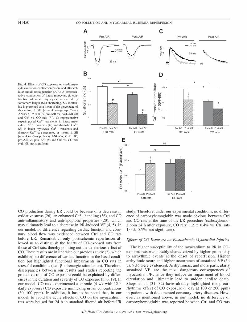
Effects of CO Exposure on CardiomyocyteExcitation-Contraction Coupling Before and After A/R
At the cellular level, prolonged CO exposure induced im-pairments of ventricular myocyte function (Fig. 4). Indeed,before A/R, SL shortening (Fig. 4, A and B) as well as Ca2�
transient (Fig. 4, C and D) were significantly impaired in COrats compared with Ctrl rats. In addition, diastolic cytosolicCa2� was markedly higher in CO compared with Ctrl rats (Fig.4E). Consecutively to A/R, SL shortening was decreased inboth groups (Fig. 4, A and B) and remained significantly lowerin CO compared with Ctrl rats. This decreased SL shorteningwas associated with a decreased Ca2� transient in both groupsconsecutive to A/R. It is of note that Ca2� transient from COrats remained lower than Ca2� transient from Ctrl rats (Fig. 4,C and D). Following A/R, diastolic Ca2� increased in the twoexperimental groups; however, for this parameter, statisticalsignificance was reported only in Ctrl rats. Consequently,
Table 1. Myocardial function during regionalischemia-reperfusion protocol in Ctrl and CO rats
LVDP, mmHg�dP/dtmax,
mmHg/s�dP/dtmax,
mmHg/s
BaselineCtrl rats 91.79 � 7.47 3,365 � 304 �1,944 � 123CO rats 93.85 � 12.11 2,932 � 369 �1,994 � 219
30 min of IschemiaCtrl rats 65.50 � 4.82 2,304 � 487 �1,447 � 199CO rats 53.29 � 5.66 1,757 � 240 �1,111 � 162
5 min of ReperfusionCtrl rats 61.28 � 3.05 2,169 � 318 �1,255 � 103CO rats 41.86 � 3.61* 1,372 � 276* �902 � 131*
30 min of ReperfusionCtrl rats 63.31 � 5.15 2,376 � 331 �1,344 � 114CO rats 43.71 � 5.66* 1,541 � 364* �887 � 216*
60 min of ReperfusionCtrl rats 50.99 � 1.85 1,996 � 320 �1,063 � 81CO rats 36.10 � 6.62* 1,332 � 365 �761 � 115*
90 min of ReperfusionCtrl rats 43.54 � 1.33 1,747 � 376 �920 � 120CO rats 31.78 � 9.76 1,203 � 331 �703 � 121
120 min of ReperfusionCtrl rats 39.85 � 2.52 1,609 � 258 �832 � 110CO rats 24.52 � 5.91* 1,028 � 237* �570 � 169*
Values are means � SE; LVDP, left ventricular developed pressure; �dP/dtmax, maximal derivative of left ventricular pressure; �dP/dtmin, minimalderivative of left ventricular pressure; Ctrl rats, rats exposed to filtered standardair; CO rats, rats exposed to carbon monoxide. *P � 0.05 vs. Ctrl rats.
Fig. 2. Effects of CO exposure on the timecourse of changes in left ventricular functionparameters during a 30-min ischemia and 120min of reperfusion. Change in left ventricledeveloped pressure (A), maximal (�dP/dtmax,B), and minimal (�dP/dtmax, C) derivative ofleft ventricular pressure over time, and coro-nary blood flow (D). Data are presented asmean fraction of baseline � SE (n � 6 rats/group, repeated-measures ANOVA, *P �0.05, Ctrl vs. CO rats).
H1448 CO POLLUTION AND MYOCARDIAL ISCHEMIA-REPERFUSION
AJP-Heart Circ Physiol • VOL 298 • MAY 2010 • www.ajpheart.org
following A/R, no difference was observed concerning intra-cellular diastolic Ca2� between Ctrl and CO rats (Fig. 4E).
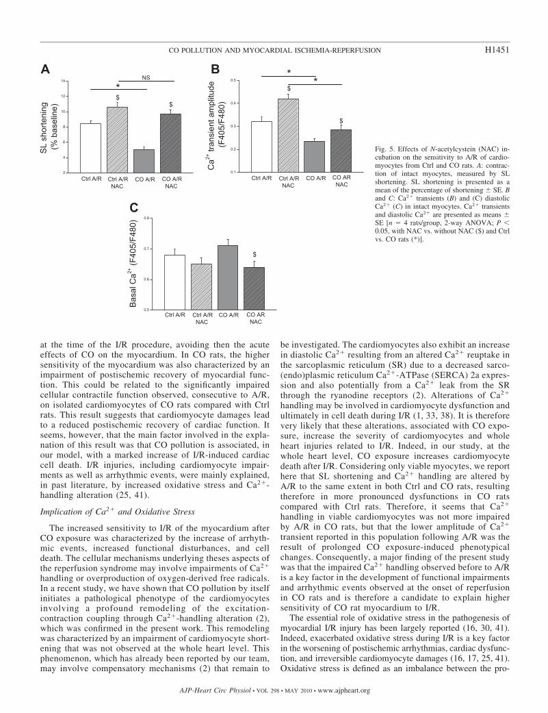
Following A/R, the incubation of NAC (a nonspecific anti-oxidant) reduced the impairment of cardiac cell contraction inboth Ctrl and CO rats. In addition, NAC infusion partly bluntedthe higher sensitivity of CO rat cardiomyocytes to A/R, sincein this condition no difference was reported between Ctrl andCO groups regarding SL shortening (Fig. 5A). As far as thealterations of Ca2� handling are concerned, the incubation ofNAC improved Ca2� transient in both Ctrl and CO rats.However, the difference between the two experimental popu-lations was still reported (Fig. 5B). Finally, diastolic cytosolicCa2� was significantly reduced in CO rat cardiomyocytes inthe presence of NAC compared with the corresponding con-trols (Fig. 5C). In the presence of NAC, no difference wasreported regarding this variable between Ctrl and CO rats.
Effects of CO Exposure on Myocardial Antioxidant EnzymeActivity
In CO rats, following 4 wk of exposure to simulated urbanCO pollution, cardiac SOD and GPx activities were signifi-cantly lower compared with Ctrl rats (Fig. 6, A and B). Nosignificant change in catalase activity was observed in thismodel of CO exposure (Fig. 6C).
DISCUSSION
To the best of our knowledge, our study is the first toinvestigate the effects of prolonged exposure to simulatedurban environmental CO pollution on myocardial sensitivity toI/R. The major result is that prolonged exposure to simulatedurban CO pollution worsens myocardial I/R injuries, promot-ing a major increase in the severity of arrhythmic events, animpairment of myocardial function observed at both global andcellular levels, and an increase in the infarct size. Thesephenomena could be mainly related to hidden effects of COexposure on myocardial phenotype, including 1) an impair-ment of cellular Ca2� handling and 2) an altered antioxidantstatus of the myocardium.
CO Exposure and Myocardial I/R
Eventhough CO urban pollution has been associated withincreased cardiovascular disease and cardiac mortality (9, 13,29), we demonstrate here that chronic CO exposure renders theheart more vulnerable to I/R. Previous experimental studieshave only documented the effect of acute CO exposure or toCO-releasing molecules used as preconditioning strategies toprotect the myocardium against I/R injury (3, 6, 19). It was alsoreported that endogenous CO production could protect theheart from I/R injuries. These beneficial effects of endogenous
Fig. 3. Effects of CO exposure on I/R-inducedcellular death. A: representative sections of rathearts stained with triphenyltetrazolium chloride(TTC) after a regional ischemia of 30 and 120 minreperfusion from the isolated heart experiments inCtrl and CO rats. B: risk zone expressed as apercentage of heart area (left) and infarct zoneexpressed as a percentage of risk zone (right).Data are presented as means � SE (n � 6/group,one-way ANOVA, *P � 0.05, Ctrl vs. CO rats).C: lactate dehydrogenase (LDH) release in coro-nary effluents during stabilization and reperfusion,and used as a marker of cell death. D: thiobarbi-turic acid-reactive substances (TBARS) concen-tration in left ventricular (LV) tissues after 30min of postischemic reperfusion, and used asa marker of lipid peroxidation. Data are presentedas means � SE (n � 6 rats/group, repeated-measures ANOVA, *P � 0.05, Ctrl vs. CO rats).
H1449CO POLLUTION AND MYOCARDIAL ISCHEMIA-REPERFUSION
AJP-Heart Circ Physiol • VOL 298 • MAY 2010 • www.ajpheart.org
CO production during I/R could be because of a decrease inoxidative stress (26), an enhanced Ca2� handling (36), and COanti-inflammatory and anti-apoptotic properties (20), whichmay ultimately lead to a decrease in I/R-induced VF (4, 5). Inour model, no difference regarding cardiac function and coro-nary blood flow was evidenced between Ctrl and CO ratsbefore I/R. Remarkably, only postischemic reperfusion al-lowed us to distinguish the hearts of CO-exposed rats fromthose of Ctrl rats, thereby pointing out the deleterious effect ofCO. These results are in line with our previous study (2), whichexhibited no difference of cardiac function in the basal condi-tion but highlighted functional impairments in CO rats instressful conditions (i.e., �-adrenergic stimulation). Therefore,discrepancies between our results and studies reporting theprotective role of CO exposure could be explained by differ-ences in the duration and severity of CO exposure (3, 6, 19). Inour model, CO rats experimented a chronic (4 wk with 12 hdaily exposure) CO exposure mimicking urban concentrations(30–100 ppm). In addition, it has to be noted that, in ourmodel, to avoid the acute effects of CO on the myocardium,rats were housed for 24 h in standard filtered air before I/R
study. Therefore, under our experimental conditions, no differ-ence of carboxyhemoglobin was made obvious between Ctrland CO rats at the time of the I/R procedure (carboxyhemo-globin 24 h after exposure, CO rats: 1.2 � 0.4% vs. Ctrl rats1.0 � 0.5%; not significant).
Effects of CO Exposure on Postischemic Myocardial Injuries
The higher susceptibility of the myocardium to I/R in CO-exposed rats was notably characterized by higher propensityto arrhythmic events at the onset of reperfusion. Higherarrhythmic score and higher occurrence of sustained VF (54vs. 9%) were evidenced. Arrhythmias, and more particularlysustained VF, are the most dangerous consequences ofmyocardial I/R, since they induce an impairment of bloodcirculation and ultimately lead to sudden cardiac death.Sheps et al. (31, 32) have already highlighted the proar-rhythmic effect of CO exposure (1 day at 100 or 200 ppm)in patients with documented coronary artery diseases. How-ever, as mentioned above, in our model, no difference ofcarboxyhemoglobin was reported between Ctrl and CO rats
Fig. 4. Effects of CO exposure on cardiomyo-cyte excitation-contraction before and after cel-lular anoxia-reoxygenation (A/R). A: represen-tative contraction of intact myocytes. B: con-traction of intact myocytes, measured bysarcomere length (SL) shortening. SL shorten-ing is presented as a mean of the percentage ofshortening � SE [n � 4 rats/group, 2-wayANOVA; P � 0.05, pre-A/R vs. post-A/R (#)and Ctrl vs. CO rats (*)]. C: representativesuperimposed Ca2� transients in intact myo-cytes. Ca2� transients (D) and diastolic Ca2�
(E) in intact myocytes. Ca2� transients anddiastolic Ca2� are presented as means � SE[n � 4 rats/group, 2-way ANOVA; P � 0.05,pre-A/R vs. post-A/R (#) and Ctrl vs. CO rats(*)]. NS, not significant.
H1450 CO POLLUTION AND MYOCARDIAL ISCHEMIA-REPERFUSION
AJP-Heart Circ Physiol • VOL 298 • MAY 2010 • www.ajpheart.org
at the time of the I/R procedure, avoiding then the acuteeffects of CO on the myocardium. In CO rats, the highersensitivity of the myocardium was also characterized by animpairment of postischemic recovery of myocardial func-tion. This could be related to the significantly impairedcellular contractile function observed, consecutive to A/R,on isolated cardiomyocytes of CO rats compared with Ctrlrats. This result suggests that cardiomyocyte damages leadto a reduced postischemic recovery of cardiac function. Itseems, however, that the main factor involved in the expla-nation of this result was that CO pollution is associated, inour model, with a marked increase of I/R-induced cardiaccell death. I/R injuries, including cardiomyocyte impair-ments as well as arrhythmic events, were mainly explained,in past literature, by increased oxidative stress and Ca2�-handling alteration (25, 41).
Implication of Ca2� and Oxidative Stress
The increased sensitivity to I/R of the myocardium afterCO exposure was characterized by the increase of arrhyth-mic events, increased functional disturbances, and celldeath. The cellular mechanisms underlying theses aspects ofthe reperfusion syndrome may involve impairments of Ca2�
handling or overproduction of oxygen-derived free radicals.In a recent study, we have shown that CO pollution by itselfinitiates a pathological phenotype of the cardiomyocytesinvolving a profound remodeling of the excitation-contraction coupling through Ca2�-handling alteration (2),which was confirmed in the present work. This remodelingwas characterized by an impairment of cardiomyocyte short-ening that was not observed at the whole heart level. Thisphenomenon, which has already been reported by our team,may involve compensatory mechanisms (2) that remain to
be investigated. The cardiomyocytes also exhibit an increasein diastolic Ca2� resulting from an altered Ca2� reuptake inthe sarcoplasmic reticulum (SR) due to a decreased sarco-(endo)plasmic reticulum Ca2�-ATPase (SERCA) 2a expres-sion and also potentially from a Ca2� leak from the SRthrough the ryanodine receptors (2). Alterations of Ca2�
handling may be involved in cardiomyocyte dysfunction andultimately in cell death during I/R (1, 33, 38). It is thereforevery likely that these alterations, associated with CO expo-sure, increase the severity of cardiomyocytes and wholeheart injuries related to I/R. Indeed, in our study, at thewhole heart level, CO exposure increases cardiomyocytedeath after I/R. Considering only viable myocytes, we reporthere that SL shortening and Ca2� handling are altered byA/R to the same extent in both Ctrl and CO rats, resultingtherefore in more pronounced dysfunctions in CO ratscompared with Ctrl rats. Therefore, it seems that Ca2�
handling in viable cardiomyocytes was not more impairedby A/R in CO rats, but that the lower amplitude of Ca2�
transient reported in this population following A/R was theresult of prolonged CO exposure-induced phenotypicalchanges. Consequently, a major finding of the present studywas that the impaired Ca2� handling observed before to A/Ris a key factor in the development of functional impairmentsand arrhythmic events observed at the onset of reperfusionin CO rats and is therefore a candidate to explain highersensitivity of CO rat myocardium to I/R.
The essential role of oxidative stress in the pathogenesis ofmyocardial I/R injury has been largely reported (16, 30, 41).Indeed, exacerbated oxidative stress during I/R is a key factorin the worsening of postischemic arrhythmias, cardiac dysfunc-tion, and irreversible cardiomyocyte damages (16, 17, 25, 41).Oxidative stress is defined as an imbalance between the pro-
Fig. 5. Effects of N-acetylcystein (NAC) in-cubation on the sensitivity to A/R of cardio-myocytes from Ctrl and CO rats. A: contrac-tion of intact myocytes, measured by SLshortening. SL shortening is presented as amean of the percentage of shortening � SE. Band C: Ca2� transients (B) and (C) diastolicCa2� (C) in intact myocytes. Ca2� transientsand diastolic Ca2� are presented as means �SE [n � 4 rats/group, 2-way ANOVA; P �0.05, with NAC vs. without NAC ($) and Ctrlvs. CO rats (*)].
H1451CO POLLUTION AND MYOCARDIAL ISCHEMIA-REPERFUSION
AJP-Heart Circ Physiol • VOL 298 • MAY 2010 • www.ajpheart.org
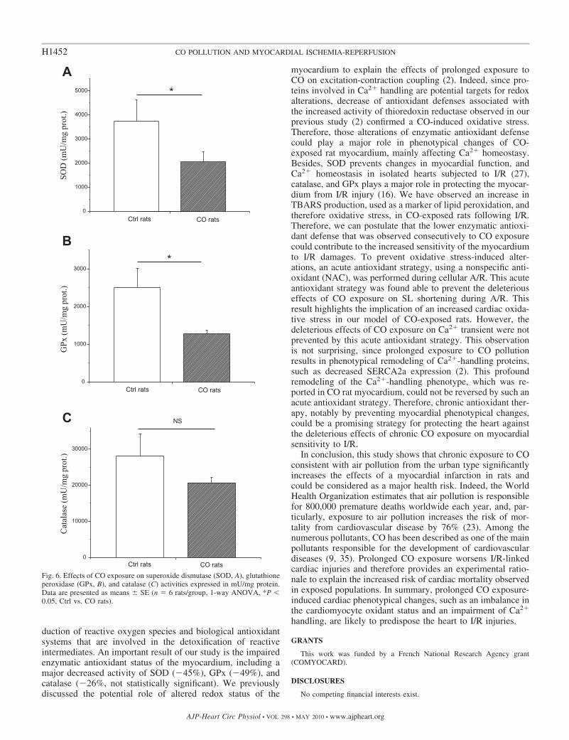
duction of reactive oxygen species and biological antioxidantsystems that are involved in the detoxification of reactiveintermediates. An important result of our study is the impairedenzymatic antioxidant status of the myocardium, including amajor decreased activity of SOD (�45%), GPx (�49%), andcatalase (�26%, not statistically significant). We previouslydiscussed the potential role of altered redox status of the
myocardium to explain the effects of prolonged exposure toCO on excitation-contraction coupling (2). Indeed, since pro-teins involved in Ca2� handling are potential targets for redoxalterations, decrease of antioxidant defenses associated withthe increased activity of thioredoxin reductase observed in ourprevious study (2) confirmed a CO-induced oxidative stress.Therefore, those alterations of enzymatic antioxidant defensecould play a major role in phenotypical changes of CO-exposed rat myocardium, mainly affecting Ca2� homeostasy.Besides, SOD prevents changes in myocardial function, andCa2� homeostasis in isolated hearts subjected to I/R (27),catalase, and GPx plays a major role in protecting the myocar-dium from I/R injury (16). We have observed an increase inTBARS production, used as a marker of lipid peroxidation, andtherefore oxidative stress, in CO-exposed rats following I/R.Therefore, we can postulate that the lower enzymatic antioxi-dant defense that was observed consecutively to CO exposurecould contribute to the increased sensitivity of the myocardiumto I/R damages. To prevent oxidative stress-induced alter-ations, an acute antioxidant strategy, using a nonspecific anti-oxidant (NAC), was performed during cellular A/R. This acuteantioxidant strategy was found able to prevent the deleteriouseffects of CO exposure on SL shortening during A/R. Thisresult highlights the implication of an increased cardiac oxida-tive stress in our model of CO-exposed rats. However, thedeleterious effects of CO exposure on Ca2� transient were notprevented by this acute antioxidant strategy. This observationis not surprising, since prolonged exposure to CO pollutionresults in phenotypical remodeling of Ca2�-handling proteins,such as decreased SERCA2a expression (2). This profoundremodeling of the Ca2�-handling phenotype, which was re-ported in CO rat myocardium, could not be reversed by such anacute antioxidant strategy. Therefore, chronic antioxidant ther-apy, notably by preventing myocardial phenotypical changes,could be a promising strategy for protecting the heart againstthe deleterious effects of chronic CO exposure on myocardialsensitivity to I/R.
In conclusion, this study shows that chronic exposure to COconsistent with air pollution from the urban type significantlyincreases the effects of a myocardial infarction in rats andcould be considered as a major health risk. Indeed, the WorldHealth Organization estimates that air pollution is responsiblefor 800,000 premature deaths worldwide each year, and, par-ticularly, exposure to air pollution increases the risk of mor-tality from cardiovascular disease by 76% (23). Among thenumerous pollutants, CO has been described as one of the mainpollutants responsible for the development of cardiovasculardiseases (9, 35). Prolonged CO exposure worsens I/R-linkedcardiac injuries and therefore provides an experimental ratio-nale to explain the increased risk of cardiac mortality observedin exposed populations. In summary, prolonged CO exposure-induced cardiac phenotypical changes, such as an imbalance inthe cardiomyocyte oxidant status and an impairment of Ca2�
handling, are likely to predispose the heart to I/R injuries.
GRANTS
This work was funded by a French National Research Agency grant(COMYOCARD).
DISCLOSURES
No competing financial interests exist.
Fig. 6. Effects of CO exposure on superoxide dismutase (SOD, A), glutathioneperoxidase (GPx, B), and catalase (C) activities expressed in mU/mg protein.Data are presented as means � SE (n � 6 rats/group, 1-way ANOVA, *P �0.05, Ctrl vs. CO rats).
H1452 CO POLLUTION AND MYOCARDIAL ISCHEMIA-REPERFUSION
AJP-Heart Circ Physiol • VOL 298 • MAY 2010 • www.ajpheart.org
REFERENCES
1. Abdallah Y, Gkatzoflia A, Gligorievski D, Kasseckert S, Euler G,Schluter KD, Schafer M, Piper HM, Schafer C. Insulin protects cardi-omyocytes against reoxygenation-induced hypercontracture by a survivalpathway targeting SR Ca2� storage. Cardiovasc Res 70: 346–353, 2006.
2. Andre L, Boissiere J, Reboul C, Perrier P, Zalvidea S, Thireau J,Meyer G, Bideaux P, Obert P, Cazorla O, Richard S. Carbon monoxidepollution promotes cardiac remodeling and ventricular arrythmia inhealthy rats. Am J Respir Crit Care Med In press.
3. Bagul A, Hosgood SA, Kaushik M, Nicholson ML. Carbon monoxideprotects against ischemia-reperfusion injury in an experimental model ofcontrolled nonheartbeating donor kidney. Transplantation 85: 576–581,2008.
4. Bak I, Papp G, Turoczi T, Varga E, Szendrei L, Vecsernyes M, Joo F,Tosaki A. The role of heme oxygenase-related carbon monoxide andventricular fibrillation in ischemic/reperfused hearts. Free Radic Biol Med33: 639–648, 2002.
5. Bak I, Szendrei L, Turoczi T, Papp G, Joo F, Das DK, de Leiris J, DerP, Juhasz B, Varga E, Bacskay I, Balla J, Kovacs P, Tosaki A. Hemeoxygenase-1-related carbon monoxide production and ventricular fibrilla-tion in isolated ischemic/reperfused mouse myocardium. FASEB J 17:2133–2135, 2003.
6. Bak I, Varadi J, Nagy N, Vecsernyes M, Tosaki A. The role ofexogenous carbon monoxide in the recovery of post-ischemic cardiacfunction in buffer perfused isolated rat hearts. Cell Mol Biol (Noisy-le-grand) 51: 453–459, 2005.
7. Barnett AG, Williams GM, Schwartz J, Best TL, Neller AH, Petro-eschevsky AL, Simpson RW. The effects of air pollution on hospitaliza-tions for cardiovascular disease in elderly people in Australian and NewZealand cities. Environ Health Perspect 114: 1018–1023, 2006.
8. Beers RF Jr, Sizer IW. A spectrophotometric method for measuring thebreakdown of hydrogen peroxide by catalase. J Biol Chem 195: 133–140,1952.
9. Bell ML, Peng RD, Dominici F, Samet JM. Emergency HospitalAdmissions for Cardiovascular Diseases and Ambient Levels of CarbonMonoxide. Results for 126 United States Urban Counties, 1999–2005.Circulation 120: 949–955, 2009.
10. Bevan MA, Proctor CJ, Baker-Rogers J, Warren ND. Exposure tocarbon monoxide, respirable syspended particulates, and volatile organiccompounds while commuting by bicycle. Environ Sci Technol 25: 788–779, 1991.
11. Bhatnagar A. Environmental cardiology: studying mechanistic linksbetween pollution and heart disease. Circ Res 99: 692–705, 2006.
12. Brook RD, Franklin B, Cascio W, Hong Y, Howard G, Lipsett M,Luepker R, Mittleman M, Samet J, Smith SC Jr, Tager I. Air pollutionand cardiovascular disease: a statement for healthcare professionals fromthe Expert Panel on Population and Prevention Science of the AmericanHeart Association. Circulation 109: 2655–2671, 2004.
13. Burnett RT, Cakmak S, Brook JR, Krewski D. The role of particulatesize and chemistry in the association between summertime ambient airpollution and hospitalization for cardiorespiratory diseases. EnvironHealth Perspect 105: 614–620, 1997.
14. Bye A, Sorhaug S, Ceci M, Hoydal MA, Stolen T, Heinrich G, TjonnaAE, Najjar SM, Nilsen OG, Catalucci D, Grimaldi S, Contu R,Steinshamn S, Condorelli G, Smith GL, Ellingsen O, Waldum H,Wisloff U. Carbon monoxide levels experienced by heavy smokers impairaerobic capacity and cardiac contractility and induce pathological hyper-trophy. Inhal Toxicol 20: 635–646, 2008.
15. Chen TM, Shofer S, Gokhale J, Kuschner WG. Outdoor air pollution:overview and historical perspective. Am J Med Sci 333: 230–234, 2007.
16. Dhalla NS, Temsah RM, Netticadan T. Role of oxidative stress incardiovascular diseases. J Hypertens 18: 655–673, 2000.
17. Dumitrescu C, Biondi R, Xia Y, Cardounel AJ, Druhan LJ, AmbrosioG, Zweier JL. Myocardial ischemia results in tetrahydrobiopterin (BH4)oxidation with impaired endothelial function ameliorated by BH4. ProcNatl Acad Sci USA 104: 15081–15086, 2007.
18. Flohe L, Gunzler WA. Assays of glutathione peroxidase. MethodsEnzymol 105: 114–121, 1984.
19. Fujimoto H, Ohno M, Ayabe S, Kobayashi H, Ishizaka N, Kimura H,Yoshida K, Nagai R. Carbon monoxide protects against cardiac ische-mia–reperfusion injury in vivo via MAPK and Akt–eNOS pathways.Arterioscler Thromb Vasc Biol 24: 1848–1853, 2004.
20. Li Volti G, Rodella LF, Di Giacomo C, Rezzani R, Bianchi R, Borsani E,Gazzolo D, Motterlini R. Role of carbon monoxide and biliverdin in renalischemia/reperfusion injury. Nephron Exp Nephrol 104: e135–e139, 2006.
21. Lowry OH, Rosebrough NJ, Farr AL, Randall RJ. Protein measure-ment with the Folin phenol reagent. J Biol Chem 193: 265–275, 1951.
22. Marklund S. Spectrophotometric study of spontaneous disproportionationof superoxide anion radical and sensitive direct assay for superoxidedismutase. J Biol Chem 251: 7504–7507, 1976.
23. Miller KA, Siscovick DS, Sheppard L, Shepherd K, Sullivan JH,Anderson GL, Kaufman JD. Long-term exposure to air pollution andincidence of cardiovascular events in women. N Engl J Med 356: 447–458,2007.
24. Mou YA, Reboul C, Andre L, Lacampagne A, Cazorla O. Late exercisetraining improves non-uniformity of transmural myocardial function inrats with ischaemic heart failure. Cardiovasc Res 81: 555–564, 2009.
25. Murphy E, Steenbergen C. Mechanisms underlying acute protectionfrom cardiac ischemia-reperfusion injury. Physiol Rev 88: 581–609, 2008.
26. Nakao A, Neto JS, Kanno S, Stolz DB, Kimizuka K, Liu F, Bach FH,Billiar TR, Choi AM, Otterbein LE, Murase N. Protection againstischemia/reperfusion injury in cardiac and renal transplantation withcarbon monoxide, biliverdin and both. Am J Transplant 5: 282–291, 2005.
27. Netticadan T, Temsah R, Osada M, Dhalla NS. Status of Ca2�/calmodulin protein kinase phosphorylation of cardiac SR proteins inischemia-reperfusion. Am J Physiol Cell Physiol 277: C384–C391, 1999.
28. Ruiz-Meana M, Garcia-Dorado D. Translational cardiovascular medi-cine (II) Pathophysiology of ischemia-reperfusion injury: new therapeuticoptions for acute myocardial infarction. Rev Esp Cardiol 62: 199–209,2009.
29. Samoli E, Touloumi G, Schwartz J, Anderson HR, Schindler C, ForsbergB, Vigotti MA, Vonk J, Kosnik M, Skorkovsky J, Katsouyanni K.Short-term effects of carbon monoxide on mortality: an analysis within theAPHEA project. Environ Health Perspect 115: 1578–1583, 2007.
30. Schulz R, Kelm M, Heusch G. Nitric oxide in myocardial ischemia/reperfusion injury. Cardiovasc Res 61: 402–413, 2004.
31. Sheps DS, Herbst MC, Hinderliter AL, Adams KF, Ekelund LG,O’Neil JJ, Goldstein GM, Bromberg PA, Ballenger M, Davis SM.Effects of 4 percent and 6 percent carboxyhemoglobin on arrhythmiaproduction in patients with coronary artery disease. Res Rep Health EffInst 1–58, 1991.
32. Sheps DS, Herbst MC, Hinderliter AL, Adams KF, Ekelund LG,O’Neil JJ, Goldstein GM, Bromberg PA, Dalton JL, Ballenger MN.Production of arrhythmias by elevated carboxyhemoglobin in patients withcoronary artery disease. Ann Intern Med 113: 343–351, 1990.
33. Siegmund B, Schlack W, Ladilov YV, Balser C, Piper HM. Halothaneprotects cardiomyocytes against reoxygenation-induced hypercontracture.Circulation 96: 4372–4379, 1997.
34. Stern FB, Halperin WE, Hornung RW, Ringenburg VL, McCammonCS. Heart disease mortality among bridge and tunnel officers exposed tocarbon monoxide. Am J Epidemiol 128: 1276–1288, 1988.
35. Stieb DM, Szyszkowicz M, Rowe BH, Leech JA. Air pollution andemergency department visits for cardiac and respiratory conditions: amulti-city time-series analysis (Abstract). Environ Health 8: 25, 2009.
36. Szabo ME, Gallyas E, Bak I, Rakotovao A, Boucher F, de Leiris J,Nagy N, Varga E, Tosaki A. Heme oxygenase-1-related carbon monox-ide and flavonoids in ischemic/reperfused rat retina. Invest Ophthalmol VisSci 45: 3727–3732, 2004.
37. Tanguy S, Boucher F, Besse S, Ducros V, Favier A, de Leiris J. Traceelements and cardioprotection: increasing endogenous glutathione perox-idase activity by oral selenium supplementation in rats limits reperfusion-induced arrhythmias. J Trace Elem Med Biol 12: 28–38, 1998.
38. Venetucci LA, Trafford AW, O’Neill SC, Eisner DA. The sarcoplasmicreticulum and arrhythmogenic calcium release. Cardiovasc Res 77: 285–292, 2008.
39. Walker MJ, Curtis MJ, Hearse DJ, Campbell RW, Janse MJ, YellonDM, Cobbe SM, Coker SJ, Harness JB, Harron DW. The LambethConventions: guidelines for the study of arrhythmias in ischaemia infarc-tion, and reperfusion. Cardiovasc Res 22: 447–455, 1988.
40. Wright GR, Jewczyk S, Onrot J, Tomlinson P, Shephard RJ. Carbonmonoxide in the urban atmosphere: hazards to the pedestrian and thestreet-worker. Arch Environ Health 30: 123–129, 1975.
41. Zhao X, Chen YR, He G, Zhang A, Druhan LJ, Strauch AR, Zweier JL.Endothelial nitric oxide synthase (NOS3) knockout decreases NOS2 induc-tion, limiting hyperoxygenation and conferring protection in the postischemicheart. Am J Physiol Heart Circ Physiol 292: H1541–H1550, 2007.
H1453CO POLLUTION AND MYOCARDIAL ISCHEMIA-REPERFUSION
AJP-Heart Circ Physiol • VOL 298 • MAY 2010 • www.ajpheart.org
Related Documents