Yagel Simcha (Orcid ID: 0000-0002-1412-5862) Simplifying Imaging of the Abdominal Fetal Precordial Venous System Simcha Yagel, Sarah M Cohen, Dan V Valsky Division of Obstetrics and Gynecology, Hadassah-Hebrew University Medical Center, Jerusalem, Israel Corresponding author: Prof. Simcha Yagel Division of Obstetrics and Gynecology Hadassah-Hebrew University Medical Centers, Mt. Scopus Jerusalem, Israel 91240 Email: [email protected] Key Words: ductus venosus; hepatic veins; IVC; left portal vein; splenic vein, umbilical vein Short title: Simpler imaging of the precordial venous system This article is protected by copyright. All rights reserved. This article has been accepted for publication and undergone full peer review but has not been through the copyediting, typesetting, pagination and proofreading process which may lead to differences between this version and the Version of Record. Please cite this article as doi: 10.1002/uog.19053

Welcome message from author
This document is posted to help you gain knowledge. Please leave a comment to let me know what you think about it! Share it to your friends and learn new things together.
Transcript
Yagel Simcha (Orcid ID: 0000-0002-1412-5862)
Simplifying Imaging of the Abdominal Fetal Precordial Venous System
Simcha Yagel, Sarah M Cohen, Dan V Valsky
Division of Obstetrics and Gynecology, Hadassah-Hebrew University Medical Center, Jerusalem, Israel
Corresponding author: Prof. Simcha Yagel Division of Obstetrics and Gynecology Hadassah-Hebrew University Medical Centers, Mt. Scopus Jerusalem, Israel 91240 Email: [email protected]
Key Words: ductus venosus; hepatic veins; IVC; left portal vein; splenic vein, umbilical vein
Short title: Simpler imaging of the precordial venous system
This article is protected by copyright. All rights reserved.
This article has been accepted for publication and undergone full peer review but has not beenthrough the copyediting, typesetting, pagination and proofreading process which may lead todifferences between this version and the Version of Record. Please cite this article as doi:10.1002/uog.19053
The fetal precordial veins comprise a complex system of vessels delivering
oxygenated blood from the placenta through the umbilical vein to the developing fetus. This
system draws most of its nomenclature from the adult venous system. However, the
direction of flow, as well as the relative size and prominence of the component vessels,
differ from those in the postnatal circulation. Understanding the unique characteristics of
the fetal circulation and its differences from the postnatal system can aid the sonographer in
identifying normal1 and anomalous2 anatomy (Figure 1). The system can be challenging to
image partly because of these differences, and partly because of individual variations in
anatomy. Using a systematic approach may assist the operator to obtain these planes and
target vessels confidently. Our aim here is to present our approach to imaging the fetal
precordial venous system, based on three successive imaging planes.
In the fetus, the umbilical vein delivers oxygen- and nutrient- rich blood from the
placenta to the left portal vein; the ductus venosus shunts this blood away from the portal
system to the IVC and onward to the right heart. The proximal portion of the left portal
vein, known also as the pars transversa or portal sinus (PS), extends to the point of
bifurcation of the main portal vein to the right portal vein. This blood flows into the main
portal vein and from there to the right portal vein and the hepatic circulation. In our
opinion, therefore, the intra-abdominal portion of the umbilical vein should be termed the
left portal vein from the bifurcations of the LPV (Figure 1a).
The blood flow direction in the left portal vein, therefore, is reversed from that in the
adult (Figure 1b). This shunt system at the left portal vein, situated between the main portal
vein junction and the ductus venosus, is known as the watershed of the fetal circulation3, 4. It
has been shown to be a sensitive gauge of hemodynamic changes in the fetus. The greater
volume and higher oxygen content of blood flowing through the left lobe of the liver as
compared to the right may result in the relatively larger left than right lobe during fetal life5.
Shortly following delivery the umbilical vein and ductus venosus atrophy and form the
This article is protected by copyright. All rights reserved.
ligamentum teres (round ligament) and the ligamentum venosum, respectively, and the
proportional sizes of the liver lobes is reversed.
While most anomalies of the venous system are rare, and some isolated anomalies
may be innocuous, such as persistent right umbilical vein6-11, others may have serious
consequences. Agenesis of the ductus venosus or abnormal development of the portal
system12-18 for example, may have grave prognoses. Indeed, it would appear that some
malformations of the venous system are not as rare as formerly believed, and that more will
be diagnosed if practitioners are cognizant of their sonographic appearance and associated
anomalies1, 2, 19. Whenever anomalies of the cardinal, vitelline, or umbilical systems are
diagnosed, they should prompt thorough investigation of the other segments of the
cardiovascular system. When a cardiac defect is suspected, venous system examination is
essential to identify or exclude possible associated lesions. Pulse Doppler scanning of the
venous system can be a sensitive tool in obstetric management of intra-uterine growth
restriction or other fetal distress20-29, as well as assisting in gauging the impact of any
anomaly on the system11, 14-16, 30, 31.
When the fetal precordial venous system is examined, whether in the context of
suspected anomaly in one of its component veins, or a cardiac or other malformation, or
compromised fetal well-being, the examination should include the course of the umbilical
vein, the ductus venosus, left and right portal veins and portal sinus, the splenic vein, hepatic
veins, and the inferior vena cava (IVC). The examiner should be able to confirm normal
anatomy and rule out anomalies13, 19, 32.
The three planes approach to the fetal venous system
We recently described19 a systematic approach to scanning the fetal venous system,
based on 2DUS color Doppler scanning of the fetal abdomen in three planes: two transverse
and one longitudinal (Figure 2A-C). The more caudal plane (A) is a ventral or lateral
This article is protected by copyright. All rights reserved.
transverse plane through the fetal abdomen at the level of the left portal sinus. This plane
shows the umbilical vein, left portal vein, portal sinus, anterior right portal vein, posterior
right portal vein, main portal vein, and splenic vein and artery. The latter was included
because it is always seen with the splenic vein. Configuration of the target vessels may
show slight individual variation. Tilting the transducer slightly to capture the main portal vein
may assist in orienting the scan.
To obtain the second plane (B), the transducer is moved cephalad to a ventral or slightly
lateral transverse plane that images the trident: the right, middle, and left hepatic veins
where they drain into the IVC, and the IVC itself.
The third plane (C), is obtained by rotating the transducer toward a longitudinal anterior-
posterior plane, in order to image the umbilical vein, left portal vein, ductus venosus, IVC,
and left hepatic vein where they converge proximal to the right atrium. Acquiring it may
require the operator to adjust the rotation and tilt of the transducer by a few degrees, to
visualize all of the target vessels. This plane corresponds to the classic sagittal ductus
venous plane proposed by Kiserud33. Using these successive planes the operator can
systematically image the elements of the fetal precordial venous system. The component
veins can be investigated with pulse Doppler, as described below. Color and power Doppler
settings should be optimized according to the vascularity and flow velocity in the region of
interest.
Anomalies diagnosed in the scanning planes
Anomalies may be encountered in all of the component veins of the precordial venous
system. We present six cases (Figure 3a-f), showing the anomalous appearance of each of
the three scanning planes that led to their diagnosis. Figure 3a-b show two anomalies
diagnosed in the A Plane (compare Figure 2A). Figure 3a is a case of complete agenesis of
the portal venous system (CAPVS). The figure shows the absence of the usual constellation
This article is protected by copyright. All rights reserved.
of vessels, except for the remnant of the system (arrow). Figure 3b is a case of portal sinus
varix anomaly.
The normal B Plane shows the trident of the hepatic veins (Figure 2b); Figure 3c-d show
shunt anomalies from the portal system to the hepatic veins. Figure 3c shows two shunts to
the left and middle hepatic veins; Figure 3d shows a left porto-hepatic shunt, with the left
portal vein flowing into the left hepatic vein.
Figure 3e-f show cases of agenesis of the ductus venosus, visualized in the C Plane. Figure 3e
is a right porto-hepatic shunt, with blood shunted directly from the left portal vein to the
right hepatic vein. Figure 3f shows another case of agenesis of the ductus venosus with a
wide shunt draining into the IVC.
Doppler waveforms of the normal fetal venous system
The pulse Doppler waveforms of the fetal precordial veins reflect the heart cycle.
Their typical three-peak form mirrors the ventricular systolic phase (s), passive diastolic
phase (d), and active diastolic phase (a). DV blood flows forward throughout the entire
heart cycle, differing from the IVC and hepatic veins and thus assuring constant high quality
blood supply to the heart. A venous system preload index34-37, similar to that devised for the
arterial system, has been proposed. It was consistently shown to gradually decrease with
progression of pregnancy35, 38. Doppler sampling sites are shown in Figure 2c, and normal
waveforms of the most commonly investigated vessels are shown in Figure 4.
The umbilical vein is evaluated in the intra-abdominal portion of the vein (Figure 2).
Pulsatile flow may be considered a normal feature until 15 weeks’ gestation, before the low
resistance placental vascular bed is established by the second trophoblast invasion39 and
even later. In addition, the waveform characteristics are influenced by the diameter and
stiffness of the vessel. In the second half of pregnancy, UV pulsations are associated with
fetal breathing movements. The sampling site will also impact the appearance of pulsations.
This article is protected by copyright. All rights reserved.
Moving from the free loop UV to the intrahepatic, porto–umbilical connection, the
retrograde atrial contraction waveform propagation becomes more pronounced and the
incidence of pulsations markedly increases40.
The ductus venosus forms a direct shunt between the left portal vein and the inferior
vena cava. The DV can be sampled at its inlet in a near-sagittal scan, with a large sample
volume at a low angle of insonation33 or in an oblique transverse section of the fetal
abdomen (Figure 2). Changes in DV flow are seen in hypoxia and hypovolemia in
experimental animal models and in human fetuses25, 41-45. (Figure 3) Venous compensatory
mechanisms aim to improve the placental supply to the heart by increasing the proportion
of DV shunting from the portal sinus. Normally at 20 weeks’ gestation about 30% of blood is
shunted through the DV, while at 30 weeks about 20% of blood is shunted29, 46-48. In small-for-
gestational age fetuses a much higher proportion of blood is shunted through the DV, and
earlier placental compromise will show a more pronounced shunt and distension of the DV20,
29. The degree of shunting in IUGR fetuses is positively correlated with the severity of
placental insufficiency as reflected by the UA diastolic flow. Ductus venosus anomalies can
be characterized by their pulse Doppler waveforms. This is beyond the scope of this paper;
the reader is referred to our review2 and other work14, 30, 49, 50.
The IVC is usually sampled in the fetal abdomen, caudal to the hepatic confluence and
DV outlet, to avoid interference from neighboring vessels (Figure 2). It is normal to observe
a negative a-wave in the IVC because of the vessel’s normally lower velocities51-54. Since the
IVC is familiar to pediatricians as the preferred vessel for postnatal evaluation of SGA
neonates, it is often sampled in the fetus29.
The trident-shaped hepatic veins (Figure 2b), although readily available for Doppler
investigation, have not been widely studied27, 55-59. The signs of cardiac compromise
observed in the hepatic vein are similar to those apparent in the DV and IVC29, 57. Hepatic
venous Doppler has been shown to differentiate between types of extrasystoles27.
This article is protected by copyright. All rights reserved.
Because of its position at the focal point of oxygenated blood flow from the umbilical
vein toward the liver and the ductus venosus, the left portal vein has been described as the
watershed of the fetal venous circulation and suggested as a simple marker of circulatory
compromise4, 46, 60-62. The vessel is sampled in the left portal branch between the ductus
venosus inlet and the junction with the main portal stem (Figure 2). In the compromised
fetus, flow in the LPV is reduced, and may become pulsatile, bi-directional, or reversed. In
hemodynamically compromised fetuses, Kiserud et al4 showed that accentuation of LPV
peak velocity mirrored the DV a-wave. Reverse flow in the LPV shows significant correlation
with increased RI in the UA3.
For more extensive discussion of the etiology and appearance of altered venous
system Doppler waveforms in IUGR and other cardiovascular compromise, we refer the
reader to our review2 and other work12, 20, 22, 49, 50.
In summary, we provide here a simple approach to evaluating the fetal precordial
venous system. Visualization of the described planes may require some finesse on the part
of the operator, but acquisition of these planes readily becomes familiar. Anomalies of this
system may be less rare than previously believed, and familiarity with the normal
configuration of these veins is imperative to reach a diagnosis. In addition, Doppler
investigation of the component veins provides important information on the well-being of
fetuses with IUGR or other cardiac compromise.
This article is protected by copyright. All rights reserved.
REFERENCES
1. Yagel S, Kivilevitch Z, Cohen SM, Valsky DV, Messing B, Shen O and Achiron R. The fetal venous system, part I: normal embryology, anatomy, hemodynamics, ultrasound evaluation and Doppler investigation. Ultrasound Obstet Gynecol 2010; 35: 741-750. 2. Yagel S, Kivilevitch Z, Cohen SM, Valsky DV, Messing B, Shen O and Achiron R. The fetal venous system, Part II: ultrasound evaluation of the fetus with congenital venous system malformation or developing circulatory compromise. Ultrasound Obstet Gynecol 2010; 36: 93-111. 3. Kilavuz O, Vetter K, Kiserud T and Vetter P. The left portal vein is the watershed of the fetal venous system. J Perinat Med 2003; 31: 184-187. 4. Kiserud T, Kilavuz O and Hellevik LR. Venous pulsation in the fetal left portal branch: the effect of pulse and flow direction. Ultrasound Obstet Gynecol 2003; 21: 359-364. 5. Gross BH, Harter LP and Filly RA. Disproportionate left hepatic lobe size in the fetus: ultrasonic demonstration. J Ultrasound Med 1982; 1: 79-81. 6. Shen O, Tadmor OP and Yagel S. Prenatal diagnosis of persistent right umbilical vein. Ultrasound Obstet Gynecol 1996; 8: 31-33. 7. Weichert J, Hartge D, Germer U, Axt-Fliedner R and Gembruch U. Persistent right umbilical vein: a prenatal condition worth mentioning? Ultrasound Obstet Gynecol 2011; 37: 543-548. 8. Blazer S, Zimmer EZ and Bronshtein M. Persistent intrahepatic right umbilical vein in the fetus: a benign anatomic variant. Obstet Gynecol 2000; 95: 433-436. 9. Wolman I, Gull I, Fait G, Amster R, Kupferminc MJ, Lessing JB and Jaffa AJ. Persistent right umbilical vein: incidence and significance. Ultrasound Obstet Gynecol 2002; 19: 562-564. 10. Martinez R, Gamez F, Bravo C, Sanchez P, Orizales C, Ortiz L and De Leon-Luis J. Perinatal outcome after ultrasound prenatal diagnosis of persistent right umbilical vein. Eur J Obstet Gynecol Reprod Biol 2013; 168: 36-39. 11. Lide B, Lindsley W, Foster MJ, Hale R and Haeri S. Intrahepatic Persistent Right Umbilical Vein and Associated Outcomes: A Systematic Review of the Literature. J Ultrasound Med 2016; 35: 1-5. 12. Berg C, Kamil D, Geipel A, Kohl T, Knopfle G, Hansmann M and Gembruch U. Absence of ductus venosus-importance of umbilical venous drainage site. Ultrasound Obstet Gynecol 2006; 28: 275-281. 13. Yagel S, Kivilevitch Z, Cohen SM, Valsky DV, Messing B, Shen O and Achiron R. The fetal venous system, Part II: ultrasound evaluation of the fetus with congenital venous system malformation or developing circulatory compromise. Ultrasound Obstet Gynecol; 36: 93-111. 14. Shen O, Valsky DV, Messing B, Cohen SM, Lipschuetz M and Yagel S. Shunt diameter in agenesis of the ductus venosus with extrahepatic portosystemic shunt impacts on prognosis. Ultrasound Obstet Gynecol 2011; 37: 184-190. 15. Moaddab A, Tonni G, Grisolia G, Bonasoni MP, Araujo Junior E, Rolo LC, Prefumo F, de la Fuente S, Sepulveda W, Ayres N and Ruano R. Predicting outcome in 259 fetuses with
This article is protected by copyright. All rights reserved.
agenesis of ductus venosus - a multicenter experience and systematic review of the literature (.). J Matern Fetal Neonatal Med 2016; 29: 3606-3614. 16. Thomas JT, Petersen S, Cincotta R, Lee-Tannock A and Gardener G. Absent ductus venosus--outcomes and implications from a tertiary centre. Prenat Diagn 2012; 32: 686-691. 17. Staboulidou I, Pereira S, Cruz Jde J, Syngelaki A and Nicolaides KH. Prevalence and outcome of absence of ductus venosus at 11(+0) to 13(+6) weeks. Fetal Diagn Ther 2011; 30: 35-40. 18. Yagel S, Cohen, SM, Rosenak, D, Messing, B, Lipschuetz, M, Valsky, DV. The importance of portal system evaluation and shunt dimensions in cases of agenesis of ductus venosus. Ultrasound Obstet Gynecol 2009; 34: 123. 19. Yagel S, Cohen SM, Valsky DV, Shen O, Lipschuetz M and Messing B. Systematic examination of the fetal abdominal precordial veins: a cohort study. Ultrasound Obstet Gynecol 2015; 45: 578-583. 20. Kiserud T, Kessler J, Ebbing C and Rasmussen S. Ductus venosus shunting in growth-restricted fetuses and the effect of umbilical circulatory compromise. Ultrasound Obstet Gynecol 2006; 28: 143-149. 21. Kiserud T, Eik-Nes SH, Blaas HG, Hellevik LR and Simensen B. Ductus venosus blood velocity and the umbilical circulation in the seriously growth-retarded fetus. Ultrasound Obstet Gynecol 1994; 4: 109-114. 22. Turan OM, Turan S, Gungor S, Berg C, Moyano D, Gembruch U, Nicolaides KH, Harman CR and Baschat AA. Progression of Doppler abnormalities in intrauterine growth restriction. Ultrasound Obstet Gynecol 2008; 32: 160-167. 23. Baschat AA. Venous Doppler evaluation of the growth-restricted fetus. Clin Perinatol 2011; 38: 103-112, vi. 24. Baschat AA, Gembruch U, Weiner CP and Harman CR. Qualitative venous Doppler waveform analysis improves prediction of critical perinatal outcomes in premature growth-restricted fetuses. Ultrasound Obstet Gynecol 2003; 22: 240-245. 25. Wada N, Tachibana D, Kurihara Y, Nakagawa K, Nakano A, Terada H, Tanaka K, Fukui M, Koyama M and Hecher K. Alterations in time intervals of ductus venosus and atrioventricular flow velocity waveforms in growth-restricted fetuses. Ultrasound Obstet Gynecol 2015; 46: 221-226. 26. Pisaneschi S, Strigini FA, Sanchez AM, Begliuomini S, Casarosa E, Ripoli A, Ghirri P, Boldrini A, Fink B, Genazzani AR, Coceani F and Simoncini T. Compensatory feto-placental upregulation of the nitric oxide system during fetal growth restriction. PLoS One 2012; 7: e45294. 27. Saemundsson Y, Johansson C, Wenling S and Gudmundsson S. Hepatic venous Doppler in the evaluation of fetal extrasystoles. Ultrasound Obstet Gynecol 2011; 37: 179-183. 28. Baschat AA. Examination of the fetal cardiovascular system. Semin Fetal Neonatal Med 2011; 16: 2-12.
This article is protected by copyright. All rights reserved.
29. Kiserud T. Venous flow in intrauterine growth restriction and cardiac decompensation. In Fetal Cardiology, S. Yagel, Silverman, N.H., Gembruch, U. (ed). Informa Healthcare: New York, 2009, 547-560. 30. Achiron R and Kivilevitch Z. Fetal umbilical-portal-systemic venous shunt: in-utero classification and clinical significance. Ultrasound Obstet Gynecol 2016; 47: 739-747. 31. Alonso-Gamarra E, Parron M, Perez A, Prieto C, Hierro L and Lopez-Santamaria M. Clinical and radiologic manifestations of congenital extrahepatic portosystemic shunts: a comprehensive review. Radiographics 2011; 31: 707-722. 32. Yagel S, Kivilevitch Z, Cohen SM, Valsky DV, Messing B, Shen O and Achiron R. The fetal venous system, part I: normal embryology, anatomy, hemodynamics, ultrasound evaluation and Doppler investigation. Ultrasound Obstet Gynecol; 35: 741-750. 33. Kiserud T, Eik-Nes SH, Blaas HG and Hellevik LR. Ultrasonographic velocimetry of the fetal ductus venosus. Lancet 1991; 338: 1412-1414. 34. Rizzo G, Capponi A, Talone PE, Arduini D and Romanini C. Doppler indices from inferior vena cava and ductus venosus in predicting pH and oxygen tension in umbilical blood at cordocentesis in growth-retarded fetuses. Ultrasound Obstet Gynecol 1996; 7: 401-410. 35. Baschat AA. Relationship between placental blood flow resistance and precordial venous Doppler indices. Ultrasound Obstet Gynecol 2003; 22: 561-566. 36. Baschat AA, Guclu S, Kush ML, Gembruch U, Weiner CP and Harman CR. Venous Doppler in the prediction of acid-base status of growth-restricted fetuses with elevated placental blood flow resistance. Am J Obstet Gynecol 2004; 191: 277-284. 37. Ott WJ. Value of inferior vena cava Doppler waveform analysis for prediction of neonatal outcome. Am J Perinatol 1999; 16: 429-434. 38. Hecher K, Campbell S, Snijders R and Nicolaides K. Reference ranges for fetal venous and atrioventricular blood flow parameters. Ultrasound Obstet Gynecol 1994; 4: 381-390. 39. Rizzo G, Arduini D and Romanini C. Umbilical vein pulsations: a physiologic finding in early gestation. Am J Obstet Gynecol 1992; 167: 675-677. 40. van Splunder IP, Huisman TW, Stijnen T and Wladimiroff JW. Presence of pulsations and reproducibility of waveform recording in the umbilical and left portal vein in normal pregnancies. Ultrasound Obstet Gynecol 1994; 4: 49-53. 41. Behrman RE, Lees MH, Peterson EN, De Lannoy CW and Seeds AE. Distribution of the circulation in the normal and asphyxiated fetal primate. Am J Obstet Gynecol 1970; 108: 956-969. 42. Edelstone DI, Rudolph AM and Heymann MA. Effects of hypoxemia and decreasing umbilical flow liver and ductus venosus blood flows in fetal lambs. Am J Physiol 1980; 238: H656-663. 43. Itskovitz J, LaGamma EF and Rudolph AM. Effects of cord compression on fetal blood flow distribution and O2 delivery. Am J Physiol 1987; 252: H100-109. 44. Meyers RL, Paulick RP, Rudolph CD and Rudolph AM. Cardiovascular responses to acute, severe haemorrhage in fetal sheep. J Dev Physiol 1991; 15: 189-197.
This article is protected by copyright. All rights reserved.
45. Paulick RP, Meyers RL, Rudolph CD and Rudolph AM. Venous and hepatic vascular responses to indomethacin and prostaglandin E1 in the fetal lamb. Am J Obstet Gynecol 1990; 163: 1357-1363. 46. Kessler J, Rasmussen S, Godfrey K, Hanson M and Kiserud T. Longitudinal study of umbilical and portal venous blood flow to the fetal liver: low pregnancy weight gain is associated with preferential supply to the fetal left liver lobe. Pediatr Res 2008; 63: 315-320. 47. Kiserud T, Rasmussen S and Skulstad S. Blood flow and the degree of shunting through the ductus venosus in the human fetus. Am J Obstet Gynecol 2000; 182: 147-153. 48. Bellotti M, Pennati G, De Gasperi C, Battaglia FC and Ferrazzi E. Role of ductus venosus in distribution of umbilical blood flow in human fetuses during second half of pregnancy. Am J Physiol Heart Circ Physiol 2000; 279: H1256-1263. 49. Turan OM, Turan S, Sanapo L, Willruth A, Berg C, Gembruch U, Harman CR and Baschat AA. Reference ranges for ductus venosus velocity ratios in pregnancies with normal outcomes. J Ultrasound Med 2014; 33: 329-336. 50. Strizek B, Zamprakou A, Gottschalk I, Roethlisberger M, Hellmund A, Muller A, Gembruch U, Geipel A and Berg C. Prenatal Diagnosis of Agenesis of Ductus Venosus: A Retrospective Study of Anatomic Variants, Associated Anomalies and Impact on Postnatal Outcome. Ultraschall Med 2017. 51. Reed KL, Appleton CP, Anderson CF, Shenker L and Sahn DJ. Doppler studies of vena cava flows in human fetuses. Insights into normal and abnormal cardiac physiology. Circulation 1990; 81: 498-505. 52. Rizzo G, Arduini D and Romanini C. Inferior vena cava flow velocity waveforms in appropriate- and small-for-gestational-age fetuses. Am J Obstet Gynecol 1992; 166: 1271-1280. 53. Huisman TW, Stewart PA, Wladimiroff JW and Stijnen T. Flow velocity waveforms in the ductus venosus, umbilical vein and inferior vena cava in normal human fetuses at 12-15 weeks of gestation. Ultrasound Med Biol 1993; 19: 441-445. 54. Huisman TW, Stewart PA and Wladimiroff JW. Flow velocity waveforms in the fetal inferior vena cava during the second half of normal pregnancy. Ultrasound Med Biol 1991; 17: 679-682. 55. Bellotti M, Pennati G, De Gasperi C, Bozzo M, Battaglia FC and Ferrazzi E. Simultaneous measurements of umbilical venous, fetal hepatic, and ductus venosus blood flow in growth-restricted human fetuses. Am J Obstet Gynecol 2004; 190: 1347-1358. 56. Axt-Fliedner R, Wiegank U, Fetsch C, Friedrich M, Krapp M, Georg T and Diedrich K. Reference values of fetal ductus venosus, inferior vena cava and hepatic vein blood flow velocities and waveform indices during the second and third trimester of pregnancy. Arch Gynecol Obstet 2004; 270: 46-55. 57. Hofstaetter C, Gudmundsson S, Dubiel M and Marsal K. Fetal right hepatic venous blood velocimetry in normal and high-risk pregnancies. European Journal of Ultrasound 1996; 4: 153-160.
This article is protected by copyright. All rights reserved.
58. Kaji T, Maeda K, Suto M, Sato M and Irahara M. Simultaneous recordings of pulsed wave Doppler signals in hepatic vein and descending aorta using dual Doppler: a novel method for evaluating fetal arrhythmias. Ultrasound Obstet Gynecol 2012; 39: 357-359. 59. Gindes L, Pretorius DH, Romine LE, Kfir M, D'Agostini D, Hull A and Achiron R. Three-dimensional ultrasonographic depiction of fetal abdominal blood vessels. J Ultrasound Med 2009; 28: 977-988. 60. Kessler J, Rasmussen S and Kiserud T. The fetal portal vein: normal blood flow development during the second half of human pregnancy. Ultrasound Obstet Gynecol 2007; 30: 52-60. 61. Minniti S, Visentini S and Procacci C. Congenital anomalies of the venae cavae: embryological origin, imaging features and report of three new variants. Eur Radiol 2002; 12: 2040-2055. 62. Kessler J, Rasmussen S and Kiserud T. The left portal vein as an indicator of watershed in the fetal circulation: development during the second half of pregnancy and a suggested method of evaluation. Ultrasound Obstet Gynecol 2007; 30: 757-764. 63. Kiserud T and Acharya G. The fetal circulation. Prenat Diagn 2004; 24: 1049-1059.
LEGENDS TO FIGURES
Figure 1:
Schematics of the precordial venous system in the fetus (A) and the adult (B), modified from
Kiserud et al.63 UV, umbilical vein; LPV, left portal vein; DV, ductus venosus; MPV, main portal
vein; RPV, right portal vein; St, stomach; Sp, spine. Arrows indicate direction of blood flow.
Figure 2:
The fetal precordial venous system in three planes.
Frame A: Lateral transverse plane through the fetal abdomen from right to left. The splenic
artery is shown (SA, blue jet) as it is always observed with the splenic vein. (ARPV, anterior
This article is protected by copyright. All rights reserved.
right portal vein; PRPV, posterior right portal vein; MPV, main portal vein; LPVs, left portal
veins; Ao, aorta; SV, splenic vein; SA, splenic artery; St, stomach; Rt, right; Lt, left; Sp, spine.
Frame B: Anterior-posterior transverse plane through the upper fetal abdomen showing the
normal "trident sign" of the hepatic veins. LHV, left hepatic vein; MHV, middle hepatic vein;
RHV, right hepatic vein; Ao, aorta; Rt, right; Lt, left; Sp, spine.
Frame C: Typical longitudinal plane showing the umbilical vein (UV); left portal vein (LPV);
left hepatic vein (LHV); right portal vein (RPV); ductus venosus (DV); and inferior vena cava
(IVC). The DV is best seen in this plane, where it arises from the left portal vein. Doppler
sample sites are marked with gates.
Figure 3:
Examples of anomalies diagnosed at plane A of the venous system:
a) Complete absence of the portal venous system having only a remnant vessel.
Compare with normal A plane, inset.
b) Portal sinus varix (PSV) with significantly dilated portal sinus (SV, splenic vein).
Examples of anomalies diagnosed at plane B of the venous system:
c) Porto-hepatic shunts: arrows indicate two anomalous vessels draining into the
hepatic veins (LHV, left hepatic vein; MHV middle hepatic vein; RHV, right hepatic
vein). These anomalies may also be referred to as intrahepatic porto-systemic shunts
(IPS).
d) Left porto-hepatic shunt (arrow) arising from the left portal vein draining into the
hepatic vein (HV) at the junction with the IVC.
Examples of anomalies diagnosed at plane C of the venous system:
e) Right porto-hepatic shunt showing the left portal vein (LPV) draining into the right
hepatic vein (RHV).
This article is protected by copyright. All rights reserved.

f) Agenesis of the ductus venosus anomaly with the umbilical vein (UV) draining directly
into the inferior vena cava (IVC) with wide shunt.
Figure 4:
Characteristic Doppler waveforms of the studied vessels. (a) umbilical vein; (b) splenic artery
and vein (note that the arterial and venous waves appear together: as mentioned, the
splenic artery and vein are always imaged together); (c) ductus venosus; (d) left hepatic
vein; (e) inferior vena cava. Reprinted with permission from reference19
This article is protected by copyright. All rights reserved.
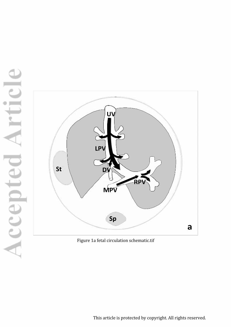
Figure 1a fetal circulation schematic.tif
This article is protected by copyright. All rights reserved.
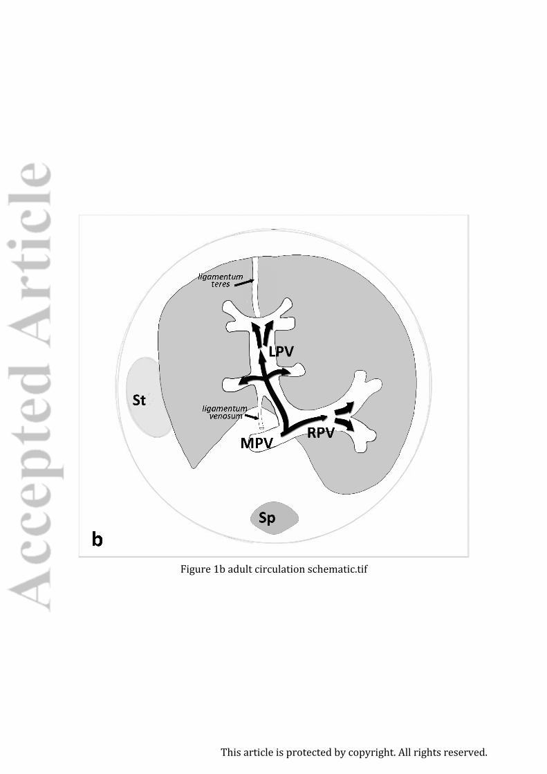
Figure 1b adult circulation schematic.tif
This article is protected by copyright. All rights reserved.
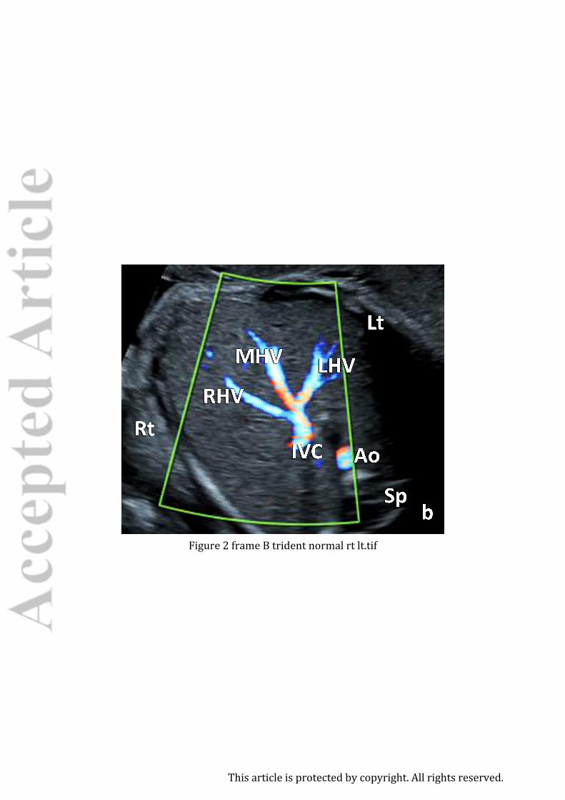
Figure 2 frame B trident normal rt lt.tif
This article is protected by copyright. All rights reserved.
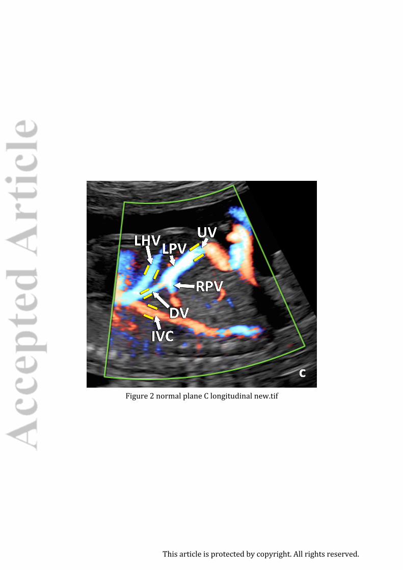
Figure 2 normal plane C longitudinal new.tif
This article is protected by copyright. All rights reserved.
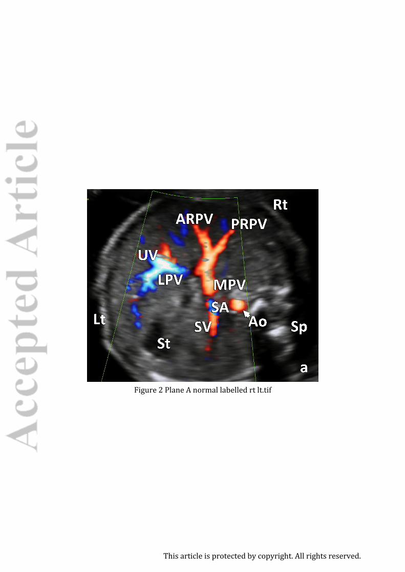
Figure 2 Plane A normal labelled rt lt.tif
This article is protected by copyright. All rights reserved.
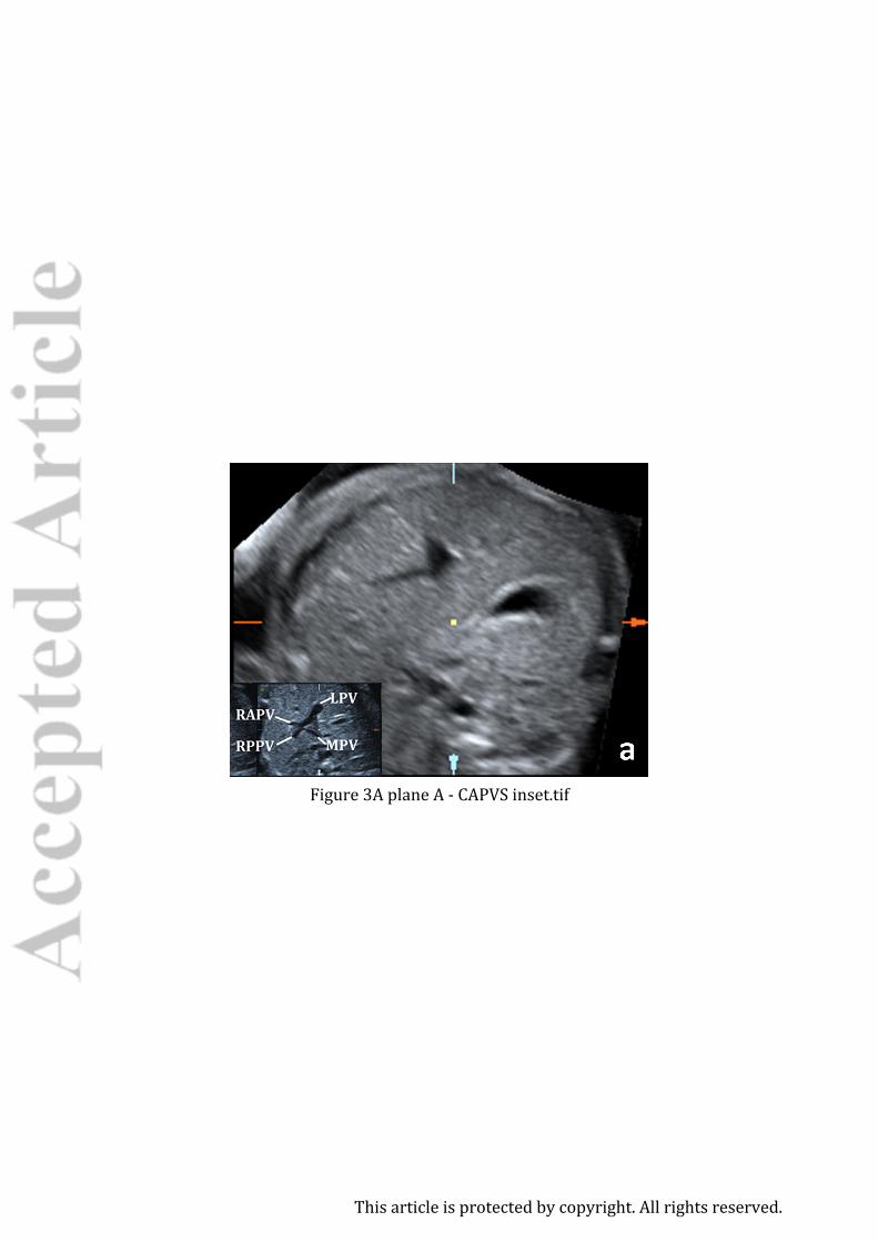
Figure 3A plane A - CAPVS inset.tif
This article is protected by copyright. All rights reserved.
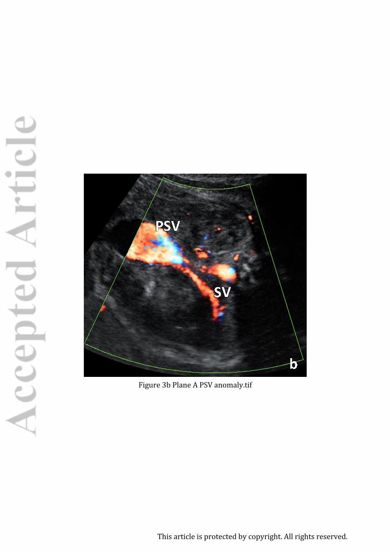
Figure 3b Plane A PSV anomaly.tif
This article is protected by copyright. All rights reserved.
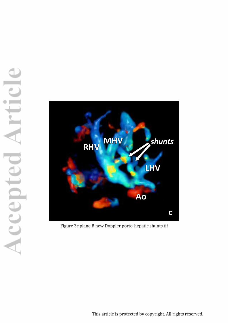
Figure 3c plane B new Doppler porto-hepatic shunts.tif
This article is protected by copyright. All rights reserved.
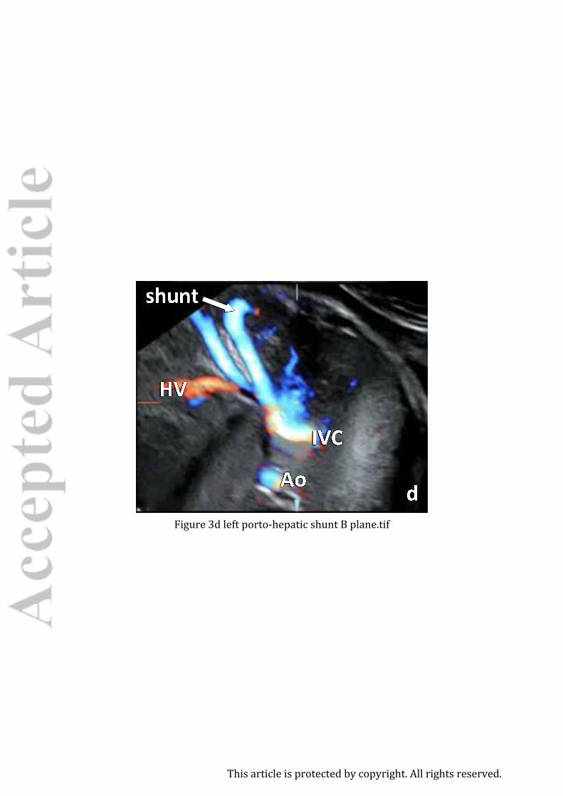
Figure 3d left porto-hepatic shunt B plane.tif
This article is protected by copyright. All rights reserved.
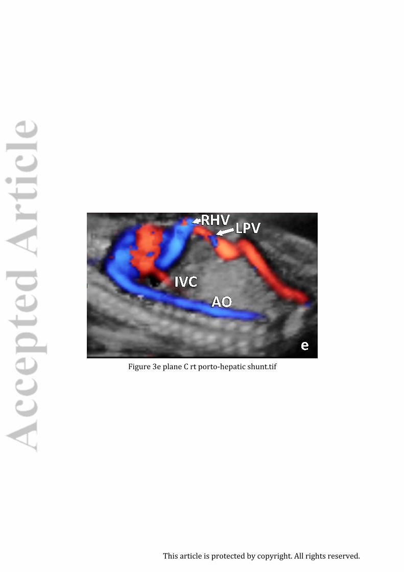
Figure 3e plane C rt porto-hepatic shunt.tif
This article is protected by copyright. All rights reserved.
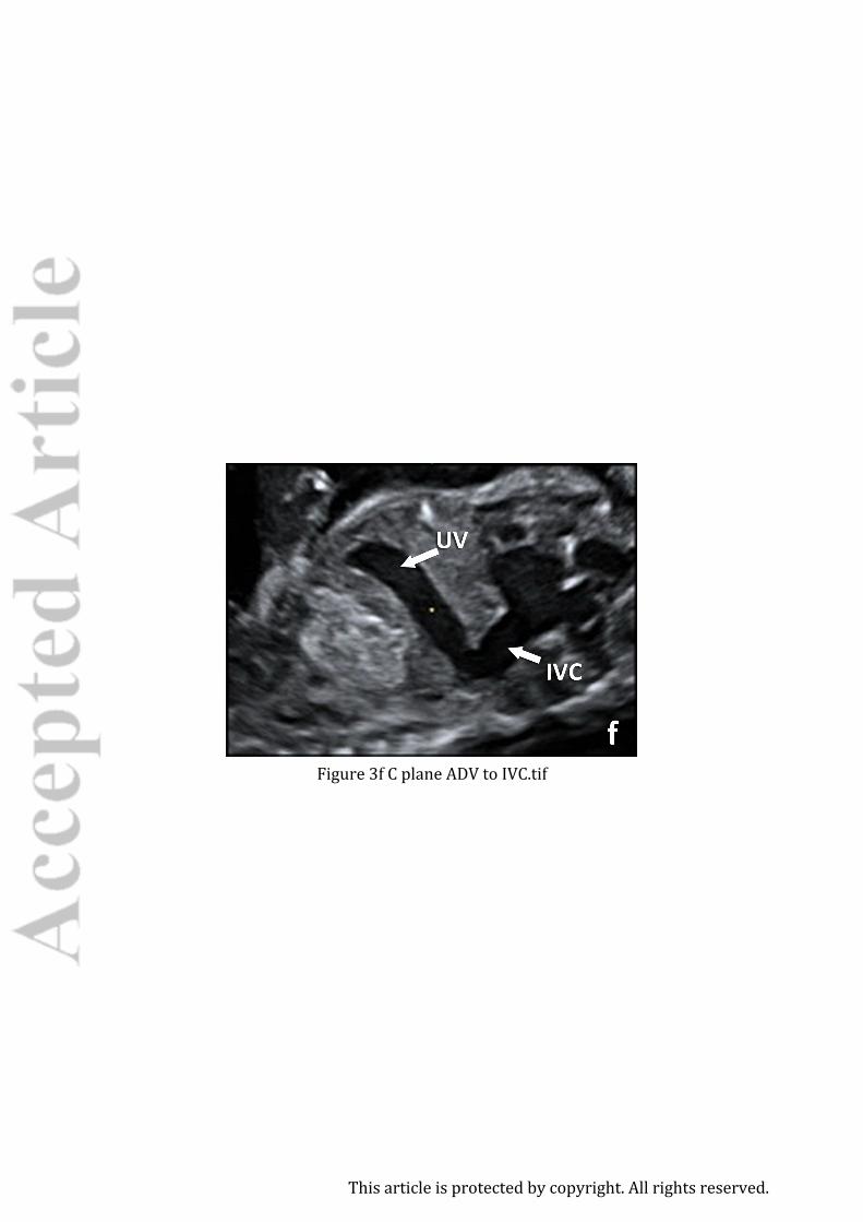
Figure 3f C plane ADV to IVC.tif
This article is protected by copyright. All rights reserved.
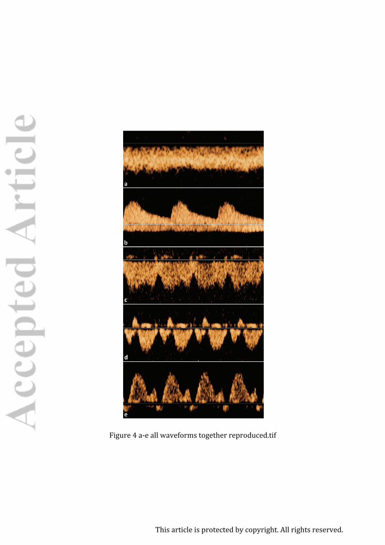
Figure 4 a-e all waveforms together reproduced.tif
This article is protected by copyright. All rights reserved.
Related Documents