IEEE TRANSACTIONS ON MEDICAL IMAGING, VOL. XX, NO. XX, XXXX 2020 1 SimCVD: Simple Contrastive Voxel-Wise Representation Distillation for Semi-Supervised Medical Image Segmentation Chenyu You, Yuan Zhou, Ruihan Zhao, Lawrence Staib, James S. Duncan, Life Fellow, IEEE Abstract — Automated segmentation in medical image analysis is a challenging task that requires a large amount of manually labeled data. However, most existing learning- based approaches usually suffer from limited manually an- notated medical data, which poses a major practical prob- lem for accurate and robust medical image segmentation. In addition, most existing semi-supervised approaches are usually not robust compared with the supervised counter- parts, and also lack explicit modeling of geometric struc- ture and semantic information, both of which limit the segmentation accuracy. In this work, we present SimCVD, a simple contrastive distillation framework that significantly advances state-of-the-art voxel-wise representation learn- ing. We first describe an unsupervised training strategy, which takes two views of an input volume and predicts their signed distance maps of object boundaries in a contrastive objective, with only two independent dropout as mask. This simple approach works surprisingly well, performing on the same level as previous fully supervised methods with much less labeled data. We hypothesize that dropout can be viewed as a minimal form of data augmentation and makes the network robust to representation collapse. Then, we propose to perform structural distillation by dis- tilling pair-wise similarities. We evaluate SimCVD on two popular datasets: the Left Atrial Segmentation Challenge (LA) and the NIH pancreas CT dataset. The results on the LA dataset demonstrate that, in two types of labeled ratios (i.e., 20% and 10%), SimCVD achieves an average Dice score of 90.85% and 89.03% respectively, a 0.91% and 2.22% improvement compared to previous best results. Our method can be trained in an end-to-end fashion, showing the promise of utilizing SimCVD as a general framework for downstream tasks, such as medical image synthesis, enhancement, and registration. Index Terms— Medical image segmentation, contrastive learning, knowledge distillation, geometric constraints. I. I NTRODUCTION Medical image segmentation is a popular task in both machine learning and medical imaging communities [1]–[5]. C. You is with the Department of Electrical Engineering, Yale Univer- sity, New Haven, CT 06510 USA (e-mail:[email protected]). Y. Zhou is with the Department of Radiology & Biomedical Imaging, Yale University, New Haven, CT 06510 USA. R. Zhao is with the Department of Electrical and Computer Engineer- ing, The University of Texas at Austin, TX 78712 USA. L. H. Staib and J. S. Duncan are with Departments of Radi- ology & Biomedical Imaging, Biomedical Engineering, and Electri- cal Engineering, Yale University, New Haven, CT 06511 USA (e- mail:[email protected]). Compared to traditional segmentation approaches, deep neural network based segmentation methods have achieved much stronger performance in recent years with huge advances in representation learning [6]–[13]. However, previous state-of- the-art approaches are mostly trained with a large amount of labeled data, which pose significant practical challenges in many medical segmentation tasks where there is a scarcity of labeled data due to the heavy burden of annotating images. In recent years, a wide variety of semi-supervised methods [14]–[26] have been designed to tackle these issues, which learn from limited labeled data along with a large amount of unlabeled data, achieving significant improvements in ac- curacy and greatly reducing the labeling cost. The common paradigms include adversarial learning, knowledge distilla- tion, and self-supervised learning. Contrastive learning, a sub- area of self-supervised learning, has recently been noted as a promising direction since it has shown great promise in learning useful representations with limited human supervi- sion [25], [27]–[30]. This is often best understood as pulling together semantically similar (positive) samples and pushing apart non-similar (negative) samples in a shared latent space. The representations uncovered by these contrastive objectives are capable of boosting the performance of any vision system especially in scenarios where the amount of annotated data available for the downstream tasks is extremely low, which is well suited for medical image analysis. Despite advances in semi-supervised learning benchmarks, previous methods still face several major challenges: (1) Suboptimal performance: although prior works have achieved promising segmentation accuracy in the setting of limited annotations, semi-supervised models are usually not robust due to some information loss, compared with fully-supervised counterparts; (2) Geometric information loss: previous seg- mentation networks are poor at characterizing geometry, i.e., leveraging the intrinsic geometric structure of the images, such as the object boundary. As a consequence, it is often hard to accurately recognize object contours; and (3) Generalization ability: considering the limited amount of training data, train- ing deep models is usually deficient due to over-fitting and co-adapting [31], [32]. In this work, we address the question: can we advance state-of-the-art voxel-wise representation learning in a more extreme few-annotation-setting for medical image segmenta- tion? To this end, we present SimCVD, a sim ple c ontrastive arXiv:2108.06227v4 [cs.CV] 21 Mar 2022

Welcome message from author
This document is posted to help you gain knowledge. Please leave a comment to let me know what you think about it! Share it to your friends and learn new things together.
Transcript
IEEE TRANSACTIONS ON MEDICAL IMAGING, VOL. XX, NO. XX, XXXX 2020 1
SimCVD: Simple Contrastive Voxel-WiseRepresentation Distillation for Semi-Supervised
Medical Image SegmentationChenyu You, Yuan Zhou, Ruihan Zhao, Lawrence Staib, James S. Duncan, Life Fellow, IEEE
Abstract— Automated segmentation in medical imageanalysis is a challenging task that requires a large amountof manually labeled data. However, most existing learning-based approaches usually suffer from limited manually an-notated medical data, which poses a major practical prob-lem for accurate and robust medical image segmentation.In addition, most existing semi-supervised approaches areusually not robust compared with the supervised counter-parts, and also lack explicit modeling of geometric struc-ture and semantic information, both of which limit thesegmentation accuracy. In this work, we present SimCVD, asimple contrastive distillation framework that significantlyadvances state-of-the-art voxel-wise representation learn-ing. We first describe an unsupervised training strategy,which takes two views of an input volume and predicts theirsigned distance maps of object boundaries in a contrastiveobjective, with only two independent dropout as mask.This simple approach works surprisingly well, performingon the same level as previous fully supervised methodswith much less labeled data. We hypothesize that dropoutcan be viewed as a minimal form of data augmentationand makes the network robust to representation collapse.Then, we propose to perform structural distillation by dis-tilling pair-wise similarities. We evaluate SimCVD on twopopular datasets: the Left Atrial Segmentation Challenge(LA) and the NIH pancreas CT dataset. The results onthe LA dataset demonstrate that, in two types of labeledratios (i.e., 20% and 10%), SimCVD achieves an averageDice score of 90.85% and 89.03% respectively, a 0.91% and2.22% improvement compared to previous best results. Ourmethod can be trained in an end-to-end fashion, showingthe promise of utilizing SimCVD as a general frameworkfor downstream tasks, such as medical image synthesis,enhancement, and registration.
Index Terms— Medical image segmentation, contrastivelearning, knowledge distillation, geometric constraints.
I. INTRODUCTION
Medical image segmentation is a popular task in bothmachine learning and medical imaging communities [1]–[5].
C. You is with the Department of Electrical Engineering, Yale Univer-sity, New Haven, CT 06510 USA (e-mail:[email protected]).
Y. Zhou is with the Department of Radiology & Biomedical Imaging,Yale University, New Haven, CT 06510 USA.
R. Zhao is with the Department of Electrical and Computer Engineer-ing, The University of Texas at Austin, TX 78712 USA.
L. H. Staib and J. S. Duncan are with Departments of Radi-ology & Biomedical Imaging, Biomedical Engineering, and Electri-cal Engineering, Yale University, New Haven, CT 06511 USA (e-mail:[email protected]).
Compared to traditional segmentation approaches, deep neuralnetwork based segmentation methods have achieved muchstronger performance in recent years with huge advances inrepresentation learning [6]–[13]. However, previous state-of-the-art approaches are mostly trained with a large amount oflabeled data, which pose significant practical challenges inmany medical segmentation tasks where there is a scarcityof labeled data due to the heavy burden of annotating images.
In recent years, a wide variety of semi-supervised methods[14]–[26] have been designed to tackle these issues, whichlearn from limited labeled data along with a large amountof unlabeled data, achieving significant improvements in ac-curacy and greatly reducing the labeling cost. The commonparadigms include adversarial learning, knowledge distilla-tion, and self-supervised learning. Contrastive learning, a sub-area of self-supervised learning, has recently been noted asa promising direction since it has shown great promise inlearning useful representations with limited human supervi-sion [25], [27]–[30]. This is often best understood as pullingtogether semantically similar (positive) samples and pushingapart non-similar (negative) samples in a shared latent space.The representations uncovered by these contrastive objectivesare capable of boosting the performance of any vision systemespecially in scenarios where the amount of annotated dataavailable for the downstream tasks is extremely low, which iswell suited for medical image analysis.
Despite advances in semi-supervised learning benchmarks,previous methods still face several major challenges: (1)Suboptimal performance: although prior works have achievedpromising segmentation accuracy in the setting of limitedannotations, semi-supervised models are usually not robustdue to some information loss, compared with fully-supervisedcounterparts; (2) Geometric information loss: previous seg-mentation networks are poor at characterizing geometry, i.e.,leveraging the intrinsic geometric structure of the images, suchas the object boundary. As a consequence, it is often hard toaccurately recognize object contours; and (3) Generalizationability: considering the limited amount of training data, train-ing deep models is usually deficient due to over-fitting andco-adapting [31], [32].
In this work, we address the question: can we advancestate-of-the-art voxel-wise representation learning in a moreextreme few-annotation-setting for medical image segmenta-tion? To this end, we present SimCVD, a simple contrastive
arX
iv:2
108.
0622
7v4
[cs
.CV
] 2
1 M
ar 2
022
2 IEEE TRANSACTIONS ON MEDICAL IMAGING, VOL. XX, NO. XX, XXXX 2020
.$% .&'(
.)*+
Segmentation
Noise ,!"
Noise ,!#
SDM
SDM
.)%-
Contrastive Learning
.
.
/!,%#
/!,&#
/!! ,'#
/!,%"
/!,&"
/!! ,'"
……
. . Projection Head
Positive Instance
Negative Instance
.&'(012)0"!"
"!#
Student Network
Teacher Network
Input Volume
Probability Map 3!#
Probability Map 3!"Projection Head
MLP
Adaptive Average Pooling
Alpha Dropout
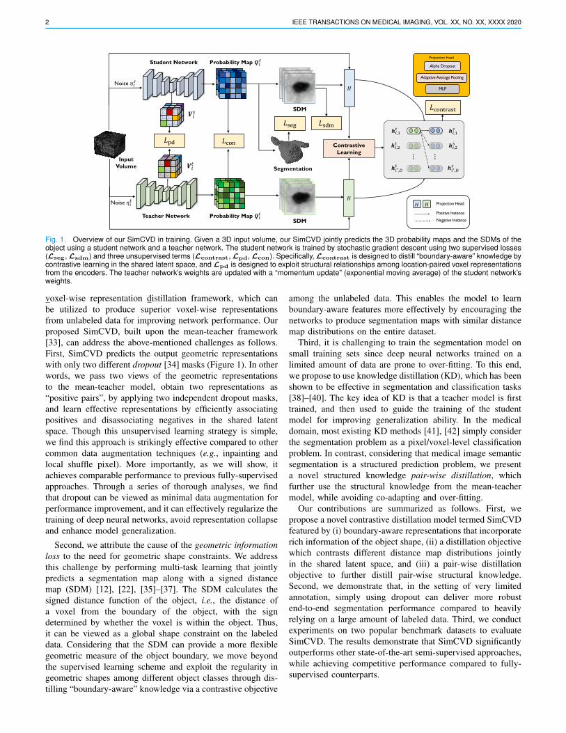
Fig. 1. Overview of our SimCVD in training. Given a 3D input volume, our SimCVD jointly predicts the 3D probability maps and the SDMs of theobject using a student network and a teacher network. The student network is trained by stochastic gradient descent using two supervised losses(Lseg, Lsdm) and three unsupervised terms (Lcontrast, Lpd, Lcon). Specifically, Lcontrast is designed to distill “boundary-aware” knowledge bycontrastive learning in the shared latent space, and Lpd is designed to exploit structural relationships among location-paired voxel representationsfrom the encoders. The teacher network’s weights are updated with a “momentum update” (exponential moving average) of the student network’sweights.
voxel-wise representation distillation framework, which canbe utilized to produce superior voxel-wise representationsfrom unlabeled data for improving network performance. Ourproposed SimCVD, built upon the mean-teacher framework[33], can address the above-mentioned challenges as follows.First, SimCVD predicts the output geometric representationswith only two different dropout [34] masks (Figure 1). In otherwords, we pass two views of the geometric representationsto the mean-teacher model, obtain two representations as“positive pairs”, by applying two independent dropout masks,and learn effective representations by efficiently associatingpositives and disassociating negatives in the shared latentspace. Though this unsupervised learning strategy is simple,we find this approach is strikingly effective compared to othercommon data augmentation techniques (e.g., inpainting andlocal shuffle pixel). More importantly, as we will show, itachieves comparable performance to previous fully-supervisedapproaches. Through a series of thorough analyses, we findthat dropout can be viewed as minimal data augmentation forperformance improvement, and it can effectively regularize thetraining of deep neural networks, avoid representation collapseand enhance model generalization.
Second, we attribute the cause of the geometric informationloss to the need for geometric shape constraints. We addressthis challenge by performing multi-task learning that jointlypredicts a segmentation map along with a signed distancemap (SDM) [12], [22], [35]–[37]. The SDM calculates thesigned distance function of the object, i.e., the distance ofa voxel from the boundary of the object, with the signdetermined by whether the voxel is within the object. Thus,it can be viewed as a global shape constraint on the labeleddata. Considering that the SDM can provide a more flexiblegeometric measure of the object boundary, we move beyondthe supervised learning scheme and exploit the regularity ingeometric shapes among different object classes through dis-tilling “boundary-aware” knowledge via a contrastive objective
among the unlabeled data. This enables the model to learnboundary-aware features more effectively by encouraging thenetworks to produce segmentation maps with similar distancemap distributions on the entire dataset.
Third, it is challenging to train the segmentation model onsmall training sets since deep neural networks trained on alimited amount of data are prone to over-fitting. To this end,we propose to use knowledge distillation (KD), which has beenshown to be effective in segmentation and classification tasks[38]–[40]. The key idea of KD is that a teacher model is firsttrained, and then used to guide the training of the studentmodel for improving generalization ability. In the medicaldomain, most existing KD methods [41], [42] simply considerthe segmentation problem as a pixel/voxel-level classificationproblem. In contrast, considering that medical image semanticsegmentation is a structured prediction problem, we presenta novel structured knowledge pair-wise distillation, whichfurther use the structural knowledge from the mean-teachermodel, while avoiding co-adapting and over-fitting.
Our contributions are summarized as follows. First, wepropose a novel contrastive distillation model termed SimCVDfeatured by (i) boundary-aware representations that incorporaterich information of the object shape, (ii) a distillation objectivewhich contrasts different distance map distributions jointlyin the shared latent space, and (iii) a pair-wise distillationobjective to further distill pair-wise structural knowledge.Second, we demonstrate that, in the setting of very limitedannotation, simply using dropout can deliver more robustend-to-end segmentation performance compared to heavilyrelying on a large amount of labeled data. Third, we conductexperiments on two popular benchmark datasets to evaluateSimCVD. The results demonstrate that SimCVD significantlyoutperforms other state-of-the-art semi-supervised approaches,while achieving competitive performance compared to fully-supervised counterparts.

AUTHOR et al.: PREPARATION OF PAPERS FOR IEEE TRANSACTIONS ON MEDICAL IMAGING 3
II. RELATED WORK
Semi-Supervised Medical Image Segmentation In recentyears, substantial efforts [12], [14]–[17], [19], [21], [43]–[50],[50]–[64] have been devoted to incorporating unlabeled datato improve network performance due to limited annotations.Yu et al. [21] investigated an uncertainty map based on themean-teacher framework [33] to guide the student networkto capture better features. Li et al. [22] proposed to usesigned distance fields for boundary prediction to improvethe performance. Also, Luo et al. [24] proposed a dual-task-consistency (DTC) model for semi-supervised medical imagesegmentation by jointly predicting the pixel-wise segmentationmaps and the global-level level set representations on theunlabeled data. Our method aims at a more practical andchallenging scenario: we train our model in a more extremefew-annotation setting that relies only on a small number ofannotations, while achieving superior segmentation accuracy.Contrastive Learning Self-supervised learning (SSL) [62],[65]–[67] has provided robust benefits to vision tasks bylearning effective visual representations from unlabeled datain an unsupervised setting. It is based on a commonly-heldbelief that superior performance gains can be achieved throughimproved representation learning. Recently, contrastive learn-ing, a type of self-supervised learning, has received a lot ofinterests [25], [27], [30], [65], [68]–[73]. The key idea ofcontrastive learning is to learn powerful representations thatoptimize similarity constraints to discriminate similar pairs(positive) and dissimilar pairs (negative) within a dataset. Theprimary stream of subsequent work focuses on the choice ofdissimilar pairs, which is critical to the quality of learnedrepresentations. The loss function used to quantify the con-trast is chosen from several options, such as InfoNCE [74],Triplet [75], and so on. Recent studies [68], [70] introducedmemory bank or momentum contrast to use more negativesamples for contrast computation. In the context of medicalimaging, Chaitanya et al. [25] extended a contrastive learningframework to extract global and local cues in a stage-wise way,which requires human intervention and extensive training time.In contrast to Chaitanya et al. [25], our unified work focuseson explicit modeling of the intrinsic geometric structure of thesemantic objects in an end-to-end manner, and hence is able torecognize object boundaries more effectively and efficiently.Knowledge Distillation The idea of knowledge distillationis to minimize the KL-divergence between the output dis-tributions of the teacher model and the student model, andthus avoid over-fitting. KD has been applied to a variety oftasks [76]–[81], including image classification [38], [82]–[84]and semantic segmentation [40], [85]. Recent works [82], [83]found that the student model can outperform the teacher modelwhen they share the same network architecture. Zhang et al.[86] proposed to collaboratively train multiple student modelswith co-distillation, which improves performance of thoseindividual models. At the same time, in the context of medicalimaging, among those existing state-of-the-art KD methods,the self-ensemble mean-teacher framework [33] is widelyexplored for image segmentation. Different from the existingmethods that separately exploit class probabilities for each
voxel, we consider knowledge distillation as a structured pre-diction problem by matching the relational similarity amongall pairs of voxels from the encoded feature maps of the mean-teacher model. We have found that our approach significantlyimproves learning better voxel-wise representations.
III. METHOD
In this section, we introduce SimCVD, a semi-supervisedsegmentation network, which is built from scratch by effec-tively leveraging scarce labeled data and ample unlabeled datafor improving end-to-end voxel-wise representation learning(See Figure 1). We first overview our proposed SimCVD andthen describe the task formulation of SimCVD. Finally, wedetail each component of SimCVD in the following subsec-tions.
A. OverviewWe aim to construct an end-to-end voxel-wise contrastive
distillation algorithm to learn boundary-aware representationsin the setting of extremely few annotations for volumetricmedical imaging segmentation. Although the accuracy of su-pervised models is usually higher than that of semi-supervisedmodels, the former requires much more labeled data than thelatter. In many clinical situations, we only have few annotateddata but a large amount of unlabeled data. This situationnecessitates a semi-supervised segmentation algorithm thatcan utilize the unlabeled data to improve the segmentationperformance.
To this end, we propose a novel contrastive distillationframework to advance state-of-the-art voxel-wise representa-tion learning. In particular, our base multi-task segmentationnetwork tackles two tasks simultaneously: classification andregression. Specifically, the segmentation network takes theinput volume batch and jointly predicts the probability maps(classification) and the SDMs of the object (regression). Toobtain better representations, we propose to perform structureddistillation in the latent feature space, followed by contrastingthe boundary-aware features in the prediction space, to learnmore effective boundary-aware representations from 3D unla-beled data by regularizing the embedding space and exploringthe geometric and spatial context of training voxels. At testtime, we remove the mean teacher and two projection heads,and only the student network is deployed for the medicalsegmentation tasks.
B. Task FormulationIn this work, we consider a set of training data (3D images)
including N labeled data and M unlabeled data, where N �M . For simplicity, we denote the small set of labeled data asDl =
{(Xi,Yi,Y
sdmi
)}Ni=1
, and abundant unlabeled data asDu = {Xi}N+M
i=N+1, where Xi ∈ RH×W×D is the volumeinput, Yi ∈ {0, 1}H×W×D is the ground-truth label, andYsdm
i ∈ RH×W×D is the computed ground truth SDMs fromYi, which measures the distance from each voxel to the objectboundary. Every 3D image Xi consists of a set of 2D imageslices Xi = [xi,1, ...,xi,D] where xi,j ∈ RH×W .
4 IEEE TRANSACTIONS ON MEDICAL IMAGING, VOL. XX, NO. XX, XXXX 2020
Our proposed SimCVD framework consists of a mean-teacher network, Ft(X; θt), and a student network, Fs(X; θs).Inspired by recent work [14], [33], the optimization of thesetwo networks can be achieved with an exponential movingaverage (EMA) which uses a weighted combination of theparameters of the student network and the parameters of theteacher network to update the latter. This strategy has beenwidely shown to improve training stability and the model’sfinal performance. Motivated by this idea, our training strategyis divided into two steps. At each iteration, we first optimizethe student network Fs by stochastic gradient descent. Thenwe update the teacher weights θt using an exponential movingaverage of the student weights θs.
The inputs to the two networks are perturbed versions of thesame image. That is, given a volume input Xi, we first adddifferent perturbations (i.e., affine transformation and randomcrop) to generate two different images Xt
i and Xsi . We then
feed Ft and Fs with these two corresponding augmentedimages to obtain two confidence score (probability) maps Qt
i
and Qsi .
Before we present our proposed SimCVD in detail, we firstdescribe our base architecture below.
C. Base Architecture
Our base architecture adopts V-Net [21] as the networkbackbone, which consists of an encoder network et :RH×W×D → RH′×W ′×D′×De and a decoder network dt :RH′×W ′×D′×De → [0, 1]H×W×D × [−1, 1]H×W×D for theteacher network, and similarly es, ds for the student network,i.e., Ft = dt ◦ et and Fs = ds ◦ es. H ′,W ′, D′ are the size ofthe hidden pattern and De is the encoded feature dimension.Inspired by previous work on medical imaging segmentation[12], [22], we incorporate multi-task learning into F to jointlyperform both classification and regression tasks.
Given input Xi, the classification branch is designed togenerate the probability map Qs
i ∈ [0, 1]H×W×D, and theregression branch is designed to predict the SDM Qs,sdm
i ∈[−1, 1]H×W×D. The design of the regression branch is simpleyet effective, only including the hyperbolic tangent function.This design brings two clear benefits: (1) we can eventu-ally encode rich geometric structure information to improvesegmentation accuracy, and (2) we can implicitly enforcecontinuity and smoothness terms for better segmentation maps.Similarly, we have outputs Qt
i and Qt,sdmi from the teacher
network.
Supervised Loss Lsup For training on labeled data, we definethe supervised loss as:
Lsup =1
N
N∑i=1
Lseg(Qsi ,Yi) +
α
N
N∑i=1
Lmse(Qs,sdmi ,Ysdm
i ),
(1)
where Lseg denotes the segmentation loss (Dice and Cross-entropy) [21], and Lmse is the mean squared error loss. α isa hyperparameter. Note that the SDM loss [12] is imposed asgeometric constraints in training.
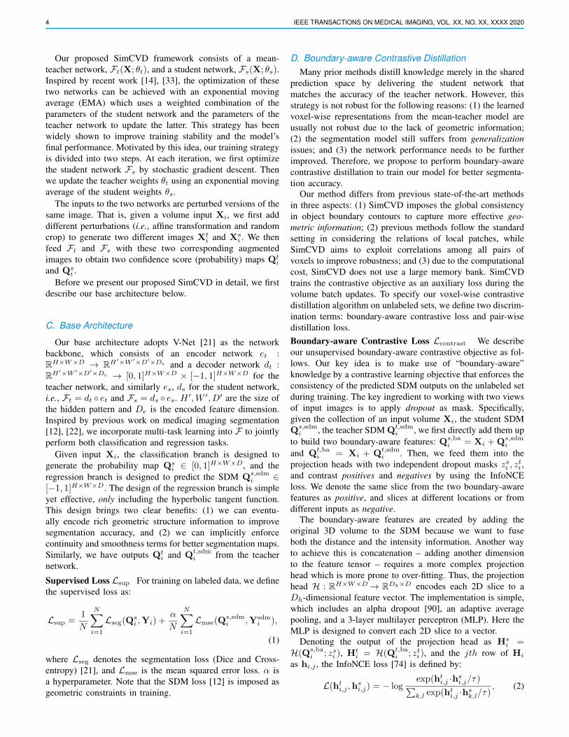
D. Boundary-aware Contrastive DistillationMany prior methods distill knowledge merely in the shared
prediction space by delivering the student network thatmatches the accuracy of the teacher network. However, thisstrategy is not robust for the following reasons: (1) the learnedvoxel-wise representations from the mean-teacher model areusually not robust due to the lack of geometric information;(2) the segmentation model still suffers from generalizationissues; and (3) the network performance needs to be furtherimproved. Therefore, we propose to perform boundary-awarecontrastive distillation to train our model for better segmenta-tion accuracy.
Our method differs from previous state-of-the-art methodsin three aspects: (1) SimCVD imposes the global consistencyin object boundary contours to capture more effective geo-metric information; (2) previous methods follow the standardsetting in considering the relations of local patches, whileSimCVD aims to exploit correlations among all pairs ofvoxels to improve robustness; and (3) due to the computationalcost, SimCVD does not use a large memory bank. SimCVDtrains the contrastive objective as an auxiliary loss during thevolume batch updates. To specify our voxel-wise contrastivedistillation algorithm on unlabeled sets, we define two discrim-ination terms: boundary-aware contrastive loss and pair-wisedistillation loss.Boundary-aware Contrastive Loss Lcontrast We describeour unsupervised boundary-aware contrastive objective as fol-lows. Our key idea is to make use of “boundary-aware”knowledge by a contrastive learning objective that enforces theconsistency of the predicted SDM outputs on the unlabeled setduring training. The key ingredient to working with two viewsof input images is to apply dropout as mask. Specifically,given the collection of an input volume Xi, the student SDMQs,sdm
i , the teacher SDM Qt,sdmi , we first directly add them up
to build two boundary-aware features: Qs,bai = Xi + Qs,sdm
i
and Qt,bai = Xi + Qt,sdm
i . Then, we feed them into theprojection heads with two independent dropout masks zsi , z
ti ,
and contrast positives and negatives by using the InfoNCEloss. We denote the same slice from the two boundary-awarefeatures as positive, and slices at different locations or fromdifferent inputs as negative.
The boundary-aware features are created by adding theoriginal 3D volume to the SDM because we want to fuseboth the distance and the intensity information. Another wayto achieve this is concatenation – adding another dimensionto the feature tensor – requires a more complex projectionhead which is more prone to over-fitting. Thus, the projectionhead H : RH×W×D→ RDh×D encodes each 2D slice to aDh-dimensional feature vector. The implementation is simple,which includes an alpha dropout [90], an adaptive averagepooling, and a 3-layer multilayer perceptron (MLP). Here theMLP is designed to convert each 2D slice to a vector.
Denoting the output of the projection head as Hsi =
H(Qs,bai ; zsi ), Ht
i = H(Qt,bai ; zti), and the jth row of Hi
as hi,j , the InfoNCE loss [74] is defined by:
L(hti,j ,h
si,j) = − log
exp(hti,j ·hs
i,j/τ)∑k,l exp(ht
i,j ·hsk,l/τ)
, (2)
AUTHOR et al.: PREPARATION OF PAPERS FOR IEEE TRANSACTIONS ON MEDICAL IMAGING 5
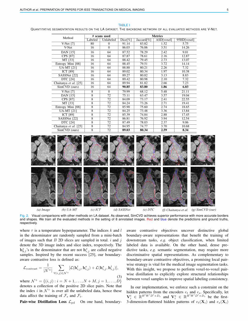
TABLE IQUANTITATIVE SEGMENTATION RESULTS ON THE LA DATASET. THE BACKBONE NETWORK OF ALL EVALUATED METHODS ARE V-NET.
Method # scans used MetricsLabeled Unlabeled Dice[%] Jaccard[%] ASD[voxel] 95HD[voxel]
V-Net [7] 80 0 91.14 83.82 1.52 5.75V-Net 16 0 86.03 76.06 3.51 14.26
DAN [15] 16 64 87.52 78.29 2.42 9.01CPS [87] 16 64 87.87 78.61 2.16 12.87MT [33] 16 64 88.42 79.45 2.73 13.07
Entropy Mini [88] 16 64 88.45 79.51 3.72 14.14UA-MT [21] 16 64 88.88 80.21 2.26 7.32
ICT [89] 16 64 89.02 80.34 1.97 10.38SASSNet [22] 16 64 89.27 80.82 3.13 8.83
DTC [24] 16 64 89.42 80.98 2.10 7.32Chaitanya et al. [25] 16 64 89.94 81.82 2.66 7.23
SimCVD (ours) 16 64 90.85 83.80 1.86 6.03V-Net [7] 8 0 79.99 68.12 5.48 21.11DAN [15] 8 72 75.11 63.47 3.57 19.04CPS [87] 8 72 84.09 73.17 2.41 22.55MT [33] 8 72 84.24 73.26 2.71 19.41
Entropy Mini [88] 8 72 85.90 75.60 2.74 18.65UA-MT [21] 8 72 84.25 73.48 3.36 13.84
ICT [89] 8 72 85.39 74.84 2.88 17.45SASSNet [22] 8 72 86.81 76.92 3.94 12.54
DTC [24] 8 72 87.49 78.03 2.37 9.06Chaitanya et al. [25] 8 72 84.95 74.77 3.70 10.68
SimCVD (ours) 8 72 89.03 80.34 2.59 8.34
(a) Image (b) UA-MT (c) ICT (d) SASSNet (e) DTC (f) Chaitanya et al. (g) SimCVD (our)
Fig. 2. Visual comparisons with other methods on LA dataset. As observed, SimCVD achieves superior performance with more accurate bordersand shapes. We train all the evaluated methods in the setting of 8 annotated images. Red and blue denote the predictions and ground truths,respectively.
where τ is a temperature hyperparameter. The indices k and lin the denominator are randomly sampled from a mini-batchof images such that B 2D slices are sampled in total. i and jdenote the 3D image index and slice index, respectively. Thehsk,l’s in the denominator that are not hs
i,j are called negativesamples. Inspired by the recent success [25], our boundary-aware contrastive loss is defined as:
Lcontrast =1
|N+|∑
∀(i,j)∈N+
[L(hti,j ,h
si,j) + L(hs
i,j ,hti,j)],
(3)where N+ = {(i, j) : i = N + 1, . . . , N +M, j = 1, . . . , D}denotes a collection of the positive 2D slice pairs. Note thatthe index i in N+ is over all the unlabeled data, hence thesedata affect the training of Fs and Ft.Pair-wise Distillation Loss Lpd On one hand, boundary-
aware contrastive objectives uncover distinctive globalboundary-aware representations that benefit the training ofdownstream tasks, e.g. object classification, when limitedlabeled data is available. On the other hand, dense pre-dictive tasks, e.g. semantic segmentation, may require morediscriminative spatial representations. As complementary toboundary-aware contrastive objectives, a promising local pair-wise strategy is vital for the medical image segmentation tasks.With this insight, we propose to perform voxel-to-voxel pair-wise distillation to explicitly explore structural relationshipsbetween voxel samples to improve spatial labeling consistency.
In our implementation, we enforce such a constraint on thehidden patterns from the encoders et and es. Specifically, letVt
i ∈ RH′W ′D′×De and Vsi ∈ RH′W ′D′×De be the first-
3-dimension-flattened hidden patterns of et(Xi) and es(Xi)
6 IEEE TRANSACTIONS ON MEDICAL IMAGING, VOL. XX, NO. XX, XXXX 2020
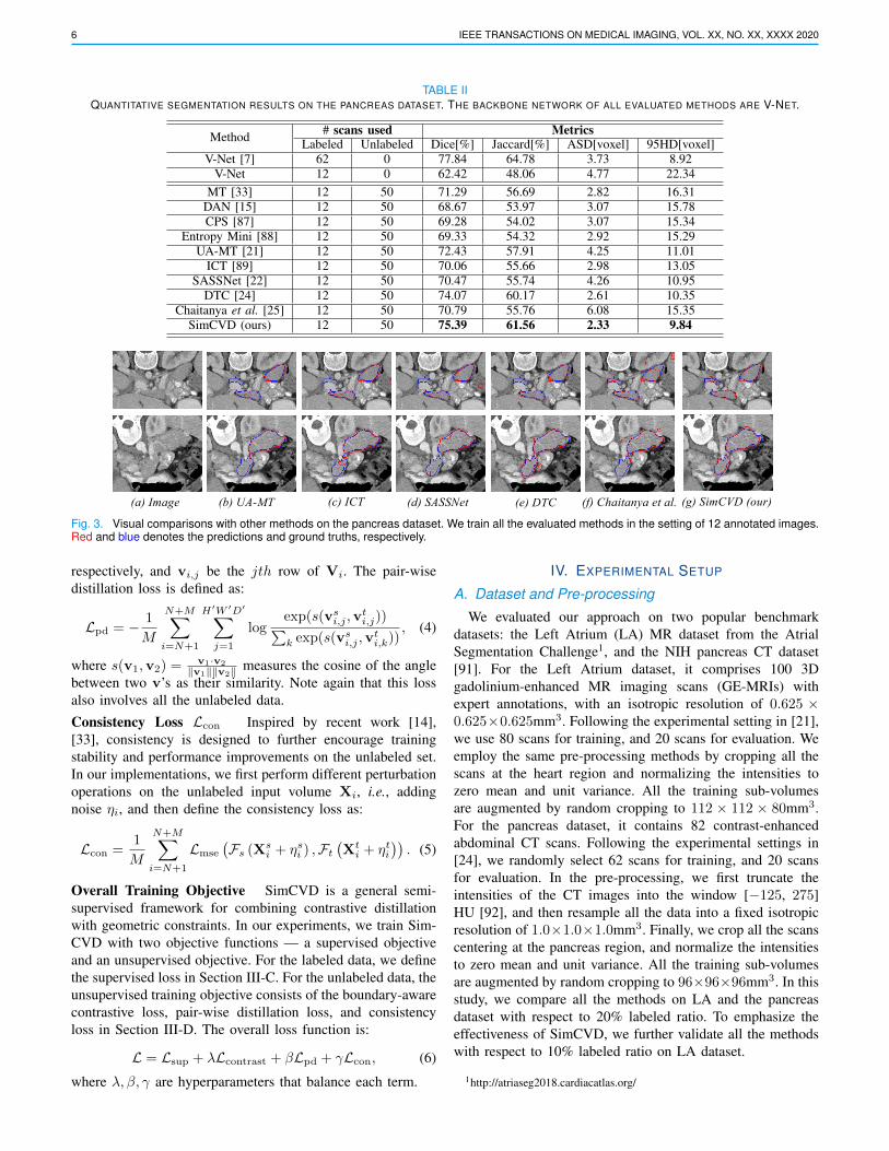
TABLE IIQUANTITATIVE SEGMENTATION RESULTS ON THE PANCREAS DATASET. THE BACKBONE NETWORK OF ALL EVALUATED METHODS ARE V-NET.
Method # scans used MetricsLabeled Unlabeled Dice[%] Jaccard[%] ASD[voxel] 95HD[voxel]
V-Net [7] 62 0 77.84 64.78 3.73 8.92V-Net 12 0 62.42 48.06 4.77 22.34
MT [33] 12 50 71.29 56.69 2.82 16.31DAN [15] 12 50 68.67 53.97 3.07 15.78CPS [87] 12 50 69.28 54.02 3.07 15.34
Entropy Mini [88] 12 50 69.33 54.32 2.92 15.29UA-MT [21] 12 50 72.43 57.91 4.25 11.01
ICT [89] 12 50 70.06 55.66 2.98 13.05SASSNet [22] 12 50 70.47 55.74 4.26 10.95
DTC [24] 12 50 74.07 60.17 2.61 10.35Chaitanya et al. [25] 12 50 70.79 55.76 6.08 15.35
SimCVD (ours) 12 50 75.39 61.56 2.33 9.84
(a) Image (b) UA-MT (c) ICT (d) SASSNet (e) DTC (f) Chaitanya et al. (g) SimCVD (our)
Fig. 3. Visual comparisons with other methods on the pancreas dataset. We train all the evaluated methods in the setting of 12 annotated images.Red and blue denotes the predictions and ground truths, respectively.
respectively, and vi,j be the jth row of Vi. The pair-wisedistillation loss is defined as:
Lpd = − 1
M
N+M∑i=N+1
H′W ′D′∑j=1
logexp(s(vs
i,j ,vti,j))∑
k exp(s(vsi,j ,v
ti,k))
, (4)
where s(v1,v2) = v1·v2
‖v1‖‖v2‖ measures the cosine of the anglebetween two v’s as their similarity. Note again that this lossalso involves all the unlabeled data.
Consistency Loss Lcon Inspired by recent work [14],[33], consistency is designed to further encourage trainingstability and performance improvements on the unlabeled set.In our implementations, we first perform different perturbationoperations on the unlabeled input volume Xi, i.e., addingnoise ηi, and then define the consistency loss as:
Lcon =1
M
N+M∑i=N+1
Lmse
(Fs (Xs
i + ηsi ) ,Ft
(Xt
i + ηti)). (5)
Overall Training Objective SimCVD is a general semi-supervised framework for combining contrastive distillationwith geometric constraints. In our experiments, we train Sim-CVD with two objective functions — a supervised objectiveand an unsupervised objective. For the labeled data, we definethe supervised loss in Section III-C. For the unlabeled data, theunsupervised training objective consists of the boundary-awarecontrastive loss, pair-wise distillation loss, and consistencyloss in Section III-D. The overall loss function is:
L = Lsup + λLcontrast + βLpd + γLcon, (6)
where λ, β, γ are hyperparameters that balance each term.
IV. EXPERIMENTAL SETUP
A. Dataset and Pre-processing
We evaluated our approach on two popular benchmarkdatasets: the Left Atrium (LA) MR dataset from the AtrialSegmentation Challenge1, and the NIH pancreas CT dataset[91]. For the Left Atrium dataset, it comprises 100 3Dgadolinium-enhanced MR imaging scans (GE-MRIs) withexpert annotations, with an isotropic resolution of 0.625 ×0.625×0.625mm3. Following the experimental setting in [21],we use 80 scans for training, and 20 scans for evaluation. Weemploy the same pre-processing methods by cropping all thescans at the heart region and normalizing the intensities tozero mean and unit variance. All the training sub-volumesare augmented by random cropping to 112 × 112 × 80mm3.For the pancreas dataset, it contains 82 contrast-enhancedabdominal CT scans. Following the experimental settings in[24], we randomly select 62 scans for training, and 20 scansfor evaluation. In the pre-processing, we first truncate theintensities of the CT images into the window [−125, 275]HU [92], and then resample all the data into a fixed isotropicresolution of 1.0×1.0×1.0mm3. Finally, we crop all the scanscentering at the pancreas region, and normalize the intensitiesto zero mean and unit variance. All the training sub-volumesare augmented by random cropping to 96×96×96mm3. In thisstudy, we compare all the methods on LA and the pancreasdataset with respect to 20% labeled ratio. To emphasize theeffectiveness of SimCVD, we further validate all the methodswith respect to 10% labeled ratio on LA dataset.
1http://atriaseg2018.cardiacatlas.org/

AUTHOR et al.: PREPARATION OF PAPERS FOR IEEE TRANSACTIONS ON MEDICAL IMAGING 7
B. Implementation DetailsIn this study, all evaluated methods are implemented in
PyTorch, and trained for 6000 iterations on an NVIDIA1080Ti GPU with a batch size of 4. For data augmentation,we use standard data augmentation techniques (i.e., randomrotation, flipping, and cropping). We set the hyper-parametersα,λ,β, γ, τ as 0.1, 0.5, 0.1, 0.1, 0.5, respectively. For the pro-jection head, we set p = 0.1 in the AlphaDropout layer, andoutput size 128 × 128 for AdaptiveAvgPool2d. We use SGDoptimizer with a momentum of 0.9 and a weight decay of0.0005 to optimize network parameters. The initial learningrate is set as 0.01 and divided by 10 every 3000 iterations.For EMA updates, we follow the experimental setting in[21], where the EMA decay rate α is set to 0.999. We usethe time-dependent Gaussian warming-up function Ψcon(t)=
exp(−5 (1− t/tmax)
2)
to ramp up parameters, where t andtmax denote the current and the maximum training step,respectively. For fairness, we do not adopt any post-processingstep.
In the testing stage, we adopt four metrics to evaluate thesegmentation performance: Dice coefficient (Dice), JaccardIndex (Jaccard), 95% Hausdorff Distance (95HD), and Aver-age Symmetric Surface Distance (ASD). Following [21], [24],[93], we adopt a sliding window strategy, which uses a stridewith 18×18×4 for the LA and 16×16×16 for the pancreas.
V. RESULTS
A. Experiments: Left AtriumWe compare SimCVD with published results from previous
state-of-the-art semi-supervised segmentation methods, includ-ing V-Net [7], MT [33], DAN [15], CPS [87], Entropy Mini[88], UA-MT [21], ICT [89], SASSNet [22], DCT [24], andChaitanya et al. [25] on the LA dataset in two labeled ratiosettings (i.e., 10% and 20%).
The quantitative results on the LA dataset are shown inTable I. SimCVD substantially improved the segmentationaccuracy in both 10% and 20% labeled cases. The results arevisualized in Fig 2. Specifically, in the setting of 20% labeledratio, our proposed SimCVD raises the previous best averageresults from 89.94% to 90.85% and from 81.82% to 83.80% interms of Dice and Jaccard, even achieving comparable perfor-mance to the fully supervised baseline. Using the 10% labeledratio, SimCVD further advances the state-of-the-art resultsfrom 87.49% to 89.03% in Dice. The gains in Jaccard, ASD,and 95HD are also substantial, achieving 80.34%, 2.59, and8.34, respectively. This suggests that: (1) taking voxel sampleswith a contrastive objective yields better voxel embeddings; (2)incorporating pair-wise spatial labeling consistency can boostthe performance by accessing more structural knowledge; and(3) utilizing a geometric constraint (i.e., SDM) is capableof helping identify more accurate boundaries. Leveraging allthese aspects, we can observe consistent performance gains.
B. Experiments: PancreasTo further evaluate the effectiveness of SimCVD, we com-
pare our model on the pancreas CT dataset. Experimental
TABLE IIIABLATION ON (a) MODEL COMPONENT: SIMCVD W/O SDM; SIMCVDW/O ADAPTIVE MAX POOLING; (b) LOSS FORMULATION: SIMCVD W/OLcontrast ; SIMCVD W/O Lpd ; SIMCVD W/O Lsdm , COMPARED TO
THE BASELINE AND OUR PROPOSED SIMCVD.Method Metrics p-value (vs. SimCVD, [%])Dice[%] Jaccard[%] ASD[voxel] 95HD[voxel]Baseline (UA-MT) 84.24 73.26 2.71 19.41 0.019
(a) SimCVD w/o SDM 88.24 79.07 4.19 11.43 2.47SimCVD w/ Adaptive Max Pooling 88.32 79.26 3.79 14.69 0.33
(b)SimCVD w/o Lcontrast + Lpd 84.97 74.49 6.13 19.98 0.018SimCVD w/o Lcontrast 85.13 74.57 5.97 16.61 2.9e-6SimCVD w/o Lpd 88.11 78.89 2.89 12.58 2.02SimCVD w/o Lsdm 88.85 80.03 2.71 9.02 5.14SimCVD 89.03 80.34 2.59 8.34 -
results on the pancreas CT dataset are summarized in Table II.We observe that our model consistently outperforms all pre-vious methods, achieving up to 6.72% absolute improvementsin Dice. As shown in Figs. 2 and 3, our method is capable ofpredicting high-quality object segmentation, considering thefact that the improvement in such a setting is difficult. Thisdemonstrates: (1) the necessity of comprehensively consider-ing both boundary-aware contrast and pair-wise distillation;and (2) the efficacy of global shape information. Compared tothe previous strong models, our approach achieves large im-provements on all the datasets, demonstrating its effectiveness.
VI. ABLATION STUDY
In this section, we conduct extensive studies to better under-stand SimCVD. We justify the inner working of SimCVD fromtwo perspectives: (1) boundary-aware contrastive distillation(Section VI-A), and (2) the projection head (Section VI-B).In these studies, we evaluate our proposed method on the LAdataset with 10% labeled ratio (8 labeled and 72 unlabeled).
A. Analysis on Boundary-aware Contrastive Distillation
Ablation on Model Component In the model formu-lation, our motivation is to advance state-of-the-art voxel-wise representations by capturing the geometric and semanticinformation in 3D space. Rather than transferring knowledgeacross confidence score maps directly, our SimCVD distills“boundary-aware” knowledge from the teacher network. Tovalidate the idea of boundary-aware contrastive distillation, wecompare SimCVD to an ablative baseline (i.e., SimCVD w/oSDM). Table III (a) compares each component of SimCVDin the 10% labeled setting. First, we observe that removingthe SDMs in training hurts the segmentation performanceby −0.79%, −1.27%, −1.6, and −3.09 absolute differencesin terms of Dice, Jaccard, ASD, and 95HD. This confirmsour intuition that the learned boundary-aware representationsprovide a good prior for improving segmentation accuracy.We also find that using adaptive max pooling strategy (i.e.,SimCVD w/ adaptive max pooling) largely degrades the seg-mentation performance. Our segmentation results demonstratethat SimCVD is an effective approach, outperforming the bestprevious method with +0.71%, +1.08%, +1.20, and +6.35absolute differences in terms of Dice, Jaccard, ASD, and95HD. We hypothesize that it is because “w/ adaptive maxpooling” leads to information loss during training.Ablation on Loss Formulation In the loss formulation,our main idea is to pull closer similar (positive) pairs upon
8 IEEE TRANSACTIONS ON MEDICAL IMAGING, VOL. XX, NO. XX, XXXX 2020
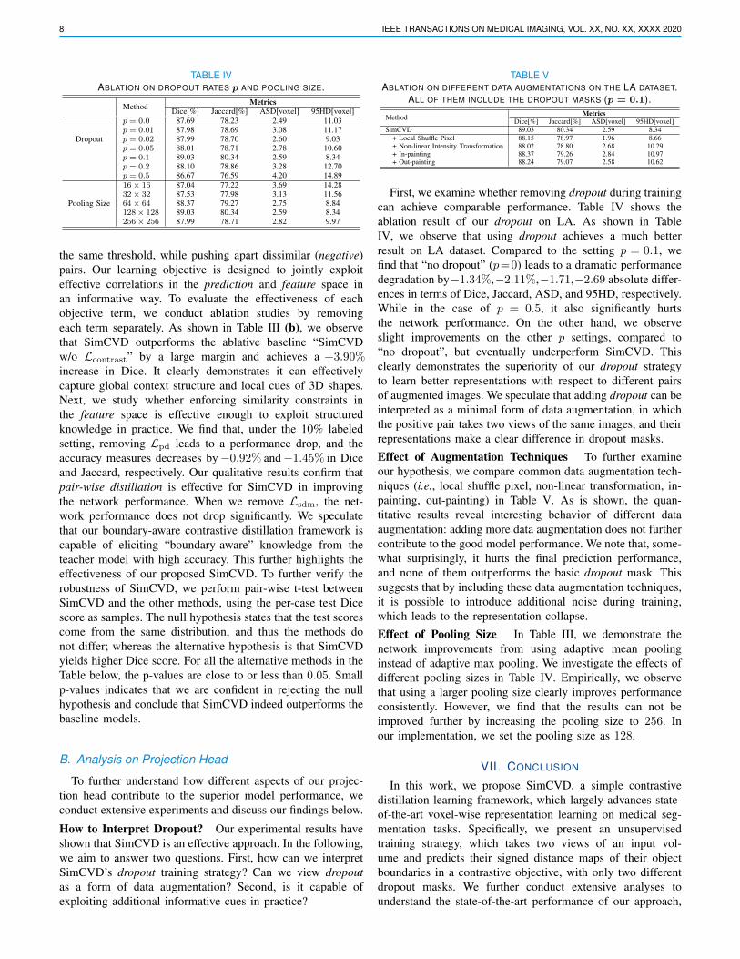
TABLE IVABLATION ON DROPOUT RATES p AND POOLING SIZE.
Method MetricsDice[%] Jaccard[%] ASD[voxel] 95HD[voxel]
Dropout
p = 0.0 87.69 78.23 2.49 11.03p = 0.01 87.98 78.69 3.08 11.17p = 0.02 87.99 78.70 2.60 9.03p = 0.05 88.01 78.71 2.78 10.60p = 0.1 89.03 80.34 2.59 8.34p = 0.2 88.10 78.86 3.28 12.70p = 0.5 86.67 76.59 4.20 14.89
Pooling Size
16× 16 87.04 77.22 3.69 14.2832× 32 87.53 77.98 3.13 11.5664× 64 88.37 79.27 2.75 8.84128× 128 89.03 80.34 2.59 8.34256× 256 87.99 78.71 2.82 9.97
the same threshold, while pushing apart dissimilar (negative)pairs. Our learning objective is designed to jointly exploiteffective correlations in the prediction and feature space inan informative way. To evaluate the effectiveness of eachobjective term, we conduct ablation studies by removingeach term separately. As shown in Table III (b), we observethat SimCVD outperforms the ablative baseline “SimCVDw/o Lcontrast” by a large margin and achieves a +3.90%increase in Dice. It clearly demonstrates it can effectivelycapture global context structure and local cues of 3D shapes.Next, we study whether enforcing similarity constraints inthe feature space is effective enough to exploit structuredknowledge in practice. We find that, under the 10% labeledsetting, removing Lpd leads to a performance drop, and theaccuracy measures decreases by−0.92% and−1.45% in Diceand Jaccard, respectively. Our qualitative results confirm thatpair-wise distillation is effective for SimCVD in improvingthe network performance. When we remove Lsdm, the net-work performance does not drop significantly. We speculatethat our boundary-aware contrastive distillation framework iscapable of eliciting “boundary-aware” knowledge from theteacher model with high accuracy. This further highlights theeffectiveness of our proposed SimCVD. To further verify therobustness of SimCVD, we perform pair-wise t-test betweenSimCVD and the other methods, using the per-case test Dicescore as samples. The null hypothesis states that the test scorescome from the same distribution, and thus the methods donot differ; whereas the alternative hypothesis is that SimCVDyields higher Dice score. For all the alternative methods in theTable below, the p-values are close to or less than 0.05. Smallp-values indicates that we are confident in rejecting the nullhypothesis and conclude that SimCVD indeed outperforms thebaseline models.
B. Analysis on Projection Head
To further understand how different aspects of our projec-tion head contribute to the superior model performance, weconduct extensive experiments and discuss our findings below.
How to Interpret Dropout? Our experimental results haveshown that SimCVD is an effective approach. In the following,we aim to answer two questions. First, how can we interpretSimCVD’s dropout training strategy? Can we view dropoutas a form of data augmentation? Second, is it capable ofexploiting additional informative cues in practice?
TABLE VABLATION ON DIFFERENT DATA AUGMENTATIONS ON THE LA DATASET.
ALL OF THEM INCLUDE THE DROPOUT MASKS (p = 0.1).
Method MetricsDice[%] Jaccard[%] ASD[voxel] 95HD[voxel]
SimCVD 89.03 80.34 2.59 8.34+ Local Shuffle Pixel 88.15 78.97 1.96 8.66+ Non-linear Intensity Transformation 88.02 78.80 2.68 10.29+ In-painting 88.37 79.26 2.84 10.97+ Out-painting 88.24 79.07 2.58 10.62
First, we examine whether removing dropout during trainingcan achieve comparable performance. Table IV shows theablation result of our dropout on LA. As shown in TableIV, we observe that using dropout achieves a much betterresult on LA dataset. Compared to the setting p = 0.1, wefind that “no dropout” (p=0) leads to a dramatic performancedegradation by−1.34%,−2.11%,−1.71,−2.69 absolute differ-ences in terms of Dice, Jaccard, ASD, and 95HD, respectively.While in the case of p = 0.5, it also significantly hurtsthe network performance. On the other hand, we observeslight improvements on the other p settings, compared to“no dropout”, but eventually underperform SimCVD. Thisclearly demonstrates the superiority of our dropout strategyto learn better representations with respect to different pairsof augmented images. We speculate that adding dropout can beinterpreted as a minimal form of data augmentation, in whichthe positive pair takes two views of the same images, and theirrepresentations make a clear difference in dropout masks.Effect of Augmentation Techniques To further examineour hypothesis, we compare common data augmentation tech-niques (i.e., local shuffle pixel, non-linear transformation, in-painting, out-painting) in Table V. As is shown, the quan-titative results reveal interesting behavior of different dataaugmentation: adding more data augmentation does not furthercontribute to the good model performance. We note that, some-what surprisingly, it hurts the final prediction performance,and none of them outperforms the basic dropout mask. Thissuggests that by including these data augmentation techniques,it is possible to introduce additional noise during training,which leads to the representation collapse.Effect of Pooling Size In Table III, we demonstrate thenetwork improvements from using adaptive mean poolinginstead of adaptive max pooling. We investigate the effects ofdifferent pooling sizes in Table IV. Empirically, we observethat using a larger pooling size clearly improves performanceconsistently. However, we find that the results can not beimproved further by increasing the pooling size to 256. Inour implementation, we set the pooling size as 128.
VII. CONCLUSION
In this work, we propose SimCVD, a simple contrastivedistillation learning framework, which largely advances state-of-the-art voxel-wise representation learning on medical seg-mentation tasks. Specifically, we present an unsupervisedtraining strategy, which takes two views of an input vol-ume and predicts their signed distance maps of their objectboundaries in a contrastive objective, with only two differentdropout masks. We further conduct extensive analyses tounderstand the state-of-the-art performance of our approach,
AUTHOR et al.: PREPARATION OF PAPERS FOR IEEE TRANSACTIONS ON MEDICAL IMAGING 9
and demonstrate the importance of learning distinct boundary-aware representations and using dropout as the minimal dataaugmentation technique. We also propose to perform structuraldistillation by distilling pair-wise similarities, which achievesgood performance improvements. Our experimental resultsshow that SimCVD obtained new state-of-the-art results ontwo benchmarks in an extreme few-annotation setting.
We believe that our unsupervised training framework pro-vides a new perspective on data augmentation along withunlabeled 3D medical data. We also plan to extend our methodto solve multi-class medical image segmentation tasks.
REFERENCES
[1] L. H. Staib and J. S. Duncan, “Model-based deformable surface findingfor medical images,” IEEE Transactions on Medical Imaging, vol. 15,no. 5, pp. 720–731, 1996.
[2] J. Yang, L. H. Staib, and J. S. Duncan, “Neighbor-constrained segmen-tation with level set based 3-d deformable models,” IEEE Transactionson Medical Imaging, vol. 23, no. 8, pp. 940–948, 2004.
[3] J. Yang and J. S. Duncan, “3D image segmentation of deformable objectswith joint shape-intensity prior models using level sets,” Medical ImageAnalysis, vol. 8, no. 3, pp. 285–294, 2004.
[4] A. Chakraborty, L. H. Staib, and J. S. Duncan, “Deformable boundaryfinding in medical images by integrating gradient and region informa-tion,” IEEE Transactions on Medical Imaging, vol. 15, no. 6, pp. 859–870, 1996.
[5] L. H. Staib and J. S. Duncan, “Boundary finding with parametrically de-formable models,” IEEE Transactions on Pattern Analysis and MachineIntelligence, vol. 14, no. 11, pp. 1061–1075, 1992.
[6] O. Ronneberger, P. Fischer, and T. Brox, “U-net: Convolutional networksfor biomedical image segmentation,” in MICCAI, pp. 234–241, Springer,2015.
[7] F. Milletari, N. Navab, and S.-A. Ahmadi, “V-net: Fully convolutionalneural networks for volumetric medical image segmentation,” in 3DV,pp. 565–571, IEEE, 2016.
[8] W. Bai, O. Oktay, M. Sinclair, H. Suzuki, M. Rajchl, G. Tarroni,B. Glocker, A. King, P. M. Matthews, and D. Rueckert, “Semi-supervised learning for network-based cardiac MR image segmentation,”in MICCAI, pp. 253–260, Springer, 2017.
[9] P.-A. Ganaye, M. Sdika, and H. Benoit-Cattin, “Semi-supervised learn-ing for segmentation under semantic constraint,” in MICCAI, pp. 595–602, Springer, 2018.
[10] C. You, J. Yang, J. Chapiro, and J. S. Duncan, “Unsupervised wassersteindistance guided domain adaptation for 3D multi-domain liver segmen-tation,” in Interpretable and Annotation-Efficient Learning for MedicalImage Computing, pp. 155–163, Springer, 2020.
[11] Y. Wang, X. Wei, F. Liu, J. Chen, Y. Zhou, W. Shen, E. K. Fishman, andA. L. Yuille, “Deep distance transform for tubular structure segmentationin ct scans,” in CVPR, pp. 3833–3842, 2020.
[12] Y. Xue, H. Tang, Z. Qiao, G. Gong, Y. Yin, Z. Qian, C. Huang, W. Fan,and X. Huang, “Shape-aware organ segmentation by predicting signeddistance maps,” in AAAI, 2020.
[13] X. Li, L. Yu, H. Chen, C.-W. Fu, L. Xing, and P.-A. Heng,“Transformation-consistent self-ensembling model for semisupervisedmedical image segmentation,” IEEE Transactions on Neural Networksand Learning Systems, 2020.
[14] S. Laine and T. Aila, “Temporal ensembling for semi-supervised learn-ing,” arXiv preprint arXiv:1610.02242, 2016.
[15] Y. Zhang, L. Yang, J. Chen, M. Fredericksen, D. P. Hughes, and D. Z.Chen, “Deep adversarial networks for biomedical image segmentationutilizing unannotated images,” in MICCAI, pp. 408–416, Springer, 2017.
[16] X. Li, L. Yu, H. Chen, C.-W. Fu, and P.-A. Heng, “Semi-supervisedskin lesion segmentation via transformation consistent self-ensemblingmodel,” arXiv preprint arXiv:1808.03887, 2018.
[17] D. Nie, Y. Gao, L. Wang, and D. Shen, “Asdnet: Attention based semi-supervised deep networks for medical image segmentation,” in MICCAI,pp. 370–378, Springer, 2018.
[18] S. Qiao, W. Shen, Z. Zhang, B. Wang, and A. Yuille, “Deep co-trainingfor semi-supervised image recognition,” in ECCV, pp. 135–152, 2018.
[19] G. Bortsova, F. Dubost, L. Hogeweg, I. Katramados, and M. de Bruijne,“Semi-supervised medical image segmentation via learning consistencyunder transformations,” in MICCAI, pp. 810–818, Springer, 2019.
[20] K. Li, W. Zhou, H. Li, and M. A. Anastasio, “Assessing the impact ofdeep neural network-based image denoising on binary signal detectiontasks,” IEEE transactions on medical imaging, vol. 40, no. 9, pp. 2295–2305, 2021.
[21] L. Yu, S. Wang, X. Li, C.-W. Fu, and P.-A. Heng, “Uncertainty-awareself-ensembling model for semi-supervised 3D left atrium segmenta-tion,” in MICCAI, pp. 605–613, Springer, 2019.
[22] S. Li, C. Zhang, and X. He, “Shape-aware semi-supervised 3D semanticsegmentation for medical images,” in MICCAI, pp. 552–561, Springer,2020.
[23] J. Zhu, Y. Li, Y. Hu, K. Ma, S. K. Zhou, and Y. Zheng, “Rubik’scube+: A self-supervised feature learning framework for 3D medicalimage analysis,” Medical Image Analysis, p. 101746, 2020.
[24] X. Luo, J. Chen, T. Song, and G. Wang, “Semi-supervised medical imagesegmentation through dual-task consistency,” in AAAI, 2020.
[25] K. Chaitanya, E. Erdil, N. Karani, and E. Konukoglu, “Contrastivelearning of global and local features for medical image segmentationwith limited annotations,” in NeurIPS, 2020.
[26] C. You, R. Zhao, L. Staib, and J. S. Duncan, “Momentum contrastivevoxel-wise representation learning for semi-supervised volumetric med-ical image segmentation,” arXiv preprint arXiv:2105.07059, 2021.
[27] T. Chen, S. Kornblith, M. Norouzi, and G. Hinton, “A simple frameworkfor contrastive learning of visual representations,” in ICML, 2020.
[28] R. D. Hjelm, A. Fedorov, S. Lavoie-Marchildon, K. Grewal, P. Bachman,A. Trischler, and Y. Bengio, “Learning deep representations by mutualinformation estimation and maximization,” in ICLR, 2019.
[29] W. Bai, C. Chen, G. Tarroni, J. Duan, F. Guitton, S. E. Petersen, Y. Guo,P. M. Matthews, and D. Rueckert, “Self-supervised learning for cardiacMR image segmentation by anatomical position prediction,” in MICCAI,pp. 541–549, Springer, 2019.
[30] J. Peng, P. Wang, C. Desrosiers, and M. Pedersoli, “Self-paced con-trastive learning for semi-supervised medical image segmentation withmeta-labels,” in NeurIPS, 2021.
[31] C. Yang, L. Xie, C. Su, and A. L. Yuille, “Snapshot distillation: Teacher-student optimization in one generation,” in CVPR, pp. 2859–2868, 2019.
[32] J. Zhuang, J. Cai, R. Wang, J. Zhang, and W.-S. Zheng, “Deep knn formedical image classification,” in MICCAI, pp. 127–136, Springer, 2020.
[33] A. Tarvainen and H. Valpola, “Mean teachers are better role mod-els: Weight-averaged consistency targets improve semi-supervised deeplearning results,” in NeurIPS, pp. 1195–1204, 2017.
[34] N. Srivastava, G. Hinton, A. Krizhevsky, I. Sutskever, and R. Salakhut-dinov, “Dropout: a simple way to prevent neural networks from over-fitting,” The Journal of Machine Learning Research, vol. 15, no. 1,pp. 1929–1958, 2014.
[35] S. Perera, N. Barnes, X. He, S. Izadi, P. Kohli, and B. Glocker, “Motionsegmentation of truncated signed distance function based volumetricsurfaces,” in WACV, pp. 1046–1053, IEEE, 2015.
[36] S. Dangi, C. A. Linte, and Z. Yaniv, “A distance map regularized cnn forcardiac cine MR image segmentation,” Medical Physics, vol. 46, no. 12,pp. 5637–5651, 2019.
[37] J. J. Park, P. Florence, J. Straub, R. Newcombe, and S. Lovegrove,“Deepsdf: Learning continuous signed distance functions for shaperepresentation,” in CVPR, pp. 165–174, 2019.
[38] G. Hinton, O. Vinyals, and J. Dean, “Distilling the knowledge in a neuralnetwork,” arXiv preprint arXiv:1503.02531, 2015.
[39] A. Romero, N. Ballas, S. E. Kahou, A. Chassang, C. Gatta, and Y. Ben-gio, “Fitnets: Hints for thin deep nets,” arXiv preprint arXiv:1412.6550,2014.
[40] Y. Liu, K. Chen, C. Liu, Z. Qin, Z. Luo, and J. Wang, “Structuredknowledge distillation for semantic segmentation,” in CVPR, 2019.
[41] F. Yu, J. Zhao, Y. Gong, Z. Wang, Y. Li, F. Yang, B. Dong, Q. Li, andL. Zhang, “Annotation-free cardiac vessel segmentation via knowledgetransfer from retinal images,” in MICCAI, 2019.
[42] K. Li, S. Wang, L. Yu, and P.-A. Heng, “Dual-teacher++: Exploitingintra-domain and inter-domain knowledge with reliable transfer forcardiac segmentation,” IEEE Transactions on Medical Imaging, 2020.
[43] Y. He, G. Yang, Y. Chen, Y. Kong, J. Wu, L. Tang, X. Zhu, J.-L. Dillenseger, P. Shao, S. Zhang, et al., “Dpa-densebiasnet: Semi-supervised 3D fine renal artery segmentation with dense biased networkand deep priori anatomy,” in MICCAI, pp. 139–147, Springer, 2019.
[44] Z. Zhou, V. Sodha, M. M. R. Siddiquee, R. Feng, N. Tajbakhsh, M. B.Gotway, and J. Liang, “Models genesis: Generic autodidactic models for3D medical image analysis,” in MICCAI, pp. 384–393, Springer, 2019.
[45] L. Yang, R. P. Ghosh, J. M. Franklin, S. Chen, C. You, R. R. Narayan,M. L. Melcher, and J. T. Liphardt, “Nuset: A deep learning tool forreliably separating and analyzing crowded cells,” PLoS computationalbiology, 2020.
10 IEEE TRANSACTIONS ON MEDICAL IMAGING, VOL. XX, NO. XX, XXXX 2020
[46] X. Chen, S. Sun, N. Bai, K. Han, Q. Liu, S. Yao, H. Tang, C. Zhang,Z. Lu, Q. Huang, et al., “A deep learning-based auto-segmentationsystem for organs-at-risk on whole-body computed tomography imagesfor radiation therapy,” Radiotherapy and Oncology, 2021.
[47] S. Shanlin, H. Kun, K. Deying, T. Hao, Y. Xiangyi, and X. Xiaohui,“Topology-preserving shape reconstruction and registration via neuraldiffeomorphic flow,” in CVPR, 2022.
[48] H. Tang, X. Chen, Y. Liu, Z. Lu, J. You, M. Yang, S. Yao, G. Zhao,Y. Xu, T. Chen, et al., “Clinically applicable deep learning frameworkfor organs at risk delineation in ct images,” Nature Machine Intelligence,2019.
[49] H. Tang, X. Liu, K. Han, X. Xie, X. Chen, H. Qian, Y. Liu, S. Sun,and N. Bai, “Spatial context-aware self-attention model for multi-organsegmentation,” in WACV, 2021.
[50] X. Zhang, D. G. Martin, M. Noga, and K. Punithakumar, “Fullyautomated left atrial segmentation from mr image sequences usingdeep convolutional neural network and unscented kalman filter,” inInternational Conference on Bioinformatics and Biomedicine, IEEE,2018.
[51] X. Zhang, M. Noga, D. G. Martin, and K. Punithakumar, “Fullyautomated left atrium segmentation from anatomical cine long-axismri sequences using deep convolutional neural network with unscentedkalman filter,” Medical Image Analysis, 2021.
[52] H. Tang, C. Zhang, and X. Xie, “Nodulenet: Decoupled false positive re-duction for pulmonary nodule detection and segmentation,” in MICCAI,2019.
[53] X. Zhang, M. Noga, and K. Punithakumar, “Fully automated deep learn-ing based segmentation of normal, infarcted and edema regions frommultiple cardiac mri sequences,” in Myocardial Pathology SegmentationCombining Multi-Sequence CMR Challenge, pp. 82–91, Springer, 2020.
[54] S. Sun, Y. Liu, N. Bai, H. Tang, X. Chen, Q. Huang, Y. Liu, and X. Xie,“Attentionanatomy: A unified framework for whole-body organs at risksegmentation using multiple partially annotated datasets,” in ISBI, IEEE,2020.
[55] X. Yan, H. Tang, S. Sun, H. Ma, D. Kong, and X. Xie, “After-unet: Axialfusion transformer unet for medical image segmentation,” in WACV,2022.
[56] H. Tang, X. Liu, S. Sun, X. Yan, and X. Xie, “Recurrent mask refinementfor few-shot medical image segmentation,” in ICCV, 2021.
[57] J. Ma, Z. Wei, Y. Zhang, Y. Wang, R. Lv, C. Zhu, C. Gaoxiang,J. Liu, C. Peng, L. Wang, et al., “How distance transform maps boostsegmentation cnns: an empirical study,” in Medical Imaging with DeepLearning, PMLR, 2020.
[58] J. Castillo-Navarro, B. Le Saux, A. Boulch, and S. Lefevre, “Onauxiliary losses for semi-supervised semantic segmentation,” in ECMLPKDD, 2020.
[59] B. Murugesan, K. Sarveswaran, S. M. Shankaranarayana, K. Ram,J. Joseph, and M. Sivaprakasam, “Psi-net: Shape and boundary awarejoint multi-task deep network for medical image segmentation,” inEMBC, IEEE, 2019.
[60] Y. Xia, D. Yang, Z. Yu, F. Liu, J. Cai, L. Yu, Z. Zhu, D. Xu,A. Yuille, and H. Roth, “Uncertainty-aware multi-view co-training forsemi-supervised medical image segmentation and domain adaptation,”Medical Image Analysis, 2020.
[61] H. Zheng, L. Lin, H. Hu, Q. Zhang, Q. Chen, Y. Iwamoto, X. Han, Y.-W. Chen, R. Tong, and J. Wu, “Semi-supervised segmentation of liverusing adversarial learning with deep atlas prior,” in MICCAI, pp. 148–156, Springer, 2019.
[62] X. Zhuang, Y. Li, Y. Hu, K. Ma, Y. Yang, and Y. Zheng, “Self-supervisedfeature learning for 3D medical images by playing a rubik’s cube,” inMICCAI, pp. 420–428, Springer, 2019.
[63] A. Taleb, W. Loetzsch, N. Danz, J. Severin, T. Gaertner, B. Bergner,and C. Lippert, “3D self-supervised methods for medical imaging,” inNeurIPS, pp. 18158–18172, 2020.
[64] C. You, R. Zhao, F. Liu, S. Chinchali, U. Topcu, L. Staib, and J. S.Duncan, “Class-aware generative adversarial transformers for medicalimage segmentation,” arXiv preprint arXiv:2201.10737, 2022.
[65] R. Hadsell, S. Chopra, and Y. LeCun, “Dimensionality reduction bylearning an invariant mapping,” in CVPR, 2006.
[66] C. Doersch, A. Gupta, and A. A. Efros, “Unsupervised visual represen-tation learning by context prediction,” in ICCV, pp. 1422–1430, 2015.
[67] M. Noroozi and P. Favaro, “Unsupervised learning of visual representa-tions by solving jigsaw puzzles,” in ECCV, pp. 69–84, Springer, 2016.
[68] Z. Wu, Y. Xiong, X. Y. Stella, and D. Lin, “Unsupervised featurelearning via non-parametric instance discrimination,” in CVPR, 2018.
[69] Y. Tian, D. Krishnan, and P. Isola, “Contrastive multiview coding,” arXivpreprint arXiv:1906.05849, 2019.
[70] I. Misra and L. v. d. Maaten, “Self-supervised learning of pretext-invariant representations,” in CVPR, pp. 6707–6717, 2020.
[71] M. Federici, A. Dutta, P. Forre, N. Kushman, and Z. Akata, “Learningrobust representations via multi-view information bottleneck,” in ICLR,2020.
[72] N. Chen, C. You, and Y. Zou, “Self-supervised dialogue learning forspoken conversational question answering,” in INTERSPEECH, 2021.
[73] C. You, N. Chen, and Y. Zou, “Self-supervised contrastive cross-modality representation learning for spoken question answering,” arXivpreprint arXiv:2109.03381, 2021.
[74] A. v. d. Oord, Y. Li, and O. Vinyals, “Representation learning withcontrastive predictive coding,” arXiv preprint arXiv:1807.03748, 2018.
[75] X. Wang and A. Gupta, “Unsupervised learning of visual representationsusing videos,” in ICCV, pp. 2794–2802, 2015.
[76] C. You, N. Chen, F. Liu, D. Yang, and Y. Zou, “Towards data distil-lation for end-to-end spoken conversational question answering,” arXivpreprint arXiv:2010.08923, 2020.
[77] C. You, N. Chen, and Y. Zou, “Contextualized attention-based knowl-edge transfer for spoken conversational question answering,” in INTER-SPEECH, 2021.
[78] C. You, N. Chen, and Y. Zou, “MRD-Net: Multi-Modal ResidualKnowledge Distillation for Spoken Question Answering,” in IJCAI,2021.
[79] C. You, N. Chen, and Y. Zou, “Knowledge distillation for improvedaccuracy in spoken question answering,” in ICASSP, 2021.
[80] H. Ma, T. Chen, T.-K. Hu, C. You, X. Xie, and Z. Wang, “Undistillable:Making a nasty teacher that cannot teach students,” in ICLR, 2020.
[81] H. Ma, T. Chen, T.-K. Hu, C. You, X. Xie, and Z. Wang, “Good studentsplay big lottery better,” arXiv preprint arXiv:2101.03255, 2021.
[82] T. Furlanello, Z. Lipton, M. Tschannen, L. Itti, and A. Anandkumar,“Born again neural networks,” in ICML, 2018.
[83] C. Yang, L. Xie, S. Qiao, and A. Yuille, “Knowledge distillationin generations: More tolerant teachers educate better students,” arXivpreprint arXiv:1805.05551, 2018.
[84] Y. Li, J. Yang, Y. Song, L. Cao, J. Luo, and L.-J. Li, “Learning fromnoisy labels with distillation,” in ICCV, pp. 1910–1918, 2017.
[85] J. Xie, B. Shuai, J.-F. Hu, J. Lin, and W.-S. Zheng, “Improv-ing fast segmentation with teacher-student learning,” arXiv preprintarXiv:1810.08476, 2018.
[86] Y. Zhang, T. Xiang, T. M. Hospedales, and H. Lu, “Deep mutuallearning,” in CVPR, pp. 4320–4328, 2018.
[87] X. Chen, Y. Yuan, G. Zeng, and J. Wang, “Semi-supervised semanticsegmentation with cross pseudo supervision,” in CVPR, 2021.
[88] T.-H. Vu, H. Jain, M. Bucher, M. Cord, and P. Perez, “Advent:Adversarial entropy minimization for domain adaptation in semanticsegmentation,” in CVPR, pp. 2517–2526, 2019.
[89] V. Verma, K. Kawaguchi, A. Lamb, J. Kannala, Y. Bengio, andD. Lopez-Paz, “Interpolation consistency training for semi-supervisedlearning,” in IJCAI, 2019.
[90] G. Klambauer, T. Unterthiner, A. Mayr, and S. Hochreiter, “Self-normalizing neural networks,” in NeurIPS, pp. 972–981, 2017.
[91] H. R. Roth, A. Farag, E. Turkbey, L. Lu, J. Liu, and R. M. Summers,“Data from pancreas-ct. the cancer imaging archive,” 2016.
[92] Y. Zhou, Z. Li, S. Bai, C. Wang, X. Chen, M. Han, E. Fishman, and A. L.Yuille, “Prior-aware neural network for partially-supervised multi-organsegmentation,” in ICCV, pp. 10672–10681, 2019.
[93] X. Luo, “SSL4MIS.” https://github.com/HiLab-git/SSL4MIS, 2020.
Related Documents