Shu Proteins Promote the Formation of Homologous Recombination Intermediates that are Processed by Sgs1-Rmi1-Top3 Hocine W. Mankouri, Hien-Ping Ngo & Ian D. Hickson * Cancer Research UK laboratories, Weatherall Institute of Molecular Medicine, University of Oxford, John Radcliffe Hospital, Oxford, OX3 9DS, UK. * Corresponding author. Mailing address: Cancer Research UK laboratories, Weatherall Institute of Molecular Medicine, University of Oxford, John Radcliffe Hospital, Oxford, OX3 9DS, UK. Phone: (+44) 1865 222417. Fax (+44) 1865 222431. E-mail: [email protected] Running head: Shu proteins promote recombination repair Keywords: DNA damage checkpoint / DNA replication / Rad51 / RecQ helicase / Topoisomerase Abbreviations: Double Strand Break (DSB), Fluorescent-Activated Cell Sorting (FACS), Homologous Recombination Repair (HRR), Hydroxyurea (HU) 1 http://www.molbiolcell.org/content/suppl/2007/08/01/E07-05-0490.DC1.html Supplemental Material can be found at:

Welcome message from author
This document is posted to help you gain knowledge. Please leave a comment to let me know what you think about it! Share it to your friends and learn new things together.
Transcript
Shu Proteins Promote the Formation of Homologous Recombination Intermediates that are
Processed by Sgs1-Rmi1-Top3
Hocine W. Mankouri, Hien-Ping Ngo & Ian D. Hickson *
Cancer Research UK laboratories, Weatherall Institute of Molecular Medicine, University of Oxford, John
Radcliffe Hospital, Oxford, OX3 9DS, UK.
* Corresponding author. Mailing address: Cancer Research UK laboratories, Weatherall Institute of
Molecular Medicine, University of Oxford, John Radcliffe Hospital, Oxford, OX3 9DS, UK. Phone: (+44)
1865 222417. Fax (+44) 1865 222431. E-mail: [email protected]
Running head: Shu proteins promote recombination repair
Keywords: DNA damage checkpoint / DNA replication / Rad51 / RecQ helicase / Topoisomerase
Abbreviations: Double Strand Break (DSB), Fluorescent-Activated Cell Sorting (FACS), Homologous
Recombination Repair (HRR), Hydroxyurea (HU)
1 http://www.molbiolcell.org/content/suppl/2007/08/01/E07-05-0490.DC1.html
Supplemental Material can be found at:
ABSTRACT
CSM2, PSY3, SHU1 and SHU2 (collectively referred to as the SHU genes) were identified in S.
cerevisiae as four genes in the same epistasis group that suppress various sgs1 and top3 mutant phenotypes
when mutated. Although the SHU genes have been implicated in homologous recombination repair (HRR),
their precise role(s) within this pathway remains poorly understood. Here, we have identified a specific
role for the Shu proteins in a Rad51/Rad54-dependent HRR pathway(s) to repair MMS-induced lesions
during S-phase. We show that, whereas mutation of RAD51 or RAD54 prevented the formation of MMS-
induced HRR intermediates (X-molecules) arising during replication in sgs1 cells, mutation of SHU genes
attenuated the level of these structures. Similar findings were also observed in shu1 cells in which Rmi1 or
Top3 function was impaired. We propose a model in which the Shu proteins act in HRR to promote the
formation of HRR intermediates that are processed by the Sgs1-Rmi1-Top3 complex.
2
INTRODUCTION
Homologous Recombination Repair (HRR) is a well-conserved cellular process for the repair of
ssDNA gaps and double strand DNA breaks (DSBs) that can arise during DNA synthesis or as a result of
replication fork stalling during S-phase. Although many of the key proteins involved in HRR have been
identified (reviewed in Paques and Haber, 1999; Sung et al., 2003; West, 2003; Krogh and Symington,
2004), in some cases their precise function(s) remains to be identified. Moreover, the mechanisms for
suppression of inappropriate HRR in S-phase are only poorly defined. It is likely that HRR must be
carefully regulated and/or executed in dividing cells, as inappropriate or excessive HR can lead to genome
rearrangements and cancer in mammals. This is exemplified by the cancer-predisposition disorder,
Bloom’s syndrome, which is caused by mutation in the human BLM gene (Reviewed in German, 1993).
Since the BLM protein, in conjunction with its associated proteins, hTOPOIIIα and hRMI1 (Johnson et al.,
2000; Wu et al., 2000; Yin et al., 2005), can catalyze dissolution of HRR intermediates in vitro (Wu and
Hickson, 2003; Plank et al, 2006; Raynard et al., 2006; Wu et al., 2006), it is likely that unprocessed and/or
aberrantly processed HRR intermediates at least partly contribute to the cellular defects in Bloom’s
syndrome. Indeed, Bloom’s syndrome cells classically demonstrate elevated levels of sister chromatid
exchanges, mitotic recombination and genome instability. Mutation of the BLM, hTOPOIIIα or hRMI1
homologues in S. cerevisiae (SGS1, TOP3, or RMI1, respectively) similarly causes sensitivity to genotoxic
agents, hyper-recombination, and synthetic lethality with mutations in other genes also implicated in HRR
(e.g. MUS81 and SRS2) (Gangloff et al., 1994; Watt et al., 1996; Gangloff et al., 2000; Mullen et al., 2001;
Fabre et al., 2002; Chang et al., 2005; Mullen et al., 2005). Furthermore, unresolved HRR intermediates
have been directly visualized by 2D gel electrophoresis in cells lacking Sgs1, or in cells with impaired
Top3 function (Liberi et al., 2005; Mankouri and Hickson, 2006). Interestingly, many of the
cellular/phenotypic defects observed in sgs1, top3 or rmi1 cells can be suppressed by deletion of genes that
control the early steps of HRR (e.g. RAD52, RAD51, RAD55, RAD57 and RAD54) (Gangloff et al., 2000;
Fabre et al., 2002; Oakley et al., 2002; Shor et al., 2002; Chang et al., 2005; Mullen et al., 2005). Taken
together, these observations suggest that excessive, unscheduled, or incomplete HRR can create toxic DNA
repair intermediates, and highlights the requirement for cells to carefully regulate HRR during S-phase.
3
CSM2, PSY3, SHU1 and SHU2 (collectively referred to as the SHU genes) were identified recently
as four novel genes within the same epistasis group that, when mutated, can suppress various defects in
sgs1 or top3 mutants (Shor et al., 2005). Interestingly, the single csm2, psy3, shu1 or shu2 mutants all
demonstrate similar phenotypes (a mutator phenotype, and moderate sensitivity to MMS and cross-linking
agents), and mutation of all four does not cause any additive effects (Shor et al., 2005). Taken together
with the demonstration that all four SHU gene products interact in a 2-hybrid assay, it has been proposed
that these proteins exist in a multimeric complex that fails to function when any one member is missing
(Shor et al., 2005). It is likely that the Shu complex is involved in some aspect of HRR, since mutation of
RAD52, which abolishes all types of HRR (Symington, 2002), is epistatic to shu mutations for MMS
sensitivity and a mutator phenotype (Shor et al., 2005). However, it is unlikely that the Shu proteins are
bona fide members of the core HRR machinery. Unlike canonical HRR mutants (e.g. rad52, rad51, rad54,
rad55, and rad57), the shu mutants are not appreciably sensitive to HU or gamma-rays (Shor et al., 2005).
Therefore, unlike classic HRR proteins, Shu proteins are apparently not essential for the repair of DSBs or
collapsed replication forks. Nevertheless, mutation of the SHU genes does affect some aspect of HRR,
since DNA damage-induced Rad52 foci persist for longer in the nuclei of shu1 cells exposed to MMS as
compared to wild-type cells (Shor et al., 2005).
Further investigation of the SHU genes in S. cerevisiae is likely to be relevant to all eukaryotes,
since putative PSY3 and SHU2 homologues have recently been identified in S. pombe (rld1+and sws1+,
respectively) and human cells (RAD51D and SWS1, respectively) (Martin et al., 2006). RAD51D is one of
the so-called human RAD51 paralogs, based on some shared sequence similarity to RAD51 (Cartwright et
al., 1998a; Kawabata and Saeki, 1998; Pittman et al., 1998). Interestingly, like psy3 and shu2 mutations in
S. cerevisiae, mutation of rld1+ or sws1+ in S. pombe also causes sensitivity to MMS (but not to other types
of DNA damage), and rescues various cellular defects caused by mutation of the RecQ helicase gene,
rqh1+, in S. pombe (Martin et al., 2006). Furthermore, Sws1 associates in vivo with Rld1 and a novel
protein, Rlp1, which shows sequence similarity to another of the human RAD51 paralogs, XRCC2
(Cartwright et al., 1998b; Liu et al., 1998; Martin et al., 2006). Therefore, Sws1, Rld1 and Rlp1 appear to
be part of a multimeric complex in S. pombe, which is similar to the complex proposed to exist in S.
cerevisiae comprising Csm2, Psy3, Shu1 and Shu2 (Shor et al., 2005). Interestingly, SWS1 and XRCC2
associate with RAD51D in human cells (Braybrooke et al., 2000; Martin et al., 2006), and RAD51D can
4
bind to BLM (Braybrooke et al., 2003) suggesting that Shu-like complexes also exist in human cells, and
therefore probably perform an evolutionarily conserved role. Ablation of SWS1 in human cells reduces the
number of RAD51 foci in both control and IR-treated human cells (Martin et al., 2006), suggesting that
SWS1, like its yeast counterparts, is involved in some aspect of HRR.
It is likely that a modulation of HRR is responsible for the suppression of sgs1/top3 phenotypes by
shu mutations (S. cerevisiae), or rqh1 phenotypes by rld1/rlp1/sws1 mutations (S. pombe). In S. cerevisiae,
mutation of SHU1 suppresses the increased rate of recombination and elevated (spontaneous) Rad52 foci in
sgs1 and top3 cells (Shor et al., 2005). A similar scenario exists in S. pombe, where sws1 and rlp1
mutations reduce the increased recombination rate and elevated number of nuclei containing spontaneous
Rad22 foci in rqh1 mutants, without apparently affecting the outcome of HRR (Martin et al., 2006). Taken
together, these data are consistent with the Shu complex in S. cerevisiae, and the Sws1, Rld1 and Rlp1
complex in S. pombe, somehow modulating HRR in cells lacking RecQ helicases or Topoisomerase III.
However, the molecular mechanisms underlying this suppression remain to be clarified.
We sought to identify the mechanism by which the shu mutations suppress sgs1 and top3 defects
in S. cerevisiae. We demonstrate that, in addition to suppressing sgs1 or top3 phenotypes (Shor et al.,
2005), shu1 mutation also suppresses the poor growth caused by the deletion of RMI1, or the
overexpression of a dominant-negative allele of TOP3 (TOP3Y356F). Consistent with a role for the Shu
proteins in HRR (Shor et al., 2005), we demonstrate that Shu1 acts in the Rad51/Rad54-dependent HRR
repair of MMS lesions, upstream of the Sgs1-Rmi1-Top3 complex. Interestingly, we demonstrate that,
unlike mutation of RAD51 (or RAD54), mutation of SHU genes does not prevent unresolved HRR
intermediates from persisting in cells compromised for Sgs1, Rmi1 or Top3. However, the level of
unresolved HRR intermediates was attenuated to some extent by shu mutations. We propose that Shu
proteins perform a non-essential role in HRR to promote the formation of HRR intermediates that are
substrates for Sgs1-Rmi1-Top3.
5
MATERIALS AND METHODS
S. cerevisiae strains and plasmids
All strains were isogenic derivaties of T344 or BY4741. All strains carrying gene deletions were
either obtained from the yeast deletion collection (EUROSCARF, University of Frankfurt, Germany), or
constructed using a polymerase chain reaction (PCR)-based gene disruption method (Wach et al., 1994).
The rmi1Δ haploid strain was obtained by sporulating a heterozygous RMI1+/- diploid strain. All haploid
rmi1 strains used for our experimental analyses were confirmed to demonstrate a slow-growth phenotype
(Chang et al., 2005; Mullen et al., 2005). The pYES2-TOP3 and pYES2-TOP3Y356F plasmids have been
described previously (Oakley et al., 2002; Mankouri and Hickson, 2006).
Growth conditions, Cell Synchronization and Flow Cytometry analysis
Strains were grown and synchronized with α–factor mating pheromone as described previously
(Mankouri and Hickson, 2006). Following release from α–factor arrest, all experiments were performed at
25°C. Release from MMS-treatment was achieved by centrifugation, washing, and resuspension of cells in
drug-free medium. Cell cycle progression was monitored using flow cytometry (fluorescent-activated cell-
sorting; FACS) as described previously (Mankouri and Hickson, 2006).
Two-dimensional (2D) Gel Electrophoresis.
The hexadecyltrimethylammonium bromide (CTAB) method of DNA extraction and two-
dimensional gel procedures were described previously (Brewer and Fangman, 1987; Allers and Lichten,
2000; Lopes et al., 2003; Liberi et al., 2006). DNA was digested with NciI and NcoI before running the
first-dimension gels. Quantification of X-shaped molecules was performed using ImageQuant analysis
software. “Object Average” mode of background correction was used, and then each X-molecule was
normalized with respect to the monomer (1N) spot, to provide an arbitrary value of X-molecule intensity.
6
RESULTS
SHU mutations are epistatic with rad51 and rad54 for suppression of TOP3Y356F-induced poor growth
We demonstrated previously that overexpression of a catalytically-dead mutant of TOP3,
TOP3Y356F, causes a dominant-negative top3-like phenotype (Oakley et al., 2002; Mankouri and Hickson,
2006). Since the SHU genes (CSM2, PSY3, SHU1 and SHU2) were initially identified as suppressors of
top3 (Shor et al., 2005), we investigated if SHU1 mutation could also suppress the poor growth caused by
overexpression of TOP3Y356F in the T344 strain (Hovland et al., 1989). Consistent with the demonstration
that shu mutations suppress top3 poor growth (Shor et al., 2005), we found that mutation of SHU1 also
partially suppressed the poor growth caused by overexpression of TOP3Y356F (Figure 1A). Interestingly, we
noted that the partial suppression of TOP3Y356F–induced poor growth was reminiscent of that previously
caused by mutation of the HRR gene, RAD51 (Mankouri and Hickson, 2006). We therefore asked if a
combination of shu1 and rad51 mutations caused additive suppression of TOP3Y356F-induced poor growth.
For comparison, we also compared the effects of mutating RAD54, which functions in RAD51-dependent
HRR (Rattray and Symington, 1995; Zhang et al., 2007), and also partially-suppresses top3 poor growth
when mutated (Oakley et al., 2002; Shor et al., 2002). Interestingly, we observed that rad51, rad54 and
shu1 mutations all demonstrated a similar partial ability to suppress TOP3Y356F-induced poor growth (Figure
1A). Furthermore, this suppression was not additive in rad51shu1 or rad54shu1 double mutants.
Therefore, we propose that SHU1, RAD51 and RAD54 are epistatic for suppression of TOP3Y356F-induced
poor growth. These data are consistent with a proposed role for the Shu proteins in RAD52-dependent
HRR (Shor et al., 2005). However, since mutation of RAD52 abolishes all types of HRR repair
(Symington, 2002), some of which are Rad51-independent, our data implies that, more specifically, Shu1 is
involved in Rad51- and Rad54-dependent branch of HRR.
Shu1 functions in RAD51- and RAD54-dependent homologous recombination repair of MMS-induced
lesions
We investigated further the genetic relationship between SHU1 and the HRR genes, RAD51 and
RAD54. Rad51 catalyses the early strand invasion step of HRR, whereas Rad54 likely acts at multiple
stages during HRR (reviewed in (Sung et al., 2003; Tan et al., 2003; Heyer et al., 2006)). However, Rad54
7
probably acts alongside Rad51 early in HRR, since Rad54 stimulates Rad51 in DNA pairing reactions
(Petukhova et al., 1998; Zhang et al., 2007). We confirmed that rad51 and rad54 mutants demonstrate
(similar) sensitivity to MMS (Figure 1B). We also confirmed that, in agreement with Shor et al. (2005), all
four shu mutants were sensitive to MMS, but were not sensitive to HU in our (T344) strain background (see
Figure 3A). Interestingly, we observed that rad51shu1 and rad54shu1 mutants did not demonstrate any
additive sensitivity to MMS (Figure 1B and Figure 3C). Rather, if anything, mutation of SHU1 produced a
very slight suppression of the MMS-sensitivity of rad51 or rad54 mutants. Similar results were also
observed in the BY4741 genetic background (data not shown; St Onge et al., 2007). Since rad51shu1 or
rad54shu1 strains were no more sensitive to MMS than rad51 or rad54 strains, we conclude that the Shu
proteins act in the same pathway as Rad51 and Rad54 for repair of MMS-induced DNA lesions.
Our previous data demonstrated that the mutation of RAD51 causes an impaired ability to traverse
S-phase in the presence of MMS (Mankouri and Hickson, 2006). This phenotype is a consequence of a
persistent activation of the DNA damage checkpoint, since this phenotype can be overridden by addition of
caffeine (Mankouri and Hickson, 2006). Interestingly, mutation of RAD52 also impairs S-phase
progression in the presence of MMS (Oakley et al., 2002), and substitution of phosphorylation sites
targeted by checkpoint kinases in Rad55 causes defects in cell cycle resumption following MMS-treatment
(Herzberg et al., 2006). Taken together, these findings suggest that impaired S-phase progression in the
presence of MMS may be a general property of HRR-defective mutants. To explore this possibility further,
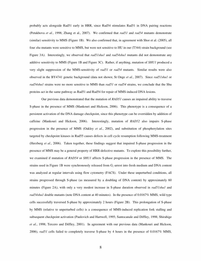
we examined if mutation of RAD54 or SHU1 affects S-phase progression in the presence of MMS. The
strains used in Figure 1B were synchronously released from G1-arrest into fresh medium and DNA content
was analyzed at regular intervals using flow cytometry (FACS). Under these unperturbed conditions, all
strains progressed through S-phase (as measured by a doubling of DNA content) by approximately 60
minutes (Figure 2A), with only a very modest increase in S-phase duration observed in rad51shu1 and
rad54shu1 double mutants (note DNA content at 40 minutes). In the presence of 0.0167% MMS, wild type
cells successfully traversed S-phase by approximately 2 hours (Figure 2B). This prolongation of S-phase
by MMS (relative to unperturbed cells) is a consequence of MMS-induced replication fork stalling and
subsequent checkpoint activation (Paulovich and Hartwell, 1995; Santocanale and Diffley, 1998; Shirahige
et al., 1998; Tercero and Diffley, 2001). In agreement with our previous data (Mankouri and Hickson,
2006), rad51 cells failed to completely traverse S-phase by 4 hours in the presence of 0.0167% MMS,
8

demonstrating a mid-S-phase DNA content at 4 hours (Figure 2B). Interestingly, we observed that rad54
and shu1 mutants resembled rad51 mutants, and also failed to traverse S-phase by 4 hours in 0.0167%
MMS (Figure 2B). Furthermore, like rad51 cells (Mankouri and Hickson, 2006), the impaired S-phase
progression phenotype of MMS-treated rad54 or shu1 mutants could be overridden by addition of caffeine
(Figure 2C). Therefore, these data imply that a more persistent or robust checkpoint activation occurs
following MMS-treatment in cells lacking Rad51, Rad54 or Shu1. Similar results were also observed for
the other shu mutant strains (data not shown), consistent with the similar phenotypes caused by mutation of
any one of the SHU genes (Figure 3A; Shor et al., 2005). Interestingly, rad51shu1 or rad54shu1 double
mutants did not demonstrate additive impaired S-phase progression phenotypes in the presence of MMS,
consistent with the proposed epistasis between these genes (Figure 1). Furthermore, the impaired S-phase
progression in MMS-treated rad51shu1 or rad54shu1 double mutants could be overridden by caffeine
(Figure 2C). We propose that Rad51, Rad54 and Shu1 all act at a similar step in a common pathway for the
repair of MMS-induced DNA lesions during S-phase, and that perturbation/inactivation of this pathway
leads to accumulation of a HRR substrate that causes persistent checkpoint activation.
shu mutations suppress sgs1 HU-sensitivity, and are epistatic to sgs1 for MMS-sensitivity
Since shu mutations suppress various phenotypic defects caused by mutation of SGS1 (Shor et al.,
2005), we further investigated the relationship between the SHU genes and SGS1. Previous data
demonstrated that mutation of SHU genes suppresses the HU-sensitivity of sgs1 cells (Shor et al., 2005).
However, in the study of Shor et al. (2005) it was shown that csm2sgs1 and shu1sgs1 mutants exhibited
additive sensitivity to MMS, suggesting that Sgs1 and the Shu proteins function in separate pathways for
repair of MMS-induced DNA lesions. We verified that our sgs1 strain demonstrated sensitivity to both HU
and MMS, whereas mutation of SHU genes conferred sensitivity to MMS alone (Figure 3A). We also
verified that, in agreement with Shor et al. (2005), mutation of all four shu genes (similarly) suppressed the
HU-sensitivity of sgs1 cells (Figure 3A). However, in contrast to Shor et al. (2005), we observed that shu
mutations were epistatic to sgs1 for MMS-sensitivity, since shu sgs1 double mutants resembled the shu
single mutants on plates containing MMS (Figure 3A). Although shu1 mutation appears to suppress sgs1
MMS-sensitivity at low doses of MMS (0.0040%) (see Figure 3C), we observed that shu (and shusgs1
double) mutants were more sensitive to MMS than sgs1 mutants at higher doses (0.0080%) (Figure 3A).
9
To further investigate this apparent discrepancy, and to test for any strain-dependent variations, we
investigated the genetic relationship between SHU1 and SGS1 in a third strain background, BY4741. In
this background, mutation of SHU1 again suppressed sgs1 HU-sensitivity (data not shown). However, in
agreement with a recent independent study (St Onge et al., 2007), we observed that mutation of SHU1
suppressed sgs1 MMS-sensitivity in the BY4741 genetic background (data not shown). We propose that
this apparent inconsistency between our T344 and BY4741 shu1sgs1 strains can be reconciled by the fact
that shu mutants are more sensitive to MMS than are sgs1 strains in the T344 background, whereas sgs1
mutants are more sensitive to MMS than are shu mutants in the BY4741 background. In both of the strain
backgrounds that we analyzed, the phenotype of the shu sgs1 double mutant mimics the phenotype of the
corresponding shu single mutant. Therefore, the additive sensitivity of shu1sgs1 double mutants to MMS as
reported by Shor et al. (2005) likely reflects a strain-specific effect. Taken together, our data implies that,
despite apparent strain-dependent differences in MMS sensitivity, shu mutations are epistatic to sgs1 for
MMS-sensitivity. Moreover, our data are consistent with the Shu proteins acting upstream of Sgs1.
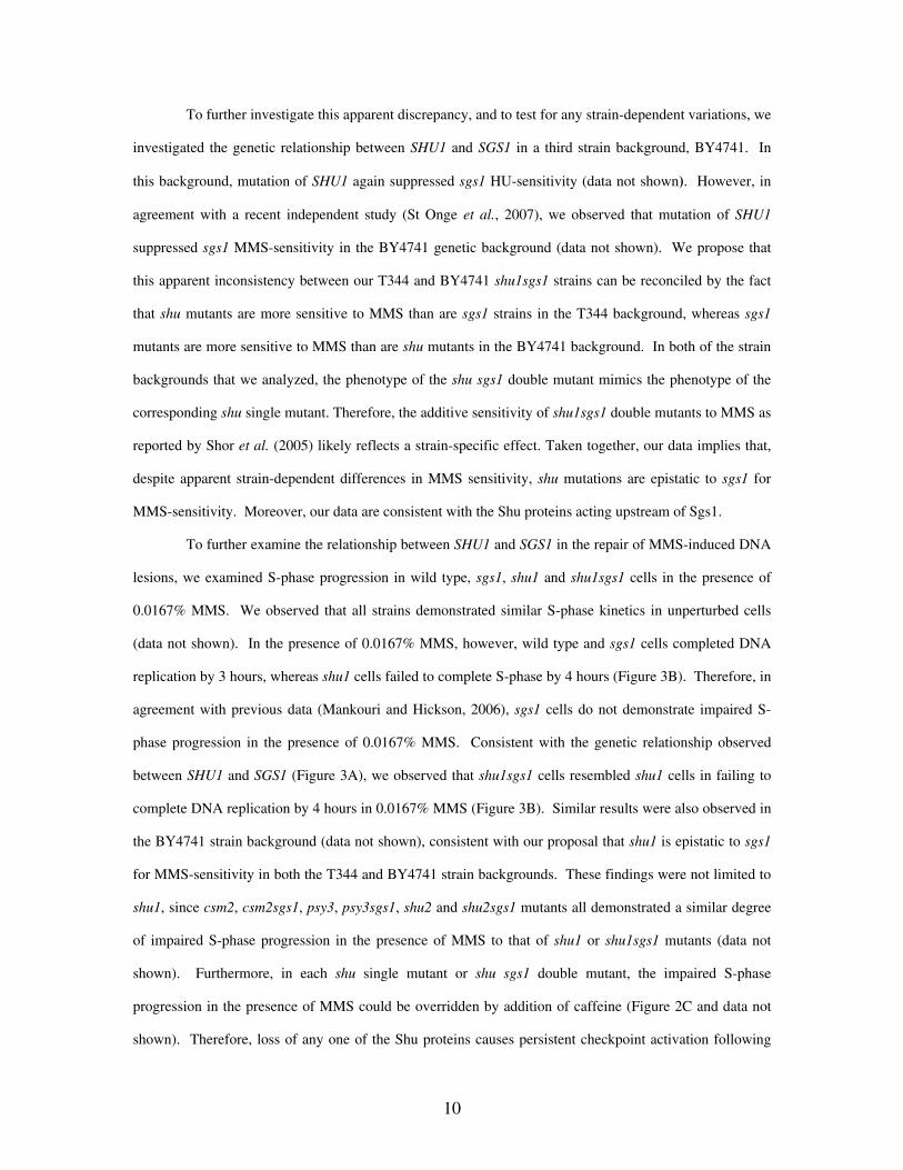
To further examine the relationship between SHU1 and SGS1 in the repair of MMS-induced DNA
lesions, we examined S-phase progression in wild type, sgs1, shu1 and shu1sgs1 cells in the presence of
0.0167% MMS. We observed that all strains demonstrated similar S-phase kinetics in unperturbed cells
(data not shown). In the presence of 0.0167% MMS, however, wild type and sgs1 cells completed DNA
replication by 3 hours, whereas shu1 cells failed to complete S-phase by 4 hours (Figure 3B). Therefore, in
agreement with previous data (Mankouri and Hickson, 2006), sgs1 cells do not demonstrate impaired S-
phase progression in the presence of 0.0167% MMS. Consistent with the genetic relationship observed
between SHU1 and SGS1 (Figure 3A), we observed that shu1sgs1 cells resembled shu1 cells in failing to
complete DNA replication by 4 hours in 0.0167% MMS (Figure 3B). Similar results were also observed in
the BY4741 strain background (data not shown), consistent with our proposal that shu1 is epistatic to sgs1
for MMS-sensitivity in both the T344 and BY4741 strain backgrounds. These findings were not limited to
shu1, since csm2, csm2sgs1, psy3, psy3sgs1, shu2 and shu2sgs1 mutants all demonstrated a similar degree
of impaired S-phase progression in the presence of MMS to that of shu1 or shu1sgs1 mutants (data not
shown). Furthermore, in each shu single mutant or shu sgs1 double mutant, the impaired S-phase
progression in the presence of MMS could be overridden by addition of caffeine (Figure 2C and data not
shown). Therefore, loss of any one of the Shu proteins causes persistent checkpoint activation following
10
treatment with MMS, regardless of whether Sgs1 is present or not. Taken together with the fact that shu
mutations are epistatic to sgs1 for MMS sensitivity (Figure 3A), these findings are consistent with a role for
the Shu complex upstream of Sgs1 in the same pathway to repair MMS-induced DNA lesions during S-
phase.
To further investigate the relationship between Shu1, Sgs1 and the HRR pathway, we examined
the effects of deleting RAD51 and RAD54. Interestingly, we observed that, similar to what was observed
when SHU1 was mutated (Figure 3B), mutation of RAD51 or RAD54 impaired the S-phase progression of
sgs1 mutants (Supplementary Figure 1). Consistent with our proposal that the impaired S-phase
progression phenotype of a shu1 mutant is not additive with rad51 or rad54 mutations, we observed that
rad51shu1sgs1 and rad54shu1sgs1 triple mutants demonstrated similar rates of (impaired) S-phase
progression in the presence of MMS to that of rad51sgs1, rad54sgs1 or shu1sgs1 double mutants
(Supplementary Figure 1). Furthermore, rad51shu1sgs1 and rad54shu1sgs1, triple mutants were no more
sensitive to MMS than rad51 or rad54 single mutants, respectively (Figure 3C). This apparent lack of
additive phenotypes is consistent with the proposal that Rad51/Rad54, Shu1 and Sgs1 are all epsitatic for
MMS-sensitivity. We conclude that Rad51, Rad54, Shu1 and Sgs1 all function in the same (HRR) pathway
to repair MMS lesions during S-phase, and that Sgs1 acts at a later stage than Rad51, Rad54, or Shu1.
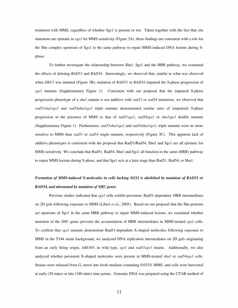
Formation of MMS-induced X-molecules in cells lacking SGS1 is abolished by mutation of RAD51 or
RAD54, and attenuated by mutation of SHU genes
Previous studies indicated that sgs1 cells exhibit persistent, Rad51-dependent, HRR intermediates
on 2D gels following exposure to MMS (Liberi et al., 2005). Based on our proposal that the Shu proteins
act upstream of Sgs1 in the same HRR pathway to repair MMS-induced lesions, we examined whether
mutation of the SHU genes prevents the accumulation of HRR intermediates in MMS-treated sgs1 cells.
To confirm that sgs1 mutants demonstrate Rad51-dependent X-shaped molecules following exposure to
MMS in the T344 strain background, we analyzed DNA replication intermediates on 2D gels originating
from an early firing origin, ARS305, in wild type, sgs1 and rad51sgs1 strains. Additionally, we also
analyzed whether persistent X-shaped molecules were present in MMS-treated shu1 or rad54sgs1 cells.
Strains were released from G1 arrest into fresh medium containing 0.033% MMS, and cells were harvested
at early (30 mins) or late (180 mins) time points. Genomic DNA was prepared using the CTAB method of
11

DNA extraction to restrain branch migration of joint (X-shaped) molecules, as described previously (Lopes
et al., 2003). We observed that origin firing at ARS305 was detectable after 30 minutes in all strains, as
evidenced by the appearance of bubbles, Y-molecules and the so-called origin-associated X-structures
(Figure 4A). Previous studies have indicated that the origin-associated X-structures are normal DNA
replication intermediates that are not dependent on Rad51 or Rad52 for their formation and are not,
therefore, HRR intermediates (Lopes et al., 2003). In each strain, the relative ratio between bubbles, Y-
molecules and X-structures was similar at 30 minutes, suggesting that origin firing occurs normally in shu1,
sgs1, rad51sgs1 and rad54sgs1 mutants. After 180 minutes, all of the ARS305 replication intermediates
detectable at 30 minutes were substantially diminished, or had disappeared, in wild type cells, consistent
with replication fork progression beyond the boundaries of this genomic region by this time. Similar
results were observed in shu1 cells, suggesting that mutation of SHU1 does not noticeably affect DNA
replication at ARS305 (Figure 4A). In contrast, we observed that, although bubbles and Y-molecules had
largely disappeared from the ARS305 region at 180 minutes, abnormal DNA replication intermediates (X-
molecules) persisted in MMS-treated sgs1 cells (Figure 4A). In agreement with previous data (Liberi et al.,
2005), we also observed that these X-molecules were not detectable in rad51sgs1 cells (Figure 4A).
Interestingly, we also observed a similar effect when RAD54 was mutated, since rad54sgs1 mutants also
did not exhibit persistent MMS-induced X-molecules (Figure 4A). Therefore, we conclude that persistent
X-molecules in MMS-treated sgs1 cells are both Rad51- and Rad54-dependent. This finding is consistent
with Rad54 acting at an early stage in Rad51-dependent HRR upstream of Sgs1 (Rattray and Symington,
1995; Petukhova et al., 1998; Zhang et al., 2007).
Since sgs1, but not wild type (or shu1), strains exhibit persistent MMS-induced X-molecules after
180 mins in 0.033% MMS (Figure 4A), we examined the effects of deleting SHU genes on X-molecule
signal intensity in sgs1 strains at this time point. We verified that, similar to what was observed in shu1
mutants (Figure 4A), all replication intermediates apparently disappeared from ARS305 in csm2, psy3, or
shu2 single mutants by 3 hours following release from G1-arrest into medium containing MMS. Therefore,
DNA replication appears normal, and MMS-induced X-molecules are not detectable at ARS305 in csm2,
psy3, or shu2 single mutants following a 3 hour exposure to MMS (data not shown). However, we
observed that MMS-induced X-molecules were detectable at ARS305 in csm2sgs1, psy3sgs1, shu1sgs1 or
shu2sgs1 mutants (Figure 4B). However, these X-molecules were diminished in intensity by
12

approximately 70% in all shusgs1 double mutants, relative to those observed in an sgs1 single mutant
(Figure 4B). This effect was not simply a consequence of altered DNA replication kinetics at ARS305,
since origin firing (bubble formation at 30 minutes) and replication fork progression (disappearance of
bubbles and Y-arcs at 90-180 minutes) at ARS305 were indistinguishable in sgs1 and shu1sgs1 cells (Figure
4C). Therefore, we conclude that mutation of SHU genes does not abolish, but does attenuate, the level of
MMS-induced X-molecules at ARS305 in MMS-treated sgs1 cells.
To determine if the low level of MMS-induced X-molecules in shu1sgs1 cells were nevertheless
HRR intermediates, we investigated the effects of deleting RAD51 or RAD54 in shu1sgs1 cells. We found
that origin firing at ARS305 and S-phase progression were indistinguishable in MMS-treated shu1sgs1,
rad51shu1sgs1 and rad54shu1sgs1 strains (data not shown and Supplementary Figure 1). After 180
minutes of MMS-treatment, we again observed that persistent X-molecules were detectable in MMS-
treated sgs1 cells, and that these were attenuated in shu1sgs1 cells (Figure 4D). However, we found that
the MMS-induced X-molecules were absent from rad51shu1sgs1 or rad54shu1sgs1 mutants, indicating
that the attenuated X-molecules in shu1sgs1 cells are RAD51- and RAD54-dependent HRR structures.
(Figure 4D). Taken together, the data presented in Figure 4 indicate that, whereas acute formation of
MMS-induced persistent X-molecules in sgs1 cells is abolished by mutation of RAD51 or RAD54, these
structures are not completely abolished by mutation of SHU genes. However, since the level of MMS-
induced X-molecules is attenuated in shusgs1 double mutants, we propose that mutation of the SHU genes
affects either the rate of formation/removal, or the nature, of the X-molecules in sgs1 cells.
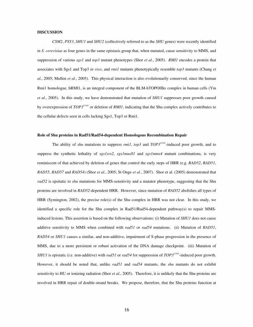
Mutation of shu1 attenuates persistent MMS-induced X-molecules in cells overexpressing TOP3Y356F
We demonstrated previously that overexpression of TOP3Y356F causes Rad51-dependent X-
molecules to persist during S-phase following exposure to MMS. To determine if the attenuation of sgs1
MMS-induced X-molecules by shu mutations was specific for sgs1 X-molecules, or general for both sgs1
and TOP3Y356F-induced X-molecules, we examined if mutation of SHU1 affects MMS-induced X-molecule
persistence in cells overexpressing TOP3Y356F. For this, wild-type and shu1 strains overexpressing TOP3Y356F
were released from G1 arrest into medium containing 0.033% MMS. In agreement with our previous data
(Mankouri and Hickson, 2006), wild type cells overexpressing TOP3Y356F demonstrated persistent MMS-
induced X-molecules at 180 mins after G1-release (Figure 5A & 5B). These structures were absent in
13

MMS-treated rad51 or rad54 cells overexpressing TOP3Y356F, verifying that Rad51 and Rad54 are required
for their formation (data not shown; Mankouri and Hickson, 2006). Persistent MMS-induced X-molecules
were detected in shu1 cells overexpressing TOP3Y356F, although their level was attenuated relative to those
observed in wild type cells overexpressing TOP3Y356F (Figure 5A & 5B). Furthermore, no MMS-induced X-
molecules were detectable in rad51shu1 TOP3Y356F strains, suggesting that these X-molecules are also
Rad51-dependent HRR intermediates (Figure 5B). Since these results are qualitatively very similar to
those observed in shu1sgs1 cells (Figure 4B & 4C), we propose that mutation of SHU1 attenuates MMS-
induced X-molecule formation when Top3 function is impaired by a similar mechanism to that occurring in
shu1sgs1 cells.
Mutation of SHU1 suppresses growth defects and MMS-sensitivity caused by mutation of RMI1, and
attenuates X-molecules in MMS-treated rmi1 cells
The yeast and human Rmi1 proteins have been proposed to be integral components of multi-
enzyme complexes containing Sgs1 and Top3, and BLM and hTOPOIIIα respectively (Johnson et al.,
2000; Wu et al., 2000; Chang et al., 2005; Mullen et al., 2005; Yin et al., 2005). We examined, therefore,
if mutation of SHU1 also suppress rmi1 phenotypes. Since rmi1 mutants demonstrate a top3-like poor
growth phenotype that is readily suppressed by mutations in SGS1, we generated haploid rmi1 mutants by
sporulating a BY4741 RMI1+/- diploid strain (see Methods). We verified that mutation of RMI1 caused
poor growth which was completely suppressed by mutation of SGS1 (data not shown), or partially
suppressed by RAD51 (Figure 5C). Interestingly, we also found that mutation of SHU1 also partially
suppressed the poor growth, and MMS-sensitivity of rmi1 mutants (Figure 5C).
Based on its interactions with Sgs1 and Top3 (Chang et al., 2005; Mullen et al., 2005), we
examined if rmi1 mutants also demonstrate persistent X-molecules in the presence of MMS. We observed
that, similar to cells lacking SGS1, or overexpressing TOP3Y356F (Figures 4-5; Liberi et al., 2005; Mankouri
and Hickson, 2006), rmi1 cells demonstrated persistent X-molecules at ARS305 following exposure to
MMS (Figure 5D). We also observed that, whereas mutation of RAD51 eliminated the MMS-induced X-
molecules at ARS305 in rmi1 cells, mutation of SHU1 again only attenuated their level (Figure 5D).
Therefore, the persistent X-molecules detected in rmi1 cells likely represent Rad51-dependent HRR
intermediates, similar or identical to those identified in cells lacking Sgs1, or cells overexpressing TOP3Y356F
14

(Figures 4-5 & Liberi et al., 2005; Mankouri and Hickson, 2006). Furthermore, since mutation of SHU1
only attenuated these MMS-induced X-molecules, we propose that the Shu proteins normally promote the
formation of Rad51/Rad54-dependent HRR intermediates that are ultimately resolved by the Sgs1-Rmi1-
Top3 complex.
15
DISCUSSION
CSM2, PSY3, SHU1 and SHU2 (collectively referred to as the SHU genes) were recently identified
in S. cerevisiae as four genes in the same epistasis group that, when mutated, cause sensitivity to MMS, and
suppression of various sgs1 and top3 mutant phenotypes (Shor et al., 2005). RMI1 encodes a protein that
associates with Sgs1 and Top3 in vivo, and rmi1 mutants phenotypically resemble top3 mutants (Chang et
al., 2005; Mullen et al., 2005). This physical interaction is also evolutionarily conserved, since the human
Rmi1 homologue, hRMI1, is an integral component of the BLM-hTOPOIIIα complex in human cells (Yin
et al., 2005). In this study, we have demonstrated that mutation of SHU1 suppresses poor growth caused
by overexpression of TOP3Y356F or deletion of RMI1, indicating that the Shu complex actively contributes to
the cellular defects seen in cells lacking Sgs1, Top3 or Rmi1.
Role of Shu proteins in Rad51/Rad54-dependent Homologous Recombination Repair
The ability of shu mutations to suppress rmi1, top3 and TOP3Y356F-induced poor growth, and to
suppress the synthetic lethality of sgs1srs2, sgs1mus81 and sgs1mms4 mutant combinations, is very
reminiscent of that achieved by deletion of genes that control the early steps of HRR (e.g. RAD52, RAD51,
RAD55, RAD57 and RAD54) (Shor et al., 2005; St Onge et al., 2007). Shor et al. (2005) demonstrated that
rad52 is epsitatic to shu mutations for MMS-sensitivity and a mutator phenotype, suggesting that the Shu
proteins are involved in RAD52-dependent HRR. However, since mutation of RAD52 abolishes all types of
HRR (Symington, 2002), the precise role(s) of the Shu complex in HRR was not clear. In this study, we
identified a specific role for the Shu complex in Rad51/Rad54-dependent pathway(s) to repair MMS-
induced lesions. This assertion is based on the following observations: (i) Mutation of SHU1 does not cause
additive sensitivity to MMS when combined with rad51 or rad54 mutations. (ii) Mutation of RAD51,
RAD54 or SHU1 causes a similar, and non-additive, impairment of S-phase progression in the presence of
MMS, due to a more persistent or robust activation of the DNA damage checkpoint. (iii) Mutation of
SHU1 is epistatic (i.e. non-additive) with rad51 or rad54 for suppression of TOP3Y356F-induced poor growth.
However, it should be noted that, unlike rad51 and rad54 mutants, the shu mutants do not exhibit
sensitivity to HU or ionizing radiation (Shor et al., 2005). Therefore, it is unlikely that the Shu proteins are
involved in HRR repair of double-strand breaks. We propose, therefore, that the Shu proteins function at
16
an early stage in a Rad51/Rad54-dependent HRR sub-pathway specifically to repair certain types of DNA
lesions (probably ssDNA gaps; see below) that arise during S-phase.
Suppression of sgs1, rmi1 and top3/TOP3Y356F phenotypes by mutation of SHU genes
Since mutation of SHU1 or CSM2 suppresses hyper-recombination in sgs1 and top3 cells (Shor et
al., 2005), we examined if shu mutations prevent unprocessed HRR intermediates from accumulating in
cells compromised for Sgs1, Top3 or Rmi1. Previous data demonstrated that unresolved Rad51-dependent
HRR intermediates are detectable in MMS-treated sgs1, top3 and TOP3Y356F cells (Liberi et al., 2005;
Mankouri and Hickson, 2006). In this study, we also demonstrated that rmi1 mutants exhibit persistent,
Rad51-dependent, MMS-induced X-molecules, suggesting that the accumulation of unprocessed HRR
intermediates is a common property of strains mutated for SGS1, TOP3 or RMI1. It should be noted,
however, that we do not know presently if the X-molecules arising in MMS-treated sgs1, rmi1 and
TOP3Y356F strains are identical, or if they represent different types of HRR intermediates that cannot readily
be distinguished by the 2D gel technique. One possibility, based on the known enzymatic functions of
BLM, hTOPOIII and RMI1 (Wu and Hickson, 2003; Plank et al., 2006; Raynard et al., 2006; Wu et al.,
2006), is that X-molecules arising in sgs1 mutants are double Holliday junctions, whereas those arising in
TOP3Y356F, or rmi1, mutants are hemicatenanes formed by the Sgs1-dependent convergent branch migration
of double Holliday junctions.
Surprisingly, we found that whereas mutation of RAD51 prevented persistent MMS-induced HRR
intermediates in sgs1, rmi1 and TOP3Y356F cells, mutation of SHU1 did not. However, mutation of SHU1
attenuated the level of MMS-induced HRR intermediates in sgs1, rmi1 and TOP3Y356F cells. This finding is
consistent with the report that shu1 suppresses the elevated recombination rate in sgs1 and top3 cells (Shor
et al., 2005). Taken together with the fact that the shu mutations are epistatic to sgs1 for MMS-sensitivity,
and also suppress the poor growth caused by deletion of RMI1 or TOP3, our data are consistent with an
early role for the Shu proteins in HRR to promote the formation of HR intermediates that are resolved by
the Sgs1-Rmi1-Top3 complex. We therefore propose that, in the absence of the Shu proteins, the
maturation of Rad51-filaments into Sgs1-Rmi1-Top3 substrates (e.g. double Holliday junctions) occurs
inefficiently (Figure 6). If true, this suggests that the Shu proteins likely function as non-essential HRR
accessory factors that facilitate efficient and timely HRR. These findings are consistent with the proposal
17
that Shu proteins may represent so-called ‘Rad51 paralogs’ (Martin et al., 2006), since these have been
demonstrated to facilitate the action of Rad51 in HRR (Takata et al., 2001).
Of wider implication, putative SHU2 and PSY3 homologues have recently been identified in S.
pombe (Sws1+ and Rld1+, respectively), and human cells (SWS1 and RAD51D, respectively) (Martin et al.,
2006), suggesting that Shu-like complexes are evolutionarily conserved. Consistent with a conserved role
in HRR, ablation of SWS1 in human cells reduces the number of Rad51 foci in both control, and IR-treated,
cells (Martin et al., 2006). It will therefore be of great interest to determine if SWS1 or RAD51D ablation
can prevent the defects seen in Bloom’s syndrome cells.
ACKNOWLEDGEMENTS
We thank Drs P. McHugh and L. Wu and various members of the Hickson laboratory for helpful comments
and criticisms. We also thank the Cancer Research UK peptide synthesis laboratory for providing α-factor.
This work was funded by Cancer Research UK.
18
REFERENCES
Allers, T., and Lichten, M. (2000). A method for preparing genomic DNA that restrains branch migration
of Holliday junctions. Nucleic Acids Res. 28, e6.
Braybrooke, J.P., Li, J.L., Wu, L., Caple, F., Benson, F.E., and Hickson, I.D. (2003). Functional interaction
between the Bloom's syndrome helicase and the RAD51 paralog, RAD51L3 (RAD51D). J. Biol. Chem.
278, 48357-48366.
Braybrooke, J.P., Spink, K.G., Thacker, J., and Hickson, I.D. (2000). The RAD51 family member,
RAD51L3, is a DNA-stimulated ATPase that forms a complex with XRCC2. J. Biol. Chem. 275, 29100-
29106.
Brewer, B.J., and Fangman, W.L. (1987). The localization of replication origins on ARS plasmids in S.
cerevisiae. Cell 51, 463-471.
Cartwright, R., Dunn, A.M., Simpson, P.J., Tambini, C.E., and Thacker, J. (1998a). Isolation of novel
human and mouse genes of the recA/RAD51 recombination-repair gene family. Nucleic Acids Res. 26,
1653-1659.
Cartwright, R., Tambini, C.E., Simpson, P.J., and Thacker, J. (1998b). The XRCC2 DNA repair gene from
human and mouse encodes a novel member of the recA/RAD51 family. Nucleic Acids Res. 26, 3084-3089.
Chang, M., Bellaoui, M., Zhang, C., Desai, R., Morozov, P., Delgado-Cruzata, L., Rothstein, R., Freyer,
G.A., Boone, C., and Brown, G.W. (2005). RMI1/NCE4, a suppressor of genome instability, encodes a
member of the RecQ helicase/Topo III complex. EMBO J. 24, 2024-2033.
Fabre, F., Chan, A., Heyer, W.D., and Gangloff, S. (2002). Alternate pathways involving Sgs1/Top3,
Mus81/ Mms4, and Srs2 prevent formation of toxic recombination intermediates from single-stranded gaps
created by DNA replication. Proc. Natl. Acad. Sci. USA 99, 16887-16892.
Gangloff, S., McDonald, J.P., Bendixen, C., Arthur, L., and Rothstein, R. (1994). The yeast type I
topoisomerase Top3 interacts with Sgs1, a DNA helicase homolog: a potential eukaryotic reverse gyrase.
Mol. Cell. Biol. 14, 8391-8398.
Gangloff, S., Soustelle, C., and Fabre, F. (2000). Homologous recombination is responsible for cell death in
the absence of the Sgs1 and Srs2 helicases. Nat. Genet. 25, 192-194.
German, J. (1993). Bloom syndrome: a mendelian prototype of somatic mutational disease. Medicine 72,
19
393-406.
Herzberg, K., Bashkirov, V.I., Rolfsmeier, M., Haghnazari, E., McDonald, W.H., Anderson, S.,
Bashkirova, E.V., Yates, J.R., 3rd, and Heyer, W.D. (2006). Phosphorylation of Rad55 on serines 2, 8, and
14 is required for efficient homologous recombination in the recovery of stalled replication forks. Mol.
Cell. Biol. 26, 8396-8409.
Heyer, W.D., Li, X., Rolfsmeier, M., and Zhang, X.P. (2006). Rad54: the Swiss Army knife of homologous
recombination? Nucleic Acids Res. 34, 4115-4125.
Hovland, P., Flick, J., Johnston, M., and Sclafani, R.A. (1989). Galactose as a gratuitous inducer of GAL
gene expression in yeasts growing on glucose. Gene 83, 57-64.
Johnson, F.B., Lombard, D.B., Neff, N.F., Mastrangelo, M.A., Dewolf, W., Ellis, N.A., Marciniak, R.A.,
Yin, Y., Jaenisch, R., and Guarente, L. (2000). Association of the Bloom syndrome protein with
topoisomerase IIIalpha in somatic and meiotic cells. Cancer Res. 60, 1162-1167.
Kawabata, M., and Saeki, K. (1998). Sequence analysis and expression of a novel mouse homolog of
Escherichia coli recA gene. Biochim. Biophys. Acta 1398, 353-358.
Krogh, B.O., and Symington, L.S. (2004). Recombination proteins in yeast. Annu. Rev. Genet. 38, 233-
271.
Liberi, G., Cotta-Ramusino, C., Lopes, M., Sogo, J., Conti, C., Bensimon, A., and Foiani, M. (2006).
Methods to study replication fork collapse in budding yeast. Methods Enzymol. 409, 442-462.
Liberi, G., Maffioletti, G., Lucca, C., Chiolo, I., Baryshnikova, A., Cotta-Ramusino, C., Lopes, M.,
Pellicioli, A., Haber, J.E., and Foiani, M. (2005). Rad51-dependent DNA structures accumulate at damaged
replication forks in sgs1 mutants defective in the yeast ortholog of BLM RecQ helicase. Genes Dev. 19,
339-350.
Liu, N., Lamerdin, J.E., Tebbs, R.S., Schild, D., Tucker, J.D., Shen, M.R., Brookman, K.W., Siciliano,
M.J., Walter, C.A., Fan, W., Narayana, L.S., Zhou, Z.Q., Adamson, A.W., Sorensen, K.J., Chen, D.J.,
Jones, N.J., and Thompson, L.H. (1998). XRCC2 and XRCC3, new human Rad51-family members,
promote chromosome stability and protect against DNA cross-links and other damages. Mol. Cell 1, 783-
793.
Lopes, M., Cotta-Ramusino, C., Liberi, G., and Foiani, M. (2003). Branch migrating sister chromatid
junctions form at replication origins through Rad51/Rad52-independent mechanisms. Mol. Cell 12, 1499-
20
1510.
Mankouri, H.W., and Hickson, I.D. (2006). Top3 processes recombination intermediates and modulates
checkpoint activity after DNA damage. Mol. Biol. Cell 17, 4473-4483.
Martin, V., Chahwan, C., Gao, H., Blais, V., Wohlschlegel, J., Yates, J.R., 3rd, McGowan, C.H., and
Russell, P. (2006). Sws1 is a conserved regulator of homologous recombination in eukaryotic cells. EMBO
J. 25, 2564-2574.
Mullen, J.R., Kaliraman, V., Ibrahim, S.S., and Brill, S.J. (2001). Requirement for three novel protein
complexes in the absence of the Sgs1 DNA helicase in Saccharomyces cerevisiae. Genetics 157, 103-118.
Mullen, J.R., Nallaseth, F.S., Lan, Y.Q., Slagle, C.E., and Brill, S.J. (2005). Yeast Rmi1/Nce4 controls
genome stability as a subunit of the Sgs1-Top3 complex. Mol. Cell. Biol. 25, 4476-4487.
Oakley, T.J., Goodwin, A., Chakraverty, R.K., and Hickson, I.D. (2002). Inactivation of homologous
recombination suppresses defects in topoisomerase III-deficient mutants. DNA repair 1, 463-482.
Paques, F., and Haber, J.E. (1999). Multiple pathways of recombination induced by double-strand breaks in
Saccharomyces cerevisiae. Microbiol. Mol. Biol. Rev. 63, 349-404.
Paulovich, A.G., and Hartwell, L.H. (1995). A checkpoint regulates the rate of progression through S phase
in S. cerevisiae in response to DNA damage. Cell 82, 841-847.
Petukhova, G., Stratton, S., and Sung, P. (1998). Catalysis of homologous DNA pairing by yeast Rad51 and
Rad54 proteins. Nature 393, 91-94.
Pittman, D.L., Weinberg, L.R., and Schimenti, J.C. (1998). Identification, characterization, and genetic
mapping of Rad51d, a new mouse and human RAD51/RecA-related gene. Genomics 49, 103-111.
Plank, J.L., Wu, J., and Hsieh, T.S. (2006). Topoisomerase IIIalpha and Bloom’s helicase can resolve a
mobile double Holliday junction substrate through convergent branch migration. Proc. Natl. Acad. Sci.
USA 103, 11118-11123.
Rattray, A.J., and Symington, L.S. (1995). Multiple pathways for homologous recombination in
Saccharomyces cerevisiae. Genetics 139, 45-56.
Raynard, S., Bussen, W., and Sung, P. (2006). A double Holliday junction dissolvasome comprising BLM,
topoisomerase IIIalpha, and BLAP75. J. Biol. Chem. 281, 13861-13864.
Santocanale, C., and Diffley, J.F. (1998). A Mec1- and Rad53-dependent checkpoint controls late-firing
origins of DNA replication. Nature 395, 615-618.
21
Shirahige, K., Hori, Y., Shiraishi, K., Yamashita, M., Takahashi, K., Obuse, C., Tsurimoto, T., and
Yoshikawa, H. (1998). Regulation of DNA-replication origins during cell-cycle progression. Nature 395,
618-621.
Shor, E., Gangloff, S., Wagner, M., Weinstein, J., Price, G., and Rothstein, R. (2002). Mutations in
homologous recombination genes rescue top3 slow growth in Saccharomyces cerevisiae. Genetics 162,
647-662.
Shor, E., Weinstein, J., and Rothstein, R. (2005). A genetic screen for top3 suppressors in Saccharomyces
cerevisiae identifies SHU1, SHU2, PSY3 and CSM2: four genes involved in error-free DNA repair.
Genetics 169, 1275-1289.
St Onge, R.P., Mani, R., Oh, J., Proctor, M., Fung, E., Davis, R.W., Nislow, C., Roth, F.P., and Giaever, G.
(2007). Systematic pathway analysis using high-resolution fitness profiling of combinatorial gene deletions.
Nat. Genet. 39, 199-206.
Sung, P., Krejci, L., Van Komen, S., and Sehorn, M.G. (2003). Rad51 recombinase and recombination
mediators. J. Biol. Chem. 278, 42729-42732.
Symington, L.S. (2002). Role of RAD52 epistasis group genes in homologous recombination and double-
strand break repair. Microbiol. Mol. Biol. Rev. 66, 630-670.
Takata, M., Sasaki, M.S., Tachiiri, S., Fukushima, T., Sonoda, E., Schild, D., Thompson, L.H., and Takeda,
S. (2001). Chromosome instability and defective recombinational repair in knockout mutants of the five
Rad51 paralogs. Mol. Cell. Biol. 21, 2858-2866.
Tan, T.L., Kanaar, R., and Wyman, C. (2003). Rad54, a Jack of all trades in homologous recombination.
DNA Repair 2, 787-94.
Tercero, J.A., and Diffley, J.F. (2001). Regulation of DNA replication fork progression through damaged
DNA by the Mec1/Rad53 checkpoint. Nature 412, 553-557.
Wach, A., Brachat, A., Pohlmann, R., and Philippsen, P. (1994). New heterologous modules for classical or
PCR-based gene disruptions in Saccharomyces cerevisiae. Yeast 10, 1793-1808.
Watt, P.M., Hickson, I.D., Borts, R.H., and Louis, E.J. (1996). SGS1, a homologue of the Bloom's and
Werner's syndrome genes, is required for maintenance of genome stability in Saccharomyces cerevisiae.
Genetics 144, 935-945.
West, S.C. (2003). Molecular views of recombination proteins and their control. Nat. Rev. Mol. Cell. Biol.
22

4, 435-445.
Wu, L., Bachrati, C.Z., Ou, J., Xu, C., Yin, J., Chang, M., Wang, W., Li, L., Brown, G.W., and Hickson,
I.D. (2006). BLAP75/RMI1 promotes the BLM-dependent dissolution of homologous recombination
intermediates. Proc. Natl. Acad. Sci. USA 103, 4068-4073.
Wu, L., Davies, S.L., North, P.S., Goulaouic, H., Riou, J.F., Turley, H., Gatter, K.C., and Hickson, I.D.
(2000). The Bloom's syndrome gene product interacts with topoisomerase III. J. Biol. Chem. 275, 9636-
9644.
Wu, L., and Hickson, I.D. (2003). The Bloom's syndrome helicase suppresses crossing over during
homologous recombination. Nature 426, 870-874.
Yin, J., Sobeck, A., Xu, C., Meetei, A.R., Hoatlin, M., Li, L., and Wang, W. (2005). BLAP75, an essential
component of Bloom's syndrome protein complexes that maintain genome integrity. EMBO J. 24, 1465-
1476.
Zhang, Z., Fan, H.Y., Goldman, J.A., and Kingston, R.E. (2007). Homology-driven chromatin remodeling
by human RAD54. Nat. Struct. Mol. Biol. (14), 397-405.
23

FIGURE LEGENDS
Figure 1
SHU1, RAD51 and RAD54 are epistatic for suppression of TOP3Y356F-induced poor growth, and MMS-
sensitivity
(A) Wild-type, shu1, rad51, rad51shu1, rad54 and rad54shu1 strains transformed with pYES2-TOP3 or
pYES2-TOP3Y356F were diluted to equivalent densities and spotted onto control plates, or plates containing
2% galactose (to induce expression from the pYES2 GAL1 promoter). Spots from left to right represent
serial 1 in 10 dilutions of yeast cultures. Plates were grown at 30°C. The large single colonies that arise in
strains overexpressing TOP3Y356F represent suppressors (probably SGS1-linked) as seen previously (Oakley
et al., 2002; Mankouri and Hickson, 2006).
(B) Wild-type, shu1, rad51, rad51shu1, rad54 and rad54shu1 strains were diluted to equivalent densities
and spotted onto control plates (no drug), or plates containing MMS. Spots from left to right represent
serial 1 in 10 dilutions of yeast cultures. Plates were grown at 30°C.
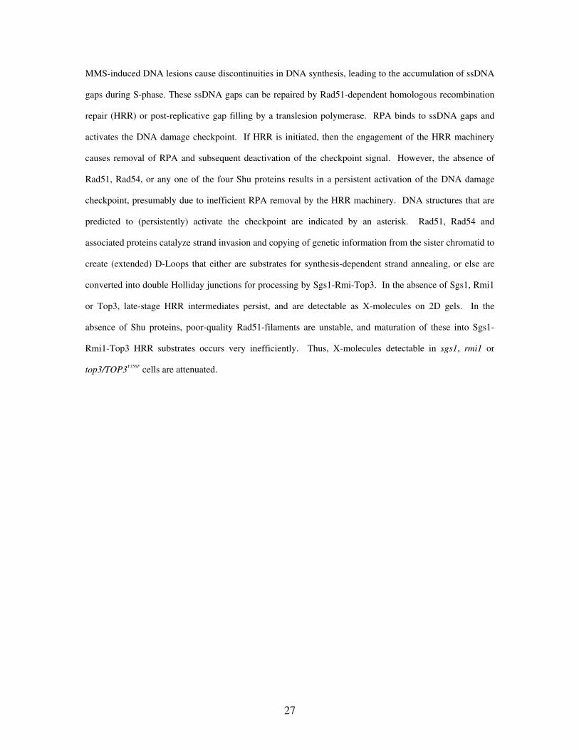
Figure 2
Shu1 functions in a Rad51/Rad54-dependent pathway to repair/tolerate MMS-induced lesions during
S-phase
(A & B) Wild-type, shu1, rad51, rad51shu1, rad54 and rad54shu1 strains were released from G1–arrest
into fresh medium (A) or medium containing 0.0167% MMS (B). DNA content was analyzed by flow
cytometry at the indicated times. The shaded peaks represent experimental data, whereas the unshaded
peak is a reference to indicate a normal G2/M peak (at 2 hours release from G1-arrest). The positions of the
1n (G1) and 2n (G2/M) peaks are indicated below.
(C) After 3 hours treatment with 0.0167% MMS, 1ml aliquots of cells were taken and either caffeine was
added to a final concentration of 5mg/ml, or an equivalent amount of fresh medium was added. Samples
were taken 1 hour later for analysis of DNA content by flow cytometry.
24
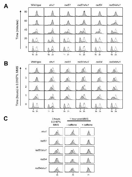
Figure 3
shu1 mutation suppresses sgs1 HU sensitivity, and is epistatic to sgs1 for MMS sensitivity
(A) Wild-type, sgs1, csm2, csm2sgs1, psy3, psy3sgs1, shu1, shu1sgs1, shu2 and shu2sgs1 strains were
diluted to equivalent densities and spotted onto control (no drug) plates, or plates containing indicated
doses of MMS or HU. Spots from left to right represent serial 1 in 10 dilutions of yeast cultures. Plates
were grown at 30°C.
(B) Wild-type, shu1, sgs1 and shu1sgs1 strains were released from G1–arrest into fresh medium containing
0.0167% MMS. DNA content was analyzed by flow cytometry at the indicated times, as per Figure 2
(C) Wild-type, sgs1, shu1, shu1sgs1, rad51, rad51sgs1, rad51shu1, rad51shu1sgs1, rad54, rad54sgs1,
rad54shu1 and rad54shusgs1 strains were diluted to equivalent densities and spotted onto control (no drug)
plates, or plates containing MMS. Spots from left to right represent serial 1 in 10 dilutions of yeast
cultures. Plates were grown at 30°C.
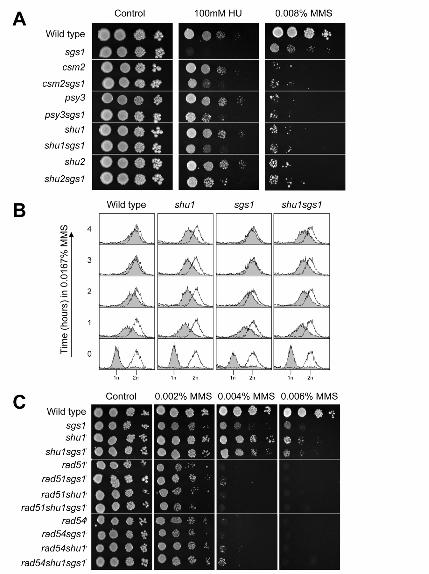
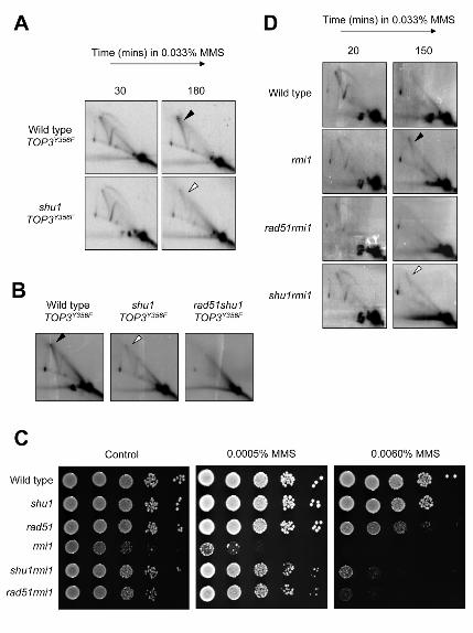
Figure 4
Mutation of SHU genes attenuate persistent X-molecules in MMS-treated sgs1 cells
(A) Wild-type, shu1, sgs1, rad51sgs1, and rad54sgs1 strains were released from G1–arrest into fresh
medium containing 0.033% MMS. DNA replication intermediates were analyzed by 2D gel
electrophoresis at the times indicated. DNA samples were analyzed with a probe for the early-firing
ARS305 replication origin. The diagrammatic representation on the top right of panel denotes DNA
structures that can be identified by the 2D gel technique. For quantification of replication intermediates
(middle and lower panels on the right), each signal (bubbles, Ys and Xs) was normalized to its
corresponding monomer (1N) spot. The arrowhead indicates the position of MMS-induced persistent X-
molecules.
(B) DNA replication intermediates at ARS305 were analyzed in sgs1, csm1sgs1, psy3sgs1, shu1sgs1 and
shu2sgs1 mutants following 3 hours treatment with 0.033% MMS following release from G1–arrest. For
quantification of X-molecules (right), each X-molecule signal was normalized to its corresponding
monomer (1N) spot.
25
(C) A time-course of DNA replication at ARS305 is shown for sgs1 and shu1sgs1 mutants following release
from G1–arrest into fresh medium containing 0.033% MMS.
(D) DNA replication intermediates at ARS305 were analyzed in sgs1, shu1sgs1, rad51shu1sgs1 and
rad54shu1sgs1 mutants following 3 hours treatment with 0.033% MMS following release from G1–arrest.
The filled arrowhead indicates the position of MMS-induced persistent X-molecules, whereas the white
arrowhead indicates the position of attenuated X-molecules.
Figure 5
Mutation of SHU1 attenuates Rad51-dependent, MMS-induced X-molecules in rmi1 cells, and in cells
overexpressing TOP3Y356F
A) Wild-type and shu1 strains transformed with pYES2-TOP3Y356F were arrested in G1 with α–factor and
simultaneously treated with 2% galactose (to induce overexpression from the pYES2 GAL1 promoter).
Cultures were released into fresh medium containing 0.033% MMS, and DNA replication intermediates
were analyzed by 2D gel electrophoresis at the times indicated. DNA samples were analyzed with a probe
for the early-firing ARS305 replication origin. The filled arrowhead indicates the position of MMS-induced
persistent X-molecules, whereas the white arrowhead indicates the position of attenuated X-molecules.
(B) DNA replication intermediates at ARS305 were analyzed in wild type, shu1 and rad51shu1 cells
overexpressing TOP3Y356F following 3 hours treatment with 0.033% MMS after release from G1–arrest.
C) Wild-type, shu1, rad51, rmi1, rad51rmi1 and shu1rmi1 strains were diluted to equivalent densities and
spotted onto control plates (no drug), or plates containing MMS. Spots from left to right represent serial 1
in 10 dilutions of yeast cultures. Plates were grown at 30°C.
D) Wild-type, rmi1, rad51rmi1, and shu1rmi1 strains were released from G1–arrest into fresh medium
containing 0.033% MMS. DNA replication intermediates were analyzed by 2D gel electrophoresis at the
times indicated. DNA samples were analyzed with a probe for the early-firing ARS305 replication origin.
Figure 6
Proposed role of the Shu complex in promoting the formation of HRR intermediates that are
processed by Sgs1-Rmi1-Top3
26
MMS-induced DNA lesions cause discontinuities in DNA synthesis, leading to the accumulation of ssDNA
gaps during S-phase. These ssDNA gaps can be repaired by Rad51-dependent homologous recombination
repair (HRR) or post-replicative gap filling by a translesion polymerase. RPA binds to ssDNA gaps and
activates the DNA damage checkpoint. If HRR is initiated, then the engagement of the HRR machinery
causes removal of RPA and subsequent deactivation of the checkpoint signal. However, the absence of
Rad51, Rad54, or any one of the four Shu proteins results in a persistent activation of the DNA damage
checkpoint, presumably due to inefficient RPA removal by the HRR machinery. DNA structures that are
predicted to (persistently) activate the checkpoint are indicated by an asterisk. Rad51, Rad54 and
associated proteins catalyze strand invasion and copying of genetic information from the sister chromatid to
create (extended) D-Loops that either are substrates for synthesis-dependent strand annealing, or else are
converted into double Holliday junctions for processing by Sgs1-Rmi-Top3. In the absence of Sgs1, Rmi1
or Top3, late-stage HRR intermediates persist, and are detectable as X-molecules on 2D gels. In the
absence of Shu proteins, poor-quality Rad51-filaments are unstable, and maturation of these into Sgs1-
Rmi1-Top3 HRR substrates occurs very inefficiently. Thus, X-molecules detectable in sgs1, rmi1 or
top3/TOP3Y356F cells are attenuated.
27


Related Documents