The Neurocircuitry of Fear, Stress, and Anxiety Disorders Lisa M Shin* ,1,2 and Israel Liberzon 3,4 1 Department of Psychology, Tufts University, Medford, MA, USA; 2 Department of Psychiatry, Massachusetts General Hospital/Harvard Medical School, Boston, MA, USA; 3 Psychiatry Service, Ann Arbor Veterans Affairs Medical Center, Ann Arbor, MI, USA; 4 Department of Psychiatry, University of Michigan, Ann Arbor, MI, USA Anxiety disorders are a significant problem in the community, and recent neuroimaging research has focused on determining the brain circuits that underlie them. Research on the neurocircuitry of anxiety disorders has its roots in the study of fear circuits in animal models and the study of brain responses to emotional stimuli in healthy humans. We review this research, as well as neuroimaging studies of anxiety disorders. In general, these studies have reported relatively heightened amygdala activation in response to disorder-relevant stimuli in post-traumatic stress disorder, social phobia, and specific phobia. Activation in the insular cortex appears to be heightened in many of the anxiety disorders. Unlike other anxiety disorders, post-traumatic stress disorder is associated with diminished responsivity in the rostral anterior cingulate cortex and adjacent ventral medial prefrontal cortex. Additional research will be needed to (1) clarify the exact role of each component of the fear circuitry in the anxiety disorders, (2) determine whether functional abnormalities identified in the anxiety disorders represent acquired signs of the disorders or vulnerability factors that increase the risk of developing them, (3) link the findings of functional neuroimaging studies with those of neurochemistry studies, and (4) use functional neuroimaging to predict treatment response and assess treatment-related changes in brain function. Neuropsychopharmacology Reviews (2010) 35, 169–191; doi:10.1038/npp.2009.83; published online 22 July 2009 Keywords: amygdala; fMRI; PET; anterior cingulate; insula; hippocampus INTRODUCTION Anxiety disorders are marked by excessive fear (and avoidance), often in response to specific objects or situations and in the absence of true danger, and they are extremely common in the general population. According to a recent epidemiological study, the lifetime prevalence of any anxiety disorder is 28.8% (Kessler et al, 2005). Anxiety disorders are associated with impaired workplace perfor- mance and hefty economic costs (Greenberg et al, 1999), as well as an increased risk of cardiovascular morbidity and mortality (Albert et al, 2005; Kawachi et al, 1994; Smoller et al, 2007). Given that anxiety disorders are a significant problem in the community, recent neuroimaging research has focused on determining the brain circuits that underlie them to inform the use of existing treatments and guide the possible development of new treatments. In the future, neuroimaging studies of anxiety disorders may also prove to be clinically helpful in the prediction of treatment response. Given that excessive fear is a key component of anxiety disorders, it is not surprising that the search for the neurocircuitry of anxiety disorders has its roots in and has been closely intertwined with studies of fear circuits in animal models. A large volume of experimental work has examined the neurocircuitry associated with fear responses, mainly in rodents, using primarily fear conditioning, inhibitory avoidance, and fear-potentiated startle models. Key components of fear circuitry including the amygdala (and its subnuclei), nucleus accumbens (including bed nucleus of stria terminalis BNST), hippocampus, ventro- medial hypothalamus, periaqueductal gray, a number of brain stem nuclei, thalamic nuclei, insular cortex, and some prefrontal regions (mainly infralimbic cortex) have been identified in these studies (for recent reviews see Davis, 2006; Maren, 2008; Quirk and Mueller, 2008). These regions have their respective roles in the various components of fear processing such as the perception of threat or of unconditioned stimuli, the pairing of an unconditioned stimulus and conditioned response (learning/conditioning), the execution of efferent components of fear response, and Received 15 April 2009; revised 20 June 2009; accepted 21 June 2009 *Correspondence: Dr LM Shin, Department of Psychology, Tufts University, 490 Boston Avenue, Medford, MA 02155, USA, Tel: + 617 627 2251, Fax: + 617 627 3181, E-mail: [email protected] Neuropsychopharmacology REVIEWS (2010) 35, 169–191 & 2010 Nature Publishing Group All rights reserved 0893-133X/10 $32.00 ............................................................................................................................................................... www.neuropsychopharmacology.org 169 REVIEW .............................................................................................................................................. Neuropsychopharmacology REVIEWS

Welcome message from author
This document is posted to help you gain knowledge. Please leave a comment to let me know what you think about it! Share it to your friends and learn new things together.
Transcript
The Neurocircuitry of Fear, Stress, and AnxietyDisorders
Lisa M Shin*,1,2 and Israel Liberzon3,4
1Department of Psychology, Tufts University, Medford, MA, USA; 2Department of Psychiatry, Massachusetts General
Hospital/Harvard Medical School, Boston, MA, USA; 3Psychiatry Service, Ann Arbor Veterans Affairs Medical Center,
Ann Arbor, MI, USA; 4Department of Psychiatry, University of Michigan, Ann Arbor, MI, USA
Anxiety disorders are a significant problem in the community, and recent neuroimaging research has focused on determining
the brain circuits that underlie them. Research on the neurocircuitry of anxiety disorders has its roots in the study of fear
circuits in animal models and the study of brain responses to emotional stimuli in healthy humans. We review this research, as
well as neuroimaging studies of anxiety disorders. In general, these studies have reported relatively heightened amygdala
activation in response to disorder-relevant stimuli in post-traumatic stress disorder, social phobia, and specific phobia.
Activation in the insular cortex appears to be heightened in many of the anxiety disorders. Unlike other anxiety disorders,
post-traumatic stress disorder is associated with diminished responsivity in the rostral anterior cingulate cortex and adjacent
ventral medial prefrontal cortex. Additional research will be needed to (1) clarify the exact role of each component of the fear
circuitry in the anxiety disorders, (2) determine whether functional abnormalities identified in the anxiety disorders represent
acquired signs of the disorders or vulnerability factors that increase the risk of developing them, (3) link the findings of
functional neuroimaging studies with those of neurochemistry studies, and (4) use functional neuroimaging to predict
treatment response and assess treatment-related changes in brain function.
Neuropsychopharmacology Reviews (2010) 35, 169–191; doi:10.1038/npp.2009.83; published online 22 July 2009
Keywords: amygdala; fMRI; PET; anterior cingulate; insula; hippocampus
����������������������������������������������
INTRODUCTION
Anxiety disorders are marked by excessive fear (andavoidance), often in response to specific objects orsituations and in the absence of true danger, and they areextremely common in the general population. According toa recent epidemiological study, the lifetime prevalence ofany anxiety disorder is 28.8% (Kessler et al, 2005). Anxietydisorders are associated with impaired workplace perfor-mance and hefty economic costs (Greenberg et al, 1999), aswell as an increased risk of cardiovascular morbidity andmortality (Albert et al, 2005; Kawachi et al, 1994; Smolleret al, 2007). Given that anxiety disorders are a significantproblem in the community, recent neuroimaging researchhas focused on determining the brain circuits that underliethem to inform the use of existing treatments and guide thepossible development of new treatments. In the future,neuroimaging studies of anxiety disorders may also prove
to be clinically helpful in the prediction of treatmentresponse.
Given that excessive fear is a key component of anxietydisorders, it is not surprising that the search for theneurocircuitry of anxiety disorders has its roots in and hasbeen closely intertwined with studies of fear circuits inanimal models. A large volume of experimental work hasexamined the neurocircuitry associated with fear responses,mainly in rodents, using primarily fear conditioning,inhibitory avoidance, and fear-potentiated startle models.Key components of fear circuitry including the amygdala(and its subnuclei), nucleus accumbens (including bednucleus of stria terminalis BNST), hippocampus, ventro-medial hypothalamus, periaqueductal gray, a number ofbrain stem nuclei, thalamic nuclei, insular cortex, and someprefrontal regions (mainly infralimbic cortex) have beenidentified in these studies (for recent reviews see Davis,2006; Maren, 2008; Quirk and Mueller, 2008). These regionshave their respective roles in the various components offear processing such as the perception of threat or ofunconditioned stimuli, the pairing of an unconditionedstimulus and conditioned response (learning/conditioning),the execution of efferent components of fear response, andReceived 15 April 2009; revised 20 June 2009; accepted 21 June 2009
*Correspondence: Dr LM Shin, Department of Psychology, TuftsUniversity, 490 Boston Avenue, Medford, MA 02155, USA,Tel: + 617 627 2251, Fax: + 617 627 3181, E-mail: [email protected]
Neuropsychopharmacology REVIEWS (2010) 35, 169–191& 2010 Nature Publishing Group All rights reserved 0893-133X/10 $32.00...............................................................................................................................................................
www.neuropsychopharmacology.org 169
REVIEW
..............................................................................................................................................
Neuropsychopharmacology REVIEWS
the modulation of fear responses through potentiation,contextual modulation, or extinction. Some key findingsfrom animal literature, such as the central role ofamygdaloid nuclei in the acquisition of fear conditioningand expression of fear responses, the involvement of thehippocampus in contextual processing, and the importanceof the infralimbic cortex in extinction recall, have beenreplicated across different studies and laboratories. Thesebasic components of fear circuitry are well preserved acrossspecies and likely support similar functions in humans.Animal work using in vivo electrophysiological recording,tracing and lesions/reversible inactivation techniques wasindispensable in acquiring this knowledge. Some recentwork had even suggested that there might be separate fearand anxiety systems orchestrated through the centralnucleus of the amygdala and the BNST, respectively (Davis,2006). These types of findings are particularly exciting asthey might allow for a better focus on the neurocircuitsinvolved in pathological anxiety.
On the other hand, other important issues, such as theexact neuroanatomical region that stores fear memorytraces, or the exact role of a particular process (eg, the roleof reconsolidation in fear memory, Nader and Hardt, 2009),or of a particular region (eg, the insular cortex) areintensely debated and actively studied. Nevertheless, thebasic fear-related neurocircuitry identified in rodents is auseful place to start examining anxiety-related neurocircui-try in humans. It is important to note that the exact roles ofmany brain regions are yet to be firmly established andcould differ across species. Even regions such as theamygdala, hippocampus, and nucleus accumbens might beinvolved in different, additional, or even unique tasks inhumans (eg, the role of the hippocampus in explicit verbalmemory in humans). Finally, there are major differencesbetween human anxiety/anxiety disorders and fear con-ditioning models in animals. These differences include thefrequent absence of clear unconditioned stimuli (US) inhuman anxiety disorders, and the central roles of avoidanceand cognitive components (eg, anticipatory anxiety) inhumans. These unique characteristics of anxiety disorderssuggest potential involvement of other brain regions inaddition to those identified in rodents, such as areas ofprefrontal cortex that are more unique to humans. Thus,although animal studies are indispensable in understanding
basic fear neurocircuitry, in vivo human studies are criticalfor understanding the neurocircuitry of anxiety disorders.
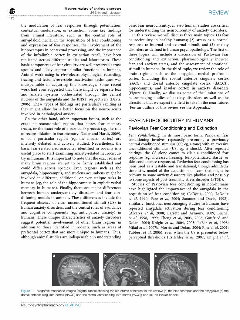
In this review, we will discuss three main topics: (1) fearneurocircuitry in healthy humans; (2) stress as a normalresponse to internal and external stimuli, and (3) anxietydisorders as defined in human psychopathology. The first ofthese topics will include a discussion of Pavlovian fearconditioning and extinction, pharmacologically inducedfear and anxiety states, and the assessment of emotionalstimuli in humans. In the third topic, we review the role ofbrain regions such as the amygdala, medial prefrontalcortex (including the rostral anterior cingulate cortex(rACC) and dorsal anterior cingulate cortex (dACC)),hippocampus, and insular cortex in anxiety disorders(Figure 1). Finally, we discuss some of the limitations ofneuroimaging studies of anxiety disorders as well as thedirections that we expect the field to take in the near future.(For an outline of this review see the Appendix.)
FEAR NEUROCIRCUITRY IN HUMANS
Pavlovian Fear Conditioning and Extinction
Fear conditioning. In its most basic form, Pavlovian fearconditioning involves repeatedly presenting a previouslyneutral conditioned stimulus (CS; eg, a tone) with an aversiveunconditioned stimulus (US; eg, a shock). After repeatedpairings, the CS alone comes to elicit a conditioned fearresponse (eg, increased freezing, fear-potentiated startle, orskin conductance responses). Pavlovian fear conditioning hasbeen used as a testable and translational, though admittedlysimplistic, model of the acquisition of fears that might berelevant to some anxiety disorders like phobias and possiblyto some aspects of post-traumatic stress disorder (PTSD).
Studies of Pavlovian fear conditioning in non-humanshave highlighted the importance of the amygdala in theacquisition of fear conditioning (LeDoux, 2000; LeDouxet al, 1990; Pare et al, 2004; Sananes and Davis, 1992).Similarly, functional neuroimaging studies in humans havereported amygdala activation during fear conditioning(Alvarez et al, 2008; Barrett and Armony, 2009; Buchelet al, 1998, 1999; Cheng et al, 2003, 2006; Gottfried andDolan, 2004; Knight et al, 2004, 2005; LaBar et al, 1998;Milad et al, 2007b; Morris and Dolan, 2004; Pine et al, 2001;Tabbert et al, 2006), even when the CS is presented belowperceptual thresholds (Critchley et al, 2002; Knight et al,
Insular cortexdACC
rACC
Hippocampus Amygdala
a b c
Figure 1. Magnetic resonance images (sagittal slices) showing the structures of interest in this review: (a) the hippocampus and the amygdala; (b) thedorsal anterior cingulate cortex (dACC) and the rostral anterior cingulate cortex (rACC); and (c) the insular cortex.
Neurocircuitry of anxiety disordersLM Shin and I Liberzon
...............................................................................................................................................................
170
REVIEW
..............................................................................................................................................
Neuropsychopharmacology REVIEWS

2009; Morris et al, 2001) and even when more complex USsare used (Doronbekov et al, 2005; Klucken et al, 2009). Inaddition, amygdala activity has been associated with skinconductance changes during fear conditioning (Cheng et al,2006; Furmark et al, 1997; LaBar et al, 1998; Phelps et al,2001). Interestingly, amygdala activation in humans also hasbeen observed in response to cues following (1) verbalinstructions that discriminate between cues that predictshock vs safety (even though no shock was actuallyadministered) (Phelps et al, 2001), and (2) observationalfear learning, whereby participants watch a video of anotherperson experiencing a Pavlovian fear-conditioning para-digm (Olsson et al, 2007). What exactly amygdaloidactivation represents in these latter paradigms is notentirely clear. It could suggest for example that: (1) higherorder centers that decipher the anticipated predictive valueof the cue, or that learn from observation using empathy,convey information to the amygdala, or (2) alternatively,that the human amygdala is less specific in its responses andis more sensitive to contextual modulation in the absence ofa US. These interpretations could have potentially differentimplications for the understanding of the role of theamygdala in anxiety disorders.
Fear conditioning is also associated with increasedactivation in the dACC and rACC (Alvarez et al, 2008;Buchel et al, 1998, 1999; Dunsmoor et al, 2007; Kluckenet al, 2009; LaBar et al, 1998; Marschner et al, 2008; Miladet al, 2007a, b; Morris and Dolan, 2004; Phelps et al, 2004).Activation in the dACC and rACC also occurs duringobservational fear learning (Olsson et al, 2007). In addition,dACC activation is positively correlated with differentialskin conductance responses (Milad et al, 2007a). Fearconditioning studies (involving both specific CSs andcontexts) also commonly report insular cortex activation(Alvarez et al, 2008; Buchel et al, 1999; Buchel et al, 1998;Critchley et al, 2002; Dunsmoor et al, 2007; Gottfried andDolan, 2004; Klucken et al, 2009; Knight et al, 2009;Marschner et al, 2008; Morris and Dolan, 2004; Phelps et al,2001, 2004) and hippocampal activation (Alvarez et al, 2008;Buchel et al, 1999; Knight et al, 2004, 2009; Lang et al, 2009;Marschner et al, 2008).
Extinction. Extinction learning occurs when a CS thatpreviously predicted a US no longer does so, and over time,the conditioned response (eg, freezing or elevated skinconductance responses) decreases. Extinction learning or,more likely, the later recall of this learning involves theventromedial prefrontal cortex (vmPFC) (Milad and Quirk,2002; Morgan et al, 1993; Quirk et al, 2000, 2003, 2006) inrodents. Functional neuroimaging studies of healthyhumans have reported vmPFC activation during extinction(Barrett and Armony, 2009; Gottfried and Dolan, 2004;Kalisch et al, 2006; Milad et al, 2007b) and the later recall ofextinction (Milad et al, 2007b; Phelps et al, 2004). Skinconductance measures of extinction memory are positivelycorrelated with vmPFC activation (Milad et al, 2007b;Phelps et al, 2004) and vmPFC cortical thickness(Milad et al, 2005). Activation of the amygdala and insular
cortex also may occur during extinction learning orrecall (Gottfried and Dolan, 2004; LaBar et al, 1998; Miladet al, 2007b; Phelps et al, 2004), and greater amygdalaresponses during extinction have been associatedwith higher trait anxiety (Barrett and Armony, 2009).Finally, extinction can be modulated by context (ie, thesurroundings in which extinction takes place), and thehippocampus has a role in this process. In rodents, dorsalhippocampal lesions reduce the context-dependence ofextinction (Bouton et al, 2006). In a recent fMRI study,hippocampal activation to the CS + occurred in theextinction context but not in the conditioning context(Kalisch et al, 2006). Hippocampal activation was alsopositively correlated with vmPFC activation in this study(Kalisch et al, 2006), suggesting that hippocampal–vmPFCinteractions may be important for the contextual modula-tion of extinction.
Fear States and Responses to Emotional Stimuli
Pharmacological challenge. Another way to examine themediating functional neuroanatomy of fear or anxiety is touse specific pharmacological agents to provoke suchstates in healthy individuals during PET or fMRI scanning.For example, cholecystokinin-4 (CCK-4) is associated withincreases in subjective states of fear and anxiety, as well asincreased activation in the amygdala, insular cortex,claustrum, cerebellum, brain stem, and the ACC (Benkelfatet al, 1995; Eser et al, 2009; Javanmard et al, 1999; Schuncket al, 2006). In addition, two studies reported dACCincreases during anticipatory anxiety preceding the CCKadministration (Eser et al, 2009; Javanmard et al, 1999). It isimportant to keep in mind, however, that CCK-B receptoragonists like pentagastrin also have direct effects onstress axis stimulation independent of their effects onsubjective experience of distress/fear (Abelson et al, 2005,2008). Procaine administration has been associated withelevated subjective ratings of fear/anxiety, activation of theamygdala, ACC, and insular cortex (Ketter et al, 1996;Servan-Schreiber et al, 1998), and deactivation of neocorticalstructures (Servan-Schreiber et al, 1998). Furthermore,amygdala activity was positively correlated with subjectivereports of anxiety (Ketter et al, 1996; Servan-Schreiber et al,1998). Interestingly, those subjects who did not have apanic attack in response to procaine had greater activation inthe rACC compared with those who did have a panic attack(Servan-Schreiber et al, 1998), consistent with the idea thatthe rACC may perform a regulatory or inhibitory function(Mayberg, 1997). The alpha-2 adrenergic antagonistyohimbine has likewise been associated with increasednormalized blood flow in medial prefrontal cortex, insularcortex, and cerebellum in healthy individuals (Cameron et al,2000). A major caveat in the interpretation of pharmacolo-gical challenge studies, however, is the difficulty in disen-tangling the effects that are specific to fear induction fromthe direct effect of a pharmacological agent on regional brainactivity and from the non-specific effects of the pharmaco-logical agent.
Emotional stimuli. Over the past two decades, functionalneuroimaging studies have shown that a core set of brain
Neurocircuitry of anxiety disordersLM Shin and I Liberzon...............................................................................................................................................................
171
REVIEW
..............................................................................................................................................
Neuropsychopharmacology REVIEWS

regions mediate responses to emotional stimuli in healthyhumans. (For reviews, see Phan et al, 2002; Phan et al,2004b). The relevance of these studies to fear/anxietycircuitry is two-fold: (1) A significant number of theseemotional activation paradigms utilize stimuli that depictand/or elicit fear, and (2) these studies shed light on moregeneral emotion-generating neurocircuitry. PET and fMRIstudies have reported amygdala activation in response toemotionally negative photographs (Britton et al, 2006;Hariri et al, 2002; Irwin et al, 1996; Lane et al, 1997a;Paradiso et al, 1999; Phan et al, 2003b; Reiman et al, 1997;Taylor et al, 1998), odors (Zald and Pardo, 1997) and tastes(Zald et al, 1998). Several studies have reported amygdalaactivation to positive stimuli as well (Garavan et al, 2001;Hamann and Mao, 2002; Hamann et al, 1999, 2002; Liberzonet al, 2003), which suggests that the amygdala respondsmore broadly to emotionally arousing and/or salient stimuli(Phan et al, 2004b). Reappraisal of emotionally negativephotographs is associated with reduced amygdala activation(Ochsner et al, 2002) and increased ventromedial prefrontalcortex activation (Urry et al, 2006). Finally, amygdalaactivation during encoding of emotionally arousing stimuliis correlated with the subsequent recollection of thosestimuli (Cahill et al, 1996; Dolcos et al, 2004, 2005; Hamannet al, 1999).
Medial prefrontal cortex, including the rACC, alsoactivates in response to emotional pictures (Lane et al,1997a, b; Phan et al, 2003a, b, 2004a; Reiman et al, 1997) andmay mediate self-referential processing (Kelley et al, 2002;Lane et al, 1997a; Zysset et al, 2002). Although the medialprefrontal cortex may activate regardless of task or valence,the rACC may be more likely to activate when a cognitivetask is performed during scanning (Phan et al, 2002).Ventromedial PFC responses to fear-related images havebeen negatively associated with cortisol reactivity (Rootet al, 2009). The dACC also activates in response toemotional photographs (Britton et al, 2006; Teasdale et al,1999) and aversive tastes (Zald et al, 1998). Finally, theinsular cortex is responsive to aversive stimuli (Phan et al,2004a), internally generated sadness (Lane et al, 1997b;Reiman et al, 1997) and disgust-related stimuli (Brittonet al, 2006).
Emotional facial expressions. Interestingly, the sameneurocircuitry that has been implicated in fear/anxietyresponses in humans is readily activated by stimuli that arenot intrinsically threatening, but may convey informationregarding the presence of threat in the environment orabout the fearful emotional state of others. Responses in theamygdala are readily elicited by photographs of facialexpressions, especially those of fear (Breiter et al, 1996a;Davis and Whalen, 2001; Fitzgerald et al, 2006; Morris et al,1996; Vuilleumier and Pourtois, 2007; Whalen et al, 2001),even when presented below conscious awareness (Morriset al, 1998; Whalen et al, 1998, 2004). Emotional facialexpressions have also been associated with activation in thedACC, rACC, medial frontal gyrus, and insular cortex(Fitzgerald et al, 2006; Gorno-Tempini et al, 2001; Morriset al, 1996; Phillips et al, 1997, 2004; Sabatini et al, 2009;Sprengelmeyer et al, 1998).
Brain responses to the relatively ambiguous facialexpression of surprise have been shown, in some studies,to depend on the extent to which individual subjectsinterpreted these expressions as positive or negative; morenegative interpretations were associated with greateramygdala and lower ventral medial prefrontal cortexactivation (Kim et al, 2003). These findings are consistentwith the notion that the amygdala and medial prefrontalcortex are reciprocally modulated (eg, Garcia et al, 1999).Furthermore, the experimental manipulation of the contextin which surprise facial expressions are presented altersbrain activation patterns in a similar way: surpriseexpressions associated with a negative context elicited moreamygdala activation than those associated with a positivecontext (Kim et al, 2004). These amygdala activations werepositively correlated with activation in the dACC (Kim et al,2004).
Of relevance to our later discussion of anxiety disordersare findings that suggest that healthy individuals with highscores on anxiety measures have greater amygdala andinsular cortex responses to emotional (angry, fearful, andhappy) faces and less rACC activation than participantswith normative scores on these measures (Bishop et al,2004a, b; Stein et al, 2007). Similarly, trait anxiety has beenpositively correlated with amygdala responses to neutralfaces (Somerville et al, 2004).
Summary
Studies of fear conditioning, pharmacologically inducedfear, and responses to emotional stimuli and facial expres-sions have provided evidence that the human amygdala,although responsive to multiple salient stimuli, respondsreliably and potentially preferably to stimuli that predictthreat and can be involved in mediating fear/anxiety states.Given that patients with anxiety disorders experience fearand distress in response to possible predictors of threat,the amygdala has been hypothesized to be hyperresponsivein some anxiety disorders. In the next section, we willreview the evidence related to this hypothesis.
Studies of extinction have highlighted the potentialinvolvement of the vmPFC and hippocampus in the processof learning and remembering that stimuli that used topredict threat no longer do. One possible reason forexaggerated fear, anxiety, and distress in patients withanxiety disorders is that these emotional responses fail toextinguish or that extinction learning is not recalled evenwhen specific cues no longer predict threat. Indeed, somestudies have reported impaired extinction in several anxietydisorders, such as PTSD (Blechert et al, 2007; Milad et al,2008; Orr et al, 2000; Peri et al, 2000; Wessa and Flor, 2007).Thus, the vmPFC and hippocampus are clear regions ofinterest in functional neuroimaging studies of anxietydisorders.
Finally, both the animal literature and studies reviewedabove suggest that the dACC (and its likely homologprelimbic cortex) and insular cortex respond to emotional
Neurocircuitry of anxiety disordersLM Shin and I Liberzon
...............................................................................................................................................................
172
REVIEW
..............................................................................................................................................
Neuropsychopharmacology REVIEWS

stimuli or those that predict threat. As with the amygdala,hippocampus, and vmPFC, these regions are involvedin multiple other functions; however, they might also haveimportant roles in specific aspects of anxiety. For example,the insular cortex is thought to mediate the monitoringof internal body states, and has been found to behyperresponsive in anxiety-prone individuals (Paulusand Stein, 2006). In summary, research on healthyindividuals has suggested that all of these brain regions areprime candidates to examine in patients with anxietydisorders.
STRESS
An important and often overlooked aspect of the fear/anxiety neurocircuitry is its overlap and interaction with theneurocircuitry that orchestrates the stress response. It isimportant to note that the concept of ‘stress’ used here isrelatively specific. It does not encompass general conceptsof ‘subjective distress’ or ‘performance load.’ Althoughthese are useful concepts, they are heterogeneous by natureand are not likely to be associated with a specificneurocircuitry. On the other hand, the concept of a stresssystem that leads to activation of limbic–hypothalamo–pituitary–adrenal axis (LHPA) and secretion of stresshormones like corticotropin-releasing hormone (CRH),adrenocorticotropic hormone, and cortisol is quite specificand is likely to be highly relevant to the neurocircuitry offear and anxiety. The neurocircuitry governing LHPAactivation has been the focus of intense studies in rodents,primates, and humans because it has been repeatedly linkedto the neurobiology of mood disorders (which is addressedin detail elsewhere in this volume), but the evidence linkingLHPA axis abnormalities to anxiety disorders has been lessconsistent, sometimes confusing, and often oversimplified.At the same time, some of the same brain regions areimplicated in both anxiety and stress responses, suggestingthat these responses are interrelated and can influence eachother. Furthermore, anxiety and mood disorders are highlycomorbid, suggesting that some common abnormalities inneurocircuitry might be present in both disorders. In thefollowing few paragraphs, we briefly address only thestructural overlap in neurocircuits and the effects of stresssystem activation (or stress hormones) on anxiety/fearneurocircuitry.
Epidemiologically, major depression is highly comorbidwith anxiety disorders like PTSD, panic disorder, and socialphobia (Reiger et al, 1990), and anxiety symptoms arehighly prevalent in depression (Frances et al, 1990).Furthermore, major subcortical components of the LHPAaxis (eg, hypothalamus, hippocampus, amygdala, andBNST) have also been identified as key components ofanxiety/fear neurocircuitry (albeit sometimes involvingdifferent subnuclei, for example paraventricular nuclei vsventromedial hypothalamus for LHPA and fear neurocir-cuitry, respectively). More recently with the introduction of
in vivo imaging methodologies in LHPA/stress research, therole of cortical structures like the insula and dorsal mPFC inthe activation and inhibition of stress response, respec-tively, has been reported (Liberzon and Martis, 2006) as wellas the role of subgenual ACC in self-induced sadness anddepression (Mayberg et al, 1999). Together, these findingssuggest a significant overlap in structures involved in thestress response and those involved in fear/anxiety responses(eg medial prefrontal cortex, insula, amygdala, hippocam-pus, and BNST). Finally, with respect to neurotransmittersinvolved, CRH is likely involved in the orchestration of bothLHPA axis activity and many anxiety/fear responses. (For areview see Heim and Nemeroff, 2001.)
The activation of these overlapping regions in functionalneuroimaging studies does not necessarily signify, however,activation of both the fear/anxiety response and the LHPAaxis. As a matter of fact, activation of fear/anxiety does notnecessarily activate an LHPA stress response, even in highlyfearful (phobic) individuals (Curtis et al, 1976). In turn,activation of LHPA axis is not necessarily experiencedsubjectively as fear or anxiety. For example, morningawakening, food intake, and nausea all lead to LHPAaxis activation without notable increases in subjectivesense of fear. It is becoming increasingly clear thatspecific characteristics of experience (novelty, control,social support, etc.) are more salient for LHPA axis acti-vation than degree of subjective distress or fear (Abelsonet al, 2007). These facts help to better understand thefindings of non-specific, or even sometimes counterintui-tive, findings regarding the LHPA and stress responses inanxiety disorders such as panic disorder (Abelson et al,2007) and PTSD (Yehuda, 2006; Yehuda et al, 1991). Thisalso suggests that activation in specific cortical regions likemPFC or insula cannot be readily interpreted as acomponent of the fear response, and has to be consideredwithin a context of a specific experiment, subjective report,symptoms, neuroendocrine profile, etc.
With these caveats in mind, important findings aboutstress exposure and LHPA axis activation affecting fear/anxiety responses have been accumulating. These can beseen in two general categories: (1) the immediate effects ofstress or of stress hormones on fear/anxiety responses (eg,stress or stress hormone exposure immediately precedes,or is present during the fear/anxiety responses), and (2)delayed or developmental effects, (eg, stress exposureduring developmentally sensitive periods, like early child-hood, modulates fear/anxiety responses later in life). In theformer category, it has been reported that exposure to acutestress in healthy individuals potentiates the anxietyresponse (Grillon et al, 2007). In addition, stress exposure(Trier Social Stress Test) led to enhanced galvanic skinresponses to conditioned stimuli (CS + ) during fearconditioning (Jackson et al, 2006). Interestingly, stressmodulates fear responses differentially in men and women.Differential effects of stress on fear/anxiety in females vsmales also have been demonstrated in animal studies.
Neurocircuitry of anxiety disordersLM Shin and I Liberzon...............................................................................................................................................................
173
REVIEW
..............................................................................................................................................
Neuropsychopharmacology REVIEWS

Chronic stress exposure led to impaired extinction recall offear conditioning in male but not female rats (Baran et al,2009). Stress exposure in animal studies also led toenhancement in contextual fear conditioning (Corderoet al, 2003). The effects of stress hormone exposure aresomewhat more difficult to interpret because higherendogenous cortisol levels were associated with higher skinconductance responses (SCR) (Jackson et al, 2006), whereasadministration of exogenous cortisol led to decreased SCR(Stark et al, 2006).
With respect to delayed effects of stress during thevulnerable developmental period, the findings are some-what complex. Studies of early maternal separation inrodents (Plotsky and Meaney, 1993) and variable foragingin primates (Coplan et al, 1996) have revealed long-termalteration in stress axis responses and key neurotransmittersystems (for review see Heim and Nemeroff, 2001). Inaddition, recent findings of gene-by-environment interac-tions in PTSD (Binder et al, 2008) also point toward thepossibility that early childhood experience might modifyfear/anxiety neurocircuitry and contribute to the develop-ment of anxiety disorders. Direct evidence of these effectson fear/anxiety behavior is less convincing, however, asmaternal separation in rat pups, which did alter relevantneurotransmitter systems, did not result in significantlyenhanced startle response or decreased open field explora-tion as compared with non-separated animals (Caldji et al,2000). Similarly, mixed results have been obtained in otherstudies where rats exposed to severe sporadic stress spentmore time in open arms of an elevated plus maze butdisplayed increases in defensive probe burying behavior.Furthermore, animals exposed to milder chronic stressshowed opposite changes (Pohl et al, 2007).
The character of the stress exposure (mild vs severe,prolonged vs short, predictable vs non-predictable) and thesex of the individual emerge as important variables that candefine the long-term effects of stress exposure, but moreexperimental data are clearly needed. Similarly, little isknown about the specific mechanisms by which stressexposure modulates fear/anxiety circuitry. It has beensuggested, as stated above, that early developmental stressexposure alters fear/anxiety circuitry via altered sensitivityand responsivity of the CRH and adrenergic systems, andrecent advances in morphological work had suggested apotential mechanism for the effects of stress on fearconditioning and extinction. Chronic stress decreasesdendritic branching in the hippocampus (McEwen, 2001)and mPFC (Liston et al, 2006; Radley et al, 2004), butincreases dendritic branching in the amygdala (Mitra et al,2005; Vyas et al, 2006). This pattern could lead to increasedconditioning and impaired extinction, and both of theseprocesses could contribute changes in anxiety/fear-relatedbehaviors. Future research addressing these importantquestions will be needed to fully understand the impact ofstress/LHPA axis activation on fear/anxiety and the under-lying neurocircuitry.
ANXIETY DISORDERS
Posttraumatic Stress Disorder
PTSD can develop in individuals who (1) were exposed toan event or events that involved the threat of death orserious injury and (2) reacted with intense fear, helplessnessor horror (APA, 2000). Individuals with PTSD reexperiencethe traumatic event in the form of nightmares, intrusiverecollections, flashbacks, and physiological arousal anddistress in response to reminders of trauma. These patientsmay attempt to avoid reminders of the trauma and mayexperience a restricted range of effect, especially positiveeffect. Finally, patients with PTSD report hyperarousalsymptoms, such as hypervigilance, exaggerated startle, anddifficulty sleeping or concentrating (APA, 2000).
Neurocircuitry models of PTSD implicate the amygdala,mPFC, and hippocampus (Rauch et al, 1998b, 2006).According to some models, the amygdala is hyperrespon-sive in PTSD, which may account for exaggerated fearresponses and the persistence of traumatic memories. Inaddition, portions of the vmPFC (including the rACC) arehyporesponsive and fail to inhibit the amygdala. It is notclear which of the two regions ‘drives’ the overall outcome,but a hyperresponsive amygdala and hyporesponsive mPFCmay potentially lead to deficits in extinction, emotionregulation, attention, and contextual processing (Liberzonand Sripada, 2008). Abnormal hippocampal function maycontribute to deficits in contextual processing, as well asimpairments in memory and neuroendocrine dysregulation.Although not originally included in early neurocircuitrymodels, the dACC and insular cortex may have a role inPTSD as well. Recent studies have suggested that the dACCis hyperresponsive in PTSD, perhaps underlying exagger-ated fear learning. Finally, the insular cortex appears to behyperresponsive in PTSD and other anxiety disorders,consistent with the notion that the insula may mediateanxiety proneness (Paulus and Stein, 2006; Simmons et al,2006). (For other models, see Elzinga and Bremner, 2002;Hamner et al, 1999; Layton and Krikorian, 2002.)
Amygdala. Several studies have reported increased amyg-dala activation in PTSD relative to comparison groups inresponse to trauma-related imagery (Shin et al, 1997; Shinet al, 2004a), combat-related sounds or smells (Liberzonet al, 1999; Pissiota et al, 2002; Vermetten et al, 2007),trauma-related photographs or words (Driessen et al, 2004;Hendler et al, 2003; Morey et al, 2009; Protopopescu et al,2005), fear conditioning (Bremner et al, 2005), and fearfulfacial expressions (Bryant et al, 2008b; Rauch et al, 2000;Shin et al, 2005; Williams et al, 2006). Exaggeratedamygdala activation in PTSD has also been found at rest(Chung et al, 2006; Semple et al, 2000) and during thecompletion of neutral attention and memory tasks (Bryantet al, 2005; Shin et al, 2004b). Several studies, however, havefound no differential response in the amygdala in PTSD (eg,Bremner et al, 1999a; Lanius et al, 2001) or even decreasedresponsivity to negative stimuli (Phan et al, 2006a).Interestingly, resilience to PTSD may be associated with
Neurocircuitry of anxiety disordersLM Shin and I Liberzon
...............................................................................................................................................................
174
REVIEW
..............................................................................................................................................
Neuropsychopharmacology REVIEWS

relatively decreased amygdala activation (Britton et al, 2005;Osuch et al, 2008), and amygdala lesions may reduce theoccurrence of PTSD (Koenigs et al, 2008). In support of thepotential role of amygdala in PTSD, some studies havereported that amygdala activation is positively correlatedwith PTSD symptom severity (Armony et al, 2005; Dickieet al, 2008; Pissiota et al, 2002; Protopopescu et al, 2005;Rauch et al, 2000; Shin et al, 2004a). Similarly, response tocognitive-behavioral treatment is associated with a decreasein amygdala activation (Felmingham et al, 2007; Peres et al,2007), and relatively higher pre-treatment amygdala activa-tion is predictive of a less favorable response to cognitive-behavioral therapy (Bryant et al, 2008a).
Relatively few studies have examined amygdala structureand neurochemistry in PTSD. Two studies have reportedtrends for smaller amygdala volumes in PTSD (Bremneret al, 1997; Wignall et al, 2004), but several others have not(Bonne et al, 2001; De Bellis et al, 2001a; Fennema-Notestine et al, 2002; Gilbertson et al, 2002; Gurvits et al,1996; Lindauer et al, 2004b). One recent study using PETand 11C-carfentanil has reported diminished mu-opioidreceptor binding in the extended amygdala in trauma-exposed individuals with vs without PTSD (Liberzon et al,2007b). Another study has found decreased [11C]flumazenilbinding in the left amygdala in PTSD subjects comparedwith trauma-exposed control participants, consistent withaltered GABAergic function in this disorder (Geuze et al,2008a), although two other studies have not reported thisfinding (Bremner et al, 2000a; Fujita et al, 2004).
Medial prefrontal cortex. Functional neuroimaging studiesof PTSD have reported decreased activation or failure toactivate the mPFC (including the rACC, medial frontalgyrus, and subcallosal cortex) during traumatic script-driven imagery (Bremner et al, 1999a; Britton et al, 2005;Lanius et al, 2001; Lindauer et al, 2004a; Shin et al, 1999,2004a), the presentation of trauma-related stimuli (Bremneret al, 1999b; Hou et al, 2007; Yang et al, 2004), and negative,non-traumatic stimuli (Kim et al, 2007; Lanius et al, 2003;Phan et al, 2006a; Shin et al, 2005; Williams et al, 2006).Relatively diminished activation of the mPFC in PTSD alsohas been shown during extinction (Bremner et al, 2005),emotional Stroop interference (Bremner et al, 2004; Shinet al, 2001), emotional word retrieval (Bremner et al,2003b), non-emotional cognitive tasks (Bryant et al, 2005;Semple et al, 2000) and at rest (Semple et al, 2000).Furthermore, mPFC activation appears to be inverselycorrelated with PTSD symptom severity (Britton et al, 2005;Dickie et al, 2008; Hopper et al, 2007; Kim et al, 2007; Shinet al, 2004a, 2005; Williams et al, 2006) and positivelycorrelated with pre-scan cortisol levels (Liberzon et al,2007a). Finally, increased mPFC activation following treat-ment has been positively associated with symptomaticimprovement (Felmingham et al, 2007; Lansing et al, 2005;Peres et al, 2007; Seedat et al, 2004), although not allstudies have shown this, perhaps due to paradigm-relatedmethodological differences (Bryant et al, 2008a).
Most of the findings summarized above reflect activationpeaks in rostral ACC and ventral portions of the mPFC. Incontrast, more dorsal regions of the ACC (ie, the dACC)
appear to have either normal or exaggerated responsivityin PTSD during fear conditioning, interference tasks,an auditory oddball task, and at rest (Bremner et al, 2005;Bryant et al, 2005; Felmingham et al, 2009; Pannu Hayeset al, 2009; Shin et al, 2001, 2007, in press).
The findings of several studies suggest diminishedvolumes or gray matter densities in the ACC in PTSD(Corbo et al, 2005; Kasai et al, 2008; Rauch et al, 2003;Woodward et al, 2006; Yamasue et al, 2003), and smallerACC volumes have been associated with greater PTSDsymptom severity (Woodward et al, 2006; Yamasue et al,2003). In a study of monozygotic twins discordant fortrauma exposure, diminished gray matter densities inpregenual ACC were not found in the identical twins ofthe PTSD participants, suggesting that this gray matterdensity decrease is likely an acquired sign of the disorderrather than a familial risk factor (Kasai et al, 2008).
Magnetic resonance spectroscopy (MRS) studies haverevealed diminished N-acetyl aspartate (NAA) levels in theACC in PTSD (De Bellis et al, 2001b, b; Ham et al, 2007a;Mahmutyazicioglu et al, 2005; Schuff et al, 2008). Further-more, NAA levels in the pregenual ACC were negativelycorrelated with the severity of reexperiencing symptoms(Ham et al, 2007a). Two recent studies have reporteddecreased benzodiazepine receptor binding in the mPFC inPTSD (Bremner et al, 2000a; Geuze et al, 2008a), althoughone other study failed to find this effect (Fujita et al, 2004).
Hippocampus. Some functional neuroimaging studies havereported decreased hippocampal activation during sympto-matic states (Bremner et al, 1999a) and during memorytasks that involve neutral or emotional stimuli (Astur et al,2006; Bremner et al, 2003a, b; Moores et al, 2008; Shin et al,2004b). One study found reduced glucose metabolic rate inthe hippocampus at rest (Molina et al, 2007), and anotherreported that successful treatment was related to increasedhippocampal activation (Peres et al, 2007). Other studies,however, have reported increased activation in the hippo-campus in PTSD (Geuze et al, 2007, 2008b; Sachinvala et al,2000; Semple et al, 2000; Thomaes et al, 2009; Werner et al,2009) or a positive correlation between hippocampalactivation and PTSD symptom severity (Osuch et al, 2001;Shin et al, 2004b). The direction of hippocampal functionalabnormalities appears to depend in part on the type of taskand the type of statistical analysis used.
Hippocampal volumes appear to be diminished in PTSDin some (Bossini et al, 2008; Bremner et al, 1995, 1997,2003a; Gilbertson et al, 2002; Gurvits et al, 1996; Karl et al,2006; Kitayama et al, 2005; Smith, 2005; Stein et al, 1997;Villarreal et al, 2002a; Wignall et al, 2004; Winter and Irle,2004; Woon and Hedges, 2008), but not all studies (Bonneet al, 2001; Carrion et al, 2001; De Bellis et al, 1999, 2002;Fennema-Notestine et al, 2002; Golier et al, 2005; Pedersonet al, 2004). Hippocampal volumes have been inverselyassociated with verbal memory deficits (Bremner et al,1995), combat exposure severity (Gurvits et al, 1996),dissociative symptom severity (Bremner et al, 2003a; Steinet al, 1997), depression severity (Villarreal et al, 2002a), and
Neurocircuitry of anxiety disordersLM Shin and I Liberzon...............................................................................................................................................................
175
REVIEW
..............................................................................................................................................
Neuropsychopharmacology REVIEWS

PTSD symptom severity (Bremner et al, 2003a; Gilbertsonet al, 2002; Villarreal et al, 2002a). Spectroscopy studies ofhippocampus have reported decreased NAA in the hippo-campus, often interpreted as consistent with decreasedneuronal integrity (Brown et al, 2003; Freeman et al, 1998;Ham et al, 2007a; Mohanakrishnan Menon et al, 2003;Schuff et al, 2001; Villarreal et al, 2002b). The results of twostudies suggest that hippocampal volumes may increasefollowing treatment with serotonin reuptake inhibitors(Bossini et al, 2007; Vermetten et al, 2003).
Whether decreased volumes can explain abnormalhippocampal activation in PTSD is not entirely clear,although the findings of at least two studies suggest thatfunctional abnormalities might be still present even if thevolumetric differences are controlled statistically (Bremneret al, 2003a; Shin et al, 2004b). The origin of decreasedhippocampal volumes is not known, although the results ofone twin study suggest that diminished hippocampalvolumes may be a familial risk factor for developing PTSDfollowing psychological trauma (Gilbertson et al, 2002).
With regard to neurochemistry, one recent PET studyfound decreased [11C]flumazenil binding in the hippocam-pus (as well as thalamus and cortical areas) suggestingdiminished benzodiazepine–GABAA function in the hippo-campus in PTSD (Geuze et al, 2008a).
Insular cortex. Relative to comparison groups, increasedactivation in the insular cortex has been found in PTSDduring script-driven imagery (Lanius et al, 2007; Lindaueret al, 2008), fear conditioning and extinction (Bremner et al,2005), the anticipation of negative images (Simmons et al,2008), the retrieval of emotional or neutral stimuli (Bremneret al, 2003b; Werner et al, 2009; Whalley et al, 2009),aversive smells and painful stimuli (Geuze et al, 2007;Vermetten et al, 2007), and the performance of anemotional Stroop task (Shin et al, 2001). Insular cortexactivation has been found to be positively correlated withmeasures of symptom severity (Carrion et al, 2008; Hopperet al, 2007; Osuch et al, 2001) and post-scan plasmaadrenocorticotropic hormone levels (Liberzon et al, 2007a).Although greater insular activation in PTSD has beenconfirmed by a recent voxel-wise meta-analysis (Etkin andWager, 2007), a few studies have reported either no group
differences in insular activation or relatively decreasedactivation in PTSD (Bremner et al, 1999a, 2004; Molina et al,2007; Moores et al, 2008; Phan et al, 2006a; Shin et al, 1999).
Voxel-based morphometry studies of PTSD have reportedreduced gray matter density in the insular cortex (Chen et al,2006; Corbo et al, 2005; Kasai et al, 2008). In one study,gray matter density in the insular cortex was negativelycorrelated with reexperiencing; that is, lower graymatter density was associated with greater reexperiencing(Kasai et al, 2008).
One recent PET-[11C]flumazenil study has reporteddecreased benzodiazepine–GABAA receptor binding inbilateral insular cortex in PTSD (Geuze et al, 2008a).
Summary. In general, the functional neuroimaging findingsin PTSD support the hypothesis that the amygdala ishyperresponsive and ventral portions of medial prefrontalcortex are hyporesponsive, at least in some groups of PTSDpatients. Indeed, the latter finding appears to be one of themost robust in the literature (Etkin and Wager, 2007)(Table 1). In addition, albeit in a small number of studies,reduced volumes and gray matter densities in the ACC havebeen fairly consistently reported. Furthermore, emergingevidence suggests that the dACC and insular cortex may behyperresponsive in PTSD, although insular cortex hyperre-sponsivity does not appear to be specific to PTSD (Etkinand Wager, 2007). Finally, the majority of studies havefound diminished hippocampal volumes in PTSD patients.Hippocampal function appears to be abnormal as well,although the direction of the abnormality seems to dependon the type of task completed during neuroimaging.
Panic Disorder
Panic disorder patients experience recurrent, unexpectedpanic attacks, along with a persistent concern about havingfuture attacks, or worry about the implications of theattacks, or a significant change in behavior related to theattacks (APA, 2000). A panic attack is a discrete episode ofintense fear, discomfort, and sympathetic nervous systemarousal that occurs in the absence of true danger (APA,2000). According to one of the neurocircuitry models ofpanic disorder, the ‘fear network,’ which includes the
Table 1 Summary of the Direction of Functional Neuroimaging Findings in Anxiety Disorders
rACC¼ rostral anterior cingulate cortex; dACC¼ dorsal anterior cingulate cortex.m¼ increased function in the disorder (relative to control groups).k¼ decreased function in the disorder (relative to control groups).mk¼ mixed findings.*¼ based on a very small number of studies.F¼ too little information available.
Neurocircuitry of anxiety disordersLM Shin and I Liberzon
...............................................................................................................................................................
176
REVIEW
..............................................................................................................................................
Neuropsychopharmacology REVIEWS

amygdala, hippocampus, thalamus, and brain stem struc-tures, is hypersensitive. Furthermore, frontal cortex fails toprovide top-down inhibitory input to the amygdala, leadingto exaggerated amygdala activation and unnecessaryactivation of the entire fear network, resulting in a panicattack (Coplan and Lydiard, 1998; Gorman et al, 2000). Thistype of model is similar to the models proposed for otheranxiety disorders such as PTSD. Indeed, PTSD patientsoften suffer from comorbid panic attacks (Falsetti andResnick, 1997). Although it is quite possible that PTSD andpanic disorder indeed share similar pathophysiologicalcomponents, it will be important in the future to identifyabnormalities that are disorder specific and are responsiblefor disorder-specific symptomatology.
Amygdala. Hyperactivation of the amygdala in panicdisorder has been reported in response to panic-relatedwords (van den Heuvel et al, 2005b) and neutral faces(Pillay et al, 2007). One recent PET study found greaterresting glucose metabolism in the amygdala (Sakai et al,2005), although amygdala glucose metabolism did notchange after effective treatment with cognitive-behavioraltherapy (Sakai et al, 2006). The possibility that amygdalahyperactivation is present in a subgroup of panic disorderpatients has been supported by recent studies examiningthe effect of genotypes within patients with panic disorder.These have revealed greater amygdala activation in carriersof the COMT 158val allele (Domschke et al, 2008), the5-HT1A x1019 GG allele, and the short allele of the sero-tonin transporter polymorphism (Domschke et al, 2006). Incontrast, two studies found relatively decreased amygdalaactivation during anticipatory anxiety (Boshuisen et al,2002) and in response to fearful facial expressions (Pillayet al, 2006), although the latter finding may be attributedto the fact that panic disorder patients were taking anti-depressants, which have been found to decrease amygdalaactivation (Harmer et al, 2006).
The volumetric findings in the amygdala of panic dis-order patients are very sparse. One study reported smallerbilateral amygdala volumes in panic disorder comparedwith healthy participants (Massana et al, 2003). The samegroup reported reduced levels of creatine and phos-phocreatine in the right medial temporal lobe (includingthe amygdala and part of the hippocampus) in panicdisorder. This was interpreted as potentially representing ahypermetabolic state in the right medial temporal region(Massana et al, 2002), which would be consistent with thefindings of Sakai et al (2005). Finally, with respect torelevant neurotransmission findings, SPECT and PETstudies have reported decreased GABA–benzodiazepinereceptor binding in the medial temporal lobes (Kaschkaet al, 1995; Malizia et al, 1998) and decreased 5-HT1A
receptor binding in the amygdala in panic disorder (Nashet al, 2008).
Medial prefrontal cortex. Consistent with the literature onpharmacologically induced fear and panic in healthyvolunteers, studies of panic disorder have revealed in-creased rACC activation during imagery of high vs low
anxiety situations (Bystritsky et al, 2001), anticipatoryanxiety (Boshuisen et al, 2002), and in response to happyfaces in panic disorder (Pillay et al, 2007), althoughmedication was a potential confound in the latter study.Dorsal ACC hyperresponsivity has also been reported inpanic disorder in one study (Pillay et al, 2007). Panicdisorder patients who are carriers of the COMT 158val alleleappear to have less medial prefrontal deactivation andgreater orbitofrontal activation in response to emotionalfacial expressions (Domschke et al, 2008). In contrast, panicdisorder subjects with the 5-HT1A x1019 GG allele had lessACC, medial prefrontal cortex, and orbitofrontal activationin response to fearful facial expressions (Domschke et al,2006).
Volumetric evidence is again sparse, but it appears thatboth dACC and rACC might be exhibiting similar types ofchange. Gray matter volumes appear to be reduced in therACC (Asami et al, 2008; Uchida et al, 2008) and dACC(Asami et al, 2008), and white matter integrity, as measuredby DTI, appears to be enhanced in the rACC in panicdisorder (Han et al, 2008).
Two studies have reported decreased GABAA–benzodia-zepine receptor binding in the ACC and medial prefrontalcortex, including in the dACC (Hasler et al, 2008; Maliziaet al, 1998), and one study found a negative correlationbetween panic attack symptom severity and benzodiazepinereceptor binding in dorsal medial frontal gyrus (Bremneret al, 2000b). An MRS study reported increased lactate andcholine levels in the rACC (Ham et al, 2007b), and two PETstudies have reported decreased 5-HT1A receptor binding inthe anterior cingulate in panic disorder (Nash et al, 2008;Neumeister et al, 2004).
Hippocampus. The evidence for hippocampal involvementin panic disorder comes mainly from studies of metabolismand perfusion. Two PET studies of panic disorder havereported abnormalities in the laterality of hippocampalresting glucose metabolic rates (Nordahl et al, 1990, 1998),and two PET studies have found greater resting glucosemetabolism in the hippocampus in patients with panicdisorder (Bisaga et al, 1998; Sakai et al, 2005). In contrast, aSPECT study has reported reduced perfusion in thehippocampus in panic disorder (De Cristofaro et al,1993). Two studies have reported decreased GABAA–benzodiazepine receptor binding in the hippocampus(Bremner et al, 2000b; Malizia et al, 1998), and one studyhas reported the opposite finding (Hasler et al, 2008).
Insular cortex. With regard to the insular cortex in panicdisorder, studies have reported decreased activity duringanticipatory anxiety (Boshuisen et al, 2002), increased graymatter volume (Protopopescu et al, 2006; Uchida et al,2008), decreased 5-HT1A receptor binding (Nash et al,2008), and decreased GABAA–benzodiazepine receptorbinding (Malizia et al, 1998).
Brain stem. Functional neuroimaging studies have reportedincreased glucose metabolic rates (Sakai et al, 2005) andincreased activity during anticipatory anxiety (Boshuisenet al, 2002) in the brain stem in panic disorder. Gray matter
Neurocircuitry of anxiety disordersLM Shin and I Liberzon...............................................................................................................................................................
177
REVIEW
..............................................................................................................................................
Neuropsychopharmacology REVIEWS

volume appears to be increased in the midbrain and pons(Protopopescu et al, 2006; Uchida et al, 2008). DecreasedGABAA–benzodiazepine receptor binding was reported inthe pons (Malizia et al, 1998), and two studies have founddecreased 5-HT1A receptor binding in the raphe nucleus inpanic disorder (Nash et al, 2008; Neumeister et al, 2004).
Summary. Several studies have provided evidence consis-tent with amygdala and brain stem hyperresponsivity inpanic disorder. Activation in rACC and dACC appears to beincreased, and gray matter volumes in these regions appearto be decreased. Several studies have reported decreasedGABAA–benzodiazepine and 5-HT1A receptor binding in theamygdala, medial prefrontal cortex, insular cortex, andbrain stem in panic disorder. A common limitation seen inseveral neuroimaging studies of this disorder is theinclusion of participants taking psychiatric medications(Domschke et al, 2006, 2008; Han et al, 2008; Pillay et al,2006, 2007; Uchida et al, 2008). Thus, the findings of suchstudies should be interpreted cautiously pending replicationin medication-free participants.
Social Phobia
Social phobia (or social anxiety disorder) is characterizedby a marked and persistent fear of social or performancesituations involving possible scrutiny by others (APA,2000). The fear of embarrassment and distress can lead toavoidance of social situations and impairment in social,occupational, and academic functioning. The amygdala andmedial prefrontal cortex have been considered importantregions of interest in this disorder (Amaral, 2002; Liebowitzet al, 2005; Mathew et al, 2001; Stein et al, 2002a; Stein,1998).
Amygdala. Exaggerated amygdala responses in socialphobia have been observed during public speaking (Tillforset al, 2001), the anticipation of public speaking (Lorber-baum et al, 2004; Tillfors et al, 2002), negative comments(Blair et al, 2008a), and in response to neutral, angry,contemptuous, happy, and schematic angry facial expres-sions (Birbaumer et al, 1998; Blair et al, 2008b; Cooney et al,2006; Evans et al, 2008; Gentili et al, 2008; Guyer et al, 2008;Phan et al, 2006b; Schneider et al, 1999; Stein et al, 2002b;Straube et al, 2004a, 2005; Veit et al, 2002; Yoon et al, 2007).One study found temporally delayed amygdala responses tofaces in a social phobia group compared with a healthycontrol group (Campbell et al, 2007). In addition, amygdalaresponses in social phobia have been positively correlatedwith self-reported fear increases (Tillfors et al, 2001),severity of social anxiety symptoms (Blair et al, 2008b;Evans et al, 2008; Guyer et al, 2008; Phan et al, 2006b), andstate-trait anxiety scores (Cooney et al, 2006). The geneticpredisposition for social phobia has been supported by thefinding that social phobia patients with a short allele of theserotonin transporter polymorphism had greater amygdalaresponses during public speaking compared to those withlong alleles (Furmark et al, 2004). Finally, amygdalaactivation during public speaking in social phobia appearsto decrease with successful treatment (Furmark et al, 2002,2005). In contrast, one recent study found decreased
amygdala activation during script-driven imagery ofanxiety-provoking social situations and a mental arithmetictask in social phobia (Kilts et al, 2006). One study hasreported reduced 5-HT1A receptor binding in the amygdalain this disorder (Lanzenberger et al, 2007).
Medial prefrontal cortex. Exaggerated rACC activation insocial phobia has been found in response to facialexpressions of fear (Blair et al, 2008b) and disgust (Amiret al, 2005), and in response to pictures of peers with whompatients did not want to interact (Guyer et al, 2008). Onestudy reported greater orbitofrontal cortex activation toangry vs neutral prosody (Quadflieg et al, 2008). In contrast,other studies have found decreased activation (VanAmeringen et al, 2004) and decreased glucose metabolicrates in the ventromedial prefrontal cortex (Evans et al,2009), which increased following treatment with tiagabine(Evans et al, 2009). Glutamate/creatine and NAA/creatineratios appear to be elevated in the rACC and are correlatedwith symptom severity (Phan et al, 2005).
Studies of dACC function in social phobia also have beensomewhat mixed. Greater dACC activation has beenreported in response to negative comments (Blair et al,2008a) and harsh or disgusted facial expressions (Amiret al, 2005; Phan et al, 2006b). Another study reportedgreater dorsal medial frontal activation in response to harshfaces (Stein et al, 2002b). Treatment with nefazodone-decreased activation in dACC (Kilts et al, 2006). In contrast,other studies have found decreased dACC activation toschematic angry faces (Evans et al, 2008) and in anticipationof public speaking (Lorberbaum et al, 2004), as well asdecreased glucose metabolism at rest (Evans et al, 2009).One study found temporally delayed medial prefrontalcortex/dACC responses to faces in social phobia, which mayhelp to account for the heterogeneity of findings in theliterature regarding this structure (Campbell et al, 2007).Finally, one study reported reduced 5-HT1A receptorbinding in the anterior cingulate in social phobia, althoughwhether this finding occurred in the dorsal or rostral ACCwas not specified (Lanzenberger et al, 2007).
Insular cortex. Insular cortex activation appears to beelevated in social phobia during the anticipation of publicspeaking (Lorberbaum et al, 2004) and in response toemotional facial expressions (Amir et al, 2005; Gentili et al,2008; Straube et al, 2004a, 2005; Yoon et al, 2007), includingschematic facial expressions (Evans et al, 2008; Straubeet al, 2004a). Two studies, however, found decreased insularcortex activation during public speaking (Tillfors et al,2001) and during an implicit sequence-learning task insocial phobia (Sareen et al, 2007). One study has foundreduced 5-HT1A receptor binding in the insular cortex inthis disorder (Lanzenberger et al, 2007).
Striatum. One recent study has found reduced caudateactivation during an implicit sequence-learning task ingeneralized social phobia (Sareen et al, 2007). Other studieshave found reduced D2 receptor binding and dopaminetransporter densities in the striatum in social phobia(Schneier et al, 2000; Tiihonen et al, 1997), although a
Neurocircuitry of anxiety disordersLM Shin and I Liberzon
...............................................................................................................................................................
178
REVIEW
..............................................................................................................................................
Neuropsychopharmacology REVIEWS

recent study failed to replicate these findings (Schneier et al,2009).
Summary. Exaggerated amygdala activation has been themost consistent functional neuroimaging finding in thesocial phobia literature. Although several studies havereported exaggerated rACC and insular cortex activationas well, a number of other studies have reported contra-dictory findings. Future research will have to: (1) addressquestions regarding ACC/mPFC and insular involvement,potentially utilizing cognitive activation tasks to probe ACCfunction, and (2) attempt to identify the neurocircuitryspecific to social phobia (eg, the regions that contribute tothe perception/interpretation of social stimuli as particu-larly anxiety/fear inducing).
Specific Phobia
Specific phobias are marked by excessive, unreasonable andpersistent fear of specific objects or situations such as smallanimals, flying, enclosed places, heights, and blood/injury(APA, 2000). The fear and avoidance causes significantdistress and/or impairment in occupational, academic, orsocial functioning. Specific phobia is a relatively commondisorder, with a lifetime prevalence of 7–11% (APA, 2000).Early models of the etiology of phobias centered on fearconditioning and extinction, and therefore implicated theamygdala and medial prefrontal cortex. Admittedly, suchfear conditioning models are likely to be incomplete giventhat (1) many individuals with phobias cannot recall aconditioning event, and (2) only a small number ofcommon stimuli or situations are the objects of phobias(Fyer, 1998). Nevertheless, fear conditioning and extinctionmodels have been useful in guiding neuroimaging research-ers toward examining amygdala, medial prefrontal cortex,and insular cortex function in this disorder.
Amygdala. Exaggerated amygdala activation in individualswith specific phobia has been observed in response tophobia-related pictures (Dilger et al, 2003; Goossens et al,2007a, b; Schienle et al, 2005, 2007; Straube et al, 2006b;Veltman et al, 2004; Wendt et al, 2008). In addition,treatment has been associated with decreased amygdalaactivation (Goossens et al, 2007b; Schienle et al, 2007).However, numerous studies have not reported exaggeratedamygdala activation in specific phobia (Hermann et al,2007; Larson et al, 2006; Paquette et al, 2003; Straube et al,2004b, 2007; Wik et al, 1996, 1997; Wright et al, 2003),perhaps in part due to methodological differences, such asthe use of different imaging modalities, type of stimuli(verbal vs pictorial), mode of presentation (video clips vsstills), or phobia-unrelated facial expressions. Preliminaryfindings assessing the NK1 receptor have suggestedenhanced levels of endogenous neuropeptide substance P(commonly associated with stress and negative affect) in theamygdala in phobics when presented with phobia-relevantpictures (Michelgard et al, 2007).
Medial prefrontal cortex. The findings with regard to therACC in specific phobia are mixed, with two studies
showing less rACC activation in phobia groups in responseto phobia-related vs neutral pictures (Hermann et al, 2007;Schienle et al, 2007), and two studies reporting enhancedrACC activation to phobia-related stimuli (Britton et al,2009; Pissiota et al, 2003). Activation in the rACC appears tobe positively correlated with anticipatory anxiety in phobiapatients (Straube et al, 2007). The rACC has been shown tobe thicker in participants with specific phobias relative tocontrol participants without phobias (Rauch et al, 2004).
In contrast, the results of functional neuroimaging studiesare much more consistent with regard to the dACC, whichhas been found to be hyperresponsive to phobia-relatedstimuli (Goossens et al, 2007a, b; Straube et al, 2006a, b) orthe anticipation of such stimuli (Straube et al, 2007). Inaddition, dACC activation in specific phobia decreases afterhabituation (Veltman et al, 2004) and cognitive-behavioraltreatment (Goossens et al, 2007b; Straube et al, 2006a).
Insular cortex. Recent research has reported exaggeratedinsular cortex activation in specific phobia vs controlcohorts in response to phobia- or fear-related pictures,videos, and words (Dilger et al, 2003; Goossens et al,2007a, b; Schienle et al, 2005; Straube et al, 2004b, 2006b,2007; Wendt et al, 2008), as well as fearful facial expressions(Wright et al, 2003). In addition, treatment studies havereported decreased insula activation following cognitive-behavioral treatment (Goossens et al, 2007b; Schienle et al,2007; Straube et al, 2006a). Insular cortex also appears to bethicker (bilaterally) in participants with specific phobia ascompared with healthy control participants (Rauch et al,2004).
Summary. The amygdala, dACC and insular cortex allappear to be hyperresponsive to phobia-related stimuli inspecific phobia. These abnormalities tend to normalize withsuccessful treatment. The findings are few and mixed withregard to the rACC.
Obsessive–Compulsive Disorder
Patients with obsessive–compulsive disorder (OCD) experi-ence recurrent, unwanted thoughts or images (obsessions)that cause distress, and engage in excessive ritualisticbehaviors or mental acts (compulsions) that are typicallycarried out in response to the obsessions (APA, 2000).Because OCD is covered more extensively in anotherchapter, we only very briefly discuss it here (see alsoFriedlander and Desrocher, 2006; Menzies et al, 2008). Ingeneral, abnormalities in thalamo-cortico–striatal loopshave been posited to account for the repetitive quality andthe cognitive and motor content of the obsessions andcompulsions in OCD. One neurocircuitry model of OCDposits that the striatum (caudate nucleus) functionsabnormally, leading to inefficient gating in the thalamus(Graybiel and Rauch, 2000; Rauch et al, 1998a). This maylead to hyperactivity in the orbitofrontal cortex and theanterior cingulate cortex, which may mediate intrusivethoughts and anxiety, respectively. Compulsions mayserve to recruit the striatum and to achieve thalamic gating,
Neurocircuitry of anxiety disordersLM Shin and I Liberzon...............................................................................................................................................................
179
REVIEW
..............................................................................................................................................
Neuropsychopharmacology REVIEWS

thereby neutralizing the obsessions and reducing anxiety.Thus, the fear/anxiety-related brain regions that we havefocused on so far (eg, amygdala, mPFC, insula, andhippocampus) do not appear to mediate the core OCDsymptomatology. It is interesting to note here that theneurocircuitry model of OCD is much further developed (eg,nodes, links, and directionality are better specified) than themodels of other anxiety disorders. There could be a numberof factors contributing to this: (1) OCD has been studiedlonger than other anxiety disorders; (2) The nature of OCDsymptoms (for example the predominant cognitive compo-nent) implicated neurocircuitry that is better understood oris organized in a way that renders itself easier for thedevelopment of circuitry-based models. Whatever the reasonmight be, one of the goals for future research in other anxietydisorders should be to strive to develop neurocircuitryhypotheses that are comparable in detail and specificationsto the models that are already available for OCD.
Although some functional neuroimaging studies havefound elevated amygdala activation in OCD (Breiter et al,1996b; van den Heuvel et al, 2004; Van Laere et al, 2006), themajority have not, even with facial expression paradigmsthat are known to activate the amygdala in healthy subjectsand patients with other anxiety disorders (Cannistraro et al,2004). The preponderance of neuroimaging studies haveidentified functional and/or structural abnormalities in thecomponents of thalamo–cortico–striatal loops: striatum(Bartha et al, 1998; Baxter et al, 1987, 1988; Chen et al,2004; Rauch et al, 1994, 1997; Remijnse et al, 2006; Robinsonet al, 1995; Rosenberg et al, 1997; van den Heuvel et al,2005a), thalamus (Atmaca et al, 2006; Chen et al, 2004;Fitzgerald et al, 2000; Gilbert et al, 2000), orbitofrontalcortex (Baxter et al, 1987, 1988; Chamberlain et al, 2008;Chen et al, 2004; Kang et al, 2004; Rauch et al, 1994;Remijnse et al, 2006; Valente et al, 2005), and the ACC(Ebert et al, 1997; Fitzgerald et al, 2005; Rauch et al, 1994;Valente et al, 2005). In addition, recent receptor imagingstudies in OCD have revealed reduced serotonin transporteravailability in the thalamus and midbrain (Reimold et al,2007), as well as reduced 5-HT2A receptor availability in theACC and other frontal cortical areas (Perani et al, 2008). Oneinteresting and unanswered question remains: If OCD isindeed associated with very high anxiety states, especiallylinked to specific worries or compulsions, why doesn’t thecircuitry associated with other anxiety disorders or stateanxiety in healthy controls have a more prominent role inthe expression of this anxiety?
Generalized Anxiety Disorder
Generalized anxiety disorder (GAD) is characterized byexcessive diffuse anxiety and worry that is difficult tocontrol. Patients with GAD may experience restlessness,fatigue, irritability, muscle tension, and sleep and concen-tration difficulties (APA, 2000). Relatively few neuroima-ging studies of GAD exist in the literature and some ofthe findings are conflicting; however, some studies have
implicated the amygdala and medial prefrontal cortex inthis disorder.
Amygdala. Exaggerated amygdala activation in response tofearful (McClure et al, 2007b) and masked angry facialexpressions (Monk et al, 2008) and during the anticipationof aversive photographs (Nitschke et al, 2009) has beenreported in patients with GAD. In a mixed cohort ofsubjects with GAD and social phobia, subjects who had alow tolerance for uncertainty had elevated amygdalaactivation during a decision-making task (Krain et al,2008). In a study of adolescents with GAD, amygdalaresponses were positively correlated with GAD symptomseverity (Monk et al, 2008). However, other studies have notfound exaggerated amygdala activation in GAD (Blair et al,2008b; Whalen et al, 2008). In a recent treatment study,greater pre-treatment left amygdala activation to fearfulfaces was associated with a less favorable response tovenlafaxine (Whalen et al, 2008); in contrast, a differentstudy on a pediatric GAD sample found that greater pre-treatment left amygdala activation to fearful faces wasassociated with a more favorable response to cognitive-behavioral treatment (McClure et al, 2007a). One study hasreported larger amygdala volumes in pediatric GAD (DeBellis et al, 2000a).
Medial prefrontal cortex. Although the literature iscurrently very small, it appears that medial prefrontalcortex activation may be elevated in GAD. Activation indACC and rACC appears to be elevated in response tofearful facial expressions in adolescents with GAD (McClureet al, 2007b). In a mixed cohort of subjects with GAD andsocial phobia, those with higher intolerance for uncertaintyhad elevated rACC and subgenual ACC activation during adecision-making task (Krain et al, 2008). In addition, dorsalACC activation in response to worry vs neutral statementsdeclined significantly after treatment with citalopram(Hoehn-Saric et al, 2004). Finally, greater pre-treatmentrACC activation in response to fearful facial expressions(Whalen et al, 2008) and the anticipation of emotionalstimuli (Nitschke et al, 2009) was associated with a morefavorable response to venlafaxine. Regions in lateralprefrontal cortex also appear to show exaggerated activation(Monk et al, 2006) and elevated NAA/creatine ratios in GAD(Mathew et al, 2004).
Summary. Although there is some evidence for exaggeratedamygdala and medial prefrontal cortex responses in GAD,there are too few studies to form conclusions about the roleof these structures in the pathophysiology of GAD. Futurefunctional neuroimaging studies using fearful facial expres-sions and tasks that probe medial prefrontal cortex function(eg, extinction, emotion regulation, or interference tasks)might be able to contribute the much needed additionalinformation regarding amygdala and medial prefrontalcortex function in GAD.
Summary
Overall, the findings of functional neuroimaging studiesare consistent with the notion of exaggerated amygdala
Neurocircuitry of anxiety disordersLM Shin and I Liberzon
...............................................................................................................................................................
180
REVIEW
..............................................................................................................................................
Neuropsychopharmacology REVIEWS

activation to specific stimuli in a number of anxietydisorders, especially social phobia, specific phobia, andPTSD. Facial expressions have been especially effectiveprobes of amygdala responses in social phobia and PTSD.Interestingly, the data regarding amygdala function in panicdisorder are still inconclusive, and given that relatively fewstudies have examined amygdala function in GAD, addi-tional research is needed to make meaningful comparisonsof amygdala responses between various anxiety disorders.Although increased amygdala activation has been observedin a few studies of OCD, the overall pathophysiology ofOCD appears to be localized to a different brain circuit.One potential conceptualization of these findings is thatamygdala hyperactivation is a common pathway for exag-gerated anxiety/fear that is triggered by specific stimuli.Thus anxiety disorders that manifest increased fear/anxietythat is associated with specific identifiable stimuli (eg,PTSD, social phobia, and specific phobia) will have evidenceof exaggerated amygdala reactivity. Panic attacks, on theother hand, can occur in the absence of such stimuli andmight thus involve activation of other structures withinfear/anxiety neurocircuitry (such as brain stem nuclei,periaqueductal gray, and mPFC). If this is the case, it canexplain why the degree of amygdala activation in panicdisorder may vary depending on the presence of identifiedpanic-related stimuli and/or other brain regions activated.Conceptually, the hyperresponsivity of amygdala and brainstem is consistent with cognitive and somatic manifestationof panic attacks; however, hyperactivation of the anteriorcingulate is probably more consistent with compensatory/regulatory roles rather than reflecting panic-specific patho-physiology.
Consistent with the findings of the meta-analysis of Etkinand Wager (2007), relatively diminished rACC activationhas been reported fairly consistently in PTSD, but not inother anxiety disorders. Thus, relatively diminished rACCfunction may be specific to PTSD and could reflectabnormalities in recall/contextualization of fear memories.Activation in other regions like the dACC and insular cortexappears to be elevated in PTSD and in several of the otheranxiety disorders. Exaggerated activation in these regionsmay reflect various aspects of anxiety/fear response, such asanticipatory anxiety, interoceptive components, autobio-graphic memory, or anxiety proneness (Paulus and Stein,2006; Shin et al, in press; Simmons et al, 2006). Togetherthese findings can be conceptualized as an evidence ofhyperresponsive threat detection, autobiographic memory,and somatic/physiological reactivity systems in PTSD,accompanied by the failure in regulatory regions respon-sible for safety signaling, fear extinction, and stimulusappraisal, together leading to aberrant contextual proces-sing of the threat-related stimuli.
The hippocampus was studied most frequently in PTSDand panic disorder, but rarely in the other disorders. Thus,whether similar structural and functional abnormalities inthe hippocampus occur in other anxiety disorders is
uncertain. Although the evidence for hippocampal involve-ment in memory and contextual processing is stronglysupported by animal studies, and deficits in hippocampalfunction (eg, contextual processing) are consistent with thePTSD model outlined above, more information is needed tounderstand the role of the hippocampus in anxietydisorders.
However, neuroimaging studies of anxiety disorders arenot without limitations. On the technical side, the spatialresolution of functional neuroimaging techniques is limited,and even those techniques with the best spatial resolution(fMRI) cannot accurately differentiate between very smalladjacent structures (such as subnuclei of the amygdala).This, in addition to the presence of susceptibility artifacts(in the case of fMRI studies), makes resolution ofpotentially important structures, such as the hypothalamusand brain stem nuclei, particularly problematic for studiesof fear/anxiety neurocircuitry. In addition, the temporalresolution of some of the imaging techniques used (ie, PETand SPECT) makes it difficult to detect quick and transientresponses to stimuli. On the clinical side, the use ofmedications (ie, antidepressants or benzodiazepines) inpatient groups can affect brain activation and represent aconfounding factor. However, for regions such as theamygdala, medications tend to normalize amygdala re-sponses rather than exaggerate them (Harmer et al, 2006;Paulus et al, 2005). Comorbidity represents a challenge aswell. The majority of patients with anxiety disorders havecomorbid conditions, such as depression. Although it istempting to exclude from neuroimaging studies thoseanxiety disorder patients with comorbid conditions, thisprocedure raises concerns about the ability to generalizefindings to the larger population of individuals with anxietydisorders. Recent studies have attempted to deal with thisissue by separating anxiety disorder patients into groupswith and without comorbidity (Kemp et al, 2007; Laniuset al, 2007).
FUTURE RESEARCH DIRECTIONS
Although much information that is relevant to thepathophysiology of anxiety disorders has been gained overthe last two decades, many research questions remain to beanswered. As we have noted previously, questions remainregarding the specific roles of the amygdala, medialprefrontal cortex, insula, and hippocampus in the anxietydisorders. In addition, one of the more general and basicquestions is whether the functional abnormalities identifiedin anxiety disorders represent acquired signs of thedisorders or vulnerability factors that increase the risk ofdeveloping the disorders. For example, does amygdalahyperresponsivity occur after the symptoms of socialphobia, specific phobia, or PTSD appear? Or does theamygdala hyperresponsivity precede the development ofsymptoms and increase the risk for developing them? Twotypes of studies could be used to try to address these
Neurocircuitry of anxiety disordersLM Shin and I Liberzon...............................................................................................................................................................
181
REVIEW
..............................................................................................................................................
Neuropsychopharmacology REVIEWS

questions. First, longitudinal studies that include functionalneuroimaging could assess functional activation before theonset of anxiety symptoms (or before trauma exposure inthe case of PTSD) to determine whether baseline amygdalaactivation predicts (or increases the risk for) subsequentanxiety disorder diagnoses. One recent study using a variantof this longitudinal design has yielded findings suggestingthat amygdala activation may represent a vulnerabilityfactor. In this study, amygdala activation in response tonovel faces was studied in adults who were categorized inchildhood as either behaviorally inhibited or uninhibited(Schwartz et al, 2003). Behavioral inhibition in childhood isa known risk factor for the development of social anxietylater in life (Biederman et al, 2001; Schwartz et al, 1999).Amygdala activation was greater in the inhibited group ascompared with the uninhibited group, and this findingremained even when inhibited subjects with social phobiawere removed from the analyses. Although this study doesnot provide definitive proof that exaggerated amygdalaactivation is a vulnerability factor for the development ofsocial phobia, it does lend some support to the idea.
The second type of study that can help determine whetherfunctional abnormalities are acquired characteristics orvulnerability factors involves studying the identical twins ofprobands with vs without the disorder in question. Withregard to PTSD, Roger Pitman and colleagues have studiedcombat veterans with PTSD and their combat-unexposedidentical co-twins without PTSD, as well as combat veteranswho never had PTSD and their identical combat-unexposedco-twins without PTSD. Structural and functional abnorm-alities that are observed in both the individuals with PTSDand their identical trauma-unexposed co-twins likelyrepresent familial vulnerability factors, whereas abnormal-ities that are observed only in the individuals with PTSDwould be consistent with acquired characteristics. Usingthis type of design, diminished hippocampal volumes(Gilbertson et al, 2002) and dACC hypermetabolism (Shinet al, in press) appear to be familial vulnerability factors,whereas diminished gray matter volumes in the rACCappear to be acquired characteristics (Kasai et al, 2008).
This twin design, however, is unable to determinewhether familial vulnerability is attributable to genetic vsenvironmental factors. Future studies will be needed todetermine whether the functional abnormalities that appearto be familial vulnerability factors are associated withspecific genotypes. For example, dACC hypermetabolismhas been reported in healthy carriers of the short allele ofthe serotonin transporter polymorphism (Graff-Guerreroet al, 2005). The prevalence of the short allele appears to beelevated in PTSD (Lee et al, 2005), and the finding of dACChypermetabolism in individuals with PTSD and theiridentical co-twins could be at least in part attributable tothe presence of this short allele. Future studies will evaluatethis possibility. Studies that examine links between specificgenotypes and endophenotypes (eg, neuroimaging orneuroendocrine measures) that may predispose individuals
to various anxiety disorders following environmentalmodulation (during development or in adulthood) arelikely to prove very valuable in future research.
Much more progress is urgently needed in examiningin vivo neurochemistry in anxiety disorders. SPECT, PET,and MRS studies are needed to link functional neuroima-ging data with cellular and molecular changes that might bedriving these abnormalities. Finally, another major futuredirection would entail using functional neuroimaging topredict treatment response in patients with anxietydisorders. This type of research has recently begun, andsome studies have reported that greater amygdala activationat a pre-treatment baseline predicts a less favorableresponse to (1) cognitive-behavioral therapy in PTSD(Bryant et al, 2008a), and (2) venlafaxine in GAD (Whalenet al, 2008). Future studies will be needed to determinewhether specific pre-treatment functional neuroimagingprofiles can distinguish between those who will respond tomedication vs cognitive-behavioral treatments. One ulti-mate goal of this research would be to determine whetherfunctional neuroimaging measures can be used to guidetreatment choice for individual patients.
ACKNOWLEDGEMENTS
We thank Kathryn Handwerger for her comments on thisarticle, and Anna Ogawa, Madhuri Ahluwalia, and BrittanyHoliday for their research assistance.
DISCLOSURE/CONFLICT OF INTEREST
The authors have no conflicts or disclosures.
REFERENCES
Abelson JL, Khan S, Liberzon I, Erickson TM, Young EA (2008). Effects of perceived
control and cognitive coping on endocrine stress responses to pharmacological
activation. Biol Psychiatry 64: 701–707.
Abelson JL, Khan S, Liberzon I, Young EA (2007). HPA axis activity in patients with
panic disorder: review and synthesis of four studies. Depress Anxiety 24: 66–76.
Abelson JL, Liberzon I, Young EA, Khan S (2005). Cognitive modulation of the
endocrine stress response to a pharmacological challenge in normal and panic
disorder subjects. Arch Gen Psychiatry 62: 668–675.
Albert CM, Chae CU, Rexrode KM, Manson JE, Kawachi I (2005). Phobic anxiety
and risk of coronary heart disease and sudden cardiac death among women.
Circulation 111: 480–487.
Alvarez RP, Biggs A, Chen G, Pine DS, Grillon C (2008). Contextual fear
conditioning in humans: cortical-hippocampal and amygdala contributions.
J Neurosci 28: 6211–6219.
Amaral DG (2002). The primate amygdala and the neurobiology of social behavior:
implications for understanding social anxiety. Biol Psychiatry 51: 11–17.
Amir N, Klumpp H, Elias J, Bedwell JS, Yanasak N, Miller LS (2005). Increased
activation of the anterior cingulate cortex during processing of disgust faces in
individuals with social phobia. Biol Psychiatry 57: 975–981.
American Psychiatric Association (2000). Diagnostic and Statistical Manual of
Mental Disorders, 4th edn. Text-Revision. American Psychiatric Press,
Washington, DC.
Armony JL, Corbo V, Clement MH, Brunet A (2005). Amygdala response in patients
with acute PTSD to masked and unmasked emotional facial expressions. Am J
Psychiatry 162: 1961–1963.
Asami T, Hayano F, Nakamura M, Yamasue H, Uehara K, Otsuka T et al (2008).
Anterior cingulate cortex volume reduction in patients with panic disorder.
Psychiatry Clin Neurosci 62: 322–330.
Neurocircuitry of anxiety disordersLM Shin and I Liberzon
...............................................................................................................................................................
182
REVIEW
..............................................................................................................................................
Neuropsychopharmacology REVIEWS

Astur RS, St Germain SA, Tolin D, Ford J, Russell D, Stevens M (2006).
Hippocampus function predicts severity of post-traumatic stress disorder.
Cyberpsychol Behav 9: 234–240. First study to use virtual reality and fMRI
to study brain function in PTSD.
Atmaca M, Yildirim BH, Ozdemir BH, Aydin BA, Tezcan AE, Ozler AS (2006).
Volumetric MRI assessment of brain regions in patients with refractory obsessive-
compulsive disorder. Prog Neuropsychopharmacol Biol Psychiatry 30:
1051–1057.
Baran SE, Armstrong CE, Niren DC, Hanna JJ, Conrad CD (2009). Chronic stress
and sex differences on the recall of fear conditioning and extinction. Neurobiol
Learn Mem 91: 323–332.
Barrett J, Armony JL (2009). Influence of trait anxiety on brain activity during
the acquisition and extinction of aversive conditioning. Psychol Med 39:
255–265.
Bartha R, Stein MB, Williamson PC, Drost DJ, Neufeld RW, Carr TJ et al (1998). A
short echo 1H spectroscopy and volumetric MRI study of the corpus striatum
in patients with obsessive-compulsive disorder and comparison subjects. Am J
Psychiatry 155: 1584–1591.
Baxter Jr LR, Phelps ME, Mazziotta JC, Guze BH, Schwartz JM, Selin CE (1987).
Local cerebral glucose metabolic rates in obsessive-compulsive disorder. A
comparison with rates in unipolar depression and in normal controls. Arch Gen
Psychiatry 44: 211–218. One of the first positron emission tomographic
studies of obsessive–compulsive disorder.
Baxter Jr LR, Schwartz JM, Mazziotta JC, Phelps ME, Pahl JJ, Guze BH et al
(1988). Cerebral glucose metabolic rates in nondepressed patients with
obsessive-compulsive disorder. Am J Psychiatry 145: 1560–1563.
Benkelfat C, Bradwejn J, Meyer E, Ellenbogen M, Milot S, Gjedde A et al (1995).
Functional neuroanatomy of CCK4-induced anxiety in normal healthy volunteers.
Am J Psychiatry 152: 1180–1184.
Biederman J, Hirshfeld-Becker DR, Rosenbaum JF, Herot C, Friedman D, Snidman
N et al (2001). Further evidence of association between behavioral inhibition and
social anxiety in children. Am J Psychiatry 158: 1673–1679.
Binder EB, Bradley RG, Liu W, Epstein MP, Deveau TC, Mercer KB et al (2008).
Association of FKBP5 polymorphisms and childhood abuse with risk of
posttraumatic stress disorder symptoms in adults. JAMA 299: 1291–1305.
Birbaumer N, Grodd W, Diedrich O, Klose U, Erb M, Lotze M et al (1998). fMRI
reveals amygdala activation to human faces in social phobics. Neuroreport 9:
1223–1226.
Bisaga A, Katz JL, Antonini A, Wright CE, Margouleff C, Gorman JM et al (1998).
Cerebral glucose metabolism in women with panic disorder. Am J Psychiatry
155: 1178–1183.
Bishop S, Duncan J, Brett M, Lawrence AD (2004a). Prefrontal cortical function and
anxiety: controlling attention to threat-related stimuli. Nat Neurosci 7: 184–188.
Bishop SJ, Duncan J, Lawrence AD (2004b). State anxiety modulation of
the amygdala response to unattended threat-related stimuli. J Neurosci 24:
10364–10368.
Blair K, Geraci M, Devido J, McCaffrey D, Chen G, Vythilingam M et al (2008a).
Neural response to self- and other referential praise and criticism in generalized
social phobia. Arch Gen Psychiatry 65: 1176–1184.
Blair K, Shaywitz J, Smith BW, Rhodes R, Geraci M, Jones M et al (2008b).
Response to emotional expressions in generalized social phobia and generalized
anxiety disorder: evidence for separate disorders. Am J Psychiatry 165:
1193–1202.
Blechert J, Michael T, Vriends N, Margraf J, Wilhelm FH (2007). Fear conditioning in
posttraumatic stress disorder: evidence for delayed extinction of autonomic,
experiential, and behavioural responses. Behav Res Ther 45: 2019–2033.
Bonne O, Brandes D, Gilboa A, Gomori JM, Shenton ME, Pitman RK et al (2001).
Longitudinal MRI study of hippocampal volume in trauma survivors with PTSD.
Am J Psychiatry 158: 1248–1251.
Boshuisen ML, Ter Horst GJ, Paans AM, Reinders AA, den Boer JA (2002). rCBF
differences between panic disorder patients and control subjects during
anticipatory anxiety and rest. Biol Psychiatry 52: 126–135.
Bossini L, Tavanti M, Calossi S, Lombardelli A, Polizzotto NR, Galli R et al (2008).
Magnetic resonance imaging volumes of the hippocampus in drug-naive patients
with post-traumatic stress disorder without comorbidity conditions. J Psychiatr
Res 42: 752–762.
Bossini L, Tavanti M, Lombardelli A, Calossi S, Polizzotto NR, Galli R et al (2007).
Changes in hippocampal volume in patients with post-traumatic stress disorder
after sertraline treatment. J Clin Psychopharmacol 27: 233–235.
Bouton ME, Westbrook RF, Corcoran KA, Maren S (2006). Contextual and temporal
modulation of extinction: behavioral and biological mechanisms. Biol Psychiatry
60: 352–360.
Breiter HC, Etcoff NL, Whalen PJ, Kennedy WA, Rauch SL, Buckner RL et al
(1996a). Response and habituation of the human amygdala during visual
processing of facial expression. Neuron 17: 875–887. One of two initial fMRI
studies to demonstrate amygdala responses to facial expressions.
Breiter HC, Rauch SL, Kwong KK, Baker JR, Weisskoff RM, Kennedy DN et al
(1996b). Functional magnetic resonance imaging of symptom provocation in
obsessive-compulsive disorder. Arch Gen Psychiatry 53: 595–606.
Bremner JD, Innis RB, Southwick SM, Staib L, Zoghbi S, Charney DS (2000a).
Decreased benzodiazepine receptor binding in prefrontal cortex in combat-
related posttraumatic stress disorder. Am J Psychiatry 157: 1120–1126.
Bremner JD, Innis RB, White T, Fujita M, Silbersweig D, Goddard AW et al (2000b).
SPECT [I-123]iomazenil measurement of the benzodiazepine receptor in panic
disorder. Biol Psychiatry 47: 96–106.
Bremner JD, Narayan M, Staib LH, Southwick SM, McGlashan T, Charney DS
(1999a). Neural correlates of memories of childhood sexual abuse in women
with and without posttraumatic stress disorder. Am J Psychiatry 156:
1787–1795.
Bremner JD, Randall P, Scott TM, Bronen RA, Seibyl JP, Southwick SM et al (1995).
MRI-based measurement of hippocampal volume in patients with combat-
related posttraumatic stress disorder. Am J Psychiatry 152: 973–981. The first
demonstration of diminished hippocampal volumes in PTSD.
Bremner JD, Randall P, Vermetten E, Staib L, Bronen RA, Mazure C et al (1997).
Magnetic resonance imaging-based measurement of hippocampal volume in
posttraumatic stress disorder related to childhood physical and sexual abuseFa preliminary report. Biol Psychiatry 41: 23–32.
Bremner JD, Staib LH, Kaloupek D, Southwick SM, Soufer R, Charney DS (1999b).
Neural correlates of exposure to traumatic pictures and sound in Vietnam
combat veterans with and without posttraumatic stress disorder: a positron
emission tomography study. Biol Psychiatry 45: 806–816.
Bremner JD, Vermetten E, Schmahl C, Vaccarino V, Vythilingam M, Afzal N et al
(2005). Positron emission tomographic imaging of neural correlates of a fear
acquisition and extinction paradigm in women with childhood sexual-abuse-
related post-traumatic stress disorder. Psychol Med 35: 791–806.
Bremner JD, Vermetten E, Vythilingam M, Afzal N, Schmahl C, Elzinga B et al
(2004). Neural correlates of the classic color and emotional stroop in
women with abuse-related posttraumatic stress disorder. Biol Psychiatry 55:
612–620.
Bremner JD, Vythilingam M, Vermetten E, Southwick SM, McGlashan T, Nazeer A
et al (2003a). MRI and PET study of deficits in hippocampal structure and
function in women with childhood sexual abuse and posttraumatic stress
disorder. Am J Psychiatry 160: 924–932.
Bremner JD, Vythilingam M, Vermetten E, Southwick SM, McGlashan T, Staib LH
et al (2003b). Neural correlates of declarative memory for emotionally valenced
words in women with posttraumatic stress disorder related to early childhood
sexual abuse. Biol Psychiatry 53: 879–889.
Britton JC, Gold AL, Deckersbach T, Rauch SL (2009). Functional MRI study of
specific animal phobia using an event-related emotional counting stroop
paradigm. Depress Anxiety (in press).
Britton JC, Phan KL, Taylor SF, Fig LM, Liberzon I (2005). Corticolimbic blood flow in
posttraumatic stress disorder during script-driven imagery. Biol Psychiatry 57:
832–840.
Britton JC, Phan KL, Taylor SF, Welsh RC, Berridge KC, Liberzon I (2006). Neural
correlates of social and nonsocial emotions: an fMRI study. Neuroimage 31:
397–409.
Brown S, Freeman T, Kimbrell T, Cardwell D, Komoroski R (2003). In vivo proton
magnetic resonance spectroscopy of the medial temporal lobes of former
prisoners of war with and without posttraumatic stress disorder. J Neuro-
psychiatry Clin Neurosci 15: 367–370.
Bryant RA, Felmingham K, Kemp A, Das P, Hughes G, Peduto A et al (2008a).
Amygdala and ventral anterior cingulate activation predicts treatment response
to cognitive behaviour therapy for post-traumatic stress disorder. Psychol Med
38: 555–561. An important study that demonstrates a relationship between
pre-treatment fMRI measures and clinical improvement.
Bryant RA, Felmingham KL, Kemp AH, Barton M, Peduto AS, Rennie C et al
(2005). Neural networks of information processing in posttraumatic stress
disorder: a functional magnetic resonance imaging study. Biol Psychiatry 58:
111–118.
Bryant RA, Kemp AH, Felmingham KL, Liddell B, Olivieri G, Peduto A et al (2008b).
Enhanced amygdala and medial prefrontal activation during nonconscious
processing of fear in posttraumatic stress disorder: An fMRI study. Hum Brain
Mapp 29: 517–523.
Buchel C, Dolan RJ, Armony JL, Friston KJ (1999). Amygdala-hippocampal
involvement in human aversive trace conditioning revealed through event-related
functional magnetic resonance imaging. J Neurosci 19: 10869–10876.
Buchel C, Morris J, Dolan RJ, Friston KJ (1998). Brain systems mediating aversive
conditioning: an event-related fMRI study. Neuron 20: 947–957.
Neurocircuitry of anxiety disordersLM Shin and I Liberzon...............................................................................................................................................................
183
REVIEW
..............................................................................................................................................
Neuropsychopharmacology REVIEWS

Bystritsky A, Pontillo D, Powers M, Sabb FW, Craske MG, Bookheimer SY (2001).
Functional MRI changes during panic anticipation and imagery exposure.
Neuroreport 12: 3953–3957.
Cahill L, Haier RJ, Fallon J, Alkire MT, Tang C, Keator D et al (1996). Amygdala
activity at encoding correlated with long-term, free recall of emotional
information. Proc Natl Acad Sci USA 93: 8016–8021. One of the first studies
to associate amygdala activity during the encoding of emotional material
with its subsequent recollection.
Caldji C, Francis D, Sharma S, Plotsky PM, Meaney MJ (2000). The effects of early
rearing environment on the development of GABAA and central benzodiazepine
receptor levels and novelty-induced fearfulness in the rat. Neuropsychopharma-
cology 22: 219–229.
Cameron OG, Zubieta JK, Grunhaus L, Minoshima S (2000). Effects of yohimbine
on cerebral blood flow, symptoms, and physiological functions in humans.
Psychosom Med 62: 549–559.
Campbell DW, Sareen J, Paulus MP, Goldin PR, Stein MB, Reiss JP (2007). Time-
varying amygdala response to emotional faces in generalized social phobia. Biol
Psychiatry 62: 455–463.
Cannistraro PA, Wright CI, Wedig MM, Martis B, Shin LM, Wilhelm S et al (2004).
Amygdala responses to human faces in obsessive-compulsive disorder. Biol
Psychiatry 56: 916–920.
Carrion VG, Garrett A, Menon V, Weems CF, Reiss AL (2008). Posttraumatic stress
symptoms and brain function during a response-inhibition task: an fMRI study in
youth. Depress Anxiety 25: 514–526.
Carrion VG, Weems CF, Eliez S, Patwardhan A, Brown W, Ray RD et al (2001).
Attenuation of frontal asymmetry in pediatric posttraumatic stress disorder. Biol
Psychiatry 50: 943–951.
Chamberlain SR, Menzies L, Hampshire A, Suckling J, Fineberg NA, del Campo N
et al (2008). Orbitofrontal dysfunction in patients with obsessive-compulsive
disorder and their unaffected relatives. Science 321: 421–422.
Chen S, Xia W, Li L, Liu J, He Z, Zhang Z et al (2006). Gray matter density reduction
in the insula in fire survivors with posttraumatic stress disorder: a voxel-based
morphometric study. Psychiatry Res 146: 65–72.
Chen XL, Xie JX, Han HB, Cui YH, Zhang BQ (2004). MR perfusion-weighted
imaging and quantitative analysis of cerebral hemodynamics with symptom
provocation in unmedicated patients with obsessive-compulsive disorder.
Neurosci Lett 370: 206–211.
Cheng DT, Knight DC, Smith CN, Helmstetter FJ (2006). Human amygdala activity
during the expression of fear responses. Behav Neurosci 120: 1187–1195.
Cheng DT, Knight DC, Smith CN, Stein EA, Helmstetter FJ (2003). Functional MRI
of human amygdala activity during Pavlovian fear conditioning: stimulus
processing vs response expression. Behav Neurosci 117: 3–10.
Chung YA, Kim SH, Chung SK, Chae JH, Yang DW, Sohn HS et al (2006).
Alterations in cerebral perfusion in posttraumatic stress disorder patients without
re-exposure to accident-related stimuli. Clin Neurophysiol 117: 637–642.
Cooney RE, Atlas LY, Joormann J, Eugene F, Gotlib IH (2006). Amygdala activation
in the processing of neutral faces in social anxiety disorder: is neutral really
neutral? Psychiatry Res 148: 55–59.
Coplan JD, Andrews MW, Rosenblum LA, Owens MJ, Friedman S, Gorman JM
et al (1996). Persistent elevations of cerebrospinal fluid concentrations of
corticotropin-releasing factor in adult nonhuman primates exposed to early-life
stressors: implications for the pathophysiology of mood and anxiety disorders.
Proc Natl Acad Sci USA 93: 1619–1623.
Coplan JD, Lydiard RB (1998). Brain circuits in panic disorder. Biol Psychiatry 44:
1264–1276.
Corbo V, Clement MH, Armony JL, Pruessner JC, Brunet A (2005). Size vs shape
differences: contrasting voxel-based and volumetric analyses of the anterior
cingulate cortex in individuals with acute posttraumatic stress disorder.
Biol Psychiatry 58: 119–124. Demonstrates that voxel-based morphometric
measures are not always consistent with more traditional segmentation
volumetric measures.
Cordero MI, Venero C, Kruyt ND, Sandi C (2003). Prior exposure to a single stress
session facilitates subsequent contextual fear conditioning in rats. Evidence for a
role of corticosterone. Horm Behav 44: 338–345.
Critchley HD, Mathias CJ, Dolan RJ (2002). Fear conditioning in humans: the
influence of awareness and autonomic arousal on functional neuroanatomy.
Neuron 33: 653–663.
Curtis G, Buxton M, Lippman D, Nesse R, Wright J (1976). ‘Flooding in vivo’ during
the circadian phase of minimal cortisol secretion: anxiety and therapeutic
success without adrenal cortical activation. Biol Psychiatry 11: 101–107.
Davis M (2006). Neural systems involved in fear and anxiety measured with fear-
potentiated startle. Am Psychol 61: 741–756.
Davis M, Whalen PJ (2001). The amygdala: vigilance and emotion. Mol Psychiatry
6: 13–34. An excellent review of studies of amygdala function in human and
non-human animals.
De Bellis MD, Casey BJ, Dahl RE, Birmaher B, Williamson DE, Thomas KM et al
(2000a). A pilot study of amygdala volumes in pediatric generalized anxiety
disorder. Biol Psychiatry 48: 51–57.
De Bellis MD, Hall J, Boring AM, Frustaci K, Moritz G (2001a). A pilot longitudinal
study of hippocampal volumes in pediatric maltreatment-related posttraumatic
stress disorder. Biol Psychiatry 50: 305–309.
De Bellis MD, Keshavan MS, Clark DB, Casey BJ, Giedd JN, Boring AM et al
(1999). A.E. Bennett research award. Developmental traumatology. Part II: Brain
development. Biol Psychiatry 45: 1271–1284.
De Bellis MD, Keshavan MS, Harenski KA (2001b). Anterior cingulate
N-acetylaspartate/creatine ratios during clonidine treatment in a maltreated
child with posttraumatic stress disorder. J Child Adolesc Psychopharmacol 11:
311–316.
De Bellis MD, Keshavan MS, Shifflett H, Iyengar S, Beers SR, Hall J et al (2002).
Brain structures in pediatric maltreatment-related posttraumatic stress disorder:
a sociodemographically matched study. Biol Psychiatry 52: 1066–1078.
De Bellis MD, Keshavan MS, Spencer S, Hall J (2000b). N-Acetylaspartate
concentration in the anterior cingulate of maltreated children and adolescents
with PTSD. Am J Psychiatry 157: 1175–1177.
De Cristofaro MT, Sessarego A, Pupi A, Biondi F, Faravelli C (1993). Brain perfusion
abnormalities in drug-naive, lactate-sensitive panic patients: a SPECT study.
Biol Psychiatry 33: 505–512.
Dickie EW, Brunet A, Akerib V, Armony JL (2008). An fMRI investigation of memory
encoding in PTSD: influence of symptom severity. Neuropsychologia 46:
1522–1531.
Dilger S, Straube T, Mentzel HJ, Fitzek C, Reichenbach JR, Hecht H et al (2003).
Brain activation to phobia-related pictures in spider phobic humans: an event-
related functional magnetic resonance imaging study. Neurosci Lett 348: 29–32.
Dolcos F, LaBar KS, Cabeza R (2004). Interaction between the amygdala and the
medial temporal lobe memory system predicts better memory for emotional
events. Neuron 42: 855–863.
Dolcos F, LaBar KS, Cabeza R (2005). Remembering one year later: role of the
amygdala and the medial temporal lobe memory system in retrieving emotional
memories. Proc Natl Acad Sci USA 102: 2626–2631.
Domschke K, Braun M, Ohrmann P, Suslow T, Kugel H, Bauer J et al (2006).
Association of the functional [minus sign]1019C/G 5-HT 1A polymorphism with
prefrontal cortex and amygdala activation measured with 3 T fMRI in panic
disorder. Int J Neuropsychopharmacol 9: 349–355.
Domschke K, Ohrmann P, Braun M, Suslow T, Bauer J, Hohoff C et al (2008).
Influence of the catechol-O-methyltransferase val158met genotype on amygdala
and prefrontal cortex emotional processing in panic disorder. Psychiatry Res 163:
13–20.
Doronbekov TK, Tokunaga H, Ikejiri Y, Kazui H, Hatta N, Masaki Y et al (2005).
Neural basis of fear conditioning induced by video clip: positron emission
tomography study. Psychiatry Clin Neurosci 59: 155–162.
Driessen M, Beblo T, Mertens M, Piefke M, Rullkoetter N, Silva-Saavedra A et al
(2004). Posttraumatic stress disorder and fMRI activation patterns of traumatic
memory in patients with borderline personality disorder. Biol Psychiatry 55:
603–611.
Dunsmoor JE, Bandettini PA, Knight DC (2007). Impact of continuous vs
intermittent CS-UCS pairing on human brain activation during Pavlovian fear
conditioning. Behav Neurosci 121: 635–642.
Ebert D, Speck O, Konig A, Berger M, Hennig J, Hohagen F (1997). 1H-magnetic
resonance spectroscopy in obsessive-compulsive disorder: evidence for
neuronal loss in the cingulate gyrus and the right striatum. Psychiatry Res 74:
173–176.
Elzinga BM, Bremner JD (2002). Are the neural substrates of memory the final
common pathway in posttraumatic stress disorder (PTSD)? J Affect Disord 70:
1–17.
Eser D, Leicht G, Lutz J, Wenninger S, Kirsch V, Schule C et al (2009). Functional
neuroanatomy of CCK-4-induced panic attacks in healthy volunteers. Hum Brain
Mapp 30: 511–522.
Etkin A, Wager TD (2007). Functional neuroimaging of anxiety: a meta-analysis
of emotional processing in PTSD, social anxiety disorder, and specific phobia.
Am J Psychiatry 164: 1476–1488. A thorough and novel review of functional
neuroimaging studies of anxiety disorders.
Evans KC, Simon NM, Dougherty DD, Hoge EA, Worthington JJ, Chow C et al
(2009). A PET study of tiagabine treatment implicates ventral medial prefrontal
cortex in generalized social anxiety disorder. Neuropsychopharmacology 34:
390–398.
Evans KC, Wright CI, Wedig MM, Gold AL, Pollack MH, Rauch SL (2008).
A functional MRI study of amygdala responses to angry schematic faces in social
anxiety disorder. Depress Anxiety 25: 496–505.
Falsetti SA, Resnick HS (1997). Frequency and severity of panic attack symptoms in
a treatment seeking sample of trauma victims. J Trauma Stress 10: 683–689.
Neurocircuitry of anxiety disordersLM Shin and I Liberzon
...............................................................................................................................................................
184
REVIEW
..............................................................................................................................................
Neuropsychopharmacology REVIEWS

Felmingham K, Kemp A, Williams L, Das P, Hughes G, Peduto A et al (2007).
Changes in anterior cingulate and amygdala after cognitive behavior therapy of
posttraumatic stress disorder. Psychol Sci 18: 127–129.
Felmingham KL, Williams LM, Kemp AH, Rennie C, Gordon E, Bryant RA (2009).
Anterior cingulate activity to salient stimuli is modulated by autonomic arousal in
Posttraumatic Stress Disorder. Psychiatry Res: Neuroimaging 173: 59–62.
Fennema-Notestine C, Stein MB, Kennedy CM, Archibald SL, Jernigan TL (2002).
Brain morphometry in female victims of intimate partner violence with and
without posttraumatic stress disorder. Biol Psychiatry 52: 1089–1101.
Fitzgerald DA, Angstadt M, Jelsone LM, Nathan PJ, Phan KL (2006). Beyond threat:
amygdala reactivity across multiple expressions of facial affect. Neuroimage 30:
1441–1448.
Fitzgerald KD, Moore GJ, Paulson LA, Stewart CM, Rosenberg DR (2000). Proton
spectroscopic imaging of the thalamus in treatment-naive pediatric obsessive-
compulsive disorder. Biol Psychiatry 47: 174–182.
Fitzgerald KD, Welsh RC, Gehring WJ, Abelson JL, Himle JA, Liberzon I et al (2005).
Error-related hyperactivity of the anterior cingulate cortex in obsessive-
compulsive disorder. Biol Psychiatry 57: 287–294.
Frances A, Widiger T, Fyer MR (1990). The influence of classification methods on
comorbidity. In: Maser J, Cloninger R (eds). Comorbidity of Mood and Anxiety
Disorders. American Psychiatric Press, Washington, DC.
Freeman TW, Cardwell D, Karson CN, Komoroski RA (1998). In vivo proton
magnetic resonance spectroscopy of the medial temporal lobes of subjects with
combat-related posttraumatic stress disorder. Magn Reson Med 40: 66–71.
Friedlander L, Desrocher M (2006). Neuroimaging studies of obsessive-compulsive
disorder in adults and children. Clin Psychol Rev 26: 32–49.
Fujita M, Southwick SM, Denucci CC, Zoghbi SS, Dillon MS, Baldwin RM et al
(2004). Central type benzodiazepine receptors in Gulf War veterans with
posttraumatic stress disorder. Biol Psychiatry 56: 95–100.
Furmark T, Appel L, Michelgard A, Wahlstedt K, Ahs F, Zancan S et al (2005).
Cerebral blood flow changes after treatment of social phobia with the neurokinin-
1 antagonist GR205171, citalopram, or placebo. Biol Psychiatry 58: 132–142.
Furmark T, Fischer H, Wik G, Larsson M, Fredrikson M (1997). The amygdala and
individual differences in human fear conditioning. Neuroreport 8: 3957–3960.
Furmark T, Tillfors M, Garpenstrand H, Marteinsdottir I, Langstrom B, Oreland L et al
(2004). Serotonin transporter polymorphism related to amygdala excitability and
symptom severity in patients with social phobia. Neurosci Lett 362: 189–192.
Furmark T, Tillfors M, Marteinsdottir I, Fischer H, Pissiota A, Langstrom B et al
(2002). Common changes in cerebral blood flow in patients with social phobia
treated with citalopram or cognitive-behavioral therapy. Arch Gen Psychiatry 59:
425–433.
Fyer AJ (1998). Current approaches to etiology and pathophysiology of specific
phobia. Biol Psychiatry 44: 1295–1304.
Garavan H, Pendergrass JC, Ross TJ, Stein EA, Risinger RC (2001). Amygdala
response to both positively and negatively valenced stimuli. Neuroreport 12:
2779–2783.
Garcia R, Vouimba RM, Baudry M, Thompson RF (1999). The amygdala modulates
prefrontal cortex activity relative to conditioned fear. Nature 402: 294–296. An
important study suggesting that the amygdala can attenuate medial
prefrontal cortex function.
Gentili C, Gobbini MI, Ricciardi E, Vanello N, Pietrini P, Haxby JV et al (2008).
Differential modulation of neural activity throughout the distributed neural system
for face perception in patients with social phobia and healthy subjects. Brain Res
Bull 77: 286–292.
Geuze E, van Berckel BN, Lammertsma AA, Boellaard R, de Kloet CS, Vermetten E
et al (2008a). Reduced GABAA benzodiazepine receptor binding in veterans with
post-traumatic stress disorder. Mol Psychiatry 13: 74–83, 73.
Geuze E, Vermetten E, Ruf M, de Kloet CS, Westenberg HG (2008b). Neural
correlates of associative learning and memory in veterans with posttraumatic
stress disorder. J Psychiatr Res 42: 659–669.
Geuze E, Westenberg HG, Jochims A, de Kloet CS, Bohus M, Vermetten E et al
(2007). Altered pain processing in veterans with posttraumatic stress disorder.
Arch Gen Psychiatry 64: 76–85.
Gilbert AR, Moore GJ, Keshavan MS, Paulson LA, Narula V, Mac Master FP et al
(2000). Decrease in thalamic volumes of pediatric patients with obsessive-
compulsive disorder who are taking paroxetine. Arch Gen Psychiatry 57: 449–456.
Gilbertson MW, Shenton ME, Ciszewski A, Kasai K, Lasko NB, Orr SP et al (2002).
Smaller hippocampal volume predicts pathologic vulnerability to psychological
trauma. Nat Neurosci 5: 1242–1247.
Golier JA, Yehuda R, De Santi S, Segal S, Dolan S, de Leon MJ (2005). Absence of
hippocampal volume differences in survivors of the Nazi holocaust with and
without posttraumatic stress disorder. Psychiatry Res 139: 53–64.
Goossens L, Schruers K, Peeters R, Griez E, Sunaert S (2007a). Visual presentation
of phobic stimuli: amygdala activation via an extrageniculostriate pathway?
Psychiatry Res 155: 113–120.
Goossens L, Sunaert S, Peeters R, Griez EJ, Schruers KR (2007b). Amygdala
hyperfunction in phobic fear normalizes after exposure. Biol Psychiatry 62:
1119–1125.
Gorman JM, Kent JM, Sullivan GM, Coplan JD (2000). Neuroanatomical hypothesis
of panic disorder, revised. Am J Psychiatry 157: 493–505. An influential
neurobiological account of panic attacks and panic disorder.
Gorno-Tempini ML, Pradelli S, Serafini M, Pagnoni G, Baraldi P, Porro C et al (2001).
Explicit and incidental facial expression processing: an fMRI study. Neuroimage
14: 465–473.
Gottfried JA, Dolan RJ (2004). Human orbitofrontal cortex mediates extinction
learning while accessing conditioned representations of value. Nat Neurosci 7:
1144–1152.
Graff-Guerrero A, De la Fuente-Sandoval C, Camarena B, Gomez-Martin D,
Apiquian R, Fresan A et al (2005). Frontal and limbic metabolic differences
in subjects selected according to genetic variation of the SLC6A4 gene
polymorphism. Neuroimage 25: 1197–1204.
Graybiel AM, Rauch SL (2000). Toward a neurobiology of obsessive-compulsive
disorder. Neuron 28: 343–347.
Greenberg PE, Sisitsky T, Kessler RC, Finkelstein SN, Berndt ER, Davidson JR et al
(1999). The economic burden of anxiety disorders in the 1990s. J Clin Psychiatry
60: 427–435.
Grillon C, Duncko R, Covington MF, Kopperman L, Kling MA (2007). Acute stress
potentiates anxiety in humans. Biol Psychiatry 62: 1183–1186.
Gurvits TV, Shenton ME, Hokama H, Ohta H, Lasko NB, Gilbertson MW et al (1996).
Magnetic resonance imaging study of hippocampal volume in chronic, combat-
related posttraumatic stress disorder. Biol Psychiatry 40: 1091–1099.
Guyer AE, Lau JY, McClure-Tone EB, Parrish J, Shiffrin ND, Reynolds RC et al
(2008). Amygdala and ventrolateral prefrontal cortex function during anticipated
peer evaluation in pediatric social anxiety. Arch Gen Psychiatry 65: 1303–1312.
Ham BJ, Chey J, Yoon SJ, Sung Y, Jeong DU, Ju Kim S et al (2007a). Decreased
N-acetyl-aspartate levels in anterior cingulate and hippocampus in subjects
with post-traumatic stress disorder: a proton magnetic resonance spectroscopy
study. Eur J Neurosci 25: 324–329.
Ham BJ, Sung Y, Kim N, Kim SJ, Kim JE, Kim DJ et al (2007b). Decreased GABA
levels in anterior cingulate and basal ganglia in medicated subjects with panic
disorder: a proton magnetic resonance spectroscopy (1H-MRS) study.
Prog Neuropsychopharmacol Biol Psychiatry 31: 403–411.
Hamann S, Mao H (2002). Positive and negative emotional verbal stimuli elicit
activity in the left amygdala. Neuroreport 13: 15–19.
Hamann SB, Ely TD, Grafton ST, Kilts CD (1999). Amygdala activity related to
enhanced memory for pleasant and aversive stimuli. Nat Neurosci 2: 289–293.
Hamann SB, Ely TD, Hoffman JM, Kilts CD (2002). Ecstasy and agony: activation of
the human amygdala in positive and negative emotion. Psychol Sci 13: 135–141.
Hamner MB, Lorberbaum JP, George MS (1999). Potential role of the anterior
cingulate cortex in PTSD: review and hypothesis. Depress Anxiety 9: 1–14.
Han DH, Renshaw PF, Dager SR, Chung A, Hwang J, Daniels MA et al (2008).
Altered cingulate white matter connectivity in panic disorder patients. J Psychiatr
Res 42: 399–407.
Hariri AR, Tessitore A, Mattay VS, Fera F, Weinberger DR (2002). The amygdala
response to emotional stimuli: a comparison of faces and scenes. Neuroimage
17: 317–323.
Harmer CJ, Mackay CE, Reid CB, Cowen PJ, Goodwin GM (2006). Antidepressant
drug treatment modifies the neural processing of nonconscious threat cues.
Biol Psychiatry 59: 816–820. Demonstrates that antidepressant medication
diminishes amygdala responses during functional neuroimaging.
Hasler G, Nugent AC, Carlson PJ, Carson RE, Geraci M, Drevets WC (2008).
Altered cerebral gamma-aminobutyric acid type A-benzodiazepine receptor
binding in panic disorder determined by [11C]flumazenil positron emission
tomography. Arch Gen Psychiatry 65: 1166–1175.
Heim C, Nemeroff CB (2001). The role of childhood trauma in the neurobiology of
mood and anxiety disorders: preclinical and clinical studies. Biol Psychiatry 49:
1023–1039.
Hendler T, Rotshtein P, Yeshurun Y, Weizmann T, Kahn I, Ben-Bashat D et al (2003).
Sensing the invisible: differential sensitivity of visual cortex and amygdala to
traumatic context. Neuroimage 19: 587–600.
Hermann A, Schafer A, Walter B, Stark R, Vaitl D, Schienle A (2007). Diminished
medial prefrontal cortex activity in blood-injection-injury phobia. Biol Psychol 75:
124–130.
Hoehn-Saric R, Schlund MW, Wong SH (2004). Effects of citalopram on worry and
brain activation in patients with generalized anxiety disorder. Psychiatry Res 131:
11–21.
Hopper JW, Frewen PA, van der Kolk BA, Lanius RA (2007). Neural correlates of
reexperiencing, avoidance, and dissociation in PTSD: symptom dimensions and
emotion dysregulation in responses to script-driven trauma imagery. J Trauma
Stress 20: 713–725.
Neurocircuitry of anxiety disordersLM Shin and I Liberzon...............................................................................................................................................................
185
REVIEW
..............................................................................................................................................
Neuropsychopharmacology REVIEWS

Hou C, Liu J, Wang K, Li L, Liang M, He Z et al (2007). Brain responses to symptom
provocation and trauma-related short-term memory recall in coal mining
accident survivors with acute severe PTSD. Brain Res 1144: 165–174.
Irwin W, Davidson RJ, Lowe MJ, Mock BJ, Sorenson JA, Turski PA (1996). Human
amygdala activation detected with echo-planar functional magnetic resonance
imaging. Neuroreport 7: 1765–1769. One of the first fMRI studies to assess
amygdala responses to emotional stimuli.
Jackson ED, Payne JD, Nadel L, Jacobs WJ (2006). Stress differentially modulates
fear conditioning in healthy men and women. Biol Psychiatry 59: 516–522.
Javanmard M, Shlik J, Kennedy SH, Vaccarino FJ, Houle S, Bradwejn J (1999).
Neuroanatomic correlates of CCK-4-induced panic attacks in healthy humans:
a comparison of two time points. Biol Psychiatry 45: 872–882.
Kalisch R, Korenfeld E, Stephan KE, Weiskopf N, Seymour B, Dolan RJ (2006).
Context-dependent human extinction memory is mediated by a ventromedial
prefrontal and hippocampal network. J Neurosci 26: 9503–9511.
Kang DH, Kim JJ, Choi JS, Kim YI, Kim CW, Youn T et al (2004). Volumetric
investigation of the frontal-subcortical circuitry in patients with obsessive-
compulsive disorder. J Neuropsychiatry Clin Neurosci 16: 342–349.
Karl A, Schaefer M, Malta LS, Dorfel D, Rohleder N, Werner A (2006). A meta-
analysis of structural brain abnormalities in PTSD. Neurosci Biobehav Rev 30:
1004–1031.
Kasai K, Yamasue H, Gilbertson MW, Shenton ME, Rauch SL, Pitman RK (2008).
Evidence for acquired pregenual anterior cingulate gray matter loss from a
twin study of combat-related posttraumatic stress disorder. Biol Psychiatry 63:
550–556.
Kaschka W, Feistel H, Ebert D (1995). Reduced benzodiazepine receptor binding in
panic disorders measured by iomazenil SPECT. J Psychiatr Res 29: 427–434.
Kawachi I, Colditz GA, Ascherio A, Rimm EB, Giovannucci E, Stampfer MJ et al
(1994). Prospective study of phobic anxiety and risk of coronary heart disease in
men. Circulation 89: 1992–1997.
Kelley WM, Macrae CN, Wyland CL, Caglar S, Inati S, Heatherton TF (2002).
Finding the self? An event-related fMRI study. J Cogn Neurosci 14: 785–794.
Kemp AH, Felmingham K, Das P, Hughes G, Peduto AS, Bryant RA et al (2007).
Influence of comorbid depression on fear in posttraumatic stress disorder: an
fMRI study. Psychiatry Res 155: 265–269.
Kessler RC, Berglund P, Demler O, Jin R, Merikangas KR, Walters EE (2005).
Lifetime prevalence and age-of-onset distributions of DSM-IV disorders in the
National Comorbidity Survey Replication. Arch Gen Psychiatry 62: 593–602.
Ketter TA, Andreason PJ, George MS, Lee C, Gill DS, Parekh PI et al (1996).
Anterior paralimbic mediation of procaine-induced emotional and psychosensory
experiences. Arch Gen Psychiatry 53: 59–69.
Kilts CD, Kelsey JE, Knight B, Ely TD, Bowman FD, Gross RE et al (2006). The
neural correlates of social anxiety disorder and response to pharmacotherapy.
Neuropsychopharmacology 31: 2243–2253.
Kim H, Somerville LH, Johnstone T, Alexander AL, Whalen PJ (2003). Inverse
amygdala and medial prefrontal cortex responses to surprised faces. Neuro-
report 14: 2317–2322.
Kim H, Somerville LH, Johnstone T, Polis S, Alexander AL, Shin LM et al (2004).
Contextual modulation of amygdala responsivity to surprised faces. J Cogn
Neurosci 16: 1730–1745.
Kim MJ, Chey J, Chung A, Bae S, Khang H, Ham B et al (2007). Diminished rostral
anterior cingulate activity in response to threat-related events in posttraumatic
stress disorder. J Psychiatr Res 42: 268–277.
Kitayama N, Vaccarino V, Kutner M, Weiss P, Bremner JD (2005). Magnetic
resonance imaging (MRI) measurement of hippocampal volume in posttraumatic
stress disorder: a meta-analysis. J Affect Disord 88: 79–86.
Klucken T, Kagerer S, Schweckendiek J, Tabbert K, Vaitl D, Stark R (2009). Neural,
electrodermal and behavioral response patterns in contingency aware and
unaware subjects during a picture-picture conditioning paradigm. Neuroscience
158: 721–731.
Knight DC, Nguyen HT, Bandettini PA (2005). The role of the human amygdala in the
production of conditioned fear responses. Neuroimage 26: 1193–1200.
Knight DC, Smith CN, Cheng DT, Stein EA, Helmstetter FJ (2004). Amygdala and
hippocampal activity during acquisition and extinction of human fear condition-
ing. Cogn Affect Behav Neurosci 4: 317–325.
Knight DC, Waters NS, Bandettini PA (2009). Neural substrates of explicit and
implicit fear memory. Neuroimage 45: 208–214.
Koenigs M, Huey ED, Raymont V, Cheon B, Solomon J, Wassermann EM et al
(2008). Focal brain damage protects against post-traumatic stress disorder in
combat veterans. Nat Neurosci 11: 232–237. Unique lesion study demon-
strating the importance of the amygdala and medial prefrontal cortex in the
pathophysiology of PTSD.
Krain AL, Gotimer K, Hefton S, Ernst M, Castellanos FX, Pine DS et al (2008). A
functional magnetic resonance imaging investigation of uncertainty in adoles-
cents with anxiety disorders. Biol Psychiatry 63: 563–568.
LaBar KS, Gatenby JC, Gore JC, LeDoux JE, Phelps EA (1998). Human amygdala
activation during conditioned fear acquisition and extinction: a mixed-trial fMRI
study. Neuron 20: 937–945.
Lane RD, Fink GR, Chau PM, Dolan RJ (1997a). Neural activation during selective
attention to subjective emotional responses. Neuroreport 8: 3969–3972.
Lane RD, Reiman EM, Ahern GL, Schwartz GE, Davidson RJ (1997b). Neuro-
anatomical correlates of happiness, sadness, and disgust. Am J Psychiatry 154:
926–933.
Lang S, Kroll A, Lipinski SJ, Wessa M, Ridder S, Christmann C et al (2009). Context
conditioning and extinction in humans: differential contribution of the hippo-
campus, amygdala and prefrontal cortex. Eur J Neurosci 29: 823–832.
Lanius RA, Frewen PA, Girotti M, Neufeld RW, Stevens TK, Densmore M (2007).
Neural correlates of trauma script-imagery in posttraumatic stress disorder with
and without comorbid major depression: a functional MRI investigation.
Psychiatry Res 155: 45–56.
Lanius RA, Williamson PC, Densmore M, Boksman K, Gupta MA, Neufeld RW et al
(2001). Neural correlates of traumatic memories in posttraumatic stress disorder:
a functional MRI investigation. Am J Psychiatry 158: 1920–1922. First in a
series of unique 4 Tesla fMRI studies of PTSD using script-driven imagery.
Lanius RA, Williamson PC, Hopper J, Densmore M, Boksman K, Gupta MA et al
(2003). Recall of emotional states in posttraumatic stress disorder: an fMRI
investigation. Biol Psychiatry 53: 204–210.
Lansing K, Amen DG, Hanks C, Rudy L (2005). High-resolution brain SPECT
imaging and eye movement desensitization and reprocessing in police officers
with PTSD. J Neuropsychiatry Clin Neurosci 17: 526–532.
Lanzenberger RR, Mitterhauser M, Spindelegger C, Wadsak W, Klein N, Mien LK
et al (2007). Reduced serotonin-1A receptor binding in social anxiety disorder.
Biol Psychiatry 61: 1081–1089.
Larson CL, Schaefer HS, Siegle GJ, Jackson CA, Anderle MJ, Davidson RJ (2006).
Fear is fast in phobic individuals: amygdala activation in response to fear-relevant
stimuli. Biol Psychiatry 60: 410–417.
Layton B, Krikorian R (2002). Memory mechanisms in posttraumatic stress disorder.
J Neuropsychiatry Clin Neurosci 14: 254–261.
LeDoux JE (2000). Emotion circuits in the brain. Annu Rev Neurosci 23: 155–184.
LeDoux JE, Cicchetti P, Xagoraris A, Romanski LM (1990). The lateral amygdaloid
nucleus: sensory interface of the amygdala in fear conditioning. J Neurosci 10:
1062–1069.
Lee HJ, Lee MS, Kang RH, Kim H, Kim SD, Kee BS et al (2005). Influence of the
serotonin transporter promoter gene polymorphism on susceptibility to
posttraumatic stress disorder. Depress Anxiety 21: 135–139.
Liberzon I, King AP, Britton JC, Phan KL, Abelson JL, Taylor SF (2007a). Paralimbic
and medial prefrontal cortical involvement in neuroendocrine responses to
traumatic stimuli. Am J Psychiatry 164: 1250–1258.
Liberzon I, Martis B (2006). Neuroimaging studies of emotional responses in PTSD.
Ann NY Acad Sci 1071: 87–109.
Liberzon I, Phan KL, Decker LR, Taylor SF (2003). Extended amygdala and
emotional salience: a PET activation study of positive and negative affect.
Neuropsychopharmacology 28: 726–733.
Liberzon I, Sripada CS (2008). The functional neuroanatomy of PTSD: a critical
review. Prog Brain Res 167: 151–169.
Liberzon I, Taylor SF, Amdur R, Jung TD, Chamberlain KR, Minoshima S et al
(1999). Brain activation in PTSD in response to trauma-related stimuli. Biol
Psychiatry 45: 817–826.
Liberzon I, Taylor SF, Phan KL, Britton JC, Fig LM, Bueller JA et al (2007b). Altered
central micro-opioid receptor binding after psychological trauma. Biol Psychiatry
61: 1030–1038.
Liebowitz MR, Ninan PT, Schneier FR, Blanco C (2005). Integrating neurobiology
and psychopathology into evidence-based treatment of social anxiety disorder.
CNS Spectr 10(suppl 13): 1–11; discussion 12–13; quiz 14–15.
Lindauer RJ, Booij J, Habraken JB, Uylings HB, Olff M, Carlier IV et al (2004a).
Cerebral blood flow changes during script-driven imagery in police officers with
posttraumatic stress disorder. Biol Psychiatry 56: 853–861.
Lindauer RJ, Booij J, Habraken JB, van Meijel EP, Uylings HB, Olff M et al (2008).
Effects of psychotherapy on regional cerebral blood flow during trauma imagery
in patients with post-traumatic stress disorder: a randomized clinical trial.
Psychol Med 38: 543–554.
Lindauer RJ, Vlieger EJ, Jalink M, Olff M, Carlier IV, Majoie CB et al (2004b). Smaller
hippocampal volume in Dutch police officers with posttraumatic stress disorder.
Biol Psychiatry 56: 356–363.
Liston C, Miller MM, Goldwater DS, Radley JJ, Rocher AB, Hof PR et al (2006). Stress-
induced alterations in prefrontal cortical dendritic morphology predict selec-
tive impairments in perceptual attentional set-shifting. J Neurosci 26: 7870–7874.
Lorberbaum JP, Kose S, Johnson MR, Arana GW, Sullivan LK, Hamner MB et al
(2004). Neural correlates of speech anticipatory anxiety in generalized social
phobia. Neuroreport 15: 2701–2705.
Neurocircuitry of anxiety disordersLM Shin and I Liberzon
...............................................................................................................................................................
186
REVIEW
..............................................................................................................................................
Neuropsychopharmacology REVIEWS

Mahmutyazicioglu K, Konuk N, Ozdemir H, Atasoy N, Atik L, Gundogdu S (2005).
Evaluation of the hippocampus and the anterior cingulate gyrus by proton MR
spectroscopy in patients with post-traumatic stress disorder. Diagn Interv Radiol
11: 125–129.
Malizia AL, Cunningham VJ, Bell CJ, Liddle PF, Jones T, Nutt DJ (1998). Decreased
brain GABA(A)-benzodiazepine receptor binding in panic disorder: preliminary
results from a quantitative PET study. Arch Gen Psychiatry 55: 715–720.
Maren S (2008). Pavlovian fear conditioning as a behavioral assay for hippocampus
and amygdala function: cautions and caveats. Eur J Neurosci 28: 1661–1666.
Marschner A, Kalisch R, Vervliet B, Vansteenwegen D, Buchel C (2008). Dissociable
roles for the hippocampus and the amygdala in human cued vs context fear
conditioning. J Neurosci 28: 9030–9036.
Massana G, Gasto C, Junque C, Mercader JM, Gomez B, Massana J et al (2002).
Reduced levels of creatine in the right medial temporal lobe region of panic
disorder patients detected with (1)H magnetic resonance spectroscopy. Neuro-
image 16: 836–842.
Massana G, Serra-Grabulosa JM, Salgado-Pineda P, Gasto C, Junque C, Massana
J et al (2003). Amygdalar atrophy in panic disorder patients detected by
volumetric magnetic resonance imaging. Neuroimage 19: 80–90.
Mathew SJ, Coplan JD, Gorman JM (2001). Neurobiological mechanisms of social
anxiety disorder. Am J Psychiatry 158: 1558–1567.
Mathew SJ, Mao X, Coplan JD, Smith EL, Sackeim HA, Gorman JM et al (2004).
Dorsolateral prefrontal cortical pathology in generalized anxiety disorder: a proton
magnetic resonance spectroscopic imaging study. Am J Psychiatry 161:
1119–1121.
Mayberg HS (1997). Limbic-cortical dysregulation: a proposed model of depres-
sion. J Neuropsychiatry Clin Neurosci 9: 471–481.
Mayberg HS, Liotti M, Brannan SK, McGinnis S, Mahurin RK, Jerabek PA et al
(1999). Reciprocal limbic-cortical function and negative mood: converging PET
findings in depression and normal sadness. Am J Psychiatry 156: 675–682.
McClure EB, Adler A, Monk CS, Cameron J, Smith S, Nelson EE et al (2007a). fMRI
predictors of treatment outcome in pediatric anxiety disorders. Psychopharma-
cology (Berlin) 191: 97–105.
McClure EB, Monk CS, Nelson EE, Parrish JM, Adler A, Blair RJ et al (2007b).
Abnormal attention modulation of fear circuit function in pediatric generalized
anxiety disorder. Arch Gen Psychiatry 64: 97–106.
McEwen BS (2001). Plasticity of the hippocampus: adaptation to chronic stress and
allostatic load. Ann NY Acad Sci 933: 265–277.
Menzies L, Chamberlain SR, Laird AR, Thelen SM, Sahakian BJ, Bullmore ET
(2008). Integrating evidence from neuroimaging and neuropsychological studies
of obsessive-compulsive disorder: the orbitofronto-striatal model revisited.
Neurosci Biobehav Rev 32: 525–549.
Michelgard A, Appel L, Pissiota A, Frans O, Langstrom B, Bergstrom M et al (2007).
Symptom provocation in specific phobia affects the substance P neurokinin-1
receptor system. Biol Psychiatry 61: 1002–1006.
Milad MR, Orr SP, Lasko NB, Chang Y, Rauch SL, Pitman RK (2008). Presence and
acquired origin of reduced recall for fear extinction in PTSD: results of a twin
study. J Psychiatr Res 42: 515–520.
Milad MR, Quinn BT, Pitman RK, Orr SP, Fischl B, Rauch SL (2005). Thickness of
ventromedial prefrontal cortex in humans is correlated with extinction memory.
Proc Natl Acad Sci USA 102: 10706–10711.
Milad MR, Quirk GJ (2002). Neurons in medial prefrontal cortex signal memory for
fear extinction. Nature 420: 70–74.
Milad MR, Quirk GJ, Pitman RK, Orr SP, Fischl B, Rauch SL (2007a). A role for the
human dorsal anterior cingulate cortex in fear expression. Biol Psychiatry 62:
1191–1194.
Milad MR, Wright CI, Orr SP, Pitman RK, Quirk GJ, Rauch SL (2007b). Recall of fear
extinction in humans activates the ventromedial prefrontal cortex and hippo-
campus in concert. Biol Psychiatry 62: 446–454.
Mitra R, Jadhav S, McEwen BS, Vyas A, Chattarji S (2005). Stress duration
modulates the spatiotemporal patterns of spine formation in the basolateral
amygdala. Proc Natl Acad Sci USA 102: 9371–9376.
Mohanakrishnan Menon P, Nasrallah HA, Lyons JA, Scott MF, Liberto V (2003).
Single-voxel proton MR spectroscopy of right vs left hippocampi in PTSD.
Psychiatry Res 123: 101–108.
Molina ME, Isoardi R, Prado MN, Bentolila S (2007). Basal cerebral glucose
distribution in long-term post-traumatic stress disorder. World J Biol Psychiatry
1–9.
Monk CS, Nelson EE, McClure EB, Mogg K, Bradley BP, Leibenluft E et al (2006).
Ventrolateral prefrontal cortex activation and attentional bias in response to angry
faces in adolescents with generalized anxiety disorder. Am J Psychiatry 163:
1091–1097.
Monk CS, Telzer EH, Mogg K, Bradley BP, Mai X, Louro HM et al (2008). Amygdala
and ventrolateral prefrontal cortex activation to masked angry faces in children and
adolescents with generalized anxiety disorder. Arch Gen Psychiatry 65: 568–576.
Moores KA, Clark CR, McFarlane AC, Brown GC, Puce A, Taylor DJ (2008).
Abnormal recruitment of working memory updating networks during main-
tenance of trauma-neutral information in post-traumatic stress disorder.
Psychiatry Res 163: 156–170.
Morey RA, Dolcos F, Petty CM, Cooper DA, Hayes JP, Labar KS et al (2009). The
role of trauma-related distractors on neural systems for working memory and
emotion processing in posttraumatic stress disorder. J Psychiatr Res 43: 809–817.
Morgan MA, Romanski LM, LeDoux JE (1993). Extinction of emotional learning:
contribution of medial prefrontal cortex. Neurosci Lett 163: 109–113.
Morris JS, Buchel C, Dolan RJ (2001). Parallel neural responses in amygdala
subregions and sensory cortex during implicit fear conditioning. Neuroimage 13:
1044–1052.
Morris JS, Dolan RJ (2004). Dissociable amygdala and orbitofrontal responses
during reversal fear conditioning. Neuroimage 22: 372–380.
Morris JS, Frith CD, Perrett DI, Rowland D, Young AW, Calder AJ et al (1996). A
differential neural response in the human amygdala to fearful and happy facial
expressions. Nature 383: 812–815.
Morris JS, Ohman A, Dolan RJ (1998). Conscious and unconscious emotional
learning in the human amygdala. Nature 393: 467–470.
Nader K, Hardt O (2009). A single standard for memory: the case for
reconsolidation. Nat Rev Neurosci 10: 224–234.
Nash JR, Sargent PA, Rabiner EA, Hood SD, Argyropoulos SV, Potokar JP et al
(2008). Serotonin 5-HT1A receptor binding in people with panic disorder:
positron emission tomography study. Br J Psychiatry 193: 229–234.
Neumeister A, Bain E, Nugent AC, Carson RE, Bonne O, Luckenbaugh DA et al
(2004). Reduced serotonin type 1A receptor binding in panic disorder. J Neurosci
24: 589–591.
Nitschke JB, Sarinopoulos I, Oathes DJ, Johnstone T, Whalen PJ, Davidson RJ
et al (2009). Anticipatory activation in the amygdala and anterior cingulate
in generalized anxiety disorder and prediction of treatment response. Am J
Psychiatry 166: 302–310.
Nordahl TE, Semple WE, Gross M, Mellman TA, Stein MB, Goyer P et al (1990).
Cerebral glucose metabolic differences in patients with panic disorder.
Neuropsychopharmacology 3: 261–272.
Nordahl TE, Stein MB, Benkelfat C, Semple WE, Andreason P, Zametkin A et al
(1998). Regional cerebral metabolic asymmetries replicated in an independent
group of patients with panic disorders. Biol Psychiatry 44: 998–1006.
Ochsner KN, Bunge SA, Gross JJ, Gabrieli JD (2002). Rethinking feelings: an FMRI
study of the cognitive regulation of emotion. J Cogn Neurosci 14: 1215–1229.
Olsson A, Nearing KI, Phelps EA (2007). Learning fears by observing others: the
neural systems of social fear transmission. Soc Cogn Affect Neurosci 2: 3–11.
Orr SP, Metzger LJ, Lasko NB, Macklin ML, Peri T, Pitman RK (2000). De novo
conditioning in trauma-exposed individuals with and without posttraumatic stress
disorder. J Abnorm Psychol 109: 290–298.
Osuch EA, Benson B, Geraci M, Podell D, Herscovitch P, McCann UD et al (2001).
Regional cerebral blood flow correlated with flashback intensity in patients with
posttraumatic stress disorder. Biol Psychiatry 50: 246–253.
Osuch EA, Willis MW, Bluhm R, Ursano RJ, Drevets WC (2008). Neurophysiological
responses to traumatic reminders in the acute aftermath of serious motor vehicle
collisions using [15O]-H2O positron emission tomography. Biol Psychiatry 64:
327–335.
Pannu Hayes J, Labar KS, Petty CM, McCarthy G, Morey RA (2009). Alterations in
the neural circuitry for emotion and attention associated with posttraumatic
stress symptomatology. Psychiatry Res 172: 7–15.
Paquette V, Levesque J, Mensour B, Leroux JM, Beaudoin G, Bourgouin P et al
(2003). ‘Change the mind and you change the brain’: effects of cognitive-
behavioral therapy on the neural correlates of spider phobia. Neuroimage 18:
401–409.
Paradiso S, Johnson DL, Andreasen NC, O’Leary DS, Watkins GL, Ponto LL et al
(1999). Cerebral blood flow changes associated with attribution of emotional
valence to pleasant, unpleasant, and neutral visual stimuli in a PET study of
normal subjects. Am J Psychiatry 156: 1618–1629.
Pare D, Quirk GJ, Ledoux JE (2004). New vistas on amygdala networks in
conditioned fear. J Neurophysiol 92: 1–9.
Paulus MP, Feinstein JS, Castillo G, Simmons AN, Stein MB (2005). Dose-
dependent decrease of activation in bilateral amygdala and insula by lorazepam
during emotion processing. Arch Gen Psychiatry 62: 282–288.
Paulus MP, Stein MB (2006). An insular view of anxiety. Biol Psychiatry 60: 383–387.
Pederson CL, Maurer SH, Kaminski PL, Zander KA, Peters CM, Stokes-Crowe LA
et al (2004). Hippocampal volume and memory performance in a community-
based sample of women with posttraumatic stress disorder secondary to child
abuse. J Trauma Stress 17: 37–40.
Perani D, Garibotto V, Gorini A, Moresco RM, Henin M, Panzacchi A et al (2008).
In vivo PET study of 5HT(2A) serotonin and D(2) dopamine dysfunction in drug-
naive obsessive-compulsive disorder. Neuroimage 42: 306–314.
Neurocircuitry of anxiety disordersLM Shin and I Liberzon...............................................................................................................................................................
187
REVIEW
..............................................................................................................................................
Neuropsychopharmacology REVIEWS

Peres JF, Newberg AB, Mercante JP, Simao M, Albuquerque VE, Peres MJ et al
(2007). Cerebral blood flow changes during retrieval of traumatic memories
before and after psychotherapy: a SPECT study. Psychol Med 37: 1481–1491.
Peri T, Ben-Shakhar G, Orr SP, Shalev AY (2000). Psychophysiologic assessment
of aversive conditioning in posttraumatic stress disorder. Biol Psychiatry 47:
512–519.
Phan KL, Britton JC, Taylor SF, Fig LM, Liberzon I (2006a). Corticolimbic blood flow
during nontraumatic emotional processing in posttraumatic stress disorder. Arch
Gen Psychiatry 63: 184–192.
Phan KL, Fitzgerald DA, Cortese BM, Seraji-Bozorgzad N, Tancer ME, Moore GJ
(2005). Anterior cingulate neurochemistry in social anxiety disorder: 1H-MRS at
4 Tesla. Neuroreport 16: 183–186.
Phan KL, Fitzgerald DA, Gao K, Moore GJ, Tancer ME, Posse S (2004a). Real-time
fMRI of cortico-limbic brain activity during emotional processing. Neuroreport 15:
527–532.
Phan KL, Fitzgerald DA, Nathan PJ, Tancer ME (2006b). Association between
amygdala hyperactivity to harsh faces and severity of social anxiety in generalized
social phobia. Biol Psychiatry 59: 424–429.
Phan KL, Liberzon I, Welsh RC, Britton JC, Taylor SF (2003a). Habituation of rostral
anterior cingulate cortex to repeated emotionally salient pictures. Neuropsycho-
pharmacology 28: 1344–1350.
Phan KL, Taylor SF, Welsh RC, Decker LR, Noll DC, Nichols TE et al (2003b).
Activation of the medial prefrontal cortex and extended amygdala by individual
ratings of emotional arousal: a fMRI study. Biol Psychiatry 53: 211–215.
Phan KL, Wager T, Taylor SF, Liberzon I (2002). Functional neuroanatomy of
emotion: a meta-analysis of emotion activation studies in PET and fMRI.
Neuroimage 16: 331–348.
Phan KL, Wager TD, Taylor SF, Liberzon I (2004b). Functional neuroimaging studies
of human emotions. CNS Spectr 9: 258–266.
Phelps EA, Delgado MR, Nearing KI, LeDoux JE (2004). Extinction learning in
humans: role of the amygdala and vmPFC. Neuron 43: 897–905.
Phelps EA, O’Connor KJ, Gatenby JC, Gore JC, Grillon C, Davis M (2001). Activation
of the left amygdala to a cognitive representation of fear. Nat Neurosci 4: 437–441.
Phillips ML, Williams LM, Heining M, Herba CM, Russell T, Andrew C et al (2004).
Differential neural responses to overt and covert presentations of facial
expressions of fear and disgust. Neuroimage 21: 1484–1496.
Phillips ML, Young AW, Senior C, Brammer M, Andrew C, Calder AJ et al (1997).
A specific neural substrate for perceiving facial expressions of disgust. Nature
389: 495–498.
Pillay SS, Gruber SA, Rogowska J, Simpson N, Yurgelun-Todd DA (2006). fMRI of
fearful facial affect recognition in panic disorder: the cingulate gyrus-amygdala
connection. J Affect Disord 94: 173–181.
Pillay SS, Rogowska J, Gruber SA, Simpson N, Yurgelun-Todd DA (2007).
Recognition of happy facial affect in panic disorder: an fMRI study. J Anxiety
Disord 21: 381–393.
Pine DS, Fyer A, Grun J, Phelps EA, Szeszko PR, Koda V et al (2001). Methods for
developmental studies of fear conditioning circuitry. Biol Psychiatry 50: 225–228.
Pissiota A, Frans O, Fernandez M, von Knorring L, Fischer H, Fredrikson M (2002).
Neurofunctional correlates of posttraumatic stress disorder: a PET symptom
provocation study. Eur Arch Psychiatry Clin Neurosci 252: 68–75.
Pissiota A, Frans O, Michelgard A, Appel L, Langstrom B, Flaten MA et al (2003).
Amygdala and anterior cingulate cortex activation during affective startle
modulation: a PET study of fear. Eur J Neurosci 18: 1325–1331.
Plotsky PM, Meaney MJ (1993). Early, postnatal experience alters hypothalamic
corticotropin-releasing factor (CRF) mRNA, median eminence CRF content and
stress-induced release in adult rats. Brain Res Mol Brain Res 18: 195–200.
Pohl J, Olmstead MC, Wynne-Edwards KE, Harkness K, Menard JL (2007).
Repeated exposure to stress across the childhood-adolescent period alters rats’
anxiety- and depression-like behaviors in adulthood: the importance of stressor
type and gender. Behav Neurosci 121: 462–474.
Protopopescu X, Pan H, Tuescher O, Cloitre M, Goldstein M, Engelien A et al
(2006). Increased brainstem volume in panic disorder: a voxel-based morpho-
metric study. Neuroreport 17: 361–363.
Protopopescu X, Pan H, Tuescher O, Cloitre M, Goldstein M, Engelien W et al
(2005). Differential time courses and specificity of amygdala activity in
posttraumatic stress disorder subjects and normal control subjects. Biol
Psychiatry 57: 464–473.
Quadflieg S, Mohr A, Mentzel HJ, Miltner WH, Straube T (2008). Modulation of the
neural network involved in the processing of anger prosody: the role of task-
relevance and social phobia. Biol Psychol 78: 129–137.
Quirk GJ, Garcia R, Gonzalez-Lima F (2006). Prefrontal mechanisms in extinction of
conditioned fear. Biol Psychiatry 60: 337–343.
Quirk GJ, Likhtik E, Pelletier JG, Pare D (2003). Stimulation of medial prefrontal
cortex decreases the responsiveness of central amygdala output neurons. J
Neurosci 23: 8800–8807.
Quirk GJ, Mueller D (2008). Neural mechanisms of extinction learning and retrieval.
Neuropsychopharmacology 33: 56–72.
Quirk GJ, Russo GK, Barron JL, Lebron K (2000). The role of ventromedial
prefrontal cortex in the recovery of extinguished fear. J Neurosci 20: 6225–6231.
Radley JJ, Sisti HM, Hao J, Rocher AB, McCall T, Hof PR et al (2004). Chronic
behavioral stress induces apical dendritic reorganization in pyramidal neurons of
the medial prefrontal cortex. Neuroscience 125: 1–6.
Rauch S, Whalen P, Dougherty D, Jenike M (1998a). Neurobiological models of
obsessive compulsive disorders. In: Jenike M, Baer L, Minichiello W (eds).
Obsessive-Compulsive Disorders: Practical Management. Mosby, Baston, MA.
pp 222–253.
Rauch SL, Jenike MA, Alpert NM, Baer L, Breiter HC, Savage CR et al (1994).
Regional cerebral blood flow measured during symptom provocation in
obsessive-compulsive disorder using oxygen 15-labeled carbon dioxide and
positron emission tomography. Arch Gen Psychiatry 51: 62–70.
Rauch SL, Savage CR, Alpert NM, Dougherty D, Kendrick A, Curran T et al (1997).
Probing striatal function in obsessive-compulsive disorder: a PET study of implicit
sequence learning. J Neuropsychiatry Clin Neurosci 9: 568–573.
Rauch SL, Shin LM, Phelps EA (2006). Neurocircuitry models of posttraumatic
stress disorder and extinction: human neuroimaging researchFpast, present,
and future. Biol Psychiatry 60: 376–382.
Rauch SL, Shin LM, Segal E, Pitman RK, Carson MA, McMullin K et al (2003).
Selectively reduced regional cortical volumes in post-traumatic stress disorder.
Neuroreport 14: 913–916.
Rauch SL, Shin LM, Whalen PJ, Pitman RK (1998b). Neuroimaging and the
neuroanatomy of PTSD. CNS Spectrums 3(Suppl. 2): 30–41.
Rauch SL, Whalen PJ, Shin LM, McInerney SC, Macklin ML, Lasko NB et al (2000).
Exaggerated amygdala response to masked facial stimuli in posttraumatic stress
disorder: a functional MRI study. Biol Psychiatry 47: 769–776.
Rauch SL, Wright CI, Martis B, Busa E, McMullin KG, Shin LM et al (2004). A
magnetic resonance imaging study of cortical thickness in animal phobia. Biol
Psychiatry 55: 946–952.
Reiger D, Burke J, Burke K (1990). Comorbidity of affective and anxiety disorders in
the NIHM Epidemiologic Catchment Area program. In: Maser J, Cloninger R
(eds). Comorbidity of Mood and Anxiety Disorders. American Psychiatric Press,
Washington, DC.
Reiman EM, Lane RD, Ahern GL, Schwartz GE, Davidson RJ, Friston KJ et al
(1997). Neuroanatomical correlates of externally and internally generated human
emotion. Am J Psychiatry 154: 918–925.
Reimold M, Smolka MN, Zimmer A, Batra A, Knobel A, Solbach C et al (2007).
Reduced availability of serotonin transporters in obsessive-compulsive disorder
correlates with symptom severityFa [11C]DASB PET study. J Neural Transm
114: 1603–1609.
Remijnse PL, Nielen MM, van Balkom AJ, Cath DC, van Oppen P, Uylings HB et al
(2006). Reduced orbitofrontal-striatal activity on a reversal learning task in
obsessive-compulsive disorder. Arch Gen Psychiatry 63: 1225–1236.
Robinson D, Wu H, Munne RA, Ashtari M, Alvir JM, Lerner G et al (1995). Reduced
caudate nucleus volume in obsessive-compulsive disorder. Arch Gen Psychiatry
52: 393–398.
Root JC, Tuescher O, Cunningham-Bussel A, Pan H, Epstein J, Altemus M et al
(2009). Frontolimbic function and cortisol reactivity in response to emotional
stimuli. Neuroreport 20: 429–434.
Rosenberg DR, Keshavan MS, O’Hearn KM, Dick EL, Bagwell WW, Seymour AB
et al (1997). Frontostriatal measurement in treatment-naive children with
obsessive-compulsive disorder. Arch Gen Psychiatry 54: 824–830.
Sabatini E, Penna SD, Franciotti R, Ferretti A, Zoccolotti P, Rossini PM et al (2009).
Brain structures activated by overt and covert emotional visual stimuli. Brain Res
Bull 79: 258–264.
Sachinvala N, Kling A, Suffin S, Lake R, Cohen M (2000). Increased regional
cerebral perfusion by 99mTc hexamethyl propylene amine oxime single photon
emission computed tomography in post-traumatic stress disorder. Mil Med 165:
473–479.
Sakai Y, Kumano H, Nishikawa M, Sakano Y, Kaiya H, Imabayashi E et al (2005).
Cerebral glucose metabolism associated with a fear network in panic disorder.
Neuroreport 16: 927–931.
Sakai Y, Kumano H, Nishikawa M, Sakano Y, Kaiya H, Imabayashi E et al (2006).
Changes in cerebral glucose utilization in patients with panic disorder treated
with cognitive-behavioral therapy. Neuroimage 33: 218–226.
Sananes CB, Davis M (1992). N-methyl-D-aspartate lesions of the lateral and
basolateral nuclei of the amygdala block fear-potentiated startle and shock
sensitization of startle. Behav Neurosci 106: 72–80.
Sareen J, Campbell DW, Leslie WD, Malisza KL, Stein MB, Paulus MP et al (2007).
Striatal function in generalized social phobia: a functional magnetic resonance
imaging study. Biol Psychiatry 61: 396–404.
Neurocircuitry of anxiety disordersLM Shin and I Liberzon
...............................................................................................................................................................
188
REVIEW
..............................................................................................................................................
Neuropsychopharmacology REVIEWS

Schienle A, Schafer A, Hermann A, Rohrmann S, Vaitl D (2007). Symptom
provocation and reduction in patients suffering from spider phobia: an fMRI study
on exposure therapy. Eur Arch Psychiatry Clin Neurosci 257: 486–493.
Schienle A, Schafer A, Walter B, Stark R, Vaitl D (2005). Brain activation of spider
phobics towards disorder-relevant, generally disgust- and fear-inducing pictures.
Neurosci Lett 388: 1–6.
Schneider F, Weiss U, Kessler C, Muller-Gartner HW, Posse S, Salloum JB et al
(1999). Subcortical correlates of differential classical conditioning of aversive
emotional reactions in social phobia. Biol Psychiatry 45: 863–871.
Schneier FR, Abi-Dargham A, Martinez D, Slifstein M, Hwang DR, Liebowitz MR
et al (2009). Dopamine transporters, D(2) receptors, and dopamine release in
generalized social anxiety disorder. Depress Anxiety 26: 411–418.
Schneier FR, Liebowitz MR, Abi-Dargham A, Zea-Ponce Y, Lin SH, Laruelle M
(2000). Low dopamine D(2) receptor binding potential in social phobia. Am J
Psychiatry 157: 457–459.
Schuff N, Neylan TC, Fox-Bosetti S, Lenoci M, Samuelson KW, Studholme C et al
(2008). Abnormal N-acetylaspartate in hippocampus and anterior cingulate in
posttraumatic stress disorder. Psychiatry Res 162: 147–157.
Schuff N, Neylan TC, Lenoci MA, Du AT, Weiss DS, Marmar CR et al (2001).
Decreased hippocampal N-acetylaspartate in the absence of atrophy in
posttraumatic stress disorder. Biol Psychiatry 50: 952–959.
Schunck T, Erb G, Mathis A, Gilles C, Namer IJ, Hode Y et al (2006). Functional
magnetic resonance imaging characterization of CCK-4-induced panic attack
and subsequent anticipatory anxiety. Neuroimage 31: 1197–1208.
Schwartz CE, Snidman N, Kagan J (1999). Adolescent social anxiety as an
outcome of inhibited temperament in childhood. J Am Acad Child Adolesc
Psychiatry 38: 1008–1015.
Schwartz CE, Wright CI, Shin LM, Kagan J, Rauch SL (2003). Inhibited and
uninhibited infants ‘grown up’: adult amygdalar response to novelty. Science
300: 1952–1953.
Seedat S, Warwick J, van Heerden B, Hugo C, Zungu-Dirwayi N, Van Kradenburg J
et al (2004). Single photon emission computed tomography in posttraumatic
stress disorder before and after treatment with a selective serotonin reuptake
inhibitor. J Affect Disord 80: 45–53.
Semple WE, Goyer PF, McCormick R, Donovan B, Muzic Jr RF, Rugle L et al (2000).
Higher brain blood flow at amygdala and lower frontal cortex blood flow in PTSD
patients with comorbid cocaine and alcohol abuse compared with normals.
Psychiatry 63: 65–74.
Servan-Schreiber D, Perlstein WM, Cohen JD, Mintun M (1998). Selective
pharmacological activation of limbic structures in human volunteers: a positron
emission tomography study. J Neuropsychiatry Clin Neurosci 10: 148–159.
Shin LM, Bush G, Whalen PJ, Handwerger K, Cannistraro PA, Wright CI et al
(2007). Dorsal anterior cingulate function in posttraumatic stress disorder.
J Trauma Stress 20: 701–712.
Shin LM, Kosslyn SM, McNally RJ, Alpert NM, Thompson WL, Rauch SL et al
(1997). Visual imagery and perception in posttraumatic stress disorder. A
positron emission tomographic investigation. Arch Gen Psychiatry 54: 233–241.
Shin LM, Lasko NB, Macklin ML, Karpf RD, Milad MR, Orr SP et al. Resting
metabolic activity in cingulate cortex and vulnerability to posttraumatic stress
disorder. Arch Gen Psychiatry (in press).
Shin LM, McNally RJ, Kosslyn SM, Thompson WL, Rauch SL, Alpert NM et al
(1999). Regional cerebral blood flow during script-driven imagery in childhood
sexual abuse-related PTSD: a PET investigation. Am J Psychiatry 156: 575–584.
Shin LM, Orr SP, Carson MA, Rauch SL, Macklin ML, Lasko NB et al (2004a).
Regional cerebral blood flow in amygdala and medial prefrontal cortex during
traumatic imagery in male and female Vietnam veterans with PTSD. Arch Gen
Psychiatry 61: 168–176.
Shin LM, Shin PS, Heckers S, Krangel TS, Macklin ML, Orr SP et al (2004b).
Hippocampal function in posttraumatic stress disorder. Hippocampus 14: 292–300.
Shin LM, Whalen PJ, Pitman RK, Bush G, Macklin ML, Lasko NB et al (2001). An
fMRI study of anterior cingulate function in posttraumatic stress disorder. Biol
Psychiatry 50: 932–942.
Shin LM, Wright CI, Cannistraro PA, Wedig MM, McMullin K, Martis B et al (2005). A
functional magnetic resonance imaging study of amygdala and medial prefrontal
cortex responses to overtly presented fearful faces in posttraumatic stress
disorder. Arch Gen Psychiatry 62: 273–281.
Simmons A, Strigo I, Matthews SC, Paulus MP, Stein MB (2006). Anticipation of
aversive visual stimuli is associated with increased insula activation in anxiety-
prone subjects. Biol Psychiatry 60: 402–409.
Simmons AN, Paulus MP, Thorp SR, Matthews SC, Norman SB, Stein MB (2008).
Functional activation and neural networks in women with posttraumatic stress
disorder related to intimate partner violence. Biol Psychiatry 64: 681–690.
Smith ME (2005). Bilateral hippocampal volume reduction in adults with post-
traumatic stress disorder: a meta-analysis of structural MRI studies. Hippocam-
pus 15: 798–807.
Smoller JW, Pollack MH, Wassertheil-Smoller S, Jackson RD, Oberman A, Wong
ND et al (2007). Panic attacks and risk of incident cardiovascular events among
postmenopausal women in the Women’s Health Initiative Observational Study.
Arch Gen Psychiatry 64: 1153–1160.
Somerville LH, Kim H, Johnstone T, Alexander AL, Whalen PJ (2004). Human
amygdala responses during presentation of happy and neutral faces: correlations
with state anxiety. Biol Psychiatry 55: 897–903.
Sprengelmeyer R, Rausch M, Eysel UT, Przuntek H (1998). Neural structures
associated with recognition of facial expressions of basic emotions. Proc Biol Sci
265: 1927–1931.
Stark R, Wolf OT, Tabbert K, Kagerer S, Zimmermann M, Kirsch P et al (2006).
Influence of the stress hormone cortisol on fear conditioning in humans: evidence
for sex differences in the response of the prefrontal cortex. Neuroimage 32:
1290–1298.
Stein DJ, Westenberg HG, Liebowitz MR (2002a). Social anxiety disorder and
generalized anxiety disorder: serotonergic and dopaminergic neurocircuitry.
J Clin Psychiatry 63(Suppl 6): 12–19.
Stein MB (1998). Neurobiological perspectives on social phobia: from affiliation to
zoology. Biol Psychiatry 44: 1277–1285.
Stein MB, Goldin PR, Sareen J, Zorrilla LT, Brown GG (2002b). Increased amygdala
activation to angry and contemptuous faces in generalized social phobia.
Arch Gen Psychiatry 59: 1027–1034.
Stein MB, Koverola C, Hanna C, Torchia MG, McClarty B (1997). Hippocampal
volume in women victimized by childhood sexual abuse. Psychol Med 27:
951–959.
Stein MB, Simmons AN, Feinstein JS, Paulus MP (2007). Increased amygdala and
insula activation during emotion processing in anxiety-prone subjects. Am J
Psychiatry 164: 318–327.
Straube T, Glauer M, Dilger S, Mentzel HJ, Miltner WH (2006a). Effects of cognitive-
behavioral therapy on brain activation in specific phobia. Neuroimage 29:
125–135.
Straube T, Kolassa IT, Glauer M, Mentzel HJ, Miltner WH (2004a). Effect of
task conditions on brain responses to threatening faces in social phobics: an
event-related functional magnetic resonance imaging study. Biol Psychiatry 56:
921–930.
Straube T, Mentzel HJ, Glauer M, Miltner WH (2004b). Brain activation to phobia-
related words in phobic subjects. Neurosci Lett 372: 204–208.
Straube T, Mentzel HJ, Miltner WH (2005). Common and distinct brain acti-
vation to threat and safety signals in social phobia. Neuropsychobiology 52:
163–168.
Straube T, Mentzel HJ, Miltner WH (2006b). Neural mechanisms of automatic and
direct processing of phobogenic stimuli in specific phobia. Biol Psychiatry 59:
162–170.
Straube T, Mentzel HJ, Miltner WH (2007). Waiting for spiders: brain activation
during anticipatory anxiety in spider phobics. Neuroimage 37: 1427–1436.
Tabbert K, Stark R, Kirsch P, Vaitl D (2006). Dissociation of neural responses and
skin conductance reactions during fear conditioning with and without awareness
of stimulus contingencies. Neuroimage 32: 761–770.
Taylor SF, Liberzon I, Fig LM, Decker LR, Minoshima S, Koeppe RA (1998). The
effect of emotional content on visual recognition memory: a PET activation study.
Neuroimage 8: 188–197.
Teasdale JD, Howard RJ, Cox SG, Ha Y, Brammer MJ, Williams SC et al (1999).
Functional MRI study of the cognitive generation of affect. Am J Psychiatry 156:
209–215.
Thomaes K, Dorrepaal E, Draijer NP, de Ruiter MB, Elzinga BM, van Balkom AJ et al
(2009). Increased activation of the left hippocampus region in Complex PTSD
during encoding and recognition of emotional words: a pilot study. Psychiatry
Res 171: 44–53.
Tiihonen J, Kuikka J, Bergstrom K, Lepola U, Koponen H, Leinonen E (1997).
Dopamine reuptake site densities in patients with social phobia. Am J Psychiatry
154: 239–242.
Tillfors M, Furmark T, Marteinsdottir I, Fischer H, Pissiota A, Langstrom B et al
(2001). Cerebral blood flow in subjects with social phobia during stressful
speaking tasks: a PET study. Am J Psychiatry 158: 1220–1226.
Tillfors M, Furmark T, Marteinsdottir I, Fredrikson M (2002). Cerebral blood flow
during anticipation of public speaking in social phobia: a PET study. Biol
Psychiatry 52: 1113–1119.
Uchida RR, Del-Ben CM, Busatto GF, Duran FL, Guimaraes FS, Crippa JA et al
(2008). Regional gray matter abnormalities in panic disorder: a voxel-based
morphometry study. Psychiatry Res 163: 21–29.
Urry HL, van Reekum CM, Johnstone T, Kalin NH, Thurow ME, Schaefer HS et al
(2006). Amygdala and ventromedial prefrontal cortex are inversely coupled
during regulation of negative affect and predict the diurnal pattern of cortisol
secretion among older adults. J Neurosci 26: 4415–4425.
Neurocircuitry of anxiety disordersLM Shin and I Liberzon...............................................................................................................................................................
189
REVIEW
..............................................................................................................................................
Neuropsychopharmacology REVIEWS

Valente Jr AA, Miguel EC, Castro CC, Amaro Jr E, Duran FL, Buchpiguel CA et al
(2005). Regional gray matter abnormalities in obsessive-compulsive disorder:
a voxel-based morphometry study. Biol Psychiatry 58: 479–487.
Van Ameringen M, Mancini C, Szechtman H, Nahmias C, Oakman JM, Hall GB et al
(2004). A PET provocation study of generalized social phobia. Psychiatry Res
132: 13–18.
van den Heuvel OA, Veltman DJ, Groenewegen HJ, Cath DC, van Balkom AJ,
van Hartskamp J et al (2005a). Frontal-striatal dysfunction during planning in
obsessive-compulsive disorder. Arch Gen Psychiatry 62: 301–309.
van den Heuvel OA, Veltman DJ, Groenewegen HJ, Dolan RJ, Cath DC, Boellaard
R et al (2004). Amygdala activity in obsessive-compulsive disorder with
contamination fear: a study with oxygen-15 water positron emission tomography.
Psychiatry Res 132: 225–237.
van den Heuvel OA, Veltman DJ, Groenewegen HJ, Witter MP, Merkelbach J,
Cath DC et al (2005b). Disorder-specific neuroanatomical correlates of
attentional bias in obsessive-compulsive disorder, panic disorder, and hypo-
chondriasis. Arch Gen Psychiatry 62: 922–933.
Van Laere K, Nuttin B, Gabriels L, Dupont P, Rasmussen S, Greenberg BD et al
(2006). Metabolic imaging of anterior capsular stimulation in refractory obsessive-
compulsive disorder: a key role for the subgenual anterior cingulate and ventral
striatum. J Nucl Med 47: 740–747.
Veit R, Flor H, Erb M, Hermann C, Lotze M, Grodd W et al (2002). Brain circuits
involved in emotional learning in antisocial behavior and social phobia in humans.
Neurosci Lett 328: 233–236.
Veltman DJ, Tuinebreijer WE, Winkelman D, Lammertsma AA, Witter MP, Dolan RJ
et al (2004). Neurophysiological correlates of habituation during exposure in
spider phobia. Psychiatry Res 132: 149–158.
Vermetten E, Schmahl C, Southwick SM, Bremner JD (2007). Positron tomographic
emission study of olfactory induced emotional recall in veterans with and without
combat-related posttraumatic stress disorder. Psychopharmacol Bull 40: 8–30.
Vermetten E, Vythilingam M, Southwick SM, Charney DS, Bremner JD (2003).
Long-term treatment with paroxetine increases verbal declarative memory and
hippocampal volume in posttraumatic stress disorder. Biol Psychiatry 54: 693–702.
Villarreal G, Hamilton DA, Petropoulos H, Driscoll I, Rowland LM, Griego JA et al
(2002a). Reduced hippocampal volume and total white matter volume in
posttraumatic stress disorder. Biol Psychiatry 52: 119–125.
Villarreal G, Petropoulos H, Hamilton DA, Rowland LM, Horan WP, Griego JA et al
(2002b). Proton magnetic resonance spectroscopy of the hippocampus and
occipital white matter in PTSD: preliminary results. Can J Psychiatry 47: 666–670.
Vuilleumier P, Pourtois G (2007). Distributed and interactive brain mechanisms
during emotion face perception: evidence from functional neuroimaging.
Neuropsychologia 45: 174–194.
Vyas A, Jadhav S, Chattarji S (2006). Prolonged behavioral stress enhances
synaptic connectivity in the basolateral amygdala. Neuroscience 143: 387–393.
Wendt J, Lotze M, Weike AI, Hosten N, Hamm AO (2008). Brain activation and
defensive response mobilization during sustained exposure to phobia-related
and other affective pictures in spider phobia. Psychophysiology 45: 205–215.
Werner NS, Meindl T, Engel RR, Rosner R, Riedel M, Reiser M et al (2009).
Hippocampal function during associative learning in patients with posttraumatic
stress disorder. J Psychiatr Res 43: 309–318.
Wessa M, Flor H (2007). Failure of extinction of fear responses in posttraumatic
stress disorder: evidence from second-order conditioning. Am J Psychiatry 164:
1684–1692.
Whalen PJ, Johnstone T, Somerville LH, Nitschke JB, Polis S, Alexander AL et al (2008).
A functional magnetic resonance imaging predictor of treatment response to
venlafaxine in generalized anxiety disorder. Biol Psychiatry 63: 858–863.
Whalen PJ, Kagan J, Cook RG, Davis FC, Kim H, Polis S et al (2004). Human
amygdala responsivity to masked fearful eye whites. Science 306: 2061.
Whalen PJ, Rauch SL, Etcoff NL, McInerney SC, Lee MB, Jenike MA (1998).
Masked presentations of emotional facial expressions modulate amygdala
activity without explicit knowledge. J Neurosci 18: 411–418.
Whalen PJ, Shin LM, Mcinerney SC, Fischer H, Wright CI, Rauch SL (2001). A
functional MRI study of human amygdala responses to facial expressions of fear
vs anger. Emotion 1: 70–83.
Whalley MG, Rugg MD, Smith AP, Dolan RJ, Brewin CR (2009). Incidental retrieval
of emotional contexts in post-traumatic stress disorder and depression: An fMRI
study. Brain Cogn 69: 98–107.
Wignall EL, Dickson JM, Vaughan P, Farrow TF, Wilkinson ID, Hunter MD et al
(2004). Smaller hippocampal volume in patients with recent-onset posttraumatic
stress disorder. Biol Psychiatry 56: 832–836.
Wik G, Fredrikson M, Fischer H (1996). Cerebral correlates of anticipated fear: a
PET study of specific phobia. Int J Neurosci 87: 267–276.
Wik G, Fredrikson M, Fischer H (1997). Evidence of altered cerebral blood-flow
relationships in acute phobia. Int J Neurosci 91: 253–263.
Williams LM, Kemp AH, Felmingham K, Barton M, Olivieri G, Peduto A et al (2006).
Trauma modulates amygdala and medial prefrontal responses to consciously
attended fear. Neuroimage 29: 347–357.
Winter H, Irle E (2004). Hippocampal volume in adult burn patients with and without
posttraumatic stress disorder. Am J Psychiatry 161: 2194–2200.
Woodward SH, Kaloupek DG, Streeter CC, Martinez C, Schaer M, Eliez S (2006).
Decreased anterior cingulate volume in combat-related PTSD. Biol Psychiatry
59: 582–587.
Woon FL, Hedges DW (2008). Hippocampal and amygdala volumes in children and
adults with childhood maltreatment-related posttraumatic stress disorder: a
meta-analysis. Hippocampus 18: 729–736.
Wright CI, Martis B, McMullin K, Shin LM, Rauch SL (2003). Amygdala and insular
responses to emotionally valenced human faces in small animal specific phobia.
Biol Psychiatry 54: 1067–1076.
Yamasue H, Kasai K, Iwanami A, Ohtani T, Yamada H, Abe O et al (2003). Voxel-
based analysis of MRI reveals anterior cingulate gray-matter volume reduction in
posttraumatic stress disorder due to terrorism. Proc Natl Acad Sci USA 100:
9039–9043.
Yang P, Wu MT, Hsu CC, Ker JH (2004). Evidence of early neurobiological
alternations in adolescents with posttraumatic stress disorder: a functional MRI
study. Neurosci Lett 370: 13–18.
Yehuda R (2006). Advances in understanding neuroendocrine alterations in PTSD
and their therapeutic implications. Ann NY Acad Sci 1071: 137–166.
Yehuda R, Giller EL, Southwick SM, Lowy MT, Mason JW (1991). Hypothalamic-
pituitary-adrenal dysfunction in posttraumatic stress disorder. Biol Psychiatry 30:
1031–1048.
Yoon KL, Fitzgerald DA, Angstadt M, McCarron RA, Phan KL (2007).
Amygdala reactivity to emotional faces at high and low intensity in
generalized social phobia: a 4-Tesla functional MRI study. Psychiatry Res 154:
93–98.
Zald DH, Lee JT, Fluegel KW, Pardo JV (1998). Aversive gustatory stimulation
activates limbic circuits in humans. Brain 121(Part 6): 1143–1154.
Zald DH, Pardo JV (1997). Emotion, olfaction, and the human amygdala: amygdala
activation during aversive olfactory stimulation. Proc Natl Acad Sci USA 94:
4119–4124.
Zysset S, Huber O, Ferstl E, von Cramon DY (2002). The anterior frontomedian
cortex and evaluative judgment: an fMRI study. Neuroimage 15: 983–991.
Neurocircuitry of anxiety disordersLM Shin and I Liberzon
...............................................................................................................................................................
190
REVIEW
..............................................................................................................................................
Neuropsychopharmacology REVIEWS

APPENDIX
Introduction
I. Fear neurocircuitry in healthy humans
A. Pavlovian fear conditioning and extinction
(i) Fear conditioning
(ii) Extinction
B. Fear states and responses to challenge
(i) Pharmacological challenge
(ii) Emotional stimuli
(iii) Emotional facial expressions
C. Summary
II. Stress
III. Anxiety disorders
A. Posttraumatic stress disorder
(i) Amygdala
(ii) Medial prefrontal cortex
(iii) Hippocampus
(iv) Insular cortex
(v) Summary
B. Panic disorder
(i) Amygdala
(ii) Medial prefrontal cortex
(iii) Hippocampus
(iv) Insular cortex
(v) Brainstem
(vi) Summary
C. Social phobia
(i) Amygdala
(ii) Medial prefrontal cortex
(iii) Insular cortex
(iv) Striatum
(v) Summary
D. Specific phobia
(i) Amygdala
(ii) Medial prefrontal cortex
(iii) Insular cortex
(iv) Summary
E. Obsessive–compulsive disorder
F. Generalized anxiety disorder
(i) Amygdala
(ii) Medial prefrontal cortex
(iii) Summary
G. Summary
IV. Future research directions
Neurocircuitry of anxiety disordersLM Shin and I Liberzon...............................................................................................................................................................
191
REVIEW
..............................................................................................................................................
Neuropsychopharmacology REVIEWS
Related Documents