Sex and Life Expectancy Joshua E. Seifarth, BHK; Cheri L. McGowan, PhD; and Kevin J. Milne, PhD Faculty of Human Kinetics, Department of Kinesiology, University of Windsor, Windsor, Ontario, Canada ABSTRACT Background: A sexual dimorphism in human life expectancy has existed in almost every country for as long as records have been kept. Although human life expectancy has increased each year, females still live longer, on average, than males. Undoubtedly, the reasons for the sex gap in life expectancy are multifac- eted, and it has been discussed from both sociological and biological perspectives. However, even if biological factors make up only a small percentage of the determinants of the sex difference in this phenomenon, parity in average life expectancy should not be anticipated. Objective: The aim of this review is to highlight biological mechanisms that may underlie the sexual dimorphism in life expectancy. Methods: Using PubMed, ISI Web of Knowledge, and Google Scholar, as well as cited and citing reference histories of articles through August 2012, English-language articles were identified, read, and synthesized into categories that could account for biological sex differences in human life expectancy. Results: The examination of biological mechanisms accounting for the female-based advantage in human life expectancy has been an active area of inquiry; however, it is still difficult to prove the relative importance of any 1 factor. Nonetheless, biological differences between the sexes do exist and include differences in genetic and physiological factors such as progressive skewing of X chromosome inactivation, telomere attrition, mitochondrial inheritance, hormonal and cellular responses to stress, immune func- tion, and metabolic substrate handling among others. These factors may account for at least a part of the female advantage in human life expectancy. Conclusions: Despite noted gaps in sex equality, higher body fat percentages and lower physical activity levels globally at all ages, a sex-based gap in life expectancy exists in nearly every country for which data exist. There are several biological mechanisms that may contribute to explaining why females live longer than men on average, but the complexity of the human life experience makes research examining the contribution of any single factor for the female advantage difficult. However, this information may still prove important to the development of strategies for healthy aging in both sexes. (Gend Med. 2012;9:390– 401) © 2012 Elsevier HS Journals, Inc. All rights reserved. Key words: biology, health, life expectancy, sex. INTRODUCTION Considered an extraordinary constant of human achievement, life expectancy has increased by ap- proximately 3 months per year for the past 160 years. 1 This trend of increased longevity is ob- served in both men and women, but a strong sex- ual dimorphism exists in absolute human life ex- pectancy. Women exhibit greater life expectancy than men, 2–4 and this sexual dimorphism holds true at all time periods 5 and in almost every coun- try, depending on which of the current reports is referenced. 6 According to the World Health Orga- nization (WHO), however, an examination of 193 countries for which data was available (Figure 1A) 7 reveals that the gap in life expectancy between sexes is narrowing even though females still con- tinue to live longer than males. Technological advances in health care have led to treatments for illness and disease that would have previously been debilitating or fatal (eg, hep- atitis, polio, and smallpox). Likewise, advances in both proactive and reactive health-care strategies allow early detection and more effective treatment of many illnesses, whereas pharmaceutical advance- Accepted for publication October 11, 2012. http://dx.doi.org/10.1016/j.genm.2012.10.001 © 2012 Elsevier HS Journals, Inc. All rights reserved. 1550-8579/$ - see front matter GENDER MEDICINE/VOL. 9, NO. 6, 2012 390

Welcome message from author
This document is posted to help you gain knowledge. Please leave a comment to let me know what you think about it! Share it to your friends and learn new things together.
Transcript
llebp
d
hi
letcp
suptttr
GENDER MEDICINE/VOL. 9, NO. 6, 2012
Sex and Life ExpectancyJoshua E. Seifarth, BHK; Cheri L. McGowan, PhD; and Kevin J. Milne, PhDFaculty of Human Kinetics, Department of Kinesiology, University of Windsor, Windsor, Ontario, Canada
ABSTRACTBackground: A sexual dimorphism in human life expectancy has existed in almost every country for as
ong as records have been kept. Although human life expectancy has increased each year, females still liveonger, on average, than males. Undoubtedly, the reasons for the sex gap in life expectancy are multifac-ted, and it has been discussed from both sociological and biological perspectives. However, even ifiological factors make up only a small percentage of the determinants of the sex difference in thishenomenon, parity in average life expectancy should not be anticipated.Objective: The aim of this review is to highlight biological mechanisms that may underlie the sexual
imorphism in life expectancy.Methods: Using PubMed, ISI Web of Knowledge, and Google Scholar, as well as cited and citing reference
istories of articles through August 2012, English-language articles were identified, read, and synthesizednto categories that could account for biological sex differences in human life expectancy.
Results: The examination of biological mechanisms accounting for the female-based advantage inhuman life expectancy has been an active area of inquiry; however, it is still difficult to prove the relativeimportance of any 1 factor. Nonetheless, biological differences between the sexes do exist and includedifferences in genetic and physiological factors such as progressive skewing of X chromosome inactivation,telomere attrition, mitochondrial inheritance, hormonal and cellular responses to stress, immune func-tion, and metabolic substrate handling among others. These factors may account for at least a part of thefemale advantage in human life expectancy.
Conclusions: Despite noted gaps in sex equality, higher body fat percentages and lower physical activityevels globally at all ages, a sex-based gap in life expectancy exists in nearly every country for which dataxist. There are several biological mechanisms that may contribute to explaining why females live longerhan men on average, but the complexity of the human life experience makes research examining theontribution of any single factor for the female advantage difficult. However, this information may stillrove important to the development of strategies for healthy aging in both sexes. (Gend Med. 2012;9:390–
401) © 2012 Elsevier HS Journals, Inc. All rights reserved.Key words: biology, health, life expectancy, sex.
nc
INTRODUCTIONConsidered an extraordinary constant of humanachievement, life expectancy has increased by ap-proximately 3 months per year for the past 160years.1 This trend of increased longevity is ob-erved in both men and women, but a strong sex-al dimorphism exists in absolute human life ex-ectancy. Women exhibit greater life expectancyhan men,2–4 and this sexual dimorphism holdsrue at all time periods5 and in almost every coun-ry, depending on which of the current reports iseferenced.6 According to the World Health Orga-
Accepted for publication October 11, 2012.
© 2012 Elsevier HS Journals, Inc. All rights reserved.390
ization (WHO), however, an examination of 193ountries for which data was available (Figure 1A)7
reveals that the gap in life expectancy betweensexes is narrowing even though females still con-tinue to live longer than males.
Technological advances in health care have ledto treatments for illness and disease that wouldhave previously been debilitating or fatal (eg, hep-atitis, polio, and smallpox). Likewise, advances inboth proactive and reactive health-care strategiesallow early detection and more effective treatment ofmany illnesses, whereas pharmaceutical advance-
http://dx.doi.org/10.1016/j.genm.2012.10.001
1550-8579/$ - see front matter
tpds“pl
best fit and R2 are displayed for reference.
J.E. Seifarth et al.
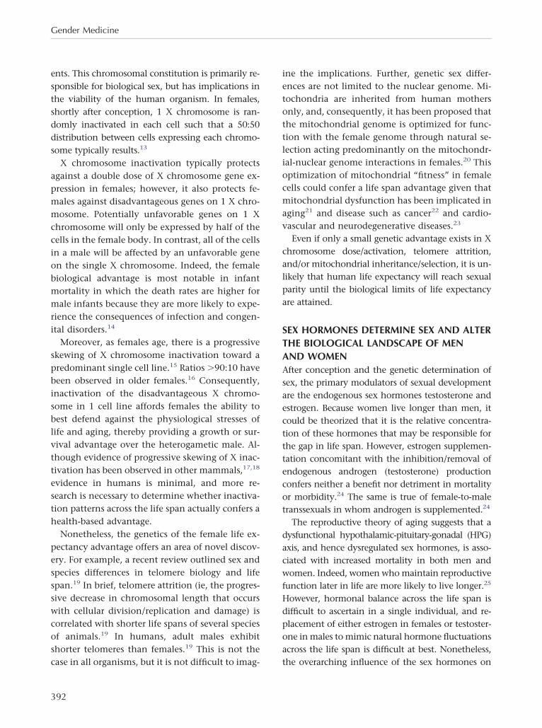
ments increase the quality of life of individuals livingwith chronic disease. These developments may beresponsible for the significant increases in life expec-tancy, improvements in mortality, and a greaterquality of life observed over time. The female advan-tage in life expectancy is obviously multifaceted, andthis complexity becomes even more apparent whenthe Global Gender Gap Index (GGI) is examined.Globally, there is no correlation between the educa-tional, economic, political, and health criteria thatmake up the GGI and the male-to-female ratio of lifeexpectancy at birth (Figure 1B). In nearly everycountry, a sex gap in life expectancy exists.
Recently, it was predicted that in parts of theUnited Kingdom, a nation that ranks high on theGGI (ie, closer to parity) and low in female mor-tality, males born in the year 2000 who reach theage of 30 will live as long as their female counter-parts.8 These predictions were primarily based onhe convergence of life expectancy trends over theast several decades and theorized to be mostlyue to modifiable societal factors such as a malemoking habits and females participating in otherunhealthy male behaviours.”8 However, it is im-ortant to note that women report greater prob-
ems in accessing health care at all ages,9 havehigher normalized levels of body fat,10 and reportlower lifetime activity levels compared with malesat all ages in nearly every country, including theUnited Kingdom.11 Consequently, although it wo-uld be unwise to instead suggest that biologicalmechanisms alone are responsible for the female ad-vantage in life span, clearly human males and fe-males differ in this regard. Here we argue that fe-males possess a biological advantage in longevitythat is likely to continue as long as the sexes exist.
FEMALES POSSESS A GENETIC ADVANTAGEAT CONCEPTIONAlthough not ubiquitous among animal species,the sexual dimorphism in longevity tends to favorthe homogametic sex (eg, human females or avianmales) over the heterogametic sex (eg, humanmales or avian females).12 Human males receive an Xchromosome from their biological mother and a Ychromosome from their biological father, whereas
1.00
0.98
0.96
0.94
0.92
0.90
0.88
mal
e:fe
mal
e lif
e ex
pect
ancy
0.86
0.84
0.82
0.80
1.000.980.960.940.920.900.88
mal
e:fe
mal
e lif
e ex
pect
ancy
at
birt
h
0.860.840.820.80
1990 2000year
2009
R2 = 0.0157
0.4 0.5 0.6 0.7Global Gender Gap Index
0.8 0.9
at birthat 60 y
A
B
Figure 1. Trends in sex differences in global life expec-tancy. (A) Male-to-female ratio of life expec-tancy at birth in 193 countries. Data wereobtained from World Health Organizationcountry statistics reported in the years 1990,2000, and 2009 and are presented as globalmeans and SE. (B) Global male-to-female ra-tio of life expectancy at birth versus theGlobal Gender Equality Index in 131 coun-tries for which data were available for bothindices. Life expectancy values were takenfrom the World Health Organization countrystatistics most recent report (2009), whereasthe Global Gender Gap index is a standarddeveloped by the World Economic Forumbased on sex equity under educational, eco-nomic, political, and health criteria.7 A valueof 1.00 would represent complete parity be-tween men and women. As such, the differ-ence between 1.00 and the reported numberrepresents the sex gap. A bivariate correlationwas performed using IBM SPSS Statistics Ver-sion .20 between the Gender Gap Index andmale:female life expectancy at birth. A signif-icant correlation was not observed. Line of
females receive an X chromosome from both par-
391
apmmcciobmmri
sp
swcosc
ietottli
calpa
dacwf
Gender Medicine
ents. This chromosomal constitution is primarily re-sponsible for biological sex, but has implications inthe viability of the human organism. In females,shortly after conception, 1 X chromosome is ran-domly inactivated in each cell such that a 50:50distribution between cells expressing each chromo-some typically results.13
X chromosome inactivation typically protectsgainst a double dose of X chromosome gene ex-ression in females; however, it also protects fe-ales against disadvantageous genes on 1 X chro-osome. Potentially unfavorable genes on 1 X
hromosome will only be expressed by half of theells in the female body. In contrast, all of the cellsn a male will be affected by an unfavorable genen the single X chromosome. Indeed, the femaleiological advantage is most notable in infantortality in which the death rates are higher forale infants because they are more likely to expe-
ience the consequences of infection and congen-tal disorders.14
Moreover, as females age, there is a progressivekewing of X chromosome inactivation toward aredominant single cell line.15 Ratios �90:10 have
been observed in older females.16 Consequently,inactivation of the disadvantageous X chromo-some in 1 cell line affords females the ability tobest defend against the physiological stresses oflife and aging, thereby providing a growth or sur-vival advantage over the heterogametic male. Al-though evidence of progressive skewing of X inac-tivation has been observed in other mammals,17,18
evidence in humans is minimal, and more re-search is necessary to determine whether inactiva-tion patterns across the life span actually confers ahealth-based advantage.
Nonetheless, the genetics of the female life ex-pectancy advantage offers an area of novel discov-ery. For example, a recent review outlined sex andspecies differences in telomere biology and lifespan.19 In brief, telomere attrition (ie, the progres-ive decrease in chromosomal length that occursith cellular division/replication and damage) is
orrelated with shorter life spans of several speciesf animals.19 In humans, adult males exhibithorter telomeres than females.19 This is not the
ase in all organisms, but it is not difficult to imag-392
ne the implications. Further, genetic sex differ-nces are not limited to the nuclear genome. Mi-ochondria are inherited from human mothersnly, and, consequently, it has been proposed thathe mitochondrial genome is optimized for func-ion with the female genome through natural se-ection acting predominantly on the mitochondr-al-nuclear genome interactions in females.20 This
optimization of mitochondrial “fitness” in femalecells could confer a life span advantage given thatmitochondrial dysfunction has been implicated inaging21 and disease such as cancer22 and cardio-vascular and neurodegenerative diseases.23
Even if only a small genetic advantage exists in Xhromosome dose/activation, telomere attrition,nd/or mitochondrial inheritance/selection, it is un-ikely that human life expectancy will reach sexualarity until the biological limits of life expectancyre attained.
SEX HORMONES DETERMINE SEX AND ALTERTHE BIOLOGICAL LANDSCAPE OF MENAND WOMENAfter conception and the genetic determination ofsex, the primary modulators of sexual developmentare the endogenous sex hormones testosterone andestrogen. Because women live longer than men, itcould be theorized that it is the relative concentra-tion of these hormones that may be responsible forthe gap in life span. However, estrogen supplemen-tation concomitant with the inhibition/removal ofendogenous androgen (testosterone) productionconfers neither a benefit nor detriment in mortalityor morbidity.24 The same is true of female-to-maletranssexuals in whom androgen is supplemented.24
The reproductive theory of aging suggests that aysfunctional hypothalamic-pituitary-gonadal (HPG)xis, and hence dysregulated sex hormones, is asso-iated with increased mortality in both men andomen. Indeed, women who maintain reproductive
unction later in life are more likely to live longer.25
However, hormonal balance across the life span isdifficult to ascertain in a single individual, and re-placement of either estrogen in females or testoster-one in males to mimic natural hormone fluctuationsacross the life span is difficult at best. Nonetheless,
the overarching influence of the sex hormones on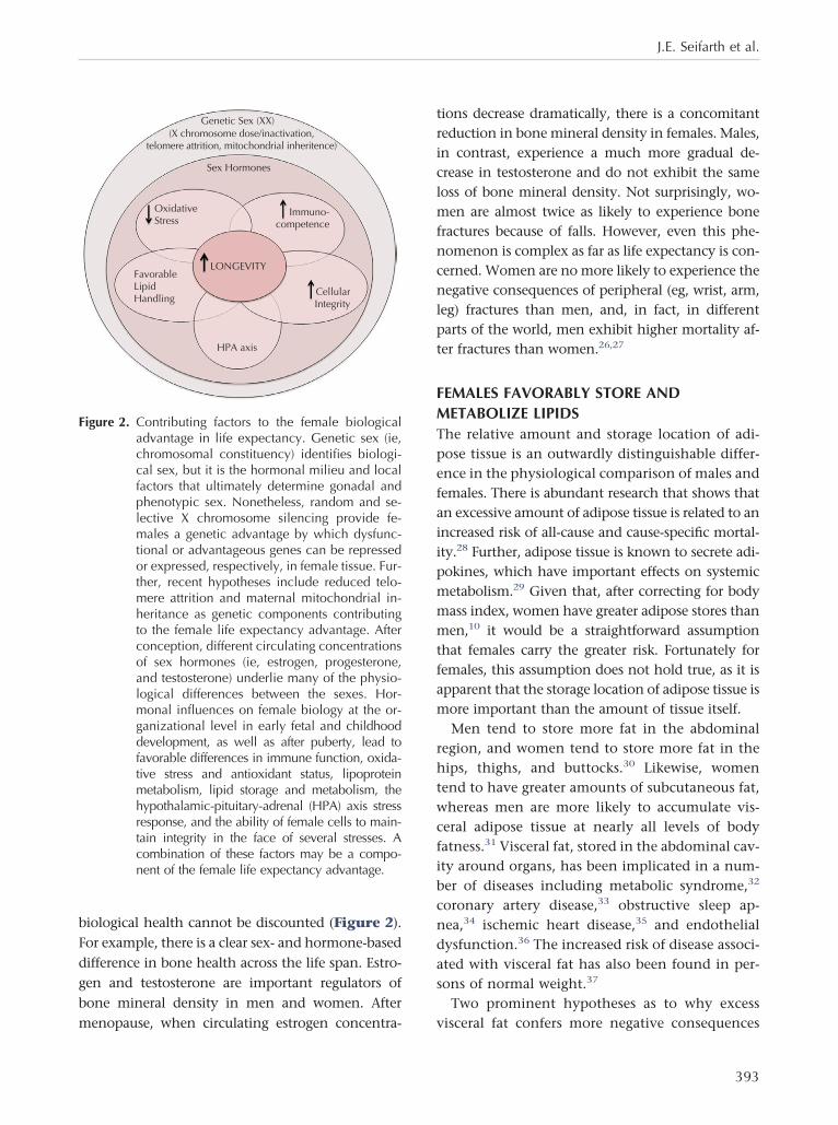
mm
J.E. Seifarth et al.
biological health cannot be discounted (Figure 2).For example, there is a clear sex- and hormone-baseddifference in bone health across the life span. Estro-gen and testosterone are important regulators ofbone mineral density in men and women. After
Genetic Sex (XX)
Sex Hormones
OxidativeStress
Immuno-competence
CellularIntegrity
HPA axis
LONGEVITYFavorableLipidHandling
(X chromosome dose/inactivation,telomere attrition, mitochondrial inheritence)
Figure 2. Contributing factors to the female biologicaladvantage in life expectancy. Genetic sex (ie,chromosomal constituency) identifies biologi-cal sex, but it is the hormonal milieu and localfactors that ultimately determine gonadal andphenotypic sex. Nonetheless, random and se-lective X chromosome silencing provide fe-males a genetic advantage by which dysfunc-tional or advantageous genes can be repressedor expressed, respectively, in female tissue. Fur-ther, recent hypotheses include reduced telo-mere attrition and maternal mitochondrial in-heritance as genetic components contributingto the female life expectancy advantage. Afterconception, different circulating concentrationsof sex hormones (ie, estrogen, progesterone,and testosterone) underlie many of the physio-logical differences between the sexes. Hor-monal influences on female biology at the or-ganizational level in early fetal and childhooddevelopment, as well as after puberty, lead tofavorable differences in immune function, oxida-tive stress and antioxidant status, lipoproteinmetabolism, lipid storage and metabolism, thehypothalamic-pituitary-adrenal (HPA) axis stressresponse, and the ability of female cells to main-tain integrity in the face of several stresses. Acombination of these factors may be a compo-nent of the female life expectancy advantage.
menopause, when circulating estrogen concentra- v
tions decrease dramatically, there is a concomitantreduction in bone mineral density in females. Males,in contrast, experience a much more gradual de-crease in testosterone and do not exhibit the sameloss of bone mineral density. Not surprisingly, wo-men are almost twice as likely to experience bonefractures because of falls. However, even this phe-nomenon is complex as far as life expectancy is con-cerned. Women are no more likely to experience thenegative consequences of peripheral (eg, wrist, arm,leg) fractures than men, and, in fact, in differentparts of the world, men exhibit higher mortality af-ter fractures than women.26,27
FEMALES FAVORABLY STORE ANDMETABOLIZE LIPIDSThe relative amount and storage location of adi-pose tissue is an outwardly distinguishable differ-ence in the physiological comparison of males andfemales. There is abundant research that shows thatan excessive amount of adipose tissue is related to anincreased risk of all-cause and cause-specific mortal-ity.28 Further, adipose tissue is known to secrete adi-pokines, which have important effects on systemicmetabolism.29 Given that, after correcting for body
ass index, women have greater adipose stores thanen,10 it would be a straightforward assumption
that females carry the greater risk. Fortunately forfemales, this assumption does not hold true, as it isapparent that the storage location of adipose tissue ismore important than the amount of tissue itself.
Men tend to store more fat in the abdominalregion, and women tend to store more fat in thehips, thighs, and buttocks.30 Likewise, womentend to have greater amounts of subcutaneous fat,whereas men are more likely to accumulate vis-ceral adipose tissue at nearly all levels of bodyfatness.31 Visceral fat, stored in the abdominal cav-ity around organs, has been implicated in a num-ber of diseases including metabolic syndrome,32
coronary artery disease,33 obstructive sleep ap-nea,34 ischemic heart disease,35 and endothelialdysfunction.36 The increased risk of disease associ-ated with visceral fat has also been found in per-sons of normal weight.37
Two prominent hypotheses as to why excess
isceral fat confers more negative consequences393
m
ewVar
fwvicheibg
liaAalUwm
namdtgg
mpweetRnecdustlg
ptwA
Gender Medicine
than excess subcutaneous fat implicate the differ-ing anatomic locations and secretions of the 2tissues. Visceral fat stores have been shown to se-crete greater amounts of inflammatory interleu-kin-6,38 vascular endothelial growth factor,39 plas-
inogen activator inhibitor-1,39 and C-reactiveprotein.40 The combined result of these secretionsis an increased incidence of metabolic syndrome,37
systemic inflammation,38 and greater mortality inmen and women.41 The implications of the differ-nce between adipose tissues become more severehen the location of visceral fat is considered.isceral fat accumulates around the organs in thebdominal cavity and is subject to an increasedate of lipolysis due to a greater amount of �-ad-
renergic receptors and less �-adrenergic inhibi-tion.42 An increase in free fatty acid (FFA) releaserom these tissues causes an influx of FFA to the liverhere it circulates immediately through the portalein. The increased FFA exposure has been shown toncrease prehepatic insulin production and to de-rease hepatic insulin clearance, exposing the liver toigher concentrations of insulin, leading to periph-ral hyperinsulinemia and reduced insulin sensitiv-ty.43 Further, visceral adiposity is associated withoth hepatic and peripheral insulin resistance re-ardless of sex.44 Decreased insulin sensitivity is a
risk factor and symptom of type 2 diabetes, whichleads to a lower quality of life and possible futurecomplications.
Considering men tend to store greater amountsof fat in the abdominal region and are more likelyto experience abdominal obesity,30 it would seemogical that they would be more likely to havencreased portal vein FFA concentrations and haven increased incidence of the negative outcomes.n interesting trend emerges when collectively ex-mining sex-specific studies of diabetes preva-ence. Early investigations noted that in thenited States, the only major cause of mortalityhere women exhibit higher rates was diabetesellitus.45 Further, it was the only serious chronic
condition in which women exhibited higher mor-bidity than males. This is in contrast to more re-cent investigations that showed that more womenhave diabetes than men, but men have a greater
prevalence of diabetes than women.46 This dy- r394
amic occurs because diabetes becomes more prev-lent with age, and women live to older ages thanen, thereby leading to a greater amount of el-
erly females with diabetes. It seems that althoughhe absolute number of women with diabetes isreater, relatively, men experience diabetes to areater degree.In addition to the storage of adipose tissue, fe-ales tend to preferentially metabolize fats during
hysical stresses, such as exercise.47 Although thisould allow women to spare carbohydrates during
xercise, leading to an advantage in long-durationxercise, the ability to metabolize fats preferen-ially also appears important to cellular survival.ecently, it was shown that when male and femaleeurons are subjected to starvation, male neuronsxperience significantly greater autophagy (ie,ontrolled digestion of cytoplasmic material) andeath, whereas female neurons switch to a greaterse of fats and survive.48 In humans, the relation-hip between the ability of female tissues to switcho a greater proportion of fat use during metabo-ism and prolonged life is a novel area of investi-ation for future research.
SEX DIFFERENCES IN LIPOPROTEINMETABOLISMA sexual dimorphism in body fat distribution is com-plemented by a sex difference in systemic lipid trans-port and storage. For example, blood HDL concen-trations are maintained in females after puberty, butdecrease in males, whereas LDL concentrations arelower in females across the life span with HDLs beinglarger in size.25 Moreover, LDL concentrations havebeen shown to decrease abruptly, whereas HDL con-centrations increase progressively, with increasedHDL apolipoprotein A-I and A-II levels, in estrogen-supplemented menopausal women.49
In contrast, supplementation with androgens inostmenopausal women has been shown to havehe opposite effect: LDL concentrations increase,hereas HDL and HDL apolipoprotein A-I and-II concentrations decrease.50 Likewise, androgen
supplementation in healthy men may initially de-crease HDL-C and continue depression for the du-ration of testosterone supplementation.51 Similar
esults were found with androgen supplementa-
srimrsaopb
ascdt
ssccs
ian
iid
J.E. Seifarth et al.
tion in older men with low to normal endogenoustestosterone levels.52 Although supplementationtudies may not directly mimic in vivo effects, theesults of these investigations provide interestingmplications about the opposing effects of sex hor-
ones on lipoprotein metabolism. Even though aecent genome-wide association report removedome of the associations behind increased HDLnd reduced cardiovascular disease risk,53 epidemi-logical associations of this characteristic with im-roved health and reduced mortality risk cannote ignored.
SEXUALLY DIMORPHIC MECHANISMS OFCOMBATTING OXIDATIVE STRESSThe free radical theory of aging states that freeradicals produced during cellular respiration causecumulative oxidative damage resulting in agingand death.54 Estrogens may provide antioxidantbenefits due to their phenolic structure,55,56 butlso because of a role in antioxidant gene expres-ion. This theory seems plausible because mito-hondria from female rats exhibit higher antioxi-ant gene expression and lower oxidative damagehan those from males.57,58 Further, removal of the
ovaries leads to increased mitochondrial oxidantproduction, whereas estrogen replacement abol-ishes this increase.57,58
Additional evidence of the role of oxidativetress in aging and a possible dimorphism betweenexes comes from the measurement of 16s mito-hondrial rRNA. 16s rRNA has been shown to de-rease with age59 and in the presence of oxidativetress.60 An investigation of sex differences in mi-
tochondrial rRNA expression in rats indicates sig-nificantly higher expression in females (700%higher than age-matched males), providing sup-port for the hypothesis that female mitochondriafunction like that of younger males.57 Continuedresearch to determine whether these findings holdtrue in humans may reveal a key sex differencethat contributes to differences in longevity.
In addition to estrogen, female rats have beenshown to exhibit higher basal levels of the cellu-lar antioxidants manganese superoxide dismutase(MnSOD) and glutathione peroxidase in an estro-
gen-dependent manner.61,62 As a result, female tis-sues are better protected against oxidative damage.The combination of the antioxidant properties ofestrogen and associated up-regulated antioxidantgenes in females could lead to more favorable han-dling of the cumulative oxidative attacks that occurover the life span. For example, aging is associatedwith chronic states of molecular inflammation andconcomitant oxidative damage to cellular integrity.Because skeletal muscle is high in mitochondrialdensity and oxygen flux, it is potentially exposed tohigher amounts of oxidative stress. Males generallypossess a greater proportion of fat free mass (notablyskeletal muscle), exhibit a higher resting metabolicrate independent of body composition and fitness,63
and therefore potentially have a greater metabolicflux of oxygen and electrons across the life span. Theaging-related loss of muscle mass (termed sarcope-nia) is a major occurrence in the elderly and mayhave oxidative injury– related causes. In elderly hu-mans, women exhibit less sarcopenia than oldermen.64 Although sarcopenia is not directly lethal, its highly correlated with disability and mortality,nd it has been proposed that this observable phe-omenon be used as a biomarker of aging.65
SEX DIFFERENCES IN IMMUNE FUNCTIONA functioning immune system is paramount toadequate health and quality of life. Sex hormonesplay an important role in the sexually dimorphicnature of human immunocompetence. In general,estrogens are considered humoral immunity en-hancers, whereas androgens and progesterone areconsidered natural immunosuppressants.66 In fact,laboratory studies of mice have shown that malesare more susceptible to infection than females,and this is attributed to differences in endogenoussex hormones.67,68 Estrogen functions to modulatemmunity via stimulating the production of anti-nflammatory cytokines69 and inhibiting the pro-uction of proinflammatory cytokines.70,71 How-
ever, a recent investigation in humans found thatwomen may be at higher risk for M2 macrophage–mediated autoimmune disorders due to the ability ofestrogen to increase T-helper type 2 responses.39 Thefemale advantage in infection resistance may serveas a disadvantage when an immune response is ini-
tiated against host cells. As such, males may be more395
i
at
dh
si
cpme
sbmser
dceli
Gender Medicine
susceptible to infection, but females are more suscep-tible to autoimmune diseases. Although there arenegative implications for both sexes, this tradeoffmay still favor women. Mortality rates due to cardio-vascular disease and cancer are orders of magnitudehigher than the best indication of rates of autoim-mune disease mortality in women,72 and infectiousdisease, but not autoimmune disease, is a leadingcause of death globally,73 even though there is poordentification of autoimmunity-related deaths.
There is also support for the hypothesis that lowerndrogen levels lead to a more robust immune sys-em.74,75 Compared with immune responses across 3
strains of gonadectomized male mice and normalmale mice injected with various antigens, femalemice exhibited a greater immune response, but go-nadectomy significantly increased the immune re-sponse of male mice.74 More recently, male gonad-ectomy caused a more female-like immune responseto bleomycin-induced pulmonary fibrosis comparedwith control mice, whereas androgen supplementa-tion in female mice yielded a more male-like re-sponse.75 These findings provide support for thepossibility that decreased androgens may have amore beneficial immunological effect than increasedestrogens.
HORMONAL AND CELLULAR MANAGEMENTOF STRESSStress, although an integral part of life, can havenegative effects on the health and well-being of anindividual. For example, an athlete uses progres-sive stresses on the body (eg, longer running du-ration, increased resistance training loads) to elicitphysiological adaptations that will lead to greaterperformance (eg, higher oxygen consumption, in-creased strength). When the body experienceslong-term exposure to a stressor or can no longersuccessfully defend against or adapt to physiolog-ical or psychological stressors, disease risk is in-creased. At the outset, it would appear that womenexperience greater amounts of stress and suffer to alarger degree from it. Women subjectively self-reportmore stress than men in response to a social stresstask76 and exhibit a higher incidence of stress-related
epression.77 However, physiological studies of the
ypothalamic-pituitary-adrenal (HPA) axis stress re-396
ponse to psychosocial stress tests provide contrast-ng data.78,79
A collection of investigations examined the freeortisol response to anticipation of an upcomingsychological stressor, speaking and mental arith-etic in front of an audience, and bicycle ergom-
ter exercise to exhaustion.78 Although the exer-cise protocol showed similar changes in the meancortisol level between sexes, the psychologicalstressor of speaking and mental arithmetic in frontof an audience produced a markedly greater meancortisol level response in males. In fact, the mereprospect of an upcoming psychological stress taskproduced a cortisol response in men, but not inwomen.78 These findings provide insight into aexual dimorphism of the HPA axis stress response,ut because the subjects were of adult age, the dataay be subject to the sociological influence of
ex-based roles throughout the life span. Recentvidence suggests that differences in cortisol stressesponse may be abolished with age,80 which may
coincide with a narrowing of differences in endog-enous sex hormones. Support for a physiologicalmechanism underlying the apparent difference inHPA axis stress response has been provided by astudy of young children. An examination of thecortisol response to corticotropin-releasing hor-mone ingestion in male and female children 7 to13 years of age found that males exhibited a sig-nificantly higher cortisol response.79
At the organ and cellular levels, several notableifferences in the ability of female cells to deal withellular perturbations have been documented. Forxample, female rodent hearts exhibit significantlyess infarcted area and dysfunction than males afterschemia-reperfusion insult.61,81 Because cardiovas-
cular disease is 1 of the top 2 killers of both men andwomen worldwide, this observation is notable. Thisobservation has been attributed to increased basallevels of cellular antioxidants (MnSOD, noted previ-ously), as well as heat shock or stress proteins (HSPs).With respect to the latter, HSPs, in particular the70-kDa HSP (Hsp70), represent some of the mosthighly conserved and cytoprotective proteins stud-ied to date. The expression of Hsp70 is increased inseveral tissues in response to a wide variety of stresses
and subsequently protects those tissues from future
sHltptHrwi
wbutv
aetlapwttnectnhs
J.E. Seifarth et al.
insults. The major circulating estrogen, 17�-estra-diol, appears to inhibit the stress-induced increase inHsp70.82,83 However, 17�-estradiol itself has beenhown to elevate cardiac and skeletal muscle levels ofsp70, a result that may account for a higher basal
evel of this protein in females.84,85 HSPs can beransported (through either coordinated secretion orassive release) into the circulation and play impor-ant roles in inflammation and immunoregulation.owever, the function of these extracellular proteins
emains less clear. For example, in healthy adults,omen exhibit twice the concentrations of circulat-
ng Hsp70 as men,86 but centenarians and their off-spring exhibit very low levels of circulating Hsp70.87
In fact, the human Hsp70 gene is an aging biomarkercandidate. In human females, genetic polymor-phisms in different Hsp70 genes appear to favor lon-ger life. More research needs to be completed todetermine the significance of these findings, but, col-lectively, they point to a sexual dimorphism in thehormonal and cellular response to stress, whichcould affect at least part of the female life expectancyadvantage.
WOMEN LIVE LONGER AND BETTERIncreased female longevity in the face of the adver-sities described here may serve as an indicator ofmore favorable innate biological health comparedwith males. If the longer life expectancy of females isaccompanied by an equal or greater quality of life inaging compared with men, then this would be evenmore favorable. Although it was previously repor-ted that females exhibit greater morbidity thanmales,88,89 these investigations must be interpreted
ith caution. It has been shown that increased mor-idity among females is only consistently observedsing psychological measures of distress, whereashis relationship is less apparent, and sometimes re-ersed, in physical symptoms and conditions.90
Moreover, after controlling for marital status, age,living arrangements, psychiatric symptoms, and roleobligations, sex differences in health across manydemographics are shifted to a sex-neutral or femaleadvantage.90–92 As such, when all factors are consid-ered, it appears that human females have an advan-tage over males in not only life expectancy, but
physiological health–related quality of life.CONCLUSIONThe human species have experienced an almostconstant increase in life span along with ever-improving qualities of life. Advances in healthcare and technology have assisted in treatmentsof illness and disease that would have previouslybeen debilitating or fatal while improving qual-ity of life for those with chronic and/or terminaldisease. Although this trend holds true for bothsexes, there is an apparent sexual dimorphism inboth life expectancy that has existed as long asrecords have been kept.6 Recently, it was pre-dicted that male children born in parts of theUnited Kingdom in the year 2000 would matchin life span or even outlive their female counter-parts once they reached 30 years of age.8 This isn interesting prediction because there are sev-ral biological mechanisms that would suggesthe sex gap in life expectancy will continue asong as the sexes exist. Even if biological mech-nisms are estimated to make up only a smallortion of the cause of the life expectancy gap,e believe that it is safe to theorize that al-
hough males may approach female life expec-ancy, parity is unattainable currently and in theear future. The complexity of the human lifexperience makes an examination of the primaryontributors to life expectancy difficult. None-heless, an examination of the sex differences inot only human life expectancy, but humanealth in aging will prove beneficial to bothexes.
ACKNOWLEDGMENTSMr. Seifarth was responsible for drafting the articleand revisions. Dr. McGowan was responsible forediting the article draft and final approval of themanuscript. Dr. Milne was responsible for draft-ing, editing, revisions, and final approval of themanuscript.
CONFLICTS OF INTERESTThe authors have indicated that they have noconflicts of interest regarding the content of this
article.397

1
1
1
1
1
2
2
2
2
2
2
2
2
Gender Medicine
REFERENCES1. Oeppen J, Vaupel JW. Demography. Broken limits
to life expectancy. Science. 2002;296:1029–1031.
2. Glei DA, Horiuchi S. The narrowing sex differen-
tial in life expectancy in high-income populations:
effects of differences in the age pattern of mortal-
ity. Popul Stud (Camb). 2007;61:141–159.
3. Trovato F, Lalu NM. Narrowing sex differentials in
life expectancy in the industrialized world: early
1970’s to early 1990’s. Soc Biol. 1996;43:20–37.
4. Wolfson MC. Health-adjusted life expectancy.
Health Rep. 1996;8:41–46.
5. Austad SN. Why women live longer than men: sex
differences in longevity. Gend Med. 2006;3:79–92.
6. World Health Organization. Global Health Obser-
vatory Data Repository. http://www.who.int/
research/en/. Accessed January 5, 2012.
7. Hausmann R, Tyson LD, Bekhouche Y, Zahidi S.
The Global Gender Gap Report 2011. World Eco-
nomic Forum; 2011.
8. Mayhew LA, Smith D. Gender Convergence in Hu-
man Survival and the Postponement of Death. Re-
port No. 200. London, UK: Cass Business School,
2012.
9. Rustgi SD, Doty MM, Collins SR. Women at risk:
why many women are forgoing needed health
care. An analysis of the Commonwealth Fund 2007
Biennial Health Insurance Survey. Issue Brief
(Commonw Fund). 2009;52:1–12.
10. Power ML, Schulkin J. Sex differences in fat stor-
age, fat metabolism, and the health risks from
obesity: possible evolutionary origins. Br J Nutr.
2008;99:931–940.
11. World Health Organization. Global Strategy on
Diet, Physical Activity and Health. http://www.who.
int/dietphysicalactivity/factsheet_inactivity/en/
index.html. Accessed January 5, 2012.
12. Liker A, Szekely T. Mortality costs of sexual selec-
tion and parental care in natural populations of
birds. Evolution. 2005;59:890–897.
13. Christensen K, Orstavik KH, Vaupel JW. The X
chromosome and the female survival advantage:
an example of the intersection between genetics,
epidemiology and demography. Ann N Y Acad Sci.
2001;954:175–183.
14. Drevenstedt GL, Crimmins EM, Vasunilashorn S,
Finch CE. The rise and fall of excess male infant
398
mortality. Proc Natl Acad Sci U S A. 2008;105:
5016–5021.
5. Christensen K, Kristiansen M, Hagen-Larsen H, et
al. X-linked genetic factors regulate hematopoietic
stem-cell kinetics in females. Blood. 2000;95:2449–
2451.
6. Sharp A, Robinson D, Jacobs P. Age- and tissue-
specific variation of X chromosome inactivation
ratios in normal women. Hum Genet. 2000;107:
343–349.
7. Abkowitz JL, Ott RL, Nakamura JM, et al. Feline
glucose-6-phosphate dehydrogenase cellular mo-
saicism. Application to the study of retrovirus-in-
duced pure red cell aplasia. J Clin Invest 1985;75:
133–140.
8. Abkowitz JL, Taboada M, Shelton GH, et al. An X
chromosome gene regulates hematopoietic stem
cell kinetics. Proc Natl Acad Sci U S A. 1998;95:
3862–3866.
9. Barrett EL, Richardson DS. Sex differences in telo-
meres and lifespan. Aging Cell. 2011;10:913–921.
0. Tower J. Sex-specific regulation of aging and
apoptosis. Mech Ageing Dev. 2006;127:705–718.
1. Trifunovic A, Larsson NG. Mitochondrial dysfunc-
tion as a cause of ageing. J Intern Med. 2008;263:
167–178.
2. Brandon M, Baldi P, Wallace DC. Mitochondrial
mutations in cancer. Oncogene. 2006;25:4647–
4662.
3. DiMauro S, Andreu AL. Mutations in mtDNA: are
we scraping the bottom of the barrel? Brain
Pathol. 2000;10:431–441.
4. Gooren LJ, Giltay EJ, Bunck MC. Long-term treat-
ment of transsexuals with cross-sex hormones: ex-
tensive personal experience. J Clin Endocrinol
Metab. 2008;93:19–25.
5. Yonker JA, Chang V, Roetker NS, et al. Hypotha-
lamic-pituitary-gonadal axis homeostasis predicts
longevity. Age (Dordr). 2011 Dec 4. [Epub ahead of
print].
6. Center JR, Nguyen TV, Schneider D, et al. Mortal-
ity after all major types of osteoporotic fracture in
men and women: an observational study. Lancet.
1999;353:878–882.
7. Jacobsen SJ, Goldberg J, Miles TP, et al. Race and
sex differences in mortality following fracture of
the hip. Am J Public Health. 1992;82:1147–50.
3
3
3
3
4
4
4
4
4
4
4
4
4
4
5
5
5
J.E. Seifarth et al.
28. Abdullah A, Wolfe R, Stoelwinder JU, et al. The
number of years lived with obesity and the risk of
all-cause and cause-specific mortality. Int J Epide-
miol. 2011;40:985–996.
29. Trayhurn P, Wood IS. Adipokines: inflammation
and the pleiotropic role of white adipose tissue.
Br J Nutr. 2004;92:347–355.
30. Nielsen S, Guo Z, Johnson CM, et al. Splanchnic
lipolysis in human obesity. J Clin Invest. 2004;113:
1582–1588.
31. Lemieux S, Prud’homme D, Bouchard C, et al. Sex
differences in the relation of visceral adipose tis-
sue accumulation to total body fatness. Am J Clin
Nutr. 1993;58:463–467.
32. Albu JB, Murphy L, Frager DH, et al. Visceral fat
and race-dependent health risks in obese nondi-
abetic premenopausal women. Diabetes. 1997;46:
456–462.
33. Nakamura T, Tokunaga K, Shimomura I, et al. Con-
tribution of visceral fat accumulation to the devel-
opment of coronary artery disease in non-obese
men. Atherosclerosis. 1994;107:239–246.
34. Shinohara E, Kihara S, Yamashita S, et al. Visceral
fat accumulation as an important risk factor for
obstructive sleep apnoea syndrome in obese sub-
jects. J Intern Med. 1997;241:11–18.
35. Matsuzawa Y, Shimomura I, Nakamura T, et al.
Pathophysiology and pathogenesis of visceral fat
obesity. Diabetes Res Clin Pract. 1994;24(Suppl):
S111–S116.
6. Romero-Corral A, Sert-Kuniyoshi FH, Sierra-John-
son J, et al. Modest visceral fat gain causes endo-
thelial dysfunction in healthy humans. J Am Coll
Cardiol. 2010;56:662–666.
7. Goodpaster BH, Krishnaswami S, Harris TB, et al.
Obesity, regional body fat distribution, and the
metabolic syndrome in older men and women.
Arch Intern Med. 2005;165:777–783.
8. Fontana L, Eagon JC, Trujillo ME, et al. Visceral fat
adipokine secretion is associated with systemic
inflammation in obese humans. Diabetes. 2007;56:
1010–1013.
9. Fairweather D, Cihakova D. Alternatively activated
macrophages in infection and autoimmunity. J Au-
toimmun. 2009;33:222–230.
0. Fox CS, Massaro JM, Hoffmann U, et al. Abdominal
visceral and subcutaneous adipose tissue com-
partments: association with metabolic risk factors
in the Framingham Heart Study. Circulation. 2007;
116:39–48.
1. Bigaard J, Frederiksen K, Tjonneland A, et al. Waist
circumference and body composition in relation
to all-cause mortality in middle-aged men and
women. Int J Obes (Lond). 2005;29:778–784.
2. Bjorntorp P. “Portal” adipose tissue as a generator
of risk factors for cardiovascular disease and dia-
betes. Arteriosclerosis. 1990;10:493–496.
3. Peiris AN, Mueller RA, Smith GA, et al. Splanchnic
insulin metabolism in obesity. Influence of body
fat distribution. J Clin Invest. 1986;78:1648–1657.
4. Miyazaki Y, Glass L, Triplitt C, et al. Abdominal fat
distribution and peripheral and hepatic insulin re-
sistance in type 2 diabetes mellitus. Am J Physiol
Endocrinol Metab. 2002;283:E1135–E1143.
5. Verbrugge LM. Sex differences in complaints and
diagnoses. J Behav Med. 1980;3:327–355.
6. Wild S, Roglic G, Green A, et al. Global prevalence
of diabetes: estimates for the year 2000 and pro-
jections for 2030. Diabetes Care. 2004;27:1047–
1053.
7. Tarnopolsky MA. Sex differences in exercise me-
tabolism and the role of 17-beta estradiol. Med Sci
Sports Exerc. 2008;40:648–654.
8. Du L, Hickey RW, Bayir H, et al. Starving neurons
show sex difference in autophagy. J Biol Chem.
2009;284:2383–2396.
9. Applebaum-Bowden D, McLean P, Steinmetz A, et
al. Lipoprotein, apolipoprotein, and lipolytic en-
zyme changes following estrogen administration
in postmenopausal women. J Lipid Res. 1989;30:
1895–1906.
0. Taggart HM, Applebaum-Bowden D, Haffner S, et
al. Reduction in high density lipoproteins by ana-
bolic steroid (stanozolol) therapy for postmeno-
pausal osteoporosis. Metabolism. 1982;31:1147–
1152.
1. Bagatell CJ, Heiman JR, Matsumoto AM, et al. Met-
abolic and behavioral effects of high-dose, exog-
enous testosterone in healthy men. J Clin Endo-
crinol Metab. 1994;79:561–567.
2. Emmelot-Vonk MH, Verhaar HJ, Nakhai Pour HR,
et al. Effect of testosterone supplementation on
functional mobility, cognition, and other parame-
399

6
6
6
6
6
6
7
7
7
7
7
7
Gender Medicine
ters in older men: a randomized controlled trial.
JAMA. 2008;299:39–52.
53. Voight BF, Peloso GM, Orho-Melander M, et al.
Plasma HDL cholesterol and risk of myocardial
infarction: a mendelian randomisation study. Lan-
cet. 2012;380;572–580.
54. Harman D. Aging: a theory based on free radical
and radiation chemistry. J Gerontol. 1956;11:298–
300.
55. Behl C, Skutella T, Lezoualc’h F, et al. Neuroprotec-
tion against oxidative stress by estrogens: structure-
activity relationship. Mol Pharmacol. 1997;51:535–
541.
56. Ozacmak VH, Sayan H. The effects of 17beta es-
tradiol, 17alpha estradiol and progesterone on ox-
idative stress biomarkers in ovariectomized fe-
male rat brain subjected to global cerebral
ischemia. Physiol Res. 2009;58:909–912.
57. Borras C, Sastre J, Garcia-Sala D, et al. Mitochon-
dria from females exhibit higher antioxidant gene
expression and lower oxidative damage than
males. Free Radic Biol Med. 2003;34:546–552.
58. Guevara R, Santandreu FM, Valle A, et al. Sex-
dependent differences in aged rat brain mito-
chondrial function and oxidative stress. Free Radic
Biol Med. 2009;46:169–175.
59. Calleja M, Pena P, Ugalde C, et al. Mitochondrial
DNA remains intact during Drosophila aging, but
the levels of mitochondrial transcripts are signifi-
cantly reduced. J Biol Chem. 1993;268:18891–18897.
60. Crawford DR, Lauzon RJ, Wang Y, et al. 16S mito-
chondrial ribosomal RNA degradation is associ-
ated with apoptosis. Free Radic Biol Med. 1997;22:
1295–1300.
61. Brown DA, Lynch JM, Armstrong CJ, et al. Suscep-
tibility of the heart to ischaemia-reperfusion in-
jury and exercise-induced cardioprotection are
sex-dependent in the rat. J Physiol. 2005;564:619–
630.
62. Borras C, Gambini J, Gomez-Cabrera MC, et al.
17beta-oestradiol up-regulates longevity-related,
antioxidant enzyme expression via the ERK1 and
ERK2[MAPK]/NFkappaB cascade. Aging Cell. 2005;
4:113–118.
63. Arciero PJ, Goran MI, Poehlman ET. Resting met-
abolic rate is lower in women than in men. J Appl
Physiol. 1993;75:2514–2520.
400
4. Baumgartner RN, Koehler KM, Gallagher D, et al.
Epidemiology of sarcopenia among the elderly in
New Mexico. Am J Epidemiol. 1998;147:755–763.
5. Fisher AL. Of worms and women: sarcopenia and
its role in disability and mortality. J Am Geriatr Soc.
2004;52:1185–1190.
6. Cutolo M, Wilder RL. Different roles for andro-
gens and estrogens in the susceptibility to auto-
immune rheumatic diseases. Rheum Dis Clin
North Am. 2000;26:825–839.
7. Sano A, Miyaji M, Nishimura K. Studies on the
relationship between the estrous cycle of BALB/c
mice and their resistance to Paracoccidioides
brasiliensis infection. Mycopathologia. 1992;119:
141–145.
8. Yamamoto Y, Saito H, Setogawa T, Tomioka H. Sex
differences in host resistance to Mycobacterium
marinum infection in mice. Infect Immun. 1991;59:
4089–4096.
9. Lambert KC, Curran EM, Judy BM, et al. Estrogen
receptor alpha (ERalpha) deficiency in macro-
phages results in increased stimulation of CD4� T
cells while 17beta-estradiol acts through ERalpha
to increase IL-4 and GATA-3 expression in CD4� T
cells independent of antigen presentation. J Im-
munol. 2005;175:5716–5723.
0. Roberts CW, Satoskar A, Alexander J. Sex steroids,
pregnancy-associated hormones and immunity to
parasitic infection. Parasitol Today. 1996;12:382–
388.
1. Arenas IA, Armstrong SJ, Xu Y, Davidge ST.
Chronic tumor necrosis factor-alpha inhibition
enhances NO modulation of vascular function in
estrogen-deficient rats. Hypertension. 2005;46:
76–81.
2. Walsh SJ, Rau LM. Autoimmune diseases: a lead-
ing cause of death among young and middle-aged
women in the United States. Am J Public Health
2000;90:1463–1466.
3. World Health Organization. Causes of Death 2008
Summary Tables. Geneva, Switzerland: WHO, 2011.
4. Eidinger D, Garrett TJ. Studies of the regulatory
effects of the sex hormones on antibody forma-
tion and stem cell differentiation. J Exp Med. 1972;
136:1098–1116.
5. Voltz JW, Card JW, Carey MA, et al. Male sex
hormones exacerbate lung function impairment
J.E. Seifarth et al.
after bleomycin-induced pulmonary fibrosis. Am J
Respir Cell Mol Biol. 2008;39:45–52.
76. Horowitz M, Wilner N, Alvarez W. Impact of Event
Scale: a measure of subjective stress. Psychosom
Med. 1979;41:209–218.
77. Krause N. Stress and sex differences in depressive
symptoms among older adults. J Gerontol. 1986;
41:727–731.
78. Kirschbaum C, Wust S, Faig HG, Hellhammer DH.
Heritability of cortisol responses to human corti-
cotropin-releasing hormone, ergometry, and psy-
chological stress in humans. J Clin Endocrinol
Metab. 1992;75:1526–1530.
79. Dahl RE, Siegel SF, Williamson DE, et al. Cortico-
tropin releasing hormone stimulation test and
nocturnal cortisol levels in normal children. Pedi-
atr Res. 1992;32:64–68.
80. Rubin RT, Rhodes ME, O’Toole S, Czambel RK.
Sexual diergism of hypothalamo-pituitary-adrenal
cortical responses to low-dose physotigmine in
elderly vs. young women and men. Neuropsycho-
pharmacology. 2002;26:672–681.
81. Thorp DB, Haist JV, Leppard J, et al. Exercise train-
ing improves myocardial tolerance to ischemia in
male but not in female rats. Am J Physiol Regul
Integr Comp Physiol. 2007;293:R363–R371.
82. Paroo Z, Haist JV, Karmazyn M, Noble EG. Exercise
improves postischemic cardiac function in males
but not females: consequences of a novel sex-
specific heat shock protein 70 response. Circ Res.
2002;90:911–917.
83. Milne KJ, Thorp DB, Krause M, Noble EG. Core
temperature is a greater influence than endoge-
nous 17beta-estradiol on the exercise-induced ac-
cumulation of myocardial heat shock protein
Windsor, Ontario, Canada N9B3P4. E-mail: kjmilne@uwin
mRNA. Can J Physiol Pharmacol. 2011 Nov 1. [Epub
ahead of print].
84. Bombardier E, Vigna C, Iqbal S, et al. Effects of
ovarian sex hormones and downhill running on
fiber-type-specific HSP70 expression in rat soleus.
J Appl Physiol. 2009;106:2009–2015.
85. Voss MR, Stallone JN, Li M, et al. Gender differ-
ences in the expression of heat shock proteins:
the effect of estrogen. Am J Physiol Heart Circ
Physiol. 2003;285:H687–H692.
86. Pockley AG, Shepherd J, Corton JM. Detection of
heat shock protein 70 (Hsp70) and anti-Hsp70 an-
tibodies in the serum of normal individuals. Im-
munol Invest. 1998;27:367–377.
87. Terry DF, McCormick M, Andersen S, et al. Car-
diovascular disease delay in centenarian offspring:
role of heat shock proteins. Ann N Y Acad Sci.
2004;1019:502–505.
88. Wingard DL. The sex differential in morbidity,
mortality, and lifestyle. Annu Rev Public Health.
1984;5:433–458.
89. Sweeting H. Reversals of fortune? Sex differences
in health in childhood and adolescence. Soc Sci
Med. 1995;40:77–90.
90. Macintyre S, Hunt K, Sweeting H. Gender differ-
ences in health: are things really as simple as they
seem? Soc Sci Med. 1996;42:617–624.
91. Kaplan RM, Anderson JP, Wingard DL. Gender dif-
ferences in health-related quality of life. Health
Psychol. 1991;10:86–93.
92. Gove WR, Hughes M. Possible causes of the ap-
parent sex differences in physical health: an em-
pirical investigation. Am Sociol Rev. 1979;44:
126–146.
Address correspondence to: Kevin J. Milne, PhD, Faculty of Human Kinetics, The University of Windsor,
dsor.ca
401
Related Documents