Sequencing of the smallest Apicomplexan genome from the human pathogen Babesia microti y Emmanuel Cornillot 1, *, Kamel Hadj-Kaddour 1 , Amina Dassouli 1 , Benjamin Noel 2 , Vincent Ranwez 3 , Benoıˆt Vacherie 2 , Yoann Augagneur 4 , Virginie Bre `s 1 , Aurelie Duclos 2 , Sylvie Randazzo 1 , Bernard Carcy 1 , Franc ¸ oise Debierre-Grockiego 5 , Ste ´ phane Delbecq 1 , Karina Moubri-Me ´ nage 1 , Hosam Shams-Eldin 6 , Sahar Usmani-Brown 4 , Fre ´ de ´ ric Bringaud 7 , Patrick Wincker 2 , Christian P. Vivare `s 8 , Ralph T. Schwarz 6 , Theo P. Schetters 9 , Peter J. Krause 10 , Andre ´ Gorenflot 1 , Vincent Berry 11 , Vale ´ rie Barbe 2 and Choukri Ben Mamoun 4, * 1 Laboratoire de Biologie Cellulaire et Mole ´ culaire (LBCM-EA4558), UFR Pharmacie, Universite ´ Montpellier 1, 15, av. Charles Flahault, 34093 Montpellier cedex 5, 2 Genoscope (CEA) and CNRS UMR 8030, Universite ´ d’Evry, 2 rue Gaston Cre ´ mieux, 91057 Evry, 3 Institut des Sciences de l’Evolution (ISEM, UMR 5554 CNRS), Universite ´ Montpellier II, Place E. Bataillon—34095 Montpellier cedex 5, and Montpellier SupAgro, UMR AGAP, av. Agropolis—TA A96/03 - 34398 Montpellier cedex 5, France, 4 Department of Internal Medicine, Section of Infectious Diseases, Yale School of Medicine, 15 York St., New Haven, CT 06520, USA, 5 UMR1282 Infectiologie et Sante ´ Publique, Universite ´ de Tours, F-37000 Tours, France and INRA, F-37380 Nouzilly, France, 6 Institut fu ¨ r Virologie, Zentrum fu ¨ r Hygiene und Infektionsbiologie, Philipps-Universita ¨ t Marburg, Hans-Meerwein-Strasse, 35043 Marburg, Germany, 7 Centre de Re ´ sonance Magne ´ tique des Syste ` mes Biologiques (RMSB, UMR 5536), Universite ´ Bordeaux Segalen, CNRS, 146 rue Le ´ o Saignat, 33076 Bordeaux, 8 Clermont Universite ´ , Universite ´ Blaise Pascal, Laboratoire Microorganismes: Ge ´ nome et Environnement, BP10448, F-63000 Clermont-Ferrand, France, 9 Microbiology R&D Department, Intervet/Schering-Plough Animal Health, 5830 AA Boxmeer, The Netherlands, 10 Yale School of Public Health and Yale School of Medicine, 60 College St., New Haven, CT 06520, USA and 11 Equipe Me ´ thodes et Algorithmes pour la Bioinformatique, LIRMM (UMR 5506 CNRS), Universite ´ Montpellier II, Place E Bataillon—34095 Montpellier, France Received March 26, 2012; Revised June 22, 2012; Accepted June 25, 2012 ABSTRACT We have sequenced the genome of the emerging human pathogen Babesia microti and compared it with that of other protozoa. B. microti has the smallest nuclear genome among all Apicomplexan parasites sequenced to date with three chromo- somes encoding 3500 polypeptides, several of which are species specific. Genome-wide phylogen- etic analyses indicate that B. microti is significantly distant from all species of Babesidae and Theileridae and defines a new clade in the phylum Apicomplexa. Furthermore, unlike all other Apicomplexa, its mitochondrial genome is circular. Genome-scale reconstruction of functional networks revealed that B. microti has the minimal metabolic requirement for intraerythrocytic protozoan parasitism. B. microti multigene families differ from those of other protozoa in both the copy number and organization. Two lateral transfer events with significant metabolic implications occurred during the evolution of this parasite. The genomic sequencing of B. microti identified several targets suitable for the develop- ment of diagnostic assays and novel therapies for human babesiosis. INTRODUCTION Babesia microti is the principal cause of human babesiosis and one of the most common transfusion-transmitted *To whom correspondence should be addressed. Tel: +33 0 4 11 75 96 86; Fax: +33 0 4 11 75 97 30; Email: [email protected] Correspondence may also be addressed to Choukri Ben Mamoun. Tel/Fax: +1 203 737 1972; Email: [email protected] yEric Pre´cigout initiated the project but left us suddenly in 2006. The authors wish it to be known that, in their opinion, the first three authors should be regarded as joint First Authors. 9102–9114 Nucleic Acids Research, 2012, Vol. 40, No. 18 Published online 24 July 2012 doi:10.1093/nar/gks700 ß The Author(s) 2012. Published by Oxford University Press. This is an Open Access article distributed under the terms of the Creative Commons Attribution Non-Commercial License (http://creativecommons.org/licenses/ by-nc/3.0), which permits unrestricted non-commercial use, distribution, and reproduction in any medium, provided the original work is properly cited.

Welcome message from author
This document is posted to help you gain knowledge. Please leave a comment to let me know what you think about it! Share it to your friends and learn new things together.
Transcript

Sequencing of the smallest Apicomplexan genomefrom the human pathogen Babesia microtiy
Emmanuel Cornillot1,*, Kamel Hadj-Kaddour1, Amina Dassouli1, Benjamin Noel2,
Vincent Ranwez3, Benoıt Vacherie2, Yoann Augagneur4, Virginie Bres1, Aurelie Duclos2,
Sylvie Randazzo1, Bernard Carcy1, Francoise Debierre-Grockiego5, Stephane Delbecq1,
Karina Moubri-Menage1, Hosam Shams-Eldin6, Sahar Usmani-Brown4,
Frederic Bringaud7, Patrick Wincker2, Christian P. Vivares8, Ralph T. Schwarz6,
Theo P. Schetters9, Peter J. Krause10, Andre Gorenflot1, Vincent Berry11,
Valerie Barbe2 and Choukri Ben Mamoun4,*
1Laboratoire de Biologie Cellulaire et Moleculaire (LBCM-EA4558), UFR Pharmacie, Universite Montpellier 1, 15,av. Charles Flahault, 34093 Montpellier cedex 5, 2Genoscope (CEA) and CNRS UMR 8030, Universite d’Evry, 2rue Gaston Cremieux, 91057 Evry, 3Institut des Sciences de l’Evolution (ISEM, UMR 5554 CNRS), UniversiteMontpellier II, Place E. Bataillon—34095 Montpellier cedex 5, and Montpellier SupAgro, UMR AGAP, av.Agropolis—TA A96/03 - 34398 Montpellier cedex 5, France, 4Department of Internal Medicine, Section ofInfectious Diseases, Yale School of Medicine, 15 York St., New Haven, CT 06520, USA, 5UMR1282Infectiologie et Sante Publique, Universite de Tours, F-37000 Tours, France and INRA, F-37380 Nouzilly,France, 6Institut fur Virologie, Zentrum fur Hygiene und Infektionsbiologie, Philipps-Universitat Marburg,Hans-Meerwein-Strasse, 35043 Marburg, Germany, 7Centre de Resonance Magnetique des SystemesBiologiques (RMSB, UMR 5536), Universite Bordeaux Segalen, CNRS, 146 rue Leo Saignat, 33076 Bordeaux,8Clermont Universite, Universite Blaise Pascal, Laboratoire Microorganismes: Genome et Environnement,BP10448, F-63000 Clermont-Ferrand, France, 9Microbiology R&D Department, Intervet/Schering-Plough AnimalHealth, 5830 AA Boxmeer, The Netherlands, 10Yale School of Public Health and Yale School of Medicine, 60College St., New Haven, CT 06520, USA and 11Equipe Methodes et Algorithmes pour la Bioinformatique,LIRMM (UMR 5506 CNRS), Universite Montpellier II, Place E Bataillon—34095 Montpellier, France
Received March 26, 2012; Revised June 22, 2012; Accepted June 25, 2012
ABSTRACT
We have sequenced the genome of the emerginghuman pathogen Babesia microti and compared itwith that of other protozoa. B. microti has thesmallest nuclear genome among all Apicomplexanparasites sequenced to date with three chromo-somes encoding �3500 polypeptides, several ofwhich are species specific. Genome-wide phylogen-etic analyses indicate that B. microti is significantlydistant from all species of Babesidae and Theileridaeand defines a new clade in the phylum Apicomplexa.Furthermore, unlike all other Apicomplexa, itsmitochondrial genome is circular. Genome-scalereconstruction of functional networks revealed that
B. microti has the minimal metabolic requirement forintraerythrocytic protozoan parasitism. B. microtimultigene families differ from those of otherprotozoa in both the copy number and organization.Two lateral transfer events with significant metabolicimplications occurred during the evolution of thisparasite. The genomic sequencing of B. microtiidentified several targets suitable for the develop-ment of diagnostic assays and novel therapies forhuman babesiosis.
INTRODUCTION
Babesia microti is the principal cause of human babesiosisand one of the most common transfusion-transmitted
*To whom correspondence should be addressed. Tel: +33 0 4 11 75 96 86; Fax: +33 0 4 11 75 97 30; Email: [email protected] may also be addressed to Choukri Ben Mamoun. Tel/Fax: +1 203 737 1972; Email: [email protected] Precigout initiated the project but left us suddenly in 2006.
The authors wish it to be known that, in their opinion, the first three authors should be regarded as joint First Authors.
9102–9114 Nucleic Acids Research, 2012, Vol. 40, No. 18 Published online 24 July 2012doi:10.1093/nar/gks700
� The Author(s) 2012. Published by Oxford University Press.This is an Open Access article distributed under the terms of the Creative Commons Attribution Non-Commercial License (http://creativecommons.org/licenses/by-nc/3.0), which permits unrestricted non-commercial use, distribution, and reproduction in any medium, provided the original work is properly cited.

pathogens in the United States (1–3). The parasite has aworldwide distribution and has been cited as an emerginghealth threat in the United States by the National Academyof Sciences (4). B. microti is primarily transmitted tohumans by the tick vector, Ixodes scapularis, but also peri-natally and through blood transfusion. The mortality rateassociated with human babesiosis is estimated to be between3 and 28%. Most severe cases occur in people over the ageof 50 years or those who are asplenic, have cancer or HIV orwho are on an immunosuppressive therapy. The majority ofpatients experience mild to moderate malaria-likesymptoms; however, in severe cases, the disease may beassociated with respiratory failure, multi-organ system dys-function or coma (1–4). Both host and parasite factors con-tribute to these symptoms, but the exact pathogenicmechanisms remain unknown (5,6). No evidence ofextraerythrocytic cellular parasitism exists for B. microti.Although the parasite has been classified as a member ofthe Babesia genus, the accuracy of this classification hasbeen debated for >50 years with some experts suggestingthat it belongs to the Theileria genus.
We describe here the first sequence of the B. microtigenome from a patient isolate propagated in gerbils.Sequence analysis revealed important information aboutthe genome organization, gene content and metaboliccapacities of this parasite and provides new insights intoits pathophysiology. Furthermore, the study identifiednew targets for the development of diagnostic assays andnovel therapies for this important human pathogen.Phylogenetic analysis using a large pool of coding se-quences (CDS) strongly suggest that B. microti defines anew taxonomic genus among Apicomplexa distinct fromBabesia and Theileria species.
MATERIALS AND METHODS
Strains and genome sequencing
The B. microti R1 isolate was obtained from an adult malepatient who experienced severe B. microti infection thatrequired hospital admission. Although the patient washospitalized in France, he lived in the United States in aBabesia-endemic area. Prior to the onset of his illness, thepatient was in general good health, non-splenectomizedand with no other evidence of immune suppression.Because the patient had fever, purpura, laboratoryevidence of hemolytic anemia and kidney failure,B. microti infection was suspected and subsequently con-firmed by blood smear, serology, rodent inoculation andpolymerase chain reaction (PCR) among other molecularmethods as outlined in the Results and Discussion section.The patient was treated with clindamycin and quinineafter which he fully recovered.
The R1 and the Gray strain control were propagatedin immune-compromised gerbils or hamsters. B. microtiR1 DNA was extracted from agarose gel plugs and frag-mented by mechanical shearing producing 3 and 10kbinserts that were subsequently cloned into the pcDNA2.1(www.invitrogen.com) and pCNS (pSU18 derived)plasmids, respectively. In addition, a large insert (30 kb) bac-terial artificial chromosome library was constructed by
cloning Sau3A partially digested genomic fragments intothe pBelo-BAC11 vector. Vector DNAs were purified andend-sequenced using dye-terminator chemistry on ABI3730sequencers. We collected 71 882, 59 193 and 3343 sequences,respectively, from each DNA library. A first assembly wasperformed using Arachne (http://www.broadinstitute.org)resulting in 522 contigs of >500nt (N50=38216) and104 scaffolds of >2kbp (N50=951 194). We retainedonly the clones containing contigs with >4 kb because theB. microti R1 DNA was contaminated with Gerbil DNA.The corresponding reads were assembled using the Phred/Phrap/Consed software package (www.phrap.com) asdescribed by Vallenet et al. (7). We then obtained 139contigs (N50=183364) with a cumulated size of6 425 753bp. Three main scaffolds were determined andattributed to the chromosomes. Contigs corresponding tomitochondrial DNAs were identified by Basic LocalAlignment Search Tool (BLAST). Primer walks, PCRsand in vitro transposition technology (TemplateGeneration System
TM
II Kit; Finnzyme, Espoo, Finland)were used to obtain complete chromosome sequences. Atotal of 10 328 sequences were used for gap closure andquality assessment. Themitochondrial genome organizationwas confirmed by PCR and by sequencing full clone inserts.PFGE analyses were performed in 0.5�Tris-HCl, Borate,EDTA (TBE) at 10�C at 4V/cm using Gene Navigator
TM
(Pharmacia). 2D Pulsed Field Gel Electrophoresis (PFGE)analyses were performed as previously described (8).
Genome annotation
Integration of resources using GAZEThe automatic gene prediction pipeline (SupplementaryFigure S1) was modified from a standard annotationpipeline (9). Programs were used with default options fol-lowing standard procedures. A semi-automatic procedurewas used to generate a first training set of 690 gene modelsfrom chromosome I sequence. A similar approach waspreviously used to analyze the Encephalitozoon cuniculigenome (10). To use these curated annotations in thedata integration step, transcript sequences of the geneswere mapped on the final genomic assembly usingBLAST-like alignment tool (BLAT) (11), best match(best BLAT score) per gene models were selected, andeach best match was realigned using Est2Genome (12) inorder to identify exon/intron boundaries. The mappingwas used to calibrate the ab initio SNAP (13) gene predic-tion software and as an entry for GAZE (14). The sameapproach was previously used with Apicomplexan ESTdata. A collection of 426 440 public messenger RNAs(mRNAs) from the clade of Apicomplexa (downloadedfrom the EMBL database, release 98) were first alignedwith the B. microti genome assembly using BLAT. Torefine BLAT alignment, we used Est2Genome (12).BLAT alignments were made using default parametersbetween translated genomic and translated ESTs (11).For protein sequences, the UniProt database (15) was
used to detect conserved proteins between B. microti andother species. Apicomplexan proteins in the UniProtdatabase were first aligned with the B. microti sequencesusing BLAT (11), and alignments with a BLAT score over
Nucleic Acids Research, 2012, Vol. 40, No. 18 9103

20 were selected. Subsequently, we extracted the genomicregions where no protein hit had been found by BLATand then realigned the UniProt protein with more permis-sive parameters. Alignments larger than 50 residues andwith >20 identity matches were selected. Each match wasthen refined using GeneWise (16) in order to identify exon/intron boundaries.Most of the genome comparisons were performed with
repeat-masked sequences. For this purpose, we searchedand sequentially masked several kinds of repeats including(i) Apicomplexan-known repeats available in Repbase(instead of the human data) using the RepeatMaskerprogram (17), (ii) tandem repeats using the TRFprogram (18) and (iii) ab initio repeat detection usingRepeatScout (19). From this pipeline, 1.2% of theassembled bases were masked. No further analysis of therepeated sequences has been performed.All the resources described here were used to automat-
ically build B. microti gene models using GAZE (14).Individual predictions from each of the programs(SNAP, GeneWise and Est2genome) were broken downinto segments (coding, intron and intergenic) and signals(start codon, stop codon, splice acceptor, splice donor,transcript start and transcript stop). Exons predictedby SNAP, GeneWise and Est2genome were used ascoding segments. Introns predicted by GeneWise andEst2genome were used as intron segments. Intergenicsegments were created from the span of each mRNAwith a negative score (to prevent genes splitting byGAZE). Predicted repeats were used as intron andintergenic segments in order to avoid prediction of genesencoding proteins in such regions. A weight was assignedto each resource. More importance was given to align-ments than to ab initio predictions. Chromosome Icurated gene models were associated with stronger param-eters to enable their consideration by GAZE. This weightacts as a multiplier for the score of each informationsource prior to processing by GAZE automaton. Allsignals were given a fixed score, but segment scores werecontext sensitive: coding segment scores were linked to thepercentage identity (%ID) of the alignment, while intronicsegment scores were linked to the %ID of the flankingexons. Finally, gene predictions created by GAZE werefiltered following their scores and lengths. When appliedto the entire assembled sequence, GAZE predicted 1426gene models.
Annotation procedureGlimmerHMM gene prediction software was used inparallel with GAZE annotation. Parameters and trainingwere performed following published recommendations(20). The training was performed on each chromosomeindependently. GlimmerHMM predicts 3923 putativeCDS. The gene models have been curated according to asemi-automatic procedure (Supplementary Figure S2).BLASTX homology search (21) covering the wholegenome was performed as described by Katinka et al.(10) and stored in an AceDB v4.9.38 database (http://www.acedb.org/index.shtml). Artemis software (22) wasused as a graphic interface (Supplementary Figure S3).
Subtelomeric sequence organization
The miropeat v2.01 software (http://www.genome.ou.edu/miropeats.html) and icatools v2.5 package (http://www.littlest.co.uk/software/pub/bioinf/freeold) were compiledon an Ubuntu Linux platform. The coordinates wererecovered from postscript output file. Ghostview wasused for the graphic representation of the miropeatoutput (http://pages.cs.wisc.edu/�ghost/). The genomeof B. microti was aligned to itself using BLASTN (21) toconfirm and to refine the structure of thesubtelomere-specific duplicated sequences (expect valuethreshold at 0.0001).
Functional annotation
The functional annotation of the B. microti CDS was per-formed by BLAST sequence comparison (21) againstUniProt and by searching for orthologues in definedspecies. Orthologous genes were identified using the bidir-ectional best hit method (BBH). The analysis was per-formed with three Apicomplexan parasites Plasmodiumfalciparum, Babesia bovis and Theileria annulata as wellas with the yeast Saccharomyces cerevisiae and thehuman pathogen Leishmania major. Homology searcheswere performed at the protein level using BLASTP withdefault options (21). Sequence data were obtained fromthe Kyoto Encyclopedia of Genes and Genomes database(KEGG) (23), PiroplasmDB and PlasmoDB (24). BBHscores were collected in a relational database andcombined with the metabolic information provided byKEGG (23) and the Malaria Parasite MetabolicPathway (MPMP, http://sites.huji.ac.il/malaria/) data-bases (Supplementary Table S1).
The functional annotation was scored using differentparameters combining the results of different BBHanalyses. A first score was given by the number of organ-isms having orthologs for each B. microti CDS (parametero2[0,5]). The reliability of the annotation was alsoassessed by the count of KEGG orthology group identifi-cation numbers (ko) associated with each ortholog (release58.1, 1 June 2011). Two parameters were used in this case:the number of KEGG ko number per B. microti CDS (k)and the number of different ko (d). Three levels of anno-tation were used, each associated with a different valid-ation procedure (Supplementary Table S2): (i) the konumber was directly inferred when o and k values werehigh; (ii) for intermediate values of d and k, the annota-tion was the only information stored (the protein wasdeclared as ‘valid’ and annotation was deduced fromBLAST best homologues’ description) and (iii) for low oor k scores and also for high d values, we considered thatthe annotation of a B. microti protein could not beinferred from those of its orthologous sequences (‘NoAnnotation’ denoted by ‘NA’).
Several metabolic charts of the KEGG and MPMPdatabases were analyzed for this study. B. microti genesencoding key enzymes and ‘missing’ genes in a given meta-bolic pathway were further analyzed by gene-specific bio-informatics approaches. Central metabolism has beenreconstructed by taking into account the carbon andredox balance.
9104 Nucleic Acids Research, 2012, Vol. 40, No. 18
Genome-wide phylogenetic analyses
Taxon samplingThe Apicomplexan phylogeny was analyzed based on ataxon sampling including nine species representing differ-ent families in the Apicomplexa phylum (25) and oneoutgroup species, Tetrahymena thermophila. The annota-tion of B. microti CDS (Supplementary Table S1) wasinitiated to generate the complete set of data. Additionalsequences were directly obtained from KEGG (release58.1, 1 June 2011).
Dataset assemblyTo perform phylogenetic analyses, we assigned CDS ofB. microti to existing KEGG gene families (23). Weretained CDS having one orthologous sequence in eachtree of the three Apicomplexan parasites (described inSupplementary Table S1) and associated with only oneKEGG ko identification number (d=1). Using thisanalysis, 1002 CDS were obtained. We filtered those kofamilies to retain only those containing one, and only one,sequence for: (i) each of the eight Apicomplexan genomesin the KEGG database, (ii) B. microti genome and (iii) T.thermophila. This filtration step reduced the B. microtisequence set to 316 CDS. For each of the 316 orthologygroups, the sequences of these 10 organisms were collectedand aligned using MUSCLE v3.8.31 (26) (with defaultoptions). Spurious alignment sites were then removedusing the ‘automated’ option of TrimAL v1.3 (27) thatestablishes optimal thresholds based on the alignmentcharacteristics.
Tree inferencesPhylogenetic analyses were conducted on each of the 316genes to infer the evolutionary history of each KEGG kofamily. The resulting trees were inferred by maximum like-lihood, using RAxML v7.2.8 (28). Inferences were per-formed starting from 10 distinct maximum parsimonytrees randomly chosen. The WAG protein model (29)using empirical base frequencies and a discrete Gammalaw with four categories to model heterogeneity of evolu-tionary rate among sites were chosen. Branch supportswere estimated through full bootstrap analyses, asopposed to the faster RAxML bootstrap approximations.Both supermatrix and supertree analyses were computedusing SeqCat.pl and SeqConverter.pl (http://www.molekularesystematik.uni-oldenburg.de/33997.html#Sequences) scripts to concatenate the 316 trimmedalignments into a nexus ‘Supermatrix’ of 129 571 proteinsites. The Supermatrix was analyzed by RAxML, with thesame parameters mentioned above. To obtain thesupertree with the Matrix Representation withParsimony (MRP) method (30), a matrix for the 316protein trees was computed with MRTOOLS (31),weighting each clade of the protein trees according to itsbootstrap support. Accordingly, clades that may havearisen from a low signal to noise ratio (low bootstrap)had less influence on the inferred supertree than morereliable clades (high bootstrap). The matrix was thenbootstrapped and analyzed with a parsimony criterion inthe PAUP* software (32). Supertree analysis was also
conducted by resorting to phySIC_IST (33), a more con-servative supertree method that allows congruent studyamong clades of individual gene histories with respect totheir bootstrap supports.
Supertree inferred by the PhySIC_IST methodThe PhySIC_IST method (33) first establishes a congru-ence analysis of the gene trees and subsequently modifiesthese trees to remove pieces of topological signal in eachgene tree that conflict with the majority of other genetrees. The 316 gene trees were curated by discardingclades supported by a boostrap value below a thresholdwere discarded, merging their two delimiting nodes in thegene tree where they belong. Then for each triplet of taxa,the frequencies of occurrence of the three possibletopologies for the triplet were compared on the basis ofthe clades remaining after the bootstrap elimination step.The P value of a �2 test was used to determine whether atopology for a triplet observed in a gene tree was ananomaly. In our study, a threshold value (STC=1-p)was used to eliminate abnormal triplet topologies.STC=1 means that no triplet topology will be rejected,even if it appears in one gene tree and is in contradictionwith all other gene trees. The usual values are STC=0.95or STC=0.8. Then clades of each gene tree inducingrejected triplets were eliminated. Once the gene treeswere curated with this procedure, a deduced supertree intotal agreement with the modified gene trees was obtained.More information on this method is available on http://www.atgc-montpellier.fr/physic_ist/.
RESULTS AND DISCUSSION
B. microti genome sequence and analysis
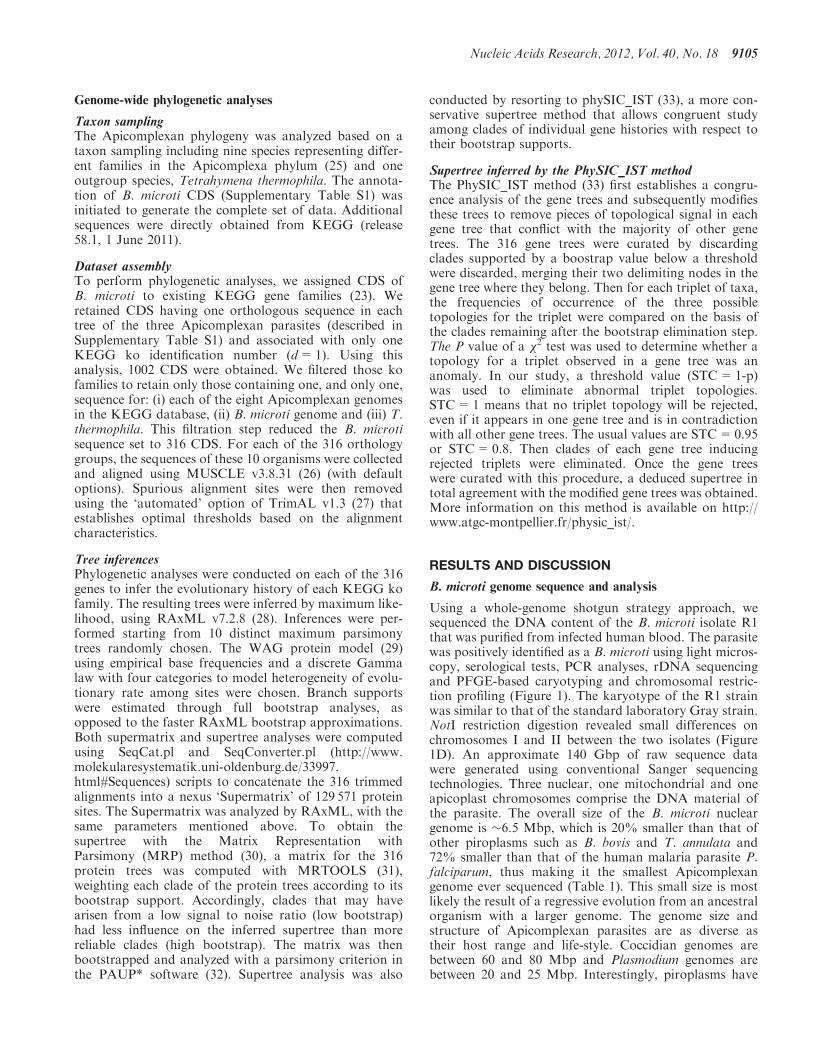
Using a whole-genome shotgun strategy approach, wesequenced the DNA content of the B. microti isolate R1that was purified from infected human blood. The parasitewas positively identified as a B. microti using light micros-copy, serological tests, PCR analyses, rDNA sequencingand PFGE-based caryotyping and chromosomal restric-tion profiling (Figure 1). The karyotype of the R1 strainwas similar to that of the standard laboratory Gray strain.NotI restriction digestion revealed small differences onchromosomes I and II between the two isolates (Figure1D). An approximate 140 Gbp of raw sequence datawere generated using conventional Sanger sequencingtechnologies. Three nuclear, one mitochondrial and oneapicoplast chromosomes comprise the DNA material ofthe parasite. The overall size of the B. microti nucleargenome is �6.5 Mbp, which is 20% smaller than that ofother piroplasms such as B. bovis and T. annulata and72% smaller than that of the human malaria parasite P.falciparum, thus making it the smallest Apicomplexangenome ever sequenced (Table 1). This small size is mostlikely the result of a regressive evolution from an ancestralorganism with a larger genome. The genome size andstructure of Apicomplexan parasites are as diverse astheir host range and life-style. Coccidian genomes arebetween 60 and 80 Mbp and Plasmodium genomes arebetween 20 and 25 Mbp. Interestingly, piroplasms have
Nucleic Acids Research, 2012, Vol. 40, No. 18 9105
some of the smallest Apicomplexan genomes (Table 1).The genome size and chromosome number of B. microtiare in the range of what has been described in otherpiroplasms (34–36). B. microti belongs to the so-calledsmall Babesia and infects only the erythrocyte of its mam-malian host (37) where it undergoes one or two divisionsprior to parasite egress and invasion by merozoites of newred blood cells. The ability of the parasite to invade andreplicate in lymphocytes of the vertebrate host has beensuggested but was never confirmed (38). Therefore, thelimited cellular host range of B. microti may account forthe dramatic reduction its genome size and structure.More than 98% of the nuclear genome of B. microti has
been assembled and annotated. Approximately 3500 CDSare present in the genome, making it the smallest set ofgenes found in Apicomplexa. Comparison of theB. microti genome with that of other piroplasmsrevealed an unexpected diversity. Notably, B. bovis andTheileria parva genomes contain �7% more genes andT. annulata 16% more genes than B. microti (Table 1).No synteny blocks encompassing more than five genescould be found between B. microti and otherApicomplexa. Between 60 and 80% of the additionalgenes found in B. bovis and T. parva are members oflarge multigenic families (e.g. vesa and SmORF or svspand Tpr, respectively). These families do not exist or
have not expanded in the B. microti genome. B. microticontains only one large lineage-specific multigenic familyencoding the sero-reactive antigen BMN (39,40).Twenty-four bmn genes are found in the B. microtigenome. The gene structure of B. microti is substantiallydivergent from that of other Apicomplexa. The number ofintrons is high with nearly 70% of B. microti genes inter-rupted with short introns of 20–25 bp in length.
Functional annotation revealed that 60% of the pre-dicted proteins of B. microti share homology withproteins of known or putative functions and about halfof them have an assigned biological function in theKEGG database (Supplementary Table S1).Approximately 12% of the proteins with unknownfunction are specific to B. microti. Two rDNA units arefound in tandem on chromosome III and 2 rRNA 5Sencoding genes and 44 tRNA genes are also present andthese are distributed throughout the three chromosomes.
The DNA sequence of B. microti has an average of 36%G+C content. The G+C content is much lower atchromosome ends. An A+T rich sequence of 1 kbp ispresent in four copies in the genome: one on both chromo-some I and II and two on chromosome III. This sequencemay function as a centromeric region (CEN,Supplementary Figure S4). Analysis of the subtelomericregions revealed the presence of a small set of multigene
R1 isolate Gray strain
First dimensionS
econd dimension
Kbp
400-
300-
200-
100-
50-
25-
R1 Gra
y R1 Gra
y
A
C
D
B
R1 Gra
y
B. dive
rgen
s
B. microti
- 430 bp
Mbp
2.0
1.5
KIII
KIIKI
+/- 15 kbp
+/- 15 kbp
Figure 1. Babesia microti strain R1 characterization. (A) Light-microscopy analysis of B. microti infected blood. Left: blood smear. Right: immuno-fluorescence analysis using serum from a hamster infected with the B. microti Gray standard laboratory strain. Similar serum was used for serologicalassays. (B) PCR amplification of the ssu gene using PIRO-A and PIRO-B primers (77). These primers amplify a 436 bp fragment in B. microti Grayand R1 strains and a 408 bp fragment in B. divergens Rouen strain. The integrity of the PCR fragment was confirmed by DNA sequencing. (C) B.microti karyotype. PFGE conditions used are: left, 0.7% agarose, 400 s pulses for 55 h; right, 1% agarose, 200 s pulses for 65 h. Length polymorphismbetween the R1 isolate and the Gray strain is observed on chromosome 1 and 2. (D) 2D-PFGE NotI Restriction Fragment Length Polymorphism(RFLP) analysis of B. microti. Each chromosome length polymorphism results from a single RFLP of 15 kbp (see triangle and star for chromosome Iand II respectively). The genome structure of Gray and R1 strain may differ from each other only with a single recombination event. PFGEconditions used are: 1.2 % agarose gel, pulse conditions, 5 s for 8 h, 15 s for 7 h and 30 s for 6 h, tension, 6.5V/cm.
9106 Nucleic Acids Research, 2012, Vol. 40, No. 18

families associated with repeated blocks of sequences of0.8–3 kbp (Figure 2). Some of these genes are members ofthe B. microti sero-reactive antigen family (bmn) (39,40).The presence of genes encoding surface antigens is acommon feature of many eukaryotic genomes and itrepresent a important source of antigenic variation foreukaryotic parasites (41,42). B. microti subtelomericregions contain pseudo-genes but also four copies of asequence homologous to members of the Theileriaspecific Tpr multigenic family and three copies of asequence homologous to members of the B. bovis vesamultigene family (Figure 2). Tpr- and vesa-like genes ofB. microti are divergent from those found in otherpiroplasms and the presence of both multigene familiesin the same genome is unique to B. microti. Interestingly,whereas in Theileria the Tpr genes are not associated withthe telomeres and are dispersed throughout the chromo-somes, in B. microti Tpr-like sequences are found near thetelomeres. Moreover, members of the bmn gene family arepresent near the telomeres as well as in the coding core ofthe chromosomes. Four of the B. microti repeated telo-meric sequences are found in the middle of chromosomeIII (IIIc cluster, Figure 2), suggesting that this chromo-some likely derived from a fusion event between two an-cestral chromosomes.
Unlike other Apicomplexa that have a linear mitochon-drial genome, B. microti has a circular 11 kbp mitochon-drial genome with two inverted repeats of 2.5 kbp
encompassing almost half of its size (Figure 3). Thegenetic information carried in this genome is similar tothat found in other Apicomplexa and encodes threeproteins, cytb of the cytochrome bc complex (complexIII) and coxI and coxIII of the cytochrome c oxidase(complex IV). The ribosomal lsu and ssu genes ofB. microti are fragmented. Their presence and organiza-tion suggest a distinct evolution from that of otherApicomplexa. The six ribosomal genes encoding lsu frag-ments show an organization similar to that of previouslyannotated mitochondrial genomes from other piroplasms(43). Interestingly, unlike other species of the Babesia andTheileria families that lack detectable ssu genes, two ribo-somal genes encoding ssu fragments were found in theB. microti mitochondrial genome.
B. microti defines a new Apicomplexan family
For many years, the classification of B. microti as amember of either the Babesia or Theileria families hasbeen controversial. Although many taxonomists have clas-sified B. microti as a member of the Babesia family, trans-mission electron microscopy analyses and recentmolecular studies using two different genetic markers sug-gested that this organism may be distinct from the otherspecies of this family (44–47). This limited set of molecularmarkers was, however, insufficient to ascertain the taxo-nomic position of this parasite among Apicomplexa. Toaddress the evolution of B. microti among Apicomplexa, a
Table 1. Genomic features of B. microti and five other Apicomplexa
Feature B. microti B. bovis T. parva T. annulata P. falciparum C. parvum
GenomeSize (Mbp)a 6.5 8.2 8.3 8.4 23.3 9.1Number of chromosomes 3 4 4 4 14 8G+C content (%) 36 41.5 34.1 32.5 19.3 30.8
GenesNumber of genesb 3513 3706 3796 4082 5324 3805Mean gene length (bp) 1327 1503 1407 1602 2326 1844Mean gene length including introns (bp) 1471 1609 1654 1802 2590 1851Gene density (bp per gene) 1816 2194 2059 2199 4374 2411Coding regions (%) 73 68 68 73 53 76Coding regions including introns (%) 81 73 80 82 59 77Number of genes with introns (%) 70 60 75 71 54 4
ExonsNumber per gene 3.3 2.8 2.7 3.9 2.6 1.1Mean length (bp) 397 547 514 416 904 1748Total length (%) 73 68 68 73 53 77
IntronsNumber per gene 2.3 1.7 2.6 2.9 1.6 0.1Number per gene presenting intron 3.4 2.9 3.5 4 2.9 1.3Mean length (bp) 61 60 94 70 167 96Total length (%) 8 5 12 9 6 0.02
Intergenic regionsMean length (bp) 346 585 405 398 1784 561Total length (%) 19 27 20 18 41 23
RNAsNumber of tRNA genesc 44 70 71 47 72 45Number of 5S rRNA genes 2 ND 1 3 3 6Number of 5.8S/18S/28S rRNA units 2 5 8 1 13 9
Quantitative data have been calculated using Artemis genome browser.aEstimated size including gaps.bData from Plasmodb 8.1 data for Apicomplexa. B. microti: this work.cData from piroplasmoDB v1.1 and plasmoDB v8.1.
Nucleic Acids Research, 2012, Vol. 40, No. 18 9107

set of 316 genes was selected from a total of 1002 CDSthat share significant homology with CDS in the genomesof B. bovis, T. annulata and P. falciparum. These 316 genesbelong to KEGG orthology groups and are present assingle copy genes in the B. microti genome as well asthat of eight Apicomplexan genomes selected forgenome-scale phylogenetic analysis. These includeB. bovis, T. annulata, T. parva, P. falciparum, Plasmodiumknowlesi, Plasmodium vivax, Toxoplasma gondii andCryptosporidium parvum. To locate the root of the phylo-genetic tree, T. thermophila was used as an outgroupspecies. Two independent phylogenetic analysesproduced in the same phylogenetic tree, placingB. microti at the root of piroplasms and separating itfrom the B. bovis and Theileria clades (Figure 4).Approximately 88% of the selected proteins support theevolutionary separation of B. microti from B. bovis andTheileria species. The data indicate that Babesia is a para-phyletic group, the taxonomy of which must be revised.Furthermore, this analysis revealed that B. microti hasevolved early in the evolution of piroplasms. Thesefindings emphasize the need to create a new genus forthe B. microti group of strains.
Coding coreChromosomesequence extremity
Iavesa Tprbmn
Ib
IIa
p
vesa TM nmbnmb
IIb
IIIa
vesaBmn
Tpr nmbnmbnmb bmn
Bmn
IIIa
IIIb
bmn Tpr bmn
bmnbmn
IIIcbmn bmnbmn Tpr
0 1 2 4
kbpS2
S3 S6
S4S1
S5
Unduplicated
S7
S8
Figure 2. Mosaic organization of the Babesia microti chromosome extremities. Chromosome ends are labeled according to Figure S2. These regionsare characterized by the presence of duplicated sequences scattered among the different chromosome extremities (S1–S8). Limits of sequencehomologies have been calculated using miropeat and BLASTN analyses. Annotated genes are indicated on the figure. Most repeated genes arepart of the bmn gene family and included truncated genes and pseudogenes. Several bmn genes are in transition regions between two duplicatedsequences. The S2 sequence encoding a putative VESA antigen is repeated three times. The S4 sequence encodes Tpr orthologues and is repeated fourtimes, two copies of which on chromosome ends IIb and IIIa are significantly shorter. The GC content in the chromosome ends is significantly lowerthan in the coding core. The sequence between S1 and S4 at extremity Ib encodes a putative sugar transporter (TM). The sequence is not duplicatedbut does not show any base composition bias compared to adjacent regions. It was not possible to precisely map the recombination sites associatedwith the rearrangements that took place at chromosome ends (average resolution of 100 bp).
ls 5
lsu4lsu6
lsu2
lsu5
Babesia microti
lsu6
IRIRmitochondrial genome
11.1 kb
lsu3
ssu6ssu7
lsu1
Figure 3. Circular organization of the Babesia microti mitochondrialgenome. IR: inverted repeats; cox1: cytochrome c oxidase subunit 1,cox3: cytochrome c oxidase subunit 3, cytb: cytochrome bc complexsubunit. The numbering of the ribosomal lsu and ssu genes fragmentsis performed according to the P. falciparum nomenclature.
9108 Nucleic Acids Research, 2012, Vol. 40, No. 18

The minimal Apicomplexan metabolism of B. microti
Reconstruction of B. microti metabolism from its genomicinformation suggests high dependence of the parasite onglucose fermentation for energy production and redoxregulation (Figure 5). Genes encoding enzymes requiredfor b-oxidation and hemoglobin degradation were notfound in the genome of this organism. The analysisfurther indicates that the parasite may not synthesizeheme or fatty acids de novo and lacks both mitochondrialand apicoplast pyruvate dehydrogenases. Furthermore,the function of the apicoplast seems to be limited to theproduction of the precursors of isoprenoids through the2-C-methyl-D-erythritol 4-phosphate/1-deoxy-D-xylulose5-phosphate pathway (MEP/DOXP) pathway, a meta-bolic function used by P. falciparum during its intraery-throcytic development (48). A ferredoxin/ferrodoxin:NAD oxidoreductase system is also present in theapicoplast and may provide electrons to the MEP/DOXP pathway resulting in the formation of isopentenyldiphosphate (IPP) and dimethylallyl diphosphate(DMAPP). Export of IPP and DMAPP to the cytosolhas been shown to be an essential step in the synthesisof ubiquinones and dolichol and plays a critical role inPlasmodium development (49). B. microti lacks a mito-chondrial superoxide dismutase, suggesting a minimallyactive respiratory chain. Consistent with this model, themitochondrial antioxidant system is underdeveloped. AF0F1-ATPase made of a reduced number of subunits isfound in the genome, suggesting that oxidative phosphor-ylation might take place in this organism. The geneencoding frataxin, an enzyme involved in the assemblyof iron-sulfur clusters, was not identified in the genome,indicating that it is either lacking or highly divergent fromknown eukaryotic orthologs.
Another unique feature of B. microti central metabol-ism is the acquisition of two genes by lateral genetransfer: Bmldh encoding lactate dehydrogenase (LDH)and Bmtpk encoding thiamine pyrophosphokinase(TPK). Bmldh and Bmtpk genes lack introns and areadjacent to each other on chromosome I but inopposite orientation. This chromosomal site is at thejunction between the subtelomere and the codingregion, a genomic location known in many organismsto be susceptible to double-stranded breaks and thus
suitable for insertion of foreign DNA. Reverse tran-scriptase–PCR analyses show that Bmldh is transcribedduring the blood stage of the parasite, whereas theBmtpk is not (Supplementary Figure S5). InApicomplexa, LDH activity is believed to haveoriginated from a duplication of the LDH-like malatedehydrogenase (mdh) gene (50–52). However, sequenceand phylogenetic analyses showed that the B. microtiLDH is not related to Apicomplexan LDH. Both theprotein and the gene share strong homology with theirmammalian counterparts, supporting a lateral genetransfer mode of acquisition from the host(Supplementary Figure S6A). Equally unique amongpiroplasms, B. microti expresses a TPK protein(BmTPK) with 70% identity to TPKs from Bartonellaspecies, which are transmitted by the same tick vectorthat transmits B. microti. BmTPK shares very lowsequence similarity with TPKs from other eukaryotes(Supplementary Figure S6B). Together, these findingssuggest that the Bmtpk gene may have been acquiredfrom bacteria by B. microti through lateral transfer, anevent that likely occurred during co-infection either inthe tick vector or in a mammalian host following atick bite. The presence of the Bmtpk gene in theB. microti genome may enable the parasite to producethiamine pyrophosphate (ThiaPP), an essential cofactorfor key enzymes of the tricarboxylic acid (TCA) cycle.The organization of the glycolytic machinery and TCA
cycle of piroplasm suggests a central role of the innermembrane of the mitochondria in parasite metabolismunder anaerobic and microaerophilic conditions (53).B. microti has several novel metabolic pathways consistentwith this model (Figure 5). In B. microti, no mdh genecould be identified, suggesting that the malate:quinoneoxydoreductase (MQO) alone is responsible for the pro-duction of malate. In the cytosol, the electron acceptor,oxaloacetate may be obtained either from glycolysis or theTCA cycle. A glycerol-3-phosphate (G3P)/dihydroxyacet-one phosphate (DHAP) shuttle might provide electrons tofeed MQO for malate production. However, there is noevidence that this electron transfer is fully responsible forthe regulation of the redox balance in the cell. It is likelythat B. microti produces pyruvate during its life cycle inaddition to lactate and malate.
Figure 4. Babesia microti defines a new clade in the Apicomplexan phylum. The tree is inferred using a maximum likelihood approach on aconcatenated alignment of 316 single-copy genes. Tetrahymena thermophila was included as outgroup. The same tree topology is inferred by asupertree approach compiling the 316 trees inferred from the 316 genes. Labels indicate the boostrap support from both the Supermatrix andSupertree analyses and the level of tree supporting the clade (%).
Nucleic Acids Research, 2012, Vol. 40, No. 18 9109

Genome analysis has also revealed that B. microtiencodes a glutamine synthetase (GLS). The presence ofthe GLS enzyme in B. microti is unique among piroplasmsand could provide the parasite with an alternative routefor nitrogen assimilation and ATP production. GLS andglutamate dehydrogenase (GDH) enzymes are essential toconvert glutamine and glutamate into 2-oxoglutarate(2OG) in order to feed the TCA cycle. Interestingly, theGLS and GDH enzymes of B. microti lack a signalpeptide, suggesting that 2OG synthesis may take place inthe cytosol followed by subsequent transport into themitochondria where the TCA cycle might work in a re-ductive way from 2OG to malate. Because of the absenceof TPK activity during intraerythrocytic development, theacquisition of ThiaPP from the host might be critical to
equilibrate the redox balance of the mitochondria throughthe activity of oxoglutarate dehydrogenase. Based on theavailable genomic information, the oxidative TCA cycle ofB. microti appears to be similar to that of P. falciparum(54). Complex III and IV of the respiratory chain are alsopresent in B. microti. The proton gradient might be usedby the F0F1-ATPase to produce ATP when the respiratorychain is highly active.
Glycosylphosphatidylinositol biosynthesis andglycosylphosphatidylinositol proteome
Glycosylphosphatidylinositol (GPI) anchors are glycoli-pids attached to many cell surface glycoproteins of lowerand higher eukaryotes including important cell surfaceparasite antigens (55–59). They have been shown to play
Figure 5. An integrated model for central metabolism of Babesia microti. Arrows show the direction of net fluxes. Lactate and malate are expectedto be the major end products of the central metabolism. The gene encoding the lactate dehydrogenase is likely obtained by lateral transfer from amammalian host. The apicoplast is devoted to the production of isoprenoids precursors. Numbers in brackets represent the biochemical steps in thereaction. Dashed arrows connect two reactions using the same metabolite. For simplicity, the membranes of the apicoplast and the outer membraneof the mitochondrion is not shown. Abbreviation used are: (1) Metabolites: 1,3BPGA, 1,3-bisphosphoglycerate; 2OG, 2-oxoglutarate; Ace-R: acetate/acetyl-CoA, c, cytochrome c; CoQ, Coenzyme-Q, ubiquinone; DHAP, dihydroxyacetone phosphate; e-, electrons; FBP, fructose 1,6-bisphosphate;G3P, glycerol-3-phosphate; GAP, glyceraldehyde-3-phosphate; Glu, glutamate; Gln, glutamine; OAA, oxaloacetate, PEP, phosphoenolpyruvate.(2) Enzymes (in red): ACS, AMP-forming acetyl-CoA synthetase; DHOD, dihydroorotate dehydrogenase; GLS, glutamine synthetase; GPDH,glycerol-3-phosphate dehydrogenase (FAD- or NAD-dependent enzymes); LDH, lactate dehydrogenase; MQO, malate:quinone oxidoreductase;NDH2, type II NADH:quinone oxidoreductase; OGDH, oxoglutarate dehydrogenase; PEPC, PEP carboxylase; PEPCK, PEP carboxykinase;PDH, pyruvate dehydrogenase; PK, pyruvate kinase; SDH, succinate dehydrogenase; TH, transhydrogenase; III, complex III of the respiratorychain; IV, complex IV of the respiratory chain; V, F0F1-ATP synthase.
9110 Nucleic Acids Research, 2012, Vol. 40, No. 18

a critical function in the pathogenesis of Apicomplexanparasites (60–62). GPI anchors are synthesized in astepwise manner in the membrane of the endoplasmic re-ticulum and attached to the C-terminal end of proteinsfollowing translation. The GPI biosynthetic pathways ineukaryotes are highly conserved and produce a core struc-ture of ethanolaminephosphate-6Mana1–2Mana1–6Mana1–4GlcNa1–6-D-myoinositol-1-HPO4-lipid,where the lipid is diacylglycerol, alkylacylglycerol orceramide. This minimal GPI structure may be embellishedwith various side chain modifications such as additionalsugars or ethanolamine phosphate in a species-specificmanner. In addition to GPI, various intermediate mol-ecules of the biosynthesis pathway are also accumulatedin the cytosol of parasites and some of them are secretedand can induce inflammatory responses (63–65). Genesencoding enzymes of the GPI biosynthetic pathway werefound in the B. microti genome indicating that the synthe-sis of GPIs occurs in this organism (Supplementary TableS3). The pagp1 deacylase was not found in the B. microtigenome, suggesting that GPI anchors made by thisparasite are likely to contain fully acylated inositol.Unlike Plasmodium or Toxoplasma species, whichexpress three mannosyltransferases and harbor GPIswith three mannose residues in their core structure (66),the B. microti genome encodes only two mannosy-ltransferases: PIGM and PIGV, and no homologues ofPIGB mannosyltransferase could be found. Thesefindings indicate that the core structure of the B. microtiGPI anchors may be different from that found in othereukaryotes. The implications of such a unique GPI struc-ture on host immune response and parasite evasion remainto be determined.
Twelve putative GPI-anchored merozoite surfaceprotein-encoding genes have been identified in theB. microti genome. All genes lack introns and theirencoded proteins have no homologs in other organisms,including other Apicomplexa.
New insights into babesiosis therapy
Babesiosis therapy has generally been based on empiricaltrials and medical consensus that combines antimalarialdrugs such as quinine or atovaquone with antibioticssuch as clindamycin or azithromycin (67–69). Treatmentfailure due to suspected parasite drug resistance has beenreported (70). Genome analysis indicates that B. microtilacks the proteases necessary to digest host hemoglobin.This result along with the lack of hemozoin formation bythis parasite may explain the ineffectiveness of chloro-quine in Babesiosis therapy and suggests that other com-pounds of the aminoquinoline family are unlikely to beeffective. Conversely, because the B. microti genomeencodes a 1-deoxy-D-xylulose 5-phosphate reductoiso-merase (Dxr) enzyme, the parasite may be sensitive tothe antibiotic fosmidomycin. Of the three major metabolicpathways in the apicoplast, the DOXP pathway seems tobe the most conserved among Apicomplexa (71). Sequenceanalyses revealed the presence of DOXP genes in severalApicomplexa parasites including Babesia sp, Plasmodium,T. gondii, Theileria and Neospora caninum. The encoded
Dxr proteins and their fosmidomycin binding sites are wellconserved (72–75). Studies in P. falciparum and Babesiadivergens have shown that both fosmidomycin andFR900098 inhibit DOXR reductoisomerase and blockparasite proliferation (75). These compounds have noeffect on the growth of T. gondii or Theileria, eventhough both parasites express Dxr enzymes, whichin vitro have been shown to be inhibited by Fosmidomycinand FR900098. Cell biological analyses demonstrated thatthe effectiveness of these drugs against Plasmodium andBabesia species is largely due to the new permeationpathways (NPP) induced by the parasites on the erythro-cyte membrane (71,73). Although we do not yet knowwhether B. microti induces NPP-like pathways on itshost plasma membrane and whether it expresses trans-porters on the plasma and apicoplast membranescapable of transporting these drugs, our metabolic recon-struction analysis suggests that such transport mechan-isms may exist in B. microti. Therefore, we predict thatboth fosmidomycin and FR900098 are likely to inhibitB. microti development and thus could be effective drugcandidates to treat human babesiosis. Fosmidomycin iscurrently in phase two clinical trials for treatment ofuncomplicated malaria and has been shown to have anexcellent safety profile in humans (75). Studies aimed toassay the sensitivity of B. microti to fosmidomycin andFR900098 in mice and hamsters are warranted and willset the stage for the advancement of this class of com-pounds for the treatment of human babesiosis. TheB. microti genome also encodes dihydropteroatesynthase and dihydrofolate reductase enzymes that arethe target of sulfadoxine and pyrimethamine, suggestingthat antifolates may be useful in the treatment of humanbabesiosis (76).In conclusion, sequencing of the B. microti genome
reveals new insights into the evolution of this parasiteand other Apicomplexan pathogens and opens newavenues for future design of improved diagnostic assaysand antimicrobial drugs. Erythrocytes are the only cellsinvaded by this parasite (37). Thus, the unique and rudi-mentary metabolism of this human pathogen indicates thatthe genome of B. microti contains the minimal genomicrequirement for successful intraerythrocytic parasitism.
ACCESSION NUMBERS
The genome sequence data have been submitted to EMBLand are available under accession numbers: FO082868,FO082871, FO082872 and FO082874.
SUPPLEMENTARY DATA
Supplementary Data are available at NAR Online:Supplementary Tables 1–4, Supplementary Figures 1–6and Phylogenic trees.
ACKNOWLEDGMENTS
We thank Ronan Peyroutou and Zoe Gallice foranalyzing the raw sequencing data. B.N., B.V., A.D.,
Nucleic Acids Research, 2012, Vol. 40, No. 18 9111

P.W. and V.B. sequenced, assembled and finished thesequence. K.H.-K., A.D. and B.N. annotated thegenome sequence. E.C., K.H.-K., Y.A., V.R. andC.B.M. performed general functional annotation. S.R.,B.C., S.D., K.M.-M., C.P.V. and A.G. isolated andcharacterized the R1 strain. V.R. and V.B. performedphylogenetic analysis. E.C. and F.B. reconstructed thecentral metabolism. F.D.-G., H.S.-E., R.T.S., E.C., V.B.and K.H.-K. analyzed the GPI biosynthesis pathway andGPI proteome. P.J.K., V.B., E.C. and C.B.M. analyzedrug targets. T.P.S. strongly supported this work. E.C.and C.B.M. wrote the article. Most authors discussedthe results and commented on the manuscript.
FUNDING
Intervet Schering Plough Animal Health; French minis-try of research; French Agence Nationale de laRecherche ‘Investissements d’avenir/Bioinformatique’(ANR-10-BINF-01-02, ‘Ancestrome’ to V.R.); NationalInstitutes of Health [AI007603 to C.B.M.]; the BurroughsWellcome Fund award [1006267 to C.B.M.]. Funding foropen access charge: Intervet Schering Plough AnimalHealth; French ministry of research.
Conflict of interest statement. None declared.
REFERENCES
1. Hatcher,J.C., Greenberg,P.D., Antique,J. and Jimenez-Lucho,V.E.(2001) Severe babesiosis in Long Island: review of 34 cases andtheir complications. Clin. Infect. Dis., 32, 1117–1125.
2. Krause,P.J., Gewurz,B.E., Hill,D., Marty,F.M., Vannier,E.,Foppa,I.M., Furman,R.R., Neuhaus,E., Skowron,G., Gupta,S.et al. (2008) Persistent and relapsing babesiosis inimmunocompromised patients. Clin. Infect. Dis., 46, 370–376.
3. Leiby,D.A. (2011) Transfusion-transmitted Babesia spp.: bull’s-eyeon Babesia microti. Clin. Microbiol. Rev., 24, 14–28.
4. Institut of Medecine (US) Committee on Lyme Disease and OtherTick-borne Diseases: The State of the Science. (2011) Prevention.In: Institute of Medicine of the National Academy of Sciences.Critical Needs and Gaps in Understanding Prevention,Amelioration, and Resolution of Lyme and Other Tick-BorneDiseases: The Short-Term and Long-Term Outcomes: WorkshopReport. National Academies Press, Washington, DC, pp. 155–176.
5. Hemmer,R.M., Ferrick,D.A. and Conrad,P.A. (2000) Role of Tcells and cytokines in fatal and resolving experimental babesiosis:protection in TNFRp55-/- mice infected with the human BabesiaWA1 parasite. J. Parasitol., 86, 736–742.
6. Krause,P.J., Daily,J., Telford,S.R. 3rd, Vannier,E., Lantos,P. andSpielman,A. (2007) Shared features in the pathobiology ofbabesiosis and malaria. Trends. Parasitol., 23, 605–610.
7. Vallenet,D., Nordmann,P., Barbe,V., Poirel,L., Mangenot,S.,Bataille,E., Dossat,C., Gas,S., Kreimeyer,A., Lenoble,P. et al.(2008) Comparative analysis of Acinetobacters: three genomes forthree lifestyles. PLoS One, 3, e1805.
8. Brugere,J.-F., Cornillot,E., Metenier,G. and Vivares,C.P. (2000)In-gel DNA radiolabelling and two-dimensional pulsed field gelelectrophoresis procedures suitable for fingerprinting and mappingsmall eukaryotic genomes. Nucleic Acids Res., 28, E48.
9. French-Italian Public Consortium for Grapevine GenomeCharacterization. (2007) The grapevine genome sequence suggestsancestral hexaploidization in major angiosperm phyla. Nature,449, 463–467.
10. Katinka,M.D., Duprat,S., Cornillot,E., Metenier,G., Thomarat,F.,Prensier,G., Barbe,V., Peyretaillade,E., Brottier,P., Wincker,P.et al. (2001) Genome sequence and gene compaction of theeukaryote parasite Encephalitozoon cuniculi. Nature, 414, 450–453.
11. Kent,W.J. (2002) BLAT—The BLAST-like alignment tool.Genome Res., 4, 656–664.
12. Mott,R. (1997) EST_GENOME: a program to align spliced DNAsequences to unspliced genomic DNA. CABIOS, 13, 477–478.
13. Korf,I. (2004) Gene finding in novel genomes. BMCBioinformatics, 5, 59.
14. Howe,K.L., Chothia,T. and Durbin,R. (2002) GAZE: a genericframework for the integration of gene-prediction data by dynamicprogramming. Genome Res., 12, 1418–1427.
15. UniProt Consortium. (2010) The Universal Protein Resource(UniProt) in 2010. Nucleic Acids Res., 38(Database issue),D142–D148.
16. Birney,E., Clamp,M. and Durbin,R. (2004) GeneWise andGenomewise. Genome Res., 14, 988–995.
17. Tarailo-Graovac,M. and Chen,N. (2009) UNIT 4.10 UsingRepeatMasker to identify repetitive elements in genomicsequences. Current Protoc Bioinform, 25, 4.10.1–4.10.14.
18. Benson,G. (1999) Tandem repeats finder: a program to analyseDNA sequences. Nucleic Acids Res., 27, 573–580.
19. Price,A.L., Jones,N.C. and Pevzner,P.A. (2005) De novoidentification of repeat families in large genomes. Bioinformatics,21(Suppl. 1), i351–i358.
20. Majoros,W.H., Pertea,M. and Salzberg,S.L. (2004) TigrScan andGlimmerHMM: two open source ab initio eukaryotic gene-finders.Bioinformatics, 20, 2878–2879.
21. Altschul,S.F., Gish,W., Miller,W., Myers,E.W. and Lipman,D.J.(1990) Basic local alignment search tool. J. Mol. Biol., 215, 403–410.
22. Rutherford,K., Parkhill,J., Crook,J., Horsnell,T., Rice,P.,Rajandream,M.A. and Barrell,B. (2000) Artemis: sequencevisualization and annotation. Bioinformatics, 16, 944–945.
23. Kanehisa,M., Goto,S., Sato,Y., Furumichi,M. and Tanabe,M.(2012) KEGG for integration and interpretation of large-scalemolecular data sets. Nucleic Acids Res., 40, D109–D114.
24. Aurrecoechea,C., Brestelli,J., Brunk,B.P., Dommer,J., Fischer,S.,Gajria,B., Gao,X., Gingle,A., Grant,G., Harb,O.S. et al. (2009)PlasmoDB: a functional genomic database for malaria parasites.Nucleic Acids Res., 37(Database issue), D539–D543.
25. Kuo,C.H., Wares,J.P. and Kissinger,J.C. (2008) TheApicomplexan whole-genome phylogeny: an analysis ofincongruence among gene trees. Mol. Biol. Evol., 25, 2689–2698.
26. Edgar,R.C. (2004) MUSCLE: multiple sequence alignment withhigh accuracy and high throughput. Nucleic Acids Res., 32,1792–1797.
27. Capella-Gutierrez,S., Silla-Martinez,J.M. and Gabaldon,T. (2009)TrimAl: a tool for automated alignment trimming in large-scalephylogenetic analyses. Bioinformatics, 25, 1972–1973.
28. Stamatakis,A. (2006) RAxML-VI-HPC: maximumlikelihood-based phylogenetic analyses with thousands of taxa andmixed models. Bioinformatics, 22, 2688–2690.
29. Whelan,S. and Goldman,N. (2001) A general empirical model ofprotein evolution derived from multiple protein families using amaximum-likelihood approach. Mol. Biol. Evol., 18, 691–699.
30. Baum,B.R. and Ragan,M.A. (2004) The MRP method.In: ORP Bininda-Emonds. (ed.), Phylogenetic Supertrees:Combining Information to Reveal the Tree of Life. KluwerAcademic, Dordrecht, The Netherlands, pp. 17–34.
31. Ranwez,V., Criscuolo,A. and Douzery,E.J. (2010) SuperTriplets: atriplet-based supertree approach to phylogenomics. Bioinformatics,26, i115–i123.
32. Swofford,D.L. (2002) PAUP*. Phylogenetic Analysis UsingParsimony (*and Other Methods) Version 4. Sinauer Associates,Sunderland, MA.
33. Scornavacca,C., Berry,V., Lefort,V., Douzery,E.J. and Ranwez,V.(2008) PhySIC_IST: cleaning source trees to infer moreinformative supertrees. BMC Bioinformatics, 9, 413.
34. Depoix,D., Carcy,B., Jumas-Bilak,E., Pages,M., Precigout,E.,Schetters,T.P., Ravel,C. and Gorenflot,A. (2002) Chromosomenumber, genome size and polymorphism of European and SouthAfrican isolates of large Babesia parasites that infect dogs.Parasitology, 125, 313–321.
35. Pain,A., Renauld,H., Berriman,M., Murphy,L., Yeats,C.A.,Weir,W., Kerhornou,A., Aslett,M., Bishop,R., Bouchier,C. et al.(2005) Genome of the host-cell transforming parasite Theileriaannulata compared with T. parva. Science, 309, 131–133.
9112 Nucleic Acids Research, 2012, Vol. 40, No. 18

36. Brayton,K.A., Lau,A.O., Herndon,D.R., Hannick,L.,Kappmeyer,L.S., Berens,S.J., Bidwell,S.L., Brown,W.C.,Crabtree,J., Fadrosh,D. et al. (2007) Genome sequence of Babesiabovis and comparative analysis of Apicomplexan hemoprotozoa.PLoS Pathog., 3, 1401–1413.
37. Hunfeld,K.P., Hildebrandt,A. and Gray,J.S. (2008) Babesiosis:recent insights into an ancient disease. Int. J. Parasitol., 38,1219–1237.
38. Uilenberg,G. (2006) Babesia—a historical overview. Vet.Parasitol., 138, 3–10.
39. Homer,M.J., Bruinsma,E.S., Lodes,M.J., Moro,M.H.,Telford,S.R. 3rd, Krause,P.J., Reynolds,L.D., Mohamath,R.,Benson,D.R., Houghton,R.L. et al. (2000) A polymorphicmultigene family encoding an immunodominant protein fromBabesia microti. J. Clin. Microbiol., 38, 362–368.
40. Lodes,M.J., Houghton,R.L., Bruinsma,E.S., Mohamath,R.,Reynolds,L.D., Benson,D.R., Krause,P.J., Reed,S.G. andPersing,D.H. (2000) Serological expression cloning of novelimmunoreactive antigens of Babesia microti. Infect. Immun., 68,2783–2790.
41. Barry,J.D., Ginger,M.L., Burton,P. and McCulloch,R. (2003)Why are parasite contingency genes often associated withtelomeres? Int. J. Parasitol., 33, 29–45.
42. Lopez-Rubio,J.J., Riviere,L. and Scherf,A. (2007) Sharedepigenetic mechanisms control virulence factors in protozoanparasites. Curr. Opin. Microbiol., 10, 560–568.
43. Hikosaka,K., Watanabe,Y., Tsuji,N., Kita,K., Kishine,H.,Arisue,N., Palacpac,N.M., Kawazu,S., Sawai,H., Horii,T. et al.(2010) Divergence of the mitochondrial genome structure in theapicomplexan parasites, Babesia and Theileria. Mol. Biol. Evol.,27, 1107–1116.
44. Rudzinska,M.A. (1976) Ultrastructure of intraerythrocytic Babesiamicroti with emphasis on the feeding mechanism. J. Protozool.,23, 224–233.
45. Goethert,H.K. and Telford,S.R. 3rd (2003) What is Babesiamicroti? Parasitology, 127, 301–309.
46. Nakajima,R., Tsuji,M., Oda,K., Zamoto-Niikura,A., Wei,Q.,Kawabuchi-Kurata,T., Nishida,A. and Ishihara,C. (2009) Babesiamicroti-group parasites compared phylogenetically by completesequencing of the CCTeta gene in 36 isolates. J. Vet. Med. Sci.,71, 55–68.
47. Lack,J.B., Reichard,M.V. and Van Den Bussche,R.A. (2012)Phylogeny and evolution of the Piroplasmida as inferred from18S rRNA sequences. Int. J. Parasitol., 42, 353–363.
48. Jomaa,H., Wiesner,J., Sanderbrand,S., Altincicek,B.,Weidemeyer,C., Hintz,M., Turbachova,I., Eberl,M., Zeidler,J.,Lichtenthaler,H.K. et al. (1999) Inhibitors of the nonmevalonatepathway of isoprenoid biosynthesis as antimalarial drugs. Science,285, 1573–1576.
49. Yeh,E. and DeRisi,J.L. (2011) Chemical rescue of malariaparasites lacking an apicoplast defines organelle function inblood-stage Plasmodium falciparum. PLoS Biol., 9, e1001138.
50. Madern,D. (2002) Molecular evolution within the L-malate andL-lactate dehydrogenase super-family. J. Mol. Evol., 54, 825–840.
51. Zhu,G. and Keithly,J.S. (2002) Alpha-proteobacterial relationshipof apicomplexan lactate and malate dehydrogenases. J. EukaryotMicrobiol., 49, 255–261.
52. Madern,D., Cai,X., Abrahamsen,M.S. and Zhu,G. (2004)Evolution of Cryptosporidium parvum lactate dehydrogenase frommalate dehydrogenase by a very recent event of gene duplication.Mol. Biol. Evol., 21, 489–497.
53. Seeber,F., Limenitakis,J. and Soldati-Favre,D. (2008)Apicomplexan mitochondrial metabolism: a story of gains, lossesand retentions. Trends Parasitol., 24, 468–478.
54. Olszewski,K.L., Mather,M.W., Morrisey,J.M., Garcia,B.A.,Vaidya,A.B., Rabinowitz,J.D. and Llinas,M. (2010) Branchedtricarboxylic acid metabolism in Plasmodium falciparum. Nature,466, 774–778.
55. Pays,E. (1991) Genetics of antigenic variation in Africantrypanosomes. Res. Microbiol., 142, 731–735.
56. Carcy,B., Precigout,E., Schetters,T. and Gorenflot,A. (2006)Genetic basis for GPI-anchorbmerozoite surface antigenpolymorphism of Babesia and resulting antigenic diversity. Vet.Parasitol., 138, 33–49.
57. Gilson,P.R., Nebl,T., Vukcevic,D., Moritz,R.L., Sargeant,T.,Speed,T.P., Schofield,L. and Crabb,B.S. (2006) Identification andstoichiometry of glycosylphosphatidylinositol-anchored membraneproteins of the human malaria parasite Plasmodium falciparum.Mol. Cell. Proteomics, 5, 1286–1299.
58. Iyer,J., Gruner,A.C., Renia,L., Snounou,G. and Preiser,P.R.(2007) Invasion of host cells by malaria parasites: a tale of twoprotein families. Mol. Microbiol., 65, 231–249.
59. Naderer,T., Vince,J.E. and McConville,M.J. (2004) Surfacedeterminants of Leishmania parasites and their role in infectivityin the mammalian host. Curr. Mol. Med., 4, 649–665.
60. Schofield,L. and Hackett,F. (1993) Signal transduction in hostcells by a glycosylphosphatidylinositol toxin of malaria parasites.J. Exp. Med., 177, 145–153.
61. Tachado,S.D., Gerold,P., Schwarz,R.T., Novakovic,S.,McConville,M. and Schofield,L. (1997) Signal transduction inmacrophages by glycosylphosphatidylinositols of Plasmodium,Trypanosoma, and Leishmania: activation of protein tyrosinekinases and protein kinase C by inositolglycan and diacylglycerolmoieties. Proc. Natl Acad. Sci. USA, 94, 4022–4027.
62. Debierre-Grockiego,F. and Schwarz,R.T. (2010) Immunologicalreactions in response to apicomplexanglycosylphosphatidylinositols. Glycobiology, 20, 801–811.
63. Striepen,B., Zinecker,C.F., Damm,J.B., Melgers,P.A., Gerwig,G.J.,Koolen,M., Vliegenthart,J.F., Dubremetz,J.F. and Schwarz,R.T.(1997) Molecular structure of the ‘‘low molecular weight antigen’’of Toxoplasma gondii: a glucose alpha 1-4 N-acetylgalactosaminemakes free glycosyl-phosphatidylinositols highly immunogenic. J.Mol. Biol., 266, 797–813.
64. Gerold,P., Jung,N., Azzouz,N., Freiberg,N., Kobe,S. andSchwarz,R.T. (1999) Biosynthesis of glycosylphosphatidylinositolsof Plasmodium falciparum in a cell-free incubation system: inositolacylation is needed for mannosylation ofglycosylphosphatidylinositols. Biochem. J., 344, 731–738.
65. Debierre-Grockiego,F., Desaint,C., Fuentes,V., Poussin,M.,Socie,G., Azzouz,N., Schwarz,R.T., Prin,L. and Gouilleux-Gruart,V. (2003) Evidence for glycosylphosphatidylinositol (GPI)-anchored eosinophil-derived neurotoxin (EDN) on humangranulocytes. FEBS Lett., 537, 111–116.
66. Stevens,V.L. (1995) Biosynthesis of glycosylphosphatidylinositolmembrane anchors. Biochem. J., 310, 361–370.
67. Wittner,M., Rowin,K.S., Tanowitz,H.B., Hobbs,J.F., Saltzman,S.,Wenz,B., Hirsch,R., Chisholm,E. and Healy,G.R. (1982)Successful chemotherapy of transfusion babesiosis. Ann. Intern.Med., 96, 601–604.
68. Krause,P.J., Lepore,T., Sikand,V.K., Gadbaw,J. Jr, Burke,G.,Telford,S.R. 3rd, Brassard,P., Pearl,D., Azlanzadeh,J.,Christianson,D. et al. (2000) Atovaquone and azithromycin forthe treatment of babesiosis. N. Engl. J. Med., 343, 1454–1458.
69. Wormser,G.P., Dattwyler,R.J., Shapiro,E.D., Halperin,J.J.,Steere,A.C., Klempner,M.S., Krause,P.J., Bakken,J.S., Strle,F.,Stanek,G. et al. (2006) The clinical assessment, treatment, andprevention of lyme disease, human granulocytic anaplasmosis, andbabesiosis: clinical practice guidelines by the Infectious DiseasesSociety of America. Clin. Infect. Dis., 43, 1089–1134.
70. Wormser,G.P., Prasad,A., Neuhaus,E., Joshi,S., Nowakowski,J.,Nelson,J., Mittleman,A., Aguero-Rosenfeld,M., Topal,J. andKrause,P.J. (2010) Emergence of resistance toazithromycin-atovaquone in immunocompromised patients withBabesia microti infection. Clin. Infect. Dis., 50, 381–386.
71. Nair,S.C., Brooks,C.F., Goodman,C.D., Strurm,A.,McFadden,G.I., Sundriyal,S., Anglin,J.L., Song,Y., Moreno,S.N.and Striepen,B. (2011) Apicoplast isoprenoid precursor synthesisand the molecular basis of fosmidomycin resistance inToxoplasma gondii. J. Exp. Med., 208, 1547–1559.
72. Clastre,M., Goubard,A., Prel,A., Mincheva,Z., Viaud-Massuart,M.C., Bout,D., Rideau,M., Velge-Roussel,F. andLaurent,F. (2007) The methylerythritol phosphate pathway forisoprenoid biosynthesis in coccidia: presence and sensitivity tofosmidomycin. Exp. Parasitol., 116, 375–384.
73. Lizundia,R., Werling,D., Langsley,G. and Ralph,S.A. (2009)Theileria apicoplast as a target for chemotherapy. Antimicrob.Agents. Chemother., 53, 1213–1217.
Nucleic Acids Research, 2012, Vol. 40, No. 18 9113

74. Seeber,F. and Soldati-Favre,D. (2010) Metabolic pathways in theapicoplast of apicomplexa. Int. Rev. Cell Mol. Biol., 281,161–228.
75. Baumeister,S., Wiesner,J., Reichenberg,A., Hintz,M., Bietz,S.,Harb,O.S., Roos,D.S., Kordes,M., Friesen,J., Matuschewski,K.et al. (2011) Fosmidomycin uptake into Plasmodium and Babesia-infected erythrocytes is facilitated by parasite-induced newpermeability pathways. PLoS One, 6, e19334.
76. Raoult,D., Soulayrol,L., Toga,B., Dumon,H. and Casanova,P.(1987) Babesiosis, pentamidine, and cotrimoxazole. Ann. Intern.Med., 107, 944.
77. Olmeda,A.S., Armstrong,P.M., Rosenthal,B.M., Valladares,B., delCastillo,A., de Armas,F., Miguelez,M., Gonzalez,A., RodriguezRodriguez,J.A., Spielman,A. et al. (1997) A subtropical case ofhuman babesiosis. Acta. Trop., 67, 229–234.
9114 Nucleic Acids Research, 2012, Vol. 40, No. 18
Related Documents