Page 1/25 Morphology and phylogeny of Neokamalomyces indicus —an addition of Septoria -like new genus and species to Mycosphaerellaceae from India Sanjay Yadav Centre of Advanced Study in Botany, Institute of Science, Banaras Hindu University, Varanasi, U.P., India 221005 Sanjeet Kumar Verma Centre of Advanced Study in Botany, Institute of Science, Banaras Hindu University, Varanasi, U.P., India 221005 Raghvendra Singh ( [email protected] ) Centre of Advanced Study in Botany, Institute of Science, Banaras Hindu University, Varanasi, U.P., India 221005 Vinay Kumar Singh Centre of Advanced Study in Botany, Institute of Science, Banaras Hindu University, Varanasi, U.P., India 221005 Balmukund Chaurasia 754-D, Ramjanaki Nagar, Mirzapur Pachpedwa, West Basharatpur, Gorakhpur, U.P., India 273004 Paras Nath Singh National Fungal Culture Collection of India (NFCCI), Biodiversity and Palaeobiology Group MACS’ Agharkar Research Institute Shambhu Kumar KSCSTE-Kerala Forest Research Institute Research Article Keywords: Anamorph, Capnodiales, Dothideomycetes, Plant pathogen, Systematics Posted Date: May 23rd, 2022 DOI: https://doi.org/10.21203/rs.3.rs-1677203/v1 License: This work is licensed under a Creative Commons Attribution 4.0 International License. Read Full License

Welcome message from author
This document is posted to help you gain knowledge. Please leave a comment to let me know what you think about it! Share it to your friends and learn new things together.
Transcript
Page 1/25
Morphology and phylogeny of Neokamalomyces indicus—an addition ofSeptoria-like new genus and species to Mycosphaerellaceae from IndiaSanjay Yadav
Centre of Advanced Study in Botany, Institute of Science, Banaras Hindu University, Varanasi, U.P., India 221005Sanjeet Kumar Verma
Centre of Advanced Study in Botany, Institute of Science, Banaras Hindu University, Varanasi, U.P., India 221005Raghvendra Singh ( [email protected] )
Centre of Advanced Study in Botany, Institute of Science, Banaras Hindu University, Varanasi, U.P., India 221005Vinay Kumar Singh
Centre of Advanced Study in Botany, Institute of Science, Banaras Hindu University, Varanasi, U.P., India 221005Balmukund Chaurasia
754-D, Ramjanaki Nagar, Mirzapur Pachpedwa, West Basharatpur, Gorakhpur, U.P., India 273004Paras Nath Singh
National Fungal Culture Collection of India (NFCCI), Biodiversity and Palaeobiology Group MACS’ Agharkar Research InstituteShambhu Kumar
KSCSTE-Kerala Forest Research Institute
Research Article
Keywords: Anamorph, Capnodiales, Dothideomycetes, Plant pathogen, Systematics
Posted Date: May 23rd, 2022
DOI: https://doi.org/10.21203/rs.3.rs-1677203/v1
License: This work is licensed under a Creative Commons Attribution 4.0 International License. Read Full License
Page 2/25
AbstractA new hyaline coelomycetous fungus was discovered on living leaves of Ficus benghalensis (Moraceae) is described and illustrated.Morphologically, it is similar to Septoria or Septoria-like genus, but based on cultural characteristics and multigene (LSU-RPB2-ITS) phylogeneticanalysis, this strain represents an additional lineage in Mycosphaerellaceae. Hence, a new genus and species as Neokamalomyces indicus isproposed.
IntroductionFungal species belonging to Septoria Sacc., are among the most common and widespread leaf-spotting coelomycetous fungi worldwide. The typespecies S. cytisi, Sutton (1980) circumscribed Septoria as follows: Mycelium immersed. Conidiomata pycnidial, immersed, globose. Conidiophoresreduced to conidiogenous cells. Conidiogenous cells holoblastic. Each locus has a broad, �at, unthickened scar, discrete, hyaline, smooth,ampulliform. Conidia hyaline and multi-septate.
This genus Septoria is enormously large, and during the past 150 years more than 2000 taxa have been recognized to this asexual genus (Verkleyand Priest 2000; Verkley et al. 2004b). Presently, Septoria s.lat. represents a polyphyletic assemblage of genera that cluster mostly in theMycosphaerellaceae have mycosphaerella-like sexual states, although fungi with septoria-like morphology have also evolved outside this family(Crous et al. 2009a, b).
Morphological characters in Septoria and septoria-like species are generally conserved, and limited. Due lack of speci�c morphological characters,the taxonomy of Septoria is largely dependent on associated host, leading to big number of species identi�able by host plant, and by variation insome supplementary characters like conidial length, width and septation (Jørstad 1965, 1967; Sutton 1980; Priest 2006). Extensive host inoculationexperiments by Beach (1919) and Teterevnikova-Babayan (1987) have proven that identi�cation of Septoria spp. are not restricted to a singlespeci�c host (i.e., several taxa have broader host ranges).
Based on multi-locus phylogeny, it has been proven that Septoria show both poly- and paraphyletic nature and showed that some septoria-likespecies are more closely related to Ramularia than to the majority of the other Septoria species (Verkley et al. 2004b, 2013, 2016; Feau et al. 2006;Quaedvlieg et al. 2011, 2013; Groenewald et al. 2013).
To date, there have been several studies focused on diversity of phytopathogenic fungi in India, related to the genera of Mycosphaerellaceae (Singhet al. 2007, 2008, 2011, 2012, 2013, 2014a, b, 2020a, 2022; Kumar et al. 2013, 2014; Awasthi et al. 2015, 2016; Kharwar et al. 2015; Kumar and Singh2015, 2016; Singh and Kumar 2017; Kushwaha et al. 2020). However, all previous studies have relied exclusively on morphological data, and veryfew records are supported by cultures and DNA sequence data (Singh et al. 2020b; Verma et al. 2021a, b; Yadav et al. 2021).
In this regard, the objective of the present study was to characterize septoria-like species obtained from infected leaves of Ficus benghalensiscollected from north India, based on morphology, cultural characteristics, and phylogenetic analyses. Molecular phylogenetic analyses showed thatthis species could not be placed in any of the allied genera described in Mycosphaerellaceae. Thus, the aim of this paper is to describe a new genusfor this new strain.
Materials And MethodsCollection, isolation and morphological characterization
Living leaves with distinct symptoms were collected during a �eld survey during July 2019, from Har Ki Pauri region of Haridwar, Uttarakhand, India.The collected samples were placed in separate paper bags and brought to the laboratory for detailed study. The sun dried and pressed leafspecimens were placed in air tight polyethylene bags and then kept in a paper envelop along with collection details. The holotype material isdeposited in the Ajrekar Mycological Herbarium (AMH), Agharkar Research Institute (ARI), Pune, India and isotype is retained in the MycologicalHerbarium of the Department of Botany of Banaras Hindu University, Varanasi, U.P., India (MH-BHU).
Using a �ame-sterilized inoculation needle, conidia were collected from the infected leaf and suspended in sterile water on sterile microscopic slides.The conidial suspension was checked in the microscope for the presence of conidia. The conidial suspension was further diluted in sterile water andpipetted onto Water Agar medium (WA), and spread uniformly over the surface. Inoculated plates were incubated at 25 °C in the dark for 24 h. Thegerminating conidia were individually transferred onto fresh Potato Dextrose Agar (PDA) plates and incubate at 25 °C in the dark for the growth.Puri�ed ex-type culture is deposited in National Fungal Culture Collection of India (NFCCI), Pune, India. The growth rates and colony characters wererecorded on PDA after 28-days at 25 °C.
Specimen’s slides for microscopic examination were prepared by free hand sections using razor blade passing through fruiting bodies from freshlycollected materials. Fungal propagules were mounted on slides in clear lacto-phenol cotton blue mixture as well as clear glycerine and observedunder an Olympus Compound Microscope (CH20i). The measurements were done by Magnus Camera (MIPS CMOS) and software at magni�cations450× and 1000×. Measurements were made of 30 of each morphological feature. The photo plates were prepared by using Adobe Photoshop v.7.0.
Page 3/25
DNA extraction, PCR ampli�cation, and sequencing
The mycelium of actively growing fungal cultures was scraped from the surface of PDA plates using a sterile scalpel blade. Harvested myceliumapproximately 150–200 mg of wet-weight was transferred to 2 ml microcentrifuge tubes kept in liquid nitrogen for one minute and then grinded to a�ne powder using micropestle. From powdered form, DNA was extracted using HiPurA™ Fungal DNA Puri�cation Kit (Himedia) following themanufacturers’ protocol. Isolated DNA was visualized by electrophoresis in 1% agarose gel (w/v) stained with Ethidium bromide and viewed underGel Documentation System (BIO-RAD: Universal Hood II). DNA concentration was quanti�ed by using NanoDrop Microvolume Spectrophotometers(Thermo Scienti�cTM NanoDropTM One/OneC Microvolume UV-Vis Spectrophotometer with Wi-Fi).
Fragments containing the region encoding the ITS1-5.8S nrDNA-ITS2 (ITS) region, 28S nrDNA (LSU) and DNA-directed RNA polymerase II subunit(RPB2) were ampli�ed using primers ITS1/ITS4 (White et al. 1990), LROR/LR7 (Vilgalys and Hester 1990; Rehner and Samuels 1994) and RBP2-5f2/fRBP2-7cR (Liu et al.1999; Sung et al. 2007) respectively. PCRs mixtures included the following ingredients for each 50 µL reaction: 5 µL PCRbuffer containing MgCl2, 1 µL each forward and reverse primer (10 pmol), 1 µl dNTPs (10 mM), 0.25 µL Taq DNA polymerase (5 Unit/µL) and 5 µL of
DNA template (20.00 ng/µL). The PCRs were carried out in Thermal Cycler (BIO-RAD: T100TM Thermal Cycler). Conditions for the PCR ampli�cationconsisted of an initial denaturation at 95 °C for 5 min; followed by 35 cycles of denaturation at 94 °C for 1 min; annealing at 52 °C for ITS, 48 °C forLSU and 55 °C for RPB2 for 1 min and extension at 72 °C for 1 min. The �nal extension step was done at 72 °C for 8 min. The ampli�ed ampliconswere run in 1.2% agarose gel and visualised in Gel Documentation system (BIO-RAD: Universal Hood II) for the product size and purity. Sequencingwas done at SciGenom Labs Private Ltd., Kerala by the Sanger sequencing method using BigDye® Terminator v3.1 Cycle sequencing Kit and ABI3100 DNA analyzer.
Sequence alignment and phylogenetic analysis
The obtained ITS, LSU and RPB2 sequences from the present collection (N. indicus) were assembled and edited with Chromas v.2.6.6. The manuallyedited sequences were submitted in NCBI GenBank (MT731962, MT731328 and OL773682 for ITS, LSU and RPB2 respectively) and was subjectedto a megablast search of the NCBI GenBank nucleotide database and sequences of related strains were retrieved (Table 1). Sequence alignmentswere generated using MAFFT v.6.864b (Katoh and Toh 2010) whereas BioEdit v.7.0.9 (Hall 2007) and MEGA-X v.10.1.8 (Kumar et al. 2018) wereused to manually check and edit the aligned sequences.
The phylogenetic methods used in this study included a Bayesian inference (BI) analysis performed with MrBayes 3.2.6 (Ronquist et al. 2012),maximum likelihood (ML) analysis performed with RAxML v.8.2.10 (Stamatakis 2014) and a maximum parsimony (MP) analysis performed withPAUP 4.0b10 (Swofford 2003). A phylogeny using N. indicus sequences from the current study and other reference sequences in GenBank wasgenerated based on the multilocus alignment (ITS, LSU and RPB2). The tree was rooted with Cylindroseptoria ceratoniae (CBS 477.69). Presentedtree was obtained with the ML approach. Tree reconstruction, visualization and editing were done using FigTree v.1.4.4 and TreeGraph_2.15.0.
Bayesian inference was also implemented with the GTR+I+G model. Bayesian inference was calculated using a Markov chain Monte Carlo (MCMC)algorithm with Bayesian posterior probabilities (Rannala and Yang 1996). The analysis lasted 3600000 generations till the standard deviation ofsplit frequency was below 0.01. The �rst 25% of generated trees representing the burn-in phase were discarded, and the remaining trees were used tocalculate posterior probabilities of the majority rule consensus tree. ML analysis was performed using a GTR model of site substitution, includingGAMMA+P-Invar model of rate heterogeneity and a proportion of invariant sites (Stamatakis 2014). The ML support values were evaluated with abootstrapping method of 1000 replicates. For the maximum parsimony analysis, a heuristic search option with 100 random sequence additions andtree bisection and reconnection (TBR) as the branch-swapping algorithm was used. Alignment gaps were treated as �fth character states, and allcharacters were unordered and of equal weight. Maxtrees were set up to 5000, branches of zero length were collapsed, and all multiple, equally mostparsimonious trees were saved. The robustness of the most parsimonious trees obtained was evaluated by 1000 bootstrap replications (Hillis andBull 1993). Other measures calculated included tree length (TL), consistency index (CI), retention index (RI), rescaled consistency index (RC),homoplasy index (HI) and G-�t.
There were a total of 2101 positions in the �nal dataset in LSU, RPB2 and ITS concatenated sequence including gaps to resolved phylogeneticposition of species belonging to the Septoria (-like) complex along with new collection. A total of 133 isolates representing 130 species weresubjected to DNA analysis belongs to 104 genera of Mycosphaerellaceae including out groups. The gene boundaries were: 1–744 bp for LSU, 745–1393 bp for RPB2 and 1394–2101 for ITS.
ResultsPhylogeny
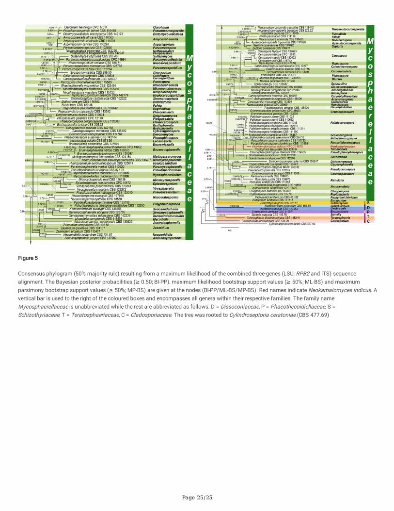
Comparing BLAST search results among sequences from the three loci (ITS, LSU, RPB2), the highest matches were all sequences ofParapallidocercospora. Phylogenetic analysis was based on LSU-RPB2-ITS concatenated sequences, our new collection Neokamalomycesindicus clustered closer (BI-PP/ML-BS/MP-BS: 1/100/100) to Parapallidocercospora colombiensis (Crous & M.J. Wingf.) Videira & Crous andParapallidocercospora thailandica (Crous, Himaman & M.J. Wingf.) Videira & Crous (Videira et al. 2017) and separated as sister lineage to the
Page 4/25
Parapallidocercospora clade (Fig. 5). Since the differences in morphology are signi�cant enough for retaining our new collection (a coelomycete) asdistinct from Parapallidocercospora (a hyphomycete) and represent a new lineage within the Mycosphaerellaceae. Hence a new genus and a newspecies are proposed. Phylogenetic trees generated from Bayesian analyses, ML, and MP produced trees with similar overall topology. A best scoringRAxML tree is presented in Fig. 5, with the Likelihood value of −57161.450108. The most parsimonious tree showed length = 16941 steps,consistency index = 0.165516, retention index = 0.477472, rescaled consistency index = 0.079029, homoplasy index = 0.834484 and G-�t is384.467564. From the analyzed characters, 727 were constant, 342 were variable and parsimony-uninformative, and 1032 were parsimony-informative. The overall parsimony phylogeny con�rmed the same species clades as those obtained in the Bayesian phylogeny.
Taxonomy
Neokamalomyces Sanjay & Raghv. Singh, gen. nov.Figs. 1–4
MycoBank: MB 843,767.
Etymology: Pre�x ‘Neo’ means new and genus su�x ‘kamalomyces’ based on the living legends Professor Kamal (DDU Gorakhpur University,Gorakhpur, India), a renowned mycologist and monographer of Cercosporoid Fungi of India.
Diagnosis: Differs from Parapallidocercospora by its very well developed pycnidial conidiomata with a central ostiolum; conidiophores hyaline,reduced to conidiogenous cells, lining the inner cavity; conidiogenous cells compactly aggregated; conidia hyaline to light olivaceous.
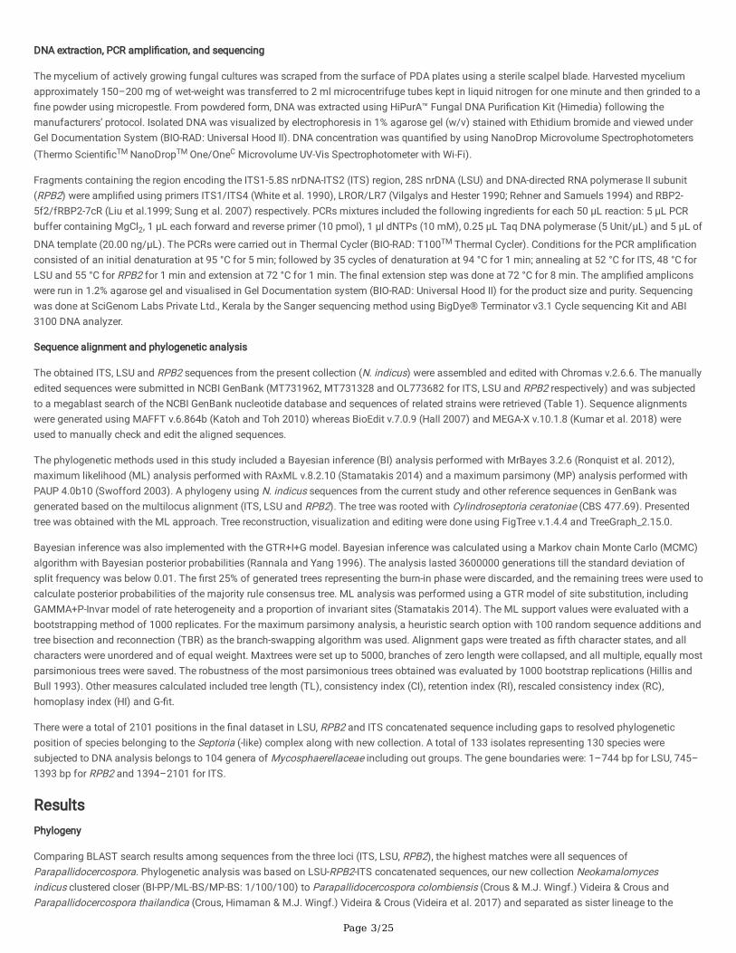
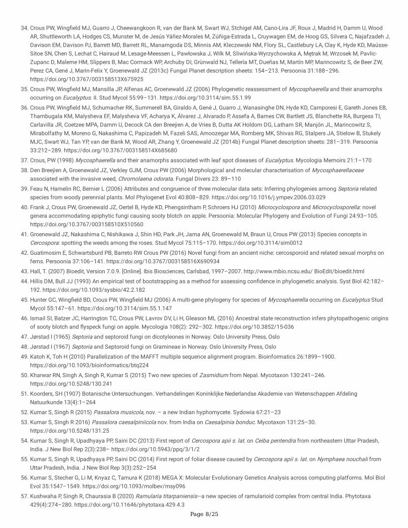
Description: Conidiomata pycnidial, brown to darkish brown, subepidermal, epigenous, numerous in each lesion, immersed to erumpent, subgloboseto globose, with a central ostiolum, releasing a hyaline conidial mass; the outer cells with brown, somewhat thickened walls, the inner cells hyaline,thin-walled. Ostiole single, circular, central. Conidiophores hyaline, reduced to conidiogenous cells, lining the inner cavity. Conidiogenous cellshyaline, tightly aggregated, cylindrical and tapering gradually toward the apex, ampulliform or lageniform with a relatively long neck, holoblastic,proliferating sympodially, smooth; scars unthickened. Conidia cylindrical, weakly to strongly curved, or �exuous, gradually attenuated to a roundedapex, gradually or more abruptly attenuated into a broadly truncate base, septate, not or indistinctly constricted around the septa, hyaline to lightolivaceous, hila unthickened to slightly thickened. Sexual morph not seen.
Type species: Neokamalomyces indicus Sanjay & Raghv. Singh
Neokamalomyces indicusSanjay & Raghv. Singh, sp. nov. Figs. 1–4
MycoBank number: MB 843,768.
Etymology: indicus, referring to India, the country where the fungus was discovered.
Type: India, Uttarakhand, Haridwar, Har Ki Pauri, 29.9567°N 78.1710°E, on living leaves of Ficus benghalensis L. (Moraceae), July 2019, coll. SanjayYadav, holotype (AMH 10233), isotype (MH-BHU 13), ex-type living culture (NFCCI 4870).
Diagnosis: Differs from Parapallidocercospora colombiensis by its presence of only internal mycelium, colonies epigenous, conidiomatapycnidial type, conidiophores develop from the inner lining of conidiomatal wall, hyaline, shorter and reduced to conidiogenous cells, conidia hyalineto light olivaceous and always smooth.
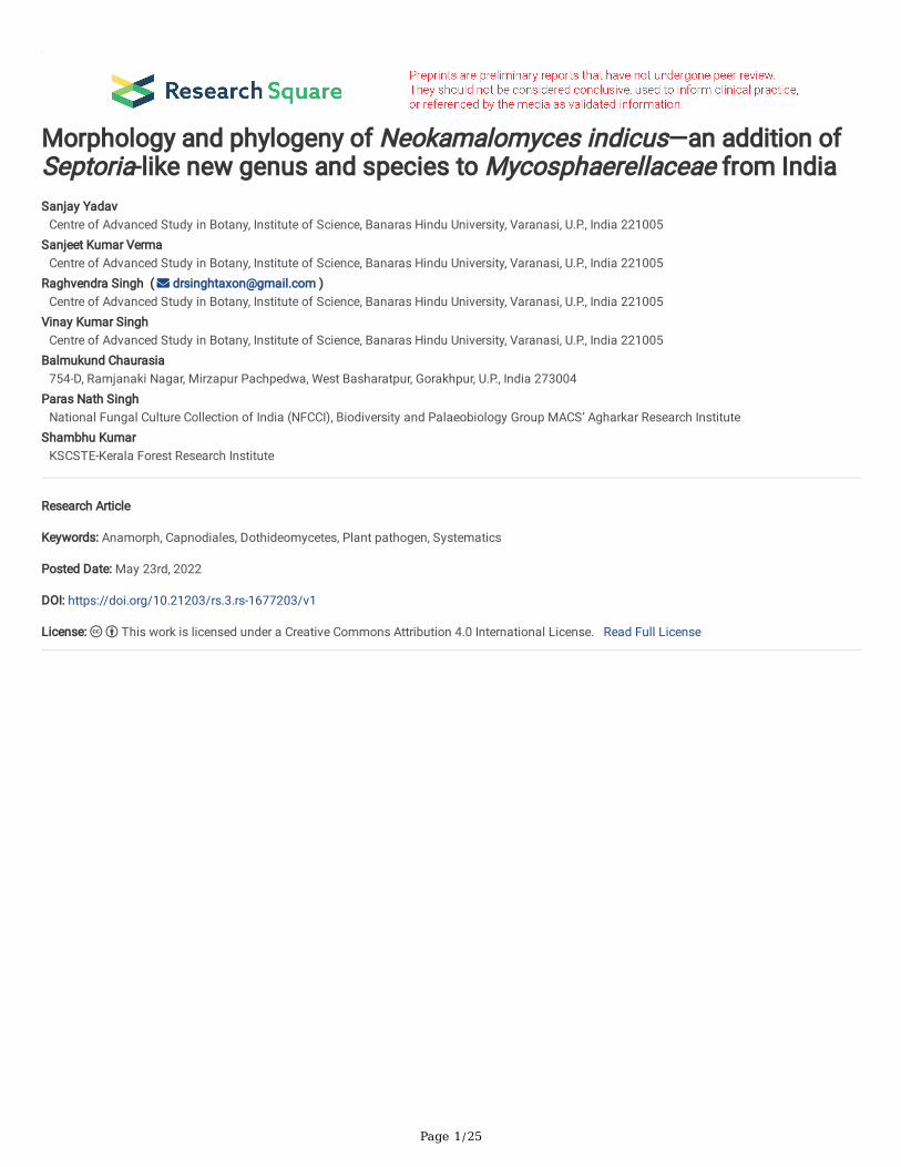
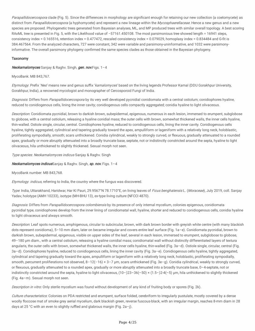
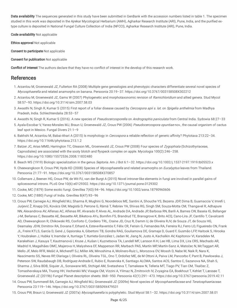
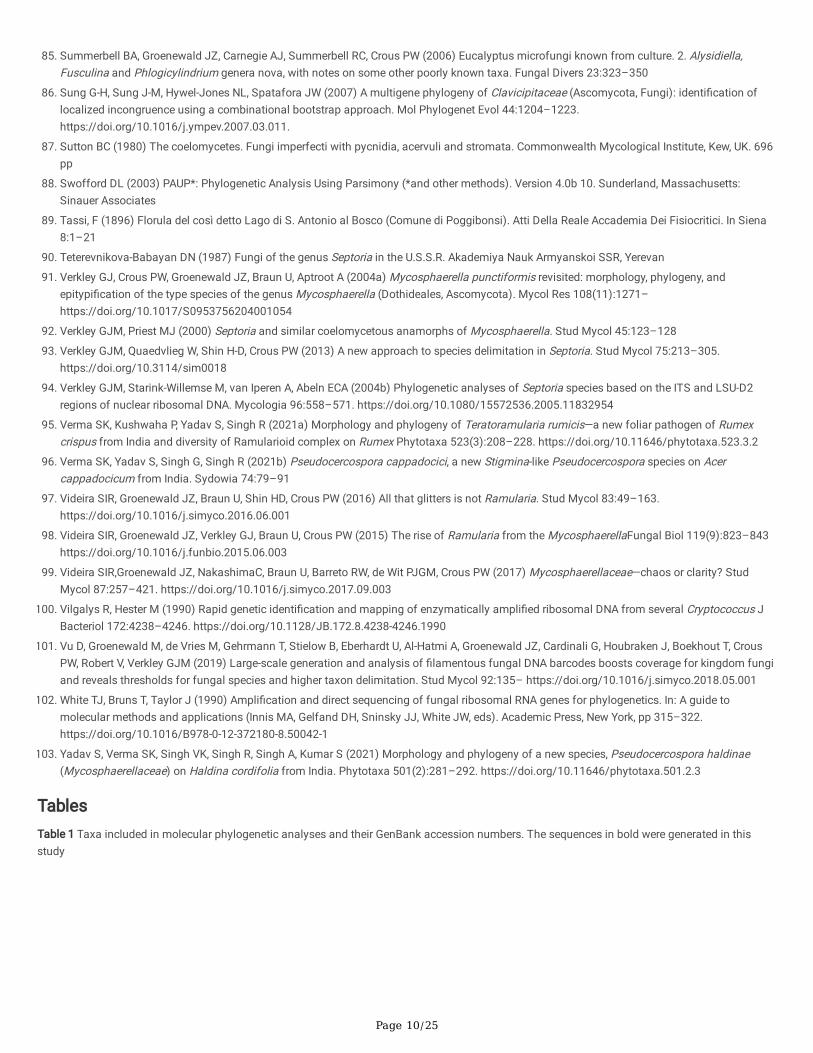
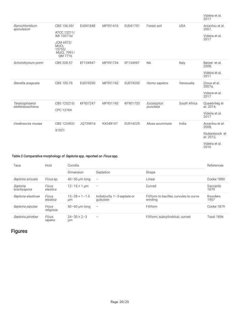
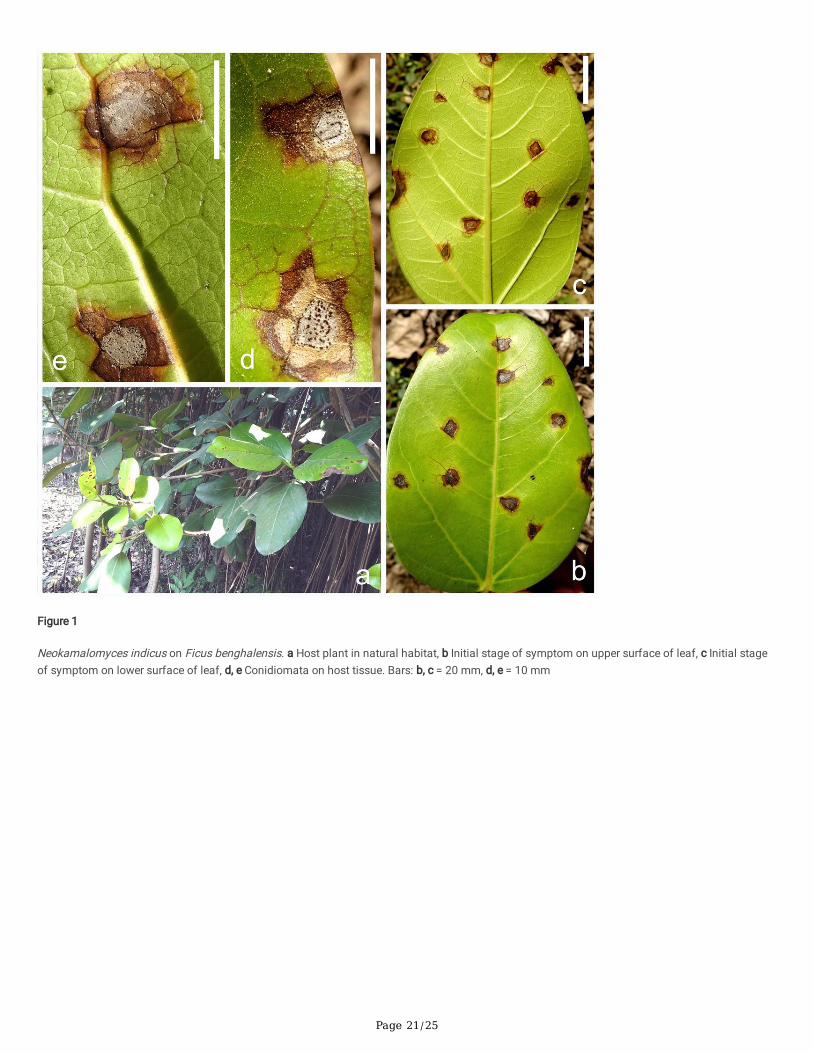
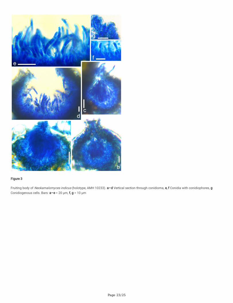
Description: Leaf spots numerous, amphigenous, circular to subcircular, brown, with dark brown border with greyish white centre (with many blackishdots represent conidioma), 5–10 mm diam, later on became irregular and covers entire leaf surface (Fig. 1a–e). Conidiomata pycnidial, brown todarkish brown, subepidermal, epigenous, visible on upper sides of the leaf, several in each lesion, immersed to erumpent, subglobose to globose,49–180 µm diam., with a central ostiolum, releasing a hyaline conidial mass; conidiomatal wall without distinctly differentiated layers of texturaangularis, the outer cells with brown, somewhat thickened walls, the inner cells hyaline, thin-walled (Fig. 3a–d). Ostiole single, circular, central (Fig.3a–d). Conidiophores hyaline, reduced to conidiogenous cells, lining the inner cavity (Fig. 3a–e). Conidiogenous cells hyaline, tightly aggregated,cylindrical and tapering gradually toward the apex, ampulliform or lageniform with a relatively long neck, holoblastic, proliferating sympodially,smooth, percurrent proliferations not observed, 8–12(–16) × 3–7 μm, scars unthickened (Fig. 3e–g). Conidia cylindrical, weakly to strongly curved,or �exuous, gradually attenuated to a rounded apex, gradually or more abruptly attenuated into a broadly truncate base, 0–4-septate, not orindistinctly constricted around the septa, hyaline to light olivaceous, (10–)23–36(–50) × (1.5–)2-4(–5) μm, hila unthickened to slightly thickened(Fig. 4a–m). Sexual morph not seen.
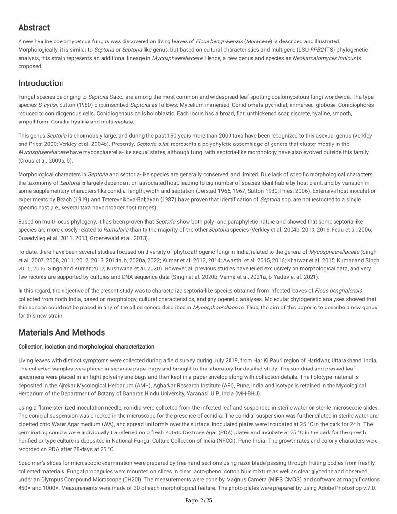
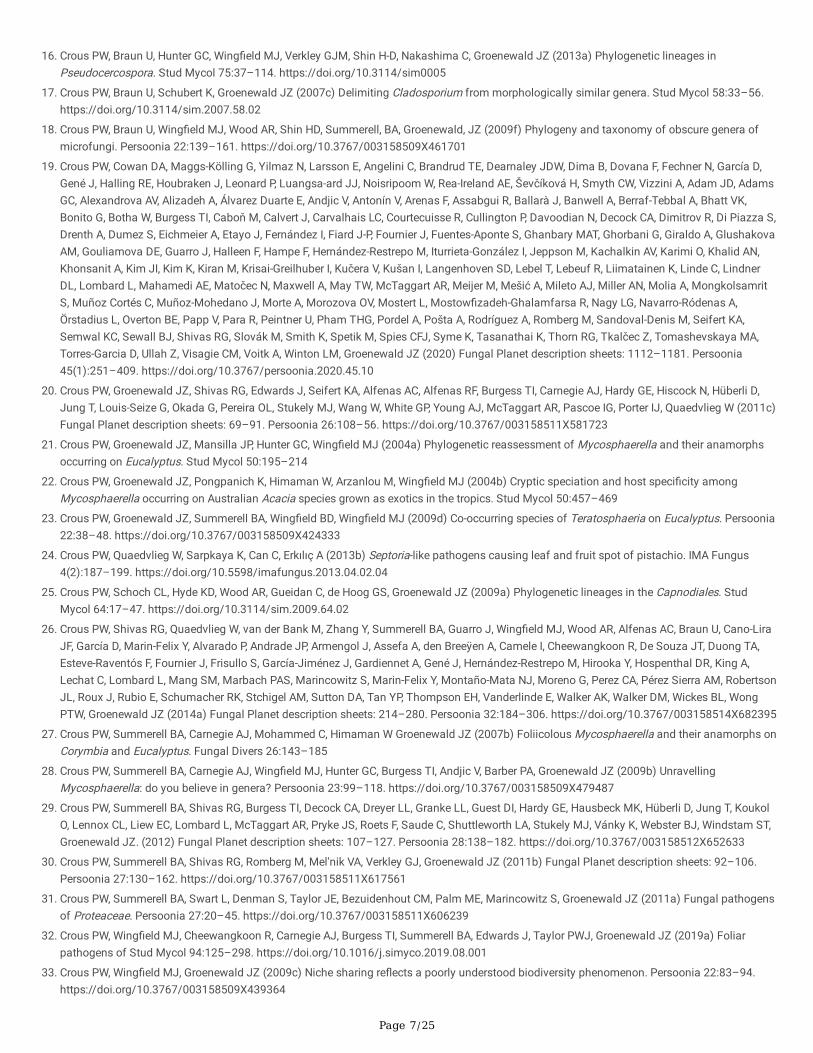
Description in vitro: Only sterile mycelium was found without development of any kind of fruiting body or spores (Fig. 2k).
Culture characteristics: Colonies on PDA restricted and erumpent, surface folded, cerebriform to irregularly pustulate, mostly covered by a densewoolly �occose mat of smoke grey aerial mycelium, dark blackish green, reverse fuscous-black, with an irregular margin, reaches 8-mm diam in 28days at 25 °C with an even to slightly ru�ed and glabrous margin (Fig. 2a–j).

Page 5/25
DiscussionPresent collectionshares a super�cial morphological resemblance with Septoria and septoria-like fungi (Crous et al. 2009a, b) due to the presence ofpycnidial type conidiomata that is internally lined by conidiophores. But on the basis of multi-gene analyses, it is quite distant from the Septoria andseptoria-like clade. Phylogenetic analyses based on the LSU, RPB2 and ITS sequence data retrieved from the ex-epitype culture of different genera ofMycosphaerellaceae along with Septoria and septoria-like fungi were chosen to establish exact phylogenetical position of present collection.Neokamalomyces indicus is morphologically indistinguishable from previous broad concepts of Septoria s. lat., just based on morphology. However,the phylogenetic position of N. indicus, quite distant from the Septoria s. lat. clade, does now allow to retain this species in the latter genus. Theseresults justify the introduction of a new genus for this lineage, viz., Neokamalomyces.
Based on both LSU and RPB2 as well as LSU, RPB2 and ITS sequences, the phylogenetic position has been shown to be closely related toParapallidocercospora with high bootstrap support (BI-PP/ML-BS/MP-BS: 1/100/100). The phylogenetic results in this study, agreed with theprevious placement of Pallidocercospora and Parapallidocercospora along with Nothophaeocryptopus, Pseudophaeophleospora,Scolecostigmina and Trochophora (Videira et al. 2017)(Fig. 5).
Based on multi-gene analyses, Pallidocercospora was established by Crous et al. (2013a) to accommodate cercospora-like species, but notcongeneric with Cercospora and is typi�ed by P. heimii (Crous) Crous (Crous et al. 2013a) and designated this genus based on its pale browncercosporoid like conidia, which are generally referred to as the Mycosphaerella heimii complex (Crous et al. 2004a, 2013a). However, they did notsynonymise Pseudocercospora colombiensis and Pseudocercospora thailandica under Pallidocercospora, even though these two species clusteredwith other Pallidocercospora species (Crous et al. 2013a). Subsequently, these two species have been stated as Pallidocercospora colombiensis andP. thailandica (Crous et al. 2013a; Pérez et al. 2013; Quaedvlieg et al. 2014), although the species combinations have not been formally establishedand thus, this name was invalid.
Later on, based on LSU, RPB2 and ITS sequence data, Parapallidocercospora was established with a type species Parapallidocercosporacolombiensis as a separated genus by Videira et al. (2017) in order to accommodate two species, Pseudocercospora colombiensis (foliar pathogenof Eucalyptus; Crous 1998), and Pseudocercospora thailandica (foliar pathogen of Acacia; Crous et al. 2004b). Morphologically, these taxa appearas a typical member of Pseudocercospora s. str. and are very di�cult to identify without the use of molecular sequence data. Mycosphaerellacolombiensis Crous & M.J. Wingf. and Pseudocercospora colombiensis Crous & M.J. Wingf. stand for sexual and asexual morphs of P.colombiensis (Crous 1998). In this study, both these species are clustered together in a very well-supported clade in the phylogenetic analyses (Fig.5) and are closely related to Nothophaeocryptopus, Pallidocercospora, Scolecostigmina and Trochophora. Currently, only 2 species names are validlyaccepted to Parapallidocercospora (https://www.mycobank.org, queried 8 March 2022).
Parapallidocercospora, however, is easily distinguished from Neokamalomyces by its lack of pycnidial type conidiomata; mycelium internal as wellas external; conidiophores brown, septate and arises singly from super�cial mycelium or aggregated in loose to dense fascicles arises from theupper cells of a brown stromata like structure and conidia that are brown and smooth to �nely verruculose. Parapallidocercospora also showsformation of ascomata and spermogonia intermixed with the ascomata or with the asexual morph that is totally lacking in Neokamalomyces.Therefore, despite the very high bootstrap support, the distinctive morphology justi�es the introduction of a new genus for this lineage, viz.,Neokamalomyces. The genus Neokamalomyces is currently monotypic based on Neokamalomyces indicus, a pathogen of Ficus benghalensis.Similarly, despite the low bootstrap support, the distinctive morphology observed in Trochophora justi�es that it is retained as a separate clade.
Presently, there are �ve species of Septoria that have been described from Ficus, namely, Septoria arcuata Cooke, S. brachyspora Sacc., S.elasticae Koord., S. pipulae Cookeand S. pirottae Tassi. The comparative morphology of these species is provided in table 2 indicates that they areclearly different from novel strain N. indicus by either having narrower conidia or shape of conidia.
DeclarationsAcknowledgments The authors are indebted to anonymous reviewers for helpful comments and the curator of AMH and NFCCI for acceptingmaterial and providing an accession number there off. We express our deep gratitude to Dr Shaun Pennycook (Landcare Research, Auckland, NewZealand) for nomenclatural review. We are also thankful to the Head, CAS in Botany, Banaras Hindu University, Varanasi for Instrumental facilities.
Author’s contribution All authors contributed to the conception and design of the study. SY collected sample, cultivated pure culture, isolated DNAand prepared samples for sequencing. SKV and VKS developed morphological features and surveyed concerned literatures. RS developed photoplates, performed phylogenetic analyses and developed discussion part of the manuscript. BC, PNS and SK wrote the �rst draft of the manuscript.All authors contributed to previous drafts of the manuscript and read and approved the �nal draft of the manuscript.
Funding RS thanks Science & Engineering Research Board (SERB), Department of Science & Technology (DST), Govt. of India (Scheme No.CRG/2020/006053) and Institution of Eminence (IoE) Scheme, Ministry of Human Resource and Development (MHRD), Govt. of India (SchemeNo.6031) for providing �nancial support.
Page 6/25
Data availability The sequences generated in this study have been submitted in GenBank with the accession numbers listed in table 1. The specimenstudied in this work was deposited in the Ajrekar Mycological Herbarium (AMH), Agharkar Research Institute (ARI), Pune, India, and the puri�ed ex-type culture is deposited in National Fungal Culture Collection of India (NFCCI), Agharkar Research Institute (ARI), Pune, India.
Code availability Not applicable
Ethics approval Not applicable
Consent to participate Not applicable
Consent for publication Not applicable
Con�ict of interest The authors declare that they have no con�ict of interest in the develop of this research work.
References1. Arzanlou M, Groenewald JZ, Fullerton RA (2008) Multiple gene genealogies and phenotypic characters differentiate several novel species of
Mycosphaerella and related anamorphs on banana. Persoonia 20:19–37. https://doi.org/10.3767/003158508X302212
2. Arzanlou M, Groenewald JZ, Gams W (2007) Phylogenetic and morphotaxonomic revision of Ramichloridium and allied genera. Stud Mycol58:57–93. https://doi.org/10.3114/sim.2007.58.03
3. Awasthi N, Singh R, Kumar S (2015) First report of a foliar disease caused by Cercospora apii s. lat. on Spigelia anthelmia from MadhyaPradesh, India. Schlechtendalia 28:53–57
4. Awasthi N, Singh R, Kumar S (2016). A new species of Pseudocercosporella on Andrographis paniculata from Central India. Sydowia 68:27–33
5. Ayala-Escobar V, Yanez-Morales MJ, Braun U, Groenewald JZ, Crous PW (2006) Pseudocercospora opuntiae nov., the causal organism of cactusleaf spot in Mexico. Fungal Divers 21:1–9
�. Bakhshi M, Arzanlou M, Babai-Ahari A (2015) Is morphology in Cercospora a reliable re�ection of generic a�nity? Phytotaxa 213:22–34.https://doi.org/10.11646/phytotaxa.213.1.2
7. Batzer JC, Arias MMD, Harrington TC, Gleason ML, Groenewald JZ, Crous PW (2008) Four species of Zygophiala (Schizothyriaceae,Capnodiales) are associated with the sooty blotch and �yspeck complex on apple. Mycologia 100(2):246–258.https://doi.org/10.1080/15572536.2008.11832480
�. Beach WS (1919) Biologic specialization in the genus Septoria. Am J Bot 6:1–32. https://doi.org/10.1002/j.1537-2197.1919.tb05523.x
9. Cheewangkoon R, Crous PW, Hyde KD (2008) Species of Mycosphaerella and related anamorphs on Eucalyptus leaves from Thailand.Persoonia 21:77–91. https://doi.org/10.3767/003158508X370857
10. Collemare J, Beenen HG, Crous PW, de Wit PJ, van der Burgt A (2015) Novel introner-like elements in fungi are involved in parallel gains ofspliceosomal introns. PLoS One 10(6):e0129302. https://doi.org/10.1371/journal.pone.0129302
11. Cooke, MC (1879) Some exotic fungi. Grevillea 7(43):94–96. https://doi.org/10.1002/asna.18790960606
12. Cooke, MC (1880) Fungi of India. Grevillea 8(47):93–96
13. Crous PW, Carnegie AJ, Wing�eld MJ, Sharma R, Mughini G, Noordeloos ME, Santini A, Shouche YS, Bezerra JDP, Dima B, Guarnaccia V, Imre� I,Jurjević Ž, Knapp DG, Kovács GM, Magistà D, Perrone G, Rämä T, Rebriev YA, Shivas RG, Singh SM, Souza-Motta CM, Thangavel R, AdhapureNN, Alexandrova AV, Alfenas AC, Alfenas RF, Alvarado P, Alves AL, Andrade DA, Andrade JP, Barbosa RN, Barili A, Barnes CW, Baseia IG, BellangerJ-M, Berlanas C, Bessette AE, Bessette AR, Biketova AYu, Bom�m FS, Brandrud TE, Bransgrove K, Brito ACQ, Cano-Lira JF, Cantillo T, CavalcantiAD, Cheewangkoon R, Chikowski RS, Conforto C, Cordeiro TRL, Craine JD, Cruz R, Damm U, de Oliveira RJV, de Souza JT, de Souza HG,Dearnaley JDW, Dimitrov RA, Dovana F, Erhard A, Esteve-Raventós F, Félix CR, Ferisin G, Fernandes RA, Ferreira RJ, Ferro LO, Figueiredo CN, FrankJL, Freire KTLS, García D, Gené J, Gęsiorska A, Gibertoni TB, Gondra RAG, Gouliamova DE, Gramaje D, Guard F, Gusmão LFP, Haitook S, HirookaY, Houbraken J, Hubka V, Inamdar A, Iturriaga T, Iturrieta-González I, Jadan M, Jiang N, Justo A, Kachalkin AV, Kapitonov VI, Karadelev M,Karakehian J, Kasuya T, Kautmanová I, Kruse J, Kušan I, Kuznetsova TA, Landell MF, Larsson K-H, Lee HB, Lima DX, Lira CRS, Machado AR,Madrid H, Magalhães OMC, Majerova H, Malysheva EF, Mapperson RR, Marbach PAS, Martín MP, Martín-Sanz A, Matočec N, McTaggart AR,Mello JF, Melo RFR, Mešić A, Michereff SJ, Miller AN, Minoshima A, Molinero-Ruiz L, Morozova OV, Mosoh D, Nabe M, Naik R, Nara K,Nascimento SS, Neves RP, Olariaga I, Oliveira RL, Oliveira TGL, Ono T, Ordoñez ME, de M Ottoni A, Paiva LM, Pancorbo F, Pant B, Pawłowska J,Peterson SW, Raudabaugh DB, Rodríguez-Andrade E, Rubio E, Rusevska K, Santiago ALCMA, Santos ACS, Santos C, Sazanova NA, Shah S,Sharma J, Silva BDB, Siquier JL, Sonawane MS, Stchigel AM, Svetasheva T, Tamakeaw N, Telleria MT, Tiago PV, Tian CM, Tkalčec Z,Tomashevskaya MA, Truong HH, Vecherskii MV, Visagie CM, Vizzini A, Yilmaz N, Zmitrovich IV, Zvyagina EA, Boekhout T, Kehlet T, Læssøe T,Groenewald JZ (2019b) Fungal Planet description sheets: 868–950. Persoonia 42(1):291–473. https://doi.org/10.3767/persoonia.2019.42.11
14. Crous PW, Summerell BA, Carnegie AJ, Wing�eld MJ, Groenewald JZ (2009e) Novel species of Mycosphaerellaceae and Teratosphaeriaceae.Persoonia 23:119–146. https://doi.org/10.3767/003158509X479531
15. Crous PW, Braun U, Groenewald JZ (2007a) Mycosphaerella is polyphyletic. Stud Mycol 58:1–32. https://doi.org/10.3114/sim.2007.58.01
Page 7/25
1�. Crous PW, Braun U, Hunter GC, Wing�eld MJ, Verkley GJM, Shin H-D, Nakashima C, Groenewald JZ (2013a) Phylogenetic lineages inPseudocercospora. Stud Mycol 75:37–114. https://doi.org/10.3114/sim0005
17. Crous PW, Braun U, Schubert K, Groenewald JZ (2007c) Delimiting Cladosporium from morphologically similar genera. Stud Mycol 58:33–56.https://doi.org/10.3114/sim.2007.58.02
1�. Crous PW, Braun U, Wing�eld MJ, Wood AR, Shin HD, Summerell, BA, Groenewald, JZ (2009f) Phylogeny and taxonomy of obscure genera ofmicrofungi. Persoonia 22:139–161. https://doi.org/10.3767/003158509X461701
19. Crous PW, Cowan DA, Maggs-Kölling G, Yilmaz N, Larsson E, Angelini C, Brandrud TE, Dearnaley JDW, Dima B, Dovana F, Fechner N, García D,Gené J, Halling RE, Houbraken J, Leonard P, Luangsa-ard JJ, Noisripoom W, Rea-Ireland AE, Ševčíková H, Smyth CW, Vizzini A, Adam JD, AdamsGC, Alexandrova AV, Alizadeh A, Álvarez Duarte E, Andjic V, Antonín V, Arenas F, Assabgui R, Ballarà J, Banwell A, Berraf-Tebbal A, Bhatt VK,Bonito G, Botha W, Burgess TI, Caboň M, Calvert J, Carvalhais LC, Courtecuisse R, Cullington P, Davoodian N, Decock CA, Dimitrov R, Di Piazza S,Drenth A, Dumez S, Eichmeier A, Etayo J, Fernández I, Fiard J-P, Fournier J, Fuentes-Aponte S, Ghanbary MAT, Ghorbani G, Giraldo A, GlushakovaAM, Gouliamova DE, Guarro J, Halleen F, Hampe F, Hernández-Restrepo M, Iturrieta-González I, Jeppson M, Kachalkin AV, Karimi O, Khalid AN,Khonsanit A, Kim JI, Kim K, Kiran M, Krisai-Greilhuber I, Kučera V, Kušan I, Langenhoven SD, Lebel T, Lebeuf R, Liimatainen K, Linde C, LindnerDL, Lombard L, Mahamedi AE, Matočec N, Maxwell A, May TW, McTaggart AR, Meijer M, Mešić A, Mileto AJ, Miller AN, Molia A, MongkolsamritS, Muñoz Cortés C, Muñoz-Mohedano J, Morte A, Morozova OV, Mostert L, Mostow�zadeh-Ghalamfarsa R, Nagy LG, Navarro-Ródenas A,Örstadius L, Overton BE, Papp V, Para R, Peintner U, Pham THG, Pordel A, Pošta A, Rodríguez A, Romberg M, Sandoval-Denis M, Seifert KA,Semwal KC, Sewall BJ, Shivas RG, Slovák M, Smith K, Spetik M, Spies CFJ, Syme K, Tasanathai K, Thorn RG, Tkalčec Z, Tomashevskaya MA,Torres-Garcia D, Ullah Z, Visagie CM, Voitk A, Winton LM, Groenewald JZ (2020) Fungal Planet description sheets: 1112–1181. Persoonia45(1):251–409. https://doi.org/10.3767/persoonia.2020.45.10
20. Crous PW, Groenewald JZ, Shivas RG, Edwards J, Seifert KA, Alfenas AC, Alfenas RF, Burgess TI, Carnegie AJ, Hardy GE, Hiscock N, Hüberli D,Jung T, Louis-Seize G, Okada G, Pereira OL, Stukely MJ, Wang W, White GP, Young AJ, McTaggart AR, Pascoe IG, Porter IJ, Quaedvlieg W (2011c)Fungal Planet description sheets: 69–91. Persoonia 26:108–56. https://doi.org/10.3767/003158511X581723
21. Crous PW, Groenewald JZ, Mansilla JP, Hunter GC, Wing�eld MJ (2004a) Phylogenetic reassessment of Mycosphaerella and their anamorphsoccurring on Eucalyptus. Stud Mycol 50:195–214
22. Crous PW, Groenewald JZ, Pongpanich K, Himaman W, Arzanlou M, Wing�eld MJ (2004b) Cryptic speciation and host speci�city amongMycosphaerella occurring on Australian Acacia species grown as exotics in the tropics. Stud Mycol 50:457–469
23. Crous PW, Groenewald JZ, Summerell BA, Wing�eld BD, Wing�eld MJ (2009d) Co-occurring species of Teratosphaeria on Eucalyptus. Persoonia22:38–48. https://doi.org/10.3767/003158509X424333
24. Crous PW, Quaedvlieg W, Sarpkaya K, Can C, Erkılıç A (2013b) Septoria-like pathogens causing leaf and fruit spot of pistachio. IMA Fungus4(2):187–199. https://doi.org/10.5598/imafungus.2013.04.02.04
25. Crous PW, Schoch CL, Hyde KD, Wood AR, Gueidan C, de Hoog GS, Groenewald JZ (2009a) Phylogenetic lineages in the Capnodiales. StudMycol 64:17–47. https://doi.org/10.3114/sim.2009.64.02
2�. Crous PW, Shivas RG, Quaedvlieg W, van der Bank M, Zhang Y, Summerell BA, Guarro J, Wing�eld MJ, Wood AR, Alfenas AC, Braun U, Cano-LiraJF, García D, Marin-Felix Y, Alvarado P, Andrade JP, Armengol J, Assefa A, den Breeÿen A, Camele I, Cheewangkoon R, De Souza JT, Duong TA,Esteve-Raventós F, Fournier J, Frisullo S, García-Jiménez J, Gardiennet A, Gené J, Hernández-Restrepo M, Hirooka Y, Hospenthal DR, King A,Lechat C, Lombard L, Mang SM, Marbach PAS, Marincowitz S, Marin-Felix Y, Montaño-Mata NJ, Moreno G, Perez CA, Pérez Sierra AM, RobertsonJL, Roux J, Rubio E, Schumacher RK, Stchigel AM, Sutton DA, Tan YP, Thompson EH, Vanderlinde E, Walker AK, Walker DM, Wickes BL, WongPTW, Groenewald JZ (2014a) Fungal Planet description sheets: 214–280. Persoonia 32:184–306. https://doi.org/10.3767/003158514X682395
27. Crous PW, Summerell BA, Carnegie AJ, Mohammed C, Himaman W Groenewald JZ (2007b) Foliicolous Mycosphaerella and their anamorphs onCorymbia and Eucalyptus. Fungal Divers 26:143–185
2�. Crous PW, Summerell BA, Carnegie AJ, Wing�eld MJ, Hunter GC, Burgess TI, Andjic V, Barber PA, Groenewald JZ (2009b) UnravellingMycosphaerella: do you believe in genera? Persoonia 23:99–118. https://doi.org/10.3767/003158509X479487
29. Crous PW, Summerell BA, Shivas RG, Burgess TI, Decock CA, Dreyer LL, Granke LL, Guest DI, Hardy GE, Hausbeck MK, Hüberli D, Jung T, KoukolO, Lennox CL, Liew EC, Lombard L, McTaggart AR, Pryke JS, Roets F, Saude C, Shuttleworth LA, Stukely MJ, Vánky K, Webster BJ, Windstam ST,Groenewald JZ. (2012) Fungal Planet description sheets: 107–127. Persoonia 28:138–182. https://doi.org/10.3767/003158512X652633
30. Crous PW, Summerell BA, Shivas RG, Romberg M, Mel'nik VA, Verkley GJ, Groenewald JZ (2011b) Fungal Planet description sheets: 92–106.Persoonia 27:130–162. https://doi.org/10.3767/003158511X617561
31. Crous PW, Summerell BA, Swart L, Denman S, Taylor JE, Bezuidenhout CM, Palm ME, Marincowitz S, Groenewald JZ (2011a) Fungal pathogensof Proteaceae. Persoonia 27:20–45. https://doi.org/10.3767/003158511X606239
32. Crous PW, Wing�eld MJ, Cheewangkoon R, Carnegie AJ, Burgess TI, Summerell BA, Edwards J, Taylor PWJ, Groenewald JZ (2019a) Foliarpathogens of Stud Mycol 94:125–298. https://doi.org/10.1016/j.simyco.2019.08.001
33. Crous PW, Wing�eld MJ, Groenewald JZ (2009c) Niche sharing re�ects a poorly understood biodiversity phenomenon. Persoonia 22:83–94.https://doi.org/10.3767/003158509X439364
Page 8/25
34. Crous PW, Wing�eld MJ, Guarro J, Cheewangkoon R, van der Bank M, Swart WJ, Stchigel AM, Cano-Lira JF, Roux J, Madrid H, Damm U, WoodAR, Shuttleworth LA, Hodges CS, Munster M, de Jesús Yáñez-Morales M, Zúñiga-Estrada L, Cruywagen EM, de Hoog GS, Silvera C, Najafzadeh J,Davison EM, Davison PJ, Barrett MD, Barrett RL, Manamgoda DS, Minnis AM, Kleczewski NM, Flory SL, Castlebury LA, Clay K, Hyde KD, Maússe-Sitoe SN, Chen S, Lechat C, Hairaud M, Lesage-Meessen L, Pawłowska J, Wilk M, Sliwińska-Wyrzychowska A, Mętrak M, Wrzosek M, Pavlic-Zupanc D, Maleme HM, Slippers B, Mac Cormack WP, Archuby DI, Grünwald NJ, Tellería MT, Dueñas M, Martín MP, Marincowitz S, de Beer ZW,Perez CA, Gené J, Marin-Felix Y, Groenewald JZ (2013c) Fungal Planet description sheets: 154–213. Persoonia 31:188–296.https://doi.org/10.3767/003158513X675925
35. Crous PW, Wing�eld MJ, Mansilla JP, Alfenas AC, Groenewald JZ (2006) Phylogenetic reassessment of Mycosphaerella and their anamorphsoccurring on Eucalyptus. II. Stud Mycol 55:99–131. https://doi.org/10.3114/sim.55.1.99
3�. Crous PW, Wing�eld MJ, Schumacher RK, Summerell BA, Giraldo A, Gené J, Guarro J, Wanasinghe DN, Hyde KD, Camporesi E, Gareth Jones EB,Thambugala KM, Malysheva EF, Malysheva VF, Acharya K, Álvarez J, Alvarado P, Assefa A, Barnes CW, Bartlett JS, Blanchette RA, Burgess TI,Carlavilla JR, Coetzee MPA, Damm U, Decock CA den Breeÿen A, de Vries B, Dutta AK Holdom DG, Latham SR, Manjón JL, Marincowitz S,Mirabolfathy M, Moreno G, Nakashima C, Papizadeh M, Fazeli SAS, Amoozegar MA, Romberg MK, Shivas RG, Stalpers JA, Stielow B, StukelyMJC, Swart WJ, Tan YP, van der Bank M, Wood AR, Zhang Y, Groenewald JZ (2014b) Fungal Planet description sheets: 281–319. Persoonia33:212–289. https://doi.org/10.3767/003158514X685680
37. Crous, PW (1998) Mycosphaerella and their anamorphs associated with leaf spot diseases of Eucalyptus. Mycologia Memoirs 21:1–170
3�. Den Breeÿen A, Groenewald JZ, Verkley GJM, Crous PW (2006) Morphological and molecular characterisation of Mycosphaerellaceaeassociated with the invasive weed, Chromolaena odorata. Fungal Divers 23: 89–110
39. Feau N, Hamelin RC, Bernier L (2006) Attributes and congruence of three molecular data sets: Inferring phylogenies among Septoria relatedspecies from woody perennial plants. Mol Phylogenet Evol 40:808–829. https://doi.org/10.1016/j.ympev.2006.03.029
40. Frank J, Crous PW, Groenewald JZ, Oertel B, Hyde KD, Phengsintham P, Schroers HJ (2010) Microcyclospora and Microcyclosporella: novelgenera accommodating epiphytic fungi causing sooty blotch on apple. Persoonia: Molecular Phylogeny and Evolution of Fungi 24:93–105.https://doi.org/10.3767/003158510X510560
41. Groenewald JZ, Nakashima C, Nishikawa J, Shin HD, Park JH, Jama AN, Groenewald M, Braun U, Crous PW (2013) Species concepts inCercospora: spotting the weeds among the roses. Stud Mycol 75:115–170. https://doi.org/10.3114/sim0012
42. Guatimosim E, Schwartsburd PB, Barreto RW Crous PW (2016) Novel fungi from an ancient niche: cercosporoid and related sexual morphs onferns. Persoonia 37:106–141. https://doi.org/10.3767/003158516X690934
43. Hall, T. (2007) Bioedit, Version 7.0.9. [Online]. Ibis Biosciences, Carlsbad, 1997–2007. http://www.mbio.ncsu.edu/ BioEdit/bioedit.html
44. Hillis DM, Bull JJ (1993) An empirical test of bootstrapping as a method for assessing con�dence in phylogenetic analysis. Syst Biol 42:182–192. https://doi.org/10.1093/sysbio/42.2.182
45. Hunter GC, Wing�eld BD, Crous PW, Wing�eld MJ (2006) A multi-gene phylogeny for species of Mycosphaerella occurring on Eucalyptus StudMycol 55:147–61. https://doi.org/10.3114/sim.55.1.147
4�. Ismail SI, Batzer JC, Harrington TC, Crous PW, Lavrov DV, Li H, Gleason ML (2016) Ancestral state reconstruction infers phytopathogenic originsof sooty blotch and �yspeck fungi on apple. Mycologia 108(2): 292–302. https://doi.org/10.3852/15-036
47. Jørstad I (1965) Septoria and septoroid fungi on dicotyleones in Norway. Oslo University Press, Oslo
4�. Jørstad I (1967) Septoria and Septoroid fungi on Gramineae in Norway. Oslo University Press, Oslo
49. Katoh K, Toh H (2010) Parallelization of the MAFFT multiple sequence alignment program. Bioinformatics 26:1899–1900.https://doi.org/10.1093/bioinformatics/btq224
50. Kharwar RN, Singh A, Singh R, Kumar S (2015) Two new species of Zasmidium from Nepal. Mycotaxon 130:241–246.https://doi.org/10.5248/130.241
51. Koorders, SH (1907) Botanische Untersuchungen. Verhandelingen Koninklijke Nederlandse Akademie van Wetenschappen AfdelingNatuurkunde 13(4):1–264
52. Kumar S, Singh R (2015) Passalora musicola, nov. – a new Indian hyphomycete. Sydowia 67:21–23
53. Kumar S, Singh R 2016) Passalora caesalpiniicola nov. from India on Caesalpinia bonduc. Mycotaxon 131:25–30.https://doi.org/10.5248/131.25
54. Kumar S, Singh R, Upadhyaya PP, Saini DC (2013) First report of Cercospora apii s. lat. on Ceiba pentendra from northeastern Uttar Pradesh,India. J New Biol Rep 2(3):238– https://doi.org/10.5943/ppq/3/1/2
55. Kumar S, Singh R, Upadhyaya PP, Saini DC (2014) First report of foliar disease caused by Cercospora apii s. lat. on Nymphaea nouchali fromUttar Pradesh, India. J New Biol Rep 3(3):252–254
5�. Kumar S, Stecher G, Li M, Knyaz C, Tamura K (2018) MEGA X: Molecular Evolutionary Genetics Analysis across computing platforms. Mol BiolEvol 35:1547–1549. https://doi.org/10.1093/molbev/msy096
57. Kushwaha P, Singh R, Chaurasia B (2020) Ramularia titarpaniensis—a new species of ramularioid complex from central India. Phytotaxa429(4):274–280. https://doi.org/10.11646/phytotaxa.429.4.3

Page 9/25
5�. Liu YJ, Whelen S, Hall BD (1999) Phylogenetic relationships among ascomycetes: evidence from an RNA polymerase II subunit. Mol Biol Evol16:1799–1808. https://doi.org/10.1093/oxfordjournals.molbev.a026092
59. Pérez CA, Wing�eld MJ, Altier N, Blanchette RA (2013) Species of Mycosphaerellaceae and Teratosphaeriaceae on native Myrtaceae in Uruguay:evidence of fungal host jumps. Fungal boil 117:94–102. https://doi.org/10.1016/j.funbio.2012.12.002
�0. Priest MJ (2006) Fungi of Australia: Septoria. ABRS, Canberra: CSIRO publishing, Melbourne, Australia
�1. Quaedvlieg W, Binder M, Groenewald JZ, Summerell BA, Carnegie AJ, Burgess TI, Crous PW (2014) Introducing the consolidated species conceptto resolve species in the Teratosphaeriaceae. Persoonia 33:1–40. https://doi.org/10.3767/003158514X681981
�2. Quaedvlieg W, Groenewald JZ, de Jesús Yáñez-Morales M, Crous PW (2012) DNA barcoding of Mycosphaerella species of quarantineimportance to Europe. Persoonia: Mol Phylogeny Evol Fungi 29:101–115. https://doi.org/10.3767/003158512X661282
�3. Quaedvlieg W, Kema GHJ, Groenewald JZ, Verkley GJM, Seifbarghi S, Razavi M, Mirzadi Gohari A, Mehrabi R, Crous PW (2011) Zymoseptorianov.: a new genus to accommodate Septoria-like species occurring on graminicolous hosts. Persoonia 26:57–69.https://doi.org/10.3767/003158511X571841
�4. Quaedvlieg W, Verkley GJM, Shin H-D, Barreto RW, Alfenas AC, Swart WJ, Groenewald JZ, Crous PW (2013) Sizing up Septoria. Stud Mycol75:307–390. https://doi.org/10.3114/sim0017
�5. Rannala B, Yang Z (1996) Probability distribution of molecular evolutionary trees: a new method of phylogenetic inference. J Mol Evol 43:304–311. https://doi.org/10.1007/PL00006090
��. Rehner SA, Samuels GJ (1994) Taxonomy and phylogeny of Gliocladium analysed from nuclear large subunit ribosomal DNA sequences. MycolRes 98:625–634. https://doi.org/10.1016/S0953-7562(09)80409-7
�7. Ronquist F, Teslenko M, van der Mark P, Ayres D, Darling A, Ohna SH, Larget B, Liu L, Suchard MA, Huelsenbeck JP (2012) MrBayes 3.2: EffcientBayesian phylogenetic inference and model choice across a large model space. Syst Biol 61:539–542. https://doi.org/10.1093/sysbio/sys029
��. Saccardo PA (1879) Fungi Gallici lecti a cl. viris P. Brunaud, C.C. Gillet et Abb. Letendre. Michelia 1(5):500–538
�9. Schoch CL, Robbertse B, Robert V, Vu D, Cardinali G, Irinyi L, Federhen S (2014) Finding needles in haystacks: linking scienti�c names, referencespecimens and molecular data for Fungi. Database 2014:1– https://doi.org/10.1093/database/bau061
70. Schoch CL, Sung GH, López-Giráldez F, Townsend JP, Miadlikowska J, Hofstetter V, Spatafora JW (2009) The Ascomycota tree of life: a phylum-wide phylogeny clari�es the origin and evolution of fundamental reproductive and ecological traits. Syst Biol58(2):224–https://doi.org/10.1093/sysbio/syp020
71. Simon UK, Groenewald JZ, Crous PW (2009) Cymadothea trifolii, an obligate biotrophic leaf parasite of Trifolium, belongs toMycosphaerellaceae as shown by nuclear ribosomal DNA analyses. Persoonia 22:49– https://doi.org/10.3767/003158509X425350
72. Singh A, Kharwar RN, Singh R, Kumar S (2014a) A new species of Zasmidium (Mycosphaerellaceae) from India. Sydowia 66(2):309–312
73. Singh A, Kumar S, Singh R, Agrawal DK (2008) Two new species of Ramularia from Indian Sub-continents. Ind Phytopath 61(3):348–352
74. Singh G, Yadav S, Singh R, Kumar S (2022) Passalora golaghati nov. from India. Mycotaxon 137(1):89–94. https://doi.org/10.5248/137.89
75. Singh R, Chaurasia B, Shukla K, Upadhyaya PP (2012) Passalora aseptata, a new cercosporoid fungus from northeastern Uttar Pradesh, India.Mycotaxon 120:461– https://doi.org/10.5248/120.461
7�. Singh R, Kumar S (2017) Passalora rhamnaecearum nov. (Capnodiales, Mycosphaerellaceae) from India. Kavaka 48(1):50–51
77. Singh R, Kumar S, Kamal (2011) Two new species of Passalora and Pseudocercospora from northeastern Uttar Pradesh, India. Mycotaxon117:137– https://doi.org/10.5248/117.137
7�. Singh R, Kumar S, Pal VK, Upadhyaya PP, Agrawal DK (2007) New taxa of foliicolous hyphomycetes-Cercospora, Corynespora andPhaeotrichochonis from North-Eastern U.P. Ind Phytopath 60(4):506–512
79. Singh R, Kumar S, Saini DC, Upadhyaya PP, Kamal, Braun U (2013) Diversity of Passalora on Ficus. Mycol Prog 12:637–https://doi.org/10.1007/s11557-012-0870-6
�0. Singh R, Singh A, Kumar S, Upadhyaya PP, Castañeda-Ruíz RF (2014b) Two new species of Zasmidium from northeastern Uttar Pradesh, India.Nova Hedwigia 98(1–2):257– https://doi.org/10.1127/0029-5035/2013/0137
�1. Singh R, Verma SK, Yadav S, Bhojak P, Kumar S (2020b) Morphology and phylogeny of Pseudocercospora hamiltoniani—A new speciescomparable to Sirosporium from Uttarakhand, India. Phytotaxa 458(4):281–293. https://doi.org/10.11646/phytotaxa.458.4.4
�2. Singh R, Verma SK, Yadav S, Kumar S (2020a) Cercosporella bundelkhandae nov. from India. Mycotaxon 135:315–320.https://doi.org/10.5248/135.315
�3. Stamatakis A (2014) RAxML version 8: a tool for phylogenetic analysis and post-analysis of large phylogenies. Bioinformatics 30:1312–1313.https://doi.org/10.1093/bioinformatics/btu033
�4. Stukenbrock EH, Quaedvlieg W, Javan-Nikhah, M, Zala M, Crous PW, McDonald BA (2012) Zymoseptoria ardabiliae and pseudotritici, twoprogenitor species of the septoria tritici leaf blotch fungus Z. tritici (synonym: Mycosphaerella graminicola). Mycologia 104(6):1397–1407.https://doi.org/10.3852/11-374
Page 10/25
�5. Summerbell BA, Groenewald JZ, Carnegie AJ, Summerbell RC, Crous PW (2006) Eucalyptus microfungi known from culture. 2. Alysidiella,Fusculina and Phlogicylindrium genera nova, with notes on some other poorly known taxa. Fungal Divers 23:323–350
��. Sung G-H, Sung J-M, Hywel-Jones NL, Spatafora JW (2007) A multigene phylogeny of Clavicipitaceae (Ascomycota, Fungi): identi�cation oflocalized incongruence using a combinational bootstrap approach. Mol Phylogenet Evol 44:1204–1223.https://doi.org/10.1016/j.ympev.2007.03.011.
�7. Sutton BC (1980) The coelomycetes. Fungi imperfecti with pycnidia, acervuli and stromata. Commonwealth Mycological Institute, Kew, UK. 696pp
��. Swofford DL (2003) PAUP*: Phylogenetic Analysis Using Parsimony (*and other methods). Version 4.0b 10. Sunderland, Massachusetts:Sinauer Associates
�9. Tassi, F (1896) Florula del così detto Lago di S. Antonio al Bosco (Comune di Poggibonsi). Atti Della Reale Accademia Dei Fisiocritici. In Siena8:1–21
90. Teterevnikova-Babayan DN (1987) Fungi of the genus Septoria in the U.S.S.R. Akademiya Nauk Armyanskoi SSR, Yerevan
91. Verkley GJ, Crous PW, Groenewald JZ, Braun U, Aptroot A (2004a) Mycosphaerella punctiformis revisited: morphology, phylogeny, andepitypi�cation of the type species of the genus Mycosphaerella (Dothideales, Ascomycota). Mycol Res 108(11):1271–https://doi.org/10.1017/S0953756204001054
92. Verkley GJM, Priest MJ (2000) Septoria and similar coelomycetous anamorphs of Mycosphaerella. Stud Mycol 45:123–128
93. Verkley GJM, Quaedvlieg W, Shin H-D, Crous PW (2013) A new approach to species delimitation in Septoria. Stud Mycol 75:213–305.https://doi.org/10.3114/sim0018
94. Verkley GJM, Starink-Willemse M, van Iperen A, Abeln ECA (2004b) Phylogenetic analyses of Septoria species based on the ITS and LSU-D2regions of nuclear ribosomal DNA. Mycologia 96:558–571. https://doi.org/10.1080/15572536.2005.11832954
95. Verma SK, Kushwaha P, Yadav S, Singh R (2021a) Morphology and phylogeny of Teratoramularia rumicis—a new foliar pathogen of Rumexcrispus from India and diversity of Ramularioid complex on Rumex Phytotaxa 523(3):208–228. https://doi.org/10.11646/phytotaxa.523.3.2
9�. Verma SK, Yadav S, Singh G, Singh R (2021b) Pseudocercospora cappadocici, a new Stigmina-like Pseudocercospora species on Acercappadocicum from India. Sydowia 74:79–91
97. Videira SIR, Groenewald JZ, Braun U, Shin HD, Crous PW (2016) All that glitters is not Ramularia. Stud Mycol 83:49–163.https://doi.org/10.1016/j.simyco.2016.06.001
9�. Videira SIR, Groenewald JZ, Verkley GJ, Braun U, Crous PW (2015) The rise of Ramularia from the MycosphaerellaFungal Biol 119(9):823–843https://doi.org/10.1016/j.funbio.2015.06.003
99. Videira SIR,Groenewald JZ, NakashimaC, Braun U, Barreto RW, de Wit PJGM, Crous PW (2017) Mycosphaerellaceae—chaos or clarity? StudMycol 87:257–421. https://doi.org/10.1016/j.simyco.2017.09.003
100. Vilgalys R, Hester M (1990) Rapid genetic identi�cation and mapping of enzymatically ampli�ed ribosomal DNA from several Cryptococcus JBacteriol 172:4238–4246. https://doi.org/10.1128/JB.172.8.4238-4246.1990
101. Vu D, Groenewald M, de Vries M, Gehrmann T, Stielow B, Eberhardt U, Al-Hatmi A, Groenewald JZ, Cardinali G, Houbraken J, Boekhout T, CrousPW, Robert V, Verkley GJM (2019) Large-scale generation and analysis of �lamentous fungal DNA barcodes boosts coverage for kingdom fungiand reveals thresholds for fungal species and higher taxon delimitation. Stud Mycol 92:135– https://doi.org/10.1016/j.simyco.2018.05.001
102. White TJ, Bruns T, Taylor J (1990) Ampli�cation and direct sequencing of fungal ribosomal RNA genes for phylogenetics. In: A guide tomolecular methods and applications (Innis MA, Gelfand DH, Sninsky JJ, White JW, eds). Academic Press, New York, pp 315–322.https://doi.org/10.1016/B978-0-12-372180-8.50042-1
103. Yadav S, Verma SK, Singh VK, Singh R, Singh A, Kumar S (2021) Morphology and phylogeny of a new species, Pseudocercospora haldinae(Mycosphaerellaceae) on Haldina cordifolia from India. Phytotaxa 501(2):281–292. https://doi.org/10.11646/phytotaxa.501.2.3
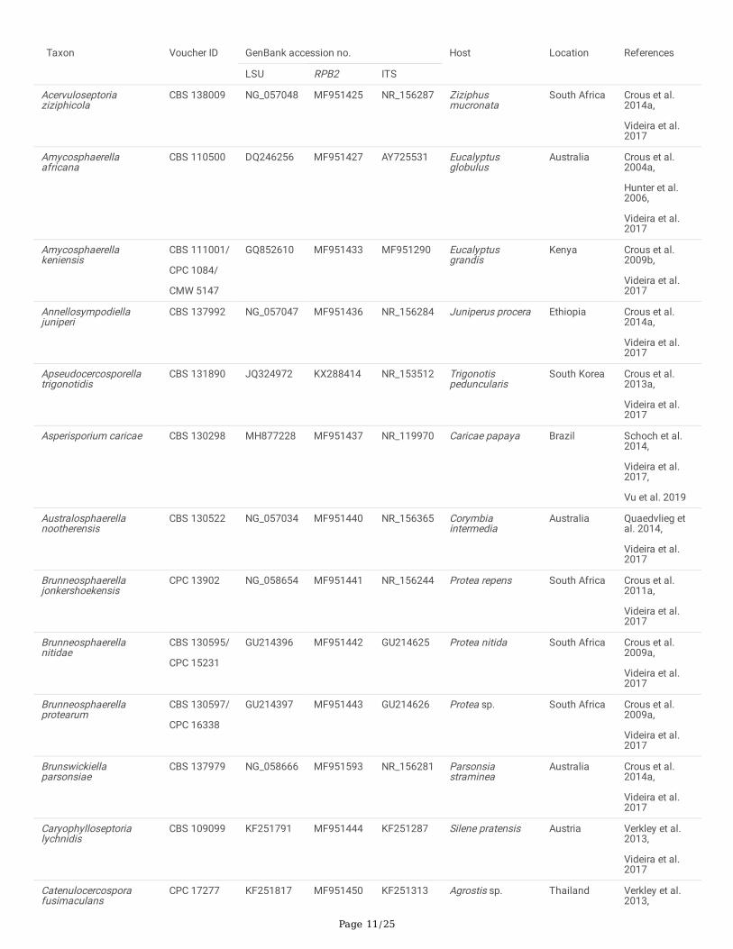
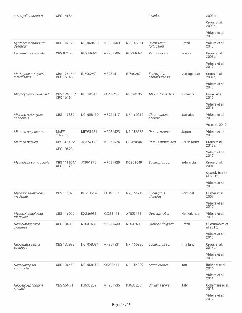
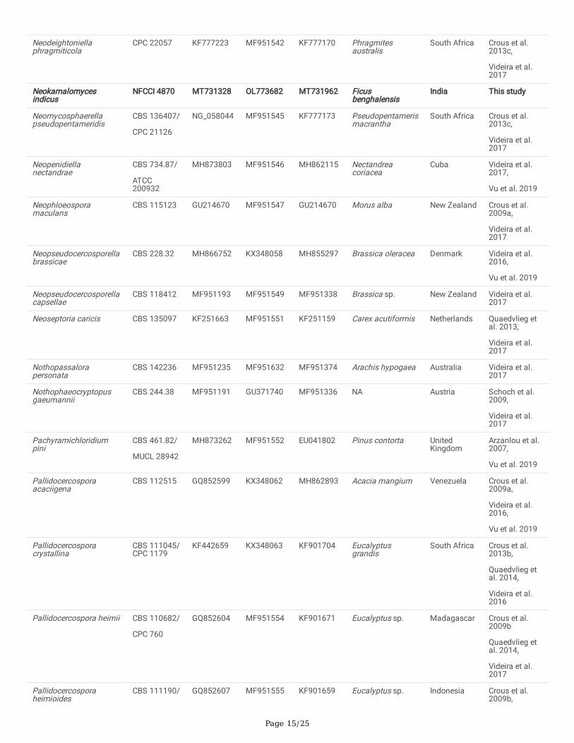
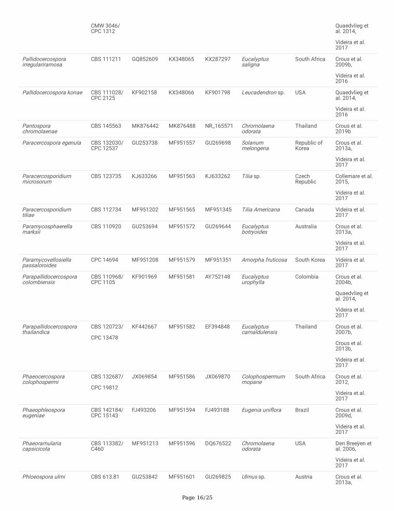
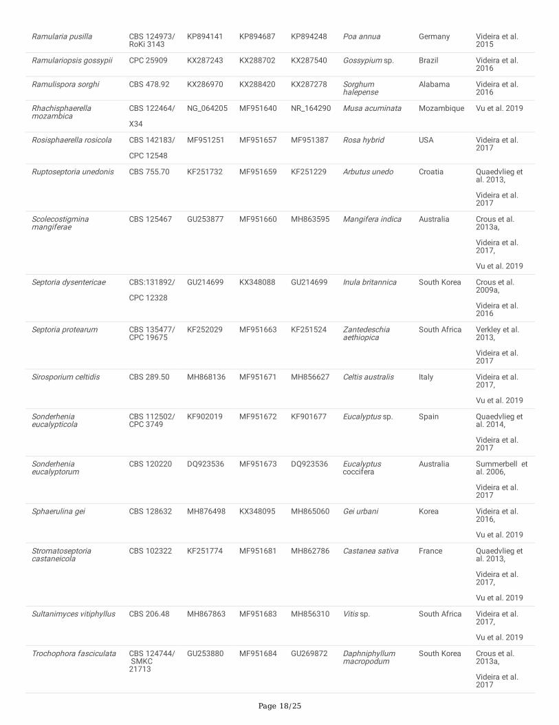
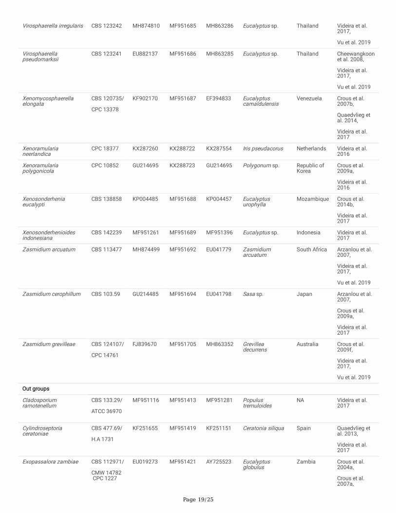
TablesTable 1 Taxa included in molecular phylogenetic analyses and their GenBank accession numbers. The sequences in bold were generated in thisstudy
Page 11/25
Taxon Voucher ID GenBank accession no. Host Location References
LSU RPB2 ITS
Acervuloseptoriaziziphicola
CBS 138009 NG_057048 MF951425 NR_156287 Ziziphusmucronata
South Africa Crous et al.2014a,
Videira et al.2017
Amycosphaerellaafricana
CBS 110500 DQ246256
MF951427 AY725531
Eucalyptusglobulus
Australia Crous et al.2004a,
Hunter et al.2006,
Videira et al.2017
Amycosphaerellakeniensis
CBS 111001/
CPC 1084/
CMW 5147
GQ852610 MF951433 MF951290 Eucalyptusgrandis
Kenya Crous et al.2009b,
Videira et al.2017
Annellosympodiellajuniperi
CBS 137992 NG_057047 MF951436 NR_156284 Juniperus procera Ethiopia Crous et al.2014a,
Videira et al.2017
Apseudocercosporellatrigonotidis
CBS 131890 JQ324972 KX288414 NR_153512 Trigonotispeduncularis
South Korea Crous et al.2013a,
Videira et al.2017
Asperisporium caricae
CBS 130298 MH877228
MF951437
NR_119970
Caricae papaya Brazil Schoch et al.2014,
Videira et al.2017,
Vu et al. 2019
Australosphaerellanootherensis
CBS 130522 NG_057034
MF951440
NR_156365
Corymbiaintermedia
Australia Quaedvlieg etal. 2014,
Videira et al.2017
Brunneosphaerellajonkershoekensis
CPC 13902 NG_058654 MF951441 NR_156244 Protea repens South Africa Crous et al.2011a,
Videira et al.2017
Brunneosphaerellanitidae
CBS 130595/
CPC 15231
GU214396 MF951442 GU214625 Protea nitida South Africa Crous et al.2009a,
Videira et al.2017
Brunneosphaerellaprotearum
CBS 130597/
CPC 16338
GU214397 MF951443 GU214626 Protea sp. South Africa Crous et al.2009a,
Videira et al.2017
Brunswickiellaparsonsiae
CBS 137979 NG_058666 MF951593 NR_156281 Parsonsiastraminea
Australia Crous et al.2014a,
Videira et al.2017
Caryophylloseptorialychnidis
CBS 109099 KF251791 MF951444 KF251287 Silene pratensis Austria Verkley et al.2013,
Videira et al.2017
Catenulocercosporafusimaculans
CPC 17277 KF251817 MF951450 KF251313 Agrostis sp. Thailand Verkley et al.2013,
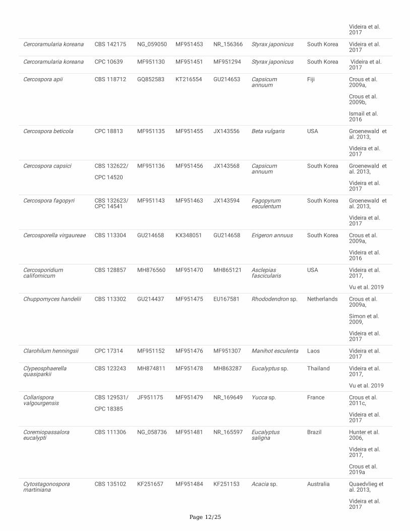
Page 12/25
Videira et al.2017
Cercoramularia koreana CBS 142175 NG_059050 MF951453 NR_156366 Styrax japonicus South Korea Videira et al.2017
Cercoramularia koreana CPC 10639 MF951130 MF951451 MF951294 Styrax japonicus South Korea Videira et al.2017
Cercospora apii CBS 118712 GQ852583
KT216554 GU214653 Capsicumannuum
Fiji Crous et al.2009a,
Crous et al.2009b,
Ismail et al.2016
Cercospora beticola CPC 18813 MF951135 MF951455 JX143556 Beta vulgaris USA Groenewald etal. 2013,
Videira et al.2017
Cercospora capsici CBS 132622/
CPC 14520
MF951136 MF951456 JX143568 Capsicumannuum
South Korea Groenewald etal. 2013,
Videira et al.2017
Cercospora fagopyri CBS 132623/CPC 14541
MF951143 MF951463 JX143594 Fagopyrumesculentum
South Korea Groenewald etal. 2013,
Videira et al.2017
Cercosporella virgaureae CBS 113304 GU214658 KX348051 GU214658 Erigeron annuus South Korea Crous et al.2009a,
Videira et al.2016
Cercosporidiumcalifornicum
CBS 128857 MH876560 MF951470 MH865121 Asclepiasfascicularis
USA Videira et al.2017,
Vu et al. 2019
Chuppomyces handelii CBS 113302 GU214437
MF951475
EU167581
Rhododendron sp. Netherlands Crous et al.2009a,
Simon et al.2009,
Videira et al.2017
Clarohilum henningsii CPC 17314 MF951152 MF951476 MF951307 Manihot esculenta Laos Videira et al.2017
Clypeosphaerellaquasiparkii
CBS 123243 MH874811 MF951478 MH863287 Eucalyptus sp. Thailand Videira et al.2017,
Vu et al. 2019
Collarisporavalgourgensis
CBS 129531/
CPC 18385
JF951175 MF951479 NR_169649 Yucca sp. France Crous et al.2011c,
Videira et al.2017
Coremiopassaloraeucalypti
CBS 111306 NG_058736
MF951481
NR_165597
Eucalyptussaligna
Brazil Hunter et al.2006,
Videira et al.2017,
Crous et al.2019a
Cytostagonosporamartiniana
CBS 135102 KF251657 MF951484 KF251153 Acacia sp. Australia Quaedvlieg etal. 2013,
Videira et al.2017
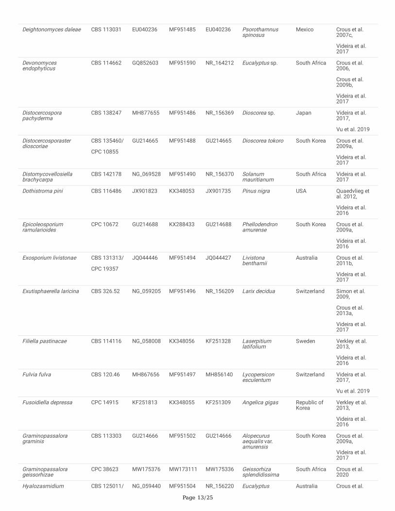
Page 13/25
Deightonomyces daleae CBS 113031 EU040236 MF951485 EU040236 Psorothamnusspinosus
Mexico Crous et al.2007c,
Videira et al.2017
Devonomycesendophyticus
CBS 114662 GQ852603
MF951590 NR_164212
Eucalyptus sp. South Africa Crous et al.2006,
Crous et al.2009b,
Videira et al.2017
Distocercosporapachyderma
CBS 138247 MH877655
MF951486
NR_156369
Dioscorea sp. Japan Videira et al.2017,
Vu et al. 2019
Distocercosporasterdioscoriae
CBS 135460/
CPC 10855
GU214665 MF951488 GU214665 Dioscorea tokoro South Korea Crous et al.2009a,
Videira et al.2017
Distomycovellosiellabrachycarpa
CBS 142178 NG_069528 MF951490 NR_156370 Solanummauritianum
South Africa Videira et al.2017
Dothistroma pini CBS 116486 JX901823 KX348053 JX901735 Pinus nigra USA Quaedvlieg etal. 2012,
Videira et al.2016
Epicoleosporiumramularioides
CPC 10672 GU214688 KX288433 GU214688 Phellodendronamurense
South Korea Crous et al.2009a,
Videira et al.2016
Exosporium livistonae CBS 131313/
CPC 19357
JQ044446 MF951494 JQ044427 Livistonabenthamii
Australia Crous et al.2011b,
Videira et al.2017
Exutisphaerella laricina CBS 326.52 NG_059205
MF951496
NR_156209
Larix decidua Switzerland Simon et al.2009,
Crous et al.2013a,
Videira et al.2017
Filiella pastinacae CBS 114116 NG_058008 KX348056 KF251328 Laserpitiumlatifolium
Sweden Verkley et al.2013,
Videira et al.2016
Fulvia fulva CBS 120.46 MH867656 MF951497 MH856140 Lycopersiconesculentum
Switzerland Videira et al.2017,
Vu et al. 2019
Fusoidiella depressa CPC 14915 KF251813 KX348055 KF251309 Angelica gigas Republic ofKorea
Verkley et al.2013,
Videira et al.2016
Graminopassaloragraminis
CBS 113303 GU214666 MF951502 GU214666 Alopecurusaequalis var.amurensis
South Korea Crous et al.2009a,
Videira et al.2017
Graminopassalorageissorhizae
CPC 38623 MW175376 MW173111 MW175336 Geissorhizasplendidissima
South Africa Crous et al.2020
Hyalozasmidium CBS 125011/ NG_059440 MF951504 NR_156220 Eucalyptus Australia Crous et al.
Page 14/25
aerohyalinosporum CPC 14636 tecti�ca 2009b,
Crous et al.2009e,
Videira et al.2017
Hyalocercosporidiumdesmodii
CBS 142179 NG_058488 MF951503 NR_156371 Desmodiumtortuosum
Brazil Videira et al.2017
Lecanosticta acicola CBS 871.95 GU214663 MF951506 GU214663 Pinus radiate France Crous et al.2009a,
Videira et al.2017
Madagascaromycesintermedius
CBS 124154/CPC 15745
FJ790297 MF951511 FJ790267 Eucalyptuscamaldulensis
Madagascar Crous et al.2009c,
Videira et al.2017
Microcyclosporella mali CBS 126136/CPC 16184
GU570547 KX288436 GU570535 Malus domestica Slovenia Frank et al.2010,
Videira et al.2016
Micronematomycescaribensis
CBS 113380 NG_058490 MF951517 NR_160215 Chromolaenaodorata
Jamaica Videira et al.2017,
Vu et al. 2019
Miuraea degenerans MAFF239265
MF951181 MF951523 NR_156373 Prunus mume Japan Videira et al.2017
Miuraea persica CBS131935/
CPC 10828
JQ324939 MF951524 GU269844 Prunus armeniaca South Korea Crous et al.2013a,
Videira et al.2017
Mycodiella sumatrensis CBS 118501/CPC 11175
JX901872 MF951525 DQ303049 Eucalyptus sp. Indonesia Crous et al.2006,
Quaedvlieg etal. 2012,
Videira et al.2017
Mycosphaerelloidesmadeirae
CBS 112895 DQ204756 KX348057 NR_154213 Eucalyptusglobulus
Portugal Hunter et al.2006,
Videira et al.2017
Mycosphaerelloidesmadeirae
CBS 116066 KX286989 KX288444 AY853188 Quercus robur Netherlands Videira et al.2016
Neoceratospermacyatheae
CPC 18580 KT037580 MF951530 KT037539 Cyathea delgadii Brazil Guatimosim etal 2016,
Videira et al.2017
Neoceratospermaeucalypti
CBS 137998 NG_058084 MF951531 NR_156285 Eucalyptus sp. Thailand Crous et al.2014a,
Videira et al.2017
Neocercosporaammicola
CBS 136450 NG_058158 KX288446 NR_154229 Ammi majus Iran Bakhshi et al.2015,
Videira et al.2016
Neocercosporidiumsmilacis
CBS 556.71 KJ633269 MF951535 KJ633265 Smilax aspera Italy Collemare et al.2015,
Videira et al.2017
Page 15/25
Neodeightoniellaphragmiticola
CPC 22057 KF777223 MF951542 KF777170 Phragmitesaustralis
South Africa Crous et al.2013c,
Videira et al.2017
Neokamalomycesindicus
NFCCI 4870 MT731328 OL773682 MT731962 Ficusbenghalensis
India This study
Neomycosphaerellapseudopentameridis
CBS 136407/
CPC 21126
NG_058044 MF951545 KF777173 Pseudopentamerismacrantha
South Africa Crous et al.2013c,
Videira et al.2017
Neopenidiellanectandrae
CBS 734.87/
ATCC200932
MH873803 MF951546 MH862115 Nectandreacoriacea
Cuba Videira et al.2017,
Vu et al. 2019
Neophloeosporamaculans
CBS 115123 GU214670 MF951547 GU214670 Morus alba New Zealand Crous et al.2009a,
Videira et al.2017
Neopseudocercosporellabrassicae
CBS 228.32 MH866752 KX348058
MH855297 Brassica oleracea Denmark Videira et al.2016,
Vu et al. 2019
Neopseudocercosporellacapsellae
CBS 118412 MF951193 MF951549 MF951338 Brassica sp. New Zealand Videira et al.2017
Neoseptoria caricis CBS 135097 KF251663 MF951551 KF251159 Carex acutiformis Netherlands Quaedvlieg etal. 2013,
Videira et al.2017
Nothopassalorapersonata
CBS 142236 MF951235 MF951632 MF951374 Arachis hypogaea Australia Videira et al.2017
Nothophaeocryptopusgaeumannii
CBS 244.38 MF951191 GU371740
MF951336 NA Austria Schoch et al.2009,
Videira et al.2017
Pachyramichloridiumpini
CBS 461.82/
MUCL 28942
MH873262 MF951552 EU041802 Pinus contorta UnitedKingdom
Arzanlou et al.2007,
Vu et al. 2019
Pallidocercosporaacaciigena
CBS 112515 GQ852599 KX348062 MH862893 Acacia mangium Venezuela Crous et al.2009a,
Videira et al.2016,
Vu et al. 2019
Pallidocercosporacrystallina
CBS 111045/CPC 1179
KF442659 KX348063 KF901704 Eucalyptusgrandis
South Africa Crous et al.2013b,
Quaedvlieg etal. 2014,
Videira et al.2016
Pallidocercospora heimii CBS 110682/
CPC 760
GQ852604 MF951554 KF901671 Eucalyptus sp. Madagascar Crous et al.2009b
Quaedvlieg etal. 2014,
Videira et al.2017
Pallidocercosporaheimioides
CBS 111190/ GQ852607 MF951555 KF901659 Eucalyptus sp. Indonesia Crous et al.2009b,
Page 16/25
CMW 3046/CPC 1312
Quaedvlieg etal. 2014,
Videira et al.2017
Pallidocercosporairregulariramosa
CBS 111211 GQ852609 KX348065 KX287297 Eucalyptussaligna
South Africa Crous et al.2009b,
Videira et al.2016
Pallidocercospora konae CBS 111028/CPC 2125
KF902158
KX348066 KF901798 Leucadendron sp. USA Quaedvlieg etal. 2014,
Videira et al.2016
Pantosporachromolaenae
CBS 145563 MK876442 MK876488 NR_165571 Chromolaenaodorata
Thailand Crous et al.2019b
Paracercospora egenula CBS 132030/CPC 12537
GU253738 MF951557 GU269698 Solanummelongena
Republic ofKorea
Crous et al.2013a,
Videira et al.2017
Paracercosporidiummicrosorum
CBS 123735 KJ633266 MF951563 KJ633262 Tilia sp. CzechRepublic
Collemare et al.2015,
Videira et al.2017
Paracercosporidiumtiliae
CBS 112734 MF951202 MF951565 MF951345 Tilia Americana Canada Videira et al.2017
Paramycosphaerellamarksii
CBS 110920 GU253694 MF951572 GU269644 Eucalyptusbotryoides
Australia Crous et al.2013a,
Videira et al.2017
Paramycovellosiellapassaloroides
CPC 14694 MF951208 MF951579 MF951351 Amorpha fruticosa South Korea Videira et al.2017
Parapallidocercosporacolombiensis
CBS 110968/CPC 1105
KF901969 MF951581 AY752148 Eucalyptusurophylla
Colombia Crous et al.2004b,
Quaedvlieg etal. 2014,
Videira et al.2017
Parapallidocercosporathailandica
CBS 120723/
CPC 13478
KF442667 MF951582 EF394848 Eucalyptuscamaldulensis
Thailand Crous et al.2007b,
Crous et al.2013b,
Videira et al.2017
Phaeocercosporacolophospermi
CBS 132687/
CPC 19812
JX069854 MF951586 JX069870 Colophospermummopane
South Africa Crous et al.2012,
Videira et al.2017
Phaeophleosporaeugeniae
CBS 142184/CPC 15143
FJ493206 MF951594 FJ493188 Eugenia uni�ora Brazil Crous et al.2009d,
Videira et al.2017
Phaeoramulariacapsicicola
CBS 113382/C460
MF951213 MF951596 DQ676522 Chromolaenaodorata
USA Den Breeÿen etal. 2006,
Videira et al.2017
Phloeospora ulmi CBS 613.81 GU253842 MF951601 GU269825 Ulmus sp. Austria Crous et al.2013a,

Page 17/25
Videira et al.2017
Pleopassalora perplexa CPC 12170 MF951219 MF951605 MF951362 Acacia sp. Indonesia Videira et al.2017
Pleuropassaloraarmatae
CBS 125420/
CPC 15419
MH875073 MF951609 MH863597 Dalbergia armata South Africa Crous et al.2009a,
Videira et al.2017,
Vu et al. 2019
Pluripassalorabougainvilleae
CBS 142237 MF951224 MF951612 MF951365 Bougainvillea sp. Australia Videira et al.2017
Polyphialoseptoriatabebuiae-serratifoliae
CBS 112650/
CPC 3944
KF251716 MF951613 KF251213 Tabebuiaserratifolia
Brazil Quaedvlieg etal. 2013,
Videira et al.2017
Polyphialoseptoriaterminaliae
CBS 135106 MH878128 MF951615 KF251214 Terminaliacatappa
Brazil Quaedvlieg etal. 2013,
Videira et al.2017,
Vu et al. 2019
Pseudocercosporacatappae
MAFF238312//MUCC 1109
MF951225
MF951616 MF951366 Terminaliacatappa
Japan Videira et al.2017
Pseudocercospora vitis CBS 132012/
CPC 11595
GU214483 KX348076 DQ073923 Vitis vinifera Republic ofKorea
Ayala et al.2006,
Crous et al.2009a,
Videira et al.2017
Pseudocercosporellabakeri
CBS 119488 KX287005 KX288462 KX287306 Ipomoea indica New Zealand Videira et al.2016
Pseudopericoniellalevispora
CBS 873.73 MH872550 MF951633 EU041780 Turpinia pomifera Sri Lanka Arzanlou et al.2007,
Videira et al.2017,
Vu et al. 2019
Pseudophaeophleospora
atkinsonii
CBS 124565/ICMP 17860
MF951236 MF951635 GU214643 Hebe sp. New Zealand Crous et al.2009a,
Videira et al.2017
Pseudozasmidiumnabiacense
CBS 125010/
CPC 12748
MH874956 MF951638 MH863455 Eucalyptus sp. Australia Videira et al.2017,
Vu et al. 2019
Ragnhildianapseudotithoniae
CBS 136442/
CPC 21688
KF777231 MF951652 KF777179 Tithoniadiversifolia
Thailand Crous et al.2013c,
Videira et al.2017
Ramularia carneola CBS 108975 KX287048 KX288507 KX287348 Scrophularianodosa
Netherlands Videira et al.2016
Ramularia endophylla CBS 113265 AY490776 KP894673 EU167569 Quercus robur Netherlands Verkley et al.2004a,
Simon et al.2009,
Videira et al.2015
Page 18/25
Ramularia pusilla CBS 124973/RoKi 3143
KP894141 KP894687 KP894248 Poa annua Germany Videira et al.2015
Ramulariopsis gossypii CPC 25909 KX287243 KX288702 KX287540 Gossypium sp. Brazil Videira et al.2016
Ramulispora sorghi CBS 478.92 KX286970 KX288420 KX287278 Sorghumhalepense
Alabama Videira et al.2016
Rhachisphaerellamozambica
CBS 122464/
X34
NG_064205 MF951640 NR_164290 Musa acuminata Mozambique Vu et al. 2019
Rosisphaerella rosicola CBS 142183/
CPC 12548
MF951251 MF951657 MF951387 Rosa hybrid USA Videira et al.2017
Ruptoseptoria unedonis CBS 755.70 KF251732 MF951659 KF251229 Arbutus unedo Croatia Quaedvlieg etal. 2013,
Videira et al.2017
Scolecostigminamangiferae
CBS 125467 GU253877 MF951660 MH863595 Mangifera indica Australia Crous et al.2013a,
Videira et al.2017,
Vu et al. 2019
Septoria dysentericae CBS:131892/
CPC 12328
GU214699 KX348088
GU214699 Inula britannica
South Korea
Crous et al.2009a,
Videira et al.2016
Septoria protearum CBS 135477/CPC 19675
KF252029 MF951663 KF251524 Zantedeschiaaethiopica
South Africa Verkley et al.2013,
Videira et al.2017
Sirosporium celtidis CBS 289.50 MH868136 MF951671 MH856627 Celtis australis Italy Videira et al.2017,
Vu et al. 2019
Sonderheniaeucalypticola
CBS 112502/CPC 3749
KF902019 MF951672 KF901677 Eucalyptus sp. Spain Quaedvlieg etal. 2014,
Videira et al.2017
Sonderheniaeucalyptorum
CBS 120220 DQ923536 MF951673 DQ923536 Eucalyptuscoccifera
Australia Summerbell etal. 2006,
Videira et al.2017
Sphaerulina gei CBS 128632 MH876498 KX348095 MH865060 Gei urbani Korea Videira et al.2016,
Vu et al. 2019
Stromatoseptoriacastaneicola
CBS 102322 KF251774 MF951681 MH862786 Castanea sativa France Quaedvlieg etal. 2013,
Videira et al.2017,
Vu et al. 2019
Sultanimyces vitiphyllus CBS 206.48 MH867863 MF951683 MH856310 Vitis sp. South Africa Videira et al.2017,
Vu et al. 2019
Trochophora fasciculata CBS 124744/ SMKC21713
GU253880 MF951684 GU269872 Daphniphyllummacropodum
South Korea
Crous et al.2013a,
Videira et al.2017
Page 19/25
Virosphaerella irregularis CBS 123242 MH874810 MF951685 MH863286 Eucalyptus sp. Thailand Videira et al.2017,
Vu et al. 2019
Virosphaerellapseudomarksii
CBS 123241 EU882137 MF951686 MH863285 Eucalyptus sp. Thailand Cheewangkoonet al. 2008,
Videira et al.2017,
Vu et al. 2019
Xenomycosphaerellaelongata
CBS 120735/
CPC 13378
KF902170 MF951687 EF394833 Eucalyptuscamaldulensis
Venezuela Crous et al.2007b,
Quaedvlieg etal. 2014,
Videira et al.2017
Xenoramularianeerlandica
CPC 18377 KX287260 KX288722 KX287554 Iris pseudacorus Netherlands Videira et al.2016
Xenoramulariapolygonicola
CPC 10852 GU214695 KX288723 GU214695 Polygonum sp. Republic ofKorea
Crous et al.2009a,
Videira et al.2016
Xenosonderheniaeucalypti
CBS 138858 KP004485 MF951688 KP004457 Eucalyptusurophylla
Mozambique Crous et al.2014b,
Videira et al.2017
Xenosonderhenioidesindonesiana
CBS 142239 MF951261 MF951689 MF951396 Eucalyptus sp. Indonesia Videira et al.2017
Zasmidium arcuatum CBS 113477 MH874499 MF951692 EU041779 Zasmidiumarcuatum
South Africa Arzanlou et al.2007,
Videira et al.2017,
Vu et al. 2019
Zasmidium cerophillum CBS 103.59 GU214485 MF951694 EU041798 Sasa sp. Japan Arzanlou et al.2007,
Crous et al.2009a,
Videira et al.2017
Zasmidium grevilleae CBS 124107/
CPC 14761
FJ839670 MF951705 MH863352 Grevilleadecurrens
Australia Crous et al.2009f,
Videira et al.2017,
Vu et al. 2019
Out groups
Cladosporiumramotenellum
CBS 133.29/
ATCC 36970
MF951116 MF951413 MF951281 Populustremuloides
NA Videira et al.2017
Cylindroseptoriaceratoniae
CBS 477.69/
H.A 1731
KF251655 MF951419 KF251151 Ceratonia siliqua Spain Quaedvlieg etal. 2013,
Videira et al.2017
Exopassalora zambiae CBS 112971/
CMW 14782 CPC 1227
EU019273 MF951421 AY725523 Eucalyptusglobulus
Zambia Crous et al.2004a,
Crous et al.2007a,
Page 20/25
Videira et al.2017
Ramichloridiumapiculatum
CBS 156.59/
ATCC 13211/IMI 100716/
JCM 6972/MUCL15753/ MUCL 7991/ QM 7716
EU041848 MF951416 EU041791 Forest soil USA Arzanlou et al.2007,
Videira et al.2017
Schizothyrium pomi CBS 228.57 EF134947 MF951734 EF134947 NA Italy Batzer et al.2008,
Videira et al.2017
Stenella araguata CBS 105.75 EU019250 MF951742 EU019250 Homo sapiens Venezuela Crous et al.2007a,
Videira et al.2017
Teratosphaeriastellenboschiana
CBS 125215/
CPC 13764
KF937247 MF951743 KF901733 Eucalyptuspunctata
South Africa Quaedvlieg etal. 2014,
Videira et al.2017
Uwebraunia musae CBS 122453/
X1021
JQ739816 KX348107 EU514225 Musa acuminata India Arzanlou et al.2008,
Stukenbrock etal. 2012,
Videira et al.2016
Table 2 Comparative morphology of Septoria spp. reported on Ficus spp.
Taxa Host Conidia References
Dimension Septation Shape
Septoria arcuata Ficus sp. 40–50 μm long – Linear Cooke 1880
Septoriabrachyspora
Ficuselastica
12–15 × 1 μm – Curved Saccardo1879
Septoria elasticae Ficuselastica
15–28 × 1–1.5µm
Indistinctly 1–3-septate orguttulate
Filiform to bacillar, curvules to curvewinding
Koorders1907
Septoria pipulae Ficusreligiosa
50–60 μm long – Filiform Cooke 1879
Septoria pirottae Ficusrepens
24–30 × 2–3μm
– Filiform, subcylindrical, curved Tassi 1896
Figures
Page 21/25
Figure 1
Neokamalomyces indicus on Ficus benghalensis. a Host plant in natural habitat, b Initial stage of symptom on upper surface of leaf, c Initial stageof symptom on lower surface of leaf, d, e Conidiomata on host tissue. Bars: b, c = 20 mm, d, e = 10 mm

Page 22/25
Figure 2
Neokamalomyces indicus on PDA (NFCCI 4870, ex-type culture). a, b Colony on PDA front view, c Colony on PDA reverse view, d–h Germinatedconidia in water droplet in cavity slide after 12-15 hours, i, j Germinated conidia on PDA (stained with cotton-blue), k Development of mycelia onPDA. Bars: a = 10 mm, b = 5 mm, c = 10 mm, d–h = 20 μm, i–k = 10 μm
Page 23/25
Figure 3
Fruiting body of Neokamalomyces indicus (holotype, AMH 10233). a–d Vertical section through conidioma, e, f Conidia with conidiophores, gConidiogenous cells. Bars: a–e = 20 μm, f, g = 10 μm
Page 24/25
Figure 4
Conidia of Neokamalomyces indicus (holotype, AMH 10233). Bars: a–k = 10 μm, l, m = 5 μm
Page 25/25
Figure 5
Consensus phylogram (50% majority rule) resulting from a maximum likelihood of the combined three-genes (LSU, RPB2 and ITS) sequencealignment. The Bayesian posterior probabilities (≥ 0.50; BI-PP), maximum likelihood bootstrap support values (≥ 50%; ML-BS) and maximumparsimony bootstrap support values (≥ 50%; MP-BS) are given at the nodes (BI-PP/ML-BS/MP-BS). Red names indicate Neokamalomyces indicus. Avertical bar is used to the right of the coloured boxes and encompasses all genera within their respective families. The family nameMycosphaerellaceae is unabbreviated while the rest are abbreviated as follows: D = Dissoconiaceae, P = Phaeothecoidiellaceae, S =Schizothyriaceae, T = Teratosphaeriaceae, C = Cladosporiaceae. The tree was rooted to Cylindroseptoria ceratoniae (CBS 477.69)
Related Documents


