Hindawi Publishing Corporation Journal of Biomedicine and Biotechnology Volume 2011, Article ID 214781, 5 pages doi:10.1155/2011/214781 Research Article Separation of Normal and Premalignant Cervical Epithelial Cells Using Confocal Light Absorption and Scattering Spectroscopic Microscopy Ex Vivo Ling Yang, 1, 2 Wen-Tao Liu, 3 Hao Wu, 4 Cheng Wang, 5 Bo Ping, 1, 2 and Da-Ren Shi 1, 2 1 Department of Pathology, Fudan University Shanghai Cancer Center, Shanghai 200032, China 2 Department of Oncology, Shanghai Medical College, Fudan University, Shanghai 200032, China 3 Department of Surgery, Ruijin Hospital, Shanghai Institute of Digestive Surgery, Shanghai Jiao Tong University, 200025 Shanghai, China 4 Department of Obstetrics and Gynecology, Shanghai First People’s Hospital, Shanghai Jiao Tong University, 200080 Shanghai, China 5 Institute of Medical Optics and Optometry, University of Shanghai for Science and Technology, 200093 Shanghai, China Correspondence should be addressed to Da-Ren Shi, [email protected] Received 16 April 2011; Accepted 25 July 2011 Academic Editor: Manoor Prakash Hande Copyright © 2011 Ling Yang et al. This is an open access article distributed under the Creative Commons Attribution License, which permits unrestricted use, distribution, and reproduction in any medium, provided the original work is properly cited. Confocal light absorption and scattering spectroscopic (CLASS) microscopy can detect changes in biochemicals and the morphology of cells. It is therefore used to detect high-grade cervical squamous intraepithelial lesion (HSIL) cells in the diagnosis of premalignant cervical lesions. Forty cervical samples from women with abnormal Pap smear test results were collected, and twenty cases were diagnosed as HSIL; the rest were normal or low-grade cervical squamous intraepithelial lesion (LSIL). The enlarged and condensed nuclei of HSIL cells as viewed under CLASS microscopy were much brighter and bigger than those of non- HSIL cells. Cytological elastic scattered light data was then collected at wavelengths between 400 and 1000 nm. Between 600 nm to 800 nm, the relative elastic scattered light intensity of HSIL cells was higher than that of the non-HSIL. Relative intensity peaks occurred at 700 nm and 800 nm. CLASS sensitivity and specificity results for HSIL and non-HSIL compared to cytology diagnoses were 80% and 90%, respectively. This study demonstrated that CLASS microscopy could effectively detect cervical precancerous lesions. Further study will verify this conclusion before the method is used in clinic for early detection of cervical cancer. 1. Introduction Cervical cancer is a very common malignancy in women. The National Cervical Cancer Coalition (NCCC) estimates that worldwide, 473,000 new cases of cervical cancer are diagnosed and 253,500 patients die of this disease annually. This makes cervical cancer the twelfth most common cancer and the fifth cause of cancer-related deaths in the world [1]. Approximately 85% of cervical cancers occur in developing countries. Human papillomavirus (HPV) infection is the most important risk factor for the development of cervical cancer [2]. Treatment of cervical cancer consists of surgery at the early stages and chemotherapy and radiation therapy for advanced disease. Prognosis of cervical cancer depends on the cancer stage. If diagnosed early, the 5-year survival rate of the patient is more than 90%. Therefore, early detection is currently the most effective means to improve survival of patients. The Papanicolaou (Pap smear screening) test followed by colposcopy is the most frequently used means to detect precancerous and cancerous changes in the cervix. However, the current most practical Pap smear screening plus cytology test has a low sensitivity, especially in discriminating high- grade squamous intraepithelial lesions (HSILs) from other abnormalities and normal tissues [3]. In routine clinical practice, inaccurate Pap smear results and colposcopic impressions often lead to overtreatment of patients [4]. Thus, development of a more sensitive and specific method to detect cervical cancer early and accurately is urgently needed. For several past decades, we have witnessed the rapid development and use of optical methods to investigate changes in cell behaviors (such as migration and intracellular

Welcome message from author
This document is posted to help you gain knowledge. Please leave a comment to let me know what you think about it! Share it to your friends and learn new things together.
Transcript

Hindawi Publishing CorporationJournal of Biomedicine and BiotechnologyVolume 2011, Article ID 214781, 5 pagesdoi:10.1155/2011/214781
Research Article
Separation of Normal and Premalignant Cervical Epithelial CellsUsing Confocal Light Absorption and Scattering SpectroscopicMicroscopy Ex Vivo
Ling Yang,1, 2 Wen-Tao Liu,3 Hao Wu,4 Cheng Wang,5 Bo Ping,1, 2 and Da-Ren Shi1, 2
1 Department of Pathology, Fudan University Shanghai Cancer Center, Shanghai 200032, China2 Department of Oncology, Shanghai Medical College, Fudan University, Shanghai 200032, China3 Department of Surgery, Ruijin Hospital, Shanghai Institute of Digestive Surgery, Shanghai Jiao Tong University,200025 Shanghai, China
4 Department of Obstetrics and Gynecology, Shanghai First People’s Hospital, Shanghai Jiao Tong University, 200080 Shanghai, China5 Institute of Medical Optics and Optometry, University of Shanghai for Science and Technology, 200093 Shanghai, China
Correspondence should be addressed to Da-Ren Shi, [email protected]
Received 16 April 2011; Accepted 25 July 2011
Academic Editor: Manoor Prakash Hande
Copyright © 2011 Ling Yang et al. This is an open access article distributed under the Creative Commons Attribution License,which permits unrestricted use, distribution, and reproduction in any medium, provided the original work is properly cited.
Confocal light absorption and scattering spectroscopic (CLASS) microscopy can detect changes in biochemicals and themorphology of cells. It is therefore used to detect high-grade cervical squamous intraepithelial lesion (HSIL) cells in the diagnosisof premalignant cervical lesions. Forty cervical samples from women with abnormal Pap smear test results were collected, andtwenty cases were diagnosed as HSIL; the rest were normal or low-grade cervical squamous intraepithelial lesion (LSIL). Theenlarged and condensed nuclei of HSIL cells as viewed under CLASS microscopy were much brighter and bigger than those of non-HSIL cells. Cytological elastic scattered light data was then collected at wavelengths between 400 and 1000 nm. Between 600 nmto 800 nm, the relative elastic scattered light intensity of HSIL cells was higher than that of the non-HSIL. Relative intensity peaksoccurred at 700 nm and 800 nm. CLASS sensitivity and specificity results for HSIL and non-HSIL compared to cytology diagnoseswere 80% and 90%, respectively. This study demonstrated that CLASS microscopy could effectively detect cervical precancerouslesions. Further study will verify this conclusion before the method is used in clinic for early detection of cervical cancer.
1. Introduction
Cervical cancer is a very common malignancy in women.The National Cervical Cancer Coalition (NCCC) estimatesthat worldwide, 473,000 new cases of cervical cancer arediagnosed and 253,500 patients die of this disease annually.This makes cervical cancer the twelfth most common cancerand the fifth cause of cancer-related deaths in the world [1].Approximately 85% of cervical cancers occur in developingcountries. Human papillomavirus (HPV) infection is themost important risk factor for the development of cervicalcancer [2]. Treatment of cervical cancer consists of surgery atthe early stages and chemotherapy and radiation therapy foradvanced disease.
Prognosis of cervical cancer depends on the cancer stage.If diagnosed early, the 5-year survival rate of the patient
is more than 90%. Therefore, early detection is currentlythe most effective means to improve survival of patients.The Papanicolaou (Pap smear screening) test followed bycolposcopy is the most frequently used means to detectprecancerous and cancerous changes in the cervix. However,the current most practical Pap smear screening plus cytologytest has a low sensitivity, especially in discriminating high-grade squamous intraepithelial lesions (HSILs) from otherabnormalities and normal tissues [3]. In routine clinicalpractice, inaccurate Pap smear results and colposcopicimpressions often lead to overtreatment of patients [4]. Thus,development of a more sensitive and specific method todetect cervical cancer early and accurately is urgently needed.
For several past decades, we have witnessed the rapiddevelopment and use of optical methods to investigatechanges in cell behaviors (such as migration and intracellular
2 Journal of Biomedicine and Biotechnology
protein movement) and for disease diagnosis; optical tech-niques enable the detection of changes in biochemical andmorphological features that are concurrent with precancer-ous conditions [5]. The advantage of optical techniques isthat in living cells visible light does no harm or is relativelybenign. Optical techniques such as confocal reflectancemicroscopy, optical coherence tomography, light-scatteringspectroscopy, and elastic scattering spectroscopy are able todistinguish and identify intrinsic optical properties of tissues.
During the transition from normal to cancerous cells,biochemical, molecular, and morphologic alterations maylead to changes in the cells’ optical properties. Indeed, recentstudies have shown that elastic light-scattering spectroscopymay permit the earliest detection of carcinogenesis. Light-scattering spectroscopy (LSS) is an optical technique inwhich quantitative information on cell organelle morphol-ogy is extracted via measurement of the spectra and angulardistribution of backscattered light. Therefore, LSS coulddetect precancerous and early cancerous changes in tissues[6].
In this study, we investigated the novel technique con-focal light absorption and scattering spectroscopic (CLASS)microscopy to detect the light scattering differences in liquid-based cervical cell samples. CLASS combines LSS withconfocal microscopy for early cancer detection [7, 8]. CLASSprovides not only size information but also the biochemicaland physical properties of the cells. It images tissue atgreater depth than conventional confocal microscopy andprovides enhanced image contrast. In addition, by utilizingbackscattered light to detect subcellular structures, CLASScan detect changes in living cells without introducingexogenous labels [5].
In the present study, we demonstrate that CLASSmicroscopy could distinguish between populations of nor-mal, low-grade squamous intraepithelial lesion (LSIL), andHSIL cells ex vivo, and therefore could be developed forclinical use in the accurate and precise diagnosis of Pap smearsamples.
2. Materials and Methods
2.1. Cell Samples. Forty women aged twenty and olderwho had a history of abnormal cervical cytology wereenrolled in this study at the Department of Pathology,Cancer Hospital of Fudan University, between 2009 and2010. Our institutional review board approved the researchprotocol for this work. A written informed consent formwas obtained from each subject. Of these 40 patients, 20had cytologically confirmed HSIL and 20 were non-HSIL. Allcytological samples were free of blood clot and stored at 4◦Cfor experimental procedures within 24 h.
2.2. Sample Preparation for CLASS Analysis. To performCLASS analysis, the cytological specimens were first putinto ThinPrep preservation solution containing deionizedwater, 0.1% Polysorbate 20 (Tween-20) detergent, and 54%ethanol. The pathologist then mapped a region of interest,designating the region that contained the worst cell lesions.
Table 1: CLASS data compared to cytology diagnosis.
Relativeintensity
HSIL > Non-HSIL(CLASS)
HSIL < Non-HSIL(CLASS)
P value
HSIL 16 4P > 0.05
Non-HSIL 2 18
Cells were suspended at 108 cells/mL to avoid overlappingnuclei in the analysis. Cells in saline were used as the control.
2.3. Spectroscopy. Cells in Thinprep or in saline (as thecontrol) were kept in an open volume container or dippedonto glass slides. The container was black on the inside inorder to minimize edge effects. Measurements were madewith a fiber optic probe placed on the surface of the cellsuspension.
To perform the CLASS analysis, we used a prototypeCLASS microscope as described in a previous report [5].After the optical signal was collected by the CLASS receiver,the data was analyzed using spectral data acquisition BWspecsoftware (B&W, Newark, DE, USA). Before every experi-ment, the light base was adjusted to minimize the signal-to-noise ratio.
2.4. Data Analysis. The data was first normalized to thesaline control samples and then comparisons were madebetween the HSIL and non-HSIL samples. SPSS 13.0 sta-tistical software was used to analyze the summarized data.Sensitivity and specificity were calculated by percentage ofcases correctly identified by CLASS as compared to thoseconfirmed by cytology, and then presented as mean ±standard deviation (SD). Student’s t-test was performed togenerate a probability P value. P < 0.05 was consideredstatistically significant.
3. Results
3.1. Study Population. From patients with abnormal Papsmear cytology results, we collected 20 cervical cytologysamples of HSIL, and 20 of non-HSIL. In the HSIL samples,the pathology report showed 6 cases with lesions of cervicalintraepithelial neoplasia (CIN) grade II, and 14 cases withlesions of CIN grade III. In the non-HSIL samples, 8 casesshowed normal cytology, and 12 cases were positive for LSIL(Table 1).
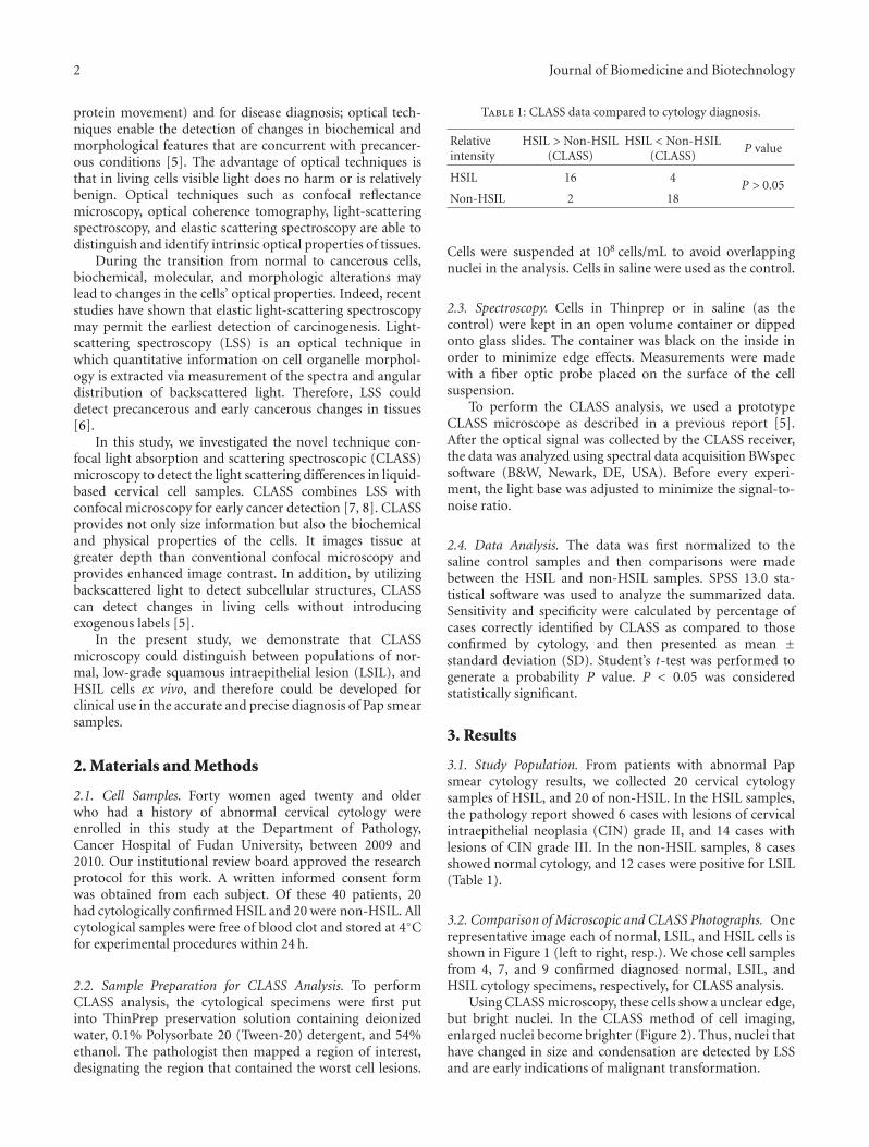
3.2. Comparison of Microscopic and CLASS Photographs. Onerepresentative image each of normal, LSIL, and HSIL cells isshown in Figure 1 (left to right, resp.). We chose cell samplesfrom 4, 7, and 9 confirmed diagnosed normal, LSIL, andHSIL cytology specimens, respectively, for CLASS analysis.
Using CLASS microscopy, these cells show a unclear edge,but bright nuclei. In the CLASS method of cell imaging,enlarged nuclei become brighter (Figure 2). Thus, nuclei thathave changed in size and condensation are detected by LSSand are early indications of malignant transformation.
Journal of Biomedicine and Biotechnology 3
(a) (b) (c)
Figure 1: Morphology of cervical epithelial cells. Pap smear cells were obtained from patients and reviewed by a pathologist. From left toright, photos show a normal, LSIL, and HSIL cell. Arrow: the nucleus.
(a) (b) (c)
Figure 2: Images of cervical epithelial cells in liquid-based thin layer samples analyzed by CLASS microscopy. The photos show from left toright a normal, LSIL, and HSIL cell. Arrow: the nucleus. The diameter of the nuclei is larger than 2 µm.
40,00035,00030,00025,00020,00015,00010,000
5,0000400 500 600 700 800 900 1,000
Rel
ativ
ein
ten
sity
Wavelength (nm)
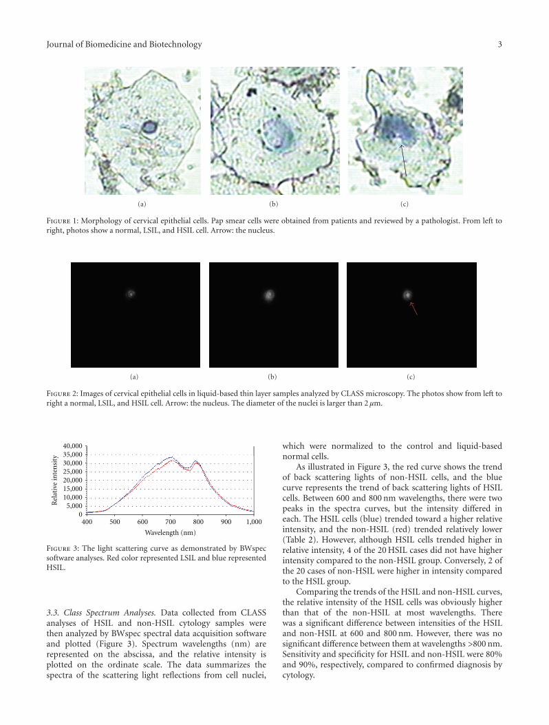
Figure 3: The light scattering curve as demonstrated by BWspecsoftware analyses. Red color represented LSIL and blue representedHSIL.
3.3. Class Spectrum Analyses. Data collected from CLASSanalyses of HSIL and non-HSIL cytology samples werethen analyzed by BWspec spectral data acquisition softwareand plotted (Figure 3). Spectrum wavelengths (nm) arerepresented on the abscissa, and the relative intensity isplotted on the ordinate scale. The data summarizes thespectra of the scattering light reflections from cell nuclei,
which were normalized to the control and liquid-basednormal cells.
As illustrated in Figure 3, the red curve shows the trendof back scattering lights of non-HSIL cells, and the bluecurve represents the trend of back scattering lights of HSILcells. Between 600 and 800 nm wavelengths, there were twopeaks in the spectra curves, but the intensity differed ineach. The HSIL cells (blue) trended toward a higher relativeintensity, and the non-HSIL (red) trended relatively lower(Table 2). However, although HSIL cells trended higher inrelative intensity, 4 of the 20 HSIL cases did not have higherintensity compared to the non-HSIL group. Conversely, 2 ofthe 20 cases of non-HSIL were higher in intensity comparedto the HSIL group.
Comparing the trends of the HSIL and non-HSIL curves,the relative intensity of the HSIL cells was obviously higherthan that of the non-HSIL at most wavelengths. Therewas a significant difference between intensities of the HSILand non-HSIL at 600 and 800 nm. However, there was nosignificant difference between them at wavelengths >800 nm.Sensitivity and specificity for HSIL and non-HSIL were 80%and 90%, respectively, compared to confirmed diagnosis bycytology.

4 Journal of Biomedicine and Biotechnology
Table 2: Relative intensity of HSIL and non-HSIL groups.
600 nm 650 nm 700 nm 750 nm 800 nm
Non-HSIL 20714 ± 210 26911 ± 326 31413 ± 168 26632 ± 292 29409 ± 221
HSIL 21982 ± 467 29393 ± 620 33206 ± 646 27651 ± 566 30874 ± 387
P < 0.05, using Student’s t-test.
4. Discussion
Cervical cancer, unlike most other cancers, is relativelyeasy to prevent and diagnose early, which contributes toreduced incidence and a better long-term survival rate.However, advanced stages of cervical cancers continue to bea serious health problem worldwide, especially in developingcountries. Indeed, the incidence and mortality rates ofcervical cancer are increasing in young women in China,although mortality for this cancer has significantly decreasedin the United States and Western Europe. This may be dueto the widespread use of the Pap smear screening test andsafer sex practices. In an effort to improve the sensitivity andspecificity of the Pap cytology test, we conducted the currentstudy using CLASS technology. Our data demonstratesthat CLASS microscopy was able to distinguish amongnormal, LSIL, and HSIL cells at specific wavelengths. CLASSmicroscopy is thus a useful and reliable technique for thedetection of early cervical epithelial abnormality and couldbe used to help pathologists diagnose early cervical cancer.
Clinically it is crucial to distinguish precancerous lesionsfrom normal and LSIL, because precancerous lesions couldprogress to invasive carcinoma if left untreated [9]. Manymethods have been developed and used preclinically andclinically for the detection of early cervical cancer [10, 11].The goals of investigations of new techniques are to reducethe cost of screening, enable the practical surveillance ofpopulations, or to improve the sensitivity to HSILs, whichare considered cervical cancer precursors. Such investigationsinclude the use of optical techniques.
Elastic light scattering spectroscopy can provide avaluable, noninvasive means to quantitatively probe tissuemorphology and even detect biochemical changes in cells.However, despite growing evidence of the clinical utility ofa variety of light scattering-based optical techniques, thebiological bases of differences in scattering signals betweennormal and neoplastic tissues are not well understood.
The rationale for using optical techniques is to detectbiochemical and morphological features that are concurrentwith precancerous conditions. Light scattering is moresensitive to morphological changes or other currently usedvisualizing techniques. Therefore, this kind of noninvasivemethod has many advantages, including the ability to mea-sure changes in nuclear size, which is one of the recognizedmorphological changes occurring during the transformationof normal cells to cancerous [9].
Some studies have demonstrated that the intensity oflight scattering increases with progression of atypia [10].The most dramatic differences in scattering occur betweennon-HSIL and HSIL cells because changes in nuclear sizeand DNA content are most pronounced between these two
categories of cells. Studies have suggested that changes inscattering properties at high CIN grades are dominated bythe effects of increased nuclear size and increased DNAcontent [11]. In fact, a complex spatial pattern is formed thatis dependent on cell size, shape, refraction index, density, andmorphology.
In this study, we used the optical method CLASS todistinguish HSIL of precancerous cervix cells from nor-mal and LSIL samples in liquid-based specimens ex vivo.We found that CLASS was able to differentiate these inconfirmed cytology samples, by the non-HSIL and HSIL-relative intensity curves generated by the BWspec software.We showed that there was a significant difference in relativeintensity between non-HSIL and HSIL cells in the 600–800 nm wavelength ranges. We could discriminate HSILfrom non-HSIL cervical epithelial cells by differences inthe curves, which represented the median light-scatteringintensities of confirmed samples. Sixteen of the 20 HSILcases demonstrated higher trends in relative intensity, and18 of the 20 non-HSIL conformed to a lower trend inrelative intensity. Therefore, in this study we established anovel model for diagnosis of liquid-based cervical cytologicalsamples through the use of CLASS microscopy. As reportedpreviously, this method is noninvasive with high sensitivity[12]. In addition, liquid-based specimens are easy to collectand preserve for analysis.
However, this study also has some limitations. Forexample, two of the LSIL cases showed high relative intensity.The reason may be due to overlapped or dividing cell nuclei.In addition, four cases of HSIL did not differ from theLSIL optimized curve. In the future, we will collect a largernumber of samples to confirm our current data beforetranslating this technique to the clinic.
Conflict of Interests
The authors have no conflict of interests.
References
[1] NCCC National Cervical Cancer Coalition, 2011, http://www.nccc-online.org/.
[2] E. P. Armstrong, “Prophylaxis of cervical cancer and relatedcervical disease: a review of the cost-effectiveness of vaccina-tion against oncogenic HPV types,” Journal of Managed CarePharmacy, vol. 16, no. 3, pp. 217–230, 2010.
[3] K. Nanda, D. C. McCrory, E. R. Myers et al., “Accuracyof the papanicolaou test in screening for and follow-up ofcervical cytologic abnormalities: a systematic review,” Annalsof Internal Medicine, vol. 132, no. 10, pp. 810–819, 2000.
Journal of Biomedicine and Biotechnology 5
[4] L. A. Dainty, J. C. Elkas, G. S. Rose, and C. M. Zahn,“Controversial topics in abnormal cervical cytology: ‘See andtreat’,” Clinical Obstetrics and Gynecology, vol. 48, no. 1, pp.193–201, 2005.
[5] H. Fang, L. Qiu, E. Vitkin et al., “Confocal light absorptionand scattering spectroscopic microscopy,” Applied Optics, vol.46, no. 10, pp. 1760–1769, 2007.
[6] J. R. Mourant, T. J. Bocklage, T. M. Powers et al., “In vivolight scattering measurements for detection of precancerousconditions of the cervix,” Gynecologic Oncology, vol. 105, no.2, pp. 439–445, 2007.
[7] V. Backman, M. B. Wallace, L. T. Perelman et al., “Detection ofpreinvasive cancer cells,” Nature, vol. 406, no. 6791, pp. 35–36,2000.
[8] L. T. Perelman, V. Backman, M. Wallace et al., “Observationof periodic fine structure in reflectance from biological tissue:a new technique for measuring nuclear size distribution,”Physical Review Letters, vol. 80, no. 3, pp. 627–630, 1998.
[9] J. R. Mourant, M. Canpolat, C. Brocker et al., “Light scatteringfrom cells: the contribution of the nucleus and the effects ofproliferative status,” Journal of Biomedical Optics, vol. 5, no. 2,pp. 131–137, 2000.
[10] R. Drezek, M. Guillaud, T. Collier et al., “Light scattering fromcervical cells throughout neoplastic progression: influence ofnuclear morphology, DNA content, and chromatin texture,”Journal of Biomedical Optics, vol. 8, no. 1, pp. 7–16, 2003.
[11] J. R. Mourant, J. P. Freyer, A. H. Hielscher, A. A. Eick, D.Shen, and T. M. Johnson, “Mechanisms of light scatteringfrom biological cells relevant to noninvasive optical-tissuediagnostics,” Applied Optics, vol. 37, no. 16, pp. 3586–3593,1998.
[12] K. Kim, R. Zang, S. C. Choi, S. Y. Ryu, and W. K. Jae, “Currentstatus of gynecological cancer in China,” Journal of GynecologicOncology, vol. 20, no. 2, pp. 72–76, 2009.

Submit your manuscripts athttp://www.hindawi.com
Hindawi Publishing Corporationhttp://www.hindawi.com Volume 2014
Anatomy Research International
PeptidesInternational Journal of
Hindawi Publishing Corporationhttp://www.hindawi.com Volume 2014
Hindawi Publishing Corporation http://www.hindawi.com
International Journal of
Volume 2014
Zoology
Hindawi Publishing Corporationhttp://www.hindawi.com Volume 2014
Molecular Biology International
GenomicsInternational Journal of
Hindawi Publishing Corporationhttp://www.hindawi.com Volume 2014
The Scientific World JournalHindawi Publishing Corporation http://www.hindawi.com Volume 2014
Hindawi Publishing Corporationhttp://www.hindawi.com Volume 2014
BioinformaticsAdvances in
Marine BiologyJournal of
Hindawi Publishing Corporationhttp://www.hindawi.com Volume 2014
Hindawi Publishing Corporationhttp://www.hindawi.com Volume 2014
Signal TransductionJournal of
Hindawi Publishing Corporationhttp://www.hindawi.com Volume 2014
BioMed Research International
Evolutionary BiologyInternational Journal of
Hindawi Publishing Corporationhttp://www.hindawi.com Volume 2014
Hindawi Publishing Corporationhttp://www.hindawi.com Volume 2014
Biochemistry Research International
ArchaeaHindawi Publishing Corporationhttp://www.hindawi.com Volume 2014
Hindawi Publishing Corporationhttp://www.hindawi.com Volume 2014
Genetics Research International
Hindawi Publishing Corporationhttp://www.hindawi.com Volume 2014
Advances in
Virolog y
Hindawi Publishing Corporationhttp://www.hindawi.com
Nucleic AcidsJournal of
Volume 2014
Stem CellsInternational
Hindawi Publishing Corporationhttp://www.hindawi.com Volume 2014
Hindawi Publishing Corporationhttp://www.hindawi.com Volume 2014
Enzyme Research
Hindawi Publishing Corporationhttp://www.hindawi.com Volume 2014
International Journal of
Microbiology
Related Documents