Premalignant and Malignant Lesions of Oral Cavity Disclaimer: The pictures used in this presentation and its content has been obtained from a number of sources. Their use is purely for academic and teaching purposes. The contents of this presentation do not have any intended commercial use. In case the owner of any of the pictures has any objection and seeks their removal please contact at [email protected] . These pictures will be removed immediately.

Welcome message from author
This document is posted to help you gain knowledge. Please leave a comment to let me know what you think about it! Share it to your friends and learn new things together.
Transcript

Premalignant
and Malignant
Lesions of Oral
Cavity Disclaimer: The pictures used in this presentation and its content has been obtained from a number of sources. Their use is purely for academic and teaching purposes. The contents of this presentation do not have any intended commercial use. In case the owner of any of the pictures has any objection and seeks their removal please contact at [email protected] . These pictures will be removed immediately.

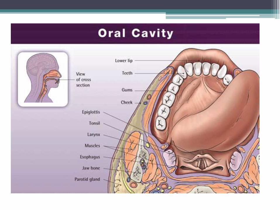
• The palate forms the roof of the mouth and intervenes between the nasal and oral cavities.
• It consists of the palatine process of the maxilla, the horizontal plates of the palatine bone.
Anatomy
• Incisive Fossa ▫ Slight depression
posterior to central incisor teeth
▫ Nasopalatine nerve • Greater palatine foramina
Medial to 3rd Molar Greater palatine
vessels and nerve • Lesser palatine foramina
Lesser Palatine nerves and vessels to soft palate
Three foramina open on the oral aspect of the hard palate
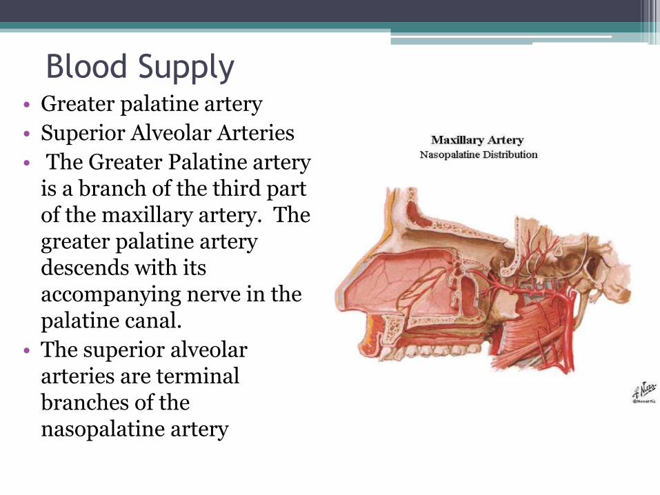
• Greater palatine artery
• Superior Alveolar Arteries
• The Greater Palatine artery is a branch of the third part of the maxillary artery. The greater palatine artery descends with its accompanying nerve in the palatine canal.
• The superior alveolar arteries are terminal branches of the nasopalatine artery
Blood Supply

• The greater palatine emerges on the hard palate from the greater palatine foramen runs forward in a groove on the inferior surface of the bony palate almost to the incisor teeth supplies the gums and the mucosa and glands of the hard palate.4
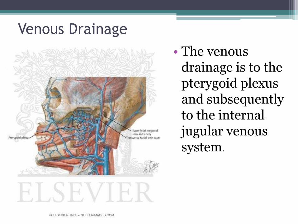
• The venous drainage is to the pterygoid plexus and subsequently to the internal jugular venous system.
Venous Drainage

• The nasopalatine nerves
• Greater Palatine Nerves
• The nasopalatine nerves are branches of the maxillary division of the trigeminal nerve.
• They enter the palate at the incisive foramen supply the anterior part of the hard palate behind the incisor teeth.5
Innervation

• Greater (and Lesser) Palatine run through the palatine canal and exit at the Great and Lesser Palatine Foramens, respectively.
• Parasympathetic postganglionic secretomotor fibres from the pterygopalatine ganglion run with the nerves to supply the palatine mucous glands.
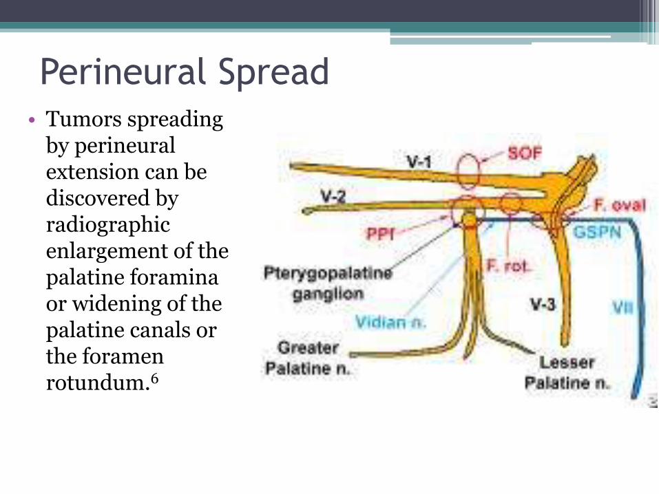
• Tumors spreading by perineural extension can be discovered by radiographic enlargement of the palatine foramina or widening of the palatine canals or the foramen rotundum.6
Perineural Spread

Oral submucous fibrosis (OSMF)
• OSMF is a high risk precancerous condition that predominantly occurs amongst Indians.
• Factors implicated in the pathogenesis of sub mucous fibrosis:
▫ Chillie consumption
▫ Areca-nut chewing, autoimmunity
▫ Genetic predisposition
• Now there is convincing epidemiologic evidence implicating areca nut as a causative factor in its pathogenesis.

Epidemiology
•The prevalence of OSMF in random samples of the population in India is up to 0.4%.
•Although hard data are not available, indications are that this disease is increasing rapidly in India
•Submucous fibrosis occurs in both sexes over a wide age range

Definition
•OSMF is a chronic mucosal condition affecting any part of the oral mucosa.
•Mucosal rigidity of varying intensity due to fibroelastic transformation of the juxtaepithelial connective tissue layer.
•The presence of palpable fibrous bands is a diagnostic criterion for submucous fibrosis.


•When the tongue is affected, it is devoid of papillae and its mobility, especially the protrusion, is impaired
•The opening of the mouth is restricted
• In severe OSMF, the patient cannot protrude the tongue beyond the incisal edges and there is a progressive closure of the oral opening
•OSMF must be diagnosed only if palpable fibrous bands are present


Clinical aspects
•The most common initial symptoms: ▫ Burning sensation of the oral mucosa aggravated by spicy food followed by either hypersalivation or dryness of the mouth.

•The most common and initial clinical sign as well as a regular feature:
▫blanching i.e., marble-like appearance of the oral mucosa.
•In advanced cases, the mucosa becomes tough and leathery, with numerous vertical fibrous bands

Natural History •Unlike precancerous lesions OSMF is
not known to regress, either spontaneously, or with the cessation of the areca-nut chewing habit
•The most serious aspect of this disease is the high risk for the development of oral cancer
•The epithelium is atrophic in this condition which renders it susceptible to the action of carcinogens

•OSMF and coexistent leukoplakia: Leukoplakia is a precancerous ! lesion; its coexistence with OSMF implies the high risk for oral cancer.
•OSMF and coexistent oral cancer: Not uncommonly (in 5% to 42% of the cases), submucous fibrosis and oral cancer coexist
•Malignant transformation: Long-term population based studies have confirmed its precancerous nature.


• No definitive and widely accepted treatment is currently available.
• Some temporary relief from the symptoms and improvement in the oral opening with medicinal treatment such as local injections of cortisone and placentrex.
• It is essential to follow-up the patients regularly.
• Patient education to discontinue the use of areca nut and tobacco in any form.

LEUKOPLAKIA
•Leukoplakia is the most common premalignant or "potentially malignant" lesion of the oral mucosa
•It is a predominantly white lesion of the oral mucosa
•The incidence and prevalence of leukoplakia vary in different parts of the world

•In general the reported prevalence ranges from 0.2 to 5% (India 0.2-4.9%)
•It is seen most frequently in middle-aged and older men.
•Gender distribution is also variable. Men are more affected in some countries.
Clinical presentation
•Leukoplakia can be either solitary or multiple
•It may appear on any site of the oral cavity, the most common sites being: buccal mucosa, alveolar mucosa, floor of the mouth, tongue, lips and palate

Classically two clinical types of
leukoplakia are recognised
• Homogeneous leukoplakia is defined as a predominantly white lesion of uniform flat and thin appearance that may exhibit shallow cracks. This type is usually asymptomatic.
• Non-homogeneous leukoplakia has been defined as a predominant white or white-and-red lesion ("eritroleukoplakia") that may be either irregularly flat, nodular ("speckled leukoplakia) or exophytic ("exophytic or verrucous)

Homogeneous leukoplakia on the dorsum and left
lateral margin of the tongue

Non-homogenous Leukoplakia
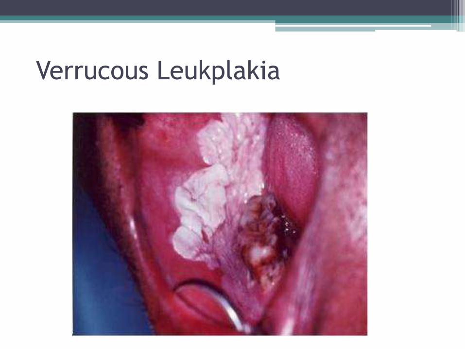
Verrucous Leukplakia

Aetiopathogenesis
•The aetiology of leukoplakia is still unclear
•Tobacco seems to be the major inductor factor, its association cannot be determined in all cases
•A variety of smokeless tobacco habits have been reported as leukoplakia inductors: e.g. snuff, chewing. These lesions have shown to have a low malignant transformation risk
•Other factors such as:
▫Alcohol
▫ Inadequate diet
▫Vitamin deficiency (e.g. vitamin A and C), areca nut (betel)
▫Chronic traumatic irritation
▫Poor oral hygiene
▫Poor socio-economic status.

Treatment • There are different treatments for leukoplakia. • However, the risk of malignant transformation is
not completely eliminated by any of the current therapies.
• Initial treatment of a white oral lesion is the elimination of the possible aetiological factors.
• Complete surgical removal (leaving free-lesion borders) is recommended in cases with epithelial dysplasia.
• Apart from surgical excision, other treatment modalities available include cryosurgery, laser surgery, retinoids, beta-carotene, bleomycin, calcipotriol, photodynamic therapy.

Prognosis and complications
•The malignant transformation rate of oral leukoplakia varies from 0 to 33%.
•Regular check-up of these patients is essential, probably every 3, 6 and then 12 months, both in treated and untreated patients

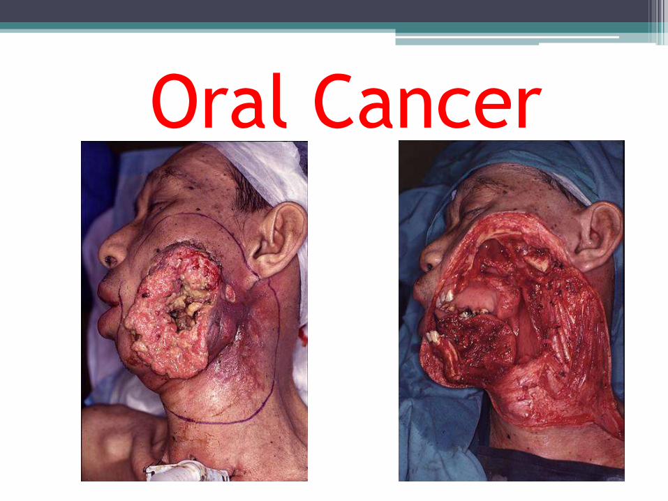
Oral Cancer

• Oral Cancer is the sixth leading cause of cancer worldwide
• The survival rate was 52%.
• Oral cancer generally are socially derived diseases.
• Tobacco and alcohol have synergistic effect
• Treatment of early oral cancer is surgery. Locally advanced T3/4 are best treated with combined surgery and Radiotherapy.
• High risk of second primary cancer

EPIDEMIOLOGY • The Oral cavity extends from vermilion border
of lips to the plane between junction of the hard palate and soft palate.
• Include: Lips and oral cavity(buccal mucosa, tongue, ginggiva, retromolar trigone, flour of mouth, hard palate)
• The incidence of oral cancer varies throughout the world. High incidence in India, France, SE Asia.
• 40% of HN cancer
• Age onset 50 yrs. Sex ratio 3:1


Risk factors
•Heavy tobacco
•Alcohol.
•Syphilis
•Viruses (EB, HSV, HPV, HIV)
•Neglect of oral dental hygiene(chronic infection, unfit dentures)
•Lichen planus, Plummer Vinson sy.
• Immunosuppression, malnutrition
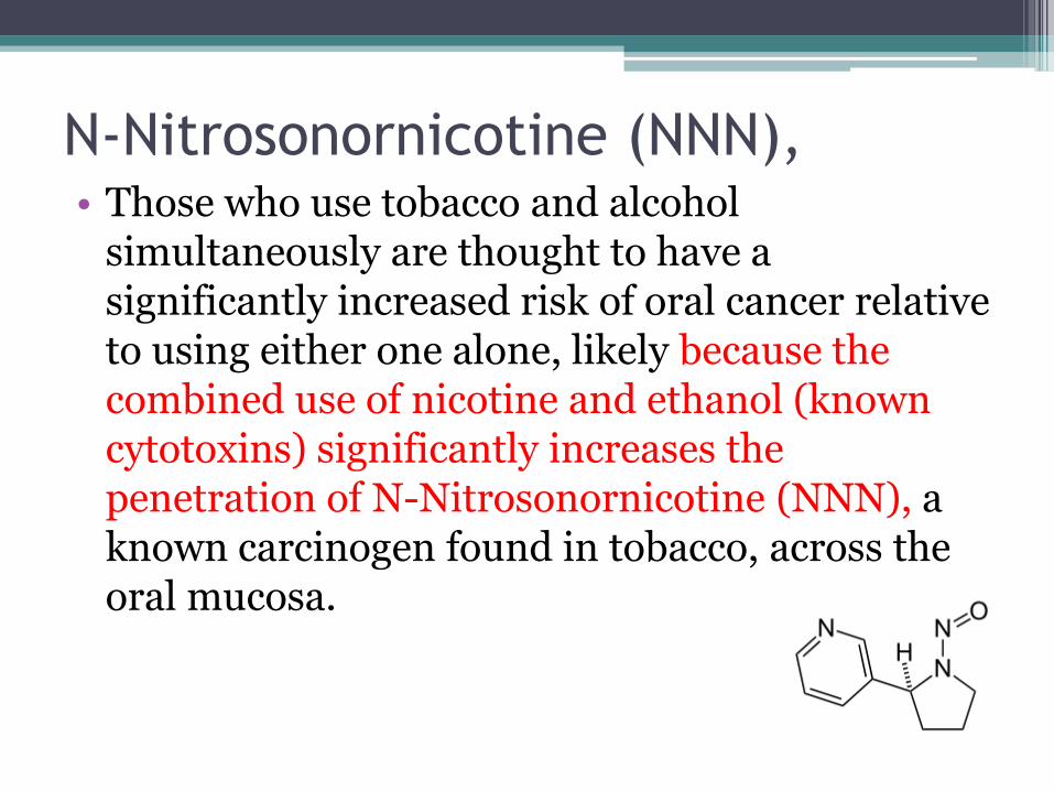
• Those who use tobacco and alcohol simultaneously are thought to have a significantly increased risk of oral cancer relative to using either one alone, likely because the combined use of nicotine and ethanol (known cytotoxins) significantly increases the penetration of N-Nitrosonornicotine (NNN), a known carcinogen found in tobacco, across the oral mucosa.
N-Nitrosonornicotine (NNN),
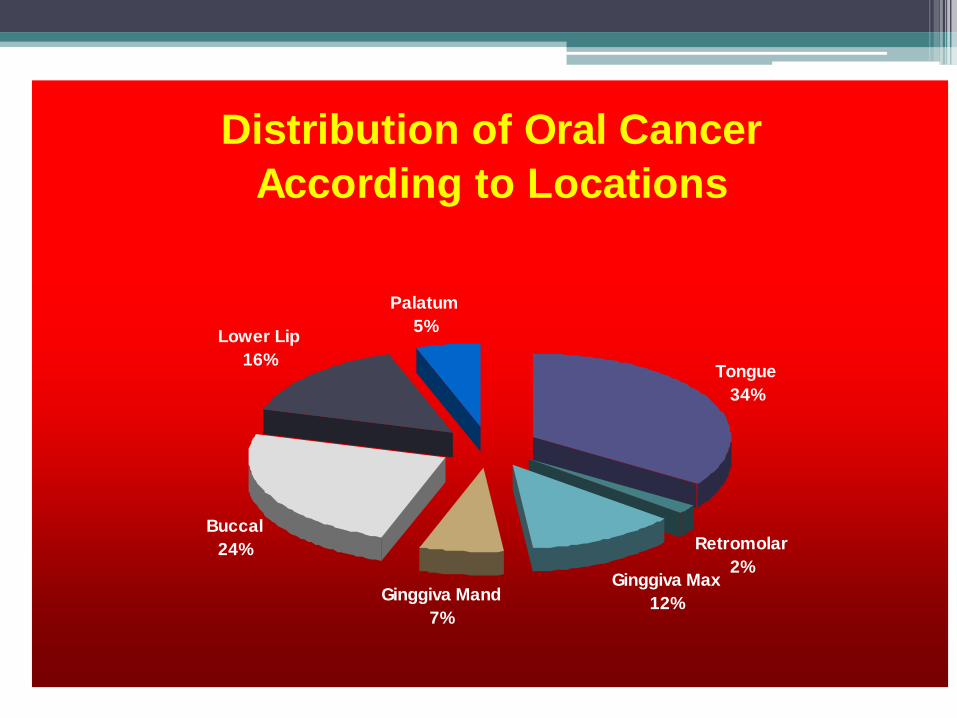
Distribution of Oral Cancer
According to Locations
Tongue
34%
Retromolar
2%Ginggiva Max
12%Ginggiva Mand
7%
Buccal
24%
Lower Lip
16%
Palatum
5%
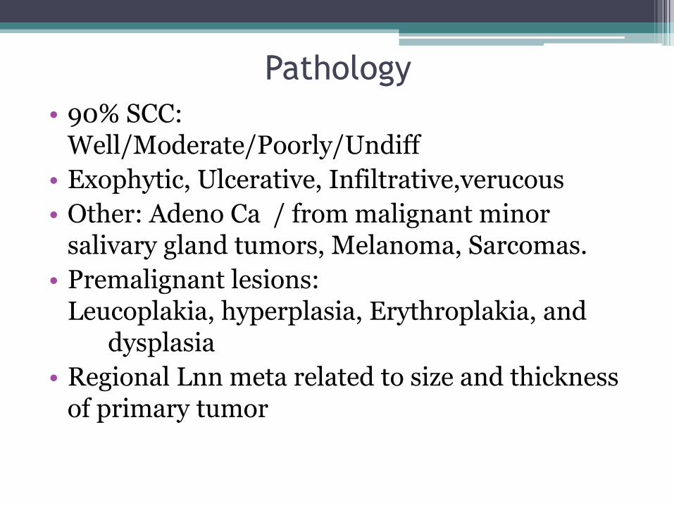
Pathology
• 90% SCC: Well/Moderate/Poorly/Undiff
• Exophytic, Ulcerative, Infiltrative,verucous
• Other: Adeno Ca / from malignant minor salivary gland tumors, Melanoma, Sarcomas.
• Premalignant lesions: Leucoplakia, hyperplasia, Erythroplakia, and dysplasia
• Regional Lnn meta related to size and thickness of primary tumor
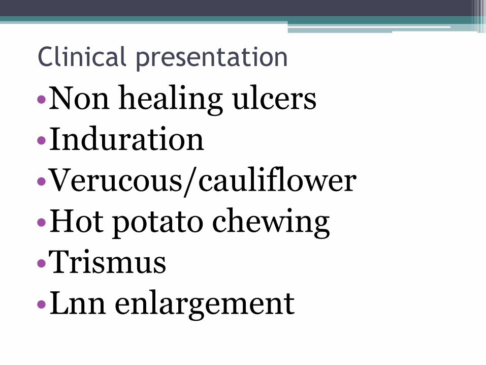
Clinical presentation
•Non healing ulcers
•Induration
•Verucous/cauliflower
•Hot potato chewing
•Trismus
•Lnn enlargement

Tumors of the soft palate
• similar to other tumors of the oral cavity ▫ 90% of all oral
cancer is squamous cell1
• 69.7% of soft palate tumors are squamous cell carcinoma2
Tumors of the hard palate
• Squamous:Non-Squamous is 1:2 to 1:4
• Varied histology with non-squamous cell tumors,
▫ minor salivary gland tumors
▫ rare cases of melanoma3
▫ sarcoma
▫ malignant lymphomas.4
Soft Palate vs. Hard Palate

Diagnosis • Clinical:
• History Detail clinical examination (used head lamp, mirror) Bimanual palpation
• Cervical Lnn examination
• Endoscopy (searching the second primary)
• Biopsy
• Staging: Panoramic photo, thorax,USG liver, or CT/MRI/PET Scan


TREATMENT • Treatment Goals:
To eradicate of the primary tumor and LN metastasis, to maintain the function, and cosmetic reconstruction.
• Factors affecting choice of treatment: Tumor factors
• Patient factors
• Resource factors
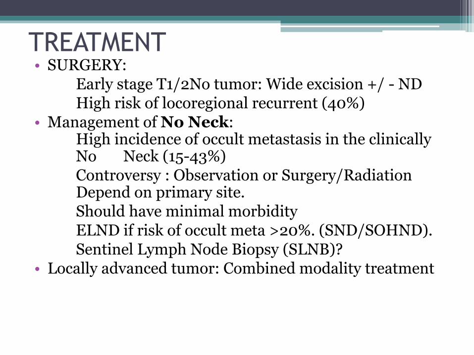
TREATMENT • SURGERY: Early stage T1/2No tumor: Wide excision +/ - ND High risk of locoregional recurrent (40%) • Management of No Neck:
High incidence of occult metastasis in the clinically No Neck (15-43%)
Controversy : Observation or Surgery/Radiation Depend on primary site.
Should have minimal morbidity ELND if risk of occult meta >20%. (SND/SOHND). Sentinel Lymph Node Biopsy (SLNB)? • Locally advanced tumor: Combined modality treatment


6 Levels of Lymph-Nodes

Selective Neck Dissection

Classification of ND
1991 Classification:
• RND
• Modified RND
• Selective ND: Supraomohyoid Lateral Posterolateral Anterior
• Extended ND
2001 Classification:
• RND
• Modified RND
• Selective ND (SND): SND (L.I-III/IV) SND (L.II-IV) SND (L.II-V) SND (L.VI)
• Extended ND
Proposed by American HN Society and AAOHNS

Selective Neck Dissection
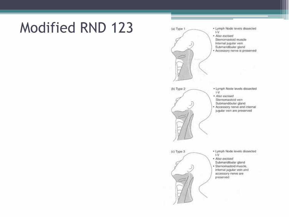
Modified RND 123

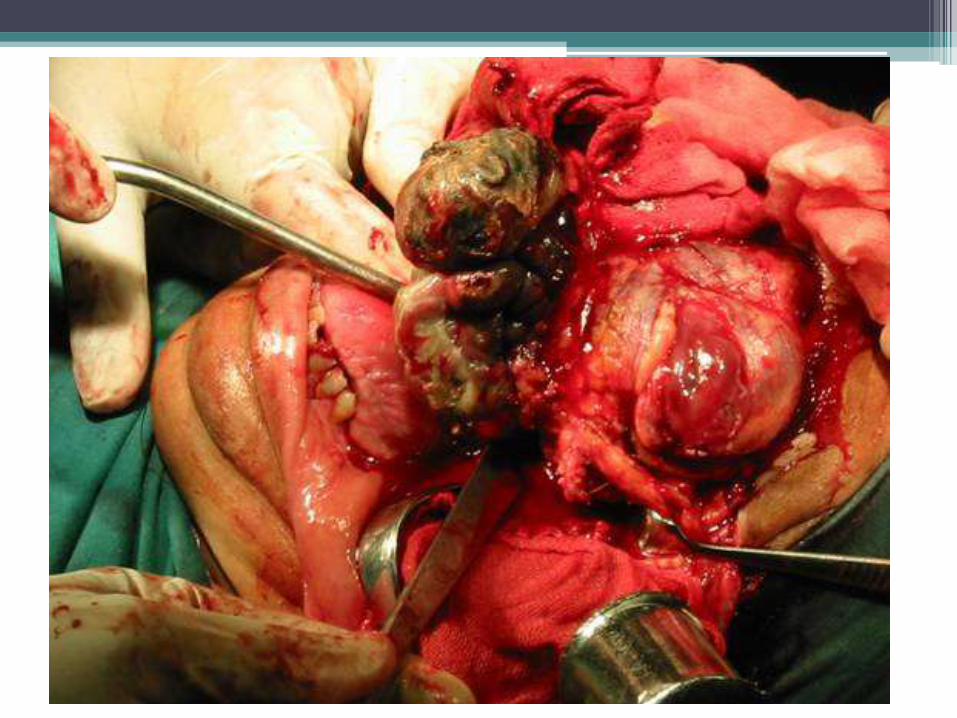
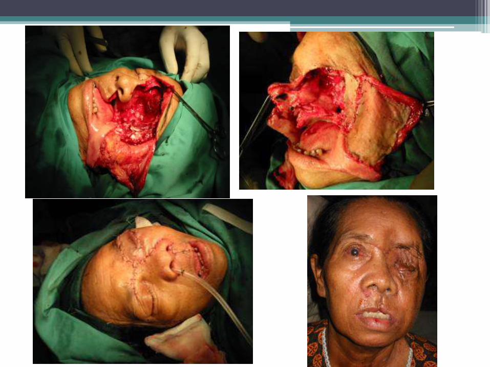
SURGICAL APPROACHES
▫ Trans-oral approach
▫ Lower cheek approach
▫ Upper cheek approach
▫ Swing mandibulotomy
▫ Visor flap
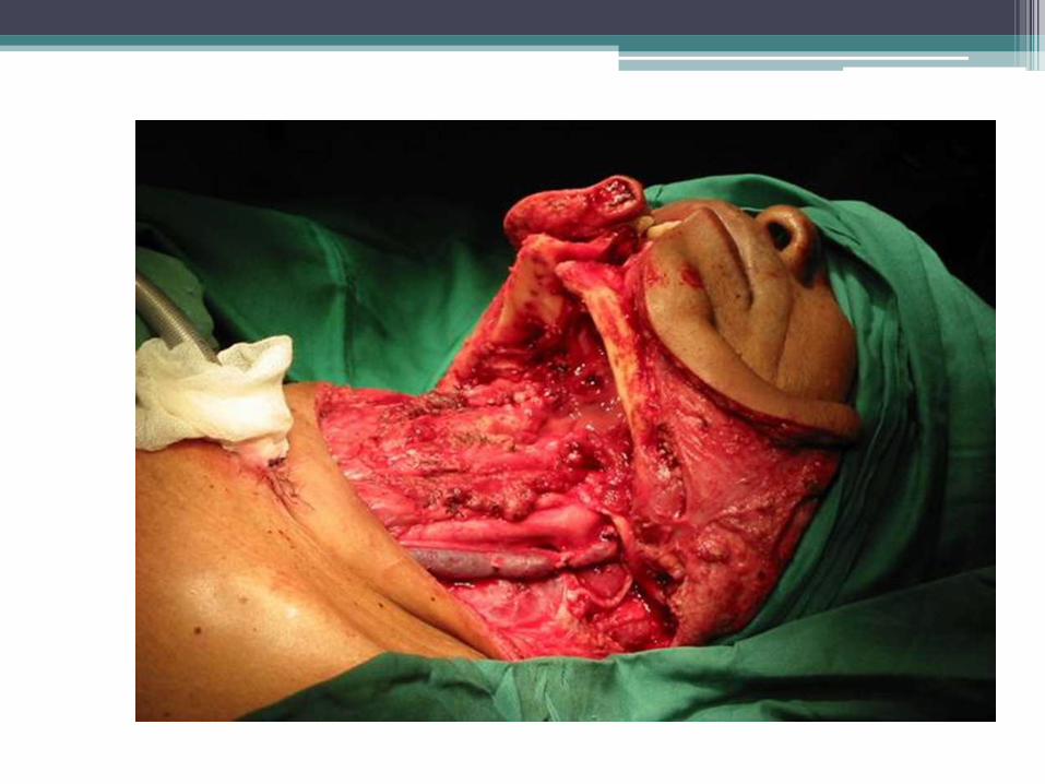


RECONSTRUCTION
• Single-stage immediate reconstruction is recommended.
• The technique: Skin grafts Pedicle flaps Alloplastic meterials Autografts
Free flaps

Adjuvant treatment
• Radiothrepy (External beam/Interstitial)
• Chemotherapy
• Concomittant Radio+Chemotherapy (Neoadjuvant)
• Palliative Chemotherapy for advanced diseases

PROGNOSIS • Location/thickness/depth of primary tumor
• Staging
• Type of histology
• Grading
• Presence of perineural spread
• Mandibular invasion
• Lnn extention (Level, size, extracaps of meta)
• Molecular markers (?)
Summary • The main problem of oral cancer is early
detection • Surgery is still the most important modality in
management of oral cancer. • Better understanding of molecular biology of
HNSCC. • Bio-molecular markers can be used in the
management of SCC oral cancer. • High risk of second primary cancer,
Chemoprevention?


Related Documents