sensors Communication Sensitive Fluorescence Assay for the Detection of Alkaline Phosphatase Based on a Cu 2+ -Thiamine System Han Zhao † , Xinfa Liu † and Changbei Ma * Citation: Zhao, H.; Liu, X.; Ma, C. Sensitive Fluorescence Assay for the Detection of Alkaline Phosphatase Based on a Cu 2+ -Thiamine System. Sensors 2021, 21, 674. https:// doi.org/10.3390/s21030674 Received: 13 December 2020 Accepted: 14 January 2021 Published: 20 January 2021 Publisher’s Note: MDPI stays neutral with regard to jurisdictional claims in published maps and institutional affil- iations. Copyright: © 2021 by the authors. Licensee MDPI, Basel, Switzerland. This article is an open access article distributed under the terms and conditions of the Creative Commons Attribution (CC BY) license (https:// creativecommons.org/licenses/by/ 4.0/). School of Life Sciences, Central South University, Changsha 410013, China; [email protected] (H.Z.); [email protected] (X.L.) * Correspondence: [email protected] † These authors contributed equally to this work. Abstract: The authors describe a novel, facile, and sensitive fluorometric strategy based on a Cu 2+ - thiamine (Cu 2+ -TH) system for the detection of alkaline phosphatase (ALP) activity and inhibition. The principle of the method is as follows. Under a basic conditions, TH, which does not exhibit a fluorescence signal, is oxidized into fluorescent thiochrome (TC) by Cu 2+ . Ascorbic acid 2-phosphate (AAP), which is the enzyme substrate, is hydrolyzed to produce ascorbic acid (AA) by ALP. The newly formed AA then reduces Cu 2+ to Cu + , which prevents the oxidation of TH by Cu 2+ ; as a result, the fluorescent signal becomes weaker. On the contrary, in the absence of ALP, AAP cannot reduce Cu 2+ ; additions of Cu 2+ and TH result in a dramatic increase of the fluorescent signal. The sensing strategy displays brilliant sensitivity with a detection limit of 0.08 U/L, and the detection is linear in the concentration range of 0.1 to 100 U/L. This approach was successfully applied to ALP activity in human serum samples, indicating that it is reliable and may be applied to the clinical diagnosis of ALP-related diseases. Keywords: alkaline phosphatase; fluorescence; Cu 2+ -thiamine; inhibitor 1. Introduction Alkaline phosphatase (ALP), an enzyme broadly found in mammalian tissues, can catalyze the hydrolysis and dephosphorylation processes of multifarious substrates, which include nucleic acids, proteins, and some small molecules [1–3]. Furthermore, research suggests that ALP is involved in cell regulation and signal transduction processes [4]. The aberrant level of ALP in serum is tightly associated with numerous serious illnesses, such as hepatitis, breast and prostatic carcinoma, liver dysfunction, osteopathy, and diabetes, and it is generally recognized as a biomarker in early clinical diagnosis [5–7]. Consequently, attention has been paid to ALP for its potential use in fast and sensitive detection. Up to now, a variety of analytical techniques, including electrochemistry, fluorometry, colorimetry, chromatography, and surface-enhanced Raman scattering have been used in the determination of ALP activity [8–17]. For example, Wu et al. established a signal amplified electrochemical method which relied on enzyme-induced metallization (EIM) for ALP analysis [18]. Despite its low detection limit, this method has other disadvantages: the preparation of modified electrodes and the sample preprocessing procedures are costly, complicated, and time-consuming. Tang et al. employed a colorimetric sensing platform for ALP activity assay by the use of Cu(II)-modulated G-quadruplex-based DNAzymes [19]. This colorimetric method has some advantages, such as simple instruments and convenient operation, however, the method is inefficient and uses probes with a complicated design. Recently, increasing attention has been paid to fluorescence-based strategies for their rapid reaction time and considerably high sensitivity. The fluorescence probes used this strategy are mainly composed of organic dyes, quantum dots (QDs), DNA-templated nanoparticles, and conjugated polyelectrolytes [20–23]. For this purpose, Qu et al. proposed a turn-on fluorescence method based on carbon dots and MnO 2 nanosheets for the detection of Sensors 2021, 21, 674. https://doi.org/10.3390/s21030674 https://www.mdpi.com/journal/sensors

Welcome message from author
This document is posted to help you gain knowledge. Please leave a comment to let me know what you think about it! Share it to your friends and learn new things together.
Transcript
sensors
Communication
Sensitive Fluorescence Assay for the Detection of AlkalinePhosphatase Based on a Cu2+-Thiamine System
Han Zhao †, Xinfa Liu † and Changbei Ma *
Citation: Zhao, H.; Liu, X.; Ma, C.
Sensitive Fluorescence Assay for the
Detection of Alkaline Phosphatase
Based on a Cu2+-Thiamine System.
Sensors 2021, 21, 674. https://
doi.org/10.3390/s21030674
Received: 13 December 2020
Accepted: 14 January 2021
Published: 20 January 2021
Publisher’s Note: MDPI stays neutral
with regard to jurisdictional claims in
published maps and institutional affil-
iations.
Copyright: © 2021 by the authors.
Licensee MDPI, Basel, Switzerland.
This article is an open access article
distributed under the terms and
conditions of the Creative Commons
Attribution (CC BY) license (https://
creativecommons.org/licenses/by/
4.0/).
School of Life Sciences, Central South University, Changsha 410013, China; [email protected] (H.Z.);[email protected] (X.L.)* Correspondence: [email protected]† These authors contributed equally to this work.
Abstract: The authors describe a novel, facile, and sensitive fluorometric strategy based on a Cu2+-thiamine (Cu2+-TH) system for the detection of alkaline phosphatase (ALP) activity and inhibition.The principle of the method is as follows. Under a basic conditions, TH, which does not exhibit afluorescence signal, is oxidized into fluorescent thiochrome (TC) by Cu2+. Ascorbic acid 2-phosphate(AAP), which is the enzyme substrate, is hydrolyzed to produce ascorbic acid (AA) by ALP. Thenewly formed AA then reduces Cu2+ to Cu+, which prevents the oxidation of TH by Cu2+; as a result,the fluorescent signal becomes weaker. On the contrary, in the absence of ALP, AAP cannot reduceCu2+; additions of Cu2+ and TH result in a dramatic increase of the fluorescent signal. The sensingstrategy displays brilliant sensitivity with a detection limit of 0.08 U/L, and the detection is linear inthe concentration range of 0.1 to 100 U/L. This approach was successfully applied to ALP activity inhuman serum samples, indicating that it is reliable and may be applied to the clinical diagnosis ofALP-related diseases.
Keywords: alkaline phosphatase; fluorescence; Cu2+-thiamine; inhibitor
1. Introduction
Alkaline phosphatase (ALP), an enzyme broadly found in mammalian tissues, cancatalyze the hydrolysis and dephosphorylation processes of multifarious substrates, whichinclude nucleic acids, proteins, and some small molecules [1–3]. Furthermore, researchsuggests that ALP is involved in cell regulation and signal transduction processes [4]. Theaberrant level of ALP in serum is tightly associated with numerous serious illnesses, suchas hepatitis, breast and prostatic carcinoma, liver dysfunction, osteopathy, and diabetes,and it is generally recognized as a biomarker in early clinical diagnosis [5–7]. Consequently,attention has been paid to ALP for its potential use in fast and sensitive detection.
Up to now, a variety of analytical techniques, including electrochemistry, fluorometry,colorimetry, chromatography, and surface-enhanced Raman scattering have been usedin the determination of ALP activity [8–17]. For example, Wu et al. established a signalamplified electrochemical method which relied on enzyme-induced metallization (EIM)for ALP analysis [18]. Despite its low detection limit, this method has other disadvantages:the preparation of modified electrodes and the sample preprocessing procedures are costly,complicated, and time-consuming. Tang et al. employed a colorimetric sensing platform forALP activity assay by the use of Cu(II)-modulated G-quadruplex-based DNAzymes [19].This colorimetric method has some advantages, such as simple instruments and convenientoperation, however, the method is inefficient and uses probes with a complicated design.Recently, increasing attention has been paid to fluorescence-based strategies for their rapidreaction time and considerably high sensitivity. The fluorescence probes used this strategyare mainly composed of organic dyes, quantum dots (QDs), DNA-templated nanoparticles,and conjugated polyelectrolytes [20–23]. For this purpose, Qu et al. proposed a turn-onfluorescence method based on carbon dots and MnO2 nanosheets for the detection of
Sensors 2021, 21, 674. https://doi.org/10.3390/s21030674 https://www.mdpi.com/journal/sensors
Sensors 2021, 21, 674 2 of 10
ALP [24]. He et al. developed a label-free fluorometric approach for the detection of ALPthrough the synthesis of ssDNA-templated fluorescent silver nanoclusters [25]. However,these strategies have unavoidable disadvantages, such as the fact that QDs have hightoxicity, dye-labeling steps are complex, the synthetic process is long, and the cost ofreagents is high. Hence, the development of a facile, economical, and timesaving assay ofALP is highly desirable.
Thiamine (TH), also known as vitamin B1, has been utilized as a non-fluorescent sub-strate [26–28]. It has attracted tremendous attention for its low cost, good water-solubility,and easy accessibility. TH does not exhibit a fluorescence signal; however, under basicconditions, it can be easily converted into fluorescent thiochrome (TC) by Cu2+ [29]. Thus,the development of a Cu2+-TH-based system may open up a new avenue for fluorescencedetection. In this study, we propose a novel Cu2+-TH-based fluorometric strategy systemfor the detection of ALP activity. The addition of ALP can catalyze the hydrolysis of ascor-bic acid 2-phosphate (AAP) into ascorbic acid (AA), which can then reduce Cu2+ to Cu+
that in turn leads to fluorescence quenching. In the absence of ALP, Cu2+ can oxidizeTH and convert it into TC, recovering the fluorescence signal. The present fluorescentmethod, which has high selectivity and sensitivity, was triumphantly utilized to detectALP in human serum samples. It is a promising strategy that can be employed in variousclinical applications.
2. Materials and Methods2.1. Materials and Reagents
Alkaline phosphatase (ALP), uracil DNA glycosylase (UDG), T4 polynucleotide ki-nase (T4 PNK), and 8-hydroxy guanine DNA glycosidase (hOGG1) were all acquiredfrom Takara Biotechnology Co., Ltd. (Dalian, China). Ascorbic acid 2-phosphate (AAP),glycine (Gly), alanine (Ala), serine (Ser), glutamic acid (Glu), histidine (His), lysozyme,streptavidin (SA), glucose (C6H12O6), thiamine, Na3VO4, and ATP were purchased fromSource Leaf Biotechnology Co., Ltd. (Shanghai, China). Cupric sulfate (CuSO4), sodiumhydroxide (NaOH), and 3′-(N-morpholino) propanesulfonic acid (MOPS) were purchasedfrom Sinopharm Chemical Reagent Co., Ltd. (Shanghai, China). All other chemicals wereof analytical grades. All of the solutions used in this study were prepared with ultrapurewater (18.2 MΩ.cm) obtained from a Milli-Q water purification system.
2.2. Apparatus
Fluorescence measurements were conducted at room temperature on an F-2700 flu-orescence spectrophotometer (Hitachi, Japan). The fluorescence emission spectra wererecorded in the range of 400–570 nm with the excitation wavelength set at 370 nm. The slitwidths for excitation and emission were both set at 10 nm.
2.3. Optimization of ALP Detection
To acquire the optimal experimental performance, a variety of reaction conditionswere optimized including the concentration of Cu2+, AAP, TH, and reaction time betweenALP and AAP. The concentration range of Cu2+, AAP, and TH were 2–50 µM, 200–800 µM,and 20–400 µM, respectively. The range of reaction time between ALP, AAP, and theCu2+-TH system was from 5 min to 45 min.
2.4. Fluorescence-Based Determination of ALP Activity
Quantitative determination of ALP activity based on the Cu(II)-TH system was carriedout as follows: 500 µM AAP was incubated with different ALP activities ranging from0 to 1500 U/L in a MOPS buffer (10 mM, pH 8.0). The mixture was incubated at 37 Cfor 30 min, and then Cu2+ (20 µM) was added to the above-mentioned solution at roomtemperature for 25 min. Subsequently, TH (100 µM) and NaOH (50 mM) were added to afinal volume of 100 µL. The mixture was vortexed and reacted for 15 min at 25 C.
Sensors 2021, 21, 674 3 of 10
2.5. Selectivity of ALP
To assess the selectivity of the proposed method, a variety of common molecules,including UDG, hOGG1, T4 PNK, SA, lysozyme, Gly, Ala, Ser, Glu, His, C6H12O6, andATP, were used as interferences for comparison. First, different molecules and 500 µM AAPwere respectively mixed with the reaction buffer (10 mM MOPS buffer, pH 8.0) at 37 C for30 min. Afterward, Cu2+ (20 µM) was incubated with the above-prepared solution at 25 Cfor 25 min. Finally, 100 µM TH and 50 mM NaOH were added into the mixture at 25 C for15 min prior to fluorescence measurement.
2.6. Inhibition Investigation
To examine the effect of Na3VO4 inhibition on ALP activity, a series of experimentswere carried out. First, various concentrations of Na3VO4 (0, 100, 300, 500, 600, 800,and 1000 µM) were mixed with ALP (100 U/L) and AAP (500 µM) in an MOPS buffer(10 mM, pH 8.0) and incubated at 37 C for 30 min. Then, Cu2+ (20 µM) was added, and thereaction solutions were allowed to incubate at ambient temperature for 25 min. Finally, TH(100 µM) and NaOH (50 mM) were added. The relative activity was obtained according tothe following equation:
Relative activity (%) =
(F(inhibitor) − F0
)(
F(no inhibitor) − F0
) × 100
where F (inhibitor) stands for the fluorescence intensity of the samples in the presenceof ALP and the inhibitor, F (no inhibitor) stands for the fluorescence intensity of thesamples with ALP but no inhibitor, F0 stands for the fluorescence intensity without ALP orthe inhibitor.
2.7. ALP Activity Assay in Human Serum Samples
To evaluate the feasibility of the proposed strategy in detecting ALP in real samples,a certain amount of ALP was spiked into human serum samples, which were diluted by100 times with a reaction buffer (10 mM MOPS, pH 8.0). The ALP activity of the samplewas subsequently determined by fluorescent measurement.
2.8. Statistical Analysis
Data analysis was performed with GraphPad Prism 8.0 statistical software. The re-sults between two groups were analyzed using two-tailed Student’s t-tests. p < 0.05 wasconsidered to indicate a significant difference.
3. Results and Discussion3.1. Principles of ALP Activity Detection
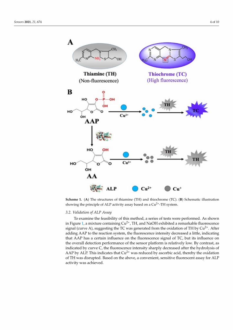
The illustration for the principle of the proposed fluorescence-based Cu2+-TH systemin the determination of ALP activity is depicted in Scheme 1. In a basic solution, TH canbe oxidized by Cu2+ to produce fluorescent thiochrome (TC). In the absence of ALP, thefluorescence signal is observed in the reaction system consisting of AAP, Cu2+, and TH.In the presence of ALP, AAP, which acts as the substrate for ALP dephosphorylation, ishydrolyzed to generate ascorbic acid. In the presence of ascorbic acid, which can reducemetal ions, Cu2+ is reduce to Cu+. As a result, the oxidation of TH is impaired, caus-ing reduced fluorescence intensity. Variation of the fluorescence response with the ALPconcentration indicates that a timesaving, facile fluorescent method for ALP detection issuccessfully developed.
Sensors 2021, 21, 674 4 of 10
Sensors 2020, 20, x FOR PEER REVIEW 4 of 10
Scheme 1. (A) The structures of thiamine (TH) and thiochrome (TC). (B) Schematic illustration showing the principle of ALP activity assay based on a Cu2+-TH system.
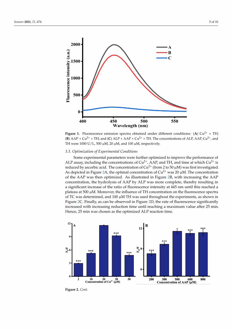
3.2. Validation of ALP Assay To examine the feasibility of this method, a series of tests were performed. As shown
in Figure 1, a mixture containing Cu2+, TH, and NaOH exhibited a remarkable fluores-cence signal (curve A), suggesting the TC was generated from the oxidation of TH by Cu2+. After adding AAP to the reaction system, the fluorescence intensity decreased a little, in-dicating that AAP has a certain influence on the fluorescence signal of TC, but its influence on the overall detection performance of the sensor platform is relatively low. By contrast, as indicated by curve C, the fluorescence intensity sharply decreased after the hydrolysis of AAP by ALP. This indicates that Cu2+ was reduced by ascorbic acid, thereby the oxida-tion of TH was disrupted. Based on the above, a convenient, sensitive fluorescent assay for ALP activity was achieved.
Scheme 1. (A) The structures of thiamine (TH) and thiochrome (TC). (B) Schematic illustrationshowing the principle of ALP activity assay based on a Cu2+-TH system.
3.2. Validation of ALP Assay
To examine the feasibility of this method, a series of tests were performed. As shownin Figure 1, a mixture containing Cu2+, TH, and NaOH exhibited a remarkable fluorescencesignal (curve A), suggesting the TC was generated from the oxidation of TH by Cu2+. Afteradding AAP to the reaction system, the fluorescence intensity decreased a little, indicatingthat AAP has a certain influence on the fluorescence signal of TC, but its influence onthe overall detection performance of the sensor platform is relatively low. By contrast, asindicated by curve C, the fluorescence intensity sharply decreased after the hydrolysis ofAAP by ALP. This indicates that Cu2+ was reduced by ascorbic acid, thereby the oxidationof TH was disrupted. Based on the above, a convenient, sensitive fluorescent assay for ALPactivity was achieved.
Sensors 2021, 21, 674 5 of 10
Figure 1. Fluorescence emission spectra obtained under different conditions: (A) Cu2+ + TH;(B) AAP + Cu2+ + TH; and (C) ALP + AAP + Cu2+ + TH. The concentrations of ALP, AAP, Cu2+, andTH were 1000 U/L, 500 µM, 20 µM, and 100 µM, respectively.
3.3. Optimization of Experimental Conditions
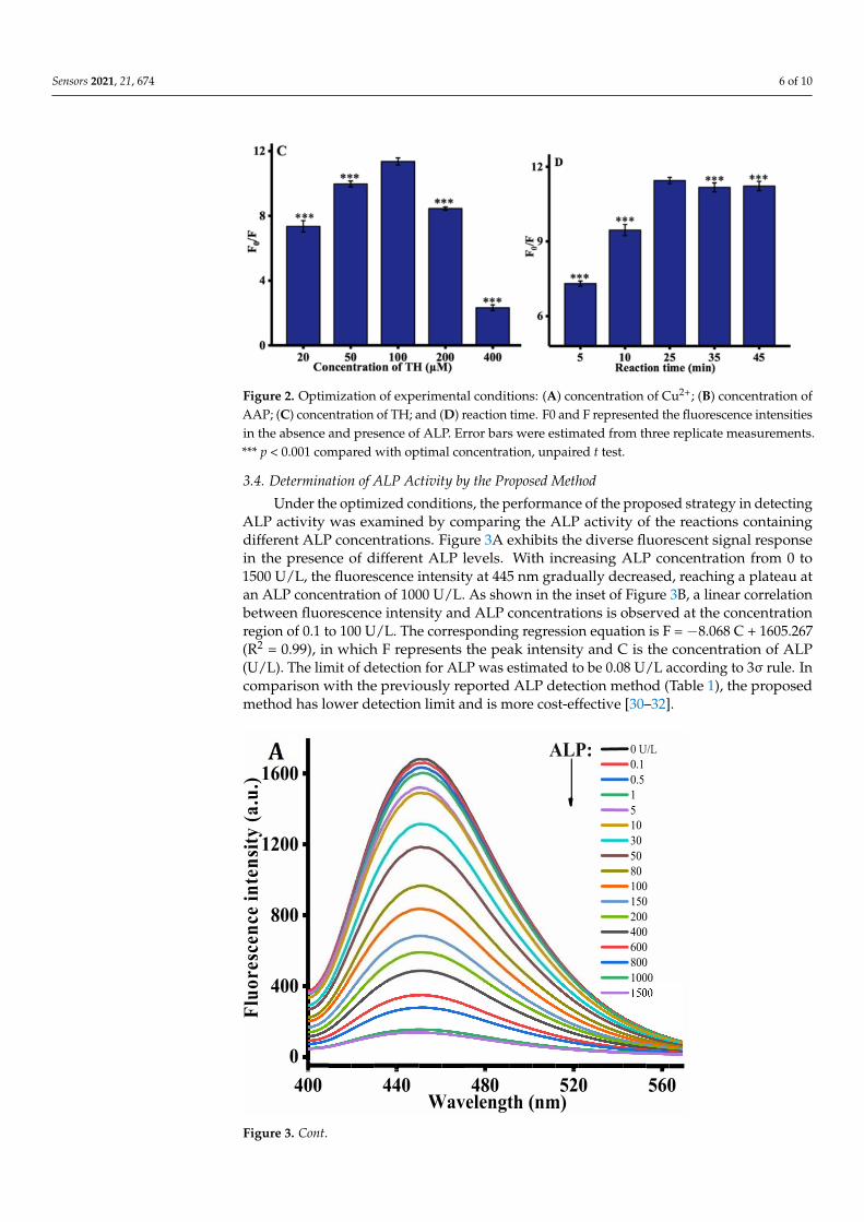
Some experimental parameters were further optimized to improve the performance ofALP assay, including the concentrations of Cu2+, AAP, and TH, and time at which Cu2+ isreduced by ascorbic acid. The concentration of Cu2+ (from 2 to 50 µM) was first investigated.As depicted in Figure 2A, the optimal concentration of Cu2+ was 20 µM. The concentrationof the AAP was then optimized. As illustrated in Figure 2B, with increasing the AAPconcentration, the hydrolysis of AAP by ALP was more complete, thereby resulting ina significant increase of the ratio of fluorescence intensity at 445 nm until this reached aplateau at 500 µM. Moreover, the influence of TH concentration on the fluorescence spectraof TC was determined, and 100 µM TH was used throughout the experiments, as shown inFigure 2C. Finally, as can be observed in Figure 2D, the rate of fluorescence significantlyincreased with increasing reduction time until reaching a maximum value after 25 min.Hence, 25 min was chosen as the optimized ALP reaction time.
Figure 2. Cont.
Sensors 2021, 21, 674 6 of 10
Figure 2. Optimization of experimental conditions: (A) concentration of Cu2+; (B) concentration ofAAP; (C) concentration of TH; and (D) reaction time. F0 and F represented the fluorescence intensitiesin the absence and presence of ALP. Error bars were estimated from three replicate measurements.*** p < 0.001 compared with optimal concentration, unpaired t test.
3.4. Determination of ALP Activity by the Proposed Method
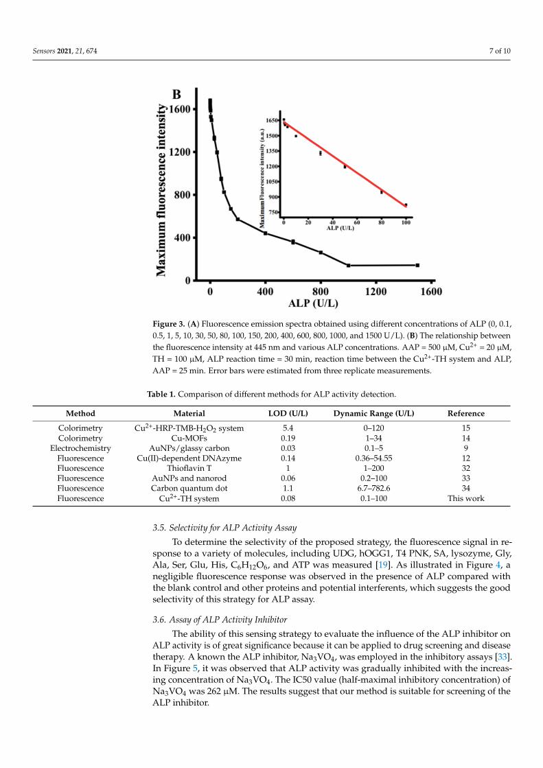
Under the optimized conditions, the performance of the proposed strategy in detectingALP activity was examined by comparing the ALP activity of the reactions containingdifferent ALP concentrations. Figure 3A exhibits the diverse fluorescent signal responsein the presence of different ALP levels. With increasing ALP concentration from 0 to1500 U/L, the fluorescence intensity at 445 nm gradually decreased, reaching a plateau atan ALP concentration of 1000 U/L. As shown in the inset of Figure 3B, a linear correlationbetween fluorescence intensity and ALP concentrations is observed at the concentrationregion of 0.1 to 100 U/L. The corresponding regression equation is F = −8.068 C + 1605.267(R2 = 0.99), in which F represents the peak intensity and C is the concentration of ALP(U/L). The limit of detection for ALP was estimated to be 0.08 U/L according to 3σ rule. Incomparison with the previously reported ALP detection method (Table 1), the proposedmethod has lower detection limit and is more cost-effective [30–32].
,_1600 .
= .
_.,
>.
:.:: 1200 r:11
= --
= ...
800 = r:11
:. 0 400 = -
ALP: -OU/L
J
-0.1
-o.s
-1
-5
-10
-30
-so
-so
-100
-150
-200
-400
-600
-800
-1000
0 .... --.......... --.... -----.----.-400 440 480 520
Wavelength (nm) 560
Figure 3. Cont.
Sensors 2021, 21, 674 7 of 10
Figure 3. (A) Fluorescence emission spectra obtained using different concentrations of ALP (0, 0.1,0.5, 1, 5, 10, 30, 50, 80, 100, 150, 200, 400, 600, 800, 1000, and 1500 U/L). (B) The relationship betweenthe fluorescence intensity at 445 nm and various ALP concentrations. AAP = 500 µM, Cu2+ = 20 µM,TH = 100 µM, ALP reaction time = 30 min, reaction time between the Cu2+-TH system and ALP,AAP = 25 min. Error bars were estimated from three replicate measurements.
Table 1. Comparison of different methods for ALP activity detection.
Method Material LOD (U/L) Dynamic Range (U/L) Reference
Colorimetry Cu2+-HRP-TMB-H2O2 system 5.4 0–120 15Colorimetry Cu-MOFs 0.19 1–34 14
Electrochemistry AuNPs/glassy carbon 0.03 0.1–5 9Fluorescence Cu(II)-dependent DNAzyme 0.14 0.36–54.55 12Fluorescence Thioflavin T 1 1–200 32Fluorescence AuNPs and nanorod 0.06 0.2–100 33Fluorescence Carbon quantum dot 1.1 6.7–782.6 34Fluorescence Cu2+-TH system 0.08 0.1–100 This work
3.5. Selectivity for ALP Activity Assay
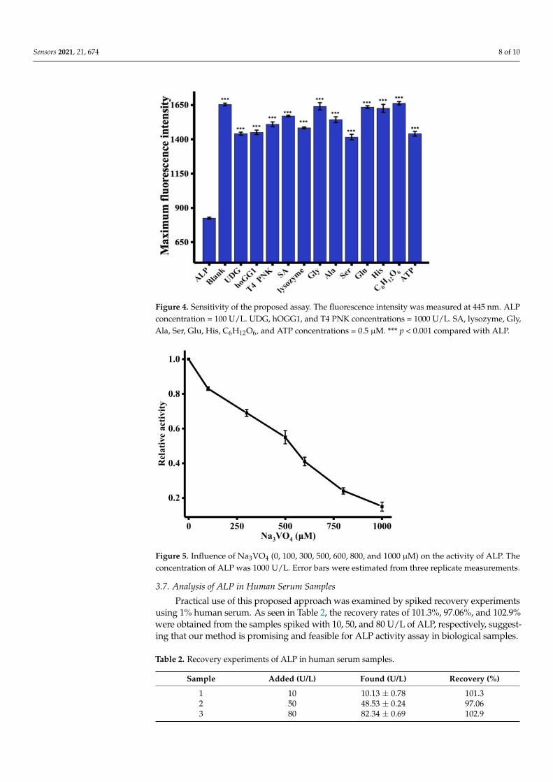
To determine the selectivity of the proposed strategy, the fluorescence signal in re-sponse to a variety of molecules, including UDG, hOGG1, T4 PNK, SA, lysozyme, Gly,Ala, Ser, Glu, His, C6H12O6, and ATP was measured [19]. As illustrated in Figure 4, anegligible fluorescence response was observed in the presence of ALP compared withthe blank control and other proteins and potential interferents, which suggests the goodselectivity of this strategy for ALP assay.
3.6. Assay of ALP Activity Inhibitor
The ability of this sensing strategy to evaluate the influence of the ALP inhibitor onALP activity is of great significance because it can be applied to drug screening and diseasetherapy. A known the ALP inhibitor, Na3VO4, was employed in the inhibitory assays [33].In Figure 5, it was observed that ALP activity was gradually inhibited with the increas-ing concentration of Na3VO4. The IC50 value (half-maximal inhibitory concentration) ofNa3VO4 was 262 µM. The results suggest that our method is suitable for screening of theALP inhibitor.
Sensors 2021, 21, 674 8 of 10
Figure 4. Sensitivity of the proposed assay. The fluorescence intensity was measured at 445 nm. ALPconcentration = 100 U/L. UDG, hOGG1, and T4 PNK concentrations = 1000 U/L. SA, lysozyme, Gly,Ala, Ser, Glu, His, C6H12O6, and ATP concentrations = 0.5 µM. *** p < 0.001 compared with ALP.
Figure 5. Influence of Na3VO4 (0, 100, 300, 500, 600, 800, and 1000 µM) on the activity of ALP. Theconcentration of ALP was 1000 U/L. Error bars were estimated from three replicate measurements.
3.7. Analysis of ALP in Human Serum Samples
Practical use of this proposed approach was examined by spiked recovery experimentsusing 1% human serum. As seen in Table 2, the recovery rates of 101.3%, 97.06%, and 102.9%were obtained from the samples spiked with 10, 50, and 80 U/L of ALP, respectively, suggest-ing that our method is promising and feasible for ALP activity assay in biological samples.
Table 2. Recovery experiments of ALP in human serum samples.
Sample Added (U/L) Found (U/L) Recovery (%)
1 10 10.13 ± 0.78 101.32 50 48.53 ± 0.24 97.063 80 82.34 ± 0.69 102.9
Sensors 2021, 21, 674 9 of 10
4. Conclusions
In summary, a label-free, sensitive, and time-saving Cu2+-TH-system-fluorometricmethod for ALP activity determination was successfully constructed. This strategy relieson the ability of AA, which is derived from the hydrolysis of AAP by ALP, to reduceCu2+ to Cu+. The oxidation of TH by Cu2+ is prevented causing the fluorescent signalto reduce. The detection limit of the proposed strategy was 0.08 U/L, which is muchlower than that of various other strategies reported in the literature. Other benefits of theCu2+-TH system include high sensitivity, low cost, and facile operation, and it also doesnot require fluorescent or nanomaterial probes. Additionally, the strategy was successfullyused to assay ALP activity in real samples, and satisfactory results were achieved. Thisfurther indicates that this approach can potentially provide a platform for the diagnosis ofALP-related diseases.
Author Contributions: Conceptualization, C.M.; formal analysis, H.Z. and X.L.; investigation, H.Z.;writing—original draft preparation, H.Z.; writing—review and editing, X.L. and C.M.; supervi-sion, C.M.; funding acquisition, X.L. All authors have read and agreed to the published version ofthe manuscript.
Funding: This research was funded by National Natural Science Foundation of China (No. 21205142), andthe Research Innovation Program for Graduates of Central South University (2018zzts384, 2019zzts453).
Institutional Review Board Statement: The study was approved by the Ethics Committee of CentralSouth University (protocol code 2020-1-11; date of approval 04/02/2020).
Informed Consent Statement: Informed consent was obtained from all subjects involved in the study.
Data Availability Statement: The data presented in this study are available on request from thecorresponding author. The data are not publicly available due to participant confidentiality.
Conflicts of Interest: The authors declare no conflict of interest.
References1. Coleman, J.E. Structure and mechanism of alkaline phosphatase. Annu. Rev. Biophys. Biomol. Struct. 1992, 21, 441–483.2. Harris, H. The human alkaline phosphatases: What we know and what we don’t know. Clin. Chim. Acta 1990, 186, 133–150.3. Tang, Z.; Chen, H.; He, H.; Ma, C. Assays for alkaline phosphatase activity: Progress and prospects. TrAC Trends Anal. Chem.
2019, 113, 32–43.4. Julien, S.G.; Dubé, N.; Hardy, S.; Tremblay, M.L. Inside the human cancer tyrosine phosphatome. Nat. Rev. Cancer 2011, 11, 35–49.5. Ooi, K.; Shiraki, K.; Morishita, Y.; Nobori, T. High-molecular intestinal alkaline phosphatase in chronic liver diseases.
J. Clin. Lab. Anal. 2007, 21, 133–139.6. Colombatto, P.; Randone, A.; Civitico, G.; Gorin, J.M.; Dolci, L.; Medaina, N.; Oliveri, F.; Verme, G.; Marchiaro, G.; Pagni, R.; et al.
Hepatitis G virus RNA in the serum of patients with elevated gamma glutamyl transpeptidase and alkaline phosphatase: Aspecific liver disease. J. Viral Hepat. 1996, 3, 301–306.
7. Lorente, J.A.; Valenzuela, H.; Morote, J.; Gelabert, A. Serum bone alkaline phosphatase levels enhance the clinical utility ofprostate specific antigen in the staging of newly diagnosed prostate cancer patients. Eur. J. Nucl. Med. Mol. Imaging 1999, 26,625–632.
8. Li, X.; Zhu, L.; Zhou, Y.; Yin, H.; Ai, S. Enhanced Photoelectrochemical Method for Sensitive Detection of Protein Kinase a ActivityUsing TiO2/g-C3N4, PAMAM Dendrimer, and Alkaline Phosphatase. Anal. Chem. 2017, 89, 2369–2376.
9. Liu, Y.Q.; Xiong, E.H.; Li, X.Y.; Li, J.J.; Zhang, X.H.; Chen, J.H. Sensitive electrochemical assay of alkaline phosphatase activitybased on TdT-mediated hemin/G-quadruplex DNAzyme nanowires for signal amplification. Biosens. Bioelectron. 2017, 87,970–975.
10. Ma, J.-L.; Yin, B.-C.; Wu, X.; Ye, B.-C. Copper-Mediated DNA-Scaffolded Silver Nanocluster On–Off Switch for Detection ofPyrophosphate and Alkaline Phosphatase. Anal. Chem. 2016, 88, 9219–9225.
11. Liu, H.S.; Ma, C.B.; Wang, J.; Wang, K.M.; Wu, K.F. A turn-on fluorescent method for determination of the activity of alkalinephosphatase based on dsDNA-templated copper nanoparticles and exonuclease based amplification. Microchim. Acta 2017, 184,2483–2488.
12. Zhao, M.; Guo, Y.; Wang, L.; Luo, F.; Lin, C.; Lin, Z.; Chen, G. A sensitive fluorescence biosensor for alkaline phosphatase activitybased on the Cu(II)-dependent DNAzyme. Anal. Chim. Acta 2016, 948, 98–103.
13. Mei, Y.; Hu, Q.; Zhou, B.; Zhang, Y.; He, M.; Xu, T.; Li, F.; Kong, J. Fluorescence quenching based alkaline phosphatase activitydetection. Talanta 2018, 176, 52–58.
Sensors 2021, 21, 674 10 of 10
14. Wang, C.; Gao, J.; Cao, Y.; Tan, H. Colorimetric logic gate for alkaline phosphatase based on copper (II)-based metal-organicframeworks with peroxidase-like activity. Anal. Chim. Acta 2018, 1004, 74–81.
15. Zhang, X.; Sun, Y.; Lin, L.; Shi, C.; Wang, G.; Zhang, X. Naked-eye sensitive detection of alkaline phosphatase (ALP) andpyrophosphate (PPi) based on a horseradish peroxidase catalytic colorimetric system with Cu(ii). Analyst 2016, 141, 5549–5554.
16. Lakra, S.; Jadhav, V.J.; Garg, S.R. Development of a Chromatographic Method for the Determination of Alkaline PhosphataseActivity in Pasteurized Milk. Food Anal. Methods 2016, 9, 2002–2009.
17. Ruan, C.; Wang, W.; Gu, B. Detection of Alkaline Phosphatase Using Surface-Enhanced Raman Spectroscopy. Anal. Chem. 2006,78, 3379–3384.
18. Wu, Z.; Zhou, C.-H.; Pan, L.-J.; Zeng, T.; Zhu, L.; Pang, D.-W.; Zhang, Z.-L. Reliable Digital Single Molecule Electrochemistry forUltrasensitive Alkaline Phosphatase Detection. Anal. Chem. 2016, 88, 9166–9172.
19. Tang, Z.; Zhang, H.; Ma, C.; Gu, P.; Zhang, G.; Wu, K.; Chen, M.; Wang, K. Colorimetric determination of the activity of alkalinephosphatase based on the use of Cu(II)-modulated G-quadruplex-based DNAzymes. Microchim. Acta 2018, 185, 109.
20. Dong, L.; Miao, Q.; Hai, Z.; Yuan, Y.; Liang, G. Enzymatic Hydrogelation-Induced Fluorescence Turn-Off for Sensing AlkalinePhosphatase in Vitro and in Living Cells. Anal. Chem. 2015, 87, 6475–6478.
21. Guo, L.; Chen, D.; Yang, M. DNA-templated silver nanoclusters for fluorometric determination of the activity and inhibition ofalkaline phosphatase. Microchim. Acta 2017, 85, 2165–2170.
22. Liu, Y.; Schanze, K.S. Conjugated Polyelectrolyte-Based Real-Time Fluorescence Assay for Alkaline Phosphatase with Pyrophos-phate as Substrate. Anal. Chem. 2008, 80, 8605–8612.
23. Qian, Z.; Chai, L.; Tang, C.; Huang, Y.; Chen, J.; Feng, H. Carbon Quantum Dots-Based Recyclable Real-Time Fluorescence Assayfor Alkaline Phosphatase with Adenosine Triphosphate as Substrate. Anal. Chem. 2015, 87, 2966–2973.
24. Qu, F.; Pei, H.; Kong, R.-M.; Zhu, S.; Xia, L. Novel turn-on fluorescent detection of alkaline phosphatase based on greensynthesized carbon dots and MnO2 nanosheets. Talanta 2017, 165, 136–142.
25. He, Y.; Jiao, B.N. Determination of the activity of alkaline phosphatase based on the use of ssDNA-templated fluorescent silvernanoclusters and on enzyme-triggered silver reduction. Microchim. Acta 2017, 184, 4167–4173.
26. Tan, H.; Li, Q.; Zhou, Z.; Ma, C.; Song, Y.; Xu, F.; Wang, L. A sensitive fluorescent assay for thiamine based on metal-organicframeworks with intrinsic peroxidase-like activity. Anal. Chim. Acta 2015, 856, 90–95.
27. Ni, P.; Chen, C.; Jiang, Y.; Zhao, Z.; Lu, Y. Fluorometric determination of sulfide ions via its inhibitory effect on the oxidation ofthiamine by Cu(II) ions. Microchim. Acta 2018, 185, 362.
28. Purbia, R.; Paria, S. A simple turn on fluorescent sensor for the selective detection of thiamine using coconut water derivedluminescent carbon dots. Biosens. Bioelectron. 2016, 79, 467–475.
29. Perezruiz, T.; Martinezlozano, C.; Tomas, V.; Ibarra, I. Flow injection fluorimetric determination of thiamine and copper based onthe formation of thiochrome. Talanta 1992, 39, 907–911.
30. Du, J.; Xiong, L.; Ma, C.; Liu, H.; Wang, J.; Wang, K. Label-free DNA hairpin probe for real-time monitoring of alkaline phosphataseactivity. Anal. Methods 2016, 8, 5095–5100.
31. Xu, A.-Z.; Zhang, L.; Zeng, H.-H.; Liang, R.-P.; Qiu, J.-D. Fluorometric determination of the activity of alkaline phosphatase basedon the competitive binding of gold nanoparticles and pyrophosphate to CePO4:Tb nanorods. Microchim. Acta 2018, 185, 288.
32. Qian, Z.S.; Chai, L.J.; Huang, Y.Y.; Tang, C.; Shen, J.J.; Chen, J.R.; Feng, H. A real-time fluorescent assay for the detection ofalkaline phosphatase activity based on carbon quantum dots. Biosens. Bioelectron. 2015, 68, 675–680.
33. Gibbons, I.R.; Cosson, M.P.; Evans, J.A.; Gibbons, B.H.; Houck, B.; Martinson, K.H.; Sale, W.S.; Tang, W.J. Potent inhibition ofdynein adenosinetriphosphatase and of the motility of cilia and sperm flagella by vanadate. Proc. Natl. Acad. Sci. USA 1978, 75,2220–2224.
Related Documents