Metal-Enhanced Fluorescence: A Novel Approach to Ultra- Sensitive Fluorescence Sensing Assay Platforms Zygmunt Gryczynski 1 , Joanna Malicka 1 , Ignacy Gryczynski 1 , Evgenia Matveeva 1 , Chris D. Geddes 2 , Kadir Aslan 2 , and Joseph R. Lakowicz 1 1 Center for Fluorescence Spectroscopy, Department of Biochemistry and Molecular Biology, University of Maryland School of Medicine, 725 West Lombard St, Baltimore, MD, 21201, USA 2 Institute of Fluorescence and Center for Fluorescence Spectroscopy, Medical Biotechnology Center, University of Maryland Biotechnology Institute, 725 West Lombard St, Baltimore, MD, 21201, USA Abstract We describe the development of a novel generic approach to fluorescence sensing based on metal- enhanced fluorescence (MEF). This work follows our initial reports of radiative decay engineering (RDE), where we experimentally demonstrated dramatic signal enhancements of fluorophores positioned close to surface-bound silver nanostructures. The attractive changes in spectral properties of fluorophores includes increased rates of excitation, increased quantum yields, decreased fluorescence lifetimes with an increased photostability, and drastically increased rates of multi- photon excitation. In this report we present a new class of fluorescent biomarkers which are strongly enhanced by metallic particles. This has afforded the development of a novel generic approach for ultra-sensitive fluorescence assay technology. The assay platform utilizes metal particles deposited on glass/quartz surfaces, covered with sub-nanometer layers of a fluorescent biomarker. As such the fluorescence signal of the composite is strongly enhanced. This readily allows easy, quantitative and inexpensive fluorescence detection of minimal traces of specific antigens. We also explore different sensing geometries, such as using evanescent wave excitation. 1. INTRODUCTION Fast and sensitive detection of physiological markers, deadly pathogens and/or chemicals, is a key element for preventive medicine, as well as an early alert system for national and personal security. For this reason novel biochip devices are of increasing importance for the rapidly emerging market of diagnostics for chronic and pathogenic diseases, as well as genetic analysis. Most of the promising detection systems, such as DNA microarrays, make use of the hybridization of an immobilized target polynucleotide with oligo- or polynucleotide probes that are labeled with reporter fluorophores. The most challenging present goals are: detection of minimal traces of physiological markers (like troponin or myoglobin in case of cardiac arrest), detection of low-abundance DNA, RNA transcripts, or proteins specific for target organisms. A successful sensing technology should offer high sensitivity and selectivity, and ideally not require any chemical or biological amplification steps like with PCR or ELISA. Recently we have begun to apply a new fluorescence technology that enables substantially enhanced fluorescence signals. 1–3 This novel approach relies on the long-range interactions of fluorophores with sub-wavelength sized metallic islands and colloids. We call this technology Metal Enhanced Fluorescence (MEF). We have predicted that fluorescence signal enhancements may be over million-fold. 1–3 We speculate that with the proper development and tuning, this technology should provide the equivalent sensitivity of PCR or ELISA, without any chemical or biological amplifications steps. NIH Public Access Author Manuscript Proc Soc Photo Opt Instrum Eng. Author manuscript; available in PMC 2009 September 3. Published in final edited form as: Proc Soc Photo Opt Instrum Eng. 2004 July 1; 5321(275): 275–282. doi:10.1117/12.530072. NIH-PA Author Manuscript NIH-PA Author Manuscript NIH-PA Author Manuscript

Welcome message from author
This document is posted to help you gain knowledge. Please leave a comment to let me know what you think about it! Share it to your friends and learn new things together.
Transcript

Metal-Enhanced Fluorescence: A Novel Approach to Ultra-Sensitive Fluorescence Sensing Assay Platforms
Zygmunt Gryczynski1, Joanna Malicka1, Ignacy Gryczynski1, Evgenia Matveeva1, Chris D.Geddes2, Kadir Aslan2, and Joseph R. Lakowicz11 Center for Fluorescence Spectroscopy, Department of Biochemistry and Molecular Biology,University of Maryland School of Medicine, 725 West Lombard St, Baltimore, MD, 21201, USA2 Institute of Fluorescence and Center for Fluorescence Spectroscopy, Medical BiotechnologyCenter, University of Maryland Biotechnology Institute, 725 West Lombard St, Baltimore, MD,21201, USA
AbstractWe describe the development of a novel generic approach to fluorescence sensing based on metal-enhanced fluorescence (MEF). This work follows our initial reports of radiative decay engineering(RDE), where we experimentally demonstrated dramatic signal enhancements of fluorophorespositioned close to surface-bound silver nanostructures. The attractive changes in spectral propertiesof fluorophores includes increased rates of excitation, increased quantum yields, decreasedfluorescence lifetimes with an increased photostability, and drastically increased rates of multi-photon excitation. In this report we present a new class of fluorescent biomarkers which are stronglyenhanced by metallic particles. This has afforded the development of a novel generic approach forultra-sensitive fluorescence assay technology. The assay platform utilizes metal particles depositedon glass/quartz surfaces, covered with sub-nanometer layers of a fluorescent biomarker. As such thefluorescence signal of the composite is strongly enhanced. This readily allows easy, quantitative andinexpensive fluorescence detection of minimal traces of specific antigens. We also explore differentsensing geometries, such as using evanescent wave excitation.
1. INTRODUCTIONFast and sensitive detection of physiological markers, deadly pathogens and/or chemicals, isa key element for preventive medicine, as well as an early alert system for national and personalsecurity. For this reason novel biochip devices are of increasing importance for the rapidlyemerging market of diagnostics for chronic and pathogenic diseases, as well as genetic analysis.Most of the promising detection systems, such as DNA microarrays, make use of thehybridization of an immobilized target polynucleotide with oligo- or polynucleotide probesthat are labeled with reporter fluorophores. The most challenging present goals are: detectionof minimal traces of physiological markers (like troponin or myoglobin in case of cardiacarrest), detection of low-abundance DNA, RNA transcripts, or proteins specific for targetorganisms. A successful sensing technology should offer high sensitivity and selectivity, andideally not require any chemical or biological amplification steps like with PCR or ELISA.
Recently we have begun to apply a new fluorescence technology that enables substantiallyenhanced fluorescence signals. 1–3 This novel approach relies on the long-range interactionsof fluorophores with sub-wavelength sized metallic islands and colloids. We call thistechnology Metal Enhanced Fluorescence (MEF). We have predicted that fluorescence signalenhancements may be over million-fold. 1–3 We speculate that with the proper developmentand tuning, this technology should provide the equivalent sensitivity of PCR or ELISA, withoutany chemical or biological amplifications steps.
NIH Public AccessAuthor ManuscriptProc Soc Photo Opt Instrum Eng. Author manuscript; available in PMC 2009 September 3.
Published in final edited form as:Proc Soc Photo Opt Instrum Eng. 2004 July 1; 5321(275): 275–282. doi:10.1117/12.530072.
NIH
-PA Author Manuscript
NIH
-PA Author Manuscript
NIH
-PA Author Manuscript

The assay platform utilizes metal particles deposited on the glass/quartz surface covered withsub-nanometer layers of a fluorescent biomarker. The fluorescence signal of the composite isenormously enhanced, which allows easy, quantitative and inexpensive fluorescence detectionof minimal traces of specific antigens. We exploit and indeed report different formats likeevanescent wave excitation, directional emission and/or multi-photon excitation.
2. OPTICAL PROPERTIES OF METALLIC PARTICLESThe theoretical aspects of the effect of metal particles on fluorophores have been studied formany years [1–4 and refs therein]; however this phenomenon is still not widely recognized influorescence sensing. For this reason we briefly review the basic concepts. Metal particles(gold and silver) exhibit strong absorption bands which are absent in the bulk metal. Theseabsorption bands, known as the surface plasmon resonances, result in strong wavelengthselective absorption and scattering and create enhanced local electromagnetic fields near thesurface of the particles. An example of gold colloid absorption is shown in Figure 1. Theplasmon resonances are highly dependent on the size and shape of the metal nanoparticles andthe dielectric properties of the surrounding environment.5,6 These near field enhancementshave given risen to surface enhanced spectroscopies such as surface enhanced Ramanspectroscopy (SERS) and surface enhanced resonance Raman scattering (SERRS). Theenhancement effect for fluorescence near the metallic particles is related to surface enhancedRaman scattering. It is well known that SERS may enhance Raman signal of chromophore bya factor 105 or in some cases even more. 7
3. FLUORESCENCE NEAR METALLIC PARTICLESFollowing excitation a fluorophore, in free space, can either emit a photon with a radiative rate,Γ, or return to ground state by a non-radiative deactivation rate, knr. For simplicity we areomitting transitions to triplet state which can be significant in some cases and chemicalprocesses leading to photodegradation which are usually slower and not needed for describingthe spectral properties. The quantum yield, Q, and lifetime, τ of the fluorophore are given by:
(1)
(2)
The presence of nanometer-size metal particles in the proximity of a fluorophore can affect itsspectral properties in two ways: firstly, illumination at the plasmon absorption of the nanometersized metal particles creates enhanced near-field intensity, Im that results in higher fluorescencedue to increased rates of excitation. We define the excitation rate enhancement factor as a ratioof enhanced excitation intensity in the presence of metal particle, Ienh, and excitation intensity,I, in the absence of particle; Gexc = Ienh/I. Secondly, the interaction between the plasmon bandand the fluorophore dipole influences both the non-radiative and radiative deactivation rates.The new radiative rate is given by: Γm = Nr Γ, and the non-radiative rate given by: knr
m =Nnr knr. We are not assuming any correlation between enhancement factors Nr and Nnr. Thenew fluorescence quantum yield, Qm and lifetime τm in the presence of metal particle andsubsequently be defined as:
Gryczynski et al. Page 2
Proc Soc Photo Opt Instrum Eng. Author manuscript; available in PMC 2009 September 3.
NIH
-PA Author Manuscript
NIH
-PA Author Manuscript
NIH
-PA Author Manuscript

(3)
(4)
The presence of the metal particle will change both the emission quantum yield and thefluorescence lifetime. The change in the quantum yield is described by the emission factorGqy and the change in the fluorescence lifetime by the lifetime factor Gτ:
(5)
(6)
Both radiative Nr and non-radiative Nnr factors can have significant effects on the fluorescenceemission. The relative contributions of these factors will depend on the separation and therelative orientation between the fluorophore and metallic particle. For very small separationsNnrknr will dominate, and fluorescence quenching is expected. For larger separations Nnrknrrapidly decreases and Nr Γ is expected to become the dominant factor resulting in fluorescenceenhancement. It is intuitive that the maximum value of quantum yield enhancement factorcannot exceed the value Gqy =1/Q (when Nr 64, Qm ~1) that corresponds to a decrease influorescence lifetime to zero. We expect significant emission enhancement factor for weakfluorophores (Q ≪ 1) and much smaller effect for high quantum yield fluorophores.
In the case of both low and high quantum yield fluorophores the fluorescence intensity can bealso increased by the change in excitation rate Gexc. It is important to stress that observedfluorescence enhancement (Gfl =GexcGqy) reflects the changes in both the excitation and decayrates (radiative and nonradiative).
(7)
The increase in radiative decay rate, as well as increases in the excitation rate in the presenceof metal particles, has until now been ill-explored in fluorescence sensing. However given theextent of fluorescence enhancement expected from these phenomena, 1–4 then was are facedwith some new possibilities for the development of more sensitive ways of assay sensing.
Figure 2 schematically shows these three main phenomena that affect the fluorescence signal.For very short separation the dominant effect is quenching of fluorescence by nearby metal[1–4]. This quenching is essentially a dumping of the excitation energy of the dipole oscillatorsto the nearby metal. For fluorophore-metal separations larger then 50 X, the field enhancementand radiative decay increase dominates the fluorophore metal interaction. In the range 50 –150 X the cumulative enhancement reaches a maximum and quickly decreases over the next
Gryczynski et al. Page 3
Proc Soc Photo Opt Instrum Eng. Author manuscript; available in PMC 2009 September 3.
NIH
-PA Author Manuscript
NIH
-PA Author Manuscript
NIH
-PA Author Manuscript

few hundred angstroms. Fluorophores located within the enhancement layer typically showmuch stronger fluorescence signals. This enhancement contains a contribution from the fieldenhancement (increase in excitation rate) and an increase in the quantum yield. For separationslarger than 200 X, the metal effects diminishes and one readily observes unmodified free spacefluorescence from the fluorophore.
4. ASSAY PLATFORM DEVELOPMENTOur experimental results to date 1–4, 8–10 indicate significant effects with silver island films,which are made by depositing sub-wavelength size silver particles on a flat glass surface. Therough silver-surface can be conveniently covered by a monolayer (or multilayer) ofbiotinylated-BSA or indeed other inert spacers (to elevate close-range quenching). Thethickness of the spacer can be adjusted in such way that captured oligomers of DNA, or anybiomolecules of interest, will be positioned within the enhancement distance. This can providea population of fluorophore-labeled DNA/proteins localized at the optimal enhancementdistance (about 7–15 nm from a silver island surface).
These thin layer samples can easily be excited in a front-face configuration. Figure 3 showsthe front-face configuration employed. The sample sandwich consists of one glass (quartz)slide covered with a silver island film, and a second cover slide, essentially used to form amicro-cuvette. In an ideal scenario, the fluorescence sensor should be located in theenhancement layer close to the silver film. The buffer solution can be used to occupy the spacebetween the slides. Our studies have shown that two high quality optical glass slides cannotbe squeezed closer than ≈ 1 micron. The enhancement layer is thought less than 500 X, and istherefore much smaller than the bulk volume. A front-face excitation through the sampleequally excites the space between the slides. In effect any background fluorescence willsignificantly contribute to the measured signal.
We have realized that a much better excitation choice for such a thin layer of fluorophoreslocated close to a transparent glass surface, is using an evanescent wave. The evanescent waveis formed between two media with different refraction indexes, when light enters the mediumof the lower refraction index, under the angle greater than the critical angle, αc. 11,12 Theintensity of an evanescent wave decreases exponentially with distance from the surface andlimits the effective excitation to a couple hundred nanometers. Figure 4 shows the schematicof evanescent wave excitation.
5. ASSAY DESIGNIn designing the format of the assay, we will take advantage of the metal surface plasmonenhancement of fluorescence and the potential benefits of evanescent wave excitation. In thisway we will obtain maximum excitation efficiency on the layer of fluorophores just above themetal-island film. The fluorescence signal will be enhanced due to the field effect near-to theisland and an increase in quantum yield of the fluorophore. Also the evanescent wave will limitthe excitation volume to the effective depth (sensing volume) below 1 micron. In the text bellowwe now describe different assay designs using as an example, a TnI protein. The approach isgeneral and applicable to other markers.
5.1. Labeled Marker ProteinThe concept of a MEF surface based immunoassay for detecting TnI can be understood fromthe schematic in Figure 5 (top). The TnI specific antibodies (SAB) are immobilized on a surfacelayer. Fluorophore labeled TnI are binding to sites of the antibodies.
Gryczynski et al. Page 4
Proc Soc Photo Opt Instrum Eng. Author manuscript; available in PMC 2009 September 3.
NIH
-PA Author Manuscript
NIH
-PA Author Manuscript
NIH
-PA Author Manuscript
5.2. Sandwich Assay FormatThe concept of the sandwich type immunoassay is also shown in Figure 5 (bottom). Theunlabeled TnI specific antibodies (SAB) are immobilized in the layer above the silver islandfilms. Addition of the sample containing natural TnI results in binding of TnI to the antibodies.
5.3. DNA Hybridization FormatThe notion of using silver island films for DNA hybridization is shown in Figure 6. Basicallya labeled fluorophore DNA strand is enhanced when binding to a complimentary strandimmobilized on the silver surface.
5.4. Experimental PlatformFigure 6 and 7 shows the practical design of an assay cartridge which we consider would beof considerable use in MEF assay sensing. The cartridge has a transparent bottom (made fromglass or plastic with high refractive index) that is coated with the silver island film. As shownin the insert on Figure 6, the island layer is covered with the layer of spacer with immobilizedSAB. Prior to measurements the SAB are filled with labeled TnI. Upon addition of sample(blood/serum) natural TnI, if present in the sample, will exchange labeled TnI. Displacedlabeled TnI will quickly diffuse from the enhancement layer and evanescent excitation field.This results in a drop in fluorescence signal that will be proportional to the concentration ofTnI, and indeed time of exposure.
The analogical concept can also be used for a sandwich format assay cartridge. The cartridgewith an immobilized layer of unlabeled antibodies (over the silver island film) will be filledwith the sample. The background signal will be detected. After a predefined time period thesample is rinsed and the cartridge washed with the buffer containing known concentration oflabeled antibodies. The labeled antibodies form a complementary layer over the bound TnI.This results in fluorescence signal increase proportional to the amount of bound TnI.
Figure 7 shows the schematic of a simple detection platform. The detection platform consistsof a cylindrical lens made from a high refractive index glass or plastic. The refractive index ofthe lens has to be greater than the refractive index of the liquid sample in the cartridge (bloodor serum). The laser beam from a laser diode is directed to the flat (horizontal) surface of thelens under the angle greater than the limiting angle, to form an evanescent wave. The cartridgecan be tightly placed on the platform. When the refractive index of the lens and bottom of thecartridge are matched, the evanescent wave will form over the sensor layer. For the blankmeasurements the cartridge can be filled with the reference solution that does not containmarker. This will effectively zero the reading. After filling the cartridge with the real samplecontaining markers, one expects to observe the signal decreasing. The extent of the drop willbe proportional to the marker level.
Figure 8 shows only the qualitative principles of the sensor. It is easy to realize that such ameasurement can be calibrated to be quantitative, but may be subject to optical artifacts. Themost important considerations will be the auto-fluorescence of the different samples andambient light.
6. CONCLUSIONSIn this short paper we have reviewed the favorable spectral effects that can be obtained byemploying fluorophore metal particle combinations. We have additionally shown how this newphenomenon, metal-enhanced fluorescence can be applied to surface assay platforms. Furtherwork developing these assay platforms is underway in our laboratories and will be reported indue course.
Gryczynski et al. Page 5
Proc Soc Photo Opt Instrum Eng. Author manuscript; available in PMC 2009 September 3.
NIH
-PA Author Manuscript
NIH
-PA Author Manuscript
NIH
-PA Author Manuscript
AcknowledgmentsThis work was supported by the National Institute of Biomedical Imaging and Bioengineering, EB-00682, EB-00981and EB-1690, National Center for Research Resources, RR-08119 and Philip Morris, USA Inc.
References1. Lakowicz JR, Gryczynski I, Shen Y, Malicka J, Gryczynski Z. Intensified fluorescence. Photonics
Spectra 2001;96:104.2. Gryczynski I, Malicka J, Shen Y, Gryczynski Z, Lakowicz JR. Multiphoton Excitation of Fluorescence
near Metallic Particles: Enhanced and Localized Excitation. J Phys Chem B 2002;106:2191–2195.3. Lakowicz JR, Shen Y, D’Auria S, Malicka J, Fang J, Gryczynski Z, Gryczynksi I. Radiative Decay
Engineering. 2. Effects of Silver Island Films on Fluorescence Intensity, Lifetimes, and ResonanceEnergy Transfer. Anal Biochem 2002;301:261–277. [PubMed: 11814297]
4. Lakowicz JR. Radiative decay engineering: Biophysical and biomedical applications. Anal Biochem2001;298:1–24. [PubMed: 11673890]
5. Link S, El-Sayed MA. Spectral properties and relaxation dynamics of surface plasmon electronicoscillations in gold and silver nanodots and nanorods. J Phys Chem B 1999;103:8410–8426.
6. Kreibig U, Genzel L. Optical absorption of small metallic particles. Surface Science 1985;156:678–700.
7. Kneipp K, Wang Y, Kneipp H, Perelman LT, Itzkan L, Dasari RR, Feld MS. Phys Rev Lett1997;78:1667.
8. Malicka J, Gryczynski I, Maliwal BP, Fang JF, Lakowicz JR. Fluorescence spectral properties ofcyanine dye labeled DNA near metallic silver particles. Biopolymers 2003;72(2):96–104. [PubMed:12583012]
9. Malicka J, Gryczynski I, Geddes CD, Lakowicz JR. Metal-enhanced emission from Indocyanine Green:A new approach to In-Vivo imaging. Jn Biomed Opt 2003;8(3):472–478.
10. Geddes CD, Gryczynski I, Malicka J, Gryczynski Z, Lakowicz JR. Metal-Enhanced Fluorescence:Potential applications in HTS. Combinatorial Chemistry and HTS 2003;6(2):109–117.
11. Gryczynski I, Gryczynski Z, Lakowicz JR. Two-Photon Excitation by the Evanescent Wave fromTotal Internal Reflection. Annal Biochem 1997;247:69–76.
12. Lakowicz JR, Gryczynski Z, Gryczynski I. On the Possibility of Evanescent Wave Excitation Distalfrom a Solid-Liquid Interface Using Light Quenching. Photochem Photobiol 1996;64:636–641.[PubMed: 8863470]
Gryczynski et al. Page 6
Proc Soc Photo Opt Instrum Eng. Author manuscript; available in PMC 2009 September 3.
NIH
-PA Author Manuscript
NIH
-PA Author Manuscript
NIH
-PA Author Manuscript
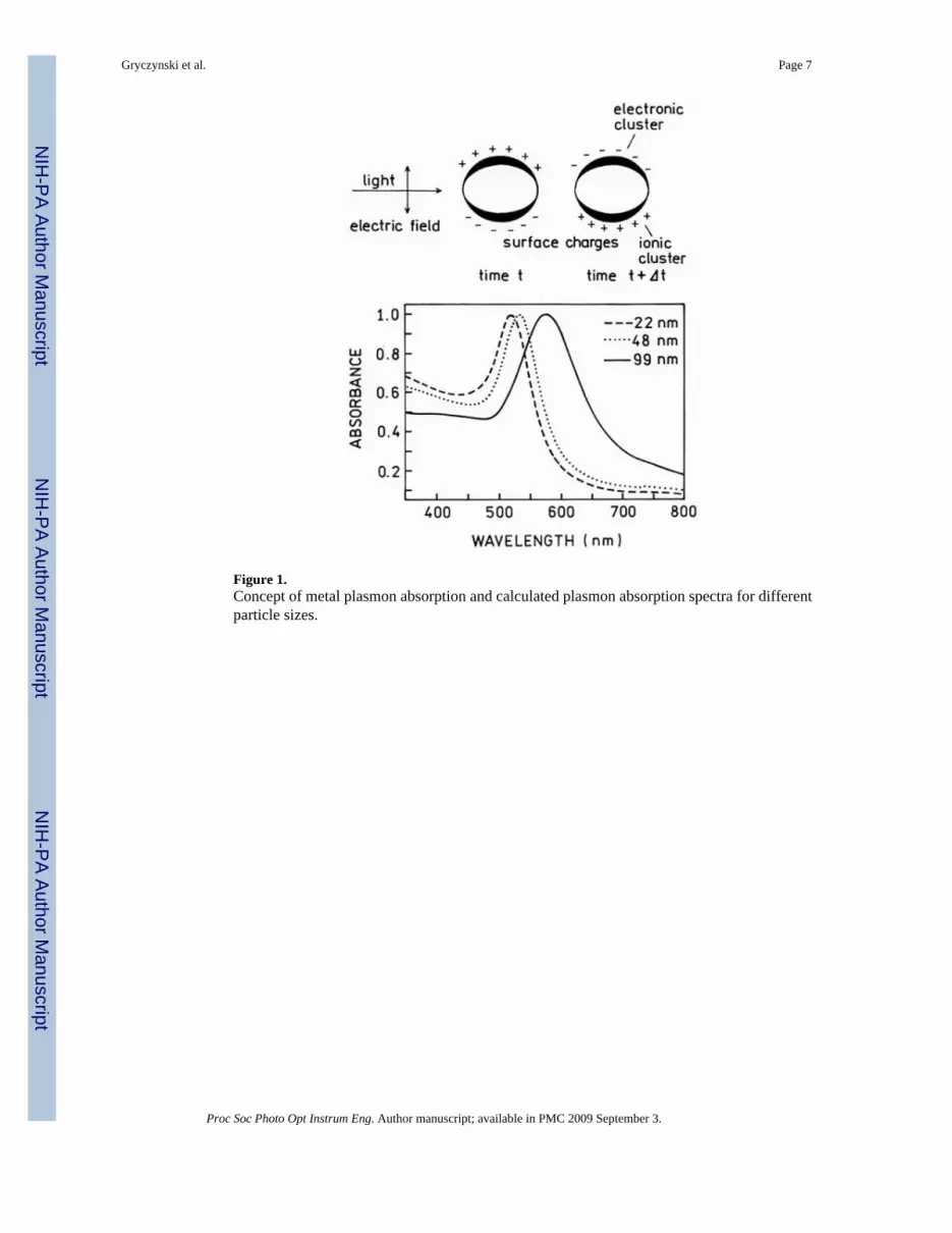
Figure 1.Concept of metal plasmon absorption and calculated plasmon absorption spectra for differentparticle sizes.
Gryczynski et al. Page 7
Proc Soc Photo Opt Instrum Eng. Author manuscript; available in PMC 2009 September 3.
NIH
-PA Author Manuscript
NIH
-PA Author Manuscript
NIH
-PA Author Manuscript
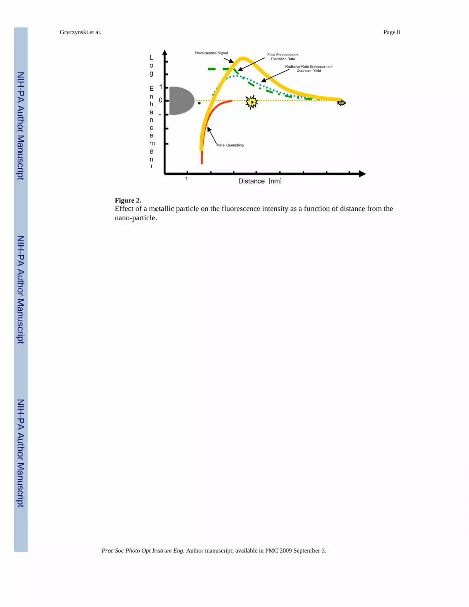
Figure 2.Effect of a metallic particle on the fluorescence intensity as a function of distance from thenano-particle.
Gryczynski et al. Page 8
Proc Soc Photo Opt Instrum Eng. Author manuscript; available in PMC 2009 September 3.
NIH
-PA Author Manuscript
NIH
-PA Author Manuscript
NIH
-PA Author Manuscript

Figure 3.Front-face configuration for excitation of a thin layer samples.
Gryczynski et al. Page 9
Proc Soc Photo Opt Instrum Eng. Author manuscript; available in PMC 2009 September 3.
NIH
-PA Author Manuscript
NIH
-PA Author Manuscript
NIH
-PA Author Manuscript

Figure 4.Top. View of experimental configuration for measurements in total internal reflectancegeometry. Bottom. Conditions for total internal reflection excitation. Insert shows thedependence of the excitation field intensity as a function of distance from the interface.
Gryczynski et al. Page 10
Proc Soc Photo Opt Instrum Eng. Author manuscript; available in PMC 2009 September 3.
NIH
-PA Author Manuscript
NIH
-PA Author Manuscript
NIH
-PA Author Manuscript
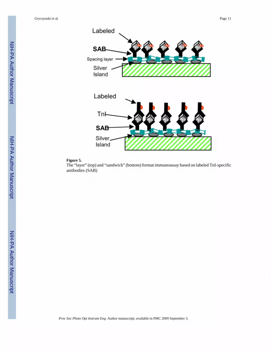
Figure 5.The “layer” (top) and “sandwich” (bottom) format immunoassay based on labeled TnI-specificantibodies (SAB)
Gryczynski et al. Page 11
Proc Soc Photo Opt Instrum Eng. Author manuscript; available in PMC 2009 September 3.
NIH
-PA Author Manuscript
NIH
-PA Author Manuscript
NIH
-PA Author Manuscript
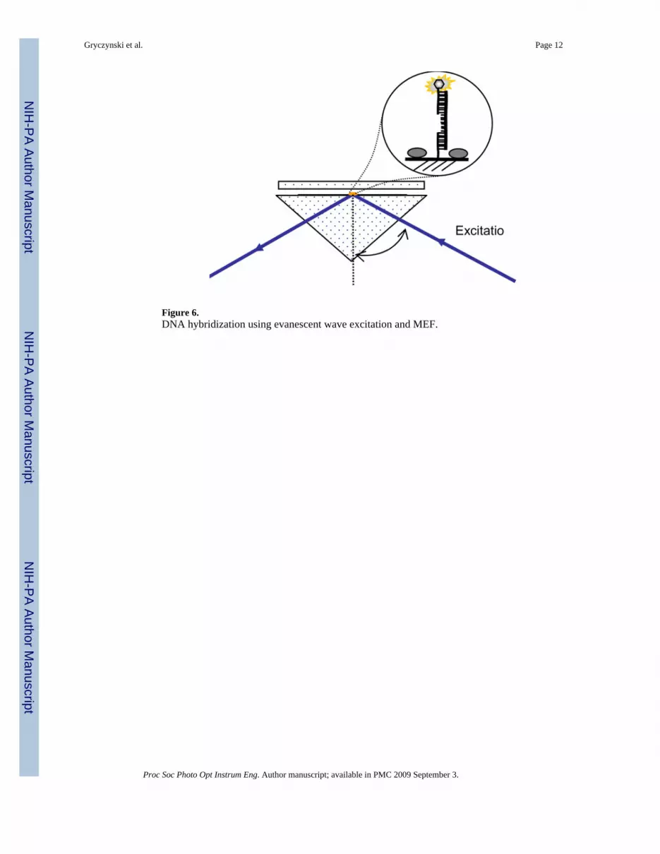
Figure 6.DNA hybridization using evanescent wave excitation and MEF.
Gryczynski et al. Page 12
Proc Soc Photo Opt Instrum Eng. Author manuscript; available in PMC 2009 September 3.
NIH
-PA Author Manuscript
NIH
-PA Author Manuscript
NIH
-PA Author Manuscript

Figure 7.Schematic of cartridge immunoassay system.
Gryczynski et al. Page 13
Proc Soc Photo Opt Instrum Eng. Author manuscript; available in PMC 2009 September 3.
NIH
-PA Author Manuscript
NIH
-PA Author Manuscript
NIH
-PA Author Manuscript
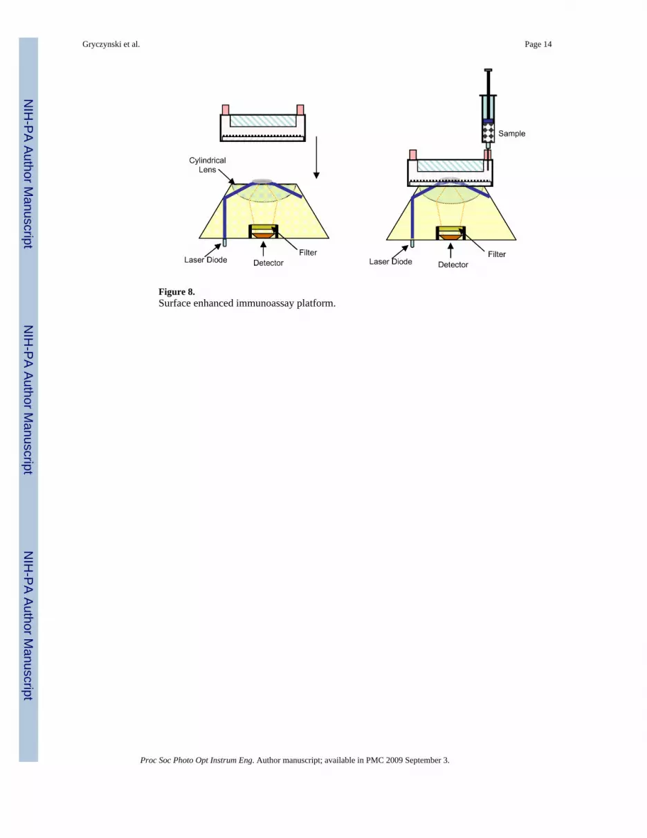
Figure 8.Surface enhanced immunoassay platform.
Gryczynski et al. Page 14
Proc Soc Photo Opt Instrum Eng. Author manuscript; available in PMC 2009 September 3.
NIH
-PA Author Manuscript
NIH
-PA Author Manuscript
NIH
-PA Author Manuscript
Related Documents
![acidification] ab197244 [Extracellular Glycolysis Assay · Glycolysis Assay [Extracellular Acidification] (ab197244) is an easy mix-and-measure, 96 or 384 well fluorescence plate](https://static.cupdf.com/doc/110x72/5ec5a381691079698166a0a6/acidification-ab197244-extracellular-glycolysis-assay-glycolysis-assay-extracellular.jpg)